electrochemical biosensors based on layer-by-layer assemblies
TRANSCRIPT
Review
Electrochemical Biosensors Based on Layer-by-Layer AssembliesWei Zhao, Jing-Juan Xu,* Hong-Yuan Chen
The Key Lab of Analytical Chemistry for Life Science (MOE), Department of Chemistry, Nanjing University, Nanjing, 210093,P. R. China*e-mail: [email protected]
Received: June 21, 2006Accepted: July 27, 2006
AbstractLayer-by-layer (LBL) assemblies, which have undergone great progress in the past decades, have been used widely inthe construction of electrochemical biosensors. The LBL assemblies provide a strategy to rationally design theproperties of immobilized films and enhance the performance of biosensors. The following review focuses on theapplication of LBL assembly technique on electrochemical enzyme biosensors, immunosensors and DNA sensors.
Keywords: Layer-by-Layer, Electrochemical biosensors, Enzyme biosensors, Immunosensors, DNA sensors
DOI: 10.1002/elan.200603630
1. Introduction
The area of electrochemical biosensors has significantdevelopment in the past few years, with the developmentof enzyme biosensors, immunosensors and DNA sensors.Devices are being successfully used in clinical chemistry,food industry and environmental fields. To achieve highspecificity, high sensitivity, rapid response and flexibility ofuse, it is clear that the research continues to focus on newassembly strategies. Recently, the use of layer-by-layer(LBL) assembly method for the design of electrochemicalbiosensors has attracted extensive attention.
The pioneering research on the sequential deposition ofcolloidal particles was reported by Iler in 1966 [1]. This workwas successfully extended and the definitive LBL assemblyapproach was introduced by Decher with consecutiveadsorption of polyanions and polycations on the surface ofa solid substrate through electrostatic force of attraction, in1991 [2]. In 1995, this new method was applied to immobilizemulticomponent protein in a polyelectrolyte based multi-layer structure and proved to be one of the most prospectivemethods for preparing biosensors [3]. This sequence wasrepeated to obtain the desired number of layers. Comparedwith other methods, the LBL assembly technique develops a“molecular architecture” with precise control of the com-position, the number of layers and the thickness of films at amolecular or nanometer level. In addition, the LBLassembly method is simple and suitable to various substratematrixes with different nature, size and topology. Theproperties of LBL assemblies can be controlled by carefullychoosing the configuration, type of polyelectrolytes, num-ber of layers, etc. The thickness of the layers and othermicrostructural attributes of these assemblies were found tobe sensitive to the type of charged species, their concen-tration, molecular weight, ionic strength and pH.
The preparation of the multilayer films usually relies onthe electrostatic attraction between the positively andnegatively charged materials. Accordingly, the choice ofmaterials and optimization of experimental variables suchas ionic strength and pH are limited. Recently, otherstrategies have been applied in the construction of LBLthin films through hydrogen-bonding interactions, charge-transfer interactions, biological interactions, etc. It would bepossible to construct many different types of multilayer thinfilms through different LBL assembly strategies. The LBLtechnique has been found as a highly useful way forassembling a number of organic and inorganic substanceswith diverse nature, including proteins [3 – 5], DNA [6],viruses [7], dendrimers [8], and nanoparticles [9 – 11]. Theorganized multilayer architectures have attracted greatinterest because of their broad range of applications in thefields of biosensors, nonlinear optical devices, surfacemodification, etc.
A variety of techniques have been used to study thecharacteristics of LBL nanostructures, such as the thicknessof the layers, the surface roughness of the films and theinterpenetration between the layers. Most of the studiesinvolved quartz crystal microbalance (QCM), which couldmonitor the assembly of multilayer films in situ continu-ously, characterizing the multilayer film growth [3, 12 – 14].X-ray reflectometry (XRR) has been used to determine theelectrodensity of the film, its thickness and the roughness ofthe interface [15]. Optical techniques such as UV-visspectroscopy can be used to quantify the loading efficiencyof the nanoparticles or proteins [16, 17]. Another opticaltechnique, surface plasmon resonance (SPR) that is basedon the optical changes occurring at interfaces between a thinmetal film and dielectric medium, was also used to study theLBL assembly in real-time measurement [18, 19]. Whenredox materials are used as LBL “modules”, electrochem-
1737
Electroanalysis 18, 2006, No. 18, 1737 – 1748 I 2006 WILEY-VCH Verlag GmbH & Co. KGaA, Weinheim
ical techniques provide valuable information. Cyclic vol-tammetry (CV) has been the most commonly used tool [12].Electrochemical impedance spectroscopy (EIS), which wasproved to be an effective method for probing the features ofsurface-modified electrodes, has been successfully used tocharacterize the formation of multilayer films on differentsubstrates [20]. Atomic force microscopy (AFM) [21] andscanning electron microscopy (SEM) [3] techniques havebeen widely used to characterize the organized structureand surface morphology of the multilayers. Other analyticaltechniques, such as X-ray photoelectron spectroscopy(XPS) [22], X-ray diffraction (XRD) [23], Fourier trans-form-infrared (FT-IR) [24, 25], single particle light scatter-ing (SPLS) [26], second harmonic generation (SHG) [27],ellipsometry [28], transmission electron microscopy (TEM)[26] and circular dichroism (CD) spectrometry [29] havealso been used to characterize the LBL assemblies and theirorganization.
This work provides an overview of the recent develop-ment in electrochemical biosensors based on LBL assemblytechnique, with emphasis on the LBL assembly methods.
2. LBL Assembly Methods
Since the first sequential adsorption of oppositely chargedcolloid reported by Iler [1], most of the reported LBLapproaches are based on the electrostatic interactionbetween oppositely charged polyelectrolytes (PE). How-ever, the LBL assembly technique is not restricted to thiskind of interlayer interactions. In the development ofelectrochemical biosensors based on LBL assembly meth-ods, other strategies such as biological affinity, covalentbinding, cross-linking have also been used. Some of theelectrochemical biosensors with different LBL assemblymethods are summarized in Table 1. Details of the differentLBL assembly methods are described below.
2.1. Electrostatic Force
The main driving force in alternate LBL films assembly isthe electrostatic interaction between oppositely chargedspecies. Initially, a base electrode should be modified with acharged layer. Generally, a negatively charged gold surfaceis prepared by immersing a cleaned gold electrode into anaqueous solution of 3-mercapto-1-propanesulfonic acid,sodium salt (MPS). This negatively charged substrate is thenimmersed in a polycation and a polyanion aqueous solution,respectively, to form a uniform and smooth negativelycharged surface as “precursor” film. Then multilayer filmsare grown by alternate dipping of the modified electrodesinto the target solution (protein, nanoparticles or DNA etc.)and/or a PE solution, respectively. Polycations that havepredominantly been used in LBL films include poly(allyl-amine) (PAA), poly(l-lysine) (PLL), poly(ethyleneimine)(PEI), poly(dimethyldiallylammonium chloride) (PDDA),poly(allylamine hydrocholoride) (PAH) and chitosan
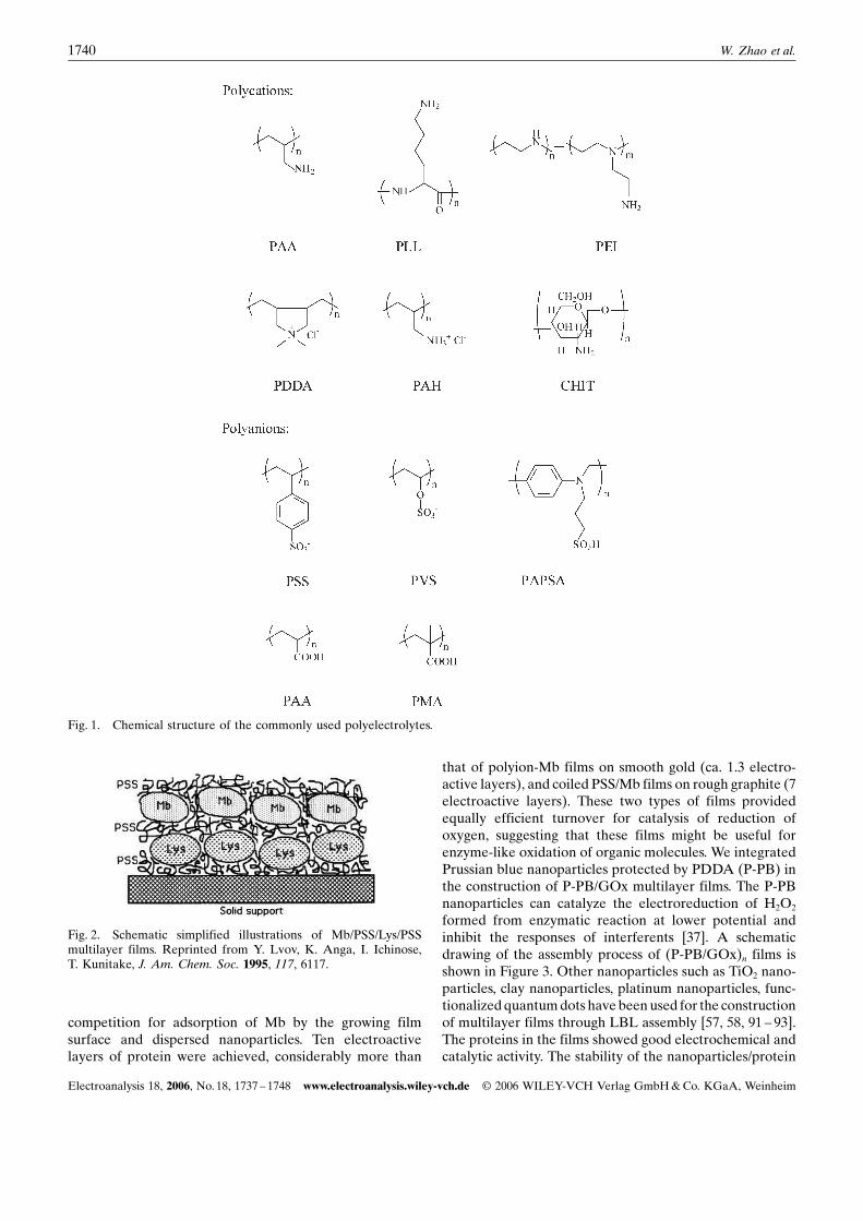
(CHIT). The most commonly used polyanions are poly(-stryrenesulfonate) (PSS), poly(vinylsulfonate) (PVS), pol-y(anilinepropanesulfonic acid) (PAPSA), poly(acrylic acid)(PAA) and poly (methacylic acid) (PMA). The chemicalstructures of the polymeric materials are shown in Figure 1.These PE multiassemblies provide excellent control of filmthickness and architecture and have been extended toimmobilize proteins, DNA and nanoparticles for the con-struction of biosensors. The LBL assembly via electrostaticattraction is simple and could be used not only on planesubstrate but spherical substrate or substrates with otherconfigurations.
2.1.1. Polyelectrolyte/Protein Films
Electrostatic LBL assembly of proteins and oppositelycharged polyelectrolytes was developed in the 1990s.Enzymes in a buffer of pH smaller or larger than theproteinNs isoelectric point can take positive or negativecharges, which make them suitably incorporated in PE films.Lvov et al. assembled multicomponent protein films bymeans of electrostatic LBL adsorption [3]. Water-solubleproteins such as cytochrome c (Cyt), myoglobin (Mb),lysozyme (Lys), histone f3, hemoglobin (Hb), glucoamylase(GA), and glucose oxidase (GOx) were used. Chargedprotein layers formed multilayers with linear polymersacting as glue or filler. The schematic illustration of thepolyelectrolyte/protein films is given in Figure 2. As an idealmodel, GOx has been firstly exploited for its incorporationin LBL assemblies and application in biosensor. In 1996, anoxygen mediated glucose biosensor was reported, based onGOx and PLL co-adsorption onto a negatively chargedmonolayer of mercaptopropionic acid [30]. One year later,Chenet al. [31] and Hodaket al. [32] simultaneously report-ed a reagentless glucose biosensor by successive alternatedeposition of ferrocene attached polypyridine or ferrocenemodified PAA (cation) polymer and anionic GOx, on Auelectrode surface, initially thiolated with negatively chargedsulfonic groups. Chen et al. [33] have used an Os-basedredox polymer for the electrochemical communication ofGOx, controlling the analytical performance of the sensorby introducing multiple bilayers of GOx and redox polymer.From then on, various biosensors based on this method havebeen developed. Recently, Calvo et al. [34] reported on the“molecular wiring” efficiency of GOx in organized self-assembled nanostructures, which comprised of enzymelayers alternating with layers of an osmium derivatizedPAA cationic polymer, acting as redox relays. Varying therelative position of the active enzyme layer in nanostruc-tures by alternating active enzyme and inactive apoenzyme,they investigated the mechanisms of electrical signalgenerated from biomolecular recognition.
Dendrimers as a new type of polymers have a highlybranched dendritic structure and unique properties such as ahigh density of active groups, good structural homogeneity,intense internal porosity and good biocompatibility. Hu andco-workers [90] reported electrostatic adsorption of hemeproteins, including Hb, Mb and catalase (Cat) alternated
1738 W. Zhao et al.
Electroanalysis 18, 2006, No. 18, 1737 – 1748 www.electroanalysis.wiley-vch.de I 2006 WILEY-VCH Verlag GmbH & Co. KGaA, Weinheim
with polyamidoamine (PAMAM) dendrimers, which has aglobular structure mimicking the three-dimensional struc-ture of biomacromolecules and good biocompatibility forLBL assembly of electroactive films. The direct electro-chemistry of proteins was realized in the PAMAM filmenvironment, and the proteins retained their near-nativestructures in the films. The pronounced lowering of reduc-tion overpotential and good sensitivity in analysis, combinedwith good stability of the films, suggested that the (PA-MAM/protein)n film has a potential applicability in devel-oping new types of electrochemical biosensors withoutmediators.
2.1.2. Nanoparticles/Protein Films
Due to the specific characteristics, nanosized materials havebeen used widely in biosensors. The surface of somenanoparticles could contain positive/negative charges underspecific process of synthesis, which facilitates the electro-static adsorption. The advantages of using the LBL assem-bly method on nanoparticles are the versatility of thetechnique, as it offers the possibility to control the size, themorphology and the composition of the stable architectures.Lvov and co-workers obtained Mb films assembled LBLwith MnO2 and SiO2 nanoparticles [11]. Films of Mb andMnO2 showed an unusual growth mechanism featuring
Table 1. Electrochemical biosensors based on various LBL assembly methods
LBL assembly methods Biorecognition molecules in multilayer films Analyte References
Electrostatic force Glucose oxidase Glucose [30 – 43]Cytochrome oxidase Cyt c [44]Horseradish peroxidase H2O2 [45 – 47]Polyphenol oxidase Dopamine [48]
Catechol [49]Uricase Uric acid [50]Catalase H2O2 [51]Cholesterol oxidase Cholesterol [52]Lactate oxidase Lactic acid [53, 54]Diaphorase/glucose-6-phosphate dehydrogenase Glucose-6-phosphate [55]Fructose dehydrogenase/horseradishperoxidase/HRP-alcohol oxidase
Fructose/H2O2/methanol [56]
Myoglobin/horseradish peroxidase Trichloroacetic acid/O2/H2O2 [57, 58]Glucose oxidase/catalase Glucose/H2O2 [59]Glucose oxidase/glucoamylase Glucose/maltose [60]Cholesterol oxidase/cholesterol esterase Cholesterol [61]Hepatitis B surface antibody Hepatitis B surface antigen [62]ds-DNA DNA damage [63]
Oxidized DNA [14]8-Oxoguanine/oxidized nucleobases [64]Oxidized DNA [65]
Avidin – biotin interaction Glucose oxidase Glucose [66, 67]Alcohol oxidase Alcohol [68]Glucose oxidase/lactate oxidase Glucose/lactic acid [69, 70]
Lectin – sugar interaction Glucose oxidase Glucose [71]Glucose oxidase/horseradishperoxidase/lactate oxidase
Glucose [72]
Glucose oxidase/horseradish peroxidase Glucose/H2O2 [73]Antibody – antigen interaction Glucose oxidase Glucose [74]Cross-linking Glucose oxidase Glucose [75, 76]
Bilirubin oxidase Bilirubin [77]Glucose oxidase/cholineoxidase/acetylcholine esterase
Glucose/acetycholine [78]
Glucose oxidase/lactate oxidase Glucose, lactic acid [79]Covalent binding Glucose oxidase Glucose [23, 80, 81]
Quinoprotein glucose dehydrogenase p-Aminophenol [82]Electrodeposition Glucose oxidase Glucose [21]Multistrategies Glucose oxidase Glucose [83]
Choline oxidase Choline [84]Cholesterol esterase Cholesterol [85]Glucose dehydrogenase/NADH Glucose [86]Horseradish peroxidase/phytic acid H2O2/O2 [87]Human IgG/goat anti-human IgG Human IgG [20]Target DNA/6-phosphate dehydrogenase Glucose-6-phosphate. [88]Alkaline phosphatese DNA/IgG [89]
1739Electrochemical Biosensors Based on LBL Assemblies
Electroanalysis 18, 2006, No. 18, 1737 – 1748 www.electroanalysis.wiley-vch.de I 2006 WILEY-VCH Verlag GmbH & Co. KGaA, Weinheim
competition for adsorption of Mb by the growing filmsurface and dispersed nanoparticles. Ten electroactivelayers of protein were achieved, considerably more than
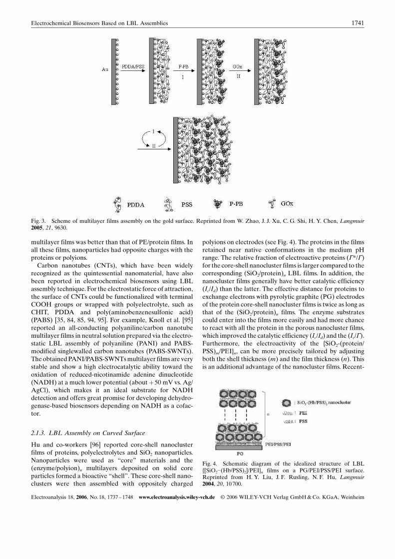
that of polyion-Mb films on smooth gold (ca. 1.3 electro-active layers), and coiled PSS/Mb films on rough graphite (7electroactive layers). These two types of films providedequally efficient turnover for catalysis of reduction ofoxygen, suggesting that these films might be useful forenzyme-like oxidation of organic molecules. We integratedPrussian blue nanoparticles protected by PDDA (P-PB) inthe construction of P-PB/GOx multilayer films. The P-PBnanoparticles can catalyze the electroreduction of H2O2
formed from enzymatic reaction at lower potential andinhibit the responses of interferents [37]. A schematicdrawing of the assembly process of (P-PB/GOx)n films isshown in Figure 3. Other nanoparticles such as TiO2 nano-particles, clay nanoparticles, platinum nanoparticles, func-tionalized quantum dots have been used for the constructionof multilayer films through LBL assembly [57, 58, 91 – 93].The proteins in the films showed good electrochemical andcatalytic activity. The stability of the nanoparticles/protein
Fig. 1. Chemical structure of the commonly used polyelectrolytes.
Fig. 2. Schematic simplified illustrations of Mb/PSS/Lys/PSSmultilayer films. Reprinted from Y. Lvov, K. Anga, I. Ichinose,T. Kunitake, J. Am. Chem. Soc. 1995, 117, 6117.
1740 W. Zhao et al.
Electroanalysis 18, 2006, No. 18, 1737 – 1748 www.electroanalysis.wiley-vch.de I 2006 WILEY-VCH Verlag GmbH & Co. KGaA, Weinheim
multilayer films was better than that of PE/protein films. Inall these films, nanoparticles had opposite charges with theproteins or polyions.
Carbon nanotubes (CNTs), which have been widelyrecognized as the quintessential nanomaterial, have alsobeen reported in electrochemical biosensors using LBLassembly technique. For the electrostatic force of attraction,the surface of CNTs could be functionalized with terminalCOOH groups or wrapped with polyelectrolyte, such asCHIT, PDDA and poly(aminobenzenesulfonic acid)(PABS) [35, 84, 85, 94, 95]. For example, Knoll et al. [95]reported an all-conducting polyaniline/carbon nanotubemultilayer films in neutral solution prepared via the electro-static LBL assembly of polyaniline (PANI) and PABS-modified singlewalled carbon nanotubes (PABS-SWNTs).The obtained PANI/PABS-SWNTs multilayer films are verystable and show a high electrocatalytic ability toward theoxidation of reduced-nicotinamide adenine dinucleotide(NADH) at a much lower potential (about þ50 mV vs. Ag/AgCl), which makes it an ideal substrate for NADHdetection and offers great promise for developing dehydro-genase-based biosensors depending on NADH as a cofac-tor.
2.1.3. LBL Assembly on Curved Surface
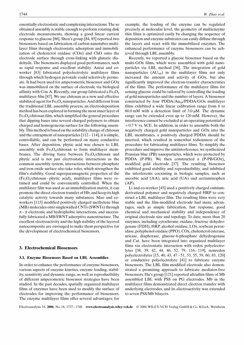
Hu and co-workers [96] reported core-shell nanoclusterfilms of proteins, polyelectrolytes and SiO2 nanoparticles.Nanoparticles were used as “core” materials and the(enzyme/polyion)n multilayers deposited on solid coreparticles formed a bioactive “shell”. These core-shell nano-clusters were then assembled with oppositely charged
polyions on electrodes (see Fig. 4). The proteins in the filmsretained near native conformations in the medium pHrange. The relative fraction of electroactive proteins (G*/G)for the core-shell nanocluster films is larger compared to thecorresponding (SiO2/protein)n LBL films. In addition, thenanocluster films generally have better catalytic efficiency(Ic/Id) than the latter. The effective distance for proteins toexchange electrons with pyrolytic graphite (PG) electrodesof the protein core-shell nanocluster films is twice as long asthat of the (SiO2/protein)n films. The enzyme substratescould enter into the films more easily and had more chanceto react with all the protein in the porous nanocluster films,which improved the catalytic efficiency (Ic/Id) and the (Ic/G).Furthermore, the electroactivity of the {SiO2-(protein/PSS)m/PEI}n, can be more precisely tailored by adjustingboth the shell thickness (m) and the film thickness (n). Thisis an additional advantage of the nanocluster films. Recent-
Fig. 3. Scheme of multilayer films assembly on the gold surface. Reprinted from W. Zhao, J. J. Xu, C. G. Shi, H. Y. Chen, Langmuir2005, 21, 9630.
Fig. 4. Schematic diagram of the idealized structure of LBL{[SiO2�(Hb/PSS)2]/PEI}n films on a PG/PEI/PSS/PEI surface.Reprinted from H. Y. Liu, J. F. Rusling, N. F. Hu, Langmuir2004, 20, 10700.
1741Electrochemical Biosensors Based on LBL Assemblies
Electroanalysis 18, 2006, No. 18, 1737 – 1748 www.electroanalysis.wiley-vch.de I 2006 WILEY-VCH Verlag GmbH & Co. KGaA, Weinheim
ly, a number of papers have reported on explorations ofmacromolecule encapsulation in polyelectrolyte capsules,which show strong potential for biosensor applications [97 –100]. The construction strategy of this three-dimensionalstructure provides a new controllable route to immobilizeproteins in films.
2.2. Biological Interaction
Another strategy for preparing enzyme multilayer films is touse the biological affinity between protein and ligand, bymeans of avidin – biotin, lectin – sugar and antibody – anti-gen interactions.
2.2.1. Avidin –Biotin Interaction
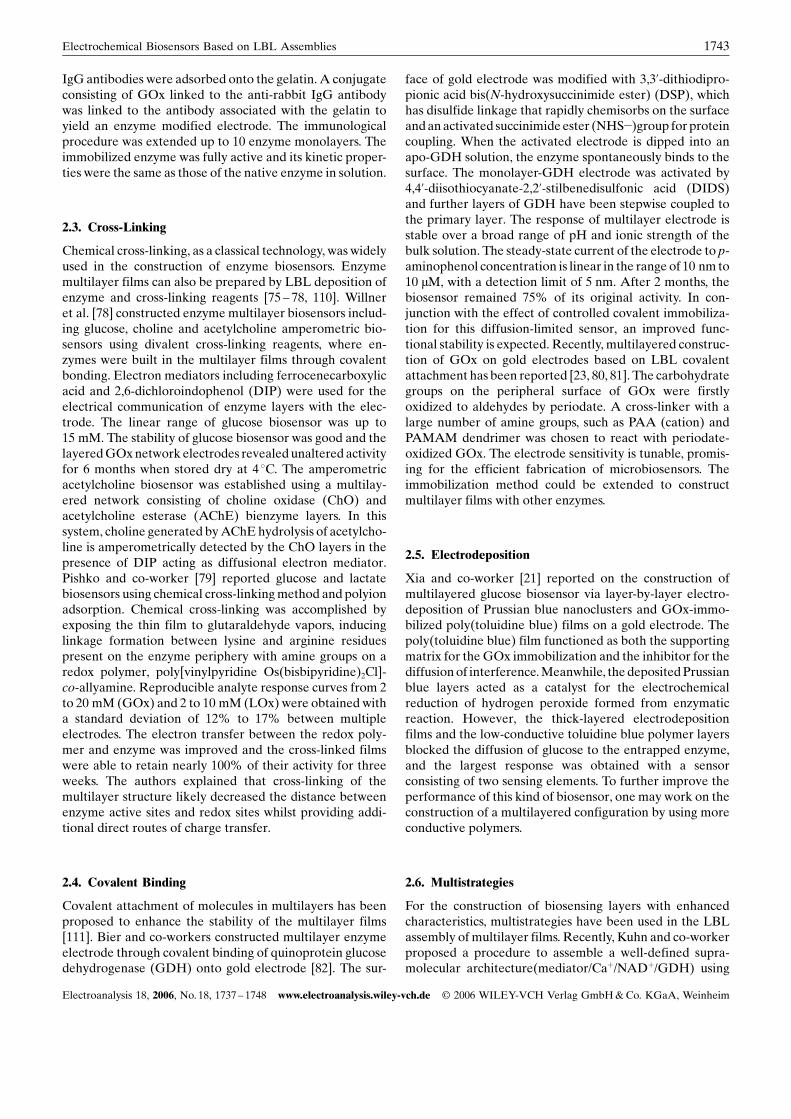
Avidin is a glycoprotein bearing a high affinity for biotin(binding constant, 1015 M�1) [101]. Avidin has nearly a cubicshape and contains four identical biotin-binding sites beingarranged in two pairs on opposite faces of the molecule(molecular weight: 68000). The molecular dimensions ofavidin are reported to be 6.0 nm� 5.5 nm� 4.0 nm, and thebinding sites are located on the 5.5� 6.0 nm face [102].Alternating and repeated deposition of avidin and biotin-labeled enzymes produced a multilayer structure composedof monomolecular layers of protein, as schematically shownin Figure 5. AnzaiNs group [66, 68, 69, 103] has integratedseveral enzymes, such as GOx, lactate oxidase (LOx), andalcohol oxidase, in the LBL structure of avidin and biotin-labeled enzymes. The enzymes were found to be catalyti-cally active in the multilayer films. The authors alsoconstructed bienzyme membrane modified biosensors viaLBL assembly. The GOx/ascorbate oxidase (AOx) andLOx/AOx multilayers were prepared on the surface of aplatinum electrode respectively, and the multilayers wereeffective to eliminate ascorbic acid interference. Thebiosensors could be used to determine the normal bloodlevel of glucose (5 mM) and lactate (1 mM) in the presenceof physiological level of ascorbic acid (0.1 mM) [70, 104].The response time of both sensors was 10 s or faster,irrespective of the number of the AOx layer, confirming thesmooth transport of substrates across the AOx layers. Anacetylcholine biosensor was constructed by optimizing theloading of choline esterase (ChE) and choline oxidase(ChOx) on the surface of a Pt and a Pt-black electrode [67].
It was found that the sensor modified with 10-layer ChOxexhibited its maximum response to acetycholine when 2additional layers of ChE were added to the surface of theChOx layer. The (ChOx)10(ChE)-modified biosensorshowed amperometric response to acetylcholine dependingon its concentration. The response time was fast for allsensors (10 s or faster). It comes from the fact that the ChOx/ChE membranes are so thin that they do not significantlyinfluence the transport of acetylchoine and products of theenzymatic reactions in the membrane. The Spectroscopicand QCM studies revealed that the avidin – biotin system-based enzyme multilayer films are composed of monomo-lecular layers of avidin and biotin-labeled enzyme.
2.2.2. Lectin – Sugar Interaction
A lectin – sugar system provided another way to constructenzyme multilayer films. Concanavalin A (Con A) ap-peared to be a promising lectin for preparing enzymemultilayer films. Con A is a lectin protein (molecular weight104000) found in Jack bean and is known to contain fouridentical binding sites to sugars such as mannose andglucose. The binding between Con A and sugar is not acovalent binding but a reversible one [105]. Lvov et al. [106,107] have prepared a LBL structure of Con A and glycogenthrough the biological affinity, but didnNt extend theapplication of the multilayer films in sugar biosensors.Scheper et al. reported on construction of multilayer filmswith Con A and glycoenzymes via sugar chains on insolublesupports [71]. The sensitivity of a flow-through glucosemonitoring cartridge, integrated into a flow injectionanalysis (FIA) system, was improved significantly byincreasing the amount of immobilized GOx via bioaffinitylayering, but the formation of more than 5 bioaffinity layerson the Sepharose matrix was ineffective in further enhanc-ing the sensitivity. Anzai and co-worker [72] reported thepreparation of bienzyme multilayer films using lectin andglycoenzymes, such as GOx and horseradish peroxidase(HRP) for biosensor applications. The enzymes are catalyti-cally active and stable in the multilayer films and thecatalytic activity of the multilayer films depended linearlyon the number of enzyme layers. The (HRP)5 þ (GOx)5
film-modified electrode can be used to determine 10�6 to10�3 M H2O2 and 10�5 to 10�2 M glucose, with nearly thesame maximum current (ca. 8.0 mA). They also successfullyintegrated mannose-labeled LOx, which contains intrinsi-cally no sugar chain, in the construction of multilayer films[73]. These results show that Con A-sugar complexation is auseful tool for constructing multilayer thin films of enzymes.
2.2.3. Antibody –Antigen Interaction
An antibody – antigen interaction was employed to organizemultilayers for electrochemical biosensors. SaveantNs group[74, 108, 109] reported an immunological method allowingthe LBL assembly of immunoglobulin G (IgG) and anti-IgG-labeled GOx onto a glass carbon electrode surface. Aglass carbon electrode was coated with gelatin, and rabbit
Fig. 5. LBL construction of enzyme layers on the electrodesurface using avidin and biotin-labeled enzyme. Reprinted from J.Anzai, H. Takeshita, Y. Kobayashi, T. Osa, T. Hoshi, Anal. Chem.1998, 70, 811.
1742 W. Zhao et al.
Electroanalysis 18, 2006, No. 18, 1737 – 1748 www.electroanalysis.wiley-vch.de I 2006 WILEY-VCH Verlag GmbH & Co. KGaA, Weinheim
IgG antibodies were adsorbed onto the gelatin. A conjugateconsisting of GOx linked to the anti-rabbit IgG antibodywas linked to the antibody associated with the gelatin toyield an enzyme modified electrode. The immunologicalprocedure was extended up to 10 enzyme monolayers. Theimmobilized enzyme was fully active and its kinetic proper-ties were the same as those of the native enzyme in solution.
2.3. Cross-Linking
Chemical cross-linking, as a classical technology, was widelyused in the construction of enzyme biosensors. Enzymemultilayer films can also be prepared by LBL deposition ofenzyme and cross-linking reagents [75 – 78, 110]. Willneret al. [78] constructed enzyme multilayer biosensors includ-ing glucose, choline and acetylcholine amperometric bio-sensors using divalent cross-linking reagents, where en-zymes were built in the multilayer films through covalentbonding. Electron mediators including ferrocenecarboxylicacid and 2,6-dichloroindophenol (DIP) were used for theelectrical communication of enzyme layers with the elec-trode. The linear range of glucose biosensor was up to15 mM. The stability of glucose biosensor was good and thelayered GOx network electrodes revealed unaltered activityfor 6 months when stored dry at 4 8C. The amperometricacetylcholine biosensor was established using a multilay-ered network consisting of choline oxidase (ChO) andacetylcholine esterase (AChE) bienzyme layers. In thissystem, choline generated by AChE hydrolysis of acetylcho-line is amperometrically detected by the ChO layers in thepresence of DIP acting as diffusional electron mediator.Pishko and co-worker [79] reported glucose and lactatebiosensors using chemical cross-linking method and polyionadsorption. Chemical cross-linking was accomplished byexposing the thin film to glutaraldehyde vapors, inducinglinkage formation between lysine and arginine residuespresent on the enzyme periphery with amine groups on aredox polymer, poly[vinylpyridine Os(bisbipyridine)2Cl]-co-allyamine. Reproducible analyte response curves from 2to 20 mM (GOx) and 2 to 10 mM (LOx) were obtained witha standard deviation of 12% to 17% between multipleelectrodes. The electron transfer between the redox poly-mer and enzyme was improved and the cross-linked filmswere able to retain nearly 100% of their activity for threeweeks. The authors explained that cross-linking of themultilayer structure likely decreased the distance betweenenzyme active sites and redox sites whilst providing addi-tional direct routes of charge transfer.
2.4. Covalent Binding
Covalent attachment of molecules in multilayers has beenproposed to enhance the stability of the multilayer films[111]. Bier and co-workers constructed multilayer enzymeelectrode through covalent binding of quinoprotein glucosedehydrogenase (GDH) onto gold electrode [82]. The sur-
face of gold electrode was modified with 3,3’-dithiodipro-pionic acid bis(N-hydroxysuccinimide ester) (DSP), whichhas disulfide linkage that rapidly chemisorbs on the surfaceand an activated succinimide ester (NHS�)group for proteincoupling. When the activated electrode is dipped into anapo-GDH solution, the enzyme spontaneously binds to thesurface. The monolayer-GDH electrode was activated by4,4’-diisothiocyanate-2,2’-stilbenedisulfonic acid (DIDS)and further layers of GDH have been stepwise coupled tothe primary layer. The response of multilayer electrode isstable over a broad range of pH and ionic strength of thebulk solution. The steady-state current of the electrode to p-aminophenol concentration is linear in the range of 10 nm to10 mM, with a detection limit of 5 nm. After 2 months, thebiosensor remained 75% of its original activity. In con-junction with the effect of controlled covalent immobiliza-tion for this diffusion-limited sensor, an improved func-tional stability is expected. Recently, multilayered construc-tion of GOx on gold electrodes based on LBL covalentattachment has been reported [23, 80, 81]. The carbohydrategroups on the peripheral surface of GOx were firstlyoxidized to aldehydes by periodate. A cross-linker with alarge number of amine groups, such as PAA (cation) andPAMAM dendrimer was chosen to react with periodate-oxidized GOx. The electrode sensitivity is tunable, promis-ing for the efficient fabrication of microbiosensors. Theimmobilization method could be extended to constructmultilayer films with other enzymes.
2.5. Electrodeposition
Xia and co-worker [21] reported on the construction ofmultilayered glucose biosensor via layer-by-layer electro-deposition of Prussian blue nanoclusters and GOx-immo-bilized poly(toluidine blue) films on a gold electrode. Thepoly(toluidine blue) film functioned as both the supportingmatrix for the GOx immobilization and the inhibitor for thediffusion of interference. Meanwhile, the deposited Prussianblue layers acted as a catalyst for the electrochemicalreduction of hydrogen peroxide formed from enzymaticreaction. However, the thick-layered electrodepositionfilms and the low-conductive toluidine blue polymer layersblocked the diffusion of glucose to the entrapped enzyme,and the largest response was obtained with a sensorconsisting of two sensing elements. To further improve theperformance of this kind of biosensor, one may work on theconstruction of a multilayered configuration by using moreconductive polymers.
2.6. Multistrategies
For the construction of biosensing layers with enhancedcharacteristics, multistrategies have been used in the LBLassembly of multilayer films. Recently, Kuhn and co-workerproposed a procedure to assemble a well-defined supra-molecular architecture(mediator/Caþ/NADþ/GDH) using
1743Electrochemical Biosensors Based on LBL Assemblies
Electroanalysis 18, 2006, No. 18, 1737 – 1748 www.electroanalysis.wiley-vch.de I 2006 WILEY-VCH Verlag GmbH & Co. KGaA, Weinheim
essentially electrostatic and complexing interactions. The soobtained assembly is stable enough to perform rotating diskelectrode measurements, showing a good linear currentresponse to glucose [86]. ShenNs group [84, 85] reported twobiosensors based on fabrication of carbon nanotubes multi-layer films through electrostatic adsorption and immobili-zation of cholesterol oxidase (COx) and ChO onto theelectrode surface through cross-linking with glutaric dia-dehyde. The biosensors displayed good performances, suchas rapid response and excellent stability. Anzai and co-worker [83] fabricated polyelectrolyte multilayer filmsthrough which hydrogen peroxide could selectively perme-ate. It had been used for amperometric biosensor and GOxwas immobilized on the surface of electrode via biologicalaffinity with Con A. Recently, our group fabricated a Fe3O4
multilayer film [87]. We use chitosan as a new and effectivestabilized agent for Fe3O4 nanoparticles. And different fromthe traditional LBL assembly process, an electrodepositionmethod has been explored in forming the most inner layer ofFe3O4/chitosan film, which simplified the general procedurethat dipping bases into several charged polymers to obtaincharged and homogeneous surface for the following assem-bly. This method is based on the solubility change of chitosanand the entrapment of nanoparticles [112 – 114], it is simple,controllable, and can be performed on many conductivebases. After deposition, phytic acid was chosen to LBLassembly with Fe3O4/chitosan to form multilayer mem-branes. The driving force between Fe3O4/chitosan andphytic acid is not just electrostatic interactions as thecommon assembly system, interactions between phosphateand iron oxide surface also take roles, which strengthen thefilmNs stability. Good superparamagnetic properties of the(Fe3O4/chitosan – phytic acid)n multilayer films were re-tained and could be conveniently controlled. When themultilayer film was used as an immobilization matrix, it canpromote the direct electron transfer of Hb, and keep its highcatalytic activity towards many substances. Mao and co-workers [115] modified positively charged methylene blue(MB) molecules onto singlewalled CNTs (SWNTs) throughp –p electronic and hydrophobic interactions, and success-fully fabricated a MB/SWNTadsorptive nanostructure. Theexcellent electroactivity and the high stability of the layerednanocomposite are envisaged to make them perspective forthe development of electrochemical biosensors.
3. Electrochemical Biosensors
3.1. Enzyme Biosensors Based on LBL Assemblies
In order to enhance the performance of enzyme biosensors,various aspects of enzyme kinetics, enzyme loading, stabil-ity, sensitivity and dynamic range, as well as reproducibilityof different amperometric biosensor strategies have beenstudied. In the past decades, spatially organized multilayerfilms of enzymes have been used to modify the surface ofelectrodes for improving the performance of biosensors.The enzyme multilayer films offer several advantages; for
example, the loading of the enzyme can be regulatedprecisely at molecular level, the geometry of multienzymethin films is optimized easily by changing the sequence ofdeposition and enzyme substrates can easily diffuse throughthe layers and react with the immobilized enzymes. Theenhanced performance of enzyme biosensors can be ach-ieved through LBL assembly.
Recently, we reported a glucose biosensor based on themulti-GOx films, which were assembled with gold nano-particles via LBL method [36]. The integration of goldnanoparticles (AUNP) in the multilayer films not onlyincreased the amount and activity of GOx, but alsosignificantly improved the electron-transfer characteristicsof the films. The performance of the multilayer films forsensing glucose could be tailored by controlling the loadingof gold nanoparticles and the number of layers. A biosensorconstructed by four PDDA/AuNP/PDDA/GOx multilayerfilms exhibited a wide linear calibration range from 0 to60.0 mM with a detection limit of 3.0 mM. The dynamicrange can be extended even up to 120 mM. However, theinterference cannot be excluded at an operating potential ofþ0.7 V vs. SCE. In addition, in order to assemble both thenegatively charged gold nanoparticles and GOx into theLBL membranes, a positively charged PDDA should beinserted, which resulted in somewhat tediousness of theprocedure for fabricating multilayer films. To simplify theprocedure and improve the antiinterference, we synthesizedPrussian blue (PB) nanoparticles, which were protected byPDDA (P-PB). We then constructed a (P-PB/GOx)nmodified gold electrode [37]. The resulting biosensorexhibited good stability and reproducibility, and inhibitedthe interferents coexisting in biologic samples, such asascorbic acid (AA), uric acid (UA) and acetaminophen(AP).
Li and co-worker [45] used a positively charged osmium-derivatized polymer and negatively charged HRP to con-struct a LBL multilayer film. The resulting films were verystable and the film-modified electrode had many advan-tages, such as simple fabrication, fast response, goodchemical and mechanical stability and independence oforiginal electrode size and topology. To date, more than 20enzymes, including cytochrome oxidase, fructose dehydro-genase (FDH), HRP, alcohol oxidase, LOx, soybean perox-idase, polyphenol oxidase (PPO), COx, cholesterol esterase,uricase, diaphorase, glucose-6-phosphate dehydrogenaseand Cat, have been integrated into organized multilayerfilms via electrostatic interaction with redox polyelectro-lytes [38, 39, 42, 44, 46, 52, 79, 116 – 119], nonredoxpolyelectrolytes [15, 40, 43, 47 – 51, 53, 55, 59, 60, 83, 120]or conductive polyelectrolyte [41] to fabricate enzymebiosensors. The LBL film modified electrode also demon-strated a promising approach to fabricate mediator-freebiosensors. HuNs group [121] reported ultrathin films of Mbassembled LBL with PSS on PG electrodes. Mb in themultilayer films demonstrated direct electron transfer withunderlying electrodes, and its electroactivity was extendedto seven PSS/Mb bilayers.
1744 W. Zhao et al.
Electroanalysis 18, 2006, No. 18, 1737 – 1748 www.electroanalysis.wiley-vch.de I 2006 WILEY-VCH Verlag GmbH & Co. KGaA, Weinheim
More sophisticated approaches have been reported,integrating more than one enzyme in the LBL assembly toachieve a co-operative mechanism. Nicolini et al. developeda cholesterol amperometric biosensors fabricated via asem-bling COx and cholesterol esterase (CE) into PE multilayers[61]. In another example, Gobi and Mizutani [52] assembledCOx by LBL assembly on a monolayer of microperoxidaseand observed a fast and reproducible response. Thebiosensor was inert and stable in the analysis of physiolog-ical samples of cholesterol with the presence of electroactiveinterferents.
3.2. Immunosensors
Recently, various kinds of immunosensors for clinical,environmental and food purposes have been developed asa result of the possibility of generating a large number ofantigens for the analysis of numerous chemical species andof the added advantage of analyzing samples without needof pretreatment. Construction of biological recognitionlayer is of essential fundamental for tailoring of suchbiosensors. Increasing the binding layer capacity and mean-while remaining its immunological active are two key issues,in terms of sensitivity. Therefore, construction of multilayerfilms with proteins via LBL method has drawn muchattention toward the preparation of immunosensors withimproved performance [20, 28, 62,122]. In order to increasethe binding layer capacity of the thin film with respect toimmunoglobulin G (IgG) detection, Furlong and co-work-ers [28] constructed alternating polyelectrolyte/anti-IgGlayers. The immunological activity of immobilized anti-IgGin the multilayer films was investigated by subsequentbinding with IgG. The sensitivity, determined using IgGmass uptake data from QCM, was found to be linearlydependent on the number of anti-IgG layers in thepolyelectrolyte film when the anti-IgG layers are separatedby one polyelectrolyte layer. In contrast, for films whereanti-IgG layers are separated by five polyelectrolyte layers,only the outer anti-IgG layer is immunologically active, dueto the formation of a dense polyelectrolyte film throughwhich antibody permeation is restricted. Bai et al. [20]reported an immunosensor based on LBL assembly ofcolloidal gold particles using cysteamine as cross-linkers.The gold particles exhibited an easier attachment of anti-body, good biocompatibility and large surface area, con-tributing to the large loading of antibody. Further coupledwith an amplification strategy to use biotin-conjugatedantibody, the sensor exhibited a linear range from 5 –400 mg/L with a detection limit of 0.5 mg/L using electro-chemical impedance measurements. Anzai and co-worker[123] prepared spatially ordered multilayer thin filmscontaining antifluoresceinisothiocyanate (anti-FITC) onthe surface of a quartz slide by the alternate deposition ofavidin and biotin-labeled anti-FITC. The anti-FITC in theoutermost four to five layers of the multilayer films can bindits antigen reversibly. Thus, the binding capability of themultilayer films is four to five times higher than that of the
antibody monolayer and was proofed to be useful inimproving the performance of immunosensors. Later on,Yang et al. [124] reported an immunosensor for IgG basedon this work, using SPR technique. The IgG protein could bedetected in a range of 0.5 – 20 mg/mL.
Recently, Yuan and co-worker [62] showed a differentway to improve the sensitivity by increasing the loading ofsignal reporters, not of antibodies. Initially, they integratedelectrochemically active Co(bpy)3þ
3 in alternation with goldnanoparticles for making a {Au/Co(bpy)3þ
3 }n multilayer filmand then developed novel electrochemical immunosensorsby immobilizing hepatitis B surface antibody (HBsAb) onthe gold nanoparticle surface. The gold nanoparticlescontributed to the performance of Co(bpy)3þ
3 and theelectrochemical response of Co(bpy)3þ
3 was tuned by thelayer number. Both amperometric and potentiometricmethods were used to detect the hepatitis B surface antigen(HBsAg). When the antibodies immobilized on the elec-trode have bound with antigens, the antigen – antibodycomplex of insulting features blocked the electron transferand accordingly, the amperometric response decreased.Meanwhile, as the isoelectric point of HBsAg is more than7.0 and thus positively charged in pH 7.0 buffer solutions,the potentiometric response increased after the antigen –antibody reaction. Both the linear range for HBsAg wasfrom 0.05 to 4.5 mg/mL with a detection limit of 0.005 mg/mLfor the amperometric system and 0.015 mg/mL for thepotentiometric system, respectively.
3.3. DNA Sensors
Oxidative DNA damage may lead to aging, cancer andmutagenesis [125]. Guanine is the most easily oxidizedamong the four DNA bases, with an oxidation potential of1.06 V versus SCE at pH 7 at mercury electrode [126]. Themajor initial product of guanine oxidation is the highlymutagenic 8-oxoguanine, which is suggested as a clinicalbiomarker for oxidative stress [127]. Guanine in singlestranded DNA is more easily oxidized than in doublestranded DNA (ds-DNA) because of better accessibility byelectrodes or oxidizing agents [128]. As DNA damage hasthe effect of unwinding the double helix, a closer access tothe nucleobases is allowed [129]. In conventional electro-chemical approaches, DNA in solution was preconcentratedon mercury electrodes by adsorption prior to analysis and, alarger voltammetric signals can be obtained if oxidativedamage occurs [128]. DNA voltammetry with high sensitiv-ity can be achieved by using electrochemical catalyticoxidation with transition metal complexes such asRu(bpy)3
2þ [130].Recently, Rusling et al. [63] constructed films of
[Ru(bpy)2(PVP)10Cl]Cl {PVP¼ poly(4-vinyl-pyridine)}and ds-DNA grown by layer-by-layer alternate electrostaticassembly. The redox polymer [Ru(bpy)2(PVP)10Cl]Cl as aninner layer in films was used to catalyze the voltammetricoxidation of guanine bases of ds-DNA in the outer layers.This film architecture provides a reagentless sensor for
1745Electrochemical Biosensors Based on LBL Assemblies
Electroanalysis 18, 2006, No. 18, 1737 – 1748 www.electroanalysis.wiley-vch.de I 2006 WILEY-VCH Verlag GmbH & Co. KGaA, Weinheim
toxicity screening of new chemicals based on detection ofDNA damage. Later on, films containing [Os(bpy)2(PVP)10
Cl]þ and [Ru(bpy)2(PVP)10Cl]þ metallopolymers were as-sembled layer-by-layer on pyrolytic graphite electrodes toobtain sensors for selectively detection of oxidized DNA[14]. These films showed reversible, independent electro-chemistry for electroactive Os3þ/Os2þ and Ru3þ/Ru2þ cen-ters, with formal potentials of 0.34 and 0.76 V vs. SCE,respectively. The combination of ruthenium and osmiummetallopolymers in the films allows for the detection of 8-oxoguanine and other oxidized nucleobases simultaneously.The catalytic Os square wave voltammetry (SWV) peak ismainly selective for 8-oxoguanine because the Os3þ selec-tively oxidized 8-oxoguanine but not guanine [64]. The otheroxidized nucleobases along with strand cleavage can bedetected from the Ru peak. Using the Os SWV peak, oneoxidize nclueobase among 6000 was detected. Rusling et al.[65] also reported that ultrathin films containing [Os(bpy)2
(PVP)10Cl]2þ and DNA layer-by-layer assembled on elec-trodes directly generated electrochemiluminescence (ECL)from oxidized DNA without using a sacrificial reductant.Based on films combining [Os(bpy)2(PVP)10Cl]2þ and[Ru(bpy)2(PVP)10Cl]2þ formed via LBL assembly, a directECL method was presented to detect DNA oxidation andnucleobase adducts from chemical damage.
In addition, the LBL assembly technique can be applied tothe DNA sequence detection, mainly for the enzyme-labeled sensing schemes. Immobilization of enzymes andmediators plays an important role in the construction ofenzyme-labeled DNA sequence biosensors, because en-zymes and mediators should be stable and easy enough to becontacted by substrates, and the immobilization procedureshould be beneficial to electron transfer between enzymesand electrode surface by redox mediator [130]. Polyelectro-lyte modified with mediators seems to be very promising aselectrical contact materials. The immobilization of media-tors modified polyelectrolyte via LBL assembly provides amore stable and much more mediator multilayer, whencompared to conventional physical adsorption of mediators.Recently, Suye et al. [88] reported an amperometric DNAsensing system based on the sandwich hybridization. Firstly,the gold electrode was modified with mercaptopropionicacid, and then PEI-Fc (ferrocene immobilized polyethyle-nimine)/alginic acid, diaphorase/PEI, and PEI/streptavidinlayers grown on the electrode surface via LBL assembly. Thecapture probe was subsequently immobilized on the elec-trode via streptavidin to make the sequence biosensor.Hybridization with the target DNA and glucose-6-phos-phate dehydrogenase labeled reporter probe was carriedout simultaneously at 56 8C. The complete hybridizationwould produce a catalytic signal to glucose-6-phosphate.The detection limit was down to femto mol order of targetDNA. To remarkably increase the sensitivity, an alternativesignal amplification scheme is to maximize the ratio ofenzyme tags per binding event. The unique properties ofCNT, particularly their huge surface area-to-weight ratio,make them extremely attractive amplification platforms.Recently, Wang et al. [89] reported a novel method for
dramatically amplifying the electrochemical detection ofproteins or DNA based on a stepwise LBL assembly ofmultilayer enzyme films on a carbon nanotube (CNT)template. The biomagnetic separation technique was used inDNA hybridization assay. Briefly, the biotinylated DNAprobe 1 was coupled to the streptavidin-coated magneticbeads and the target DNAwas then hybridized to the probe1. The resulting hybrid-conjugated magnetic beads wereagain hybridized to the biotinylated DNA probe 2. Sub-sequently, the CNT-(PDDA/ALP)4-PDDA-streptavidintags were linked to the completed sandwich-hybridizedmagnetic beads. After incubation with the a-naphthylphosphate substrate, the enzymatically liberated a-naph-thol was measured at a MWCNT-modified glassy carbonelectrode via square-wave voltammetric technique. Suchamplified assay allow detection of DNA down to 80 copies(5.4 aM), representing the lowest value reported to date forelectrical DNA detection.
3.4. ENFET Biosensors
Besides electrodes, ion-sensitive field-effect transistor (IS-FET) is another attractive transducer for electrochemicalbiosensors. ISFET, which was first developed in the early1970s, has been introduced as an alternative to the fragileglass electrode for the measurement of pH and other ionsconcentrations. ISFET has several significant advantages,such as small size, rapid response, low output impedance andlow-cost mass production. Since the first introduction of theenzyme field-effect transistor (ENFET) for penicillin,considerable efforts have been devoted to the developmentof ENFET biosensors. ENFETs are usually constructed byimmobilizing enzymes on the gate insulator of an ISFET.The sensing performance of the ENFET is greatly affectedby the integration method of the enzymes. A variety oftechniques have been introduced for the enzyme immobi-lization on the gate surface of ISFETs, such as cross-linking,entrapment in organic polymers and embedding in addi-tional membranes such as Nafion. However, the ENFETsconstructed by these methods may suffer from limitationsassociated with diffusion barriers of the substrates orleakage of enzymes. Covalent attachment of enzymes canovercome such shortages, but it may result in lose ofenzymatic activity partially. We immobilized LOx andMnO2 nanoparticles in PDDA films via LBL assemblymethod to develop lactate ENFET [54]. MnO2 nanoparti-cles were introduced as an oxidant to react with hydrogenperoxide, which results in a sensitive pH change in thesensitive membrane of the ENFET with the addition oflactate. By introducing MnO2 nanoparticles into the multi-layers, the sensitivity of the ENFET is increased to16.84 mV/mM, 50 times higher than that without MnO2
nanoparticles. The dynamic range is extended up to6.0 mM, with a detection limit of 8.0 mM. The biosensorconstructed via LBL assembly demonstrated an excellentlong-term stability.
1746 W. Zhao et al.
Electroanalysis 18, 2006, No. 18, 1737 – 1748 www.electroanalysis.wiley-vch.de I 2006 WILEY-VCH Verlag GmbH & Co. KGaA, Weinheim
4. Conclusions
Electrochemical biosensors are a wide area of research,which continues to develop at a rapid pace. As a simple,rapid, reliable and cost-effective method, the LBL assemblytechnique has drawn extremely attention over the pastdecade. Various LBL assembly strategies have been inte-grated to incorporate enzymes, proteins and DNA in theorganized multilayer films for the design of biosensors. TheLBL assembly method allows the controlling of nano-structure and nanoarchitecture properties, such as charge,thickness, polarity, roughness of the multilayer films. It willhave more applications in biosensors, biocatalysis andbiotechnology field.
5. Acknowledgements
The financial supports from the National Natural ScienceFoundation of China (NSFC) (No. 20575029, 90206037,20475025, 20435010), and the Science Foundation of JiangsuProvince (BK 2004210) are gratefully acknowledged.
6. References
[1] R. K. Iler, J. Colloid Interface Sci. 1966, 21, 569.[2] G. Decher, J.-D. Hong,Macromol. Chem., Macromol. Symp.
1991, 46, 321.[3] Y. Lvov, K. Anga, I. Ichinose, T. Kunitake, J. Am. Chem.
Soc. 1995, 117, 6117.[4] Y. Lvov, Z. Lu, J. B. Schenkman, X. Zu, J. F. Rusling, J. Am.
Chem. Soc. 1998, 120, 4073.[5] G. Decher, B. Lehr, K. Lowack, Y. Lvov, J. B. Schmitt,
Biosens. Bioelectron. 1994, 9, 677.[6] Y. Lvov, G. Decher, G. Sukhorukov, Macromolecules 1993,
26, 5396.[7] Y. Lvov, J. Haas, G. Decher, H. Mçhwald, A. Mikhailov, B.
Mtchedlishvily, E. Morgunova, B. Vainshtein, Langmuir1994, 10, 4232.
[8] S. Watanabe, S. L. Regan, J. Am. Chem. Soc. 1994, 116,8855.
[9] P. A. Fiorito, V. R. Goncales, E. A. Ponzio, S. I. Cordoba deTorresi, Chem. Commun. 2005, 366.
[10] F. Patosky, T. Gabriel, I. Willner, J. Electroanal. Chem. 1999,479, 69.
[11] Y. Lvov, B. Munge, O. Giraldo, I. Ichinose, S. Suib, J. F.Rusling, Langmuir 2000, 16, 8850.
[12] Z. Li, N. F. Hu, J. Colloid Interface Sci. 2002, 254, 257.[13] J. P. Santos, E. R. Welsh, B. P. Gaber. A. Singh, Langmuir
2001, 17, 5361.[14] A. Mugweru, B. Q. Wang, J. Rusling, Anal. Chem. 2004, 76,
5557.[15] E. S. Forzani, M. L. Teijelo, F. Nart, E. J. Calvo, V. M. Solis,
Biomacromolecules 2003, 4, 869.[16] Y. Shimazaki, M. Mitsuishi, S. Ito, M. Yamamoto, Langmuir
1998, 14, 2768.[17] C. Li, K. Mitamura, T. Imae, Macromolecules 2003, 36,
9957.[18] R. Pei, X. Cui, X. Yang, E. Wang, Biomacromolecules 2001,
2, 463.[19] O. A. Raitman, E. Katz, A. F. BVckmann, I. Willner, J. Am.
Chem. Soc. 2002, 124, 6487.
[20] M. J. Wang, L. Y. Wang, H. Yuan, X. H. Ji, C. Y. Sun, L. Ma,Y. B. Bai, T. J. Li, J. H. Li, Electroanalysis 2004, 16, 757.
[21] D. Zhang, Ke, Zhang, Y. L. Yao, X. H. Xia, H. Y. Chen,Langmuir 2004, 20, 7303.
[22] L. Cheng, J. Liu, S. Dong, Anal. Chim. Acta 2000, 417, 133.[23] S. X. Zhang, W. W. Yang, Y. M. Niu, C. Q. Sun. Anal. Chim.
Acta 2004, 523, 209.[24] F. Caruso, D. N., Furlong, K. Ariga, I. Ichinose, T. Kunitake,
Langmuir 1998, 14, 4599.[25] G. B. Sukhorukov, M. M. Montrel, A. I. Petrov, L. I. Shabar-
china, B. I. Sukhorukov, Biosens. Bioelectron. 1996, 11, 913.[26] F. Caruso, L. Mçhwald, J. Am. Chem. Soc. 1999, 121, 6039.[27] J. A. He, L. Samuelson, L. Li, S. K. Tripathy, Langmuir
1998, 14, 1674.[28] F. Caruso, K. Niikura, D. N. Furlong, Y. Okahata, Langmuir
1997, 13, 3427.[29] J. Lang, M. H. Lin, J. Phys. Chem. B 1999, 103, 11393.[30] F. Mizutani, Y. Sato, S. Yabuki, Y. Hirata, Chem. Lett. 1996,
4, 251.[31] S. F. Hou, H. Q. Fang, H. Y. Chen,Anal. Lett. 1997, 30, 1631.[32] J. Hodak, R. Etchenique, E. J. Calvo, K. Singhal, P. N.
Bartlett, Langmuir 1997, 13, 2708.[33] S. .F. Hou, K. .S. Yang, H. Q. Fang, H. Y. Chen, Talanta
1998, 47, 561.[34] E. J. Calvo, C. Danilowicz, A. Wolosiuk, J. Am. Chem. Soc.
2002, 124, 2452.[35] H. T. Zhao, H. X. Ju, Anal. Biochem. 2006, 350, 138.[36] W. Zhao, J. J. Xu, H. Y. Chen, Frontiers Biosci. 2005, 10,
1060.[37] W. Zhao, J. J. Xu, C. G. Shi, H. Y. Chen, Langmuir 2005, 21,
9630.[38] M. C. Rodriguez, G. A., Rivas, Electroanalysis 2004, 16,
1717.[39] E. J. Calvo, R. Etchnique, L. Pietrasanta, A. Wolosiuk, C.
Danilowicz, Anal. Chem. 2001, 73, 1161.[40] D. Trau, R. Renneberg, Biosens. Bioelectron. 2003, 18, 1491.[41] M. K. Ram, M. Adami, S. Paddeu, C. Nicolini, Nano-
technology 2000, 11, 112.[42] Y. Sun, J. Sun, X. Xhang, C. Sun, Y. Wang, J. Shen, Thin
Solid Films 1998, 327 – 329, 730.[43] W. J. Zhang, Y. X. Huang, H. Dai, X. Y. Wang, C. H. Fan,
G. X. Li, Anal. Biochem. 2004, 329, 85.[44] B. Lindholm-Sethson, J. C. Gonzalez, G. Puu, Langmuir
1998, 14, 6705.[45] W. Li, Z. Wang, C. Sun, M. Xian, M. Zhao, Anal. Chim.
Acta 2000, 418, 225.[46] V. Rosca, J. C. Popescu, Electrochem. Commun. 2002, 4,
904.[47] C. Sun, W. Li, Y. Sun, X. Zhang, J. Shen, Electrochim. Acta
1999, 44, 3401.[48] E. S. Forzani, V. M. Solis, E. J. Calvo, Anal. Chem. 2000, 72,
5300.[49] L. Coche-Guerente, J. Desbrieres, J. Fatisson, P. Labbe,
M. C. Rodriguez, G. Rivas, Electrochim. Acta 2005, 50,2865.
[50] T. Hoshi, H. Saiki, J. Anzai, Talanta 2003, 61, 363.[51] A. Yu, F. Caruso, Anal. Chem. 2003, 75, 3031.[52] K. V.Gobi, F. Mizutani, Sens. Actuators B 2001, 80, 272.[53] X. Wei, M. Zhang, W. Gorski, Anal. Chem. 2003, 75, 2060.[54] J. J. Xu, W. Zhao, X. L. Luo, H. Y. Chen, Chem. Commun.
2005, 792.[55] H. Zheng, H. Okada, S. Nojima, S.-I. Suye, T. Hori, Sci.
Technol. Adv. Mater. 2004, 5, 371.[56] A. Narvaez, G. Suarez, I. C. Popescu, I. Katakis, E.
Dominguez, Biosens. Bioelectron. 2000, 15, 43.[57] Y. Zhou, Z. Li, N. Hu, Y. Zeng, J. F. Rusling, Langmuir
2002, 18, 8573.
1747Electrochemical Biosensors Based on LBL Assemblies
Electroanalysis 18, 2006, No. 18, 1737 – 1748 www.electroanalysis.wiley-vch.de I 2006 WILEY-VCH Verlag GmbH & Co. KGaA, Weinheim
[58] Z. Li, N. F. Hu, J. Electroanal. Chem. 2003, 558, 155.[59] L. Shi, Y. Lu, J. Sun, J. Zhang, C. Sun, J. Liu, J. Shen,
Biomacromolecules 2003, 4, 1161.[60] Q. T. Nguyen, Z. Ping, T. Nguyen, P. Rigal, J. Membr. Sci.
2003, 213, 85.[61] M. K. Ram, P. Bertoncello, H. Ding, S. Paddeu, C. Nicolini,
Biosens. Bioelectron. 2001, 16, 849.[62] D. P. Tang, R. Yuan, Y. Q. Chai, Y. Z. Fu, J. Y. Dai, Y. Liu,
X. Zhong, Biosens. Bioelectron. 2005, 21, 539.[63] B. Wang, J. F. Rusling, Anal. Chem. 2003, 75, 4229.[64] P. A. Ropp, H. H. Thorp, Chem. Biol. 1999, 6, 599.[65] L. Dennany, R. J. Forster, B. White, M. Smyth, J. F. Rusling,
J. Am. Chem. Soc. 2004, 126, 8835.[66] T. Hoshi, J. Anzai, T. Osa, Anal. Chem. 1995, 67, 770.[67] Q. Chen, Y. Kobayashi, H. Takeshita, T. Hoshi, J. Anzai,
Electroanalysis 1998, 10, 94.[68] X. Du, J. Anzai, T. Osa, R. Motohashi, Electroanalysis 1996,
8, 813.[69] J. Anzai, Y. Kobayashi, Y. Suzuki, H. Takeshita, Q. Chen,T.
Osa, T.Hoshi, X. Du, Sens. Actuators B 1998, 52, 3.[70] J. Anzai, H. Takeshita, Y. Kobayashi, T. Osa, T. Hoshi,Anal.
Chem. 1998, 70, 811.[71] M. Farooqi, M. Saleemuddin, R. Ulber, P. Sosnitza, T.
Scheper, J. Biotechnol. 1997, 55, 171.[72] J. Anzai, Y. Kobayashi Langmuir 2000, 16, 2851.[73] Y. Kobayashi, J. Anzai, J. Electroanal. Chem. 2001, 507, 250.[74] C. Bourdillon, C. Demaille, J. Moiroux, J. M. Saveant, Acc.
Chem. Res. 1996, 29, 529.[75] I. Willner, A. Riklin, B. Shoham, D. Rivenson, E. Katz, Adv.
Mater. 1993, 5, 912.[76] I. Willner, M. Liom-Degan, S. Marx-Tibbon, E. Katz, J. Am.
Chem. Soc. 1995, 117, 6581.[77] B. Shoham, Y. Migron, A. Riklin, I. Willner, B. Tartakovsky,
Biosens. Bioelectron. 1995, 10, 341.[78] A. Riklin, I. Willner, Anal. Chem. 1995, 67, 4118.[79] K. Sirkar, A. Revzin, M. V. Pishko, Anal. Chem. 2000, 72,
2930.[80] S. X. Zhang, W. W. Yang, Y. M. Niu, C. Q. Sun Sens.
Actuators B 2004, 101, 387.[81] H. C. Yoon, M. Y. Hong, H. S. Kim, Anal. Chem. 2000, 72,
4420.[82] W. Jin, F. Bier, U. Wollenberger, F. Scheller, Biosens.
Bioelectron. 1995, 10, 823.[83] T. Hoshi, H. Saiki, S. Kuwazawa, C. Tsuchiya, Q. Chen, J. I.
Anzai, Anal. Chem. 2001, 73, 5310.[84] F. L. Qu, M. H. Yang, J. H. Jiang, G. L. Shen, R. Q. Yu,
Anal. Biochem. 2005, 344, 108.[85] M. H. Yang, Y. Yang, H. F. Yang, G. L. Shen, R. Q. Yu,
Biomaterials 2006, 27, 246.[86] N. Mano, A. Kuhn, Talanta 2005, 66, 21.[87] G. Zhao, J. J. Xu, H. Y. Chen, Electrochem. Commun., 2006,
8, 148.[88] S. Suye, T. Matsuura, T. Kimura, H. Zheng, T. Hori, Y.
Amano, H. Katayama, Microelectr. Eng. 2005, 81, 441.[89] B. Munge, G. Liu, G. Collins, J. Wang, Anal. Chem. 2005, 77,
4662.[90] L. Shen, N. F. Hu, Biomacromolecules 2005, 6, 1475.[91] N. Kimizuka, M. Tanaka, T. Kunitake, Chem. Lett. 1999, 12,
1333.[92] C. A. Constantine, K. M. Gattas-Asfura, S. V. Mello, G.
Crespo, V. Rastogi, T. C. Cheng, J. J. DeFrank, R. M.Leblanc, Langmuir 2003, 19, 9863.
[93] P. L. He, N. F.Hu, J. F. Rusling, Langmuir 2004, 20, 722.[94] M. N. Zhang, K. P. Gong, H. W. Zhang, L. Q. Mao, Biosens.
Bioelectron. 2005, 20, 270.[95] J. Y. Liu, S. J. Tian, W. Knoll, Langmuir 2005, 21, 5596.
[96] H. Y. Liu, J. F. Rusling, N. F. Hu, Langmuir 2004, 20, 10700.[97] H. G. Zhu, R. Srivastava, M. J. McShane, Biomacromole-
cules 2005, 6, 2221.[98] Z. H. An, C. Tao, G. Lu, H. Mçhwald, S. P. Zheng, Y. Cui,
J. B. Li, Chem. Mater. 2005, 17, 2514.[99] R. Heuberger, G. Sukhorukov, J. Vçrçs, M. Textor, H.
Mçhwald, Adv. Funct. Mater. 2005, 15, 357.[100] N. G. Balabushevitch, G. B. Sukhorukov, N. A. Moroz, D. V.
Volodkin, N. I. Larionova, E. Donath, H. Mohwald, Bio-technol. Bioeng. 2001, 76, 207.
[101] M. Wilchek, E. Bayer, Anal. Biochem. 1988, 171, 1.[102] L. Pugliese, A. Coda, M. Malcovati, M. Bolognesi, J. Mol.
Biol. 1993, 231, 698.[103] J. Anzai, H. Takeshita, T. Hoshi, T. Osa, Chem. Pharm. Bull.
1995, 43, 520.[104] J. Anzai, H. Takeshita, T. Hoshi, T. Osa, Denki Kagaku
1995, 63, 1141.[105] J. W. Becker, G. N. Jr. Reeke, B. A. Cunningham, G. M.
Edelman, Nature 1976, 259, 406.[106] Y. Lvov, K. Ariga, I. Ichinose, T. Kunitake, J. Chem. Soc.,
Chem. Commun. 1995, 2313.[107] Y. Lvov, K. Ariga, I. Ichinose, T. Kunitake, Thin Solid Films
1996, 284 – 285, 797.[108] C. Bourdillon, C. Demaille, J. Moiroux, J. M. Saveant, J.
Am. Chem. Soc. 1994, 116, 10328.[109] C. Bourdillon, C. Demaille, J. Moiroux, J. M. Saveant, J.
Am. Chem. Soc. 1995, 117, 11499.[110] Z. Chen, D. L. Kaplan, H. Gao, J. Kumar, K. A. Marx, S. K.
Tripathy, Mater. Sci. Eng. 1996, C4, 155.[111] I. Willner, E. Katz, Angew. Chem. Int. Ed. Engl. 2000, 39,
1180.[112] X. L. Luo, J. J. Xu, Y. Du, H. Y. Chen, Anal. Biochem. 2004,
334, 284.[113] X. L. Luo, J. J. Xu, Q. Zhang, G. J. Yang, H. Y. Chen,
Biosens. Bioelectron. 2005, 21, 190.[114] X. L. Luo, J. J. Xu, J. L. Wang, H. Y. Chen, Chem. Com-
mun., 2005, 2169.[115] Y. M. Yan, M. N. Zhang, K. P. Gong, L. I Su, Z. X. Guo,
L. Q. Mao, Chem. Mater. 2005, 17, 3457.[116] E. J. Calvo, F. Battaglini, C. Danilowicz, A. Wolosiuk, M.
Otero, Faraday Discuss. 2000, 116, 47.[117] E. S. Forzani, M. A. Otero, M. A. Perez, M. L. Teijelo, E. J.
Calvo, Langmuir 2002, 18, 4020.[118] E. S. Forzani, M. A. Perez, M. Lopez Teijelo, E. J. Calvo,
Langmuir 2002, 18, 9867.[119] E. J. Calvo, E. S. Forzani, M. Otero, Anal. Chem. 2002, 74,
3281.[120] Y. Li, Z. Chen, X. Jiang, X. Lin, Chem. Lett. 2004, 33, 564.[121] H. Ma, N. Hu, J. F. Rusling, Langmuir 2000, 16, 4969.[122] E. J. Calvo, C. Danilowicz, C. M. Lagier, J. Manrique, M.
Otero, Biosens. Bioelectron. 2004, 19, 1219.[123] T. Hoshi, H. Saiki, J. Anzai, Biosens. Bioelectron. 2000, 15,
623.[124] X. Q. Cui, R. J. Pei, Z. X. Wang, F. Yang, Y. Ma, S. J. Dong,
X. R. Yang, Biosens. Bioelectron. 2003, 18, 59.[125] B. B. Ames, M. K. Shigenaga, T. M. Hagen, Proc. Natl.
Acad. Sci. U. S. A. 1993, 90, 7915.[126] S. Steenken, S. V. Jovanovic J. Am. Chem. Soc. 1997, 119,
617.[127] M. K. Shigenaga, B. N. Ames, Free Rad. Biol. Med.1991, 10,
211.[128] E. Palecek, M. Fojta, Anal. Chem. 2001, 73, 74A.[129] J. F. Rusling, Biosens. Bioelectron. 2004, 20, 1022.[130] H. H. Thorp, Trends Biotechnol. 1998, 16, 117.[131] A. Chaubey, B. D. Malhotra, Biosens. Bioelectron. 2002, 17,
441.
1748 W. Zhao et al.
Electroanalysis 18, 2006, No. 18, 1737 – 1748 www.electroanalysis.wiley-vch.de I 2006 WILEY-VCH Verlag GmbH & Co. KGaA, Weinheim