electrical heart instability evaluation in conditions of diastolic
TRANSCRIPT

5
Electrical Heart Instability Evaluation in Conditions of Diastolic Heart Failure
Suffered by Coronary Heart Disease Patients
E.P. Tatarchenko, N.V. Pozdnyakova, O.E. Morozova and E.A. Petrushin Penza Extension Course Institute for Medical Practitioners
Russia
1. Introduction
In 1965 WHO experts singed out coronary heart disease (CHD) in a separate group. It was dictated by the growing epidemic incidence rate and high mortality from disease complications. Besides, it was urgent to take measures to treat the disease. However, the end of the XX century and the beginning of the XXI century have not brought any significant changes. The heart and vessel diseases complicated by atherosclerosis are still one of the main problems in most countries. The main reasons are high incidence of disease, stabile director disability and mortality among employable population. Each year, cardiovascular diseases cause 4.3 million deaths in Europe in general, and over 2 million in the EU, accounting for 48 and 42% of the total number of deaths respectively. Mortality from coronary heart disease among of men in the age of 65 is 3 times higher than among women. In older age mortality rate is equalized, and after 80 years of age it is 2 times higher among women.
In its essence, all the pathophysiological manifestations of coronary heart disease are caused by an imbalance between myocardial oxygen demand and oxygen delivery.
CHD may start abruptly with myocardial infarction (MI) or sudden cardiac death (SCD), but almost 50% of patients suffer immediately a chronic form of coronary heart disease. It is called exertional angina [2, 3]. In absolute figures it means approximately 30,000 – 40,000 patients with angina per 1 million population [4]. According to some reports men suffering angina live on average eight years less than those who do not have this pathology [5].
According to epidemiological studies, CHD patients suffer blood circulation blocking in most cases; it leads to approximately 90% sudden deaths. About half of patients with diagnosticated CHD die suddenly without a preceding pain syndrome.
Meanwhile, M.R.Cowie and others [2002] argue, the main risk factor for sudden death is left ventricular dysfunction. The prognosis of chronic heart failure (CHF) is still extremely serious, regardless of its etiology, but you should agree with J.N. Cohn and others [1999] who wrote that “…coronary disease may be an independent predictor of poor prognosis for patients suffering heart failure”. Traditionally, CHF was considered to have connection with systolic dysfunction, however, in recent years the main subject of research of clinicians and physiologists is mechanisms of myocardium diastolic dysfunction (DD) development and
www.intechopen.com

Novel Strategies in Ischemic Heart Disease
82
its role in the onset of heart failure. Nowadays heart failure is considered as a syndrome which develops from various pathological heart changes, neuroendocrinal regulation disorders and represents a complex of circulatory reactions because of systolic or diastolic cardiac dysfunction.
About 50% of patients with chronic heart failure die within 5 years after the onset of clinical symptoms despite the use of combination therapy. According to the Framingham study, 75% of men with CHF and 62% of women die within 5 years after establishing diagnosis. Only half of patients with CHF die from heart failure which is refractory to therapy. The second half of patients with heart failure dies suddenly because of ventricular tachyarrhythmia. Oxymortia is the main death mechanism (in 30-80% of cases) among patients with CHF of II-III of functional class [8]. These facts allowed us to formulate an assumption that heart failure is the most the arrhythmogenic factor in cardiology and the most important sign of sudden death risk [9, 10].
Thus, the prediction and solution of sudden cardiac death (SCD) problem is only possible with the full study of the structural abnormalities and functional diseases which cause life-threatening arrhythmias acoording to modern model. Pathological changes of myocardium go with various dysfunctions of electrical heart activity. They are prognostically unfavorable in terms of the occurrence of fatally dangerous rhythm disturbances. The validity of the assumption is based on the fact that electrophysiological alternation of cells and cell membranes boosts the development of electrical heart instability (EHI) after the cases of transient coronary heart disease and myocardial infarction having developed in areas of myocardial dysfunction.
Though this problem is intensively investigated, the search of pathogenic mechanisms causing electrophysiological properties disorder and associated with pathological electrocardiographic and electrophysiological phenomena going with structural remodeling of myocardium in CHD is still important.
The research objective was to study the indicators characterizing electrical instability of myocardium suffered by CHD patients who have diastolic heart failure.
According to protocol adopted by local Ethical Committee a number of patients who had suffered myocardial infarction more than a year ago took part in the research. They had a stabile clinical course of coronary heart disease and clinical implications of chronic cardiac failure syndrome with left ventricle ejection fraction of 45% during the previous month. Each patient signed an agreement to take part in our research as a volunteer. We observed the group of 128 patients (36 women and 92 men). The average age of patients was 57.3±5.6 years.
The elimination criteria were coronary revascularization or cerebral stroke during the last 6 months, symptoms of VI class cardiac failure according to NYHA classification, clinically significant cardiac defects and lung diseases, dysfunctions of liver and nephros, atrial fibrillation.
Besides standard clinical research, we did the whole complex of work including electrocardiography in 12 derivations, echocardiography, Holter monitoring, registration of average signaling electrocardiography with identification of ventricular late potential, analysis of variability of cardiac rhythm, evaluation of ventricle repolarization-interval dispersion Q-T (QTd), resolved interval Q-T (QTC).
www.intechopen.com
Electrical Heart Instability Evaluation in Conditions of Diastolic Heart Failure Suffered by Coronary Heart Disease Patients
83
We evaluated left ventricular geometry and left atrium (end-diastolic size - EDD, size - ESD; end-systolic volume - EDV, end-systolic volume - ESV; left ventricular mass index – LVMI, relative thickness of the left ventricular wall; left atrium volume change index); left ventricular systolic function (ejection fraction - EF, %; stroke index – SI, ml/m2; systolic shortening`s fraction of the front-back size aortic ventricle - ΔS,%); myocardium regional contractility (diacrisis of zone with regional contractility dysfunction). We used ultrasound cordis investigation with Doppler spectral echocardiography mode and color Doppler mapping.
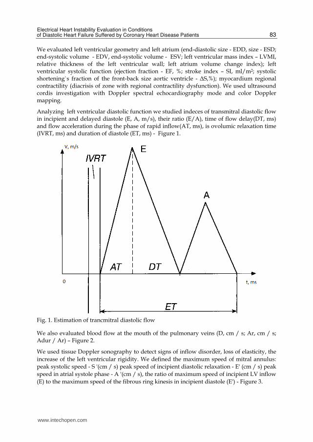
Analyzing left ventricular diastolic function we studied indeces of transmitral diastolic flow in incipient and delayed diastole (Е, А, m/s), their ratio (Е/А), time of flow delay(DT, ms) and flow acceleration during the phase of rapid inflow(АТ, ms), is ovolumic relaxation time (IVRT, ms) and duration of diastole (ET, ms) - Figure 1.
Fig. 1. Estimation of trancmitral diastolic flow
We also evaluated blood flow at the mouth of the pulmonary veins (D, cm / s; Ar, cm / s; Adur / Ar) – Figure 2.
We used tissue Doppler sonography to detect signs of inflow disorder, loss of elasticity, the
increase of the left ventricular rigidity. We defined the maximum speed of mitral annulus:
peak systolic speed - S '(cm / s) peak speed of incipient diastolic relaxation - E' (cm / s) peak
speed in atrial systole phase - A '(cm / s), the ratio of maximum speed of incipient LV inflow
(E) to the maximum speed of the fibrous ring kinesis in incipient diastole (E') - Figure 3.
www.intechopen.com

Novel Strategies in Ischemic Heart Disease
84
Fig. 2. Еstimation of bloodstream at the mouth of the pulmonary veins
S' (cm/s) – peak diastolic speed; E' (cm/s) – peak speed of incipient diastolic relaxation; A' (cm/s) – peak speed in atrial systole phase.
Fig. 3. Fibrous ring`s movement of the mitral valve in pulse-wave mode histic doppler
Using Holter monitoring ECG we took into account the nature of rhythm disturbance and asequence, we estimated the quantity of pain assessed and painless ischemic episodes, the daily duration of ischemia, the maximum depth of segment ST depression.
Time-line analysis of ventricular late potential was performed by method of M. Simson [1981]. We calculated numerical quantitative values of three indicators: the duration of the filtered complex QRS (HF QRS-Dauer), the rms amplitude of the last 40 ms of the complex QRS (RMS 40), the duration of the low amplitude signals at the end of the filtered complex QRS (LAH Fd). We took HF QRS-Dauer which is more than 114 ms, RMS 40 which is less than 25 mV, LAH Fd which is more than 38 ms as pathological parameters of the signal averaged ECG - Figure 4.
www.intechopen.com

Electrical Heart Instability Evaluation in Conditions of Diastolic Heart Failure Suffered by Coronary Heart Disease Patients
85
Fig. 4. Analysis of the signal-averaged ECG with elimination late potentials of the ventricles
Performing heart rate variability analysis we evaluated rMSSD (mean square difference between the duration of contiguous sinus intervals RR, BB 50 (proportion of contiguous sinus intervals RR which differ more than 50ms,%), SDNN (standard derivation of average duration of sinus intervals RR, ms).
Performing spectral analysis of heart rate variability analysis we estimated frequency ranges: high frequency (HF), low frequency (LF), the ratio of sympathetic and parasympathetic influences on heart rate variability (LF/HF).
We examined the following heart rate turbulence indicants:
turbulence onset (TO,%). We estimated the value-of sinus rhythm acceleration after ventricular arrhythmia;
turbulence slope (TS, ms / RR). We studied the intensity of the sinus rhythm deceleration, following its quickening.
In assessing ventricular repolarization we calculated corrected interval Q-T (QTs) using Bazeta. The dispersion of the interval Q-T (QTd) was defined as the difference between the maximal and minimal value of Q-T interval in different leads of standard ECG.
T-test of Student, entry criteria applied by static data handing. Gotten results have been presented in the form of an average arithmetical significance ± is standart devition. Differences considered reliable at p< 0,05.
In case of CHD left ventricle diastole indices and diastole functions undergo complex changes which are connected with both worsening of diastolic disorders and the development of hemodynamic adaptive responses acting through an increase in pressure in the left atrium and/or left ventricular end diastolic pressure and leading to the formation of various types of diastolic dysfunction.
www.intechopen.com

Novel Strategies in Ischemic Heart Disease
86
We divided our patients into 3 groups according to the type of diastolic dysfunction of left
ventricle: the first group (n=36) included patients with abnormal relaxation of left ventricle,
the second group (n=28) consisted of patients with pseudonormal type of diastolic
dysfunction and the third group (n=22) was formed from the patients who suffered
restrictive type of diastolic dysfunction of left ventricle.
The average duration of disease was 6.9 3.9 years (3 to 12 years). Medical history and
electrocardiographic criteria indicated macrofocal myocardial infarction (with Q tooth)
suffered by 68 patients (53.1%).9 out of 68 patients had a chronic aneurysm of the anterior
wall according to electrocardiographic and echocardiographic features. Fine-focal MI
(without Q tooth) is marked in 60 cases according to medical documentation (hospital
records, discharge summary). 82 patients (64%) had coronary heart disease accompanied by
arterial hypertension (AH). 92 patients (72%) had burdened familial history. Lipid exchange
violations were indicated in 112 cases (87.5%).
Figure 1 shows the gradation of patients with different variants of the left ventricle diastolic
dysfunction according to functional classes of chronic heart failure (CHF). 42 patients had
signs of I FC CHF, 51 patients had II FC and 35 patients had III FC.
0%
20%
40%
60%
80%
100%
I FC II FC III FC
diastolic dysfunction of abnormal relaxation type pseudonormal type of diastorestrictive type of diastolic dysfunction
Fig. 5. The gradation of patients suffering coronary heart disease with left ventricle diastolic dysfunction according to functional classes of chronic heart failure (CHF).
In cases of heart failure clinical manifestations we have received the relation of r = 0,620 (p
<0.01) of the frequency of CHF FC to the type of diastolic dysfunction. When FC increases
the disturbances of LV DF become more pronounced: restrictive type of LV DD was
detected in III FC (51% observations); in I FC it was observed on 11.4%; patients had the
type of abnormal relaxation in 9.5% and 64.3% respectively, pseudonormal type was
detected in 21.6% and 55% of cases.
The analysis of the structural and geometric parameters of left heart chambers (Table 1)
showed that group III had an increase of the dilation of the cavities of the left ventricle and
left atrium, increased left ventricle myocardial mass, reduction in contractile function
www.intechopen.com

Electrical Heart Instability Evaluation in Conditions of Diastolic Heart Failure Suffered by Coronary Heart Disease Patients
87
(ejection fraction of III group was more than 45% but less than 50%, in group I it was 50%) in
comparison with group I.
Indicators I group II group III group
Number of patients, n 59 36 33
Е, m/s 0.65±0.06* 0.81±0.03 1.07±0.05**
А, m/s 0.93±0.09* 0.65±0.02 0.36±0.03*
Е/А, 0.82±0.05* 1.25±0.02 3.15±0.08**
АТ, ms 99.47±2.1 94.1±3.2 97.3±3.3
DT, ms 267.6±11.4* 169.6±7.9 143.5±3.2**
IVRT, ms 116.8±7.4* 88.3±2.3 58.9±4.8**
Ar, cm/s 24.1±2.2˚ 36.6±1.3 55.3±1.6
SI, ml/m2 40.3±2.8 41.2±1.4 41.7±2.6
EDVI, ml/m2 82.1±5.3* 85.8±4.58 100.8±6.7
ESVI, ml/m2 37.85±2.1* 39.2±1.1 41.61±2.5
LVEF, % 55.93±2.7* 53.1±2.17 49.6±2.06
ESVILA, ml/m2 39.5±2.3* 44.37±3.2 50.43±3.61
ICVLA,% 37.65±1.5 * 34.1±2.1 24.9±2.7
LVMI, g/m2 126.35±9.7* 132.1±6.3 148.2±11.1
left ventricular RT, cm 0.44±0.06* 0.45±0.05 0.50±0.02
SI – stroke index, EDVI - end-diastolic volume index, ESVI - end systolic volume index, LVEF - left ventricular ejection fraction, ESVILA - end systolic volume index of left atrium, the ICVLA - an indicator of changes in the volume of the left atrium, LVMI - left ventricular mass index, left ventricular RT is the relative thickness of the left ventricular wall, *-p <0.05 - reliability of differences between patients with DD of abnormal relaxation type and patients with pseudonormal type of DD, ** p <0.05 - reliability of differences between the group of patients with restrictive type of DD and the groups with pseudonormal type of DD and abnormal relaxation, ˚ - p <0.05 - reliability of the indicator difference between group with DD of abnormal relaxation type and groups with DD of pseudonormal and restrictive type.
Table 1. Echocardiographic indicators of patients with coronary heart disease
We believe that one of the determinants of heart failure is diastolic dysfunction of the heart, which can be viewed both as the initial stage and as a pathophysiological link in the "vicious circle" of circulatory inefficiency, which affects negatively on the prognosis of patients with coronary heart disease [11].
The violation of left ventricular diastolic properties is essential, along with other structural and functional changes (dilation and hypertrophy), which form the concept of “remodeling”. Patients had eccentric hypertrophy (43.75% of cases) and concentric hypertrophy of LV (37.5% of cases) as predominant types of LV remodeling after MI.
www.intechopen.com

Novel Strategies in Ischemic Heart Disease
88
We established the correlation between the severity of LV DD and exponent change in the volume of the left atrium - r = -0.43 (p = 0.032), with left ventricular ejection fraction - r = -0.48 (p <0.04), with an index of end diastolic volume - r = 0,51 (p = 0.02), with left ventricular mass index - r = 0.45 (p = 0.034).
We associate the appearance and progression of LV DD with violation of diastolic relaxation on the back of acute or long-lasting ischemia with increased stiffness of the myocardium, including the location of postinfarction scar. We spotted the following law: a degree of LV DF disorder was growing in patients having macrofocal myocardial infarction versus patients having fine-focal myocardial infarction, respectively, it was 30.5% with abnormal relaxation type and 69.5% (p = 0.047), 58.3% and 41.7% (p = 0.05) with pseudonormal, 87.9% and 12.1% (p = 0.0476) with restrictive type. We believe that patients with previous Q-infarction had long-lasting deterioration of LV function during myocardial hibernating, which is characterized by a chronic reduction in cardiomyocyte contractility, while their viability is maintained. In these conditions viable myocardium which is far from infarction zona undergoes diastolic myocardial stress.
We detected the dependence of manifestation degree of diastolic dysfunction with an index of contractility WMSI - r = 0.91 (p = 0.01) among patients with a history of MI. We pointed the positive correlation of restrictive type of LV DD with Q- myocardial infarction r = 0.638 (p <0.001). In group III (restrictive type) Q-infarction occurred in 87.9% of cases, which is significantly lower in comparison with group I – 30.5%, p <0.01.
In conditions of limited blood flow the possibility of injury and death of cardiomyocytes because of their necrotization and apoptosis [12] is the main unfavorable result of myocardial hypoxia in case of coronary heart disease. However, death of cardiomyocytes is accompanied by compensatory hypertrophy of survived myocardial cell and the development of connective tissue [13]. There is an opinion that left ventricle hypertrophy may lead to myocardial dysfunction. It is an individual risk factor for high cardiovascular case rate, including ischemia myocardial infarction, heart failure, ventricular arrhythmias and sudden death.
We tend to assume that the process of cardiac remodeling affects all myocardium components. Changes occur at the level of cardiac myocytes and in the extracellular matrix. Typically, ischemic myocardial injuries develop leading to the coagulation and apoptosis. In this case, as approved by the J.Soto, G.Beller [2001], responsive genes are activated immediately, especially c-fos, and the program of "programmed death", apoptosis, starts to run. In the peripheral areas ischemic injury usually results in colliquative necrosis with cell edema and myocytolysis. It is especially true for reperfusion injury. The formation of connective tissue (fibrosis) may have redundant diffuse or interstitial nature in addition to substitution (focal), with interstitial spread perivascular.
Different biomechanical properties of myocardium in chronic ischemic zone and so-called intact myocardium generate special effects according to the type of stress concentration on their border. The type of stress concentration is connected with further progression of degenerative and sclerotic changes, significant changes in the shape and size of remodeled heart chambers. Further LV remodeling with the expansion of the cavity in postinfarction period is caused by involving of myocardium in the "transition zone" in the process of remodeling. The data obtained from pathophysiological studies suggest that patients suffering coronary heart disease have clinical picture determined by not only the
www.intechopen.com

Electrical Heart Instability Evaluation in Conditions of Diastolic Heart Failure Suffered by Coronary Heart Disease Patients
89
heterogeneity of causes, but also the diversity of adaptive and disadaptive changes in metabolism and contractile condition of cardiomyocytes [16]. The most significant adaptive myocardium reactions in response to myocardial ischemia include the “new ischemic syndromes”: hibernate, stupor.
The defining condition for the occurrence of lethal arrhythmias is considered to be structural disease of heart, which becomes electrically unstable under the influence of various functional factors. Ischemia and necrosis, hypertrophy and dilatation of the ventricles, the inflammation and swelling of the myocardial tissue can act as the structural changes that determine the development of ventricular tachycardia. According to the results of many studies [17, 18, 19] these changes constitute anatomical substrate for appearance of malignant arrhythmias with the participation of the various triggering and modulating factors.
Ventricular late potential with a stable disease course was recorded among 35 patients (27.3%) with a history of MI (Table 2). We believe that structural cardiomyocyte changes, programmed apoptosis and secondary hypertrophy, increased diastolic stiffness and active myocardium relaxation violation, reactive changes of connective tissue skeleton of the myocardium, decrease in diastolic filling and remodeling with segmental structure violation, i.e. all these processes, lead to the formation of electrical myocardium heterogeneity and cause greater frequency of recording of late potentials among CHD patients in conditions of progression of LV DD: with abnormal relaxation (18.6%), with pseudonormal type (27.8%), and restrictive type of LV DD (42.4%, p <0.05).
Indicator Variations of diastolic dysfunction
Type I Type II Type III
The number of patients, n 59 36 33
The number of patients with ventricle late potential, n
11 10 14
The number of patients with ventricular arrhythmia, n
50 35 33
analysis of ventricular extrasystole
I gradation, n/% 29/49.1% 10/27.8% 2/6.1%
IIgradation, n/% 7/11.9% 7/19.4% 5/15.1%
IIIgradation, n/% 10/16.9% 13/36.1% 12/36.4%
IV-V gradations, n/% 4/6.8% 5/13.9% 14/42.4%
analysis of Q-T interval
RRNN, ms 810 ± 64 798 ± 98 753 ± 132
QTс, ms 405.5 ± 5.2* 422.3 ± 2.6** 450.2 ± 5.4
QTd, ms 39.7 ± 2.7* 49.6 ± 3.3** 64.2 ± 3.1
I type- LV DD of abnormal relaxation type, II type - LV DD of pseudonormal type; III type - LV DD of restrictive type, VLP ventricular late potential, VA- ventricular arrhythmia, RRNN - the duration of sinus RR intervals; n /% - the absolute number of patients with given symptom/percentage of the number of patients of this group.
Table 2. The comparative analysis of ventricular arrhythmias, cardiac late potentials, QT intervals among patients with coronary heart disease
www.intechopen.com
Novel Strategies in Ischemic Heart Disease
90
Ventricular arrhythmias are considered to be one of the factors determining the poor
prognosis and high mortality among CHD patients. Ventricular arrhythmias may be the
cause of death among patients with heart failure symptoms even in conditions of adequate
control of decompensation symptoms.
Cardiac arrhythmias were detected among 99.2% of patients (Table 2), while
supraventricular arrhythmias (SA) were detected among 7% of patients, ventricular
arrhythmias were detected among 56.2% of patients, a combination of ventricular
arrhythmias (VA) with various forms of supraventricular arrhythmias (SA) were indicated
among 36% of patients. Complex forms of VA (ventricular extrasystole of grades IV-V) were
found in 23 cases (18%). Polymorphic ventricular extrasystole was detected among 35 CHD
patients with left ventricular diastolic dysfunction (27.3%). In 41 cases (32%) the revealed
ventricular extrasystole was assigned to I gradation when the frequency of episodes did not
exceed 220 per day.
Having analyzed ventricular arrhythmias we noted the following law: early, paired, volley
ventricular extrasystoles were detected among patients with slow fragmented activity more
frequently in comparison with patients without ventricular late potential, respectively, 19
(54.3%) and 4 (4.3%) with χ2 = 7.4, p <0.001.
Ventricular late potential and ventricular arrhythmias were recorded more often with an
increase in the degree of diastolic dysfunction and end-diastolic volume of left ventricle.
When DD had a restrictive type, ventricular extrasystole was found in 100% of cases and
ventricular extrasystole of IV-V grades was detected among 14 patients (42.4%), 13 of them
had a diagnosed slow fragmented activity at the end of the ventricular complex. In group I
(with abnormal left ventricular relaxation) ventricular extrasystole was observed among
84.7% of patients and complex forms of ventricular extrasystole were detected among 4
patients with abnormal rates of Holter ECG (6.8%).
The most common mechanism for tachyarrhythmias of high grades is the mechanism of re-
entry momentum. It means the presence of unilateral and delayed passage of the
depolarization wave front because of the violation of inter-cell contacts in parallel-oriented
fibers [20], proliferation heterogeneity and fragmentation of the depolarization wavefront,
which is a marked by ventricular late potential. That's what we explain the significant
prevalence of complex forms of ventricular extrasystole among patients with slow
fragmented ventricular activity (54.3%).
To study the effect of reversible ischemia on myocardial electrophysiological properties we
analyzed daily myocardial ischemia and indicators of Holter ECG among patients with
coronary heart disease having left ventricular DD. Correlation analysis revealed the
dependence of the daily duration of ischemia on the duration of filtered set of HF QRS
Dauer (r = 0.53, p = 0.01), with duration of low-amplitude signals at the end of filter-
centered complex QRS LAH Fd (r = 0.49 p = 0.03), duration of painless myocardial ischemia
with HF QRS Dauer (r = 0.57, p = 0.01). A positive correlation was obtained by estimating
the depth of segment depression in HF QRS-Dauer (r = 0.45, p = 0.03), with LAH Fd (r =
0.41, p = 0.05); number of painless ischemic episodes per day with HF QRS-Dauer (r = 0.51,
p = 0.04).
www.intechopen.com

Electrical Heart Instability Evaluation in Conditions of Diastolic Heart Failure Suffered by Coronary Heart Disease Patients
91
The results confirm that the presence of slow fragmented activity focus, marked by ventricular late potential is connected with heterogeneous areas of myocardium in CHD patients: profound disturbances in the processes of relaxation and recovery of cardiomyocytes occur in both acute and chronic ischemia because of degenerative changes and apoptosis. But it should be noted that studying the complex mechanisms of late potentials formation and the functions and metabolism of cardiac muscle, we traditionally pay much attention to cardiomyocytes. However, we tend to assume that extracellular matrix plays a insignificant role in the genesis of fragmented ventricular activity. Fibrosis is an essential component of cardiac muscle remodeling; the development of reparative (replacement) fibrosis may not only violate cardiomyocytes supply but also impede the electrical contact between them [21] in case of ischemia and myocardial necrosis.
The results of our studies have shown that myocardial dysfunction in ischemic area leads
to the inhomogeneity of repolarization processes in conditions of limited coronary blood
flow. However, the increase of QT dispersion in patients with coronary heart disease is
associated with the duration of daily myocardial ischemia (DMI). We received a positive
relationship of DMI with QTd (r = 0.485, p = 0.05). When the DMI was more than 60
minutes the duration of QTc and QTd interval was 449.2±4.1 ms and 67.2±2.7ms, p
<0.05,when the DMI was 30 to 60 min analogous figures corresponded to the values of
412.5±3,2ms and 46.8± 2,4 ms.
While studying QTd we observed higher values in patients with ventricular arrhythmia of
III-V grades (67.5±4.2 ms) in comparison with patients without ventricular arrhythmia
(44.3±2.5ms), p <0,05. The analysis revealed a dependence of the variance of QT interval
with gradation of ventricular extrasystole (r = 0.6432, p <0.01), with exponents of Holter
ECG: LAH Fd (r = 0.4831, p <0.05) HF QRS-Dauer (r = 0.5744, p <0.05).
The proportion of patients with restrictive type of LV DD with QTc interval which was over
440 ms (69%), and QTd which was more than 50 ms (72%) was significantly higher in
comparison with those values in group II, it was 30.6% and 33% (p<0.05) respectively, and
in Group I it was 19% and 17% (p<0.03) respectively.
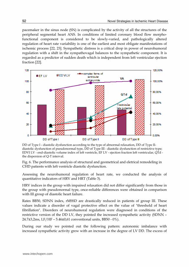
The performance analysis of electrical and structural remodeling among patients
havingcoronary heart disease with different variants of left ventricular DD allowed us to
establish a clear pattern (Figure 6): ventricular late potential and ventricular arrhythmias of
higher grades were detected more often with increasing degree of diastolic function
disturbance. Inhomogeneity of repolarization processes was heightened with an increase in
QT interval dispersion. In our view, the resulting dependence reflects inhomogeneity of
repolarization processes and depolarization of the ventricles in chronic ischemia, the
conditions for malignant arrhythmias in CHD patients having left ventricular DD were
created. We assume that the electrical stability of myocardium and its global-sustaining
capacity are integral characteristics that determine the functioning of the heart as a self-
regulating system. The violation of bioelectrical activity of the myocardium inevitably leads
to its mechanical failure.
The discussion of the pathogenesis of coronary heart disease was always connected with
neurogenic component. The theory of “punctuated antagonism” of the interaction of two
divisions of the autonomic nervous system (ANS) and their impact on the regulation of the
www.intechopen.com
Novel Strategies in Ischemic Heart Disease
92
pacemaker in the sinus node (SN) is complicated by the activity of all the structures of the
peripheral segmental heart ANS. In conditions of limited coronary blood flow morpho-
functional component is considered to be slowly-varied, and pathologically altered
regulation of heart rate variability is one of the earliest and most obligate manifestations of
ischemic process [22, 23]. Sympathetic distress is a critical drop in power of neurohumoral
regulation with a shift in the sympathovagal balances to the sympathetic component. It is
regarded as a predictor of sudden death which is independent from left ventricular ejection
fraction [22].
DD of Type I - diastolic dysfunction according to the type of abnormal relaxation, DD of Type II- diastolic dysfunction of pseudonormal type, DD of Type III - diastolic dysfunction of restrictive type; EDVI LV - end-diastolic volume index of left ventricle, EF LV - ejection fraction left ventricular, QTd - the dispersion of Q-T interval.
Fig. 6. The performance analysis of structural and geometrical and eletrical remodeling in CHD patients with left ventricle diastolic dysfunction.
Assessing the neurohumoral regulation of heart rate, we conducted the analysis of quantitative indicators of HRV and HRT (Table 3).
HRV indices in the group with impaired relaxation did not differ significantly from those in the group with pseudonormal type, once-reliable differences were obtained in comparison with III group of diastolic heart failure.
Rates BB50, SDNN index, rMSSD are drastically reduced in patients of group III. These
values indicate a disorder of vagal protective effect on the value of "threshold of heart
fibrillation". Disorders of neurohumoral regulation were diagnosed in conditions of the
restrictive version of the DD LV, they pointed the increased sympathetic activity (SDNN –
24.7±3,2ms, LF/HF – 5.46±0,61 conventional units, ВВ50 - 0%).
During our study we pointed out the following pattern: autonomic imbalance with
increased sympathetic activity grew with an increase in the degree of LV DD. The excess of
www.intechopen.com

Electrical Heart Instability Evaluation in Conditions of Diastolic Heart Failure Suffered by Coronary Heart Disease Patients
93
the balance of ANS of sympathetic type LF / HF was observed in 39% of patients in group
I who had abnormal left ventricular relaxation, it was detected in 58.3% of patients in group
II with pseudonormal type of DD LV and it was indicated in 94% cases in group III with
restrictive type, p <0.05.
And, if the HRV is a proven non-invasive method of quantitative analysis of the functioning
of ANS in patients with coronary heart disease with preserved sinus rhythm, HRT is a fairly
new noninvasive method of estimation the modulating autonomic influences on the sinus
node in patients with ventricular arrhythmias, which resulted from the progressive fading
of the carotid baroreflex [24]. Schematically, the formation of HRT can be represented by the
following sequence: compensatory pause occurs after ventricular extrasystole, and, as a
result, heart rate and blood pressure increase are caused by baroreflex, then blood pressure
increase leads to the subsequent decrease in heart rate through baroreflex.
Indicators Group I Group II Group III
The number of patients, n 59 36 33
RRNN, ms 810±64 798 ± 98 753 ± 132
analysis of heart rate variability
SDNN, ms 41.4± 2.7 36.6± 2.3 24.7± 3.2*
SDNN index, ms 22.6 ±1.9 19.3 ±1.3 8.7±1.6*
rMSSD, ms 23.1±2.1 21.1±1.4 16 ±1.3*
ВВ50, beats per minute 10.3±3.2 8.8±1.4 3.4±1.2*
LF/HF, conv. 2.93±0.54 3.51±0.68 5.16 ±0.61*
analysis of heart rate turbulence
TO more 0% , n/% 10/17 9/25 17/51.5
TS less 2,5мс/RR, n/% 9/15.2 9/25 16/48.5
RRNN - the average duration of sinus RR intervals between contractions, SDNN-standard deviation
from the mean duration of RR intervals between sinus contractions, BB50 - the number of adjacent sinus
RR intervals, which differ by more than 50 ms, SDNN index-average standard deviations, rMSSD -
square root of the sum of squared differences of successive sinus RR intervals, LF/HF - index of the
balance of sympathetic and parasympathetic nervous systems; TO is more than 0%; TS is less than 2.5
ms / RR * - p <0, 05 – the difference is reliable when indicators of group III are compared with
indicators of I and II groups.
Table 3. Quantitative indices of HRV in patients with coronary heart disease
HRT indicators reflect early acceleration of sinus rhythm (TO) and the subsequent decrease
in heart rate (TS) after ventricular arrhythmia. Studies in recent years characterized HRT as
effective method of predicting the outcomes of patients with coronary pathology, which is
www.intechopen.com

Novel Strategies in Ischemic Heart Disease
94
comparable with HRV, and in some cases it is superior to HRV in its diagnostic capabilities
[25, 26].
During our study we reported the value of TO> 0% in 36 patients (28%) from the group of
128 patients with previous MI history, we detected TS <2.5 ms / RR in 34 cases (26.6%), and
we observed the combination of pathological values of TO and TS in 19 cases (14.8%). The
frequency of registration TO> 0% and TS <2.5 ms / RR significantly dominated in the group
with restrictive type of LV DD in comparison with group I (DD type of abnormal
relaxation) and group II (DD of pseudonormal type), p <0.05.
We have established a connection of HRT violation with left atrium volume change
indicator- r = -0.43 (p = 0.032), with left ventricular ejection fraction- r = -0.49 (p =0.03), with
end-diastolic volume index - r = 0.51 (p = 0.02), with left ventricular mass index - r = 0.45 (p
= 0.04). We marked the correlation of TS with HRV indicators: SDNN (r = 0.41, p = 0.02) and
LH/HF (r = 0.332, p = 0.02). The absolute values of TO significantly correlated with QTd (r =
0.349, p = 0.04).
Pathophysiologic ground for coronary heart disease is a discrepancy between myocardial
oxygen demand and opportunities in meeting the needs in oxygen because of a limitation of
acute or chronic coronary blood flow. Admittedly, the provision of myocardial blood
through the coronary arteries occurs in the phase of diastole. That is why we suggest to
consider diastolic dysfunction not only as an initial stage, but also as a pathological link in
the "vicious circle" of the development of cardiac events in CHD patients.
Rates of diastole and its function undergo complex changes caused by the violation of
diastolic relaxation in the background of acute or chronic ischemia, with increased
stiffness of the myocardium at the site of postinfarction scar during the coronary heart
disease. Necrosis of cardiomyocytes and activation of the cascade of biochemical reactions
which occur in the postinfarction period, cause a significant change in mechanical
properties of tissues. However, we should take into account different biomechanical
properties of myocardium in the areas of chronic ischemia and so-called intact (healthy)
myocardium. The specific effects are generated on their borders according to the type of
stress concentration. They cause the further progression of degenerative and sclerotic
changes, the formation of connective tissue in conditions of limited blood flow. In
conditions of chronic ischemia, the changes occur not only in cardiomyocytes but also in
cells of connective tissue skeleton. Increased rigidity of myocardium, change in the
myocardium viscoelastic properties are the consequences of disorganization and
reorganization of connective tissue skeleton of ischemic nature. Progressive hypertrophy
and dilation of the heart are accompanied by a further violation of the diastolic left
ventricular function, then it is accompanied by systolic dysfunction, they cause an
increase in myocardial oxygen demand and a change in subendocardial blood flow,
impaired myocardial bioenergetics.
The evaluation of the changes of myocardium electrophysiological properties seems for us
to be an important aspect in the search for new diagnostic tests of severity of ischemic
myocardial damage in patients with coronary heart disease. The validity of these
approaches is based on the notion that electrophysiological alternation of cells and their
membranes is associated with remodeling after an episode of ischemia or myocardial
www.intechopen.com

Electrical Heart Instability Evaluation in Conditions of Diastolic Heart Failure Suffered by Coronary Heart Disease Patients
95
infarction and is involved in genesis of arrhythmia That is why we examined the
relationship of electrical and structural myocardium remodeling with left ventricular
diastolic function in CHD patients who had myocardial infarction history.
The analysis of ventricular late potentials and dispersion of QT interval allows to assess
arrhythmogenic substrate in patients having CHD with left ventricular diastolic
dysfunction. Persistent deterioration of left ventricular diastolic function during
myocardium hibernating among CHD patients increases the manifestation of
inhomogeneity of depolarization and repolarization processes.
The increase of the sympathetic influence has an effect on myocardium active relaxation and
diastolic phase structure during the subsequent abuse of metabolic pathways accompanied
by destabilization of connective tissue skeleton and secondary changes of contractile
myocardium. Neurohumoral regulation violations caused by diastolic dysfunction in CHD
patients are the same as when they are generated by heart failure. They are evident in
increase of sympathetic activity, reduced parasympathetic and pressosensitive activity.
Even single ventricular extrasystole can be a trigger factor of fatally dangerous arrhythmias
in conditions of chronic myocardial ischemia with arrhythmogenic substrate, which is the
marked by ventricular late potential and QTd, when autonomic nervous system is activated
according to the sympathetic type.
Complex analysis of parameters reflecting the functional conditions of myocardium and the
interrelation of electrical and structure-geometrical myocardium remodeling is necessary to
improve the diagnostics and the prognostication of risk degree among CHD patients in
conditions of increasing of left ventricle diastolic dysfunction, given the multifactorial
genesis of the problem of myocardial electrical instability
The use of integrated risk markers may contribute to the development of an integrated approach to risk stratification of SCD and arrhythmic complications prevention in CHD patients.
2. Abbreviations
ANS - autonomic nervous system
CHD - coronary heart disease
CHF - chronic heart failure
DD - diastolic dysfunction
DF - diastolic function
EDD - end-diastolic size
EDV - end-diastolic volume
ESD - end- systolic size
EHI - electrical heart instability
ESV - end- systolic volume
www.intechopen.com

Novel Strategies in Ischemic Heart Disease
96
EF - ejection fraction
HRT - heart rate turbulence
HRV - heart rate variability
IVRT – isovolumic relaxation time
LV - left ventricle
LVMI - left ventricular mass index
MI - myocardial infarction
SA - supraventricular arrhythmias
SCD - sudden cardiac death
SI – stroke index
ΔS - systolic shortening’s fraction of the front-back size aortic ventricle
VA - ventricular arrhythmia
VLP - ventricular late potential
3. References
[1] Allender S., Scarborough P., Peto V., Rayner M., Leal J., Luengo-Fernandez R., Gray A. .
European cardiovascular disease statistics 2008 // Eur. Heart Network. -2008.- Vol.
4. – P. 76–78.
[2] Kannel W.B., Feinleib M. Natural history of angina pectoris in the Framingham study.
Prognosis and survival // American Journal of Cardiology. – 1972.- Vol. 29.- P.
154-163.
[3] Pepine C.J., Abrams J., Marks R.G. et al. Characteristics of a contemporary population
with angina pectoris: TIDES Investigators // Am. J. Cardiol. – 1994.- Vol. 74.- P.
226-231.
[4] Williams S.V., Fihn S.D., Gibbons R.J. Guidelines for the management of patients with
chronic stable angina: diagnosis and risk stratification// Ann. Intern. Med. – 2001.-
Vol. 135.- P. 530-547.
[5] Oganov R.G., Maslennikova. G.Y. Demographic situation and cardiovascular diseases in
Russia: ways to solve problems// Cardiovascular therapies and prevention. .-
2007.- №6. – P.7-14.
[6] Cowie M.R., Fox K.F., Wood D.A. et al. Hospitalization of patients with heart failure: a
population–based study. //Eur Heart J. - 2002.- Vol. 23.- P. 877–85.
[7] Cohn, J.N., Johnson G. Veterans Administration Cooperative Study Group. Heart failure
with normal ejection fraction: V-HEFT Study // Circulation. - 1990.- Vol. 81.- P. 48-
53.
[8] O'Callaghan Р.А., Camm A.J. Treatment of arrhythmias in heart failure // Europ. J.
Heart Failure.- 1999.- Vol.1 (2).- P. 133-137.
www.intechopen.com

Electrical Heart Instability Evaluation in Conditions of Diastolic Heart Failure Suffered by Coronary Heart Disease Patients
97
[9] Mareev V.Y. Heart failure and ventricular arrhythmias: the problem of treatment.//
Cardiology.-1996.- № 12.- P.4–12.
[10] Cowie M.R., Mosterd A., Wood D.A. et al. The epidemiology of heart failure // Eur
Heart J. – 1997. - Vol. 18.- P. 208-255.
[11] Devereux R.B. Left ventricular diastolic dysfunction: early diastolic relaxation and late
diastolic compliance. J. Am. Coll. Cardiol 1989; 13: 337-339.
[12] Kapelko V.E. The evolution of concepts and metabolic basis of ischemic myocardial
dysfunction.// Cardiology. - 2005. - № 9. – P. 55–61.
[13] Wainwirght C.L. Matrix metalloproteinases? Oxidative stress and the acute response to
acute myocardial ischemia-reperfusion // Curr Opin Pharmacol. - 2004.- Vol. 4.-
P. 132 – 138.
[14] Molkentin J.D., Lu J-R, Antos C.L. et al. A calcineurin-dependent transcriptional
pathway for cardiac hypertrophy // Cell.- 1998. – Vol. 93.- P. 215 - 228.
[15] Soto J., Beller G. Clinical Benefit of Noninvasive Viability Studies of Patients with
Severe Ischemic Left Ventricular Dysfunction // Clin. Cardiol. – 2001. - Vol. 24. - P.
428-434.
[16] Opie L.H. Newly identified ischemic syndromes and myocardial endogenous
cytoprotection and their role in clinical cardiology in the past and future. //
Medicography. - 1999. - № 21(2). - P. 65–73.
[17] Hohnloser S.H., Franck P., Klingenheben T. et al. Open infarct artery, late potentials,
and other prognostic factors in patients after acute myocardial infarction in the
thrombolytic era: a prospective trial // Circulation. – 1994.- Vol. 90.- P. 1747-1756.
[18] Goldstein S., Brooks M. M., Ledingham R. et al. Association between ease of
suppression of ventricular arrhythmia and survival// Circulation. – 1995.- Vol. 91.-
P. 7979 – 7983.
[19] Petersen S., Peto V., Rayner M. et al. European cardiovascular disease statistics // Eur.
Heart J. – 2005. - Vol. 21. - P. 1261-1272.
[20] Kramer J.B., Saffitz J.E., Witkowsky F.V., Corr P.B. Intramural reentry as a mechanism
of ventriculartachycardia during evolving canine myocardial infarction.// Circ.
Res.- 1985 - Vol. 56 - P. 736-754.
[21] Tatarchenko E.P., Pozdnyakova N.V., Morozova O.E. Late potentials of the heart:
clinical and electrophysiological evaluation. Penza: Elma.- 2000. – P. 144 .
[22] Maisch B. Ventricular remodeling // Cardiology.- 1996.- Vol. 8(7).- P. 2-10.
[23] Bigger J., Fleiss J.L., Steiman R.C. Correlation among time and freguency domain
measures of heart period variability two week after acute myocardial infarction.
Am. J. Cardiol 1992; 69: 891 - 898.
[24] Tatarchenko E.P., Pozdnyakova N.V., Morozova O.E., Shevyryov V.A. Prognosis of
patients with coronary heart disease (clinical and instrumental aspects). Penza:
Elma.- 2002. – P. 206.
[25] Schmidt G., Malik M., Barthel P. et al. Heart-rate turbulence after ventricular premature
beats as a predictor of mortality after acute myocardial infarction // Lancet.- 1999.-
Vol.353.- P.1390-1396.
www.intechopen.com

Novel Strategies in Ischemic Heart Disease
98
[26] Ghuran A., Reid F., La Rovere M.T. et al. Heart rate turbulence-based predictors of fatal
and nonfatal cardiac arrest (The Autonomic Tone and Reflexes After Myocardial
Infarction substudy) // Am. J. Cardiol.- 2002.- Vol.89.- P.184-190
[27] Bauer A., Barthel P., Schneider R., Schmidt G. Dynamics of heart rate turbulence //
Circulation.- 2001.- Vol.104, Supplement II-339.- P.1622.
www.intechopen.com

Novel Strategies in Ischemic Heart DiseaseEdited by Dr. Umashankar Lakshmanadoss
ISBN 978-953-51-0184-0Hard cover, 450 pagesPublisher InTechPublished online 29, February, 2012Published in print edition February, 2012
InTech EuropeUniversity Campus STeP Ri Slavka Krautzeka 83/A 51000 Rijeka, Croatia Phone: +385 (51) 770 447 Fax: +385 (51) 686 166www.intechopen.com
InTech ChinaUnit 405, Office Block, Hotel Equatorial Shanghai No.65, Yan An Road (West), Shanghai, 200040, China
Phone: +86-21-62489820 Fax: +86-21-62489821
The first edition of this book will provide a comprehensive overview of ischemic heart disease, includingepidemiology, risk factors, pathogenesis, clinical presentation, diagnostic tests, differential diagnosis,treatment, complications and prognosis. Also discussed are current treatment options, protocols anddiagnostic procedures, as well as the latest advances in the field. The book will serve as a cutting-edge pointof reference for the basic or clinical researcher, and any clinician involved in the diagnosis and management ofischemic heart disease. This book is essentially designed to fill the vital gap existing between these practices,to provide a textbook that is substantial and readable, compact and reasonably comprehensive, and to providean excellent blend of "basics to bedside and beyond" in the field of ischemic heart disease. The book alsocovers the future novel treatment strategies, focusing on the basic scientific and clinical aspects of thediagnosis and management of ischemic heart disease.
How to referenceIn order to correctly reference this scholarly work, feel free to copy and paste the following:
E.P. Tatarchenko, N.V. Pozdnyakova, O.E. Morozova and E.A. Petrushin (2012). Electrical Heart InstabilityEvaluation in Conditions of Diastolic Heart Failure Suffered by Coronary Heart Disease Patients, NovelStrategies in Ischemic Heart Disease, Dr. Umashankar Lakshmanadoss (Ed.), ISBN: 978-953-51-0184-0,InTech, Available from: http://www.intechopen.com/books/novel-strategies-in-ischemic-heart-disease/electrical-heart-instability-evaluation-in-conditions-of-diastolic-heart-failure-suffered-by-coronar

© 2012 The Author(s). Licensee IntechOpen. This is an open access articledistributed under the terms of the Creative Commons Attribution 3.0License, which permits unrestricted use, distribution, and reproduction inany medium, provided the original work is properly cited.