effects of two commonly found strains of influenzaa aa ... fileinfluenza virus (infv) infection...
TRANSCRIPT

Effects of Two Commonly Found Strains of InfluenzAa AVirus on Developing Dopaminergic Neurons, in Relationto the Pathophysiology of SchizophreniaFernando Landreau1, Pablo Galeano2, Laura R. Caltana3, Luis Masciotra3, Agustın Chertcoff4,
A. Pontoriero5, Elsa Baumeister5, Marcela Amoroso6,8, Herminia A. Brusco4, Monica I. Tous1,
Vilma L. Savy5, Marıa del Rosario Lores Arnaiz6,8, Gabriel A. de Erausquin7*
1 Cultivo de Tejidos, Departamento Virologıa, Instituto Nacional de Enfermedades Infecciosas, ANLIS ‘‘Dr Carlos G. Malbran’’, Buenos Aires, Argentina, 2 Laboratorio de
Citoarquitectura y Plasticidad Neuronal, Instituto de Investigaciones ‘‘Prof. Dr. Alberto C. Taquini’’ (ININCA), Facultad de Medicina, Universidad de Buenos Aires, Buenos
Aires, Argentina, 3 Instituto de Biologıa Celular y Neurociencia ‘‘Profesor E. De Robertis’’, Facultad de Medicina, Universidad de Buenos Aires, Buenos Aires, Argentina,
4 Bioterio Central, Instituto Nacional de Produccion de Biologicos, ANLIS ‘‘Dr Carlos G. Malbran’’, Buenos Aires, Argentina, 5 Virus Respiratorios, Departamento Virologıa,
Instituto Nacional de Enfermedades Infecciosas, ANLIS ‘‘Dr Carlos G. Malbran’’, Buenos Aires, Argentina, 6 Microscopıa Electronica, Departamento Virologıa, Instituto
Nacional de Enfermedades Infecciosas, ANLIS ‘‘Dr Carlos G. Malbran’’, Buenos Aires, Argentina, 7 Roskamp Laboratory of Brain Development, Modulation and Repair,
Department of Psychiatry and Neurosciences, University of South Florida, Tampa, Florida, United States of America, 8 Facultad de Psicologıa, Universidad de Buenos Aires,
Buenos Aires, Argentina
Abstract
Influenza virus (InfV) infection during pregnancy is a known risk factor for neurodevelopment abnormalities in the offspring,including the risk of schizophrenia, and has been shown to result in an abnormal behavioral phenotype in mice. However,previous reports have concentrated on neuroadapted influenza strains, whereas increased schizophrenia risk is associatedwith common respiratory InfV. In addition, no specific mechanism has been proposed for the actions of maternal infectionon the developing brain that could account for schizophrenia risk. We identified two common isolates from the communitywith antigenic configurations H3N2 and H1N1 and compared their effects on developing brain with a mouse modified-strain A/WSN/33 specifically on the developing of dopaminergic neurons. We found that H1N1 InfV have high affinity fordopaminergic neurons in vitro, leading to nuclear factor kappa B activation and apoptosis. Furthermore, prenatal infectionof mothers with the same strains results in loss of dopaminergic neurons in the offspring, and in an abnormal behavioralphenotype. We propose that the well-known contribution of InfV to risk of schizophrenia during development may involvea similar specific mechanism and discuss evidence from the literature in relation to this hypothesis.
Citation: Landreau F, Galeano P, Caltana LR, Masciotra L, Chertcoff A, et al. (2012) Effects of Two Commonly Found Strains of Influenza Virus on DevelopingDopaminergic Neurons, in Relation to the Pathophysiology of Schizophrenia. PLoS ONE 7(12): e51068. doi:10.1371/journal.pone.0051068
Editor: Malu G. Tansey, Emory University, United States of America
Received June 30, 2011; Accepted November 1, 2012; Published December 10, 2012
Copyright: � 2012 Landreau et al. This is an open-access article distributed under the terms of the Creative Commons Attribution License, which permitsunrestricted use, distribution, and reproduction in any medium, provided the original author and source are credited.
Funding: The authors have no support or funding to report.
Competing Interests: The authors have declared that no competing interests exist.
* E-mail: [email protected]
Introduction
Influenza virus (InfV) infection during pregnancy is a known
risk factor for neurodevelopment in the offspring, increasing the
risk of schizophrenia [1–6] and autism [7–9], and resulting in an
abnormal behavioral phenotype in mice [10]. The peak of this
effect occurs during the 6th month of intrauterine development in
humans [7], and it has been shown to correlate with serologically
proven infection in the mother [1]. However, the relationship
between infection and disease remains controversial [11–14].
Experimental infection of pregnant mice with InfV results in
pathological and physiological changes in the brain of offspring
that resemble those observed in schizophrenia [10,15–19]. Such
effects of InfV on neurodevelopment appear to be at least in part
mediated through immune mechanisms [10,20–22], but infection
of pregnant mice with InfV may also result in persistent expression
of virus in fetal brain [23].
Regardless of the molecular mechanism by which infection
results in neurodevelopmental changes, deviant behavior in adult
offspring should have a correlate in structural or functional
changes in brain circuitry, and there are reasons to consider the
dopaminergic system as a potential target. Indeed, intracerebral
inoculation of InfV in adult rodents results in viral antigen
accumulation in dopaminergic neurons [24–25], and a similar
preference for dopaminergic neurons can be demonstrated in
primary neuronal cultures [26]. Direct stereotaxic introduction of
InfV in mice olfactory bulb leads to apoptosis of infected neurons
[27–28]. Yet, these findings refer to neuroadapted strains of InfV,
whereas the most common viral epidemics of InfV A are rarely
associated with encephalitis [25]. InfV A viruses are RNA viruses
characterized by two surface antigens: hemagglutinin (H) and
neuraminidase (N) [25]. Variations in these two antigens occur
independently from strain to strain, and can involve major (shift)
or minor (drift) alterations and at least in mice neurovirulence is
determined by the plasminogen binding activity of neuraminidase
[29]. Spread of a virus and tissue tropism may depend largely on
the proper match between cleavability of viral glycoproteins by
tissue endopeptidases and availability of proteases in the host, the
PLOS ONE | www.plosone.org 1 December 2012 | Volume 7 | Issue 12 | e51068
A
A

latter being the limiting factor [25]. Once again, a virus activating
protease, blood clotting factor Xa (FXa), is selectively present in
brainstem neurons, including dopaminergic neurons in the
substantia nigra pars compacta, and in white matter microglia
[25,30].
Is there any evidence that susceptibility of dopaminergic
neurons to InfV A infection may modify risk of schizophrenia?
First, the observation that parkinsonism can occur as a trait
associated with schizophrenia suggests a dopaminergic deficit [31–
35]. Functional imaging and postmortem studies support a loss of
dopaminergic function in schizophrenia [36–42] and the patients’
impairment in executive function and working memory correlates
with low dopamine availability in the prefrontal cortex [41–42].
On the other hand, psychotic symptoms correlate with excessive
release of dopamine in basal ganglia [43]. Therefore, the available
evidence points to the simultaneous lack and excess in dopami-
nergic function albeit in different brain circuits; in rodents,
prenatal damage to a subpopulation of dopaminergic neurons
projecting from the midbrain to the prefrontal cortex has been
shown to lead over time to a maladaptive, compensatory increase
in mesolimbic dopaminergic activity upon the arrival of puberty
[43–45].
Thus, we hypothesized that maternal infection with InfV during
the critical period of risk for schizophrenia (the equivalent of the
human second trimester, or embryonic days 11–16 in rodents),
should result in preferential damage to developing dopaminergic
neurons in the brainstem of offspring. We further hypothesized
that such damage depends on the antigenic configuration of
circulating InfV. In the present work we evaluated both of these
hypotheses in primary cultures of ventral mesencephalon and in a
mouse model of prenatal infection with circulating InfV strains.
Materials and Methods
Primary Cultures of Rat MesencephalonAll animal work was conducted according to relevant national
and international guidelines and approved by the Animal Studies
Committee of ANLIS Carlos G Malbran (Resolucion 20/4/2004).
Rat embryos were recovered at day 14 from timed pregant Wistar
rats, and the ventral midbrain was dissected, mechanically
dissociated, and plated on polyethylenimine (1 mg/ml)-coated
culture wells, in DMEM/F12 medium, supplemented with 5%
horse serum (US) and 2.5% fetal calf serum (FCS), at a density of
16106 cells per 6-mm-diameter well. After 2 days in culture FCS
content was reduced to 0.5% to prevent astrocyte proliferation;
medium was changed daily. Cultures are maintained for 7–11 days
in vitro. Dopaminergic neurons can be identified after fixation by
immunohistochemistry for tyrosine hydroxylase (TH). This is the
accepted standard methodology to prepare dopaminergic neurons
in culture, in part because at this developmental age, dopaminer-
gic neurons account for 10–20% of the total number of neurons,
express specific phenotypic markers and functional glutamate
receptors, and release and uptake dopamine [46–49].
MDCK CellsReplication of viruses under liquid medium and plaque
determinations are performed in MDCK cells (passage 59)
obtained from the Centers for Disease Control and Prevention
(CDC, Atlanta, GA).
Viral StrainsWe identified two common isolates from community samples of
Buenos Aires from the Instituto Malbran, with antigenic
configurations H3N2 and H1N1. The results of characterization
with ferret antisera are: A/New Caledonia/20/99-like (H1N1) (A/
NC-L/99), and A/Sydney/5/97-like (H3N2) (A/Sy-L/97). Iso-
lates were amplified in MDCK cells and frozen in lots of 100
cryotubes. We used a well characterized neurovirulent strain
obtained from the CDC (Atlanta, GA) (A/WSN/33) as a positive
control for toxicity to dopaminergic neurons [25] and behavioral
abnormalities in the offspring of infected pregnant mice [10].
Viral QuantitationTitration of plaque forming units (PFU) was performed in
MDCK cells and normalized for each lot to:
Lot 1 A/Sy-L/97 (H3N2) 3,000 PFU/ml.
Lot 1 A/NC-L/99 (H1N1) 3,000 PFU/ml.
Lot 1 A/WSN/33 (H1N1) 3,000 PFU/ml.
Influenza a Genome AmplificationViral RNA was purified from selected samples by QIAampH
Viral RNA Mini Kit.
Reverse Transcription-polymerase Chain Reaction (RT-PCR)
RT PCR was used to amplify regions of the protein coding
domains of viral RNA segment 7 to obtain finally an amplicon of
560 bp, using the QIAGENHOne Step RT-PCR Kit.
Influenza a Antigen DetectionThe antigen detection was performed by Indirect immunoflu-
orescent assay (IFA) using the standard antibody for surveillance
recommended by CDC (Atlanta, GA), namely mouse monoclonal
anti influenza a nucleoprotein (immunized with Influenza A/
Puerto Rico/8/34 (H1N1) and A/Bangkok/1/79 (H3N2) viruses),
from spleen cells from BALB/c mice fused with cells of the P3
Ag8.653 mouse myeloma cell line (AbD Serotec, USA).
In vitro InfectionFor neurotropism experiments we inoculated 50 ml of viral
suspension (150 PFU/well) in primary cultures of rat mesenceph-
alon, and examined the presence of viral infected cells at 24 h,
48 h and 72 h (after fixation with formalin), by indirect
immunofluorescence with monoclonal anti-InfV A antibodies
(CDC, Atlanta, GA) followed by fluorescein-conjugated anti
mouse (Light Diagnostics) (Figure 1). 50% Tissue Culture Infective
Dose (TCID50) was calculated for each viral strain using the Reed
and Muench Calculator [50] (Brett D. Lindenbach, 2008).
In vivo InfectionBALB-C mice were obtained from the Bioterio Central of the
Instituto Nacional de Produccion de Biologicos. Because infection
entails discomfort to the animals, we minimized it by ensuring
adequate hydration, temperature control, and cage conditions
according to strict guidelines (Instituto Malbran/CDC). Intranasal
inoculation with 30 ml (3.000 PFU/ml of InfV, 90 PFU/mouse)
strains or vehicle (PBS, pH 7.4) was performed at embrionic day
9–11 on the assumption that viremia peaks (and most likely
invades CNS) three days later (and therefore , embryonic day
14). Post-infection all animals were individually kept in filtered
cages with ad lib access to food and water, at 25uC. Seroconver-
sion was established by inhibition of hemagglutination of turkey
red blood cells at day 14 following infection. We calculated the
50% infective dosing by measuring change in lung weights post
infection in lots of 5 animals for each strain and for each of four
dilutions. ID50 (expressed as dilutions of a normalized hemagglu-
tinin unit) for each strain were: A/Sy-L/97 = 1.07E-02, A/NC-L/
Influenza and Development of Dopaminergic Neurons
PLOS ONE | www.plosone.org 2 December 2012 | Volume 7 | Issue 12 | e51068

99 = 1.41E-02, and A/WSN/33 = 2.49E-02. We did not find
lethal infections at any of the used viral loads and therefore LD50
could not be estimated for any strain. Institutional and ethical
committee approval for this protocols was obtained at both
centers. ID50 was calculated for each viral strain using the Reed
and Muench Calculator [50] (www.med.yale.edu/micropath/pdf/
Infectivity%20calculator.xls, Brett D. Lindenbach, 2008).
Behavioral StudiesPups from infected mothers were examined in a behavioral test
battery including elevated plus maze, open field, novel object, and
spontaneous activity before puberty (30 days) or in adulthood (90
days). In the latter we also carried out an object recognition task
(90 days). Behavioral tests were carried out in an experimental
room evenly illuminated provided with white noise. Animals were
handled for 5 min daily for 3 days prior to testing. Sessions were
recorded and later analyzed using a computerized video-tracking
system (Ethovision XT, version 5, Noldus Information Technol-
ogy, Wageningen, Netherlands) or ethological observation soft-
ware (JWatcher V1.0).
Elevated plus maze. Elevated plus maze testing was carried
out in a standard mice apparatus elevated 37.5 cm from the floor.
At designated times animals were placed onto the centrdal
platform facing an open arm and allowed to freely explore the
maze for 5 min. After each session the apparatus was cleaned. An
arm entry was counted when all four paws were placed into an
arm.
Open field. Animals were placed in the center of a standard
open field apparatus and total distance moved, number of rearings
and time spent in the central area were recorded for 20 minutes. A
mouse was considered to be into the central area (arbitrarily
defined as a square of 30630 cm) when its four paws were in it.
The apparatus was cleaned between sessions.
Novel object. Upon completion of the 20-min open field
session, a novel object (a metal cup), was placed in approximately
20 cm away from the mouse and the latency to contact the object
and time spent in contact with it were recorded for 10 min.
Spontaneous activity. Two days after the open field and
novel object tests, the spontaneous locomotor activity was
monitored using a webcam and quantified using a simple criteria
for number of squares crossed per unit time (24 h).
Object recognition task. For 3 consecutive days prior to this
task mice were handled once a day for 5 min and placed 10 min in
the open field to allow habituation. On the fourth day each mouse
was observed during two 5 min trials separated by an interval of
1 h during which animals were returned to their cage. In the
sample trial (T1) mice were faced with two identical objects placed
in a symmetrical position and the time exploring each object was
recorded. In the retention trial (T2) mice one of the two objects
was replaced by a novel, non-familiar object and the time
exploring each was recorded. Sets composed of three copies of the
same object were used to prevent odor cues and all combinations
and location of objects were used to prevent bias due to preference
for a particular object or location. Exploration time was computed
when the snout pointed to the object at a distance #2 cm.
Histological StudiesAfter behavioral testing mice were sacrificed and fixed, brain
sections (15 mm) were obtained in stereological series (every 8th
section), and processed for histopathological analysis that included
gross morphology with Nissl staining, stereological quantitation of
immunohistochemistry for dopaminergic neurons and reactive
astroglia in the substantia nigra and ventral tegmental area, and
electron microscopy of dopaminergic neurons stained with
tyrosine hydroxylase. Post-fixed brains stored in 16 PBS
containing 0.05% sodium azide were embedded in 3% low-
Figure 1. Indirect immunofluorescence for Influenza virus in primary mesencephalic cultures. Culture dishes infected with NewCaledonia-like (H1N1) (panels a–c) or Syndey-like (H2N3) strains (panels d–f), at 24 (a, d), 48 (b, e) and 72 hours (c, f) post inoculation. The twocommunity isolates differed from each in that the New Caledonia-like strain (A/NC-L/99) showed greater affinity at earlier times and more toxic thanSydney-like (A/Sy-L/97) virus.doi:10.1371/journal.pone.0051068.g001
Influenza and Development of Dopaminergic Neurons
PLOS ONE | www.plosone.org 3 December 2012 | Volume 7 | Issue 12 | e51068

melting agarose, sectioned cornally (40 mm) on a vibratome (Leica
VT1200) and stored in cryoprotectant (30% glycerol, 30%
ethylene glycol in 16 PBS) at 220uC until processing. Immuno-
histochemistry was performed on every 6th section. For immuno-
histochemistry sections were washed in PBS endogenous perox-
idases were blocked by incubation in 3% H2O2, and blocking of
non-specific binding was achieved by incubating in PBS contain-
ing 0.25% (w/v) Triton-X 100 and 10% normal serum. Sections
were incubated overnight at 4uC with one of the following primary
antibodies: Tyrosine Hydroxylase (TH, Pelfreeze Biologicals,
Rogers, AR; or Boehringer GmbH, Mannheim, Germany),
Nuclear Factor kappa B (NFkBp65, Abcam, Cambridge, MA),
Anti-GFAP antibody - Astrocyte Marker (ab4674, Abcam, Cam-
bridge, MA) and Anti-InfV A antibodies provided by the Instituto
Malbran (Buenos Aires, Argentina). For microglia activation we
used lectin staining (IsoB4) and antibody staining (ED-1).
Following washing, sections were then incubated with appropriate
biotinylated secondary antibody (Jackson ImmunoResearch Lab-
oratories, West Grove, PA) for 1 hr followed by incubation with
avidin-biotin-peroxidase complex (ABC Elite Kit; Vector Labora-
tories) detection system with diaminobenzidine (Ultratech HRP
Streptavidin-biotin Universal Detection System and DAB Chro-
mogen Kit, Inmunotech Co, Marseille, France). For immunoflu-
orescence labeling, sections were stained with streptavidin
conjugated Alexa Fluor 568 (working solution 1–5 mg/ml;
Molecular Probes, Eugene, OR). For detection and quantification
of apoptosis (programmed cell death) at single cell level, we used a
protocol based on labeling of DNA strand breaks (TUNEL
technology) carried out following manufacturer’s recommenda-
tions (DeadEndTM colorimetric TUNEL System, Promega,
Madison, WI).
Electron MicroscopyEmbryonic brains 3 days after maternal infection were fixed
with 3% v/v glutaraldehyde in PBS overnight, washed, postfixed
in 1.5% w/v osmium tetroxide in the same buffer for 2 hs,
contrasted with 2% w/v acuose uranyl acetate, and embedded in
Fluka Poly-Bed 812 (Sigma-Aldrich, St Louis, MO). Ultrathin
sections (90–100 nm) were collected in cupper grids and stained
with uranyl acetate and Reynolds solution, and imaged using a
Philips EM300 transmission electron microscope.
Data AnalysisAll experiments were carried out in triplicates. For stereology,
cells were counted using every 8th consecutive section, 400 mm
apart from each other, throughout the entire ventral mesenceph-
alon (substantia nigra compacta and ventral tegmental area) using
a Nikon 80i Eclipse microscope (Tokyo, Japan) equiped with
Stereologer software (SRC, Tampa, FL) and a motorized stage.
Digital photographs were taken using a Zeiss camera with
AxioVision software (Carl Zeiss Microscopy GmbH, Gottingen,
Germany). Digital images were then processed with ImageJ (NIH).
The cross sectional area of each section was determined with
Stereologer and the data were expressed as number of cells per
hemibrain. Statistical analyses were performed with SPSS software
(IBM Corporation, Somers, NY). Data are presented as mean 6
standard error of the mean (SEM). Comparisons among groups
were performed using a one or two way ANOVA as indicated,
followed by a Kruskal–Wallis post hoc test as appropriate. For
significant differences a was set at 0.05. For multivariate analysis of
behavioral performance, discriminant analysis was performed
using prenatal exposure as the classification criterion (SPSS, IBM
Corporation, Somers, NY).
Results
Community Strains of InfV have High Affinity forMesencephalic Cultures in vitro and Result in NeuronalDeath
Epidemiological reports show an association between respira-
tory infection by InfV A in pregnancy and risk of schizophrenia to
the offspring; we investigated the effects of common respiratory
InfV A strains on development of dopaminergic neurons in
primary mesencephalic cultures to test the hypothesis that InfV A
could mediate its effects on schizophrenia risk through develop-
mental damage to specific dopaminergic projections [43–45]. We
first assessed the affinity of viral strains for neurons with IFA for
InfV A community isolates using normalized viral loads. Both
strains infected neurons effectively, but A/NC-L/99 had greater
infectivity at earlier times than A/Sy-L/97 (Figure 1). This result
was consistent with the calculated TCID50 which was lowest for
A/Sy-L/97 (TCID50 for A/WSN/33 = 1.43E-03, for A/NC-L/
99 = 3.16E-03, and for A/SY-L/97 = 3.98E-02). Dopaminergic
neurons were identified by TH immunoreactivity, since loss of TH
staining correlates tightly with cell death in these cultures
[48,49,51–53].
Because control cultures undergo progressive attrition of
neurons in serum-free conditions we compared the effect of
infection to spontaneous neuronal loss over time in PBS inoculated
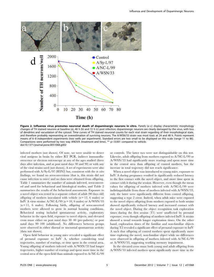
wells (Figure 2A). Following infection, TH stained neurons
undergo rapid loss of dendrites, cytosolic vacuolation and nuclear
picnosis (Figure 2C–E). Yet, since TH immunostaining does not
directly establish the presence of cell death, we carried out
TUNEL staining to detect DNA fragmentation. As expected, in
control cultures changed to serum free medium attrition by
apoptosis was detected by 24 h. By contrast, cultures inoculated
with InfV strains underwent significant apoptosis 6 hours post-
infection. Notably, apoptosis was most apparent in cultures
exposed to A/Sy-L/97 (Figure 3). This finding is discordant with
the susceptibility of TH immunoreactive neurons (where A/NC-
L/99 resulted in greater cell loss), and may be due to a different
susceptible neuronal population (non TH immunoreactive).
We have previously described that programmed cell death of
dopaminergic neurons in this culture system is mediated by NFkB
(51), and others have shown that NFkB signaling is an early and
necessary step in InfV-triggered apoptosis pathway [54]. NFkB
immunostained neurons were distinguished from glial cells on
morphological grounds (Figure 4A–B) and counted. As predicted,
we found a marked increase in neuronal NFkB immunostaining
between 3 and 6 hours post-infection with H1N1 strains, but not
with H3N2 (Figure 4C).
InfV Infection during Pregnancy Results in BehavioralAbnormalities in the Offspring
Pregnant (embryonic day 9–11) mice received intranasal
infusions containing A/NC-L/99 (H1N1), A/WSN/33 (H1N1),
or vehicle alone (control). Gross inspection of mesencephalon of
mouse embryos 72 h after mothers were infected with InfV with
Nissl staining at low magnification revealed dystrophic appearance
of neurons, with significant cytosolic vacuolation and increased
intercellular space, both of which were more apparent on offspring
of mothers infected with A/WSN/33 strains (Figure 5A, 5C, 5E).
Electron microscopy inspection of synaptic terminals revealed
marked vacuolation of embryonic neurons (Figure 5B, 5D, 5F,
arrowheads). This acute dystrophic changes at the ultrastructural
level have not been described before. Both viral strains caused
significant ultrastructural dystrophic alteration and loss of dopa-
minergic neurons in the mesencephalon of adult offspring of
Influenza and Development of Dopaminergic Neurons
PLOS ONE | www.plosone.org 4 December 2012 | Volume 7 | Issue 12 | e51068
infected mothers (not shown). Of note, we were unable to detect
viral antigens in brain by either RT PCR, indirect immunoflu-
orescence or electron microscopy at any of the ages studied (three
days after infection, and at post natal days 30 and 90) or with any
of the viral strains used (not shown). A set of experiments were also
performed with A/Sy-L/97 (H3N2) but, consistent with the in vitro
findings, we found no seroconversions (that is, this strain did not
cause infection to mice) and no data were obtained from offspring.
Table 1 summarizes the number of animals infected, seroconvert-
ed and used for behavioral and histological studies, and Table 2
summarizes the results of the behavioral assessments. Exposure to
a novel object was tested in a separate cohort of adult (90 days old)
offspring of mothers inoculated with vehicle (n = 12, 6 males) or
InfV A virus strains: A/NC-L/99 (n = 10, 6 males) or A/WSN/33
(n = 11, 6 males). Following birth, offspring of seroconverted
mothers were allowed to grow in normal housing conditions.
Behavioral testing included spontaneous activity, exploratory
behavior in the open field, exposure to novel objects, and elevated
cross maze either on post natal days 30–40 (pre-puberty) or post
natal days 90–100 (adulthood). No differences between groups
were observed in either diurnal or nocturnal spontaneous activity
(data not shown).
Open field behavior in young mice revealed a significant effect
of prenatal exposure to InfV A, whether considering total
trajectories, number of rearings, or time spent in the central area.
Young offspring of mothers infected with A/WSN/33 had longer
trajectories, higher number of rearings and spent more time in the
central area of the open field than animals exposed to A/NC-L/99
or controls. The latter two were not distinguishable on this test.
Likewise, adult offspring from mothers exposed to A/NC-L/99 or
A/WSN/33 had significantly more rearings and spent more time
in the central area than offspring of control mothers, but the
increase in total trajectory did not reach significance.
When a novel object was introduced to young mice, exposure to
InfV A during pregnancy resulted in significantly reduced latency
to the first contact with the novel object, and more time spent in
contact with it during the session. However, even though the mean
values for offspring of mothers infected with A/NC-L/99 were
indistinguishable from those of mothers infected with A/WSN/33,
only the latter were significantly different from control offspring,
suggesting a type 2 error. Indeed, when adult mice were exposed
to the novel object offspring from mothers exposed to both strains
showed significantly reduced latency and increased contact with
the novel object. During the object recognition task exploration
times during the first session (T1) were unaffected by prenatal
exposure, even though offspring of mothers infected InfV A strains
showed a trend towards longer exploration times. On the other
hand, exploration times of the familiar and non-familiar objects
during T2 revealed a significant effect of prenatal exposure to InfV
A such that offspring of control mothers spent significantly more
time exploring the novel, non-familiar object while no differences
were observed in offspring of mothers infected with A/NC-L/99
or A/WSN/33, suggesting working memory impairment.
In the elevated cross maze both young and adult offspring from
A/WSN/33 infected mothers spent significantly more time in the
Figure 2. Influenza virus promotes neuronal death of dopaminergic neurons in vitro. Panels (a–c) display characteristic morphologychanges of TH stained neurons at baseline (a), 48 h (b) and 72 h (c) post infection; dopaminergic neurons are clearly damaged by the virus, with lossof dendrites and vacuolation of the cytosol. Time curves of TH stained neuronal counts for each viral strain regarding of their morphological state,and therefore probably representing an overestimation of surviving neurons. The A/WSN/33 strain was most toxic at 24 and 48 h. Points representmeans of 6–8 independent experiments (two wells per experiment). Standard errors are two small to be displayed on this scale (range 11 to 86).Comparisons were performed by two way ANOVA (treatment and time). ** p,0.001 compared to vehicle.doi:10.1371/journal.pone.0051068.g002
Influenza and Development of Dopaminergic Neurons
PLOS ONE | www.plosone.org 5 December 2012 | Volume 7 | Issue 12 | e51068

open arms, whereas offspring of A/NC-L/99 infected mothers
were undistinguishable from controls.
Multivariate analysis of the behavioral data by discriminant
analysis revealed that the net effect of prenatal infection with InfV
virus on behavior could be described by two statistically significant
discriminant functions accounting for more than 90% of the
variance (Chi square = 24.208, d.f. = 12, p,0.05). These two
functions allowed correct classification of 100% of the offspring
of mothers infected with A/WSN/33, and of 83% of those of
mothers infected with A/NC-L/99. The reminder 17% were
indistinguishable from controls (Table 3).
In summary, maternal infection with both strains resulted in
abnormal behavior in the offspring, including time spent in the
open arm of the elevated plus maze, changes in exploratory
behavior, reduced latency to contact and increased time in contact
with a novel object, and working memory impairment when
compared to offspring of mothers exposed to vehicle. Additionally,
the behavioral alterations appear earlier in the offspring of
mothers infected with A/WSN/33.
InfV Infection during Pregnancy Results in Selective Lossof Dopaminergic Neurons in the Adult Offspring
Histopathological abnormalities in the offspring of mice infected
with InfV strains during pregnancy was carried out in two sets of
samples obtained at p30 (n = 4 per group), and at p90 (n = 4 per
group). Both viral strains caused significant dystrophic alteration
and loss of dopaminergic neurons in the mesencephalon of adult
(post-natal day 90) offspring of infected mothers (Figure 6A–I).
Quantitative stereology demonstrated a 30% loss of tyrosine
hydroxylase immunoreactive neurons in adult offspring of A/NC-
L/99 infected mothers, and a 50% loss in those of mothers
infected with A/WSN/33 (Figure 6J). When the effects of viral
strains were discriminated between TH positive neurons in the
substantia nigra pars compacta (SNpc) vs. the ventral tegmental
are (VTA) in a separate set of experiments, similar reductions were
observed in both nuclei (Figure 6J, inset). In adolescent offspring
(post natal day 30) the effects were virtually identical (not shown).
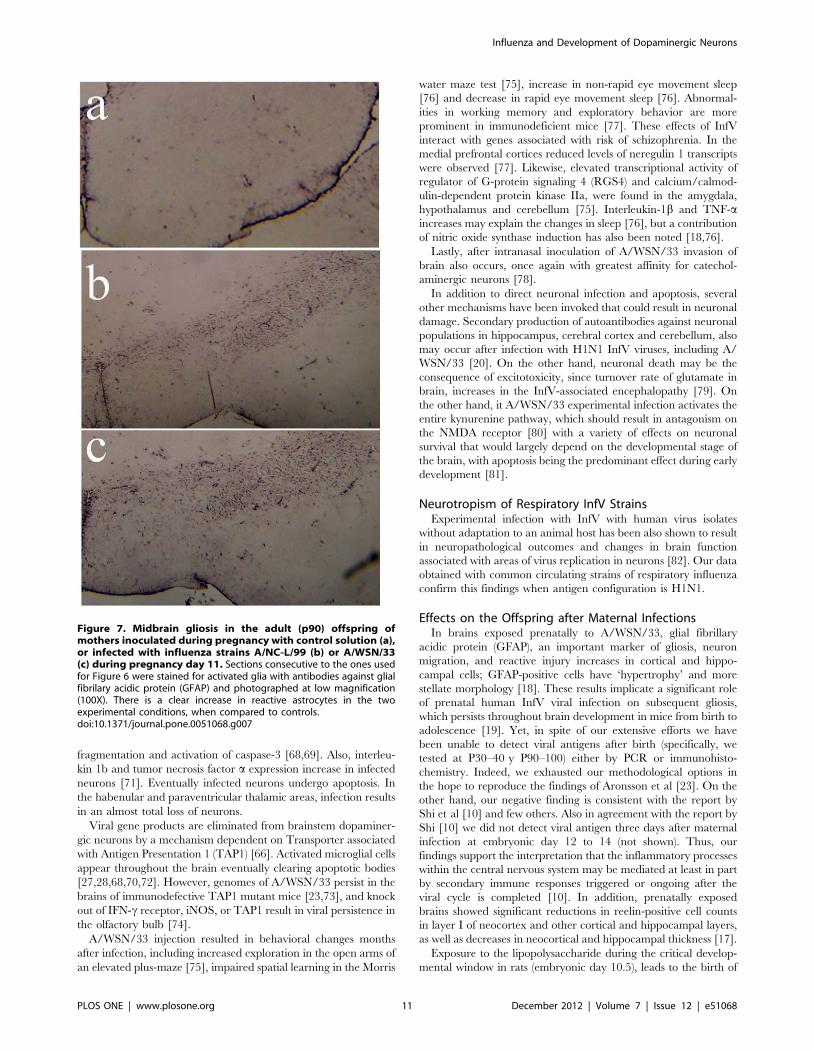
A corresponding increase in glial acidic fibrilary protein (GFAP)
immunostaining indicating gliosis was observed in the same brain
region of the adult offspring of mice infected with InfV A strains
during pregnancy (Figure 7). This increase was again most marked
for animals born to mothers infected with WSN/33 (Figure 7C),
and intermediate for those born to mothers infected with A/NC-
L/99 InfV A virus (Figure 7B).
Discussion
The three major findings of the present work provide evidence
in favor of the hypotheses tested. First, we found that circulating
Figure 3. Influenza infection causes neuronal apoptosisin vitro. Panels a and b show TUNEL staining in primary culturesfollowing inoculation of cultures (11 day in vitro) with either PBSsolution (a) or A/WSN/33 (d) at 200X. Cultures were fixed, stained forTUNEL and counted at times specified on the x axis of panel (c). Cellcounts bars represent means of 6–8 independent experiments (twowells per experiment). Error bars are SEM. Comparisons were performedby two way ANOVA (treatment and time). ** p,0.001 compared tocontrol (PBS solution).doi:10.1371/journal.pone.0051068.g003
Figure 4. Influenza infection results in NFkB activation in vitro.Panels (a) and (b) show NFkB staining in primary cultures followinginoculation of A/WSN/33 (a) or PBS solution (b). Cultures (11 dayin vitro) were inoculated with PBS, A/WSN/33, A/NC-L/99 (H1N1), A/Sy-L/97 (H2N3) influenza strains as described in the text, fixed,immunostained for NFkB and neurons were counted at times specifiedin panel (c). Cell bars represent means of 6–8 independent experiments(two wells per experiment). Error bars are SEM. Comparisons wereperformed by two way ANOVA (treatment and time). ** p,0.001compared to control (PBS solution).doi:10.1371/journal.pone.0051068.g004
Influenza and Development of Dopaminergic Neurons
PLOS ONE | www.plosone.org 6 December 2012 | Volume 7 | Issue 12 | e51068

strains of InfV infect cultured mesencephalic neurons resulting in
selective loss of dopaminergic neurons; this effect was dependent
on the antigenic configuration of the strain, it was preceded by
NFkB activation and mediated by apoptosis. Both H1N1 strains of
InfV had the greatest affinity for dopaminergic neurons, whereas
an H3N2 strain induced apoptosis preferentially in other cell
types, and did not result in NFkB activation.
Second, following maternal infection with H1N1 (but not
H3N2) InfV strains we found a persistent and a selective loss of
dopaminergic neurons in substantia nigra pars compacta and
ventral tegmental area of the offspring (Figure 6J, inset). Perhaps
not surprisingly, loss of dopaminergic neurons was more
pronounced in the adult offspring of mothers infected with the
neuroadapted A/WSN/33 than with the respiratory strain A/NC-
L/99. Likewise, neuroadapted A/WSN/33 was associated with
greater behavioral impairment than A/NC-L/99.
Third, offspring of mother infected with both InfV strains
showed marked behavioral abnormalities in exploration, anxiety
and working memory. Additionally, behavioral alterations emerge
in different neurodevelopmental stages depending on the InfV
strain, appearing in adult life in offspring of mother infected with
A/NC-L/99.
Could the specific findings with each viral strain could perhaps
be explained by differences in LD50 or TCID50, rather than
because of antigen configurations? We specifically assessed this
question and found that TCID50 of both H1N1 strains in brain
Table 1. Summary of animals used in the in vivo experiments.
dams offspring young offspring adult offspring
inoculated seroconverted Total behavior histology behavior histology
Control 38 0 29 20 4 16 4
A/NC-L/99 24 17 23 13 4 9 4
A/WSN/33 39 22 34 25 4 18 4
A/Sy-L/97 9 0 12 0 0 0 0
doi:10.1371/journal.pone.0051068.t001
Table 2. Behavioral assessments in offspring of infected mothers.
Test Control A/WSN/33 A/NC-L/99 ANOVA for Strain
30 day old (young adults)
Open Field
rearings 17.44+/22.30 24.39+/21.43 19.78+/22.62 F,1, d.f. = 2, p = n.s
trajectory (cm) 972.86+/2184.67 1791+/2232.43 944.24+/2196.17 H = 8.396, d.f. = 2, p,0.05
time in center (s) 1.34+/20.95 3.09+/20.93 1.22+/20.56 H = 8.396, d.f. = 2, p,0.05
Novel Object
Latency (s) 448.05+/240.47 284.64+/238.21 318.69+/259.1 HF = 7.933, d.f. = 2, p,0.05
Time spent in contact (s) 14.43+/25.68 32.84+/26.66 42.15+/216.52 H = 6.68, d.f. = 2, p,0.05
Elevated plus maze
Time in open arms (s) 10.05+/20.99 15.36+/21.93 8.46+/21.86 F = 4.55, d.f. = 2, p,0.05
90 day old (young adults)
Open Field
rearings 14.13+/22.22 28.56+/22.80 26.00+/23.29 H = 11.19, d.f. = 2, p,0.01
trajectory (cm) 879.79+/2174.46 1329.77+/2487.70 947.97+/2285.45 F = 0.44, d.f. = 2, p = n.s.
time in center (s) 0.70+/20.53 27.50+/28.16 30.53+/214.53 H = 11.29, d.f. = 2, p,0.01
Novel Object
Latency (s) 421.63+/245.06 199.78+/234.85 186.89+/227.33 F = 11.27, d.f. = 2, p,0.01
Time spent in contact (s) 28.44+/28.56 75.83+/218.53 74.00+/215.55 H = 6.06, d.f. = 2, p,0.05
Object Recognition
D (novel-familiar) 5.45+/21.63 0.80+/23.68 21.70+/26.16 F = 19.30, d.f. = 2, p,0.01
Elevated plus maze
Time in open arms (s) 6.13+/20.97 14.11+/21.67 5.13+/21.34 F = 12.14, d.f. = 2, p,0.01
Means are provided for for exploratory behavior, rearing and and anxiety levels in the Open Field, exploratory behavior of a novel object, and performance on anelevated cross maze test of young (post natal day 30) and adult (post natal day 90) offspring of mothers inoculated with control solution (PBS) or infected with influenzaA strains A/WSN/33 or A/NC-L/99. A separate cohort of adult offspring was tested for an object recognition task; time spent exploring the familiar and non-familiarobject was computed and the difference (D) is reported. Values represent means 6 SEM. Bold values in grayed cells represent p,0.01 compared to control. Italic valuesin grayed cells represent p,0.05 compared to control. Statistics represent ANOVA followed by Kruskal Wallis post hoc comparisons for means.doi:10.1371/journal.pone.0051068.t002
Influenza and Development of Dopaminergic Neurons
PLOS ONE | www.plosone.org 7 December 2012 | Volume 7 | Issue 12 | e51068

primary cultures was similar, but one order of magnitude higher
than the TCID50 for the H3N2 isolate. Yet, the H3N2 strain
caused more apoptosis than either H1N1 strain, indicating that
neurotoxicity is not limited by TCID50. On the other hand, the
effect of both H1N1 strains was selectively higher on dopaminergic
neurons, again indicating specificity of this effect. In vivo, all three
strains resulted in effective infection judged by increased lung
weight, such that A/NC-L/99 = A/WSN/33. A/Sy-L/97.
Indeed, no seroconversions were found after infection of the dams
with the latter strain and thus no further studies were carried out
in their offspring. In summary, differences in infectivity between
strains do not explain the specificity of their effects on different
neuronal populations (i.e., dopaminergic vs non-dopaminergic)
in vitro. Rather, H1N1 strains induced toxicity to dopaminergic
neurons at viral loads in which the H3N2 strain caused significant
apoptosis in other neuronal populations but not in dopaminergic
neurons. On the other hand, the lower infectivity of A/Sy-L/97 in
mouse explains its lack of effects in vivo. Yet, adapted and non-
adapted H1N1 strains were equally infective (by ID50) and
resulted in comparable neurodevelopmental damage to the
offspring. Lastly, differences in LD50 or TCID50 between
H1N1 and H3N2 strains (higher or lower dose) could lead to
differences in innate cell activation upon infection, chemokine or
cytokine expression leading to differences in the magnitude of the
innate and adaptive immune response and differences in the
pathogenic potential of the virus or the spread of the virus and the
infection beyond the target organ.
Influenza Virus, Schizophrenia and ParkinsonismSchizophrenia is a heterogeneous disorder with genetic,
epigenetic and environmental factors contributing to its causation
[6,11,43,55]. Yet, the precise way in which such factors contribute
to a common pathophysiology giving rise to a recognizable
syndrome remains far from established. Evidence of abnormal
development in schizophrenia includes increased incidence of
craniofacial asymmetries, dermatoglyphic irregularities, and dis-
turbed neuronal migration, which point to a noxius process acting
between the first and second trimester of pregnancy [55]. A
relationship between viral infections and the onset of psychosis has
been suspected for a long time because schizophrenia-like
psychoses occur after InfV pandemics. Even though the extent
of the association remains controversial, some patients with
schizophrenia show increased proinflammatory cytokines, acute
phase proteins, and TH-2 activity [56]. Yet again, no clear
mechanism for the contribution of these environmental events to
inherited (genetic) risk of disease has been established, even
assuming that infections could either initiate schizophrenia by
direct brain lesion or by triggering an autoimmune response
during the neurodevelopmental period on a genetically susceptible
brain [55]. Genetic association findings have contributed some-
what to clarification of the mechanism, since genes thought to
contribute risk are related to cell division and differentiation,
neuronal cell adhesion, neurotransmission and neuroplasticity,
oligodendrocyte function, and immune responses to pathogens
implicated in the disease [57]. Most notably, Disrupted in
Figure 5. Morphological changes in embryos from mothers infected with influenza Virus strains sacrificed 3 days followinginoculation of control solution (a,d), or infection with influenza A viral strains A/NC-L/99 (b,e) or A/WSN/33 (c,f) on pregnancy day11. Top panels display low magnification (X200) of the area of the substantia nigra stained with Nissl. Bottom panels show electron micrographs(X24,000) obtained from the same specimens. Infection with influenza results in significant swelling of midbrain embryonic neurons. Arrowheadsindicate vacuolar dilatations.doi:10.1371/journal.pone.0051068.g005
Influenza and Development of Dopaminergic Neurons
PLOS ONE | www.plosone.org 8 December 2012 | Volume 7 | Issue 12 | e51068

schizophrenia 1 (DISC1) controls the microtubule network that is
used by viruses as a route to the nucleus, and Neuregulin 1
activates ERBB receptors releasing a factor, EBP1, known to
inhibit the InfV transcriptase [57]. Several other genes may affect
pathogen virulence, while the pathogens in turn may affect
expression of genes and processes underlying schizophrenia; thus,
genetic association may be conditioned by the presence of the
pathogen [57].
In 1916, an epidemic of encephalitis lethargica suggested an
association between viral infections, parkinsonism and psychosis,
and a recent case series confirmed an encephalitis of the midbrain
and basal ganglia [58], even though a role for InfV in this
pathology remains controversial. In any case, once InfV A strains
adapt to the central nervous system, they tend to show great
affinity for dopaminergic neurons in the substantia nigra both in
human cases and experimental models, and on this basis it has
been proposed that InfV may play a role in the etiology of
Parkinson’s disease [59].
Thus, the net effect of InfV infections on brain pathology may
depend on genetic susceptibility, antigenic strain characteristics,
and neurodevelopmental stage. Our data support the view that in
susceptible individuals, prenatal infection may result in selective
destruction of key dopaminergic projections increasing the risk of
neuropsychiatric disorders.
Mechanism of Tissue Selectivity of InfluenzaExtracellular cleavage of the hemagglutinin by host trypsin-like
proteases is a prerequisite for the infectivity and pathogenicity of
human InfV A, and tissue specific infectivity is largely dependent
on the availability of proteases [60]. On the other hand,
neuraminidases are incorporated and detected inside neurons
with a diffuse cytoplasmic and nuclear distribution, followed later
by concentration in dendrites [61]. InfV bind to human and
murine PTX3, a protein that activates complement and facilitates
pathogen recognition by macrophages [62]. PTX3 is rapidly
induced following InfV infection and therapeutic treatment of
mice with human PTX3 promotes survival and reduces InfV load
[62]. It is worth pointing out that PTX3 is a unique transcription
factor marking the mesencephalic dopaminergic neurons at the
exclusion of other dopaminergic neurons, and it may be involved
in developmental determination of this neuronal lineage [63].
Selectivity of Neuroadapted H1N1 StrainsNeurovirulence in mice is a unique property of some InfV
strains. We included a neurovirulent strain (A/WSN/33) as a
positive control in our experiments. A brief review of the published
effects of direct infection with this InfV strain in brain is
warranted. Indeed, A/WSN/33 in culture strictly infect neurons
[26], and have highest affinity for TH-positive neurons in
substantia nigra [26]. Furthermore, intracerebral inoculation of
A/WSN/33 in mice results in early infection of neurons in
Table 3. Canonical Discriminant Analysis of Behavioral Data.
Eigenvalues
Discriminant Function Eigenvalue % of variance Cumulative % Canonical Correlation
1 63.842 91.9 91.9 0.992
2 5.632 8.1 100 0.922
Wilks’ Lambda
Test of Functions Wilks’ Lambda Chi-square d.f. significance
1 through 2 0.02 22 36.383 0.28
2 0.151 11.351 10 0.331
Functions at Group Centroids
Influenza Strain Discriminant Function
1 2
Control 24.595 2.470
A/NC-L/99 8.020 20.475
A/WSN/33 28.382 23.167
Classification Results
Influenza Strain Predicted Group Membership
Control A/NC-L/99 A/WSN/33
Control 100 0 0
A/NC-L/99 0 100 0
A/WSN/33 0 0 100
Two significant functions were identified which allowed correct classification of all animals, suggesting that neuroadapted (A/WSN/33) and respiratory (A/NC-L/99)strains of H1N1 influenza result in significant differences in severity of abnormal behavior in the offspring of mothers infected during pregnancy.doi:10.1371/journal.pone.0051068.t003
Influenza and Development of Dopaminergic Neurons
PLOS ONE | www.plosone.org 9 December 2012 | Volume 7 | Issue 12 | e51068

circumventricular regions, cerebral cortices, substantia nigra pars
compacta and the vental tegmental area, but later accumulates in
substantia nigra pars compacta and hippocampus [24,64,65].
Following direct injection of A/WSN/33 into the olfactory bulb,
InfV-infected neurons appear quickly in the anterior olfactory
nucleus, habenular, paraventricular thalamic, ventral tegmental
area, amygdala and the pyramidal layer of the hippocampus
[27,66–70].
Mice survive and the viral infection is cleared from the brain,
but a variety of neuronal changes occur over a period of weeks.
Infected neurons up regulate Fas ligand molecules leading to
activation of JNK signal transduction pathway followed by DNA
Figure 6. Midbrain dopaminergic nuclei in the adult (p90) offspring of mothers inoculated during pregnancy with control solution(a,b,c), or infected with influenza strains A/NC-L/99 (d,e,f) or A/WSN/33 (g,h,i) during pregnancy day 11. From left to right, panels showincreasing magnification of tyrosine hydroxilase immunostaining (100X, 400X, 1000X). There is a clear loss of stained neurons in the exposed animals,and at higher magnification a dystrophic appearance of surviving neurons is evident. Panel (j) displays mean 6 SEM of stereological counts ofdopaminergic (TH positive) neurons throughout the entire brainstem (n = 4/group). * p,0.05 compared to control. The insert displays the resultsdiscriminating between substantia nigra pars compacta (SNpc) and ventral tegmental area (VTA) in a separate set of 4 animals for each viral strain.doi:10.1371/journal.pone.0051068.g006
Influenza and Development of Dopaminergic Neurons
PLOS ONE | www.plosone.org 10 December 2012 | Volume 7 | Issue 12 | e51068
fragmentation and activation of caspase-3 [68,69]. Also, interleu-
kin 1b and tumor necrosis factor a expression increase in infected
neurons [71]. Eventually infected neurons undergo apoptosis. In
the habenular and paraventricular thalamic areas, infection results
in an almost total loss of neurons.
Viral gene products are eliminated from brainstem dopaminer-
gic neurons by a mechanism dependent on Transporter associated
with Antigen Presentation 1 (TAP1) [66]. Activated microglial cells
appear throughout the brain eventually clearing apoptotic bodies
[27,28,68,70,72]. However, genomes of A/WSN/33 persist in the
brains of immunodefective TAP1 mutant mice [23,73], and knock
out of IFN-c receptor, iNOS, or TAP1 result in viral persistence in
the olfactory bulb [74].
A/WSN/33 injection resulted in behavioral changes months
after infection, including increased exploration in the open arms of
an elevated plus-maze [75], impaired spatial learning in the Morris
water maze test [75], increase in non-rapid eye movement sleep
[76] and decrease in rapid eye movement sleep [76]. Abnormal-
ities in working memory and exploratory behavior are more
prominent in immunodeficient mice [77]. These effects of InfV
interact with genes associated with risk of schizophrenia. In the
medial prefrontal cortices reduced levels of neregulin 1 transcripts
were observed [77]. Likewise, elevated transcriptional activity of
regulator of G-protein signaling 4 (RGS4) and calcium/calmod-
ulin-dependent protein kinase IIa, were found in the amygdala,
hypothalamus and cerebellum [75]. Interleukin-1b and TNF-aincreases may explain the changes in sleep [76], but a contribution
of nitric oxide synthase induction has also been noted [18,76].
Lastly, after intranasal inoculation of A/WSN/33 invasion of
brain also occurs, once again with greatest affinity for catechol-
aminergic neurons [78].
In addition to direct neuronal infection and apoptosis, several
other mechanisms have been invoked that could result in neuronal
damage. Secondary production of autoantibodies against neuronal
populations in hippocampus, cerebral cortex and cerebellum, also
may occur after infection with H1N1 InfV viruses, including A/
WSN/33 [20]. On the other hand, neuronal death may be the
consequence of excitotoxicity, since turnover rate of glutamate in
brain, increases in the InfV-associated encephalopathy [79]. On
the other hand, it A/WSN/33 experimental infection activates the
entire kynurenine pathway, which should result in antagonism on
the NMDA receptor [80] with a variety of effects on neuronal
survival that would largely depend on the developmental stage of
the brain, with apoptosis being the predominant effect during early
development [81].
Neurotropism of Respiratory InfV StrainsExperimental infection with InfV with human virus isolates
without adaptation to an animal host has been also shown to result
in neuropathological outcomes and changes in brain function
associated with areas of virus replication in neurons [82]. Our data
obtained with common circulating strains of respiratory influenza
confirm this findings when antigen configuration is H1N1.
Effects on the Offspring after Maternal InfectionsIn brains exposed prenatally to A/WSN/33, glial fibrillary
acidic protein (GFAP), an important marker of gliosis, neuron
migration, and reactive injury increases in cortical and hippo-
campal cells; GFAP-positive cells have ‘hypertrophy’ and more
stellate morphology [18]. These results implicate a significant role
of prenatal human InfV viral infection on subsequent gliosis,
which persists throughout brain development in mice from birth to
adolescence [19]. Yet, in spite of our extensive efforts we have
been unable to detect viral antigens after birth (specifically, we
tested at P30–40 y P90–100) either by PCR or immunohisto-
chemistry. Indeed, we exhausted our methodological options in
the hope to reproduce the findings of Aronsson et al [23]. On the
other hand, our negative finding is consistent with the report by
Shi et al [10] and few others. Also in agreement with the report by
Shi [10] we did not detect viral antigen three days after maternal
infection at embryonic day 12 to 14 (not shown). Thus, our
findings support the interpretation that the inflammatory processes
within the central nervous system may be mediated at least in part
by secondary immune responses triggered or ongoing after the
viral cycle is completed [10]. In addition, prenatally exposed
brains showed significant reductions in reelin-positive cell counts
in layer I of neocortex and other cortical and hippocampal layers,
as well as decreases in neocortical and hippocampal thickness [17].
Exposure to the lipopolysaccharide during the critical develop-
mental window in rats (embryonic day 10.5), leads to the birth of
Figure 7. Midbrain gliosis in the adult (p90) offspring ofmothers inoculated during pregnancy with control solution (a),or infected with influenza strains A/NC-L/99 (b) or A/WSN/33(c) during pregnancy day 11. Sections consecutive to the ones usedfor Figure 6 were stained for activated glia with antibodies against glialfibrilary acidic protein (GFAP) and photographed at low magnification(100X). There is a clear increase in reactive astrocytes in the twoexperimental conditions, when compared to controls.doi:10.1371/journal.pone.0051068.g007
Influenza and Development of Dopaminergic Neurons
PLOS ONE | www.plosone.org 11 December 2012 | Volume 7 | Issue 12 | e51068
animals with fewer than normal dopaminergic neurons, reduction
in striatal dopamine, and increased TNF-a [83,84]. This
dopaminergic neuron loss is apparently permanent and increases
with age [84]. Furthermore, animals exposed to prenatal
lipopolysaccharide have increased susceptibility to environmental
toxins postnatally [84].
In summary, there is at least one strain of circulating InfV (not
pandemic) in Argentina able to selectively decrease of dopami-
nergic neurons in the offspring of dams infected during a critical
developmental period. Selective loss of dopaminergic neurons
follows activation of NFkB and apoptosis, and results in profound
behavioral abnormalities when the animals reach adulthood.
Acknowledgments
GdE is Sydney R. Baer and Stephen and Constance Lieber Investigator
and Roskamp Chair of Biological Psychiatry at University of South
Florida.
Author Contributions
Conceived and designed the experiments: FL PG LRC LM AC AP EB MA
HAB MIT VLS MRLA GAdE. Performed the experiments: FL PG LRC
LM GAdE. Analyzed the data: FL PG LRC LM MRLA GAdE.
Contributed reagents/materials/analysis tools: FL PG LRC LM AC AP
EB MA HAB MIT VLS MRLA GAdE. Wrote the paper: FL PG HAB
MRLA GAdE.
References
1. Mednick SA, Machon RA, Huttunen MO (1990) An update on the Helsinki
Influenza Project. Arch. Gen. Psychiatry 47: 292.
2. O’Callaghan E, Sham P, Takei N, Glover G, Murray RM (1991) Schizophreniaafter prenatal exposure to 1957 A2 influenza epidemic. Lancet 337: 1248–1250.
3. Adams W, Kendell RE, Hare EH, Munk-Jørgensen P (1993) Epidemiological
evidence that maternal influenza contributes to the aetiology of schizophrenia.An analysis of Scottish, English, and Danish data. Br J Psychiatry 163: 522–534.
4. Izumoto Y, Inoue S, Yasuda N (1999) Schizophrenia and the influenza
epidemics of 1957 in Japan. Biol. Psychiatry 46: 119–124.
5. Brown AS, Derkits EJ (2010) Prenatal infection and schizophrenia: a review of
epidemiologic and translational studies. Am J Psychiatry 167: 261–280.doi:10.1176/appi.ajp.2009.09030361.
6. Munk-Jørgensen P, Ewald H (2001) Epidemiology in neurobiological research:
exemplified by the influenza-schizophrenia theory. Br J Psychiatry Suppl 40:s30–32.
7. Takei N, Murray RM, Sham P, O’Callaghan E (1995) Schizophrenia risk for
women from in utero exposure to influenza. Am J Psychiatry 152: 150–151.
8. Dassa D, Takei N, Sham PC, Murray RM (1995) No association between
prenatal exposure to influenza and autism. Acta Psychiatr Scand 92: 145–149.
9. Libbey JE, Sweeten TL, McMahon WM, Fujinami RS (2005) Autistic disorderand viral infections. J. Neurovirol 11: 1–10. doi:10.1080/13550280590900553.
10. Shi L, Fatemi SH, Sidwell RW, Patterson PH (2003) Maternal influenza
infection causes marked behavioral and pharmacological changes in theoffspring. J. Neurosci 23: 297–302.
11. Crow TJ (1992) Maternal viral infection hypothesis. Br J Psychiatry 161: 570–
572.
12. Morgan V, Castle D, Page A, Fazio S, Gurrin L, et al. (1997) Influenza
epidemics and incidence of schizophrenia, affective disorders and mentalretardation in Western Australia: no evidence of a major effect. Schizophr. Res
26: 25–39. doi:10.1016/S0920-9964(97)00033-9.
13. Selten JP, Brown AS, Moons KG, Slaets JP, Susser ES, et al. (1999) Prenatalexposure to the 1957 influenza pandemic and non-affective psychosis in The
Netherlands. Schizophr. Res 38: 85–91.
14. Mino Y, Oshima I, Tsuda T, Okagami K (2000) No relationship between
schizophrenic birth and influenza epidemics in Japan. J Psychiatr Res 34: 133–138.
15. Cotter D, Takei N, Farrell M, Sham P, Quinn P, et al. (1995) Does prenatal
exposure to influenza in mice induce pyramidal cell disarray in the dorsalhippocampus? Schizophr. Res 16: 233–241.
16. Fatemi SH, Sidwell R, Akhter P, Sedgewick J, Thuras P, et al. (1998) AID-
SYN8.3.0.CO; 2–7.
17. Fatemi SH, Emamian ES, Kist D, Sidwell RW, Nakajima K, et al. (1999)
Defective corticogenesis and reduction in Reelin immunoreactivity in cortex andhippocampus of prenatally infected neonatal mice. Mol. Psychiatry 4: 145–154.
18. Fatemi SH, Cuadra AE, El-Fakahany EE, Sidwell RW, Thuras P (2000)
Prenatal viral infection causes alterations in nNOS expression in developingmouse brains. Neuroreport 11: 1493–1496.
19. Fatemi SH, Earle J, Kanodia R, Kist D, Emamian ES, et al. (2002) Prenatal viral
infection leads to pyramidal cell atrophy and macrocephaly in adulthood:
implications for genesis of autism and schizophrenia. Cell. Mol. Neurobiol 22:25–33.
20. Laing P, Knight JG, Hill JM, Harris AG, Oxford JS, et al. (1989) Influenza
viruses induce autoantibodies to a brain-specific 37-kDa protein in rabbit. Proc.Natl. Acad. Sci. U.S.A 86: 1998–2002.
21. Zuckerman L, Weiner I (2005) Maternal immune activation leads to behavioral
and pharmacological changes in the adult offspring. J Psychiatr Res 39: 311–
323. doi:10.1016/j.jpsychires.2004.08.008.
22. Zuckerman L, Rehavi M, Nachman R, Weiner I (2003) Immune activationduring pregnancy in rats leads to a postpubertal emergence of disrupted latent
inhibition, dopaminergic hyperfunction, and altered limbic morphology in theoffspring: a novel neurodevelopmental model of schizophrenia. Neuropsycho-
pharmacology 28: 1778–1789. doi:10.1038/sj.npp.1300248.
23. Aronsson F, Lannebo C, Paucar M, Brask J, Kristensson K, et al. (2002)
Persistence of viral RNA in the brain of offspring to mice infected with influenza
A/WSN/33 virus during pregnancy. J. Neurovirol 8: 353–357. doi:10.1080/13550280290100480.
24. Takahashi M, Yamada T, Nakajima S, Nakajima K, Yamamoto T, et al. (1995)
The substantia nigra is a major target for neurovirulent influenza A virus. J. Exp.Med 181: 2161–2169.
25. Takahashi M, Yamada T (2001) A possible role of influenza A virus infection for
Parkinson’s disease. Adv Neurol 86: 91–104.
26. Takahashi M, Yamada T, Nakanishi K, Fujita K, Nakajima K, et al. (1997)Influenza a virus infection of primary cultured cells from rat fetal brain.
Parkinsonism Relat. Disord 3: 97–102.
27. Mori I, Kimura Y (2000) Apoptotic neurodegeneration induced by influenza Avirus infection in the mouse brain. Microbes Infect 2: 1329–1334.
28. Mori I, Imai Y, Kohsaka S, Kimura Y (2000) Upregulated expression of Iba1
molecules in the central nervous system of mice in response to neurovirulentinfluenza A virus infection. Microbiol. Immunol 44: 729–735.
29. Goto H, Wells K, Takada A, Kawaoka Y (2001) Plasminogen-binding activity ofneuraminidase determines the pathogenicity of influenza A virus. J. Virol 75:
9297–9301. doi:10.1128/JVI.75.19.9297-9301.2001.
30. Ling ZD, Chang Q, Lipton JW, Tong CW, Landers TM, et al. (2004) Combinedtoxicity of prenatal bacterial endotoxin exposure and postnatal 6-hydroxydopa-
mine in the adult rat midbrain. Neuroscience 124: 619–628. doi:10.1016/
j.neuroscience.2003.12.017.31. Chakos MH, Mayerhoff DI, Loebel AD, Alvir JM, Lieberman JA (1992)
Incidence and correlates of acute extrapyramidal symptoms in first episode of
schizophrenia. Psychopharmacol Bull 28: 81–86.32. Caligiuri MP, Lohr JB, Jeste DV (1993) Parkinsonism in neuroleptic-naive
schizophrenic patients. Am J Psychiatry 150: 1343–1348.
33. Chatterjee A, Chakos M, Koreen A, Geisler S, Sheitman B, et al. (1995)Prevalence and clinical correlates of extrapyramidal signs and spontaneous
dyskinesia in never-medicated schizophrenic patients. Am J Psychiatry 152:1724–1729.
34. Wolff AL, O’Driscoll GA (1999) Motor deficits and schizophrenia: the evidence
from neuroleptic-naıve patients and populations at risk. J Psychiatry Neurosci24: 304–314.
35. Honer WG, Kopala LC, Rabinowitz J (2005) Extrapyramidal symptoms and
signs in first-episode, antipsychotic exposed and non-exposed patients withschizophrenia or related psychotic illness. J. Psychopharmacol. (Oxford) 19:
277–285. doi:10.1177/0269881105051539.
36. Bogerts B, Hantsch J, Herzer M (1983) A morphometric study of the dopamine-containing cell groups in the mesencephalon of normals, Parkinson patients, and
schizophrenics. Biol. Psychiatry 18: 951–969.
37. Kolomeets NS, Uranova NA (1999) Synaptic contacts in schizophrenia: studiesusing immunocytochemical identification of dopaminergic neurons. Neurosci.
Behav. Physiol 29: 217–221.
38. Benes FM, Todtenkopf MS, Taylor JB (1997) AID-SYN10.3.0.CO;2–2.39. Akil M, Pierri JN, Whitehead RE, Edgar CL, Mohila C, et al. (1999) Lamina-
specific alterations in the dopamine innervation of the prefrontal cortex in
schizophrenic subjects. Am J Psychiatry 156: 1580–1589.40. Albert KA, Hemmings HC, Adamo AIB, Potkin SG, Akbarian S, et al. (2002)
Evidence for decreased DARPP-32 in the prefrontal cortex of patients with
schizophrenia. Arch. Gen. Psychiatry 59: 705–712.41. Egan MF, Goldberg TE, Kolachana BS, Callicott JH, Mazzanti CM, et al.
(2001) Effect of COMT Val108/158 Met genotype on frontal lobe function and
risk for schizophrenia. Proc. Natl. Acad. Sci. U.S.A 98: 6917–6922.doi:10.1073/pnas.111134598.
42. Rosa A, Peralta V, Cuesta MJ, Zarzuela A, Serrano F, et al. (2004) Newevidence of association between COMT gene and prefrontal neurocognitive
function in healthy individuals from sibling pairs discordant for psychosis.
Am J Psychiatry 161: 1110–1112.43. Masciotra L, Landreau F, Conesa HA, de Erausquin GA (2005) Pathophysi-
ology of schizophrenia: a new look at the role of dopamine. Trends in
schizophrenia research. New York: Nova Biomedical Books. 27–44.44. Carter CJ, Pycock CJ (1980) Behavioural and biochemical effects of dopamine
and noradrenaline depletion within the medial prefrontal cortex of the rat. Brain
Res 192: 163–176.
Influenza and Development of Dopaminergic Neurons
PLOS ONE | www.plosone.org 12 December 2012 | Volume 7 | Issue 12 | e51068

45. Weinberger DR (1987) Implications of normal brain development for the
pathogenesis of schizophrenia. Arch. Gen. Psychiatry 44: 660–669.
46. Grilli M, Wright AG, Hanbauer I (1991) Characterization of [3H]dopamine
uptake sites and [3H]cocaine recognition sites in primary cultures of
mesencephalic neurons during in vitro development. J. Neurochem 56: 2108–2115.
47. de Erausquin G, Brooker G, Hanbauer I (1992) K(+)-evoked dopamine releasedepends on a cytosolic Ca2+ pool regulated by N-type Ca2+ channels. Neurosci.
Lett 145: 121–125.
48. de Erausquin G, Brooker G, Costa E, Hanbauer I (1994) Persistent AMPAreceptor stimulation alters [Ca2+]i homeostasis in cultures of embryonic
dopaminergic neurons. Brain Res. Mol. Brain Res 21: 303–311.
49. Valchar M, Hanbauer I (1995) Rat mesencephalic neuronal cells cultured for
different periods as a model of dopamine transporter ontogenesis. Mol.
Neurobiol 11: 111–119. doi:10.1007/BF02740689.
50. Reed LJ, Muench H (1938) A simple method of estimating fifty percent
endpoints. Am. J. Hygiene 27: 493–497.
51. de Erausquin GA, Hyrc K, Dorsey DA, Mamah D, Dokucu M, et al. (2003)
Nuclear translocation of nuclear transcription factor-kappa B by alpha-amino-3-hydroxy-5-methyl-4-isoxazolepropionic acid receptors leads to transcription of
p53 and cell death in dopaminergic neurons. Mol. Pharmacol 63: 784–790.
52. Anastasia A, de Erausquin GA, Wojnacki J, Masco DH (2007) Protection ofdopaminergic neurons by electroconvulsive shock in an animal model of
Parkinson’s disease. J. Neurochem 103: 1542–1552. doi:10.1111/j.1471-4159.2007.04856.x.
53. Anastasıa A, Torre L, de Erausquin GA, Masco DH (2009) Enriched
environment protects the nigrostriatal dopaminergic system and inducesastroglial reaction in the 6-OHDA rat model of Parkinson’s disease. J.
Neurochem 109: 755–765. doi:10.1111/j.1471-4159.2009.06001.x.
54. Ludwig S, Planz O (2008) Influenza viruses and the NF-kappaB signaling
pathway - towards a novel concept of antiviral therapy. Biol. Chem 389: 1307–
1312. doi:10.1515/BC.2008.148.
55. Fruntes V, Limosin F (2008) Schizophrenia and viral infection during
neurodevelopment: a pathogenesis model? Med. Sci. Monit 14: RA71–77.
56. Sperner-Unterweger B (2005) Biological hypotheses of schizophrenia: possible
influences of immunology and endocrinology. Fortschr Neurol Psychiatr 73
Suppl 1: S38–43. doi:10.1055/s-2005-915544.
57. Carter CJ (2009) Schizophrenia susceptibility genes directly implicated in the life
cycles of pathogens: cytomegalovirus, influenza, herpes simplex, rubella, andToxoplasma gondii. Schizophr Bull 35: 1163–1182. doi:10.1093/schbul/
sbn054.
58. Dale RC, Church AJ, Surtees RAH, Lees AJ, Adcock JE, et al. (2004)
Encephalitis lethargica syndrome: 20 new cases and evidence of basal ganglia
autoimmunity. Brain 127: 21–33. doi:10.1093/brain/awh008.
59. Takahashi M, Yamada T (1999) Viral etiology for Parkinson’s disease–a possible
role of influenza A virus infection. Jpn. J. Infect. Dis 52: 89–98.
60. Le TQ, Kawachi M, Yamada H, Shiota M, Okumura Y, et al. (2006)
Identification of trypsin I as a candidate for influenza A virus and Sendai virus
envelope glycoprotein processing protease in rat brain. Biol. Chem 387: 467–475. doi:10.1515/BC.2006.062.
61. Brask J, Chauhan A, Hill RH, Ljunggren H, Kristensson K (2005) Effects onsynaptic activity in cultured hippocampal neurons by influenza A viral proteins.
J. Neurovirol 11: 395–402. doi:10.1080/13550280500186916.
62. Reading PC, Bozza S, Gilbertson B, Tate M, Moretti S, et al. (2008) Antiviralactivity of the long chain pentraxin PTX3 against influenza viruses. J. Immunol
180: 3391–3398.
63. Smidt MP, van Schaick HS, Lanctot C, Tremblay JJ, Cox JJ, et al. (1997) A
homeodomain gene Ptx3 has highly restricted brain expression in mesencephalic
dopaminergic neurons. Proc. Natl. Acad. Sci. U.S.A 94: 13305–13310.
64. Yamada T (1996) Viral etiology of Parkinson’s disease: Focus on influenza A
virus. Parkinsonism Relat. Disord 2: 113–121.
65. Nakajima K (1997) [Neurovirulence of influenza virus in mice]. Nippon Rinsho
55: 2693–2698.
66. Mori I, Diehl AD, Chauhan A, Ljunggren HG, Kristensson K (1999) Selective
targeting of habenular, thalamic midline and monoaminergic brainstem neuronsby neurotropic influenza A virus in mice. J. Neurovirol 5: 355–362.
67. Mori I, Goshima F, Imai Y, Kohsaka S, Sugiyama T, et al. (2002) Olfactory
receptor neurons prevent dissemination of neurovirulent influenza A virus intothe brain by undergoing virus-induced apoptosis. J. Gen. Virol 83: 2109–2116.
68. Mori I, Liu B, Hossain MJ, Takakuwa H, Daikoku T, et al. (2002) Successfulprotection by amantadine hydrochloride against lethal encephalitis caused by a
highly neurovirulent recombinant influenza A virus in mice. Virology 303: 287–
296.69. Mori I, Goshima F, Koshizuka T, Koide N, Sugiyama T, et al. (2003)
Differential activation of the c-Jun N-terminal kinase/stress-activated proteinkinase and p38 mitogen-activated protein kinase signal transduction pathways in
the mouse brain upon infection with neurovirulent influenza A virus. J. Gen.Virol 84: 2401–2408.
70. Mori I, Kimura Y (2001) Neuropathogenesis of influenza virus infection in mice.
Microbes Infect 3: 475–479.71. Leyva-Grado VH, Churchill L, Wu M, Williams TJ, Taishi P, et al. (2009)
Influenza virus- and cytokine-immunoreactive cells in the murine olfactory andcentral autonomic nervous systems before and after illness onset. J. Neuroim-
munol 211: 73–83. doi:10.1016/j.jneuroim.2009.03.016.
72. Kimura Y (2000) Pathophysiological events in the central nervous system of miceduring neurovirulent influenza A virus infection. Nippon Rinsho 58: 2211–2216.
73. Aronsson F, Karlsson H, Ljunggren HG, Kristensson K (2001) Persistence of theinfluenza A/WSN/33 virus RNA at midbrain levels of immunodefective mice. J.
Neurovirol 7: 117–124.74. Aronsson F, Robertson B, Ljunggren H, Kristensson K (2003) Invasion and
persistence of the neuroadapted influenza virus A/WSN/33 in the mouse
ol factory sys tem. Vira l Immunol 16: 415–423. doi :10.1089/088282403322396208.
75. Beraki S, Aronsson F, Karlsson H, Ogren SO, Kristensson K (2005) Influenza Avirus infection causes alterations in expression of synaptic regulatory genes
combined with changes in cognitive and emotional behaviors in mice. Mol.
Psychiatry 10: 299–308. doi:10.1038/sj.mp.4001545.76. Chen L, Duricka D, Nelson S, Mukherjee S, Bohnet SG, et al. (2004) Influenza
virus-induced sleep responses in mice with targeted disruptions in neuronal orinducible nitric oxide synthases. J. Appl. Physiol 97: 17–28. doi:10.1152/
japplphysiol.01355.2003.77. Asp L, Beraki S, Aronsson F, Rosvall L, Ogren SO, et al. (2005) Gene expression
changes in brains of mice exposed to a maternal virus infection. Neuroreport 16:
1111–1115.78. Yamada T, Yamanaka I, Takahashi M, Nakajima S (1996) Invasion of brain by
neurovirulent influenza A virus after intranasal inoculation. Parkinsonism Relat.Disord 2: 187–193.
79. Kawashima H, Morishima T, Togashi T, Yokota S, Yamanaka G, et al. (2004)
Extraordinary changes in excitatory amino acid levels in cerebrospinal fluid ofinfluenza-associated encephalopathy of children. Neurochem. Res 29: 1537–
1540.80. Holtze M, Asp L, Schwieler L, Engberg G, Karlsson H (2008) Induction of the
kynurenine pathway by neurotropic influenza A virus infection. J. Neurosci. Res86: 3674–3683. doi:10.1002/jnr.21799.
81. Yuede CM, Wozniak DF, Creeley CE, Taylor GT, Olney JW, et al. (2010)
Behavioral consequences of NMDA antagonist-induced neuroapoptosis in theinfant mouse brain. PLoS ONE 5: e11374. doi:10.1371/journal.pone.0011374.
82. Rubin S, Liu D, Pletnikov M, McCullers J, Ye Z, et al. (2004) Wild-type andattenuated influenza virus infection of the neonatal rat brain. J. Neurovirol 10:
305–314. doi:10.1080/13550280490499579.
83. Ling Z, Gayle DA, Ma SY, Lipton JW, Tong CW, et al. (2002) In utero bacterialendotoxin exposure causes loss of tyrosine hydroxylase neurons in the postnatal
rat midbrain. Mov. Disord 17: 116–124.84. Carvey PM, Chang Q, Lipton JW, Ling Z (2003) Prenatal exposure to the
bacteriotoxin lipopolysaccharide leads to long-term losses of dopamine neurons
in offspring: a potential, new model of Parkinson’s disease. Front. Biosci 8: s826–837.
Influenza and Development of Dopaminergic Neurons
PLOS ONE | www.plosone.org 13 December 2012 | Volume 7 | Issue 12 | e51068