effects of solvents: preparation and characterization of...
TRANSCRIPT

©JIPBS, All rights reserved
Journal of Innovations in Pharmaceuticals and Biological Sciences
www.jipbs.com
Abstract
The nasal administration of rizatriptan benzoate has been studied using microspheres constituted by ethyl cellulose and sodium carboxymethylcellulose. Microspheres were prepared using two different solvents, dichloromethane, ethyl acetate and their mixture in 1:1 ratio. The microspheres were prepared using various drug to polymer ratio with the help of solvent evaporation technique and characterized for various parameters. In-vitro drug release/ drug diffusion studies were performed in phosphate buffer (pH 6.4). Ex-vivo study (drug permeation study) was carried out on sheep nasal mucosa. The physical properties, particle size, entrapment efficiency, mucoadhesion time and % drug release depend on the solvent used and on drug to polymer ratio. In order to further investigate the type of drug release mechanism taking place, the % drug release data were plotted according to the four different kinetic models. In-vitro drug release studies showed that peppas and matrix release characteristics were exhibited. Keywords: Solvent evaporation technique, ethyl cellulose, dichloromethane, ethyl acetate, in-vitro drug release, ex-vivo study
*Corresponding Author:Momin Mufassir M., Department of Quality Assurance Technique, Y.B. Chavan College of Pharmacy, Aurangabad-431001, Maharashtra, India. Mobile No. 7276364624 Email: [email protected]
1. Introduction
Nasal route has gained increased attention for administration of systemic and CNS delivery of active drugs such as peptides and proteins (poorly absorbed orally) and extensively metabolised in the
gastrointestinal tract or in liver (first pass metabolism), since it has a large epithelial surface area available due to numerous microvilli and highly vascularised subepithelial layer and the venous blood from the nose passes directly into the systemic circulation shortcutting the
JIPBS
Original Article
Effects of solvents: preparation and characterization of sustained release intranasal microspheres of rizatriptan benzoate
Mufassir Mushtaque M*1, Maria Saifee1, M. H. Dehghan1, Zahid Zaheer1, Sarfraz Khan1, Firoz A. Kalam Khan1
1Department of Quality Assurance Technique, Y.B. Chavan College of Pharmacy, M.S., India.
Mufassir Mushtaque M. et al, JIPBS, Vol 1 (1), 68-71, 2014
69
Innovational Publishers www.innovationalpublishers.com
liver[7,8]. But there are certain limitations of nasal drug delivery such as retention of drug delivery system placed in it and need of absorption enhancers to improve the bioavailability of administered drug.[9] In order to overcome the barriers to nasal absorption two main approaches have been employed, use of absorption enhancers such as surfactants, bile salts, cyclodextrin and phospholipids and use of mucoadhesive systems such as use of bioadhesive liquid formulations, microspheres, powders, and liquid gelling formulations that decrease the mucociliary clearance of the formulation and thereby increases residential time at the site of absorption. Nasal microspheres offer certain advantage over other dosage forms such as less cost preparation as compared to nanoparticles, easy method of preparation than niosome and liposome, are more stable than nasal emulsion and suspension and offers accurate dose delivery. Microspheres are polymeric device which allow for slow and predictable drug release over a period of time and reduces over all amount of drug needed. Mucoadhesive microspheres include microsphere and microcapsules of 1-1000 µm in diameter and consist entirely of mucoadhesive polymer or having an outer coating of it, respectively. Microsphere offer advantages such as increasing the residence time, efficient absorption, enhanced bioavailability and much more intimate contact with the mucus layer, also enable reduction in frequency of drug administration.[9]
The substance like cellulose derivative (Ethyl cellulose) used to prepare nasal microsphere increase nasal residence time by absorbing water and by forming a gel, it dehydrate epithelial cell and causes opening of tight junction.
Rizatriptan benzoate is a selective 5-hydroxytriptamine1B/1D receptor agonist an effective second generation triptan for acute treatment of moderate or severe migraine. Chemically it is N, N-dimethyl-5-(1 H-1, 2, 4-triazol-1-ylmethyl) -1H- indole-3-ethanamine monobenzoate with plasma half life of 2-3 hrs and percent bioavailability of 47% and produces its antimigraine effect by binding on 5HT1B and 5HT1D receptor on intracranial and extracranial blood vessel and causes vasoconstriction and also inhibits the release of neuropeptide and reduces the transmission to the trigeminal pain pathway.
In the present work, rizatriptan benzoate loaded ethyl cellulose-sodium carboxymethylcellulose microspheres were prepared using solvent evaporation technique. The surface morphology of microspheres, the influence of drug-polymer ratio and solvents (dichloromethane, ethyl acetate and their mixture in 1:1 ratio) on the formation of microspheres, production yield, particle size, drug entrapment efficiency, mucoadhesion time, in-vitro percent drugrelease/percent drug diffusion and ex-vivo (percent drug permeation) studies were investigated.
2. Materials and Methods
Materials
Rizatriptan benzoate was received as gift sample from Cipla Pvt Ltd. (Mumbai, India). Ethyl cellulose (Lot No: MKBD7443V, % ethoxy content: 48 and viscosity 46 mPas) and Sodium carboxymethylcellulose (sodium CMC, B No: 19010509, Mw3.27×105 Da and 500mPas viscosity as a 2%) was obtained as gift samples from Wockhardt Research
Mufassir Mushtaque M. et al, JIPBS, Vol 1 (1), 68-71, 2014
70
Innovational Publishers www.innovationalpublishers.com
Ltd (Aurangabad, India). All other reagents used were of analytical grade.
Methods
Preparation of microspheres by solvent evaporation technique:
Microspheres were prepared by solvent evaporation technique as described previously by Murthy T E G K.[1] Dichloromethane and ethyl acetate were selected as volatile solvent and ethyl cellulose as a polymer. For the preparation of microspheres, 0.1 N hydrochloric acid (HCl) (100ml) containing 0.5% of sodium carboxymethylcellulose (SCMC) was selected as continuous phase.
The microspheres were prepared by dissolving ethylcellulose in dichloromethane/ethyl acetate, as described in Table No. 1, followed by the addition of drug to the polymeric solution until uniform distribution was achieved (varying the concentration drug and polymer). It was poured as thin stream into 0.5% sodium CMC solution which was prepared in 0.1N of 100ml HCl, held at 1400 rpm. Stirring was continued until solvent was completely evaporated. Microspheres formed were collected by decantation, filtered, dried and were further subjected to various evaluation parameters. The solvents selected for preparation of microspheres were dichloromethane and ethyl acetate due to their high volatility, low toxicity.
Production yield:
The dried microspheres were weighed and their percentage yield (w/w) was determined by using following formula [15]
Particle size:
The particle sizes of prepared microspheres were measured by microscopic method. The diameter of 500 microspheres was measured from each batch and the statistical data was obtained[3].
Entrapment efficiency:
Entrapment efficiency was calculated using the following formula reported by Martinac A et al 3. Briefly, 50mg of the prepared microspheres were dissolved in 10ml of methanol and 1ml of the solution was then further diluted with phosphate buffer pH 6.4. The amount of rizatriptan benzoate was estimated spectrophotometrically based on absorbance at 225 nm (Shimadzu, UV-1700).
Mucoadhesive studiesby in vitro wash-off test[2]:
The mucoadhesive potential of each batch was determined by adapting the method reported by Harikarnpakdee S.[2] In brief, nasal mucosal tissues were carefully removed from the nasal cavity of sheep, which was obtained from the local slaughter house. The tissues were cut into the size of 1×1 cm and were mounted onto the glass slide. About 100 microspheres were spread onto the wet rinsed nasal tissue specimen, and then the glass slide was hung on one of the grove of USP tablet
Mufassir Mushtaque M. et al, JIPBS, Vol 1 (1), 68-71, 2014
71
Innovational Publishers www.innovationalpublishers.com
disintegration test apparatus. The test apparatus was operated where by nasal tissue was allowed to move upward and downward at a constant speed (20 cycles per minute) in a vessel containing 400mL phosphate buffer of pH 6.4 maintained at 37°C. Immediately, the time required for complete washing of microspheres from the tissue surface was noted.
In-vitro drug release study:
In-vitro release of rizatriptan benzoate from the microspheres was established by suspending 100mg of drug loaded microspheres in 400 mL of a phosphate buffer solution (PBS, pH 6.4) maintained at 37+0.5°Cat 100rpm. Samples (1ml) were withdrawn at regular time interval of 0, 1, 2, 3, 4, 5 and 6 hr and replaced with fresh PBS. The percentage drug dissolved at different time intervals was calculated using Lamberts-Beers equation. The results were obtained in triplicate and average value reported.[4,5] In-vitro diffusion study and ex-vivo permeation study:
The drug diffusion of different batches was determined using cellulose nitrate membrane (Sartorius Gmbh, W Germany)/sheep nasal mucosa mounted on keshary-chien diffusion cell. The treated cellulose nitrate membrane (pore size 0.2 um)/sheep nasal mucosa was fixed between donor and receptor compartment of diffusion cell to support the microspheres. Specified quantity of microspheres were placed on the membrane in the donor compartment, the receptor compartment contains PBS (pH 6.4) maintained at 37+ 0.5°C which was stirred with the help of magnetic stirrer at about 50 rpm. At predetermined time interval of 0, 1, 2, 3, 4, 5, 6hrs, 1ml of
samples were withdrawn, diluted, filtered and analysed by UV Visible spectrophotometer (SHIMADZU UV-1800, Japan) at 225 nm.[6]
Mathematical drug release models:
The different mathematical models may be applied for describing the kinetics of the drug release process from prepared microspheres. The kinetics of drug release from formulations were determined by finding the best fit of the release data to zero order, first order, Hixson–Crowell, Higuchi, and Korsmeyer–Peppas models, respectively.
Scanning electron microscopy (SEM)study:
The topological and morphological characteristics of the prepared microspheres were performed by SEM. The microspheres were mounted on metal stubs with double- sided tapes and sputter coated with gold for 90 s at 15 mA. These were then view under scanning electron microscopy (JEOL, JSM-5610 LV, Japan) using 3 kV.[6]
3. Results and Discussion:
Physical characteristics of sustained release microspheres namely, production yield (%), mean particle size, entrapment efficiency (%), mucoadhesion time are listed in Table No. 2. Production yield (%)
As concentration of polymer increases production yield decreases (Table 2), since increase in polymeric concentration make solution more viscous this was difficult to pour/inject. The % production yield of microspheres was found to be maximum for DC 03 batch

Mufassir Mushtaque M. et al, JIPBS, Vol 1 (1), 68-71, 2014
72
Innovational Publishers www.innovationalpublishers.com
(88.59%) having dichloromethane as a solvent. Lowest % yield (EA 02) was obtained with ethyl acetate solvent (71%). Since ethylacetate is less volatile then dichloromethane.
Particle size determination
It was found that as concentration of drug increases, the microsphere mean size decreases (Table 2). The reduction in size of microsphere with changing drug to polymer ratio may be due to a decrease in the viscosity of the internal phase as a result of a decrease in the concentration of solids in the polymer solution.[12] The size of microspheres also depends on the solvent selected. It was found that highly volatile solvent (DCM) gives smaller particle size as compared to ethyl acetate solvent since it is less volatile then DCM and their mixtures. The size of the microspheres was in the range of 30–250 µm, which showed wide size distribution of particles. The batches DC 03 (Figure1.a), EA 02 (Figure1.c) and DCEA 03 (Figure1.b) showed particle size below 100 µm which is favorable for intranasal administration. Batch EA 03 (Figure1.d) show particle size above 100 µm.
Entrapment efficiency (%):
The drug entrapment efficiency of rizatriptan benzoate microspheres was found to be in a range 68.01-89.80 % (Table 2). Highest drug entrapment (DC 03) was obtained when drug to polymer ratio was 2:1and dichloromethane used as solvent. The lowest entrapment (EA 01) was obtained with ethyl acetate solvent when drug to polymer ratio 1:1. The influence of variation in drug to polymer ratio on the drug entrapment efficiency was found that, greater the drug to
polymer ratio the highest the drug entrapment 12. Also the batches prepared in dichloromethane gives higher entrapment as compared to the batches prepared in ethyl acetate solvent.
Mucoadhesive studies by in vitro wash-off test:
From Table 2, it was concluded that as polymer concentration increases, mucoadhesion time also increases. Batch DC 02 (327 mins), EA 02(318 mins) and DCEA 02(318 mins) have higher concentration of polymers as compared to other batches and shows maximum mucoadhesion time.
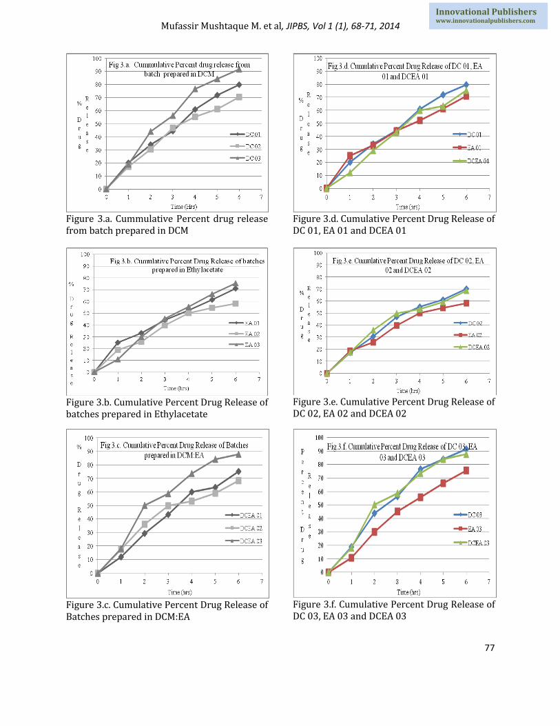
In-vitro percent drug release study:
The in-vitro drug release profile of rizatriptan benzoate is shown in the fig. 2. The maximum release of the drug was obtained for batches DC 03 (91.51%), DC 01 (79.76%) and DCEA 03 (87.80%)and minimum release was obtained for EA 02 (58.08%), DCEA 02 (68.41) and DC 02 (70.29%) for 6 h release study (Fig 3).Formulation variables such as concentration of polymer, drug and solvent selected for preparation of microsphere affects the rate and extent of drug release which is due to increase in density of polymer and increase in path length that a drug has to travel.[12,14]
The curves of release profiles in Fig. 3(a,b,c,) indicate that increasing polymer concentration resulted in thicker coated walls and greater impeding of the release of rizatriptan benzoate. Release data of all formulations indicate a sustained effect due to the encapsulation of the drug which depends on the drug to polymer ratio.
Mufassir Mushtaque M. et al, JIPBS, Vol 1 (1), 68-71, 2014
73
Innovational Publishers www.innovationalpublishers.com
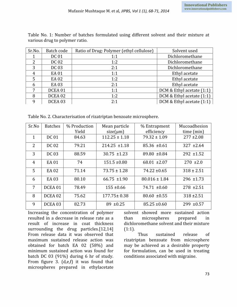
Table No. 1: Number of batches formulated using different solvent and their mixture at various drug to polymer ratio.
Table No. 2. Characterisation of rizatriptan benzoate microsphere.
Sr.No Batches % Production Yield
Mean particle size(µm)
% Entrapment efficiency
Mucoadhesion time (min)
1 DC 01 84.63 112.25 ± 1.18 79.32 ± 1.09 277 ±2.08
2 DC 02 79.21 214.25 ±1.18 85.36 ±0.61 327 ±2.64
3 DC 03 88.59 30.75 ±1.23 89.80 ±0.84 292 ±1.52
4 EA 01 74 151.5 ±0.80 68.01 ±2.07 270 ±2.0
5 EA 02 71.14 73.75 ± 1.28 74.22 ±0.65 318 ± 2.51
6 EA 03 80.10 66.75 ±1.90 80.016 ± 1.84 296 ±1.73
7 DCEA 01 78.49 155 ±0.66 74.71 ±0.60 278 ±2.51
8 DCEA 02 75.62 177.75± 0.38 80.60 ±0.55 318 ±2.51
9 DCEA 03 82.73 89 ±0.25 85.25 ±0.60 299 ±0.57
Increasing the concentration of polymer resulted in a decrease in release rate as a result of increase in coat thickness surrounding the drug particles.[12,14] From release data it was observed that maximum sustained release action was obtained for batch EA 02 (58%) and minimum sustained action was found for batch DC 03 (91%) during 6 hr of study. From figure 3. (d,e,f) it was found that microspheres prepared in ethylacetate
solvent showed more sustained action than microspheres prepared in dichloromethane solvent and their mixture (1:1).
Thus sustained release of rizatriptan benzoate from microsphere may be achieved as a desirable property for formulation, can be used in treating conditions associated with migraine.
Sr.No. Batch code Ratio of Drug: Polymer (ethyl cellulose) Solvent used 1 DC 01 1:1 Dichloromethane 2 DC 02 1:2 Dichloromethane 3 DC 03 2:1 Dichloromethane 4 EA 01 1:1 Ethyl acetate 5 EA 02 1:2 Ethyl acetate 6 EA 03 2:1 Ethyl acetate 7 DCEA 01 1:1 DCM & Ethyl acetate (1:1) 8 DCEA 02 1:2 DCM & Ethyl acetate (1:1) 9 DCEA 03 2:1 DCM & Ethyl acetate (1:1)
Mufassir Mushtaque M. et al, JIPBS, Vol 1 (1), 68-71, 2014
74
Innovational Publishers www.innovationalpublishers.com
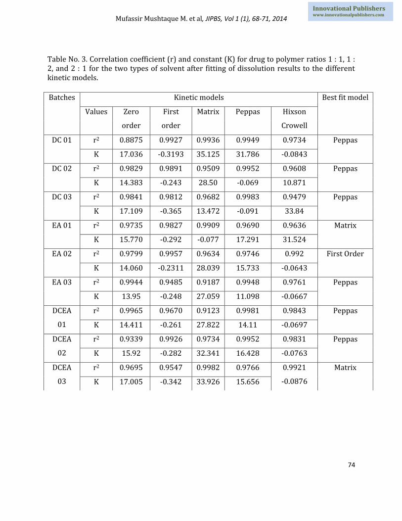
Table No. 3. Correlation coefficient (r) and constant (K) for drug to polymer ratios 1 : 1, 1 : 2, and 2 : 1 for the two types of solvent after fitting of dissolution results to the different kinetic models.
Batches Kinetic models Best fit model
Values Zero
order
First
order
Matrix Peppas Hixson
Crowell
DC 01 r2 0.8875 0.9927 0.9936 0.9949 0.9734 Peppas
K 17.036 -0.3193 35.125 31.786 -0.0843
DC 02 r2 0.9829 0.9891 0.9509 0.9952 0.9608 Peppas
K 14.383 -0.243 28.50 -0.069 10.871
DC 03 r2 0.9841 0.9812 0.9682 0.9983 0.9479 Peppas
K 17.109 -0.365 13.472 -0.091 33.84
EA 01 r2 0.9735 0.9827 0.9909 0.9690 0.9636 Matrix
K 15.770 -0.292 -0.077 17.291 31.524
EA 02 r2 0.9799 0.9957 0.9634 0.9746 0.992 First Order
K 14.060 -0.2311 28.039 15.733 -0.0643
EA 03 r2 0.9944 0.9485 0.9187 0.9948 0.9761 Peppas
K 13.95 -0.248 27.059 11.098 -0.0667
DCEA
01
r2 0.9965 0.9670 0.9123 0.9981 0.9843 Peppas
K 14.411 -0.261 27.822 14.11 -0.0697
DCEA
02
r2 0.9339 0.9926 0.9734 0.9952 0.9831 Peppas
K 15.92 -0.282 32.341 16.428 -0.0763
DCEA
03
r2 0.9695 0.9547 0.9982 0.9766 0.9921
-0.0876
Matrix
K 17.005 -0.342 33.926 15.656

Mufassir Mushtaque M. et al, JIPBS, Vol 1 (1), 68-71, 2014
75
Innovational Publishers www.innovationalpublishers.com
Figure No. 1. SEM images of drug loaded microspheres; Figure 1.a. SEM image of drug loaded microspheres of batch DC 03; Figure 1.b. SEM image of drug loaded microspheres of batch DCEA 03; Figure 1.c. SEM image of drug loaded microspheres of batch EA 02; Figure 1.d. SEM image of drug loaded microspheres of batch EA 03.

Mufassir Mushtaque M. et al, JIPBS, Vol 1 (1), 68-71, 2014
76
Innovational Publishers www.innovationalpublishers.com
In-vitro % drug diffusion and ex-vivo permeation study
The diffusion cell method has several benefits over usual in-vitro release studies. In this model, microspheres are in contact with a wetted and warm surface (filter paper) in a humid atmosphere, similar to nasal cavity. Normally, microspheres are immersed in a stirring medium, thus it seems that our results could be better extrapolated to in-vivo situations. It has found that diffusion of drug through cellulose nitrate runs parallel along with the in-vitro release of drug. All the formulation showed sustained release over 6 hr of study. Maximum diffusion was obtained for DC 03 (87.637%) and minimum diffusion of drug was obtained for EA 02 (64.47%) (Figure: 4 a). More sustained release effect is obtained when ethyl acetate is chosen as a solvent at highest polymer to drug ratio i.e. 1:2, since it increases the polymer matrix and being a hydrophobic polymer, ethylcellulose retards release of drug and formulation shows sustained action.
Permeation study was performed on dissected sheep nasal mucosa at 37 ± 0.50C and data obtained were plotted as shown in the figure 4.b.Maximum drug was permeated from DC 03 and minimum permeation was obtained for EA 02. Release kinetic study:
In order to obtain meaningful information for the release models, the drug release profiles were fitted to the four different kinetic models i.e. zero order, first order, hixson-crowel, higuchi (matrix) and korsmeyer-peppas equations and the goodness of fit of the release data was assessed. Table 3 summarizes the correlation coefficients for the different release kinetic models for ethylcellulose
microspheres formulated at 1:1, 1:2 and 2:1 drug to polymer ratios using two different solvents. Models with higher correlation coefficients were judged to be a more appropriate model for the dissolution data.
As shown in Table 3; batches DC 01, DC 02, DC 03, EA 03, DCEA 01 and DCEA 02 sows best fit model for peppas. Batches showing best fit model for matrix are EA 01 and DCEA 03 but batch EA 02 shows first order release kinetic.
SEM photomicrographs
Drug loaded microspheres of batch EA 02, DC 03 and DCEA 03 were performed and shown in Fig. 1. The drug was characterized by particles of spherical shape and heterogeneous size as shown in Fig.1, drug-loaded microspheres showed regular shape and smooth surface and no free drug was present. Drug-loaded microspheres made with EC showed a regular morphology, but also few bubble-like structures were present possibly due to rizatriptan benzoate adsorption and not entrapped into the polymeric network.
Figure 2: In-vitro percent drug release profile of microspheres for different batches.
Mufassir Mushtaque M. et al, JIPBS, Vol 1 (1), 68-71, 2014
77
Innovational Publishers www.innovationalpublishers.com
Figure 3.a. Cummulative Percent drug release from batch prepared in DCM
Figure 3.b. Cumulative Percent Drug Release of batches prepared in Ethylacetate
Figure 3.c. Cumulative Percent Drug Release of Batches prepared in DCM:EA
Figure 3.d. Cumulative Percent Drug Release of DC 01, EA 01 and DCEA 01
Figure 3.e. Cumulative Percent Drug Release of DC 02, EA 02 and DCEA 02
Figure 3.f. Cumulative Percent Drug Release of DC 03, EA 03 and DCEA 03

Mufassir Mushtaque M. et al, JIPBS, Vol 1 (1), 68-71, 2014
78
Innovational Publishers www.innovationalpublishers.com
Figure 4.a. Cumulative percent drug diffusion of batches for 6 hr of study
Figure 4.b. Cumulative percent drug permeation of batches for 6hrs of study
4. Conclusion
From the investigation it is concluded that the solvent evaporation technique can be used to obtain sustained release microspheres using ethylcellulose as a polymer which is most appropriate at an optimum stirrer speed of 1400 rpm.
1. Different formulation batches were evaluated for various parameters like production yield, entrapment efficiency, mucoadhesion time and particle size determination were found to get a better result for DC 03 batch having, production yield (80.97%), drug entrapment efficiency (85.19%), mucoadhesion time (270 min).
2. The In-vitro mucoadhesive study demonstrated that increase in polymer concentration, increases adherence of microspheres to mucus to a greater extent.
3. The In- vitro % drug release data suggest that as the amount of hydrophobic polymer increases, release of a drug decreases. The maximum drug release was obtained for DC 03 and minimum drug release was observed for EA 02 batch.
4. Sustained release for rizatriptan benzoate was obtained for EA 02 (78% of release was obtained after 6 hr of study) successfully achieved by using solvent evaporation technique.
5. Microspheres prepared by dichloromethane releases drug at faster rate, whereas microspheres prepared by ethyl acetate releases drug at slower rate. This is most evident at the same 2:1 drug to polymer ratio.
6. The release kinetic studies indicate that DC DC 01, DC 02, DC 03, EA 03, DCEA 01 and DCEA 02 followed diffusion as the main mechanism for drug release. While for EA 01, EA 02 DCEA 02and DCEA 03 shows matrix and first order release model.
Acknowledgements Authors are grateful to Mrs.Padmashreee Fatma Rafiq Zakaria, Chairman, Maulana Azad Education Trust Y B Chavan college of Pharmacy, for encouragement, support and providing facilities. The authors are thankful to Cipla. Ltd ( Mumbai, India) and Wockhardt Research Ltd (Aurangabad, India). References
1. Murthy T E G K, Chowdar K D R. Formulation and evaluation of ethyl cellulose coated-
Mufassir Mushtaque M. et al, JIPBS, Vol 1 (1), 68-71, 2014
79
Innovational Publishers www.innovationalpublishers.com
diclofenac sodium microcapsule: Effect of solvent. Indian JPS, 67(2): 216-219, 2005.
2. Harikarnpakdee S, Lipipun V, Sutanthavibul N, Ritthidej G C. Spray dried mucoadhesive microspheres: Preparation and transport through nasal cell monolayer. AAPS PharmSciTech., 7(1): 12, 2006.
3. Martinac A, Filipovic-Grcic J, Voinoch D, Perissutti B, Franceschinis E . Development and bioadhesive properties of chitosan-ethyl cellulose microspheres for nasal delivery. International Journal of Pharmaceutics, 291: 69-77, 2005.
4. Tabassi S A S, Razavi N. Preparation and characterization of albumin microspheres encapsulated with propranolol HCl. DARU. ,11(4): 137-141, 2003.
5. Gavini E, Rassu G, Muzzarelli C, Cossu M, Giunchedi P.Spray-dried microspheres based on methylpyrrolidinone chitosan as new carrier for nasal administration of metoclopramide. European Journal of Pharmaceutics and Biopharmaceutics, 68: 245–252, 2008.
6. Jain S K, Jain N K, Gupta Y, Jain A, Jain D, Chaurasia M. Mucoadhesive chitosan microspheres for non invasive and improved nasal delivery of insulin. Indian J. of Pharm Sci, 69(4): 498-504, 2007.
7. Abd El-Hameed M D, Kellaway I W. Preparation and in vitro characterisation of mucoadhesive polymeric microspheres as intra-nasal delivery systems. European Journal of Pharmaceutics and Biopharmaceutics, 44: 53-60, 1997.
8. Ramesh RP, Mahesh C P. Nasal Drug delivery in Pharmaceutical and biotechnology: present and future; e-Journal of Science & Technology. 3-5, 2009.
9. Anaísa P, Ana F, Gilberto A, Amílcar F. Intranasal Drug Delivery: How, Why and What for. J Pharm PharmaceutSci (www.cspsCanada.org).,12(3): 288 – 311, 2009.
10. Keri W, Greg L P. Rizatriptan, An Update of its Use in the Management of Migraine, Adis Drug Evaluation, Drugs., 62(10):1539-1574, 2002.
11. Illum, L, Jorgensen H, Bisgaard, H, Krogsgaard O, Rossing N. Bioadhesive microspheres as a potential nasal delivery system. Int. J. Pharm., 39:189–199, 1987.
12. Amperiadou A, Georgarakis M. Controlled release salbutamol sulphate microcapsules prepared by emulsion solvent-evaporation technique and study on the release affected parameters. International J Pharmaceutics. , 115:1-8, 1995.
13. Shirui M, Jianming C, Zhenping W, Huan L, Dianzhou B. Intranasal administration of melatonin starch microspheres. International J Pharmaceutics. , 272:37–43, 2004.
14. Al Gohary, O. and El Gamal, S., Release of furosemide from sustained release microcapsules prepared by phase separation technique. Drug Dev. Ind. Pharm., 17: 443-450, 1991.
15. Ziyaur, R., Kanchan, K., Khar, R.K., Mushir, A., Charoo, N.A., Shamsher, A. A. Characterization of 5-fluorouracil microsphere for colonic delivery. AAPS Pharm. Sci. Tech, 7 (2): E1–E9 (article 47), 2006.