effects of rna on early chick embryos cultivated in vitro
TRANSCRIPT
Acta physiol. scand. 1960. 48. 352-363
Zoophysiological Institute, University of Lund, Sweden
Effects of RNA on Early Chick Embryos Cultivated in Vitro
BY
HADAR EMANUELSSON Received 15 July 1959
Abstract EMANUELSSON, H. Effects of RNA on ear& chick embryos cultivated in vitro.
Acta physiol. scand. 1960. 48. 352-363. - The object of the present investigation has been to find out whether pure RNA can exert a stimulating action on the morphogenesis of early chick embryos. The experiments have been performed on 16 to 18-hour-old embryos cultivated in vitro. RNA from 1) area opaca of 2-day-old chick embryos 2) from heads of 6-day-old chick embryos and 3) from yolk-sacs of 6-day-old chick embryos was tested. - After 24 hours’ cultivation in vitro a marked growth and differentiation occurred in embryo-regions homologous to those from which the RNA had been isolated. Other regions, however, were rather retarded in their development and displayed disturbed mitoses. Mitotic counts demonstrated an in- creased mitotic activity in the growth-stimulated embryo-regions. In the latter a slight increase in the percentage of anaphases + telophases also was noted. - Thus it appears that the pure RNA in itself has the capacity of stimulating growth and differentiation in chick em- bryos, the localization of the effect being dependent, however, on the origin of the RNA used.
As the result of their grafting experiments on early embryos several inves- tigators have arrived at the conclusion that among other effects RNA exerts an inducing activity on embryonic morphogenesis. Most investigators ( BRACHET 1957 p. 402 et seq.) are of the opinion that it is in combination with protein that RNA can act as an inductor, but there are also indications (NIU 1956) that RNA itself can fulfil this mission.
RNA from chick embryos has been tested as inductor in newt and axolotl embryos (TIEDEMANN and TIEDEMANN 1956), but very weak inductive effect was reported.
352

EFFECTS OF RNA ON EARLY CHICK EMBRYOS 353
When CHEVREMENT and FIRKET (1 952) investigated the effect of pure RNA in low concentrations on tissue cultures, they found a stimulating effect from RNA on cell-multiplication; but this effect was mainly to be found in those cultures where conditions were suboptimal with reference to embryo extract, of which only traces had been added to the medium.
DEOTTO (1 956), however, who has studied the effect of pure RNA on various materials (hanging drop cultures of chick fibroblasts, sea urchin embryos, plant roots etc.), states that the general effect of RNA when added in low concentrations is inhibition of cell-multiplication and morphogenesis. The same author has also made experiments in which RNA-solution was injected into the amnion of chick embryos, but unfortunately crucial importance could not be attached to the latter experiments owing to the high mortality among the embryos caused by the technique employed.
In the present investigation the object for the RNA-treatment has been isolated chick embryos at the primitive streak stage. The reason for using embryos of so early age has been that these - still being in the gastrula stage - are especially sensitive to external stimulus. Accordingly it was to be expected that if the various isolated ribonucleic acids really exert any special effects on embryogenesis in the chick, these ought to become particularly manifest at this very stage.
Material and Methods The experiments were performed on chick embryos (White Leghorn), 16-18 hours
old according to the Hamburger system. The embryos were at this stage transferred into Carrel flasks and cultivated there
for exactly 24 hours on a modified Spratt’s minimum medium to which RNA was also added.
Modified Spratt’s minimum medium: (cf. Spurn 1948, HOWARD 1953)
Control embryos were cultivated on the pure minimum medium alone.
NaCl KCl CaC1, .6 H,O Na,HPO,. 2 H,O KH,P04 NaHCO, Glucose Agar
0.650 per cent 0.033 0.031 0.007 0.003 0.055 0.850 0.420
Buffer components are dissolved separately and the resulting solution is saturated with CO, before addition of other components.
Previous investigations ( EMANUELSSON 1958) have proved this minimum medium to be suitable for the cultivation of early chick embryos. Thus explanted embryos when cultivated on it for 24 hours undergo a normal differentiation, only slightly slower than in embryos not removed out of the egg.
Addition of RNA was carried out by first dissolving it in a solution made up from

354 HADAR EMANUELSSON
Table I . Base ratios of isolated ribonucleic acids. All ratios are expressed on a molar basis with respect to adenine
RNA
isolated from Area opaca
Y olk-sacs
(Embryo: 40-60 h) . . . . . . . .
(Embryo: 6 days) ..........
(Embryo: 6 days) . . . . . . . . . . Embryo heads
Guanine
2.28
1.89
1.82
Adenine
1 .o
1 .O
1 .o
Cytosine
I .63
1.65
1.41
Uracil
1.58
1 .o
0.81
the buffer components of the minimum medium. This solution was then sterilized by filtration through a Seitz filter and finally mixed with the other components, which had been sterilized by boiling just before. The pH of the complete medium was 7.4.
Preliminary experiments had shown that added RNA gave effect in concentrations from 0.06 to 0.27 mg/ml cultivation medium. In the present investigation the concentration was usually held at 0.17 mg/ml. TheRNAwas alwaysusedimmediately after it had beenisolated.
Three different ribonucleic acids were investigated. They were isolated from respec- tively 1) area opaca of chick embryos, 40-60 hours old 2) yolk-sacs of 6-day-old chick embryos 3) heads of 6-day-old chick embryos. Isolation of the RNA was made ac- cording to the method of KIRBY (1956), but some modifications, as proposed by NIU (private communication) were applied. Thus the selected tissue was rapidly chilled to 2" C (freezing was avoided) and was homogenized at the same temperature in 0.9 per cent NaCl (as opposed to distilled water in the original method) before the addition of phenol.
Furthermore, when homogenizing the tissues it was arranged that the resulting con- centration of RNA in the homogenate was maintained at about 40 pg/ml.
The isolated ribonucleic acids were analyzed to ascertain their base composition. Cf. Table I. For that purpose four separate samples from each RNA were hydrolyzed in N-HCl for 1 h at 100" C and then chromatographed on filter paper. Solvent system: isopropanol - conc. HC1- water (98: 25: 28 by vol.) All samples proved to be in good agreement.
No traces of proteins or protein derivatives in the ribonucleic acids were observed in these chromatograms.
In all experiments the explanted embryo has been a quadrangular area, dissected out of the blastoderm, and has included the former part of the primitive streak with Hensen's node. In Fig. 1 the extension of an explanted 18-hour-old embryo is out- lined.
The cultivation was carried out at 37.0" C & 0.5". Afterwards the explants were fixed in Carnoy's fluid and stained whole in Gomori's hematoxylin according to ME- LANDER and WINGSTRAND (1953). The preparations were finally mounted in Euparal or DPX.
Fig. 1. Schematic drawing of chick embryo of 18 hours of incubation. Heavy lines limit that part of the embryo which is cultivated in vitro.
EFFECTS OF RNA ON EARLY CHICK EMBRYOS 355
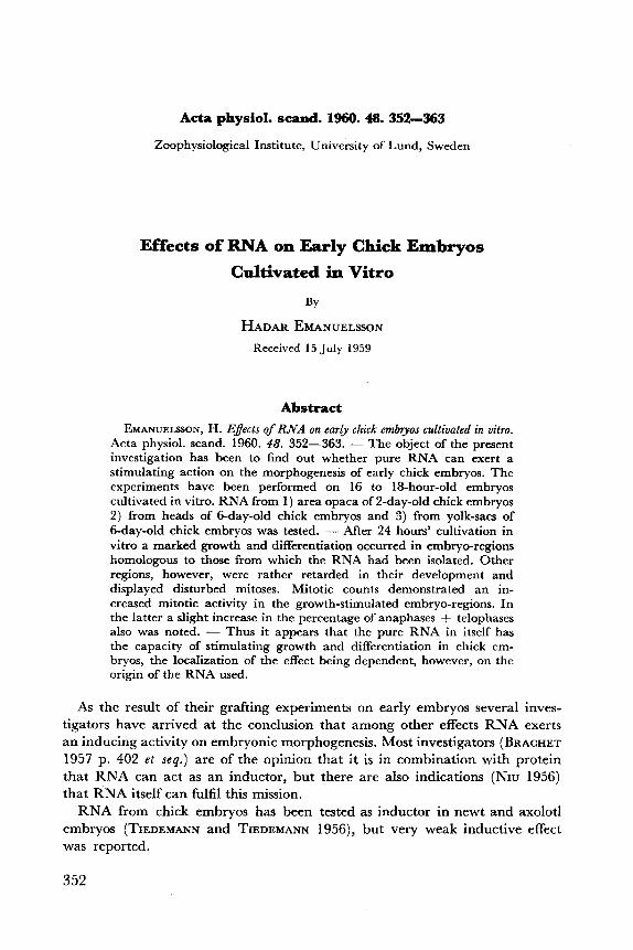
Fig. 2. Chick embryos explanted at the 16-18 hour stage and then cultivated in vitro for 24 hours. A,, A,: Controls, cultivated on the pure minimum medium B,, B,: Embryos, cultivated on minimum medium to which was added RNA from area opaca
of 40-60-hour old chick embryos C,, C,: Embryos, cultivated on minimum medium to which was added RNA from yolk-sacs
of 6-day-old chick embryos. D,, D,: Embryos, cultivated on minimum medium to which was added RNA from heads of .. .
6-day:old chick embryos. Gomori hematoxylin. Yellow filter.
356 HADAR EMANUELSSON
Results
The cultivated embryos have been classified in 4 groups, A-D. Group A: Controls, cultivated on the pure minimum medium. Group B: Embryos, cultivated on minimum medium, to which had been
added RNA from area opaca of 40 to 60 hour-old chick embryos. Group C: Embryos, cultivated on minimum medium, to which had been
added RNA from yolk-sacs of 6-day-old chick embryos. Group D: Embryos, cultivated on minimum medium, to which had been
added RNA from heads of 6-day-old chick embryos. In Fig. 2 are reproduced microphotographs, showing two embryos from
each group. Such microphotographs have been the basis for the schematic drawings in Fig. 3-Fig. 6, which are intended to give a representative picture of the morphblogy of the explants. All embryos are drawn to the same scale.
About 150 embryos from group A and 70 from each of the groups B-D have been examined. Disregarding the regular occurrence of entirely abnor- mal embryos in all the groups, successful treatment with RNA was noted for about 75 per cent of the embryos belonging to the groups B-D.
Morphological Observations
the following appearances :
Group A: These, the control embryos, are characterized by well-developed neural
tube and brain. In the latter the optical vesicles are distinctly visible. Somites
After 24 hours' cultivation in vitro the explanted embryos have acquired
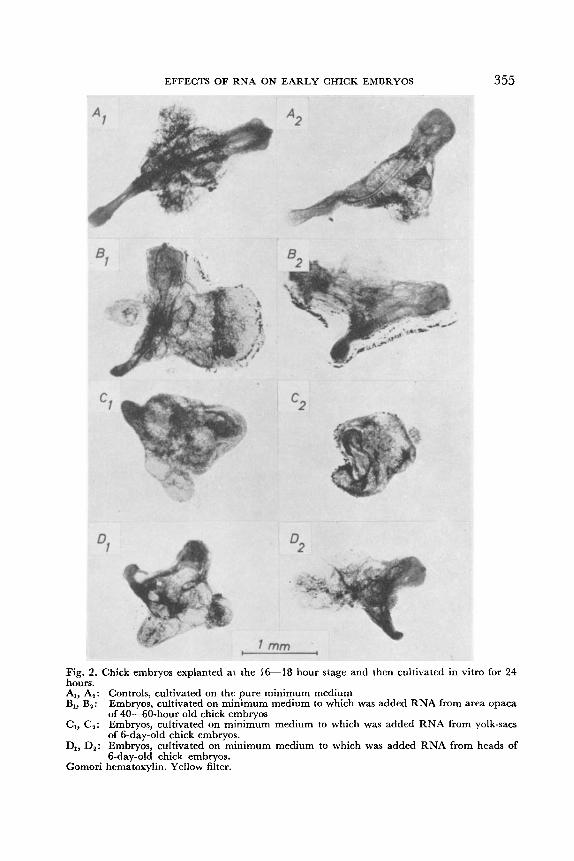
Fig. 3. Schematic drawings of chick embryos, explanted at the 16 to 18-hour stage and then cultivated in vitro for 24 hours on the pure minimum medium. The drawings, which are intended to give a representative picture of the results are based on microphotographs of the same sort as in Fig. 2.

EFFECTS OF RNA ON EARLY CHICK EMBRYOS 357
Fig. 4. Schematic drawings of chick embryos, explanted at the 16 to 18-hour stage and then cultivated in vitro for 24 hours on minimum medium to which was added RNA from area opaca of 48--60-hour-old chick embryos. The drawings, which are intended to give a rep- resentative picture of the results, are based on microphotographs of the same sort as in Fig. 2.
Fig. 5. Schematic drawings of chick embryos, explanted at the 16 to 18-hour stage and then cultivated in vitro for 24 hours on minimum medium to which was added RNA from yolk- sacs of 6-day-old chick embryos. The drawings, which are intended to give a representative picture of the results, are based on microphotographs of the same sort as in Fig. 2.
are always to be found. Outgrowth on either side of the middle part of the embryo - in the future referred to as “marginal outgrowth” - is noted, but it is usually not very pronounced.
Group B: Compared with the embryos of group A the embryos belonging to the B
group are somewhat retarded in their development of the neural tube and the brain. In most cases somites are visible, but sometimes they can be missing.
358 HADAR EMANUELSSON
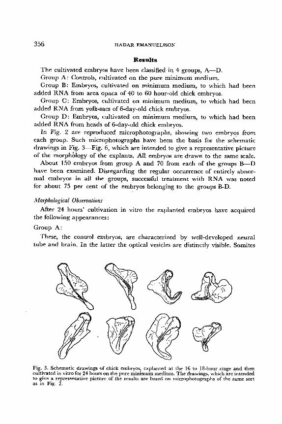
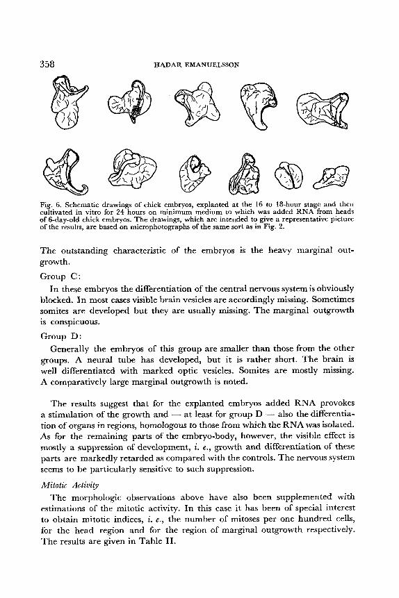
Fig. 6. Schematic drawings of chick embryos, explanted at the 16 to 18-hour stage and then cultivated in vitro for 24 hours on minimum medium to which was added RNA from heads of 6-day-old chick embryos. The drawings, which are intended to give a representative picture of the results, are based on microphotographs of the same sort as in Fig. 2.
The outstanding characteristic of the embryos is the heavy marginal out- growth.
Group C: In these embryos the differentiation of the central nervous system is obviously
blocked. In most cases visible brain vesicles are accordingly missing. Sometimes somites are developed but they are usually missing. The marginal outgrowth is conspicuous.
Group D: Generally the embryos of this group are smaller than those from the other
groups. A neural tube has developed, but it is rather short. The brain is well differentiated with marked optic vesicles. Somites are mostly missing. A comparatively large marginal outgrowth is noted.
The results suggest that for the explanted embryos added RNA provokes a stimulation of the growth and - at least for group D - also the differentia- tion of organs in regions, homologous to those from which the RNA was isolated. As for the remaining parts of the embryo-body, however, the visible effect is mostly a suppression of development, i. e . , growth and differentiation of these parts are markedly retarded as compared with the controls. The nervous system seems to be particularly sensitive to such suppression.
Mitotic Activity The morphologic observations above have also been supplemented with
estimations of the mitotic activity. In this case it has been of special interest to obtain mitotic indices, i. e. , the number of mitoses per one hundred cells, for the head region and for the region of marginal outgrowth respectively. The results are given in Table 11.

EFFECTS OF RNA ON EARLY CHICK EMBRYOS 359
Table II. Mitotic indices ( =number of mitoses per one hundred cells) for RNA-treated embryos and controls. The jigures are meam of valuesfiom covesfionding areas of four separate embryos. In each single measurement 1,000 - 2,000 cells have been counted. Investigated areas are marked off in Fig. 7
I Group A 1 Group B I Group C I Group D
Brain area. .. . . . . .. . . .. . . . Area of marginal outgrowth
On the whole the values above will confirm the morphologic results. Thus the mitotic indices indicate- that for group D there is an increased mitotic activity in the brain area, whereas the reverse condition prevails for the cor- responding area of group B and group C.
In the region of marginal outgrowth again there appears for group B a distinct increase of the mitotic activity. Contrary to this corresponding areas for group D present values which suggest a suppression of mitotic activity.
A
B
Fig. 7. Schematic drawings showing embryos from the four groups (A-D), which have been selected for estimation of the mitotic activity. Investigated areas are encircled. All embryos are drawn to the same scale. 25-593790. Acta physiol. scand. Vol. 48.

360 HADAR EMANUELSSON
Fig. 8. Microphotograph showing disturbed mitoses from the neural groove of an embryc, cultivated on minimum medium to which was added RNA from yolk-sacs of 6-day-old chick embryos. The large grey oval spots are nuclei. Smaller black globules derive from disintegrated nuclei. Gomori hematoxylin. Yellow filter.
Finally, it will seem that the values for group C remarkably enough point in favour of a suppression of mitotic activity in the region of marginal out- growth. In the latter case one would rather have expected ari increased cell division, corresponding to the morphologic observations.
As to the mitotic spectrum, i. e. , the occurrence of the various phases of mitosis, it is observed that in areas where RNA has stimulated cell-multiplica- tion the percentage of ana- and telophases is somewhat increased, as compared with values from the control embryos. For the latter the percentage of ana- phases + telophases is thus 21 f 5 for the two types of investigated areas, whereas for the brain area of embryos of group D the corresponding percentage is 30 & 2 and for the marginal outgrowth area of embryos of group B 30 & 1. Otherwise there is no difference between RNA-treated embryos and controls as regards this percentage.
Inspection of the embryo cells further reveals many disturbed mitoses and karyorrhexis in those embryo regions where morphogenesis had been blocked by the RNA-treatment. Cf. Fig. 8.
Discussion
In the study of embryogenesis and protein biosynthesis the problem of control of growth by organ-specific substances holds an outstanding position, and accordingly it has been subjected to extensive experimental investiga- tions.
EFFECTS OF RNA ON EARLY CHICK EMBRYOS 36 1
In 1941 WEISS reported thatrincorporation of liver and kidney fragments from 6-day-old chick embryos into the area vasculosa of 4-day-old chick embryos provoked a considerable increase in mass of the homologous organ of the latter as compared with controls.
Similar observations have been made by EBERT (1954), who studied the effect of chorio-allantoic grafts of spleen from adult chicks on 9-day-old host chick embryos.
On the other hand, several authors report that addition of small fragments as well as extracts from organs of older chick embryos also can have an inhibiting action on the differentiation of the homologous organs of younger embryos.
Thus WEISS (1 952) has shown that extracts of whole chick embryos have an inhibiting action on isolated heart and kidney fragments from embryonal chick cultured in vitro. This inhibition was not found if the extracts were made on embryos from which the homologous organ had first been removed.
DITTMAR, LIPP and AUGSTEIN (1 957), who have studied the action of various organ-extracts from chick embryos on fragments of the homologous organs cultured in vitro, report no special effect on the explants from the homologous organs with the exception that extracts from liver had a definitely inhibiting action on the growth of all types of explants.
Apparently it would seem as if the results quoted were conflicting, but as a matter of fact the different effects in the investigations in question are not inconsistent, and they can be satisfactorily explained if one takes into con- sideration the antagonism prevailing between growth and differentiation during embryogenesis.
As to the biochemical nature of the inducing factors involved in the growth processes just mentioned, there are, however, no definite records.
Now the present investigation actually demonstrates that pure RNA in itself can stimulate morphogenesis in chick embryos and increase cell-multi- plication. It will be remembered, however, that those ribonucleic acids which have been made use of here have been isolated according to a method that presumably preserves the properties of the native RNA better than most other isolation methods. Therefore it does not seem improbable that previous failures to prove a stimulating effect on cell-multiplication from pure RNA might have partly been due to the application of unsuitable methods for the isolation of the RNA.
Further, it appears from the present investigation that the stimulating action of RNA affects only the homologous tissues, other tissues rather being sup- pressed in their development. Considering this, it seems reasonable that the effect of added, non-specific RNA in tissue culture experiments has been inhibition.
When interpreting the results from the chick embryos in the present investiga- tion it will be remembered that the controls with which the RNA-treated
362 HADAR EMANUELSSON
embryos have been compared, have been wltivated on minimum medium exclusively. However - in spite of their complete differentiation - these controls have in fact restricted possibilities to a continued development.
Bearing this in mind, it will perhaps be less surprising that addition of RNA to the minimum medium can give rise to considerable growth changes. Of course, it would have been desirable to carry out a comparison between the RNA-treated embryos here and embryos developing in the intact egg, but that would have been misleading in many respects, as the latter are in pos- session of an entirely intact area opaca and a complete supply of nutrients. Nevertheless, it is highly interesting that the growth-stimulating effect actually produced by the pure RNA has been specific in such a way that it has been restricted to regions, homologous to those, from which the RNA has been isolated.
-4s to the mitotic activities, it is a surprise to find a low mitotic activity in the highly developed marginal outgrowth region of embryos of group C, where instead a high activity - as for embryos of group B - was to be ex- pected.
Now when investigating the mitotic activity in a limited area of the region in question all mitoses within this area, irrespective of their distribution at various levels, have been counted, i. e . , the area has been limited sideways only. This will imply that the counting might concern different kinds of tissues if they are disposed on each other. One possible explanation to the low ac- tivity in question might perhaps be then that the proportion of presumptive yolk-sac tissue in the investigated area - i. e., that very tissue which will be susceptible to stimulation - has not been big enough to produce a general impression of increased mitotic activity.
Finally, the observation that there has been a slight increase in the proportion of ana- and telophases in the RNA-treated embryos is of interest. Thus it indicates the possibility of RNA acting on cell-division by shortening the dura- tion of pro- and metaphases. The other possibility - indication of a prolonga- tion of ana- and telophases - seems less plausible in the present case.
The author wishes to express his sincere thanks to Professor IVAR AGRELL, Head of the Zoo-
The investigation has been facilitated by grants from the Kungliga Fysiografiska Sallskapet, physioIogica1 Institute, Lund, for stimulating advice and criticism during the work.
l a n d , and from the Swedish Natural Science Research Council.
References BRACHET, J., “Biochemical Cytology”. New York 1957. CH~VREMENT, M. and H. FIRKET, Action de l’acide ribonucleique sur la croissance et la mitose
DEOTTO, R., Azione degli acidi nucleinici sulla moltiplicazione cellulare. ‘Tumora 1956. 42.
DITTMAR, C. , R. LIPP and G. AUGSTEIN, Experiments on the influence of extracts of different
en culture de tissus. C. R. Assoc. Anat. 1952. 39. 95-98.
1-64.
organs on the growth of tissue cultures. 5. ges. exp. Med. 1957. 128. 600-606.
EFFECTS OF RNA ON EARLY CHICK EMBRYOS 363
EBERT, J. O., The effects of chorioallantoic transplants of adult chicken tissues on homologous
EMANUELSSON, H., Variations in nucleic acid concentration during the development of earl?-
HOWARD, E., Some effects of NaCl concentration on the development of early chick blastoderms
KIRBY, K. S., A new method for the isolation of ribonucleic acids from mammalian tissues.
MELANDER, Y. and K. G. WINGSTRAND, Gomori’s hematoxylin as a chromosome stain. Stain
NIU, M. C., in “Cellular mechanisms in differentiation and growth” Ed. by D. Rudnick, Sew
SPRATT, JR., N. T., Development of the early chick blastoderm on synthetic media. J . esp.
TIEDEMANN, H. and H. TIEDEMANN, Isolierung von Ribonukleinsaure und Nucleotiden aus Embryonalextrakt und Leber und ihr Verhalten im Induktionsversuch. Hoppe-Seyler‘s 2. physiol. Chem. 1956. 306. 132-142.
tissues of the host chick embryo. PYOC. nut. Acad. Sci. (Wash.) 1954. 40. 337-347.
chick embryos. Acta physiol. scand. 1958. 44. 336-364.
in culture. 3. cell. comp. Phjsiol. 1953. 41. 237-259.
Biochem. 3. 1956. 64 . 405-408.
Technol. 1953. 28. 217-223.
Jersey 1956. 155-171.
<00l. 1948. 107. 39-64.
WEISS, P., Self-regulation of organ growth by its own products. Science 1952. 115. 487-488. WEISS, P. and H. WANG, Growth response of the liver of embryonic chick hosts to the incorpora-
tion in the area vasculosa of liver and other organ fragments. Anat. Rec. 1941. 79. Suppl. 62.