effects of posture on dynamic back loading during a cable
TRANSCRIPT

ERGONOMICS, 2002, VOL. 45, NO. 5, 380 ± 398
Ergonomics ISSN 0014-0139 print/ISSN 1366-584 7 online # 2002 US Governmenthttp://www.tandf.co.uk/journals
DOI: 10.1080/0014013021012763 9
EVects of posture on dynamic back loading during a cable lifting task
SEAN GALLAGHER{*,
*Author for correspondence. e-mail: [email protected]
{National Institute for Occupational Safety and Health, Pittsburgh Research Laboratory, Pittsburgh, PA 15236-0070, USA
WILLIAM S. MARRAS{, and KIMBERLY KOVACS{
{Biodynamics Laboratory, Department of Industrial, Welding, and Systems Engineering, The Ohio State University, 1971 Neil Avenue, Columbus,
OH 43210, USA
KERMIT G. DAVIS}
}Department of Environmental Health, Industrial Hygiene Division, 330 Kettering Laboratory, 3223 Eden Avenue, University of Cincinnati, Cincinnati,
OH 45267, USA
This study evaluated spinal loads associated with lifting and hanging heavy mining cable in a variety of postures. This electrical cable can weigh up to 10 kg per metre and is often lifted in restricted spaces in underground coal mines. Seven male subjects performed eight cable lifting and hanging tasks, while trunk kinematic data and trunk muscle electromyograms (EMGs) were obtained. The eight tasks were combinations of four postures (standing, stooping, kneeling on one knee, or kneeling on both knees) and two levels of cable load (0 N or 100 N load added to the existing cable weight). An EMG-assisted model was used to calculate forces and moments acting on the lumbar spine. A two-way split-plot ANOVA showed that increased load (p50.05) and changes in lifting posture (p50.05) independently aVected trunk muscle recruitment and spinal loading. The increase in cable load resulted in higher EMG activity of all trunk muscles and increased axial and lateral bending moments on the spine (p50.05). Changes in posture caused more selective adjustments in muscle recruitment and aVected the sagittal plane moment (p50.05). Despite the more selective nature of trunk EMG changes due to posture, the magnitude of changes in spinal loading was often quite dramatic. However, average compression values exceeded 3400 N for all cable lifting tasks.
1. Introduction The posture adopted by the body during manual materials handling tasks is thought to have a profound impact on spinal loading. The preponderance of manual lifting research in ergonomics has concentrated on analysis of loads experienced during standing postures; however, certain occupations require that workers adopt postures that may signi®cantly alter muscle recruitment patterns and biomechanical loads on the body. For example, coal miners often work in vertically con®ned workspaces where standing erect is not possible. In such an environment, the miner is obliged to

3381
select a lifting posture from a menu of unpalatable alternatives, typically stooping (standing with a fully ¯exed trunk) or kneeling (on one or both knees). Each of these postures engenders signi®cant biomechanical disadvantages . For example, psycho-physical lifting capacity and strength are signi®cantly reduced in the kneeling posture (Gallagher et al. 1988, Smith et al. 1989, Gallagher and Unger 1990, Gallagher and Hamrick 1992, Gallagher 1997). Stooping requires severe trunk ¯exion resulting in a number of biomechanical disadvantages , including higher shear loading on the spine, reliance on passive tissues for spinal support, and reduced force output from the back muscles as a function of the muscle length-strength relationship (Floyd and Silver 1955, Potvin et al. 1991, Yingling and McGill 1999). However, the stooping posture often appears to be favoured by mineworkers, particularly when high forces need to be exerted in restricted spaces, or when enhanced mobility is needed. However, it is quite conceivable that workers who adopt the stooping posture to take advantage of its strength bene®ts may, at the same time, be subjecting themselves to potentially damaging spinal loads.
In addition to the unusual postural demands of their job, miners sometimes have to handle materials possessing rather unique characteristics. One example is the large diameter electrical cable used to power heavy mining equipment. These cables are extremely heavy due to the large amounts of copper wire and to the insulation requirements associated with the electrical current demands of this massive equipment. It is not uncommon for this cable to have a diameter of 7.5 cm and to weigh 10 kg per metre. Several metres of cable may have to be handled during a cable lifting task, and the higher the cable is lifted the greater is the load that must be supported by the worker. This progressive increase in the load being supported is unusual in manual handling activities and must place a higher demand on the muscular agonists compared to a similar lift involving a constant load. The ¯exibility of cable may cause additional concerns due to load instability, especially given the heavy weight of the material. In the mining environment, additional stressors may exist when lifting cable. For example, the cable may become quite muddy, adding signi®cantly to the weight and force requirements of the lift. Not surprisingly, handling of heavy cable has been associated with a large number of lost-time back injuries in the mining industry (Randolph 1991).
Changes in body posture (from standing to kneeling, for instance) are likely to have signi®cant impacts on the synergism of muscular activity, and it is presumed that the sensorimotor cortex would have to adjust muscle activation programmes to accomplish a speci®ed manual lifting task (Dul 1986). Previous investigations have con®rmed that signi®cant changes in trunk muscle activity do occur in restricted postures (Gallagher et al. 1988, 1994, Gallagher and Unger 1990). Increases in the weight (or load) lifted have also been shown to impact on trunk muscle recruitment (Fathallah et al. 1998, De Looze et al. 1999, Davis and Marras 2000). However, it is not well understood whether the trunk muscle activity changes due to posture and those due to increased load activity act independently or whether these in¯uences might interact. Since trunk muscle activity is thought to be indicative of spinal load (McGill and Norman 1986, Chaf n and Andersson 1991, Granata and Marras 1993), which is (in turn) associated with development of low-back disorders (Marras et al. 1993), understanding such relationships is of critical importance to those who must handle materials in restricted working postures.
Therefore, the current experiment was undertaken to address the following hypotheses.

382 S. Gallagher et al.
(1) Hypothesis 1. Changes in posture would alter trunk muscle activity and estimates of lumbar spine loading.
(2) Hypothesis 2. Increased cable load would alter trunk muscle activity and lumbar spine loading.
(3) Hypothesis 3. The main eVects described above might interact with one another.
These eVects were investigated in a task involving the lifting and hanging of large diameter electrical cables, which are commonly handled in underground coal mines.
2. Methods 22.1. Subjects Seven male subjects volunteered to perform a series of lifts using a heavy mining cable used to power heavy underground equipment. Subjects read and signed an informed consent form prior to the initiation of testing. None had a prior history of low-back disorders (LBD). The subjects’ ages ranged from 22 to 36 years, and none had prior experience with cable hanging tasks. Anthropometric data are provided in table 1.
Table 1. Anthropometric data of the subjects.
Subject Age (yrs) Weight (kg) Height (cm)
1 24 76.2 194.4 2 21 72.3 189.9 3 25 64.5 174.0 4 30 92.7 178.6 5 36 95.5 197.5 6 22 80.9 165.0 7 22 79.5 181.3 Mean 25.7 80.2 182.9 Standard deviation 5.4 10.9 11.6
Comparison of these data with the mining population suggests that these subjects were approximately the same stature, but weighed less and were somewhat younger than underground coal miners (Gallagher 1999).
2.2. Experimental design The study evaluated spinal loads during the lifting and hanging of the cable using an EMG-assisted biomechanical model. The independent variables consisted of lifting posture (with four levels) and cable load (two levels). Subjects served as blocks within which experimental conditions were randomized. The following lifting postures were manipulated in this study: kneeling on one knee (1KNEE), kneeling on two knees (2KNEE), stooping (STOOP), and standing (STAND). The weight of the entire length of cable (7.6 m) used in this study was approximately 367 N. However, the amount of this weight actually supported by the subject during a lift was only a portion of the total load and varied according to the length lifted during the task. Previous research (Gallagher et al. 2001), also employing continuous mining cable, indicated a linear increase in load according to the following equation:
Cable load …N† ˆ 24:7 ‡ 0:083 £ Height of centre of cable …mm† …1†

383
An additional 100 N (50 N load attached to both ends) was placed on the cable to simulate the eVect of an additional load (from additional lengths of cable, mud, etc.) as may be experienced in underground cable lifting tasks. While the subjects did not directly lift this additional load, the weight added to either end increased the force required to draw the cable ends together when lifting the centre portion of the cable to the hook. The added load increased the force required to lift the cable by an estimated 20±25%. The magnitude of the additional load was selected such that the load would provide an estimate of the eVect without being so large as to create an undue risk of injury to the subject. Each lift was performed three times in each load posture/load condition, for a total of 24 lifts per subject.
2.3.2 Dependent measures Spine moments and forces were estimated using an EMG-assisted biomechanical model which has been under development in the Ohio State University Biodynamics Laboratory over the past 18 years (Marras and Reilly 1988, Reilly and Marras 1989, Marras and Sommerich 1991a, b, Granata and Marras 1993, 1995, Mirka and Marras 1993, Marras and Granata 1995, 1997, Davis et al. 1998). This model provides estimates of spine loading parameters based upon measured activity of ten trunk muscles, from which estimates of muscle force and subsequent spine loading are determined (Marras and Granata 1997). Forces for each muscle are estimated using the following equation.
EMGj…t† Fj ˆ PCSAj £ Gain £ £ f…L ¡ S† £ f…F ¡ V† …2†
EMGmax¡j
where: Fj = predicted muscle force for muscle j; PCSAj = physiological cross-sectional area for muscle j; Gain = estimate of muscle stress (force/area); EMGj(t) = instantaneous integrated EMG for muscle j at time t; EMGMax-j = integrat ed EMG from maximum voluntary contraction for muscle j; f(L-S) = muscle length-strength relationship modulation factor; and f(F-V) = muscle force-velocity relationship modulation factor.
The muscle generated internal moments about the axis of rotation are predicted from the sum vector products combining the tensile muscle force for each muscle and the moment arms of each respective muscle as given by equation (2):
X
Mi ˆ rij £ Fij …3†
where: Mi = predicted internal moment for the ith plane; rij = moment-arm for muscle j in the ith plane; and Fij =vector force component for muscle j in the ith plane.
The model has been validated under forward trunk bending motions (Marras and Sommerich 1991a, b, Granata and Marras 1993, 1995), trunk twisting motions (Marras and Granata 1995), lateral bending motions (Marras and Granata 1997), and lowering tasks (Davis et al. 1998).
The spinal loads evaluated in this study include compression, anterior-posterior (A-P) shear force and lateral shear force, as well as moments acting about the

3384 S. Gallagher et al.
lumbosacral joint (L5/S1) during the lift. Maximum values (over the entire lift) were obtained for each of these parameters for each of the three repetitions within each experimental condition. The average value of the three repetitions was taken as the best estimate for these spinal load variables for each subject. Normalized electromyographi c (EMG) activity of the 10 trunk muscles was also evaluated as a function of posture and load. The values presented represent the mean of the maximum EMGs obtained for the three repetitions in each treatment combination.
2.4. Apparatus The Lumbar Motion Monitor (LMM) was used to collect the trunk motion variables. The LMM is essentially an exoskeleton of the spine in the form of a triaxial electrogoniometer that measures instantaneous three-dimensional position, velocity, and acceleration of the trunk. The lightweight design of the LMM allowed the data to be collected with minimal obstruction to the subject’s movements. More detailed information on the design, accuracy, and application of the LMM can be found in Marras et al. (1992).
EMG activity was collected through the use of bi-polar electrodes spaced 3 cm apart at the 10 major trunk muscle sites. The ten muscles of interest were the right and left pairs of the erectores spinae (RES, LES), latissimus dorsi (RLD, LLD), internal obliques (RIO, LIO), external obliques (REO, LEO), and rectus abdominis (RRA, LRA). The standard locations of electrode placement of muscles used in conjunction with the Ohio State University EMG-assisted biomechanical model are described in Mirka and Marras (1993).
A Bertec 4060A force plate (Bertec Corp., Columbus, OH, U.S.A.) and a set of electrogoniometers measured the external loads and moments placed on L5/S1
during calibration exertions. The purpose of the calibration exertions was to determine the subject-speci®c gain value to be used with the model in an `open-loop’ fashion, as required to model the current set lifting tasks. The term `open-loop’ refers to exertions that use a predetermined gain to calculate internal moments and forces, rather than calculating a speci®c gain for each exertion. The electrogoniometers measured the relative position of L5/S1 with respect to the centre of the force plate, along with the subject’s pelvic angle. The forces and moments were translated and rotated from the centre of the force plate to L5/S1 by this means as described in Fathallah et al. (1997) . The internal moments were adjusted to equal the external moments through the use of this gain factor. The value of the gain represented the maximum force output of the muscles per cross-sectional unit area for the particular subject. Gains are highly variable between subjects, depending on the degree of conditioning and natural ability, but have been found to remain stable on a within-subjects basis for this EMG-assisted model (Granata and Marras 1995).
All signals from the aforementioned equipment were collected simultaneously through customized Windows-based software developed in the Biodynamics Laboratory. The signals were collected at 100 Hz and recorded on a personal computer via an analogue-to-digita l board.
2.5. Procedure Upon arrival at the Biodynamics Laboratory, subjects were given a brief description of the study and the tasks that they would be asked to perform. Next, anthropometric measurements were taken. The surface electrodes were applied using standard placement procedures to sample the muscles of interest in accordance

385
with Marras (1990) and NIOSH (1991). Each subject was then placed into a structure that allowed maximal exertions to be performed in six directions, while a constant resistance was held against the subject (Marras and Mirka 1993). These maxima were performed to allow subsequent EMG data to be normalized. The six exertions consisted of the following: sagittal extension with the trunk at a 208 forward ¯exion angle, sagittal ¯exion at 08 ¯exion, right lateral bending at 08 ¯exion, left lateral bending at 08 ¯exion, right twist at 08 ¯exion, and left twist at 08 ¯exion. After each maximal exertion, 2 min of rest was given to reduce the eVects of fatigue (Caldwell et al. 1974).
Before beginning the experimental tasks, subjects completed a set of calibration lifts. These lifts allowed the gain of the individual to be determined for the `openloop’ exertions. During the calibration exertions, the subject lifted a 30 lb (13.6 kg) case from a sagittally symmetric position at a slow, smooth pace (controlled by the subject). The lift started with the case at the subject’s knee height and ended when the subject reached an upright position. The calibration lifts were run under `closedloop’ conditions; that is, internal moments were validated with measured external moments. Before and after each set of calibrations, data were collected to determine the position of the LMM and the relative position of the subject’s L5/S1 joint to the centre of the force plate as measured by electrogoniometers when the subject was standing erect. Along with the LMM and goniometer neutral values, the initial readings from the force plate were recorded. Figure 1 shows a subject lifting the case during a calibration exertion.
2.6. Experimental task In each posture, the subject was required to lift the cable from the ¯oor and hang it on a hook located above the cable. The centre of cable was located on the ¯oor in front of the subject for each lift, with equal lengths of the cable extending laterally to the left and right. The subject faced the cable and grabbed it with both hands and lifted the cable and hung the centre of the cable from a hook located above the cable and slightly in front of the subject. The speed of the lift was left to the subject’s discretion.
For restricted lifting postures (kneeling and stooping), the height of the hook was located 137 cm above the ¯oor. For unrestricted (standing) lifts the hook was located 178 cm above the ¯oor. These hook locations re¯ect the nature of cable hanging tasks in the underground environment. It must be recalled that the cable must always be hung from hooks attached to the `ceiling’ of the mine (or `mine roof’ as it is called). In coal mines with restricted vertical space (1.4 m, for example), a kneeling or stooping lift is compulsory (as most adults cannot stand upright in such a space). In such circumstances, the vertical excursion of the lift will necessarily be limited by the lower height of the mine roof. However, when the coal seam is thicker (say 1.8 m), hanging the cable from the mine roof must be done using a more traditional lift, ending in the upright standing position, so that the cable can be lifted all the way up to the ceiling. Thus, a `standing’ lift cannot be used in a restricted space, and restricted lifting postures (e.g. kneeling) cannot be used to lift the cable to the mine roof when the seam height is higher (due to the lack of the ability to reach the ceiling). The spine loading and muscle activity in each of the four postures were of interest to the investigators, as all of these situations occur in the mining environment. It should be realized however, when comparing results in these postures, that more cable weight is supported by the subject in standing lifts than in

386 S. Gallagher et al.
lifts involving restricted postures. The increase in load handled at the end of the lift when standing was estimated to be approximately 3.5 kg (Gallagher et al. 2001).
Figure 1. A subject performing the calibration exertions.
Subjects were given instructions regarding the postural constraints for each lifting task. For the 1KNEE condition, subjects began kneeling with one knee on the ¯oor and the shank of the opposite leg perpendicular to the ¯oor. Each subject was permitted to kneel on either the right or left knee, but was required to maintain that same position for all of the 1KNEE conditions. The 2KNEE condition required the subject to kneel on both knees. For the STOOP condition, subjects maintained a semi-squat position at a comfortable knee angle throughout the lift. The height of the hook also acted as an imaginary `ceiling’ in the STOOP condition that the subjects were not permitted to exceed. The STAND condition also began with the cable on the ¯oor; however, the subject was able to fully extend his trunk in order to hang the cable on the hook. Thus, the primary diVerence between the STAND and STOOP conditions was that the subject had a limited ability to extend his trunk in the STOOP, while complete trunk extension was permitted in the STAND. Subjects

387
were given a maximum of 5 s to complete each lift. Data were collected for the entire lift, and a laboratory assistant marked the end of the lift by depressing a button (allowing a change in voltage to be recorded in the data ®le) so that post-lift EMG data could be eliminated during subsequent analyses. Figure 2 shows a subject performing the task in each of the four postures.
2.7. Data analysis Lumbar Motion Monitor (LMM) voltage signals were converted into angles, velocities, and accelerations through customized conversion software. The kinematic data were then used in the EMG-assisted spinal loading model. The raw EMG signals were pre-ampli®ed, high-pass ®ltered at 30 Hz, low-pass ®ltered at 1000 Hz, recti®ed, and integrated via a 20-ms sliding window hardware ®lter. With the aid of a customized software program, the EMG data were normalized with respect to the maximum output of the muscles and muscle length-strength and force-velocity modulations. Finally, the EMG and kinematic data were imported into the EMG-assisted model to calculate spinal forces and moments at the lumbosacral joint (L5/ S1), which (along with the normalized EMG activity) served as the dependent variables in this study.
Univariate descriptive statistics were obtained and a split-plot analysis of variance (ANOVA) was performed for each dependent measure (Kirk 1982). For all signi®cant posture eVects, post hoc analyses, in the form of Tukey multiple pairwise comparisons (Honestly Signi®cant DiVerence [HSD]), were performed to determine the source(s) of the signi®cant eVect(s). Alpha levels (a) were set at 0.05 for all cases.
3. Results Tables 2 and 3 provide summaries of ANOVA results for EMG activity of the ten trunk muscles and spine loading estimates, respectively. Results of Tukey HSD post hoc tests for signi®cant posture eVects are also provided in these tables. No signi®cant interactions between posture and load condition were detected for any dependent measures.
3.1. EVects due to posture Changes in posture aVected the activation patterns of all trunk muscles with the exception of the rectus abdominis (p50.05); however, the change in activity for each posture typically involved a subset of trunk muscles. As can be seen in ®gure 3, the primary spine extensors (erectores spinae and latissimus dorsi) exhibited the greatest amount of activity in the STOOP posture, while the STAND and 2KNEE conditions resulted in the least activation of these muscles. The 1KNEE stance was characterized by an intermediate amount of activation of these extensor muscles. On the other hand, the internal obliques (another extensor muscle) tended to be less active in the kneeling postures than when the subject was standing or stooping. The external obliques exhibited increased activity in the STAND posture, with other postures not signi®cantly diVerent from one another.
Changes in posture also resulted in signi®cant diVerences in the forces and moments experienced by the lumbar spine during the criterion task, as reported in table 3 and as illustrated in ®gures 4 and 5. However, in all cases, compression and shear forces were quite high. Compressive forces on the spine were signi®cantly higher in the STOOP posture compared to all other positions (p50.05), while the 2KNEE lifting condition resulted in signi®cantly lower compressive forces than the

388 S. Gallagher et al.
Figure 2. Illustration of the four postures used in the study: (a) kneeling on one knee (1KNEE); (b) kneeling on two knees (2KNEE); (c) stooping (STOOP); and (d) standing (STAND).

389
Tab
le 2
. S
um
mar
y o
f F
sta
tist
ics
fro
m A
NO
VA
s ex
am
inin
g t
he
eVec
ts o
f p
ost
ure
and
lo
ad
on
no
rmal
ized
EM
G a
ctiv
ity f
or
10 t
runk
musc
les.
F
stati
stic
s h
ave
bee
n t
este
d a
gai
nst
a p
oo
led
err
or
term
wit
h 1
54
deg
rees
of
free
dom
. Res
ult
s of
Tu
key
HS
D p
ost
hoc
test
s ar
e pro
vid
ed f
or
signi®
can
tp
ost
ure
eV
ects
. LL
D
RL
D
LE
S
RE
S
LE
O
RE
O
LR
A
RR
A
LIO
R
IO
Lef
t R
igh
t L
eft-
Rig
ht
Lef
t R
ight
Lef
t R
igh
t L
eft
Rig
ht
So
urc
e
of
la
tiss
imu
s
lati
ssim
us
er
ecto
res
er
ecto
res
ex
tern
al
ex
tern
al
re
ctu
s
rect
us
in
tern
al
in
tern
al
vari
ati
on
df
d
ors
i
do
rsi
sp
inae
sp
inae
ob
liq
ues
obli
qu
es
abd
om
inis
ab
do
min
is
o
bliq
ues
ob
liq
ues
Po
sture
3
5.6
5**
3.9
2**
4.8
9**
13.
33***
9.1
0***
6.
28***
0.
61
1.4
8
9.41**
*
10.0
7***
T
ukey
HS
D
C>
BD
C
>D
C
>B
C
>A
BD
D
>A
BC
d
>A
BC
A
CD
>B
C
D>
AB
re
sult
sL
oad
1
12.6
8***
29
.72**
*
29.3
4***
77.
08***
41
.18*
**
69.
04***
28.
73**
*
38.9
4***
21.
41**
*
51.6
7***
Po
sture
6
3
0.4
3
0.6
5
0.1
7
1.
29
1.3
5
0.
12
0.
57
0.6
6
1.
61
0.3
7
Lo
ad
*Sig
ni®
can
t at
0.
05;
**S
igni®
can
t at
0.0
1;
***Sig
ni®
cant
at
0.0
01.
A=
Knee
lin
g
on
on
e k
nee
, B
=K
nee
lin
g o
n
bo
th
knee
s, C
=Sto
op
ing,
D=
Sta
nd
ing.

S. Gallagher et al.390
Tab
le 3
. A
NO
VA
res
ult
s fo
r p
redic
ted
fo
rces
and
mo
men
ts a
ctin
g o
n t
he
lum
bo
sacr
al jo
int.
F s
tati
stic
s re
po
rted
for
each
fo
rce
or
mom
ent
hav
e b
een
test
ed a
gain
st a
po
ole
d e
rror
term
wit
h 1
54 d
egre
es o
f fr
eed
om
. R
esult
s of
Tuk
ey H
SD
post
hoc
test
s ar
e p
rovi
ded
for
sign
i®ca
nt
po
sture
eV
ects
.
Forc
e/M
om
ent
com
po
nen
tS
ourc
e of
vari
ati
on
d
f
Fx
Fy
F
z
Mx
My
Mz
M
resu
ltant
Po
stu
re
3
8.5
9**
*
5.8
8**
*
36.8
1***
41
.13*
**
2.3
7
1.3
7
11.
64***
T
ukey
HS
D r
esult
s D
>B
C,
A>
B
C>
AB
C
>A
D>
B
C>
AD
>B
C
>D
>B
,A
>B
L
oad
1
14
.43**
*
27.1
5**
*
7.0
2**
3.1
4
5.4
2*
6.9
3***
6.
96**
Po
stu
re6
Lo
ad
3
0.2
5
0.2
8
0.6
1
0.5
3
0.3
8
0.1
8
0.
53
*Sig
ni®
can
t at
0.0
5;
**S
igni®
can
t at
0.0
1; *
**S
igni®
can
t at
0.0
01.
A=
Kn
eeling
o
n
on
e k
nee
, B
=K
nee
ling
o
n
bo
th
kn
ees,
C=
Sto
opin
g,
D=
Sta
ndin
g.

391
Figure 3. Normalized maximum EMG activity of 10 trunk muscles during cable lifts in the four postures studied (LLD = left latissimus dorsi, RLD = right latissimus dorsi, LES = left erectores spinae, RES = right erectores spinae, LEO = left external obliques, REO = right external obliques, LRA = left rectus abdominis, RRA = right rectus abdominis, LIO = left internal obliques, RIO = right internal obliques).
Force direction
Fx FzFy
Figure 4. Predicted peak forces acting on the lumbar spine in the four postures studied (Fx = lateral shear force, Fy = anterior-posterior shear forces, and Fz = compression).

392 S. Gallagher et al.
other postures (p50.05). STAND and 1KNEE lifting conditions resulted in compressive forces that were not signi®cantly diVerent from one another and intermediate in relation to those described above. The STOOP posture also resulted in higher A-P shear forces than the kneeling postures (p50.05), but A-P shear was not signi®cantly diVerent when stooping and standing were compared. Lateral shear forces were higher in the STAND condition than in the STOOP or 2KNEE conditions (p50.05). However, lateral shear in 1KNEE lifts was not diVerent from that in the STAND position.
Forward bending and the resultant moments were signi®cantly aVected by the posture adopted (as can be seen in table 3); however, neither lateral bending nor twisting moments were signi®cantly aVected by posture (p40.05). The forward bending moment (Mx) was signi®cantly higher in the STOOP than in other postures, and signi®cantly lower in the kneeling conditions than in the other postures (p50.05). As the forward bending moment in lifting tasks dominates the resultant moment, it is not surprising that much the same result is seen with the resultant. The only diVerence is that the resultant moment experienced when stooping was not signi®cantly diVerent from that observed when lifting on one knee. This appears to be the result of the increased lateral bending moment experienced when lifting on one knee.
3.2.
Mx My Mz Mr
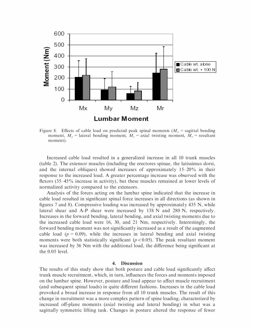
Figure 5. Predicted peak moments acting on the lumbar spine in the four postures studied (Mx = sagittal bending moment, My = lateral bending moment, Mz = axial twisting moment, Mr = resultant moment).
EVects due to increased cable load Figures 6±8 contain data describing the eVects of increased cable load on EMG data and predicted spine loading. As reported in tables 2 and 3, the 100 N increase in cable load caused signi®cant increases in the activity of all measured trunk muscles,

393
as well as in the spinal loading variables (p50.05). However, in neither case did the increase result in a diVerential response according to the posture adopted in the performance of the cable-lifting task.
Cable weight aloneCable weight + 100 N
Figure 6. EVects of cable load on normalized maximum EMG activity of 10 trunk muscles (LLD = left latissimus dorsi, RLD = right latissimus dorsi, LES = left erectores spinae, RES = right erectores spinae, LEO = left external obliques, REO = right external obliques, LRA = left rectus abdominis, RRA = right rectus abdominis, LIO = left internal obliques, RIO = right internal obliques).
Cable weight aloneCable weight + 100 N
Force direction
Fx FzFy
Figure 7. EVects of cable load on predicted peak forces acting on the lumbar spine (F = lateral shear force, F = anterior-posterior shear forces, and F = compression). x y z
394 S. Gallagher et al.
Increased cable load resulted in a generalized increase in all 10 trunk muscles (table 2). The extensor muscles (including the erectores spinae, the latissimus dorsi, and the internal obliques) showed increases of approximately 15±20% in their response to the increased load. A greater percentage increase was observed with the ¯exors (35±45% increase in activity), but these muscles remained at lower levels of normalized activity compared to the extensors.
Cable weight aloneCable weight + 100 N
Mx MzMy Mr
Figure 8. EVects of cable load on predicted peak spinal moments (Mx = sagittal bending moment, My = lateral bending moment, Mz = axial twisting moment, M = resultant r
moment).
Analysis of the forces acting on the lumbar spine indicated that the increase in cable load resulted in signi®cant spinal force increases in all directions (as shown in ®gures 7 and 8). Compressive loading was increased by approximately 435 N, while lateral shear and A-P shear were increased by 138 N and 280 N, respectively. Increases in the forward bending, lateral bending, and axial twisting moments due to the increased cable load were 16, 30, and 21 Nm, respectively. Interestingly, the forward bending moment was not signi®cantly increased as a result of the augmented cable load (p = 0.09), while the increases in lateral bending and axial twisting moments were both statistically signi®cant (p50.05). The peak resultant moment was increased by 36 Nm with the additional load, the diVerence being signi®cant at the 0.05 level.
4. Discussion The results of this study show that both posture and cable load signi®cantly aVect trunk muscle recruitment, which, in turn, in¯uences the forces and moments imposed on the lumbar spine. However, posture and load appear to aVect muscle recruitment (and subsequent spinal loads) in quite diVerent fashions. Increases in the cable load provoked a broad increase in response from all 10 trunk muscles. The result of this change in recruitment was a more complex pattern of spine loading, characterized by increased oV-plane moments (axial twisting and lateral bending) in what was a sagittally symmetric lifting task. Changes in posture altered the response of fewer

395
trunk muscles, although these were often in¯uential extensor muscles that had substantial impact on spine loading estimates. In fact, the increase in spinal compression between the least stressful posture (KNEEL2) and the most stressful (STOOP) was nearly two-fold. However, the increase in spinal loading resulting from posture did not produce higher oV-plane moments (axial twisting and lateral bending). Instead, the postural changes aVected only moments in the sagittal plane.
It was hypothesized that the demands on the trunk musculature from postural changes and increased load might interact with one another in this study. However, the eVects of these variables were remarkably independent from one another (and none of the 17 dependent measures disclosed such an interaction). This implies that, no matter which posture is adopted, the body’s response to the increased load is the same, and appears to be characterized by increased activity of all available muscular resources. Similarly, if one handles a given load but changes the posture of the body, the body’s response is a more selective adjustment of available muscle resources. Moreover, these eVects appear to be additive in terms of both muscular recruitment and the ensuing spinal load.
The eVects of increased load on trunk muscle EMG and spine loading in this study are similar to those of increased weight seen in studies of box lifting in the unrestricted standing posture (Fathallah et al. 1998, DeLooze et al. 1999, Davis and Marras 2000). Results of the current study suggest that the body’s trunk muscle recruitment response to increased load is not dependent on posture, but always appears to be associated with a broad increase in activation of both extensor and ¯exor muscle groups.
It is easy to imagine that higher load conditions will create a situation where small changes in the position of the load may quickly lead to instability of the lumbar spinal column, due to high magnitude and rapidly changing moments aVecting the spine. It may be that the broad co-contraction of trunk muscles in this situation provides an environment where muscles can respond more eVectively to rapidly changing moments and that this may help to limit excessive movement of the spine under high moment conditions. In any event, the result of the increased load results is a more intricate pattern of loading on the spine, with notably higher shear forces that may be di fcult for the spinal column to safely withstand (Yingling and McGill 1999). The abdominal muscles have variously been predicted to account for about 6±45% of the compressive load experienced by the spine (Potvin et al. 1991, Granata and Marras 1995, DeLooze et al. 1999). Their recruitment may be important in terms of increasing the stability of the spine in response to the increased load (Cholewicki and McGill 1996).
The stooping position was associated with high bilateral EMG activation of the erectores spinae and latissimus dorsi musculature, along with increased internal oblique activity. It should be noted that these are all extensor muscles of the spine, and that all appeared to be highly recruited in these tasks based on the need to balance the large external moment associated with the criterion task. It is apparent that subjects in this posture performed su fcient trunk extension to go beyond the range of the ¯exion-silence phenomenon (Floyd and Silver 1955), placing the burden of generating a signi®cant restorative moment on the extensor musculature as noted above. The result of this activation pattern was that the stooping posture consistently showed the greatest spinal loads.
The 2KNEE lifting posture in this study resulted in the lowest spinal loading. Forces were lower in all three axes when this posture was adopted. The forward
396 S. Gallagher et al.
bending moment was markedly lower than in any of the alternative postures. In addition, this was the only posture where the mean spinal compression was close to the 3400 N maximum acceptable limit recommended by NIOSH (Waters et al. 1993); however, shear forces were still high in this posture. Activity of all trunk extensors (erectores spinae, latissimus dorsi, and internal obliques) was diminished in the 2KNEE cable lifts. It appears that this posture may have allowed subjects to keep the load close to the body (thereby reducing the forward bending moment), and may have reduced this moment even further by allowing the subject to maintain a more upright trunk orientation throughout the lift, thereby decreasing the demands on the trunk extensor muscles to develop a high restorative moment. Comparison of the 1KNEE versus 2KNEE lifting postures revealed a sizeable diVerence in spinal loading, at least for the condition under study. The raised knee presents a barrier so signi®cant as to increase the moment by 43% when compared to the 2KNEE condition.
Results of this study suggest that the stooping posture should be avoided whenever possible when working in restricted workspace. Such a recommendation is difcult to implement in practice, however, due to the higher strength capabilities in this posture when compared to kneeling. When loads are heavy, workers opt to use the stoop posture because it represents a position where the body can impart considerable force in an eVort to move an object. Unfortunately, the results of this and other studies indicate that the costs in terms of spinal loading are severe, especially with respect to shear forces. Results of recent studies indicate that if one must adopt a stoop posture, it may be of bene®t to avoid the end-range of spinal motion and to attempt to maintain some degree of lordosis in the lumbar spine region (Adams and Hutton 1982, Potvin et al. 1991, McGill and Kippers 1994, McGill 1999). Doing this may allow the paraspinal muscles to remain active and reduce the anterior shear forces observed when the interspinous ligaments assume increased reliance in supporting the spine.
The magnitudes of the forces and moments associated with lifting the cable in this experiment were all quite high, and this may help to explain the high incidence of lost-time back injuries in the coal mining industry associated with workers who perform this task. The NIOSH criterion for recommended weight limit (RWL) is based upon a 3400 N maximum compressive load on the spine (Waters et al. 1993). However, recent evidence suggests that injuries resulting from anterior shear loadings (primarily to the pars interarticularis, annulus and vertebral endplates) may begin to occur at shear loadings of only 900 N (McGill 1999, Yingling and McGill 1999). The fact that the mean compression for all experimental conditions exceeded recommended values (Waters et al. 1993) testi®es to the high level of exertion required when handling such large diameter electrical cable. EVorts should be made to provide workers with mechanical assistance when performing this demanding task.
5. Conclusions Results of this experiment support the following conclusions. First, changes in posture and cable load both in¯uenced trunk muscle recruitment and spinal loading; however, these eVects were not interactive. Second, increased cable load resulted in signi®cantly increased activity of all trunk muscles, and resulted in increased oVplane (non-sagittal ) moments on the spine. Third, changes in posture resulted in modi®cations in trunk muscle recruitment that were more selective than those

397
associated with increased load. That is, changes in posture typically aVected the activity of only a few muscles, rather than the entire set. However, the muscles aVected by changes in posture often involved the powerful extensors and therefore had a large impact in terms of spine loading. Fourth, kneeling on both knees was the least stressful posture in terms of spine loading and stooping was most stressful, while standing and kneeling on one knee involved a level of spinal loading intermediate between these two. Finally, the magnitudes of compression and shear loading on the spine were always quite high when lifting the cable regardless of the load condition or posture. EVorts should be made to provide mechanical assistance in performance of this task.
References ADAMS, M. A. and HUTTON, W. C. 1982, Prolapsed intervertebral disc: a hyper¯exion injury,
Spine, 7, 184±190. CALDWELL, L. S., CHAFFIN, D. B., DUKES-DOBOS, F. N., KROEMER, K. H. E., LAUBACH, L. L.,
SNOOK, S. H . a nd W ASSERMAN, D. E. 1974, A proposed standard procedure for static muscle strength testing, American Industrial Hygiene Association Journal, 35, 201±206.
CHAFFIN, D. B . and A NDERSSON, G. B. J. 1991, Occupational Biomechanics, 2nd edn (New York: John Wiley).
CHOLEWICKI, J. and MCGILL, S. M. 1996, Mechanical stability of the in vivo lumbar spine: implications for injury and chronic low back pain, Clinical Biomechanics, 11, 1±15.
DAVIS, K. G . a nd M ARRAS, W. S. 2000, Assessment of the relationship between box weight and trunk kinematics: does a reduction in box weight necessarily correspond to a decrease in spine loading? Human Factors, 42, 195±208.
DAVIS, K. G., MARRAS, W. S. and W ATERS, T. R. 1998, Evaluation of the spinal loading during lowering and lifting, Clinical Biomechanics, 13, 141±152.
DE LOOZE, M. P., GROEN, H., HOREMANS, H., KINGMA, I. and VAN DIEEN, J. H. 1999, Abdominal muscles contribute in a minor way to peak spinal compression in lifting, Journal of Biomechanics, 32, 655±662.
DUL, J. 1986, Muscular coordination in working postures, in N. Corlett, J. Wilson. and I. Manenica (eds), The Ergonomics of Working Postures (London: Taylor & Francis), 115± 125.
FATHALLAH, F. A., MARRAS, W. S. a nd P ARNIANPOUR, M. 1998, An assessment of complex spinal loads during dynamic lifting tasks, Spine, 23, 706±716.
FATHALLAH, F. A., MARRAS, W. S., PARNIANPOUR, M. and GRANATA, K. P. 1997, A method for measuring external loads during unconstrained free-dynamic lifting, Journal of Biomechanics, 30, 975±978.
FLOYD, W. F. a nd S ILVER, P. H. S. 1955, The function of the erectores spinae muscles in certain movements and postures in man, Journal of Physiology, 129, 184±203.
GALLAGHER, S. 1997, Trunk extension strength and trunk muscle activity in standing and kneeling postures, Spine, 22, 1864±1872.
GALLAGHER, S. 1999, Ergonomics Issues in Mining, in W. Karwowski and W. S. Marras (eds), Handbook of Occupational Ergonomics (Boca Raton, FL: CRC Press), 1889±1911.
GALLAGHER, S. a nd HAMRICK, C. A. 1992, Acceptable workloads for three common mining materials, Ergonomics, 35, 1013±1031.
GALLAGHER, S. and UNGER, R. L. 1990, Lifting in four restricted lifting conditions, Applied Ergonomics, 21, 237±245.
GALLAGHER, S., MARRAS, W. S. a nd B OBICK, T. G. 1988, Lifting in stooped and kneeling postures: eVects on lifting capacity, metabolic costs, and electromyography of eight trunk muscles, International Journal of Industrial Ergonomics, 3, 65±76.
GALLAGHER, S., HAMRICK, C. A., CORNELIUS, K. M . and R EDFERN, M. S. 2 001, The e Vects of restricted workspace on lumbar spine loading, Occupational Ergonomics, 2, 201±213.
GALLAGHER, S., HAMRICK, C. A., LOVE, A. C. and MARRAS, W. S. 1994, Dynamic biomechanical modeling of symmetric and asymmetric lifting tasks in restricted postures, Ergonomics, 37, 1289±1310.

398 S. Gallagher et al.
GRANATA, K. P. and MARRAS, W. S. 1993, An EMG-assisted model of loads on the lumbar spine during asymmetric trunk extensions, Journal of Biomechanics, 26, 1429±1438.
GRANATA, K. P . a nd M ARRAS, W. S. 1995, An EMG-assisted model of trunk loading during free-dynamic lifting, Journal of Biomechanics, 28, 1309±1317.
KIRK, R. E. 1982, Experimental Design: Procedures for the Behavioral Sciences, 2nd edn (Paci®c Grove, CA: Brooks/Cole), 911 pp.
MARRAS, W. S. 1990, Industrial electromyography, International Journal of Industrial Ergonomics, 6, 89±93.
MARRAS, W. S. and GRANATA, K. P. 1995, A biomechanical assessment and model of axial twisting in the thoraco-lumbar spine, Spine, 20, 1440±1451.
MARRAS, W. S. and GRANATA, K. P. 1997, The development of an EMG-assisted model to assess spine loading during whole-body free-dynamic lifting, Journal of Electromyography and Kinesiology, 7, 259±268.
MARRAS, W. S. and MIRKA, G. A. 1993, Electromyographic studies of the lumbar trunk musculature during the generation of low-level trunk acceleration, Journal of Orthopaedic Research, 11, 811±817.
MARRAS, W. S. a nd REILLY, C. H. 1988, Networks of internal trunk loading activities under controlled trunk motion conditions, Spine, 13, 661±667.
MARRAS, W. S. and SOMMERICH, C. M. 1991a, A three dimensional motion model of loads on the lumbar spine. I. Model structure, Human Factors, 33, 129±137.
MARRAS, W. S. and SOMMERICH, C. M. 1991b, A three dimensional motion model of loads on the lumbar spine. II. Model validation, Human Factors, 33, 139±149.
MARRAS, W. S., FATHALLAH, F. A., MILLER, R. J., DAVIS, S. W. and MIRKA, G. A. 1992, Accuracy of a three-dimensional lumbar motion monitor for recording dynamic trunk motion characteristics, International Journal of Industrial Ergonomics, 9, 75±87.
MARRAS, W. S., LAVENDER, S. A., LEURGANS, S. E., RAJULU, S. L., ALLREAD, W. G., FATHALLAH, F. A. and FERGUSON, S. A. 1993, The role of dynamic three-dimensional motion in occupationally-related low back disorders. The eVects of workplace factors, trunk position, and trunk motion characteristics on risk of injury, Spine, 18, 617±628.
MCGILL, S. M. 1999, Dynamic low back models: theory and relevance in assisting the ergonomist to reduce risk of low back injury, in W. Karwowski and W. S. Marras (eds), The Occupational Ergonomics Handbook (Boca Raton, FL: CRC Press), 945±965.
MCGILL, S. M. and KIPPERS, V. 1994, Transfer of loads between lumbar tissues during the ¯exion relaxation phenomenon, Spine, 19, 2190±2196.
MCGILL, S. M . and N ORMAN, R. W. 1986, Partitioning of the L4-L5 dynamic moment into disc, ligamentous, and muscular components during lifting, Spine, 11, 666±678.
MIRKA, G. A . and MARRAS, W. S. 1993, A stochastic model of trunk muscle coactivation during trunk bending, Spine, 18, 1396±1409.
NIOSH (NATIONAL INSTITUTE FOR OCCUPATIONAL SAFETY AND HEALTH) 1991, Selected Topics in Surface Electromyography for Use in the Occupational Setting: Expert Perspectives. DHHS(NIOSH) Publication No. 91-100, NIOSH Technical Report (Cincinnati, OH: NIOSH).
POTVIN, J. R., MCGILL, S. M. and NORMAN, R. W. 1991, Trunk muscle and lumbar ligament contributions to dynamic lifts with varying degrees of trunk ¯exion, Spine, 16, 1099± 1107.
RANDOLPH, R. F. 1991, Unpublished data. Available upon request from NIOSH, PO Box 18070, Pittsburgh, PA 15236, USA.
REILLY, C. H. a nd MARRAS, W. S. 1989, SIMULIFT: a simulation model of human trunk motion during lifting, Spine, 14, 5±11.
SMITH, J. L., AYOUB, M. M., SELAN, J. L., KIM, H. K. and C HEN, H. C. 1989, Manual materials handling in unusual postures, in A. Mital (ed.), Advances in Industrial Ergonomics and Safety I (London: Taylor & Francis), 685±691.
WATERS, T. R., PUTZ-ANDERSON, V., GARG, A. and FINE, L. J. 1993, Revised NIOSH equation of the design and evaluation of manual lifting tasks, Ergonomics, 36, 749±776.
YINGLING, V. R. and MCGILL, S. M. 1999, Anterior shear of spinal motion segments: kinematics, kinetics, and resultant injuries observed in a porcine model, Spine, 24, 1882± 1889.