effects of fluoride on pancreatic digestive … · the ultrastructural changes in the pancreas...
TRANSCRIPT

Fluoride 2005;38(3):215–219 Research report 215
EFFECTS OF FLUORIDE ON PANCREATIC DIGESTIVE ENZYME ACTIVITIES AND ULTRASTRUCTURE IN YOUNG PIGS
Xiu-an Zhan,a Jian-xin Li,a Zi-rong Xu,a Min Wanga
Hangzhou, China
SUMMARY: This study was conducted to investigate the effects of fluoride onpancreatic digestive enzyme activities and ultrastructure in young pigs. Threegroups of crossbred barrows were exposed to 100, 250, and 400 mg F–/kg (fromNaF) in their diets for 50 days. Compared to a control group, the activities ofpancreatic lipase and protease (but not amylase) were significantly decreased.Pancreatic acinar cells showed markedly swollen mitochondria and loss ofmitochondrial cristae. Endoplasmic reticulum (ER) was markedly dilated and itsfolds were irregular. These results indicate that excessive fluoride in the diet caninhibit pancreatic digestive enzyme activities and cause ultrastructural changes,which may lead to a series of biochemical and pathological abnormalities.Keywords: Digestive enzymes; Endoplasmic reticulum; Lipase; Mitochondria; Pancreatic acinar cells; Protease; Young pigs.
INTRODUCTION
Prevalent in many parts of the world, chronic fluorosis is caused by excessiveingestion of fluoride over a prolonged period and endangers the health of humansas well as animals. In addition to its well-known effects on the skeleton and onteeth, fluoride can exert toxic effects on many other tissues and organs, givingrise to a broad array of symptoms and pathological changes.1 Recently, we foundthat fluoride induced excessive production of nitric oxide and reactive oxygenspecies, enhanced lipid peroxidation, and disturbed the antioxidant system ofyoung pigs; therefore oxidative damage from oxidative stress might be animportant pathway for fluoride toxicity in soft tissues.2 Owing to fluoride inenvironmental pollution and in mineral supplements such as calcium phosphateand limestone, chronic fluorosis in domestic animals is an increasingly seriousproblem resulting in considerable economic losses in animal production.
Although fluorosis has been investigated for many years, there are relativelyfew studies of its effect on the digestive system such as the pancreas, a smallorgan located near the lower part of the stomach and the beginning of the smallintestine. Enzyme secretions of the exocrine pancreas are required for hydrolysisof nutrients present in food and feed.3 The present study was undertaken to assessthe effects of fluoride on pancreatic digestive enzyme activities and onultrastructure in the pancreas.
MATERIALS AND METHODS
Animals and experimental diets: Thirty-two 50-day-old barrows with anaverage body weight of about 17 kg were acclimatized for one week and then
aFeed Science Institute, College of Animal Science, Zhejiang University, No.268 KaixuanRoad, Hangzhou, China, 310029.For correspondence: Jian-xin Li, E-mail:[email protected].
Fluoride 2005;38(3)

216 Zhan, Li, Xu, Wang
allotted randomly to four groups of eight. These animals were the same pigs weused earlier,2 which were fed a basal diet containing 6.2 mg F–/kg in the controlgroup 1, supplemented by 100, 250, and 400 mg F–/kg diet from NaF inexperimental groups 2, 3, and 4, respectively. On the 50th day of the feeding trial,all pigs were deprived of feed for 12 hr and then slaughtered under generalanesthesia using halothane.
Pancreatic digestive enzyme activities: The pancreas from the slaughtered pigswas homogenized and centrifuged. The supernatant was saved for determiningthe activities of lipase (EC 3.1.1.3), protease, and amylase (EC 3.2.1.1). Lipasewas determined at 37°C by a pH-stat titration using tributyrin as substrateaccording to the method of Erlanson-Albertsson et al.4 Protease activity wasanalyzed with the modified method of Lynn and Clevette-Radford5 usingazocasein as substrate. One lipase or protease unit is defined as the amount ofenzyme that hydrolyses 1 µmol of substrate per minute. Amylase was determinedby the iodometric method.6 One amylase unit is the amount of enzyme thathydrolyses 10 mg of starch in 30 min.
Transmission electron microscopy: The pancreas was excised and fixed with2.5% glutaraldehyde in 0.1 M phosphate buffer (pH 7.4). Each sample wasdehydrated through graded ethanol solution and then embedded in epoxy resin.Ultra-thin sections were cut and stained with uranyl acetate and lead citrate. Thesections were examined under a Jeol JEM-1230 transmission electronmicroscope.
Statistical analysis: The significance of the difference between means wasdetermined by analysis of variance (ANOVA) with p<0.05 being consideredsignificant.
RESULTS
Compared with the control group 1, the activity of pancreatic lipase in piggroups 2, 3, and 4 exhibited a significant monotonic decrease, whereas theactivity of pancreatic protease decreased more slowly and was significantly lowerin group 4. Pancreatic amylase activity, on the other hand, showed a small, non-significant paradoxical increase in group 3 before declining again in group 4(Table).
Table. Effects of fluoride on pancreatic digestive enzyme activities in young pigsa,b
Groupc 1 2 3 4
Lipase 450.20±59.31 345.93±70.76* 333.92±66.83* 355.91±63.06*
Protease 88.04±13.28 80.46±7.76 81.90±14.47 72.54±9.09*
Amylase 293.22±19.85 291.62±23.09 304.75±30.57 285.96±48.59aValues are mean±SD; n=8 per group. Compared with the control group, *p<0.01.bPancreatic digestive enzyme activities are expressed as U/g pancreas.cGroup 1 was the control, and the other three groups were the experimental ones with100, 250, and 400 mg F–/kg diet (from NaF).
Fluoride 2005;38(3)
Effects of fluoride on pancreatic enzyme activities and ultrastructure in pigs 217
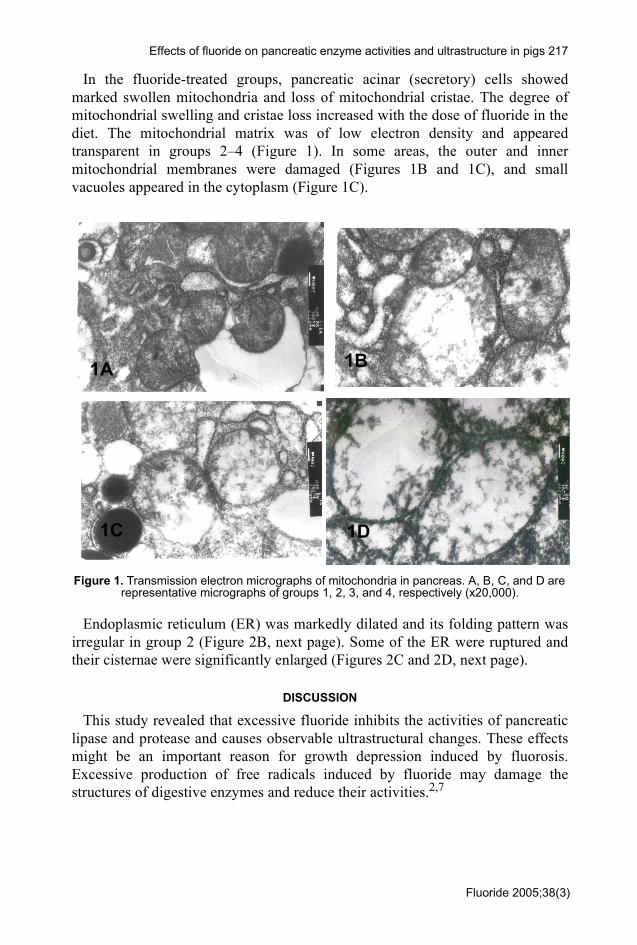
In the fluoride-treated groups, pancreatic acinar (secretory) cells showedmarked swollen mitochondria and loss of mitochondrial cristae. The degree ofmitochondrial swelling and cristae loss increased with the dose of fluoride in thediet. The mitochondrial matrix was of low electron density and appearedtransparent in groups 2–4 (Figure 1). In some areas, the outer and innermitochondrial membranes were damaged (Figures 1B and 1C), and smallvacuoles appeared in the cytoplasm (Figure 1C).
Figure 1. Transmission electron micrographs of mitochondria in pancreas. A, B, C, and D are representative micrographs of groups 1, 2, 3, and 4, respectively (x20,000).
Endoplasmic reticulum (ER) was markedly dilated and its folding pattern wasirregular in group 2 (Figure 2B, next page). Some of the ER were ruptured andtheir cisternae were significantly enlarged (Figures 2C and 2D, next page).
DISCUSSION
This study revealed that excessive fluoride inhibits the activities of pancreaticlipase and protease and causes observable ultrastructural changes. These effectsmight be an important reason for growth depression induced by fluorosis.Excessive production of free radicals induced by fluoride may damage thestructures of digestive enzymes and reduce their activities.2,7
1B
1C 1D
1A
Fluoride 2005;38(3)

218 Zhan, Li, Xu, Wang
Figure 2. Transmission electron micrographs of endoplasmic reticulum in pancreas. A, B, C, and D are representative micrographs of groups 1, 2, 3, and 4 respectively, (x25,000).
The ultrastructural changes in the pancreas observed here are in accordancewith previous findings in thyroid, liver, and kidney of fluorotic animals.8,9 Themitochondrion is an organelle that is the site of aerobic cellular respiration and isresponsible for the conversion of food and feed to usable energy.10 Itsmorphological changes would disturb the metabolism of substances in themitochondria and suppress ATP (adenosine triphosphate) synthesis. Enlargementof mitochondria in fluoride-treated pigs is believed to a compensatory processdue to ATP deficiency.8 Endoplasmic reticulum (ER) is a membrane networkwithin the cytoplasm of cells that is involved in the synthesis, modification, andtransport of cellular materials. The dilation of ER in fluoride-treated pigsindicates enhancement of detoxification reactions and its rupture would suppressprotein synthesis that might reduce the pancreatic secretions, including digestiveenzymes. The destruction of mitochondria and ER can be attributed to oxidativestress induced by fluoride, which can seriously damage the structure of cells andorganelles, especially their membranes.2,7
In conclusion, we have found that excessive fluoride in the diet can inhibitpancreatic digestive enzyme activities and damage pancreatic ultrastructure,especially mitochondria and ER, which may in turn lead to a series ofbiochemical and pathological abnormalities.
ACKNOWLEDGEMENTS
This research was supported by the National Basic Research Program ofChina (Project 2004CB117505). We also wish to thank Wei-fen Li for her skillfultechnical assistance.
2C 2D
2A 2B
Fluoride 2005;38(3)

Effects of fluoride on pancreatic enzyme activities and ultrastructure in pigs 219
REFERENCES1 Waldbott GL. Nonskeletal fluorosis [editorial review]. Fluoride 1978;11:111-4.2 Zhan XA, Xu ZR, Li JX, Wang M. Effects of fluorosis on lipid peroxidation and antioxidant
systems in young pigs. Fluoride 2005;38:157-61.3 Rinderknecht H. Pancreatic secretory enzymes. In: Go VLW, DiMagno JD, Gardner E, Lebenthal
E, Reber HA, Scheele GA, editors. The Pancreas: biology, pathobiology and disease. New York:Raven Press; 1993. p. 219-51.
4 Erlanson-Albertsson C, Larsson A, Duan R. Secretion of pancreatic lipase and colipase from ratpancreas. Pancreas 1987;2:531-5.
5 Lynn KR, Clevette-Radford NA. Purification and characterization of hevin, a serin protease fromHevea brazilliensis. Biochemistry 1984;23:963-4.
6 Harms DR, Camfield RN. An automated iodometric method for the determination of amylase. AmJ Med Technol 1966;32:341-7.
7 Liu G, Chai C, Cui L. Fluoride causing abnormally elevated serum nitric oxide levels in chicks.Environ Toxicol Pharmacol 2003;13:199-204.
8 Zhan CW, Huo DJ. Ultrastructural findings in liver, kidneys, thyroid gland and cardiac muscle ofrabbits following sodium fluoride administration. Fluoride 1988;21:32-8.
9 Liu GY, Chai CY, Kang SL. Effects of fluoride on ultrastructure of thyroid in chicks. Chin J Vet Sci2002;22:512-4 (in Chinese).
10 Enger ED, Ross FC. Concepts in biology. Cell structure and function. 10th ed. Beijing: SciencePress; 2003. p. 58-83.
Published by the International Society for Fluoride Researchhttp://homepages.ihug.co.nz/~spittle/fluoride-journal.htm
Editorial Office: 727 Brighton Road, Ocean View, Dunedin 9051, New Zealand
Fluoride 2005;38(3)