effects of core characters and veneering technique on … · it blocks light transmission and...
TRANSCRIPT

The Journal of Advanced Prosthodontics 349
Effects of core characters and veneering technique on biaxial flexural strength in porcelain fused to metal and porcelain veneered zirconia
Ju-Won Oh1, Kwang-Yeob Song1,2, Seung-Geun Ahn1,2, Ju-Mi Park1,2, Min-Ho Lee3, Jae-Min Seo1,2* 1Department of Prosthodontics, School of Dentistry and Institute of Oral Bio-Science, Chonbuk National University, Jeonju, Republic of Korea2Biomedical Research Institute, Chonbuk National University Hospital, Jeonju, Republic of Korea3Department Dental Biomaterials, Institute of Oral Bio-science, School of Dentistry, Chonbuk National University, Jeonju, Republic of Korea
PURPOSE. The purpose of this study was to assess the impact of the core materials, thickness and fabrication methods of veneering porcelain on prosthesis fracture in the porcelain fused to metal and the porcelain veneered zirconia. MATERIALS AND METHODS. Forty nickel-chrome alloy cores and 40 zirconia cores were made. Half of each core group was 0.5 mm-in thickness and the other half was 1.0 mm-in thickness. Thus, there were four groups with 20 cores/group. Each group was divided into two subgroups with two different veneering methods (conventional powder/liquid layering technique and the heat-pressing technique). Tensile strength was measured using the biaxial flexural strength test based on the ISO standard 6872:2008 and Weibull analysis was conducted. Factors influencing fracture strength were analyzed through three-way ANOVA (α≤.05) and the influence of core thickness and veneering method in each core materials was assessed using two-way ANOVA (α≤.05). RESULTS. The biaxial flexural strength test showed that the fabrication method of veneering porcelain has the largest impact on the fracture strength followed by the core thickness and the core material. In the metal groups, both the core thickness and the fabrication method of the veneering porcelain significantly influenced on the fracture strength, while only the fabrication method affected the fracture strength in the zirconia groups. CONCLUSION. The fabrication method is more influential to the strength of a prosthesis compared to the core character determined by material and thickness of the core. [ J Adv Prosthodont 2015;7:349-57]
KEY WORDS: Zirconia; Metal ceramic; Pressed ceramic; Dental porcelain; Tensile strength
http://dx.doi.org/10.4047/jap.2015.7.5.349http://jap.or.kr J Adv Prosthodont 2015;7:349-57
INTRODUCTION
Porcelain fused to metal restorations have played a crucial role in aesthetic dental restoration since the development of ceramics in 1965.1 The rigid metal alloy core enables use of a wide range of metal ceramic crowns, from single crown to the long-span fixed partial denture that supports the veneer-ing ceramic. However, the metal alloy has some limitations; it blocks light transmission and causes the umbrella effect and aesthetic disharmony.2 To overcome these limitations, all-ceramic restorations are appropriate for anterior restora-tions. Although many attempts have been made to improve its physical properties, the all-ceramic restoration is relative-ly fragile and therefore, it has been applied only to anterior
Corresponding author: Jae-Min SeoDepartment of Prosthodontics, School of Dentistry and Institute of Oral Bio-Science, Chonbuk National University, 567, Baekje-daero, Deokjin-gu, Jeonju 54896, Republic of KoreaTel. 82 63 250 2696: e-mail, [email protected], [email protected] September 17, 2014 / Last Revision January 2, 2015 / Accepted January 9, 2015
© 2015 The Korean Academy of ProsthodonticsThis is an Open Access article distributed under the terms of the Creative Commons Attribution Non-Commercial License (http://creativecommons.org/licenses/by-nc/3.0) which permits unrestricted non-commercial use, distribution, and reproduction in any medium, provided the original work is properly cited.
pISSN 2005-7806, eISSN 2005-7814
350
restorations or short-span prostheses. These limitations have spurred the adoption of new ceramic materials and development of different fabricating techniques. For exam-ple, dental porcelain materials developed by an enforcement mechanism have been introduced and heat-pressed ceram-ics using the lost-wax technique are widely known as a new fabrication technique.
Yttrium zirconia is one of the most widely used restora-tion materials to resolve the aforementioned troubles in metal alloy core and porcelain core. Following the introduc-tion of Computer aided design-Computer aided manufac-turing (CAD/CAM) technology, yttrium zirconia has gained rapid acceptance in clinical dentistry. This popularity is based on the material’s aesthetic benefit and strength. Yttrium zirconium has been used for many aesthetic resto-rations. Also, the material enhances the speed, accuracy, and reliability of manufacture. Moreover, the transformation toughening that strengthens yttrium zirconia allows zirconi-um crowns to substitute for metal ceramic crowns.3,4
Despite such advantages, use of the zirconia prosthesis has been limited by its opaque shade, which lacks visual harmony with adjacent teeth. To alleviate the problem, veneering and staining techniques have been introduced. However, those methods lead to other issues, such as unsta-ble durability of veneered ceramics and attrition of the opposing teeth. Although zirconia dental prostheses per se display high fracture strength and excellent clinical perfor-mance compared to all-ceramic restorations, high fracture rate in veneered ceramics due to the resulting mismatch in the elastic modulus and thermal expansion coefficient between the zirconia core and veneered ceramic have been reported.5
The present study investigated the effects of the two different core materials (metal core vs. zirconia core), core thickness (0.5 mm vs. 1.0 mm) and two veneering methods (conventional layering technique vs. heat-pressing tech-nique) on the fracture of the porcelain veneered prostheses using the biaxial flexure strength test and Weibull analysis.
MATERIALS AND METHODS
Forty nickel-chromium (Ni-Cr) alloy (Wiron 99; Bego, Bremen, Germany) cores and 40 zirconia (IPS e.maxZircad; IvoclarVivadent, Amherst, NY, USA) cores were prepared.
Twenty of each type of core was 0.5 mm in thickness and the remaining 20 were 1.0 mm in thickness. This comprised four groups with 20 cores per group. Each group was divided into two subgroups depending on the veneering method (conventional layering technique and heat-pressing technique). Thus, the final veneering porcelain samples included eight groups with 10 samples per group (Table 1).
Forty disk-shaped specimens having a 15 mm diameter were prepared for the metal core based on the ISO stan-dard 6872:2008(dental ceramic). The 40 waxed (Preparation wax; Bego, Bermen, Germany) specimens were divided into two groups with thicknesses of 0.7 mm (n = 20) and 1.2 mm (n = 20). Both groups were invested (CB-30; Ticonium, Gardenia, CA, USA), cast in Ni-Cr alloy (Wiron 99; Bego, Bermen, Germany) and sanded to a finish with 300, 600, 1000 and 1200 grit sand paper to a thickness of 0.5 mm or 1.0 mm.
The other forty disk-shaped specimens were prepared by milling and drying zirconia blocks (IPS e.maxZirCAD; Ivoclar Vivadent, Amherst, NY, USA) followed by sintering at 1,500°C. Like the metal core, the specimens were wet-ground with sand paper to produce the disks of 15 mm diameter and thickness of 0.5 mm (n = 20) or 1.0 mm (n = 20).
The 80 cores were sand-blasted with a pressure of 2.0 bar and ultrasonically cleaned for 10 minutes (Ultrasonic cleaner 2210; Branson, Danbury, CT, USA). For conven-tional layered veneering porcelain, opaque porcelain and dentin porcelain was prepared by mixing the powder/liquid, sintering following the manufacturer’s instruction, and manufacturing the veneering porcelain with a thickness of 1.0 mm. For the heat-pressed veneering porcelain, the top of each core was waxed in a disk-shaped wax pattern. This was followed by an ordinary burn out process and injection of the heat-pressed porcelain ingot for the veneering pro-cess to produce a thickness of 1.0 mm. Table 2 summarizes the materials and methods used in the porcelain sintering process. The samples in the M0.5C, M0.5P, Z0.5C, and Z0.5P groups had disk-shaped cores with a 15 mm diame-ter and 1.5 mm thickness, while the M1.0C, M1.0P, Z1.0C and Z1.0P groups had disk-shaped cores with a 15 mm diameter and 2 mm thickness. To reproduce the oral envi-ronment, samples were tested after 6000 cycles of thermo-cycling in distilled water with a temperature of 5ºC and 55ºC (Invertech, Kwangju, Korea). The retention period
Table 1. Group characteristics
GroupingM0.5CGroup
M1.0CGroup
M0.5P Group
M1.0PGroup
Z0.5CGroup
Z1.0CGroup
Z0.5PGroup
Z1.0PGroup
Core material Ni-CrMetal Ni-CrMetal Ni-CrMetal Ni-CrMetal Zirconia Zirconia Zirconia Zirconia
Core thickness 0.5 mm 1.0 mm 0.5 mm 1.0 mm 0.5 mm 1.0 m 0.5 mm 1.0 mm
Veneering method
Conventional- layer
Conventional- layer
Heat-press
Heat-press
Conventional- layer
Conventional- layer
Heat-press
Heat-press
J Adv Prosthodont 2015;7:349-57

The Journal of Advanced Prosthodontics 351
was 15 seconds for each reservoir.The biaxial flexural strength was measured by the piston
using the three ball method following ISO standard 6872:2008. The universal testing machine (Instron, Norwood, MA, USA) was used to measure the strength for the fracture with a crosshead speed of 1.0 mm/min (Fig. 1). The veneering porcelain was placed on the tensile surface and the core material was placed on the compressed sur-face. The thin plastic sheet (0.05 mm thick) was placed between the sample and the piston. The load for the frac-ture of the sample was recorded. The biaxial flexural strength was calculated using the equation below. The sam-ples used in the study did have a heterogeneous structure where two different materials were physically attached to each other, while keeping their own physical characteristics. Since the bilayer structure has a relatively weak interface and is more vulnerable compared to the monolithic struc-ture, it was not proper to apply the conventional equation to calculate the biaxial flexure strength. Therefore, we made
use of Roark’s formulae based on the bending theory, which reflects the characteristics of each material and ana-lyzes the stress on the tensile surface and the stress on the compressed surface separately. The formulae are :
Table 2. Brand name, firing and pressing temperature for veneering ceramics
Materials Group Firing cycleFinal temperature
(°C)Rate temperature increase (°C/Min)
Holding times (Min)
Manufacturer
IPS d.SIGNM0.5CM1.0C
OpaqueDentin
890870
8060
11
IvoclarVivadent
IPS InLine/Inline POMM0.5PM1.0P
OpaquePress
930910
10060
21
IvoclarVivadent
IPS e.max CeramZ0.5CZ1.0C
ZirlinerWashingDentin
403403403
404040
111
IvoclarVivadent
IPS e.max ZirPressZ0.5PZ1.0P
OpaquePress
930910
10060
21
IvoclarVivadent
Fig. 1. Biaxial flexural strength was measured by the universal testing machine.
R = equivalent radiusM = maximum bending momentr = radius (a = compressive side’s, b = tensile side’s)t = thicknessT = total thicknessE = elastic modulusδ=tensilestressγ=Poissonratio(γa,γb = 0.25)
The tensile stress obtained from the formula was used to compare and analyze the fracture strength of the groups. After conducting the biaxial flexural strength test, we observed the fractured samples with an optical microscope (Leica Microsystems GnbH, Wetzlar, Germany).
Since the strength of the ceramic could readily deviate from the standard distribution, Weibull analysis measured the failure probability at scale parameters and shape param-eters. The Weibull parameters of shape and scale were
Effects of core characters and veneering technique on biaxial flexural strength in porcelain fused to metal and porcelain veneered zirconia
352
obtained by making use of median rank regression method to measure unreliability. To assume a Weibull distribution and create a survival graph, Weibull calculator software (Excel; Microsoft, Redmond, WA, USA) was used.
RESULTS
SPSS version 18.0 (SPSS Inc, Chicago, IL, USA) was used to analyze the elements affecting fracture strength.
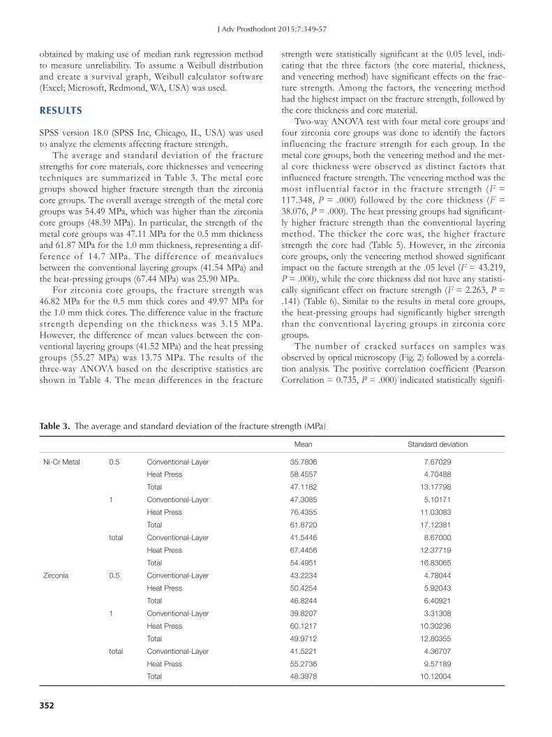
The average and standard deviation of the fracture strengths for core materials, core thicknesses and veneering techniques are summarized in Table 3. The metal core groups showed higher fracture strength than the zirconia core groups. The overall average strength of the metal core groups was 54.49 MPa, which was higher than the zirconia core groups (48.39 MPa). In particular, the strength of the metal core groups was 47.11 MPa for the 0.5 mm thickness and 61.87 MPa for the 1.0 mm thickness, representing a dif-ference of 14.7 MPa. The difference of meanvalues between the conventional layering groups (41.54 MPa) and the heat-pressing groups (67.44 MPa) was 25.90 MPa.
For zirconia core groups, the fracture strength was 46.82 MPa for the 0.5 mm thick cores and 49.97 MPa for the 1.0 mm thick cores. The difference value in the fracture strength depending on the thickness was 3.15 MPa. However, the difference of mean values between the con-ventional layering groups (41.52 MPa) and the heat pressing groups (55.27 MPa) was 13.75 MPa. The results of the three-way ANOVA based on the descriptive statistics are shown in Table 4. The mean differences in the fracture
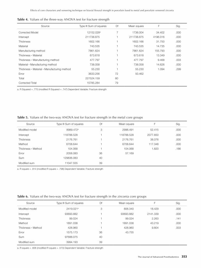
strength were statistically significant at the 0.05 level, indi-cating that the three factors (the core material, thickness, and veneering method) have significant effects on the frac-ture strength. Among the factors, the veneering method had the highest impact on the fracture strength, followed by the core thickness and core material.
Two-way ANOVA test with four metal core groups and four zirconia core groups was done to identify the factors influencing the fracture strength for each group. In the metal core groups, both the veneering method and the met-al core thickness were observed as distinct factors that influenced fracture strength. The veneering method was the most influential factor in the fracture strength (F = 117.348, P = .000) followed by the core thickness (F = 38.076, P = .000). The heat pressing groups had significant-ly higher fracture strength than the conventional layering method. The thicker the core was, the higher fracture strength the core had (Table 5). However, in the zirconia core groups, only the veneering method showed significant impact on the facture strength at the .05 level (F = 43.219, P = .000), while the core thickness did not have any statisti-cally significant effect on fracture strength (F = 2.263, P = .141) (Table 6). Similar to the results in metal core groups, the heat-pressing groups had significantly higher strength than the conventional layering groups in zirconia core groups.
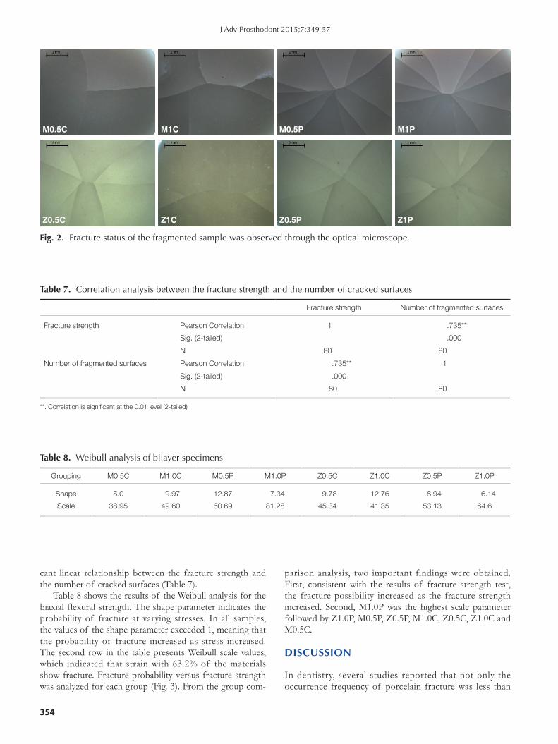
The number of cracked surfaces on samples was observed by optical microscopy (Fig. 2) followed by a correla-tion analysis. The positive correlation coefficient (Pearson Correlation = 0.735, P = .000) indicated statistically signifi-
Table 3. The average and standard deviation of the fracture strength (MPa)
Mean Standard deviation
Ni-Cr Metal 0.5 Conventional-Layer 35.7806 7.67029
Heat Press 58.4557 4.70488
Total 47.1182 13.17798
1 Conventional-Layer 47.3085 5.10171
Heat Press 76.4355 11.03083
Total 61.8720 17.12381
total Conventional-Layer 41.5446 8.67000
Heat Press 67.4456 12.37719
Total 54.4951 16.83065
Zirconia 0.5 Conventional-Layer 43.2234 4.78044
Heat Press 50.4254 5.92043
Total 46.8244 6.40921
1 Conventional-Layer 39.8207 3.31308
Heat Press 60.1217 10.30236
Total 49.9712 12.80355
total Conventional-Layer 41.5221 4.36707
Heat Press 55.2736 9.57189
Total 48.3978 10.12004
J Adv Prosthodont 2015;7:349-57
The Journal of Advanced Prosthodontics 353
Table 4. Values of the three-way ANOVA test for fracture strength
Source Type III Sum of squares Df Mean square F Sig.
Corrected Model 12152.028a 7 1736.004 34.402 .000
Intercept 211738.875 1 211738.875 4196.016 .000
Thickness 1602.166 1 1602.166 31.750 .000
Material 743.535 1 743.535 14.735 .000
Manufacturing method 7861.624 1 7861.624 155.793 .000
Thickness * Material 673.618 1 673.618 13.349 .000
Thickness * Manufacturing method 477.797 1 477.797 9.468 .003
Material * Manufacturing method 738.058 1 738.058 14.626 .000
Thickness * Material * Manufacturing method 55.230 1 55.230 1.094 .299
Error 3633.256 72 50.462
Total 227524.159 80
Corrected Total 15785.284 79
a. R Squared = .770 (modified R Squared = .747) Dependent Variable: Fracture strength
Table 5. Values of the two-way ANOVA test for fracture strength in the metal core groups
Source Type III Sum of squares Df Mean square F Sig.
Modified model 8989.472a 3 2996.491 52.415 .000
Intercept 118788.528 1 118788.528 2077.850 .000
Thickness 2176.761 1 2176.761 38.076 .000
Method 6708.644 1 6708.644 117.348 .000
Thickness * Method 104.068 1 104.068 1.820 .186
Error 2058.083 36 57.169
Sum 129836.083 40
Modified sum 11047.555 39
a. R square = .814 (modified R square = .798) Dependent Variable: Fracture strength
Table 6. Values of the two-way ANOVA test for fracture strength in the zirconia core groups
Source Type III Sum of squares Df Mean square F Sig.
Modified model 2419.021a 3 806.340 18.429 .000
Intercept 93693.882 1 93693.882 2141.339 .000
Thickness 99.024 1 99.024 2.263 .141
Method 1891.038 1 1891.038 43.219 .000
Thickness * Method 428.960 1 428.960 9.804 .003
Error 1575.173 36 43.755
Sum 97688.075 40
Modified sum 3994.193 39
a. R square = .606 (modified R square = .573) Dependent Variable: Fracture strength
Effects of core characters and veneering technique on biaxial flexural strength in porcelain fused to metal and porcelain veneered zirconia
354
cant linear relationship between the fracture strength and the number of cracked surfaces (Table 7).
Table 8 shows the results of the Weibull analysis for the biaxial flexural strength. The shape parameter indicates the probability of fracture at varying stresses. In all samples, the values of the shape parameter exceeded 1, meaning that the probability of fracture increased as stress increased. The second row in the table presents Weibull scale values, which indicated that strain with 63.2% of the materials show fracture. Fracture probability versus fracture strength was analyzed for each group (Fig. 3). From the group com-
parison analysis, two important findings were obtained. First, consistent with the results of fracture strength test, the fracture possibility increased as the fracture strength increased. Second, M1.0P was the highest scale parameter followed by Z1.0P, M0.5P, Z0.5P, M1.0C, Z0.5C, Z1.0C and M0.5C.
DISCUSSION
In dentistry, several studies reported that not only the occurrence frequency of porcelain fracture was less than
M0.5C M1C M0.5P M1P
Z0.5C Z1C Z0.5P Z1P
Fig. 2. Fracture status of the fragmented sample was observed through the optical microscope.
Table 7. Correlation analysis between the fracture strength and the number of cracked surfaces
Fracture strength Number of fragmented surfaces
Fracture strength Pearson Correlation 1.000 .735**
Sig. (2-tailed) .000
N 80.000 80.000Number of fragmented surfaces Pearson Correlation .735** 1.000
Sig. (2-tailed) .000
N 80.000 80.000
**. Correlation is significant at the 0.01 level (2-tailed)
Table 8. Weibull analysis of bilayer specimens
Grouping M0.5C M1.0C M0.5P M1.0P Z0.5C Z1.0C Z0.5P Z1.0P
Shape 5.0 9.97 12.87 7.34 9.78 12.76 8.94 6.14
Scale 38.95 49.60 60.69 81.28 45.34 41.35 53.13 64.6
J Adv Prosthodont 2015;7:349-57

The Journal of Advanced Prosthodontics 355
3% for 20 years but also survival rate was 95.5% for 7 years in the metal ceramic crown.6,7 However, the superiority of the metal ceramic crown in terms of fracture is still a con-troversial issue. For example, Sailer et al.8 reported that the chipping rate of the metal ceramic crown is four times less than the porcelain veneered zirconia prosthesis. Other stud-ies suggested that the chipping of the porcelain veneered zirconia prosthesis is the main factor leading to failure, showing the failure rate is 15.2% for 35.1 ± 13.8 months.9,10 In Contrast, Quinn et al.11 proposed that there is no differ-ence in the fracture rate between the metal ceramic crown and porcelain veneered zirconia. Therefore, in this study, we compared the porcelain veneered zirconia to the porce-lain fused to metal in terms of fracture strength with the same standard.
Recently, a number of studies have investigated how a weak interface bonding between zirconia and veneering porcelain causes clinical failure. Alhasanyah et al.12 suggest-ed that a proper size of core support in the zirconia pros-thesis improves the fracture resistance to chipping, showing that the fracture load of 1.7 mm core is as strong as that of the metal ceramic crown, whereas 0.6 mm and 1.2 mm zir-conia cores with identical whole thickness crowns have the same fracture strength and these values are smaller than values of the metal ceramic crowns. Despite the empirical findings, previous studies have some limitations. First, the thickness of veneering porcelain varies for each core. So, the studies are hindered in identifying the isolated effect of core thicknesses on fracture strength. Second, the 1.7 mm zirconia core has some limitations in clinical applications, such as the excessive tooth preparation and shade problem. To alleviate the issues, we investigated the isolated impact of core thickness on fracture strength by fixing the thick-ness of veneering porcelain and proposed a clinically appli-
cable range of a core thickness. Consistent with the previous studies, we found a posi-
tive correlation between the core thickness and the fracture strength for the metal core groups with 1.0 mm veneering ceramic. One plausible explanation for the finding is that increases in the metal core thickness decrease the flexure and tensile stress for the veneering porcelain and inhibits the interface separation. Contrary to the metal core groups, difference of the fracture strength with 0.5 mm and 1.0 mm core thickness among the zirconia core groups was not significant.This infers that the zirconia core≤ 1.0mmthick may not resist flexure. The result that the fracture strength values of the 0.5 mm metal core groups, 0.5 mm zirconia core groups and 1.0 mm zirconia core groups were markedly smaller than the fracture strengths of the 1.0 mm metal core groups supports this suggestion. Therefore, in the limitingsituationof ≤1mmthicknessof thezirconiacore, reinforcingthe veneered zirconia is required. To strengthen the veneered zirconia, choosingthe strong veneering ceramic might be one of the most effective methods.
Millen et al.13 measured the fracture toughness of the bilayer ceramic with the same thickness but with different coping to veneer thickness ratio and found that the veneer thickness rather than coping thickness is a more important factor of the fracture toughness. In particular, they showed that the veneering porcelain > 2 mm in thickness had strong fracture toughness. Consistent with the study, our findings also suggested that the veneering porcelain charac-ter (vs. coping thickness) has a more significant impact on the fracture strength although there is a positive relation-ship between the core thickness and the fracture strength of the bilayer specimen.
Accordingly, various fabrication methods of the veneer-
Fig. 3. Probability Weibull was analyzed for group comparisons.
1
0.8
0.6
0.4
0.2
0
Frac
ture
pro
babi
lity
0 10 20 30 40 50 60 70 80 90 100
Fracture strength (MPa)
M0.5P
M0.5C
Z0.5P
Z0.5C
M1.0P
M1.0C
Z1.0P
Z1.0C
Effects of core characters and veneering technique on biaxial flexural strength in porcelain fused to metal and porcelain veneered zirconia
356
ing ceramic have been introduced to enhance its strength. Among them, the heat-pressed veneering method is the preferred technique in fabrication of the veneering ceramic, while the conventional layered veneering method is still widely used. In contemporary dentistry, many studies have examined how the heat-pressed veneering technique influ-ences the fracture strength of the bilayer ceramic prosthe-sis. This method has two advantages. First, the heat-pressed porcelain on metal has a strong bonding between a metal and a ceramic compared to the conventional metal ceramic crown.14 Second, since the porosity in the ceramic prosthe-sis weakens the material’s strength, the homogeneity of the heat-pressed porcelain materials increase the physical strength.15 Besides the physical advantages, the heat-press-ing method has some clinical benefits, such that it acceler-ates the working speed of the dental technician and decreases the technique sensitivity. Therefore, by using this method, we are able to fabricate a more reliable prosthesis with a certain level.16,17 Our findings indicate that the bilayer ceramics specimen had a low value of Weibull modulus, while for the ceramic materials the Weibull modulus ranged from 5 to 20. This result indicates that even heat-pressed ceramics have a structural limit caused by a defective inter-face and/or a difference in a heat expansion coefficient between two different materials.18,19 The heat-pressing tech-nique has been used for porcelain-fused-to-metal and for porcelain veneered zirconia. Heat-pressed porcelain based zirconia prosthesis shows a significantly higher perfor-mance than the conventionally made zirconia porcelain.20,21
The defect free interface between the zirconia core and the veneering porcelain decreases the zirconia ceramic chipping and delamination.22,23 Christensen reported that the pros-thesis using heat pressing technique (vs. traditional method) had a lower fracture rate 2 years later in both zirconia and metal ceramics.24 Consistent with the previous studies, we also found some evidence that the heat pressing technique provides stronger fracture strength than the conventional layering technique. High fracture strength of the heat pressed ceramic, homogeneity of the material and decreases in the interface porosity may be influential.
Gonzaga et al.25 explained that the magnitude of the fracture strain was proportional to the number of frag-ments and the accumulated elastic energy applied to the sample increased for the higher strain. It may be that the core with low elasticity coefficient, dense veneering ceramic and strong interface have roles as storage for the strain and a strain over the capacity may cause more extensive and fritter fracture. This is similar to our experimental data and may be interpreted as an evidence to support the measured fracture strength.
In this study, the metal core groups showed higher frac-ture strength than the zirconia core group at 1.0 mm thick-ness, while the two groups had less difference in fracture strength at 0.5 mm thickness than 1.0 mm thickness. Our findings suggest that the core material influences the flex-ural strength of the bilayer prosthesis and interacts with core thickness. Despite of the clinical and empirical contri-
butions, this study has some limitations in that we used only 1.0 mm veneering porcelain. Thus, we need to investi-gate the effect of the veneering porcelain on the fracture strength by varying its thickness. Also, additional studies on the proper thickness of the zirconia ceramic with the simi-lar bending resistance to the metal ceramic should be pur-sued in the future.
CONCLUSION
Within in limitation of this in study, the following conclu-sions were drawn:
The core material, core thickness and fabrication method of the veneering porcelain affect the fracture strength and the magnitude of the impact is highest in the fabrication method of the veneering porcelain in porcelain fused to metal and porcelain veneered zirconia. The heat-pressed veneering technique provide higher fracture strength value than the conventional layering technique.
ORCID
Ju-Won Oh http://orcid.org/0000-0003-4286-255XKwang-Yeob Song http://orcid.org/0000-0003-4283-1278Seung-Geun Ahn http://orcid.org/0000-0002-9105-931XJu-Mi Park http://orcid.org/0000-0003-1910-1525Min-Ho Lee http://orcid.org/0000-0001-6142-4876Jae-Min Seo http://orcid.org/0000-0001-5095-4046
REFERENCES
1. McLean JW, Hughes TH. The reinforcement of dental por-celain with ceramic oxides. Br Dent J 1965;119:251-67.
2. Magne P, Belser U. The esthetic width in fixed porcelain res-torations. Int J Prosthodont 1999;8:106-18.
3. Guazzato M, Albakry M, Ringer SP, Swain MV. Strength, fracture toughness and microstructure of a selection of all-ceramic materials. Part II. Zirconia-based dental ceramics. Dent Mater 2004;20:449-56.
4. Denry I, Kelly JR. State of the art of zirconia for dental ap-plications. Dent Mater 2008;24:299-307.
5. Triwatana P, Nagaviroj N, Tulapornchai C. Clinical perfor-mance and failures of zirconia-based fixed partial dentures: a review literature. J Adv Prosthodont 2012;4:76-83.
6. Näpänkangas R, Raustia A. Twenty-year follow-up of metal-ceramic single crowns: a retrospective study. Int J Prosthodont 2008;21:307-11.
7. Reitemeier B, Hänsel K, Kastner C, Walter MH. Metal-ceramic failure in noble metal crowns: 7-year results of a prospective clinical trial in private practices. Int J Prosthodont 2006;19: 397-9.
8. Sailer I, Fehér A, Filser F, Gauckler LJ, Lüthy H, Hämmerle CHF. Five-year clinical results of zirconia frameworks for posterior fixed partial dentures. Int J Prosthodont 2007;20:383-8.
9. Sailer I, Fehér A, Filser F, Lüthy H, , Gauckler LJ, Scharer P, Hämmerle CHF. Prospective clinical study of zirconia poste-
J Adv Prosthodont 2015;7:349-57
The Journal of Advanced Prosthodontics 357
rior fixed partial dentures: 3-year follow-up. Quintessence Int 2006;37:685-93.
10. Agustín-Panadero R, Román-Rodríguez JL, Ferreiroa A, Solá-Ruíz MF, Fons-Font A. Zirconia in fixed prosthesis: A litera-ture review. J Clin Exp Dent 2014;6:e66-73.
11. Quinn JB, Sundar V, Parry EE, Quinn GD. Comparison of edge chipping resistance of PFM and veneered zirconia spec-imens. Dent Mater 2010;26:13-20.
12. Alhasanyah A, Vaidyanathan TK, Flinton RJ. Effect of core thickness differences on post-fatigue indentation fracture re-sistance of veneered zirconia crowns. J Prosthodont 2013;22: 383-90.
13. Millen CS, Reuben RL, Ibbetson RJ. The effect of coping/veneer thickness on the fracture toughness and residual stress of implant supported, cement retained zirconia and metal-ce-ramic crowns. Dent Mater 2012;28:e250-8.
14. Henriques B, Soares D, Silva FS. Shear bond strength of a hot pressed Au-Pd-Pt alloy-porcelain dental composite. J Mech Behav Biomed Mater 2011;4:1718-26.
15. Rice RW. Limitations of pore-stress concentrations on the mechanical properties of porous materials. J Mater Sci 1997; 32:4731-6.
16. Anusavice KJ, Phillips RW. Science of dental materials. 11th ed. St. Louis, Mo.: Saunders; 2003, p. 665-719.
17. Nakamura T, Wakabayashi K, Kawamura Y, Kinuta S, Mutobe Y, Yatani H. Analysis of internal defects in all-ce-ramic crowns using micro-focus X-ray computed tomogra-phy. Dent Mater J 2007;26:598-601.
18. Ban S, Anusavice KJ. Influence of test method on failure stress of brittle dental materials. J Dent Res 1990;69:1791-9.
19. Ban S, Hasegawa J, Anusavice KJ. Effect of loading condi-tions on bi-axial flexure strength of dental cements. Dent Mater 1992;8:100-4.
20. Lin WS, Ercoli C, Feng C, Morton D. The effect of core ma-terial, veneering porcelain, and fabrication technique on the biaxial flexural strength and weibull analysis of selected den-tal ceramics. J Prosthodont 2012;21:353-62.
21. Aboushelib MN1, de Kler M, van der Zel JM, Feilzer AJ. Effect of veneering method on the fracture and bond strength of bilayered zirconia restorations. Int J Prosthodont 2008;21:237-40.
22. Aboushelib MN, Kleverlaan CJ, Feilzer AJ. Microtensile bond strength of different components of core veneered all-ce-ramic restorations. Part II: Zirconia veneering ceramics. Dent Mater 2006;22:857-63.
23. Aboushelib MN, Kleverlaan CJ, Feilzer AJ. Microtensile bond strength of different components of core veneered all-ce-ramic restorations. Part 3: double veneer technique. J Prosthodont 2008;17:9-13.
24. Christensen GJ. PFM vs. zirconia restorations-how are they comparing clinically? Clinician’s Report. CR found 2008;11:1-2.
25. Gonzaga CC, Cesar PF, Miranda WG Jr, Yoshimura HN. Slow crack growth and reliability of dental ceramics. Dent Mater 2011;27:394-406.
Effects of core characters and veneering technique on biaxial flexural strength in porcelain fused to metal and porcelain veneered zirconia