effects of citrate on the different phases of calcium
TRANSCRIPT

Scanning Microscopy Scanning Microscopy
Volume 7 Number 1 Article 41
11-9-1992
Effects of Citrate on the Different Phases of Calcium Oxalate Effects of Citrate on the Different Phases of Calcium Oxalate
Crystallization Crystallization
H. -G. Tiselius University Hospital, Linköping
C. Berg University Hospital, Linköping
A. -M. Fornander University Hospital, Linköping
M. -A. Nilsson University Hospital, Linköping
Follow this and additional works at: https://digitalcommons.usu.edu/microscopy
Part of the Biology Commons
Recommended Citation Recommended Citation Tiselius, H. -G.; Berg, C.; Fornander, A. -M.; and Nilsson, M. -A. (1992) "Effects of Citrate on the Different Phases of Calcium Oxalate Crystallization," Scanning Microscopy: Vol. 7 : No. 1 , Article 41. Available at: https://digitalcommons.usu.edu/microscopy/vol7/iss1/41
This Article is brought to you for free and open access by the Western Dairy Center at DigitalCommons@USU. It has been accepted for inclusion in Scanning Microscopy by an authorized administrator of DigitalCommons@USU. For more information, please contact [email protected].

Scanning Microscopy, Vol. 7, No. 1, 1993 (Pages 381-390) 0891-7035/93$5 .00+ .00 Scanning Microscopy International, Chicago (AMF O'Hare), IL 60666 USA
EFFECTS OF CITRATE ON THE DIFFERENT PHASES OF CALCIUM OXALATE CRYSTALLIZATION
H.-G. Tiselius•, C. Berg, A.-M. Fornander and M.-A. Nilsson
Department of Urology and the Clinical Research Centre, University Hospital, Linkoping, Sweden
(Received for publication March 26, 1992, and in revised form November 9, 1992)
Abstract
Urinary citrate appears to be an important factor in the crystallization process of calcium oxalate and calcium phosphate. The urinary excretion of citrate was found to be significantly lower in patients with calcium oxalate stone disease as compared with normal subjects, and about 30 per cent of the calcium stone formers can be considered as hypocitraturic. The lowest excretion of citrate was recorded in urine collected during the night. Citrate has significant effects on supersaturation with respect to both calcium oxalate and calcium phosphate, it also inhibits the growth of these crystals. In addition, citrate appears to be capable of inhibiting the aggregation of crystals composed of calcium oxalate, brushite, and hydroxyapatite. The heterogenous growth of calcium oxalate on calcium phosphate is also counteracted by citrate. As a consequence of the crucial role of citrate in these processes, stone prevention with alkaline citrate has become an attractive form of treatment in patients with recurrent stone formation. Single evening dose administration of sodium potassium citrate resulted in an increased excretion of citrate, reduced levels of the calcium/citrate ratio as well as supersaturation with respect to calcium oxalate and a decreased rate of stone formation. However, conflicting results of stone preventive treatment with alkaline citrate have been reported by different groups, and long-term follow-up of patients treated in a randomized way is necessary to definitely assess the efficacy of alkaline citrate.
Key words: Citrate, calcium oxalate, calcium phosphate, renal stone disease, inhibition, crystal growth, crystal aggregation, alkaline citrate, recurrence prevention.
• Address for Correspondence: Hans-Goran Tiselius University Hospital Department of Urology S-58185 Linkoping Sweden
Telephone Number: (46) 13 223743 Fax Number: (46) 13 223570
381
Introduction
Renal stone disease represents an everyday problem to urologists and nephrologists because of the high recurrence rate encountered in these patients. Calcium oxalate (CaOx) is the type of crystal most often found in renal calcium stones. These crystals occur either in monohydrate (COM) or dihydrate form or both, and frequently together with calcium phosphate. A detailed understanding of the crystallization process that results in the formation of a renal stone is necessary for design of a rational recurrence preventive treatment.
Intensive research activities in this field during the last 15-20 years have also definitely given us valuable insights in this apparently very complicated process. A urine, sufficiently supersaturated with calcium oxalate, is certainly the most important prerequisite for starting the crystallization. It needs to be emphasized however, that a primary crystallization of calcium phosphate (CaP) is capable of inducing heterogenous crystallization of CaOx. In this way crystallization of CaOx on nuclei of CaP can occur at lower supersaturation levels than when pure CaOx crystals are formed. The crystals thus formed will grow and aggregate to large crystals and crystal masses, that constitute the renal stone. It has also been demonstrated that the number and sizes of crystals are greater in patients with stone disease than in normal subjects (Robertson et al., 1971). In order to explain the subsequent for_mation of a renal stone it is also essential to consider a retention factor. The formation of a stone might thus start with a nucleation followed by growth and aggregation of fixed or slowly moving crystal masses in the nephron. A number of urine constituents has been shown to exert effects on these different phases of the crystallization process, mainly as inhibitors of crystal growth, crystal aggregation or both. Small molecules such as pyrophosphate, citrate, and trace metals as well as macromolecules such as glycosaminoglycans, nephrocalcin, Tamm Horsfall mucoprotein and uropontin might be contributive in this respect (Robertson, 1976; Finlayson, 1977; Fleisch, 1990; Meyer, 1990; Coe et al., 1991; Hoyer, 1992).
During recent years, evidence has accumulated indicating that citrate might have a crucial role as

H.-G. Tiselius et al.
regulator of the calcium salt crystallization. Citrate can thereby affect supersaturation with respect to both CaOx and CaP by complex formation with calcium ions and inhibit growth of these two crystal types at least in diluted urine (Fleisch, 1990; Tiselius and Pomander, 1981). There have also been some observations of an inhibition of citrate on CaOx crystal aggregation. In as much as CaOx and CaP often are found together in calcium stones (Herring, 1962; Leusmann et al., 1990; Otnes, 1983; Ohman et al., 1992), maintenance of an adequate urinary citrate level might, therefore, be of great importance in the treatment of patients with calcium stone disease.
Attributable to the these properties of citrate, in addition to the reduced risk of CaOx crystallization at a pH above 6.5 (Tiselius, 1984; Ahlstrand et al., 1984), there has been a considerable interest for the use of alkaline citrate in prevention of recurrent calcium stone formation. Successful results with this form of therapy have been reported by several authors (Butz, 1982; Pak et al., 1985; Preminger et al., 1988; Schwille et al., 1987; Berg et al., 1992), but not by others (Hofbauer et al, 1992; Trinchieri et al., 1992).
The aim of this review is to summarize some experimental and clinical support for the effects of citrate in this respect and to determine the value of citrate analysis in patients with calcium stone disease.
Urinary Excretion of Citrate
A low urinary excretion of citrate is a common finding in patients with calcium stone disease. The fraction of patients with hypocitraturia varies between different reports (Butz and Knispel, 1984; Menon and Mahle, 1983; Nicar et al., 1983; Pak, 1991; Robertson eta/., 1978; Rudman eta/., 1982; Schwilleeta/., 1979; Welshman and McGeown, 1976; Rudman et al., 1982). This variation certainly reflects both different selection of patients and different normal levels.
The 24 hour excretion of citrate in men and women in a large group of patients with recurrent stone disease is shown in Figure 1. During the 24 hour period 10 per cent of the normal men excreted less than 1. 85 mmol and 10 per cent of the normal women less than 1. 75 mmol.
In our investigation, approximately 27 per cent of both stone forming men and women had a citrate excretion below these levels. Pak (1991), who used a slightly lower normal citrate limit of 1. 69 mmol per 24 hours, found that approximately 50 per cent of the investigated patients were hypocitraturic. None of these observations gave support to a sex difference in terms of citrate excretion, which previously has been observed (Hodgkinson, 1962; Marangella et al., 1987). In our measurements the mean (standard deviation, SD) citrate excretion were in normal men and women 3.10 (1.08) and 3.10 (1.04) mmol, respectively; and in stone-formers 2.73 (1.70) and 2.78 (1.49) mmol. The values between stone formers and normal subjects differed statistically for men (p < 0.05), but not for women (p > 0.05).
80
60
%
40
20
a
... SF men -o- N men
0 O'"'lj-Ct-0--Q<=-~---'-~--'--~--'--~-...J.--'----'
0 2 3 4 5 6
Urinary citrate mmol per 24 hours
Urinary citrate mmol per 24 hours
Figure 1. Cumulative frequency distribution curves of 24 hour urinary excretion of citrate in normal (N) men and stone-forming (SF) men (a) and normal women and stone-forming women (b).
In terms of creatinine related urinary excretion, 13 percent of the normal men had values below 125 mmol per mol of creatinine whereas 13 per cent of the normal women had values below 175. Corresponding fractions of stone forming men and women were 33 and 29 per cent, respectively. The mean (SD) values were 190 (68) and 287 (181) mmol per mol of creatinine for normal men and women, and 191 (95) and 271 ( 129) for stone forming men and women. There was no significant difference between stone formers and normal subjects in this respect. If we thus define the normal range
Citrate and Calcium Oxalate Crystallization
4.----------------------
.... -~ ---0 E E 3 c:: .9 ~ .... i= II) <.)
c:: 0 <.)
c:: 2 .9 E .. Ca++ pH 4 ::s -0- Ca++ pH s ·c:;
'@ .... Ca++ pH 6
u --ir Ca++ pH 7
I 0 2 3 4
Citrate mmol per liter
Figure 2. The relationship between pH and the free calcium ion concentration in a solution which contained: 200 mmol of sodium, 80 mmol of potassium, 5 mmol of calcium, 3 mmol of magnesium, 0.25 mmol of oxalate, 30 mmol of phosphate, 20 mmol of sulfate and 20 mmol of ammonium, per liter.
so that it includes 90 per cent of the normal subjects, approximately 30 per cent of stone formers can be considered as hypocitraturic. This figure is in good agreement with results reported by others.
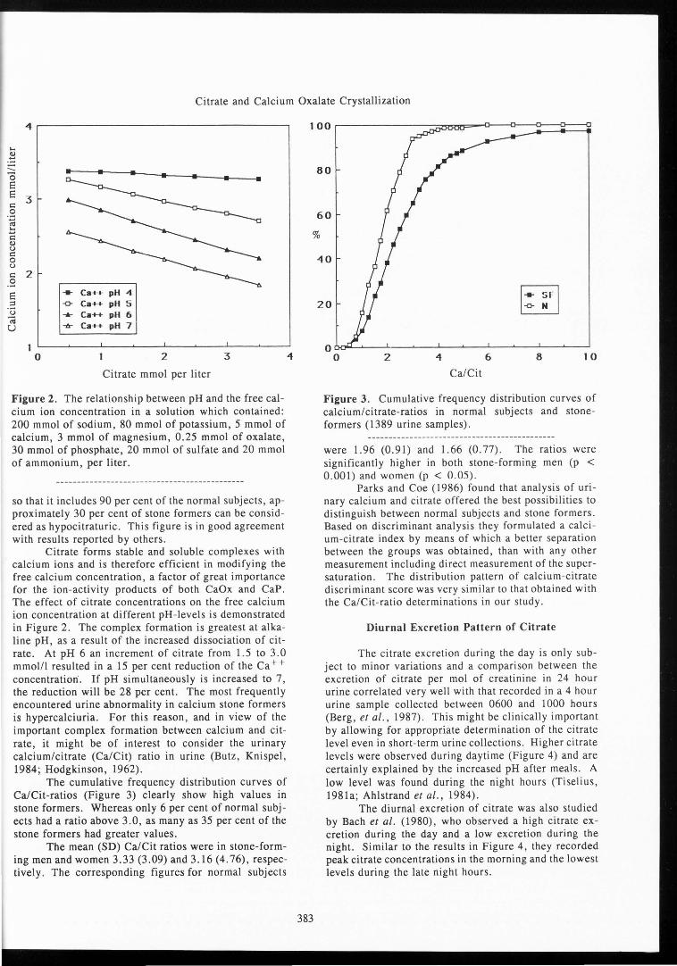
Citrate forms stable and soluble complexes with calcium ions and is therefore efficient in modifying the free calcium concentration, a factor of great importance for the ion-activity products of both Ca Ox and CaP. The effect of citrate concentrations on the free calcium ion concentration at different pH-levels is demonstrated in Figure 2. The complex formation is greatest at alkaline pH, as a result of the increased dissociation of citrate. At pH 6 an increment of citrate from 1.5 to 3.0 mmol/1 resulted in a 15 per cent reduction of the Ca++ concentration·. If pH simultaneously is increased to 7, the reduction will be 28 per cent. The most frequently encountered urine abnormality in calcium stone formers is hypercalciuria. For this reason, and in view of the important complex formation between calcium and citrate, it might be of interest to consider the urinary calcium/citrate (Ca/Cit) ratio in urine (Butz, Knispel, 1984; Hodgkinson, 1962).
The cumulative frequency distribution curves of Ca/Cit-ratios (Figure 3) clearly show high values in stone formers. Whereas only 6 per cent of normal subjects had a ratio above 3.0, as many as 35 per cent of the stone formers had greater values.
The mean (SD) Ca/Cit ratios were in stone-forming men and women 3.33 (3.09) and 3.16 (4. 76), respectively. The corresponding figures for normal subjects
80
60
%
40
383
20 [r] N
0 0 2 4 6 8 10
Ca/Cit
Figure 3. Cumulative frequency distribution curves of calcium/citrate-ratios in normal subjects and stoneformers (1389 urine samples).
were 1.96 (0.91) and 1.66 (0.77). The ratios were significantly higher in both stone-forming men (p < 0.001) and women (p < 0.05).
Parks and Coe (1986) found that analysis of urinary calcium and citrate offered the best possibilities to distinguish between normal subjects and stone formers. Based on discriminant analysis they formulated a calcium-citrate index by means of which a better separation between the groups was obtained, than with any other measurement including direct measurement of the supersaturation. The distribution pattern of calcium-citrate discriminant score was very similar to that obtained with the Ca/Cit-ratio determinations in our study.
Diurnal Excretion Pattern of Citrate
The citrate excretion during the day is only subject to minor variations and a comparison between the excretion of citrate per mo! of creatinine in 24 hour urine correlated very well with that recorded in a 4 hour urine sample collected between 0600 and 1000 hours (Berg, et al., 1987). This might be clinically important by allowing for appropriate determination of the citrate level even in short-term urine collections. Higher citrate levels were observed during daytime (Figure 4) and are certainly explained by the increased pH after meals. A low level was found during the night hours (Tiselius, 1981a; Ahlstrand et al., 1984).
The diurnal excretion of citrate was also studied by Bach et al. (1980), who observed a high citrate excretion during the day and a low excretion during the night. Similar to the results in Figure 4, they recorded peak citrate concentrations in the morning and the lowest levels during the late night hours.

H.-G. Tiselius et al.
.8 ~ ... -u ~ u "O i::
"' i::
.8 ~ !:: i:: ti) u i:: 0 u ti)
~ !:: u
6
- Ca/Cit
5 -0- Cit cone. Cs) .. Cit cone.
4
3
2
0 ~__.,_,_~_.__,_~..__.__.._,___._~......_...__L.........--L~--'--'-..L...J
1 3 5 7 9 1 1 1 3 1 5 1 7 1 9 21 23
hours
Figure 4. Diurnal variation in the urinary excretion of citrate and calcium/citrate-ratios. The citrate concentrations were calculated for a 24 hour urine volume of 1500 ml.
Effects of Citrate on CaOx Supersaturation
Variations in citrate concentrations have significant effects on the ion-activity product of CaOx (APcaox)- A relationship between changes in citrate excretion and APcaox was calculated by means of the EQUIL 2 program (Werness et al., 1985) in a solution which at pH 6.0 and in a volume of 1500 ml contained 6.5 mmol of calcium, 4.5 mmol of magnesium, 0.3 mmol of oxalate, 200 mmol of sodium, 70 mmol of potassium, 25 mmol of phosphate, 30 mmol of sulfate, and 35 mmol of ammonium. With this urine composition, an increment of 24 hour urinary citrate from 2 to 4 mmol resulted in a AP caox reduction from 1. 37 x 10·8 to 1. 26 x 10·8 (mmol/1) 2 .
Citrate was more efficient than magnesium in changing APcaox· The exponent for citrate was -0.22 and that for magnesium -0.12 (Tiselius, 1991).
Citrate affects APcaox by complex formation with calcium, and the citrate response is therefore more pronounced in alkaline solutions. This is clearly shown in Figure 5. In a similar way, citrate influences the ionactivity product of brushite, a salt that might be of importance particularly for formation of stones containing both CaOx and CaP (Pak and Holt, 1976) (Figure 6).
Effects of Citrate CaOx Crystal Growth Inhibition
It has been reported by several authors that citrate in low concentrations is capable of inhibiting CaOx crystal growth in metastably supersaturated salt solutions (Meyer and Smith, 1975; Ryall et al., 1981a; Tiselius et al., 1981). The question can be raised, however, to what extent does citrate contribute to the crystal growth
>< 1 .5 0 "' u ~
< 1.4
1 .3
1 .2 0
300
250
200 "' ~
Cl.
.. -0---6-
6
APCaOx pH 4 APCaOx pH S APCaOx pH 6 APCaOx pH 7
2 3
Citrate mmol per liter
.. AP 8ru pH 4 -o- AP Bru pH S ...._ AP Bru pH 6 -6- AP Bru pH 7
< 150
100
50
0 0 2 3
Citrate mmol per liter
Figures 5 and 6. The effects of citrate on the ionactivity product of CaOx (APcaox) (Figure 5) and brushite (AP 8 ru) (Figure 6) at pH 4, 5, 6, and 7.
inhibition in the presence of the high concentrations of powerful macromolecules in urine (Tiselius et al., 1987; Bek-Jensen and Tiselius, 1991). To address this question, direct determination of the crystallization was carried out in whole urine samples after addition of different amounts of citrate. As could be expected, increased concentrations of citrate resulted in a reduced crystallization risk, when determined by the oxalate addition required for formation of 100 crystals in the size-range of 3.5-5 µm (Tiselius, 1985).
4

Citrate and Calcium Oxalate Crystallization
70 a
60
C: 0 '-§ 50 0 "' .5 40 Q)
';;j
] 30 0 I u
~
~ 20
.. Citrate
1 0 -o- Magnesium
0 L__ __ _._ __ __,_ ___ .__ __ _L,_ __ __., __ __,
0 2 4 6
Concentration mmol/liter
5----------------------, b
4
3
2 .__ __ _.__ ___ ..,__ __ _.__ ___ ..,__ __ _.__ __ ~
0 2 4 6
Concentration mmol/liter
Figure 7. Effects of different concentrations of citrate and magnesium on the crystallization of calcium oxalate in a salt solution as determined from the fraction of 14Coxalate remaining in solution 60 minutes after supersaturation (a) and on the ion-activity product (b).
The effect of citrate, as well as that of magnesium, was also determined in salt solutions brought to crystallization by supersaturation to an ion-activity product of 4.25 x 10⁻⁸ (mol/1)2 with addition of sodium oxalate. As shown in Figure 7, there appeared to be a close relationship to the effects these compounds had on the supersaturation level in the sample. In an attempt to study this problem further, the crystal growth in samples containing 90 per cent of dialysed urine and CaOx seed crystals, was followed in the presence of various citrate
0.9---------------------,
0,8
0,7 >< ~ u
p.. -< 0,6
00 0
385
0,5
0,40L,_ __ .....__ __ ....1.... __ _,_ __ -1,2 ___ .._ _ ___,J3
Citrate mmol per liter
Figure 8. The ion-activity product of CaOx for the same precipitation of calcium oxalate in relation to the citrate concentration. Crystal growth was determined 30 minutes after addition of seed crystals by measuring the fraction of 45Ca remaining in solution.
concentrations at different levels of supersaturation accomplished by the addition of oxalate (Tiselius et al., 1987). The growth of Ca Ox was determined by measuring the amount of (45Ca) remaining in solution in the samples 30 minutes after addition of the seed. A reduced rate of calcium oxalate precipitation was recorded with increased citrate concentrations. In order to determine whether this effect on crystallization reflected a direct inhibitory action of citrate or only was the result of the altered supersaturation, a comparison of the APcaox at corresponding degrees of calcium precipitation was calculated. The AP caox was calculated with the EQUIL 2 program. As is evident from Figure 8, the same CaOx precipitation was observed at increasing levels of APcaox as the citrate concentration was increased. This indicates that citrate, even in the presence of whole urine concentrations of macromolecules, has the capacity to inhibit the growth of CaOx crystals (Tiselius, unpub-1 ished observation).
It is reasonable to assume that the effect of an increased pH on crystal growth, that has been demonstrated in diluted urine (Tiselius, 1981b), is also valid for whole urine.
Inhibition of Crystal Aggregation
It can be assumed that the process of renal stone formation includes a rapid formation of large crystal masses. This is accomplished by aggregation of crystals in a way that enables their entrapment in the distal part
H.-G. Tiselius et al.
of the collecting system. In this way, the initial crystal formation might result in a stone nidus, that can increase in size to a clinically significant stone by further growth and deposition of crystals. It has also been shown that the size of crystals and crystal aggregates is greater in stone formers than in normal subjects (Robertson et al., 1971). Several authors have also suggested that the inhibition of crystal aggregation can be the most important discriminating risk factor between normal subjects and patients with calcium stone disease (Azoury et al., 1987; Coe et al., 1991; Kok et al., 1986; Springmann et al., 1986).
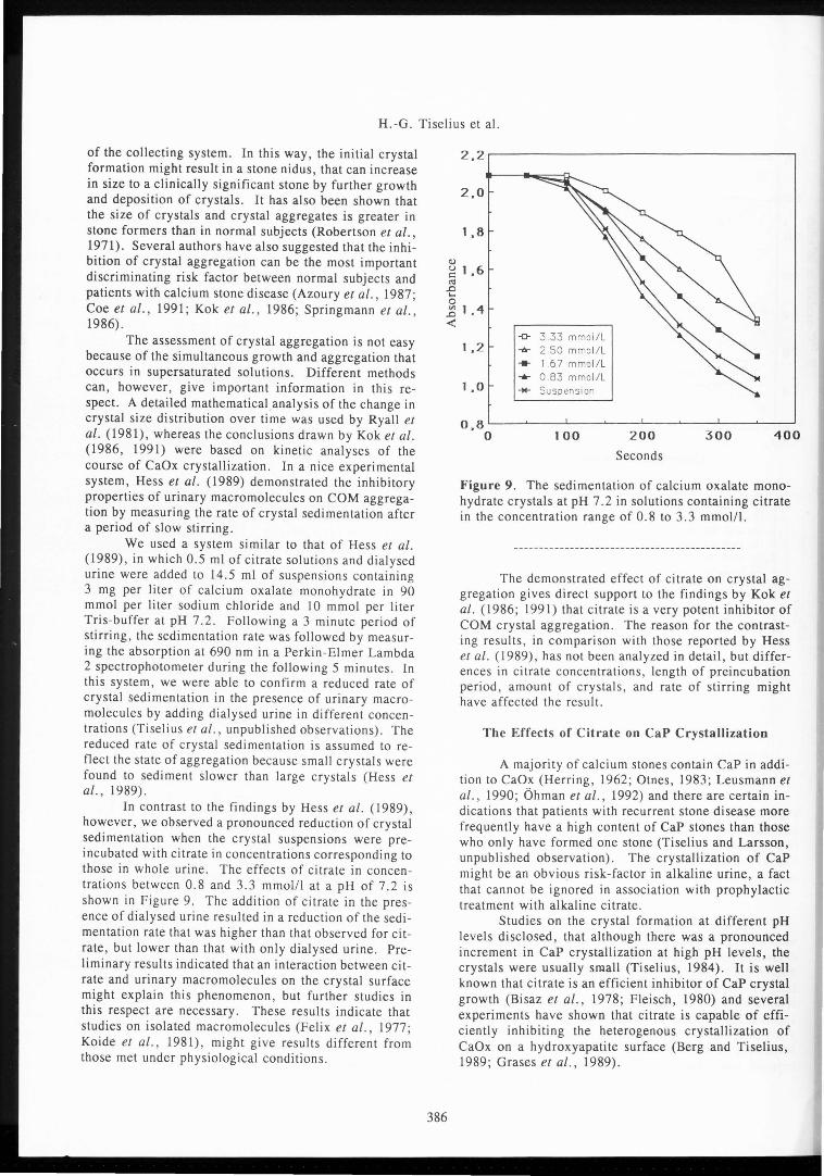
The assessment of crystal aggregation is not easy because of the simultaneous growth and aggregation that occurs in supersaturated solutions. Different methods can, however, give important information in this respect. A detailed mathematical analysis of the change in crystal size distribution over time was used by Ryall et al. (1981), whereas the conclusions drawn by Kok et al. (1986, 1991) were based on kinetic analyses of the course of CaOx crystallization. In a nice experimental system, Hess et al. (1989) demonstrated the inhibitory properties of urinary macromolecules on COM aggregation by measuring the rate of crystal sedimentation after a period of slow stirring.
We used a system similar to that of Hess et al. (1989), in which 0.5 ml of citrate solutions and dialysed urine were added to 14.5 ml of suspensions containing 3 mg per liter of calcium oxalate monohydrate in 90 mmol per liter sodium chloride and 10 mmol per liter Tris-buffer at pH 7.2. Following a 3 minute period of stirring, the sedimentation rate was followed by measuring the absorption at 690 nm in a Perkin-Elmer Lambda 2 spectrophotometer during the following 5 minutes. In this system, we were able to confirm a reduced rate of crystal sedimentation in the presence of urinary macromolecules by adding dialysed urine in different concentrations (Tiselius et al., unpublished observations). The reduced rate of crystal sedimentation is assumed to reflect the state of aggregation because small crystals were found to sediment slower than large crystals (Hess et al., 1989).
In contrast to the findings by Hess et al. (1989), however, we observed a pronounced reduction of crystal sedimentation when the crystal suspension-s were preincubated with citrate in concentrations corresponding to those in whole urine. The effects of citrate in concentrations between 0.8 and 3.3 mmol/1 at a pH of 7.2 is shown in Figure 9. The addition of citrate in the presence of dialysed urine resulted in a reduction of the sedimentation rate that was higher than that observed for citrate, but lower than that with only dialysed urine. Preliminary results indicated that an interaction between citrate and urinary macromolecules on the crystal surface might explain this phenomenon, but further studies in this respect are necessary. These results indicate that studies on isolated macromolecules (Felix et al., 1977; Koide et al., 1981), might give results different from those met under physiological conditions.
2.2.-------------------7
2.0
1 ,8
<U
~ 1 ,6 c<j
.D ... 0 E 1.4 ,,(
386
-0- 3.33 mmoi/L 1 .2 -a- 2.50 mmol/L .... 1.67 mmol/L .... 0.83 mmol/L 1 .o -M- Suspension
100 200 O.BOL__~_--1. __ ~ _ _,_ _ __,_ __ 3~0-0 _ __.__4_0~0
Seconds
Figure 9. The sedimentation of calcium oxalate monohydrate crystals at pH 7.2 in solutions containing citrate in the concentration range of 0.8 to 3.3 mmol/1.
The demonstrated effect of citrate on crystal aggregation gives direct support to the findings by Kok et al. (1986; 1991) that citrate is a very potent inhibitor of COM crystal aggregation. The reason for the contrasting results, in comparison with those reported by Hess et al. ( 1989), has not been analyzed in detail, but differences in citrate concentrations, length of preincubation period, amount of crystals, and rate of stirring might have affected the result.
The Effects of Citrate on CaP Crystallization
A majority of calcium stones contain CaP in addition to CaOx (Herring, 1962; Otnes, 1983; Leusmann et al., 1990; Ohman et al., 1992) and there are certain indications that patients with recurrent stone disease more frequently have a high content of CaP stones than those who only have formed one stone (Tiselius and Larsson, unpublished observation). The crystallization of CaP might be an obvio_us risk-factor in alkaline urine, a fact that cannot be ignored in association with prophylactic treatment with alkaline citrate.
Studies on the crystal formation at different pH levels disclosed, that although there was a pronounced increment in CaP crystallization at high pH levels, the crystals were usually small (Tiselius, 1984). It is well known that citrate is an efficient inhibitor of CaP crystal growth (Bisaz et al., 1978; Fleisch, 1980) and several experiments have shown that citrate is capable of efficiently inhibiting the heterogenous crystallization of CaOx on a hydroxyapatite surface (Berg and Tiselius, 1989; Grases et al., 1989).

Citrate and Calcium Oxalate Crystallization
There is, however, only limited information available concerning the effects of citrate on CaP crystal aggregation. For this reason, we measured the effects of citrate on the sedimentation rate of brushite and hydroxyapatite crystals in a way similar to that described above for CaOx. The results showed that citrate also efficiently reduced the sedimentation rate of these crystals (Tiselius et al., unpublished observation). This information is encouraging for the further use of alkaline citrate in the preventive treatment of patients with recurrent calcium stone disease.
Treatment of Patients with Recurrent Calcium Stone Disease with Alkaline Citrate
Several authors have successfully used alkaline citrate preparations in prevention of recurrent calcium oxalate stone formation. Different preparations were administered, such as sodium potassium citrate, potassium citrate, and magnesium citrate (Butz, 1982; Pak et al., 1985; Preminger et al., 1988; Schwille et al., 1987). A 24 hour dose of 10-43 mmol given on two or three occasions appeared to be an efficient pharmacological therapy with pronounced effects on stone formation. The rational for this therapy is the increment in urinary citrate that is achieved with these preparations. In view of the risk situation that occurs during the late night and morning hours, with a low citrate concentration, a low pH, a low urine volume and a high Ca/Cit ratio, we found it reasonable to administer alkaline citrate in a single evening dose (Berg et al., 1992). That type of administration would accordingly counteract crystallization and stone formation during this particular risk period. Single dose administration might also result in a better patients' compliance than when the drug has to be administered several times a day.
A group of 39 men and 16 women with recurrent calcium oxalate stone disease were treated with 5. 0 g ( 1 8 mmol) and 3.8 g (14 mmol) of sodium potassium citrate respectively, as a single evening dose. Their mean (SD) pretreatment 24 hour urinary excretion of citrate was 1. 7 (l.2) mmol. In these patients mean (SD) urinary citrate increased from 138 (98) to 253 (127) mmol per mo! of creatinine (p < 0.001), the Ca/Cit ratio decreased from 5.19 (3.88) to 2.07 (1.05) (p < 0.001) and the APcaoxindex, which is an estimate of the supersaturation, was reduced from 2.32 (0.83) to 1.45 (0.45) (p < 0.001). The clinical course of the disease was favorably affected by alkaline citrate and the number of stones formed during the treatment period up to 6 years [mean (SD) 3.5 (l. 7)), was significantly lower than that recorded during a period of the same length following the first observation of a renal stone. The tolerance of the treatment was very good, certainly a result of the absence of side effects and the simplicity of drug-administration.
As could be expected, the mean (SD) AP(CaP) index, used as an estimate of CaP supersaturation, increased in morning samples from 4.1 (7.5) to 14.4
387
(16.9), a reflection of the increased pH. A reliable estimate of this index could be obtained only in urine collected between 0600 and 1000 hours. There was, however, a pronounced variation in the level of AP(CaP) index, and a significant increment was recorded only in a group of patients given an evening dose of 7.5 g.
Further long-term follow-up studies with single dose administration of alkaline citrate are in progress. From these studies, it is particularly important to determine whether the effect on urinary pH, with this regimen, also is transient, as recently observed by Schwille et al. (1992), who gave 6 g of potassium citrate three times a day. Such an effect can theoretically reduce the therapeutic effect of alkaline citrate despite a maintained increment in the citrate excretion.
A lack of stone prevention was reported by Trinchieri et al. (1992) who followed 19 patients treated with 5-10 g of potassium citrate during 2 years. They assumed that this might be attributable to an unaffected high calcium excretion. In a randomized study by Hofbauer et al. (1992), 25 patients treated with three daily doses of alkaline citrate to get a pH around 7, were compared with 25 patients only given metaphylactic instructions. The citrate was significantly higher in the group with alkaline citrate therapy, but there were no differences in the course of stone formation between the two groups.
Conclusions
Urinary citrate affects CaOx as well as CaP-crystallization by reduction of the ion-activity products of these salts. The CaOx crystal growth is also inhibited by urinary citrate in a way that adds inhibitory power to that accomplished by urinary macromolecules. The heterogenous crystallization of CaOx on CaP is significantly inhibited by citrate. Increments in citrate concentrations result in a reduction of the aggregation of CaOx, brushite and hydroxyapatite crystals. Although the high pH associated with alkaline citrate treatment results in CaP crystallization, the concomitant citrate increment seems to be one of the most important factors in reducing the risk of this precipitation. Citrate thereby apparently both maintains the crystals small and prevents the heterogenous growth of CaOx on CaP. The different experimental findings are supported by the successful effects of alkaline citrate on the course of calcium stone disease. The optimal dose and best way of administration however, has to await further long term studies.
Acknowledgement
The experimental work carried out in our laboratory and presented in this review has been supported by grants from Dr. Madaus & Company, The Swedish Medical Research Council, the Research Funds of the County Councils of Ostergotland and Sormland, and Maud and Birger Gustavsson's Research Fund.

H.-G. Tiselius et al.
References
Ahlstrand C, Tiselius HG, Larsson L. (1984) Studies on crystalluria in calcium oxalate stone formers. Urological Research 12, 103-106.
Azoury R, Robertson WG, Garside J. (1987) Observations on in vitro and in vivo calcium oxalate crystalluria in primary calcium stone formers and normal subjects. British Journal of Urology 69, 211-213.
Bach D, Hesse A, Vahlensieck W. (1980) Zitratausscheidung bei Harnsteinpatienten und Gesunden unter Normal- und Standardkost (Excretion of citrate in stone formers and healthy subjects during normal and standard diet). Urologe A19, 220-225.
Bek-Jensen H, Tiselius HG. (1991) Inhibition of calcium oxalate crystallization by urinary macromolecules. Urological Research 19, 165-169.
Berg C, Tiselius HG. ( 1989) The effects of citrate on hydroxyapatite induced calcium oxalate crystallization and on the formation of calcium phosphate crystals. Urological Research 17, 167-172.
Berg C, Larsson L, Tiselius HG. (1987) Four hour urine composition in patients with calcium oxalate stone disease. British Journal of Urology 60, 301-306.
Berg C, Larsson L, Tiselius HG. (1992) The effects of a single evening dose of alkaline citrate on urine composition and calcium stone formation. Journal of Urology 148, 979-985.
Bisaz S, Felix R, Neuman WF, Fleisch H. (1978) Quantitative determination of inhibitors of calcium phosphate precipitation in whole urine. Mineral and Electrolyte Metabolism 1, 74-83.
Butz M. (1982) Oxalatsteinprophylaxe durch Alkali-Therapie. Urologe A21, 142-146.
Butz M, Knispel H. (1984) Molar calcium citrate ratio as diagnostic indicator in calcium nephrolithiasis. In: Urinary stone. Ryall RM, Brockis JG, Marshall V, Finlayson B (eds.), Churchill Livingstone, pp 219-223.
Coe FL, Nakagawa Y, Parks JH. (1991) Inhibitors within the nephron. American Journal of Kidney Diseases 17, 407-413.
Felix R, Menod A, Brage L, Hansen NM, Fleisch H. (1977) Aggregation of calcium oxalate crystals: effect of urine and various inhibitors. Urological Research s, 21-28.
Finlayson B. (1977) Calcium stones. Some physical and clinical aspects. In: Calcium Metabolism in Renal Failure and Nephrolithiasis. David DS (ed.), John Wiley & Sons, pp 337-382.
Fleisch H. (1980) Mechanisms of stone formation: Role of promoters and inhibitors. Scandinavian Journal of Urology and Nephrology, Suppl 53, 53-66.
Fleisch H. (1990) Role of inhibition and promoters of crystal nucleation, growth and aggregation in the formation of calcium stones. In: Renal tract stonemetabolic basis and clinical practice. Wickham JEA, Buck AC (eds.), Churchill Livingstone, pp 295-306.
Grases F, Gil JJ, Cante A. (1989) Urolithiasis inhibitors and calculus nucleation. Urological Research
388
17, 163-166. Herring LC. ( 1962) Observations on the analysis
of ten thousand urinary calculi. Journal of Urology 88, 545-562.
Hess B, Nakagawa Y, Coe FL. (1989) Inhibition of calcium oxalate monohydrate crystal aggregation by urine proteins. American Journal of Physiology 257, F99-F106.
Hofbauer J, Hobarth K, Eisenmenger M, Marberger M. (1992) Alkali citrate metaphylaxis for recurrent calcium oxalate stone-formers - a prospective randomized study. In: Proc. VIIth International Symposium on Urolithiasis, Cairns. Ryall RL (ed.), Flinders Med. Ctr., Bedford Park, Australia, p 87 (abstract).
Hodgkinson A. (1962) Citric acid excretion in normal adults and in patients with renal calculus. Clinical Science 23, 203-212.
Hoyer JR (1992) Uropontin is selectively incorporated into the organic matrix of urinary calcium oxalate monohydrate and brushite stones. In: Proc. Vllth International Symposium on Urolithiasis, Cairns. Ryall RL (ed.), Flinders Med. Ctr., Bedford Park, Australia, p 118 (abstract).
Koide T, Takemoto M, Itatani H, Takaha M, Sonoda T. ( 1981) Urinary macromolecular substances as natural inhibitors of calcium oxalate crystal aggregation. Investigative Urology 18, 382-386.
Kok DJ. ( 1991) The role of crystallization process in calcium oxalate urolithiasis. Doctorate Thesis. University of Leiden, Netherlands, pp 58-73.
Kok DJ, Papapoulos SE, Bijvoet OLM. (1986) Excessive crystal agglomeration with low citrate excretion in recurrent stone formers. Lancet i, 1056-1058.
Leusmann DB, Blaschke R, Schwandt W. (1990) Results of 5035 stone analyses: a contribution to epidemiology of urinary stone disease. Scandinavian Journal of Urology and Nephrology, 24, 205-210.
Marangella M, Bianco 0, Grande ML, Petrarulo M, Valente D, Vitale C, Linari F. (1987) Patterns of citrate excretion in healthy subjects and patients with idiopathic stone disease. Contributions to Nephrology 58, 34-38.
Menon M, Mahle J. (1983) Urinary citrate excretion in patients with renal calculi. Journal of Urology 129, 1158-1160.
Meyer JL, Smith LH. (1975) Growth of calcium oxalate crystals. Investigative Urology 13, 36-39.
Meyer JL. (1990) Physicochemistry of Stone formation. In: Urolithiasis - a Medical and Surgical Reference. Resnick MI, Pak CYC (eds.), WB Saunders Company, Philadelphia, pp 11-34.
Nicar MJ, Skurla C, Sakhaee K, Pak CYC. (1983) Low urinary citrate excretion in nephrolithiasis. Urology 21, 8-14.
Ohman S, Larsson L, Tiselius HG. (1992) Clinical significance of phosphate in calcium oxalate renal stones. Annals of Clinical Biochemistry 29, 59-63.
Otnes B. (1983) Crystalline composition of

Citrate and Calcium Oxalate Crystallization
urinary stones in Norwegian patients. Scandinavian Journal of Urology and Nephrology 17, 85-92.
Pak CYC. (1991) Citrate and renal calculi: New insights and future directions. American Journal of Kidney Diseases 17, 420-425.
Pak CYC, Holt K. (1976) Nucleation and growth of brushite and calcium oxalate in urine of stone-formers. Metabolism 25, 665-673.
Pak CYC, Fuller C, Sakhaee K, Preminger GM, Britton F. (1985) Long-term treatment of calcium nephrolithiasis with potassium citrate. Journal of Urology 134, 11-19.
Parks JH, Coe FL. (1986) A calcium citrate index for the evaluation of nephrolithiasis. Kidney International 30, 85-90.
Preminger GM, Sakhaee K, Pak CYC. (1988) Alkali action on the urinary crystallization of calcium salts: contrasting responses to sodium citrate and potassium citrate. Journal of Urology 139, 240-242.
Robertson WG. (1976) Physical chemical aspects of calcium stone-formation in the urinary tract. In: Urolithiasis Research. Fleisch H, Robertson WG, Smith LH, Vahlensieck W (eds.), Plenum Press, pp 25-39.
Robertson WG, Peacock M, Nordin BEC. (1971) Calcium oxalate crystalluria and urine saturation in recurrent renal stone-formers. Clinical Science 40, 365-374.
Robertson WG, Peacock M, Heyburn PJ, Marshall DH, Clark PB. (1978) Risk factors in calcium stone disease of the urinary tract. British Journal of Urology 50, 449-454.
Rudman D, Kutner MH, Redd SC II, Waters WC IV, Gerron GG, Bleier J. (1982) Hypocitraturia in calcium nephrolithiasis. Journal of Clinical Endocrinology and Metabolism 55, 1052-1057.
Ryall RL, Harnett RM, Marshall VR. (1981a) The effect of urine, pyrophosphate, citrate, magnesium and glycosaminoglycans on the growth and aggregation of calcium oxalate crystals in vitro. Clinica Chimica Acta 112, 349-356.
Ryall RL, Ryall RG, Marshall VR. (1981b) Interpretation of particle growth and aggregation patterns obtained from the Coulter Counter: a single theoretical model. Investigative Urology 18, 396-400.
Schwille PO, Scholz D, Paulus M, Engelhardt W, Sigel A. (1979) Citrate in daily and fasting urine. Investigative Urology 16, 457-462.
Schwille PO, Rumenapf G, Schwarzlander H, Kuch P, Berens H. (1987) Medium-term treatment of idiopathic recurrent calcium urolithiasis by oral sodiumpotassium citrate - a preliminary report on metabolic effects. In: Acta Medica: Inhibitors of crystallization in renal lithiasis and their clinical application. Martelli A, Buli P, Marchesini B (eds.), I.C.E.P., Roma, pp 177-182.
Schwille PO, Herrmann U, Wolf C, Berger I, Meister R. (1992) Citrate and recurrent idiopathic calcium urolithiasis. A longitudinal pilot study on the metabolic effects of oral potassium citrate administered over
the short-, medium- and long-term medication of male stone patients. Urological Research 20, 145-155.
Springmann KE, Drach GW, Gottung B, Randolph AD. (1986) Effects of human urine on aggregation of calcium oxalate crystals. Journal of Urology 135, 69-71.
Tiselius HG. (1981a) Urinary excretion of citrate in normal subjects and patients with urolithiasis. In: Urolithiasis: Clinical and basic research. Smith LH, Robertson WG, Finlayson B (eds.), Plenum Press, pp 39-44.
Tiselius HG. (1981b) The effect of pH on the urinary inhibition of calcium oxalate growth. British Journal of Urology 53, 470-474.
Tiselius HG. (1984) Urinary pH and calcium oxalate crystallization. In: Pathogenese und Klinik der Harnsteine. Fortschritte der Urologie und Nephrologie (Pathology and Clinic of Renal Stones. Progress in Urology and Nephrology). Vahlensieck W, Gasser G (eds.), Steinkopff, Darmstadt, pp 184-187.
Tiselius HG. (1985) Measurement of the risk of calcium oxalate crystallization in urine. Urological Research 13, 297-300.
Tiselius HG. (1991) Aspects on estimation of risk of calcium oxalate crystallization in urine. Urologia Internatinalis 47, 255-259.
Tiselius HG, Pomander AM. (1981) Evaluation of a routine method for determination of calcium oxalate crystal growth inhibition in diluted urine samples. Clinical Chemistry 27, 565-568.
Tiselius HG, Pomander AM, Nilsson MA. (1987) Inhibition of calcium oxalate crystallization in urine. Urological Research 15, 83-86.
Trinchieri A, Rovera F, Nespoli R, Zanetti G, Austoni E. (1992) Prophylactic treatment of recurrent calcium stones with potassium sodium citrate. In: Proc. Vllth International Symposium on Urolithiasis, Cairns. Ryall RL (ed.), Flinders Med. Ctr., Bedford Park, Australia, p 154 (abstract).
Welshman SG, McGeown MG. (1976) Urinary citrate excretion in stone-formers and normal controls. British Journal of Urology 48, 7-11.
Werness PG, Brown CM, Smith LH, Finlayson B. (1985) EQUIL 2: A basic computer program for the calculation of urinary saturation. Journal of Urology 134, 1242-1244.
Discussion with Reviewers
W .G. Robertson: The difference in the citrate excretion of stone-formers and normals in the authors' own study is very small and would be predicted to have a trivial effect on the crystallization parameters quoted. How do such small differences explain stone-formation in the authors' patients? Authors: It is true that the difference between stone formers and normal subjects in terms of mean 24 hour citrate excretion is relatively small. Citrate excretion levels below 1.0 mmol per 24 hours were, however, only found in stone formers and these patients might

H.-G. Tiselius et al.
constitute a special risk group. There is no proof that a low citrate excretion alone can explain why a person becomes a stone-former. Probably, in many patients, a low urinary citrate is rather a contributing factor to an already established risk situation, whether caused by increased concentrations of oxalate or calcium, or decreased inhibitory activities. In addition, we know that a high supersaturation occurs during the night, at which time the pH also is low. The low pH results in a further reduction of citrate excretion together with a decreased dissociation. Such mechanisms might result in much reduced activity of citrate during a period when the crystallization is unusually high.
A Hesse: Is there any evidence that citrate therapy is effective in different ways in patients forming whewellite and weddelite stones, respectively? Authors: We have so far not studied differences in
crystallization properties between these two crystai phases. All our measurements have been carried out with calcium oxalate monohydrate, but we are currently exploring the possibilities to measure at least aggregation in suspensions of calcium oxalate dihydrate. A comparison between stone composition (determined with x-ray diffraction technique) and citrate excretion indicated that male patients with less than 1.5 mmol of citrate excreted during 16 hours (between 0600 and 2200 hours) had a higher content of calcium oxalate dihydrate in their stones. According to these preliminary results, 16 per cent of the patients with a low citrate excretion had more than 70 per cent of dihydrate in their stones, compared with 8 per cent in patients with a higher citrate excretion.
A Hesse: Does citrate therapy favor stone passage after extracorporeal shock-wave (ESWL)-treatment? Authors: This is a very interesting and most important question because of the problems associated with residual fragments after shock wave lithotripsy. There are a few observations in the literature which indicate that this might be the case. We have no personal experience to support this assumption, but we have recently started a randomized study to address this question.
390
A Hesse: High citrate concentrations in urine inhibit crystal aggregation or aggregation of disintegrates after ESWL-treatment. What kind of reaction may be responsible for this observation? Authors: Citrate might either bind to the crystal surface or in any other way interact with the electrical double layer surrounding the crystals and/or disintegrates. There is also a possibility that citrate changes the physicochemical properties of aggregation promoting substances in urine (e.g., Tamm Horsfall mucoprotein). Furthermore, can the growth inhibition reduce the risk of crystal bridge formation between adjacent fragments.
B. Hess: The apparent contradiction between the authors' studies and our results (Hess et al., 1989) with respect to citrate as an inhibitor of calcium oxalate crystal aggregation, tested at almost identical conditions, might not really exist, if one considers that we never studied citrate at more than l mmolil. At this concentration, citrate inhibited by 7 + 3 per cent (mean + SD) in our system; in the authors' study, citrate at 0.83 mmol/1 marginally promoted, whereas it slightly inhibited crystal aggregation at l. 67 mmol/1. These findings seem fully compatible with our own results and demonstrate that only high physiologic concentrations of citrate inhibit calcium oxalate crystal aggregation under saturated conditions at pH 7.2. Authors: We agree completely with this comment. It is reasonable to assume that citrate concentrations above l mmol/1 are necessary to get measurable effects on the crystal aggregation in this experimental setting. For appropriate evaluation of the definite role of citrate in terms of aggregation inhibition under physiological conditions, it is certainly necessary to improve the method by decreasing the concentration of crystals in the suspension and to carry out the measurements at pH levels representative for that of urine in different parts of the collecting system. Such work is in progress.