effects of an eight-week progressive...
TRANSCRIPT
EFFECTS OF AN EIGHT-WEEK PROGRESSIVE RESISTANCE TRAINING
PROGRAM ON BALANCE IN PERSONS WITH MULTIPLE SCLEROSIS
By
GREGORY MICHAEL GUTIERREZ
A THESIS PRESENTED TO THE GRADUATE SCHOOL OF THE UNIVERSITY OF FLORIDA IN PARTIAL FULFILLMENT
OF THE REQUIREMENTS FOR THE DEGREE OF MASTER OF SCIENCE
UNIVERSITY OF FLORIDA
2005

Copyright 2005
by
GREGORY MICHAEL GUTIERREZ

ACKNOWLEDGMENTS
I would like to thank Dr. Mark Tillman, Dr. John Chow, and Dr. Lesley White for
their support and guidance in the completion of this work. I would also like to extend my
deepest gratitude to my family and friends for their encouragement and moral support
throughout this thesis project. I especially need to thank my parents; without them I
would not be the person I am today. Special thanks are extended to Dr. Mark Tillman for
his personal and professional advice throughout the years, for which I feel forever
indebted to him.
iii

TABLE OF CONTENTS page ACKNOWLEDGMENTS ................................................................................................. iii
LIST OF TABLES............................................................................................................. vi
LIST OF FIGURES .......................................................................................................... vii
ABSTRACT...................................................................................................................... vii
CHAPTER
1 INTRODUCTION ........................................................................................................1
2 LITERATURE REVIEW .............................................................................................5
Multiple Sclerosis .........................................................................................................5 Expanded Disability Status Score (EDSS) ...................................................................7 Symptoms .....................................................................................................................9 Risk of Falls................................................................................................................10 MS, Exercise and Remyelination ...............................................................................11 Balance .......................................................................................................................13
3 METHODOLOGY .....................................................................................................16
Subjects.......................................................................................................................16 Instrumentation ...........................................................................................................17
Force Platform.....................................................................................................17 Isokinetic Dynamometer .....................................................................................17
Experimental Setup.....................................................................................................17 Postural Sway ......................................................................................................18 Strength Testing...................................................................................................20 Functional Tests...................................................................................................21 Resistance Training .............................................................................................21
Data Reduction ...........................................................................................................22 Design/Analysis..........................................................................................................23
4 RESULTS...................................................................................................................24
Strength.......................................................................................................................24
iv

Balance .......................................................................................................................25 Functional Tests..........................................................................................................26
5 DISCUSSION.............................................................................................................27
Strength.......................................................................................................................27 Balance .......................................................................................................................29 Limitations..................................................................................................................31 Summary and Conclusions .........................................................................................31
APPENDIX
A INFORMED CONSENT............................................................................................33
B EXPANDED DISABILITY STATUS SCALE..........................................................43
LIST OF REFERENCES...................................................................................................45
BIOGRAPHICAL SKETCH .............................................................................................49
v

LIST OF TABLES
Table page 1 – Muscle groups being tested, the movement they produce, and the corresponding joint
angles. ..........................................................................................................................20
2 – Strength measures for the MS training group (mean ± SD). All strength (torque) measures in Nm. * denotes p<0.05. ............................................................................24
3 – Strength measures in the non –MS control training group (mean ± SD). All strength (torque) measures in Nm..............................................................................................24
4 – Mean balance measures for the MS training group. All balance measures in m. * denotes p<0.05. ............................................................................................................25
5 – Mean balance measures for the control training group. All balance measures in m. .25
vi

LIST OF FIGURES
Figure page 1 – The self-selected (E) stance. ........................................................................................19
2 – The feet apart (F) stance. .............................................................................................19
3 – The foam pad (P) stance. .............................................................................................19
4 – The semitandem (S) seen from a A) frontal view and B) sagittal view.......................20
5 – The tandem (T) stance seen from a A) frontal view and B) sagittal view. ..................20
6 – Diagram depicting the movement of the COP throughout a balance trial. ..................22
vii

Abstract of Thesis Presented to the Graduate School
of the University of Florida in Partial Fulfillment of the Requirements for the Degree of Master of Science
EFFECTS OF AN EIGHT-WEEK PROGRESSIVE RESISTANCE TRAINING PROGRAM ON BALANCE IN PERSONS WITH MULTIPLE SCLEROSIS
By
Gregory M. Gutierrez
May 2005
Chair: Mark Tillman Major Department: Department of Exercise and Sports Sciences
Multiple sclerosis (MS) is an autoimmune disorder of the central nervous system,
which leads to degeneration of the myelin sheaths that protect the neural axons. MS can
affect any part of the central nervous system, so persons with MS experience a wider
variety of symptoms than most neurological disorders, including problems with balance
and strength loss. The aim of this study was to determine if a strength training program,
designed to increase muscle strength, could improve postural sway measures in persons
with MS. Nine MS subjects and four non-MS controls participated in an eight-week
strength-training program. They were tested for isometric strength for their knee
extensors, knee flexors, plantar flexors, and dorsiflexors prior to and following the
strength-training program. Postural sway was also evaluated before and after training in
5 different stance conditions: 1) self-selected, 2) feet 6 inches apart, 3) feet 6 inches apart
on a foam pad, 4) semitandem, and 5) tandem. Four dependent variables were calculated
from the tests of postural sway: path length (PL), average speed (AS), antero-posterior
viii

amplitude (AP), and medio-lateral amplitude (ML) of the COP movement. Wilcoxon
signed rank tests were performed on all strength and balance variables to determine if
changes occurred due to the strength-training program with a conventional significance
level of 0.05. For the MS training group, the Wilcoxon signed rank tests revealed a
significant increase in PL and AS for the self-selected stance and an increase in isometric
strength in the knee flexors. The non-MS control training group had no significant
differences in strength or balance after training. The results indicate that strength training
is safe for persons with MS and may lead to an increase in muscular strength. However,
it does not appear to have a significant effect on standing balance in the stance positions
studied. A training program more specific to balance may demonstrate more significant
improvement in balance for persons with MS.
ix

CHAPTER 1 INTRODUCTION
Multiple sclerosis (MS) is the most common cause of nontraumatic neurological
disability affecting young adults in the northern hemisphere (Goodkin, 2000). MS is an
autoimmune disorder of the central nervous system that leads to widespread degeneration
of the myelin sheaths that encase axons in the central nervous system. The loss of the
protective myelin layer causes lesions to form on the axons, which can eventually
develop into hardened scleroses that inhibit the normal conduction of nerve impulses
down the axons (Herndon, 2000). The extent of axonal loss is variable, but usually
substantial, with some axonal loss occurring in every lesion (Trapp et al., 1998).
Symptoms from axonal loss cannot be alleviated. However, experimental efforts to
improve conduction in neurons without axonal loss have been promising (Herndon,
2000).
The four accepted patterns of pathology in MS as defined in 1996 are: 1) relapsing-
remitting, 2) secondary progressive, 3) chronic progressive and 4) progressive relapsing
MS (Goodkin, 2000). However, specific lines separating these disease patterns are not
completely clear thus making specific diagnoses challenging. Furthermore, the variable
nature of the disease leads to a difficulty in creating an ideal outcome assessment
measure for patients with MS. In all patterns of MS, the level of disability in patients is
typically categorized using the Expanded Disability Status Score (EDSS). The EDSS
scale was designed by John F. Kurtzke and is based on the maximum function of a
patient as limited by their neurological deficits (Kurtzke, 1955). Aside from a few
1

2
shortcomings, it serves as a familiar and quantifiable method of communication amongst
healthcare professionals concerning individuals with MS.
MS lesions occur in different areas of the central nervous system and due to this
variable distribution of demyelination, people with MS may experience a wider variety of
symptoms than any other neurological disease including balance, coordination, strength
and sensation disorders (Cattaneo et al., 2002). Furthermore, individuals with MS have
been found to have a reduced amount of skeletal muscle and a tendency to supply energy
through anaerobic pathways (Kent-Braun et al, 1997), which implies a decrease in the
number of slow-twitch muscle fibers. Along with a decrease in skeletal muscle fiber size,
persons with MS also face a reduced ability to activate muscle (Lambert et al., 2001),
which is associated with the demyelination of nerves (Kent-Braun et al., 1997). This
reduced muscle size and compromised motor unit activation cause the muscle weakness
associated with MS, which coupled with spasticity, further compromises the ability to
balance by affecting the sequencing and force of muscle contraction (Frzovic et al.,
2000).
Inability to maintain standing balance impacts a patient’s ability to perform
activities of daily living (ADLs) and puts them at an increased risk of falling and
subsequent injury, which contributes to the development of a fear of falling that may lead
to a change in quality of life (Cattaneo et al., 2002). Therefore, balance assessment and
implementation of rehabilitation strategies to improve balance is important in attempting
to maintain a favorable quality of life for persons with MS.
Patients with MS demonstrate reduced physical activity when compared to non-MS
individuals (Ng & Kent-Braun, 1997), which is usually attributed to muscle weakness

3
and fatigue, but could also be due to a patient’s fear of falling (Cattaneo et al., 2002).
When balance is compromised, even simple ADLs such as dressing, walking, and
standing become challenging, which may contribute to the anxiety and depression that
affects about 65% of patient’s with MS (Joffe et al., 1987).
The muscle weakness and fatigue demonstrated in persons with MS is consistent
with human models of disuse and provides a rationale for therapeutic intervention in the
form of exercise training as a means of reversing some of the reduced sensory and motor
functions in individuals with MS (Kent-Braun et al., 1997). Individuals with MS
experience muscle weakness and more symptomatic fatigue with exercise, however Kent-
Braun and colleagues found that they were not weaker compared to healthy individuals
when the amount of fat-free mass was taken into account. Therefore, an exercise-training
program designed to enhance muscle strength and endurance is a reasonable therapeutic
intervention in persons with MS and may be helpful in improving the functional capacity
of individuals with MS and offsetting the deleterious effects of their disease.
Furthermore, the demyelination associated with MS is not always permanent;
remyelination has been documented in MS (Chang et al., 2002). However, these
remyelinated nerves often fail to return to baseline functioning because of the decline in
activity following an acute MS attack (Herndon, 2000). Strength training may assist in
both promoting strength that may have been lost because of physical inactivity and
returning proper neural function to the remyelinated tissue. In addition, physical activity
has an important benefit in reducing the risk of secondary diseases and improving overall
health.
4
As stated earlier, persons with MS often face a reduced ability to balance, meaning
they are frequently unable to maintain the body’s position over its base of support
(Rogers et al., 2001). Quantifying a patient’s level of balance impairment is important,
therefore many different techniques have been proposed to measure a person’s ability to
maintain static or dynamic balance. In research settings, postural sway analysis is widely
accepted as a reliable way to quantify the complex and multidimensional nature of a
person’s standing balance (Tillman & Chow, 2002); however little research has been
conducted using postural sway in persons with MS. In addition, the influence of strength
training in MS patients has not been evaluated. Thus, the primary aim of this study is to
determine whether an eight-week program of progressive resistance training is tolerable
for persons with MS and if it could enhance standing balance in ambulatory individuals
with MS.

CHAPTER 2 LITERATURE REVIEW
A review of the literature revealed a significant shortage of relevant information
concerning strength training and balance in persons with MS. This work is intended to
fill that void. More specifically, it aims to provide more data regarding the effects of
resistance training on balance in individuals with MS.
Multiple Sclerosis
Multiple sclerosis (MS) is the most common progressive neurological disease in
young adults (Kraft & Wessman, 1974), usually diagnosed in individuals between the
ages of 20 and 40. MS is a degenerative inflammatory autoimmune disorder of the
central nervous system that destroys the myelin sheaths that encase and insulate the
neural axons (Chang et al., 2002; Kidd, 2001). The etiology of MS is not known,
however the most widely accepted hypothesis is that it is a virus-induced autoimmune
disorder (Herndon, 2000). The myelin in the central nervous system and the cells that
form that myelin, the oligodendrocytes, are the primary targets of attack (Herndon, 2000).
Lesions form on the myelin sheaths and can eventually develop into hardened scleroses
that inhibit the normal conduction of nerve impulses down the axons (Herndon, 2000).
MS lesions occur in different areas of the central nervous system and can range
from acute plaques with active macrophages containing lipid and myelin degenerating
products to chronic, inactive glial scars. The plaques appear to begin with the
macrophages and lymphocytes forming perivascular cuffs about the capillaries and
venules (Herndon, 2000). This is followed by diffuse infiltration by inflammatory cells,
5

6
edema, astrocytic hyperplasia, and macrophages consuming the myelin off the axons
causing an increasing number of lipid-filled macrophages and demyelinated axons
(Prineas, 1975). The extent of axonal loss is variable, but usually substantial (Trapp et
al., 1998), with some axonal loss occurring in every lesion. Experimental efforts to
improve conduction in neurons without axonal loss may produce dramatic improvements
in many symptoms that result from conduction failure in unaffected axons, however the
symptoms that result from axonal loss cannot be alleviated by these interventions
(Herndon, 2000).
There are four accepted patterns of pathology in MS that were defined in 1996,
which include: 1) relapsing-remitting, 2) secondary progressive, 3) chronic progressive
and 4) progressive relapsing MS. The relapsing-remitting form of MS is the best
understood and is more common in younger patients. Approximately 85% of MS
patients experience an exacerbation at disease onset (Goodkin, 2000). Multifocal discrete
inflammatory demyelinating lesions in both the gray and white matter of the CNS are
characteristic of this pattern (Herndon, 2000) and patients are usually stable between
exacerbations (Goodkin, 2000). Secondary progressive MS has characteristics consisting
of a combination of both relapsing-remitting and chronic progressive MS (Herndon,
2000). In these individuals, old, inactive, multifocal lesions coexist with progressive
diffuse demyelination (Herndon, 2000). Chronic progressive MS (also known as primary
progressive MS) is more typical in older patients and is less dramatic than relapsing-
remitting MS. The demyelination is diffusely scattered involving individual fibers or
small groups of fibers interspersed with normal appearing myelinated fibers. The
inflammatory infiltrates and macrophages are much more limited and diffuse than in
7
relapsing-remitting MS (Herndon, 2000). Chronic progressive MS involves a gradual
progression of disability without superimposed relapses (Goodkin, 2000). The fourth
pattern is termed progressive relapsing MS in which patients experience gradual
disability progression accompanied by one or more relapses (Goodkin, 2000). However,
specific lines separating these disease patterns are not completely clear which makes
specific diagnoses challenging.
During the process of demyelination, some conduction failure is unavoidable in the
affected fibers. Some lesions are known as clinically silent lesions, which occur when a
minority of fibers in a conduction path become demyelinated at any one time, leaving
intact conduction in the unaffected fibers in the path (Herndon, 2000). The causes of
conduction failure associated with demyelination are not completely understood, but it is
hypothesized that it may be due to 1) damage to the nodal sodium channels (Kaschow et
al., 1986a & 1986b), 2) a virtual absence of these sodium channels (Ritchie et al., 1977),
and/or 3) increased membrane capacitance in the demyelinated region (Waxman, 1995).
There is substantial evidence that the nodal membranes are damaged by various enzymes
released by the inflammatory cells that appear to produce extensive damage to the
myelin. Furthermore, an increased membrane capacitance causes the amount of current
required to depolarize the axon to be higher and therefore make impulse conduction
slower or in some cases blocked. These characteristics of demyelinated fibers help
explain some of the features of the motor fatigability and activity related failure of
neurological processes that affect individuals with MS (Herndon, 2000).
Expanded Disability Status Score (EDSS)
The very nature of the disease leads to a difficulty in creating an ideal outcome
assessment measure for patients with MS. In all patterns of MS, level of disability in
8
patients is typically categorized using the Expanded Disability Status Score (EDSS). The
Disability Status Score (DSS) was designed in 1955 by John F. Kurtzke and measures the
maximum function of a patient as limited by their neurological deficits (Kurtzke, 1955).
The DSS was expanded in 1983 to include more extensive criteria and is now known as
the Expanded Disability Status Score. The EDSS scale is based on any lack of function
in eight functional systems: 1) Pyramidal (degree of paralysis), 2) Cerebellar
(coordination of movement), 3) Brain Stem (cranial nerve functioning), 4) Sensory, 5)
Bowel and Bladder, 6) Visual (optic), 7) Cerebral (mental), and 8) Other neurological
deficits attributed to MS. The scale ranges form 0 to 10, where 0 is normal functioning
and 10 is death due to MS (Kurtzke, 1983). The scale is primarily based on the patient’s
ability to ambulate and deficiencies in any of the eight functional systems make the score
more specific to the patient’s actual disability level.
The most favorable aspects of the EDSS scale lie in the coverage of four of the
eight functional systems: the pyramidal, cerebellar, visual, and mental systems
(Coulthard-Morris, 2000). The pyramidal scale measures disability in the appendages
(i.e. paralysis in a limb). Cerebellar function is measured by the ability to coordinate
movements, which can be affected by the ataxia suffered by MS patients. Visual
impairments are characterized by a loss of visual acuity and/or temporal pallor. Finally,
mental functioning is measured by decreases in mentation leading to dementia. Although
we will not be directly testing disability level in any of the eight functional systems, the
ones that are most important for maintaining balance include the pyramidal, cerebellar,
sensory and visual systems.

9
Even though the EDSS scale is the most widely accepted MS impairment measure
and provides a familiar and quantifiable method of communication among health care
professionals, it lacks the sensitivity needed to detect the small changes in disease status
experienced by people with MS over short time periods. Furthermore, low interrater
reliability makes reproducible assessments more challenging. The EDSS scale
predominantly measures ambulation and many clinicians feel it does not adequately
assess impairment and disability in persons with MS (Coulthard-Morris, 2000). Aside
from its few shortcomings, the EDSS scale is the best measure available for quantifying
disability in persons with MS to date. However, more comprehensive scales for balance
deficits in MS would be beneficial. (See Appendix B for the complete breakdown of the
EDSS scale).
Symptoms
Without proper neural functioning, individuals with MS may suffer from a variety
of symptoms, including sensory loss in the appendages, slowly progressive motor deficit,
acute motor deficit, optic neuritis, and/or a variety of other ailments (Paty, 2000). Due to
the variable distribution of demyelination throughout the central nervous system
(Cattaneo et al., 2002), people with MS may experience a wider variety of symptoms
than any other neurological disease. These symptoms can lead to problems with balance,
coordination, walking mechanics (gait), and postural control. Much of the disability
associated with MS results from axonal destruction in very long pathways, such as the
pyramidal tract, which supplies the legs and dorsal column with efferent and afferent
signals. The imbalance and coordination issues encountered by individuals with MS are
due to the slowed conduction in these tracts of proprioceptive impulses and the inability
to monitor motor processes that pass through the demyelinated areas (Herndon, 2000). In

10
many of these individuals, symptoms are exacerbated by an increase in core body
temperature of as little as 0.5° C (Paty, 2000).
The combination of factors causes individuals with MS to have reduced skeletal
muscle fiber size, lower oxidative capacity per unit volume, and a greater tendency for
the muscle to supply energy via anaerobic pathways (Kent-Braun et al., 1997). The
variability in muscle strength in MS patients appears to be the result of reduced ability to
activate muscle (Lambert et al., 2001), in part, because of poor motor unit activation
associated with demyelination of nerves (Kent-Braun et al., 1997). Also, MS often
results in muscle atrophy and high fatigueability associated with reduced physical
function during MS relapses. Following an acute MS attack, intact motor units may not
function fully because of disuse, and coupled with spasticity, further compromise the
patient’s ability to balance themselves, affecting both the sequencing and force of muscle
contraction (Frzovic et al., 2000).
Risk of Falls
Balance assessment in conjunction with the implementation of rehabilitation
strategies is important in a clinical setting to improve mobility and reduce the risk of falls
and subsequent injury in persons with MS. Patients with MS, even those only mildly
affected, demonstrate reduced physical activity patterns compared to healthy individuals
(Ng & Kent-Braun, 1997). This reduced physical activity is usually attributed to muscle
weakness and fatigue, but could also be due to poor balance, frequent falling, fear of
falling, thermoregulatory issues and a global decline in functional capacity (Cattaneo et
al., 2002). Recreational and social activities may also be reduced, especially when
considering that leisure activities are the first lost when an illness is present (Petajan et
al., 1999).
11
In patients with compromised neurological function, falling has a multifactorial
origin and consequently there are many reasons why these individuals face an increase
risk of falling (Cattaneo et al., 2002). The role of improved balance in decreasing the risk
of falls has important implications in reducing injury and long-term disability.
Unfortunately, little research has been performed on falling behavior in persons with MS,
however the risk of falls in MS is comparable to that of the elderly (Cattaneo et al.,
2002). Gryfe and colleagues (1977) reported that 45% of adults age 65 and older
experience on average one fall per year. Furthermore, falling is the leading cause of
injury related deaths in older adults with 27.2% of injury related deaths in persons age
70-79 being attributed to falling behavior (National Safety Council, 2000). Most
published studies have found that balance impairment is an important risk factor in
predicting falling behavior (Cattaneo et al., 2002).
MS has a global impact on patients and impairs their ability to perform even the
simplest ADLs. When balance is compromised, many common activities such as
standing, dressing, and walking become challenging. Inability to maintain balance when
performing ADLs can lead to anxiety and depression, which already affects about 65% of
patient’s with MS (Joffe et al., 1987).
MS, Exercise and Remyelination
The muscle weakness and fatigue demonstrated in persons with MS is consistent
with human models of atrophy, which provide the rationale for exercise as a therapeutic
intervention to reverse reductions in functional capacity in individuals with MS (Kent-
Braun et al., 1997). Kent-Braun also found that even though individuals with MS
experience more symptomatic fatigue with exercise, they were not weaker when
compared to control subjects when differences in fat-free mass were taken into account.

12
The finding that MS patients are in fact not weaker than control subjects also supports the
idea that strength training to increase the quantity and quality of skeletal muscle is a
viable means of improving the function and quality of life in individuals with MS.
Improvements in muscle strength, endurance, range of motion, and coordination may
improve balance in individuals with MS (Armstrong et al., 1983).
An exercise-training program designed to enhance these variables may improve the
functional capacity of individuals with MS and offset the deleterious effects of their
disease. Unfortunately, little research is available on resistance training in MS, however
there is information available concerning MS and exercise, specifically aerobic exercise.
Several studies have found that even a short term aerobic exercise program can improve
aerobic fitness and fatigue, and may lead to an increased level of physical activity and an
improved perception of health status in persons with MS (Mostert & Kesselring, 2002;
Petajan et al., 1996; Gehlsen et al., 1984). This strengthens the rationale that an exercise-
training program may improve quality of life in persons with MS.
Furthermore, the demyelination associated with MS is not always permanent;
remyelination has been documented in MS (Chang et al., 2002). Bunge and colleagues
(1961) demonstrated that central nervous tissue could be remyelinated in a cat and this
was later proven to be true in other species including the tadpole, rat, mouse, rabbit and
dog (Hommes, 1980). Remyelinated areas in experimental animals show 1) an increased
number of oligodendrocytes, which contrary to traditional beliefs can proliferate
(Ludwin, 1984), 2) thin myelin sheaths of uniform thickness, and 3) short internodes
(Herndon, 2000). Demyelinated areas that become remyelinated are often unused after
an MS attack and thus do not reestablish baseline function. Furthermore, demyelination

13
of newly remyelinated areas may result in scarring that prohibits further remyelination,
creating a glial scar. The progressive accumulation of demyelination, axonal damage,
and increasing disability provides a rationale for early implementation of therapeutic
interventions (Herndon, 2000).
Following an acute MS attack, intact motor units may not function fully because of
disuse, thus neural recruitment through activity may contribute to positive neural
adaptations. Exercise training may facilitate positive neural adaptations and help regain
strength that may have been lost because of physical inactivity. Although remyelination
has been documented in MS, it will not be evaluated in this study. However, if resistance
training contributes to remyelination or improves conduction and recruitment in
remyelinated fibers, improvements in strength and function could be significant.
Moreover, improving the function of unaffected skeletal muscle may also improve
overall physical function and help attenuate disability. Furthermore, physical activity has
an important benefit in reducing the risk of heart disease and improving insulin
sensitivity.
Balance
As stated previously, maintaining balance is a major concern for persons with MS.
Balance is the ability to maintain the body’s position over its base of support (Rogers et
al., 2001). The study of human standing balance has provided insight into the basic
mechanisms of neurological integration and into biomechanics in both health and disease
(Kirby et al., 1987). For this reason, many different techniques have been proposed to
quantify a person’s ability to maintain static or dynamic balance. In clinical settings,
balance tests must be reliable and valid, use readily available equipment, and be easy to
administer and master (Smithson et al., 1998). However, in a research laboratory,

14
postural sway analysis has been widely accepted as a reliable way to quantify the
complex nature of a person’s standing balance in both healthy individuals and in special
populations (Tillman & Chow, 2002).
The center of gravity (COG) of the body shifts continuously even during quiet
standing. Postural sway is the corrective actions made by the body in an attempt to
control body position and is measured by observing the vertical projection of the COG
onto their base of support using force platform technology (Rogers et al., 2001). This
vertical projection of the COG onto the force platform is commonly referred to as the
center of pressure (COP). Increased sway as measured by the path length, speed of sway,
and the amplitudes in the sagittal and coronal planes indicates greater effort to maintain
upright position and therefore poorer balance (Rogers et al., 2003). Individuals who have
sustained multiple falls demonstrate greater postural sway than age-matched peers (Era,
1985). Analysis of postural sway is a valid measure of standing balance control in many
populations, but little research has been conducted using postural sway in persons with
MS.
Postural control is dependent on complex, integrative processing from a variety of
sensory and motor inputs (Teasdale et al., 1991) and it is therefore difficult to quantify
the origin of poor balance. There is no single global clinical test that can reflect the
complexity and multidimensional nature of balance (Horak, 1987). Instead, balance
measurements should test a patient’s ability to maintain steady standing in a variety of
different stance conditions and their ability to remain stable during and after self-
generated perturbations (Frzovic et al., 2000). Sway velocity has been found to be higher
when the feet are positioned close together resulting in a functionally small base of

15
support (i.e., semitandem, tandem, or unilateral stances), which indicates that in these
conditions there is a higher likelihood of falling and subsequent injury (Rogers et al.,
2001).
The effect of a strength-training program on balance has not been evaluated in MS
patients. However, none of the training in this study is designed to be balance specific.
The goal of this study was to evaluate the efficacy of a resistance training program on
improving balance in persons with MS without specifically concentrating on balance
training so as to provide a rehabilitative intervention available to all individuals with MS
without the need for special balance training equipment.

CHAPTER 3 METHODOLOGY
This experiment investigated the effects of resistance training on balance control in
persons with MS. Postural stability in a series of different stance positions and altered
support surface and isometric strength was measured before and after an 8-week
resistance-training program.
Subjects
Nine MS subjects (7 female and 2 male, mean ± SD, age: 43.3 ± 12.1 yrs; weight:
69.6 ± 10.3 kg; height: 1.69 ± 0.08 m; EDSS: 4.44 ± 1.67) and four non-MS controls (3
female and 1 male; age: 46.8 ± 11.4 yrs; weight: 82.0 ± 9.1 kg; height: 1.71 ± 0.07 m)
were recruited from the local Gainesville population. The subjects were examined by a
neurologist for disability status and cleared for participation prior to the outset of the
experiment. For participation in this study, the subjects were required to meet the
following criteria:
• Subjects must have been able to walk a distance of at least one city block (100m)
• Subjects could not have any coexisting orthopedic disorders, visual impairments (blindness, diplopia, blurred vision, severe nystagmus, etc.) or tremor that would adversely affect their ability to balance.
Each subject was asked to sign an informed consent agreement approved by the
Institutional Review Board of the University of Florida prior to participation. The
subjects were asked to fill out a Physical Activity Readiness Questionnaire (PAR-Q), a
RISKO: Heart Health Appraisal, and a Health Risk Questionnaire to assure that they were
healthy enough to participate in a resistance-training program.
16

17
Instrumentation
Force Platform
A Bertec 4060-10 Force Platform System (Bertec Corporation, Columbus, OH),
Peak Motus 2000 Motion Analysis System (Peak Performance Technologies,
Englewood, CO), and a Motion Analysis Hawk Realtime system (Motion Analysis
Corp., Santa Rosa, CA) were utilized to measure postural stability of each subject prior to
and after an 8-week progressive resistance-training program. The force platform is
capable of measuring forces and moments in the x, y, and z directions, which allows for
the center of pressure to be tracked in the frontal and sagittal planes. The analog data
were sampled at 40 Hz with the amplifier set at a gain of 5.
Isokinetic Dynamometer
A Kincom® isokinetic dynamometer (Model AP125, Chattecx Corp., Chattanooga,
TN) was used to perform all isometric strength testing. Isokinetic dynamometers can be
used to measure isometric force production at a preset joint angle for each exercise. The
dynamometer sampled data at 100 Hz. Even though subjects trained isotonically,
isometric testing was preferred because it has been found to be more reliable (Todd et al.,
2004) and data are readily available in the literature for comparison purposes (Chetlin et
al., 2004). Subjects were seated and restrained using shoulder and lap belts and the axis
of the joint being studied was aligned with the axis of the dynamometer. Seat position
and orientation on the dynamometer were stored in the computer database as well as on
data sheets to ensure reproducibility of body position for all testing.
Experimental Setup
Subjects performed the tests of standing balance and muscular strength in the
Biomechanics Laboratory in the Center for Exercise Science in the Florida Gym at the

18
University of Florida prior to and following an eight-week resistance-training program.
They were advised to wear comfortable clothing and footwear, although the balance
testing was performed with the subjects barefoot. Prior to data collection, the purpose of
the study and procedures were explained to the subjects and all questions were answered.
Sex, age, height, weight, and lower limb dominance (as ascertained by asking “Which
foot would you kick a ball with?”) was recorded.
Postural Sway
For the tests of static balance, the subjects were asked to stand on a force platform
for two trials lasting 20 seconds each in five different stance positions. The subjects were
asked to stand quietly with their hands at their sides in a neutral position for each 20-
second trial. All five conditions were administered in a randomized testing order and
subjects were allowed to rest as much as needed between trials. The five different stance
conditions were:
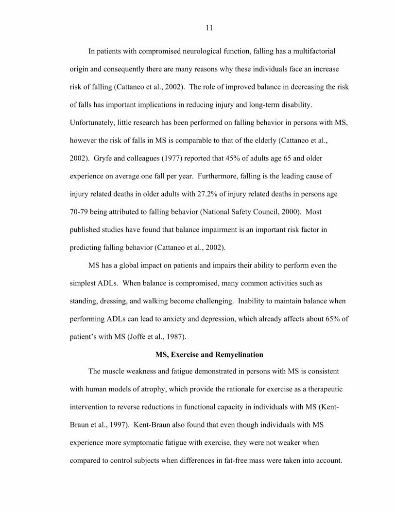
• The self-selected (E) stance – feet apart at a self selected distance (See Figure 1). Distance between the toes and heels were measured.
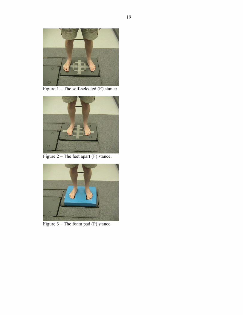
• The feet apart (F) stance – feet 15.2 cm (6 in.) apart (See Figure 2)
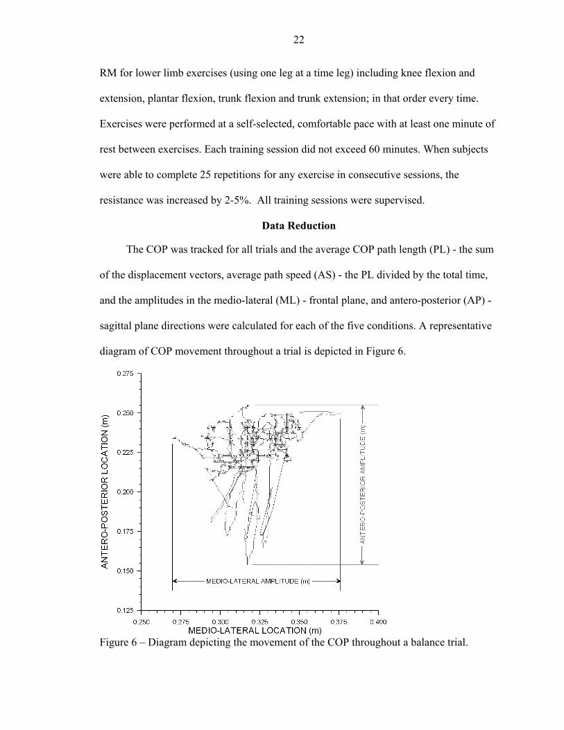
• The foam pad (P) stance – feet 15.2 cm (6 in.) apart on a foam balance pad; to simulate altered support (See Figure 3).
• The semitandem (S) stance – feet 15.2 cm (6 in.) apart and the heel of their dominant leg in line with the toe of their non-dominant leg (See Figure 4a and 4b)
• The tandem (T) stance – feet inline heel-to-toe and the dominant limb in front. (See Figure 5a and 5b)
19
Figure 1 – The self-selected (E) stance.
Figure 2 – The feet apart (F) stance.
Figure 3 – The foam pad (P) stance.

20
A B
Figure 4 – The semitandem (S) seen from a A) frontal view and B) sagittal view.
A B
Figure 5 – The tandem (T) stance seen from a A) frontal view and B) sagittal view.
Strength Testing
The subjects were tested for isometric strength prior to and following an 8-week
study period. The muscle groups and corresponding joint angles are depicted in Table 1.
The subjects were asked to contract their muscles to attempt to produce maximal force.
Muscle Group Tested Exercise Joint Angle Quadriceps Knee Extension Knee Angle = 90 Hamstrings Knee Flexion Knee Angle = 90 Ankle Plantarflexors Plantarflexion Ankle Angle = 0 (neutral) Ankle Dorsiflexors Dorsiflexion Ankle Angle = 0 (neutral) Table 1 – Muscle groups being tested, the movement they produce, and the
corresponding joint angles.
To normalize the force measurement to leg length, the highest force (F) reading
was multiplied by the moment arm (r) to determine the maximum torque (T) produced.
T = F * r

21
Functional Tests
Functional tests were also performed prior to and following the strength-training
program. These tests included a 100 ft. walk test and a 3-min step test. For the walk test,
subjects were asked to walk a distance of 100 ft as quickly and as safely as possible. The
time taken to complete the walk was recorded. For the step test, subjects were asked to
step up onto a platform 15.2 cm (6 in.) above the ground with both legs as many times as
possible in a 3-min period and total number of steps were recorded. Subjects were
allowed any assistance necessary to complete the step test.
Resistance Training
During the next eight weeks of the study period, MS and non-MS control subjects
were asked to visit the Center for Exercise Science or Living Well Fitness and Wellness
Center twice a week, either Monday/Thursday or Tuesday/Friday sessions to perform
resistance training exercises. Exercises were performed under the supervision of staff
trained in cardiopulmonary resuscitation, emergency procedures, and proper exercise
safety for individuals with disabilities. A training protocol was established using
recognized criteria for load assignment in older/disabled persons (ACSM, 2000).
During the first training session, subjects were asked to lift a submaximal load until
they could no longer complete a full repetition for each exercise (2-20 repetitions). A
predicted 1-repitition maximum (1-RM) was determined using the Kuramoto and Payne
(1996) prediction equation for older women. During the second training session, subjects
performed one set of 6-10 repetitions at 50% of the predicted 1-RM. In subsequent
sessions, subjects completed one warm-up set and one training set for each exercise.
Their warm-up consisted of five repetitions at 40% of the predicted 1-RM on each of the
weight-machines. The training set consisted of 10-15 repetitions at 70% of predicted 1-
22
RM for lower limb exercises (using one leg at a time leg) including knee flexion and
extension, plantar flexion, trunk flexion and trunk extension; in that order every time.
Exercises were performed at a self-selected, comfortable pace with at least one minute of
rest between exercises. Each training session did not exceed 60 minutes. When subjects
were able to complete 25 repetitions for any exercise in consecutive sessions, the
resistance was increased by 2-5%. All training sessions were supervised.
Data Reduction
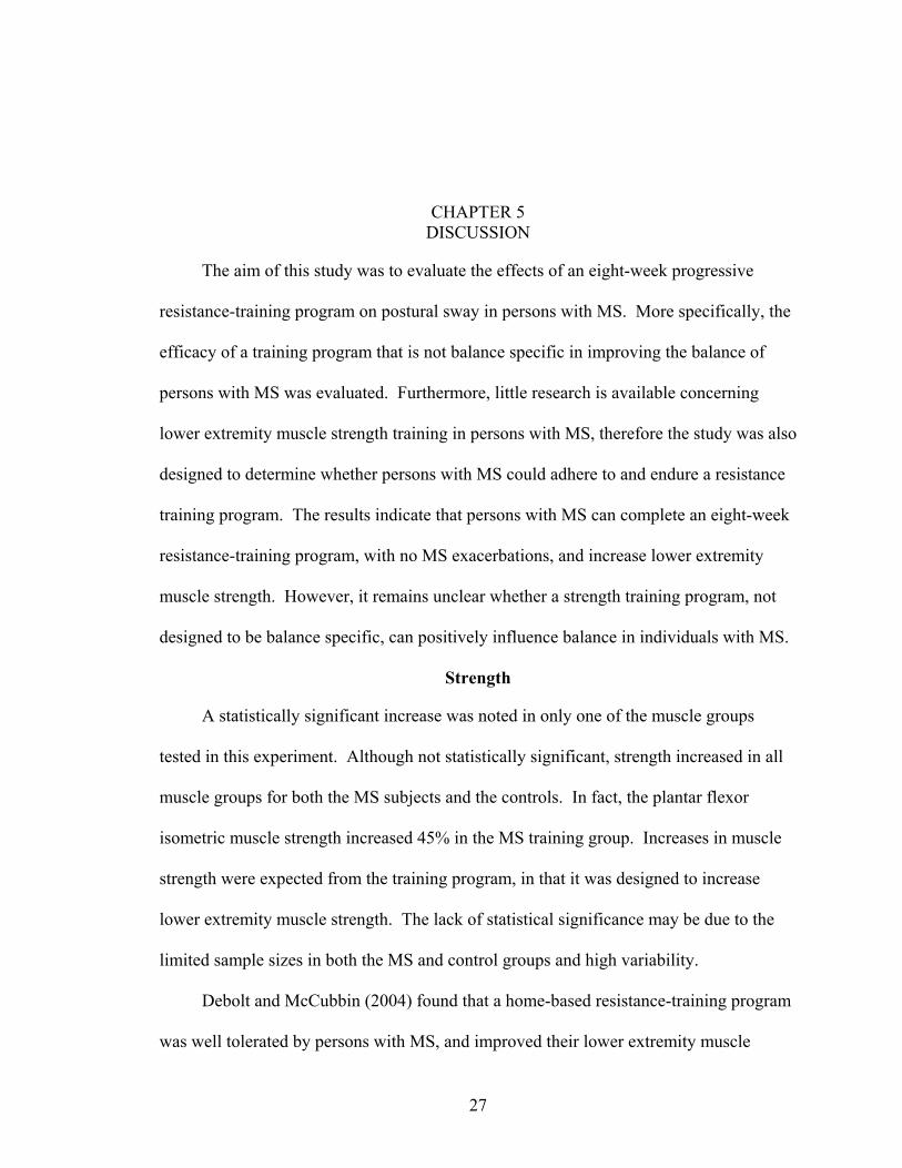
The COP was tracked for all trials and the average COP path length (PL) - the sum
of the displacement vectors, average path speed (AS) - the PL divided by the total time,
and the amplitudes in the medio-lateral (ML) - frontal plane, and antero-posterior (AP) -
sagittal plane directions were calculated for each of the five conditions. A representative
diagram of COP movement throughout a trial is depicted in Figure 6.
Figure 6 – Diagram depicting the movement of the COP throughout a balance trial.

23
Design/Analysis
This study was a pretest-posttest control group design. Descriptive statistics
(means and standard deviations) were calculated for each of the four dependent variables
(total sway path length, average sway speed, and sway amplitude in the AP and ML
directions) in each of the five stance conditions. Due to the small sample size,
nonparametric Wilcoxon signed ranks tests were performed to determine if any changes
occurred in any balance and/or strength measures following eight weeks of strength
training. Descriptive statistics were calculated for the functional tests, however no
statistical tests were performed on the data. All statistical tests were conducted with the
conventional level of significance, α=.05.

CHAPTER 4 RESULTS
All subjects completed the eight-week resistance-training program (16 sessions)
with no MS-related exacerbations reported. The protocol was occasionally adjusted when
subjects missed days between workouts for personal reasons, although adherence
remained at 100%.
Strength
Eight of the nine MS subjects were tested for strength prior to and following the
strength-training program. One subject was unable to produce muscular force from
several lower extremity muscle groups at the time of the day the pre-testing took place,
therefore he was excluded from the isometric strength analysis. The MS training group
significantly increased strength in the knee flexors (p<0.05). Although not statistically
significant, all other muscle groups also increased isometric strength (Table 2).
Stance Pre Post % Change p-value Knee Extension 66.8 ± 29.5 81.6 ± 38.7 + 22.17 % 0.069 Knee Flexion 34.9 ± 17.2 42.7 ± 14.4 + 22.02 % 0.012* Plantar Flexion 45.6 ± 28.9 68.2 ± 33.5 + 49.54 % 0.069 Dorsiflexion 25.7 ± 10.8 28.2 ± 9.5 + 9.89 % 0.484 Table 2 – Strength measures for the MS training group (mean ± SD). All strength
(torque) measures in Nm. * denotes p<0.05. Stance Pre Post % Change p-value Knee Extension 94.4 ± 24.5 112.5 ±30.3 + 19.15 % 0.068 Knee Flexion 43.5 ± 9.8 50.7 ± 18.3 + 16.54 % 0.144 Plantar Flexion 71.1 ± 24.7 92.5 ± 47.6 + 30.13 % 0.144 Dorsiflexion 42.7 ± 9.7 45.1 ± 10.7 + 5.45 % 0.144 Table 3 – Strength measures in the non –MS control training group (mean ± SD). All
strength (torque) measures in Nm.
24

25
The non-MS control training subjects displayed increases in isometric muscle
strength similar to those seen in the MS group, although again not statistically significant
(Table 3).
Balance
Several of the MS subjects could not complete certain stances for the entire 20
seconds, primarily the more difficult stances, such as the T and P stances. However,
subjects that did require assistance required a similar amount of assistance in the pre-test
and post-test. In the MS subjects, a significant increase was noted in the path length and
average speed for the E stance (p=0.028), however none of the other dependent variables
for any of the other stances changed significantly (Table 4). Furthermore, the control
subjects did not significantly change any of the dependent balance variables evaluated
(Table 5).
Stance PL pre
PL post
p AP pre
AP post
p ML pre
ML post
p
E 0.780 1.068 0.028* 0.041 0.046 0.139 0.026 0.032 0.214 F 0.841 1.001 0.066 0.051 0.050 0.767 0.033 0.036 0.374 P 1.109 1.297 0.374 0.084 0.071 0.859 0.062 0.057 0.859 S 1.100 1.234 0.441 0.054 0.044 0.374 0.067 0.045 0.767 T 1.305 1.329 0.953 0.062 0.069 0.260 0.047 0.038 0.314 Table 4 – Mean balance measures for the MS training group. All balance measures in m.
* denotes p<0.05. Stance PL
pre PL post
p AP pre
AP post
p ML pre
ML post
p
E 0.395 0.681 0.068 0.018 0.019 0.715 0.009 0.008 0.715 F 0.416 0.663 0.144 0.020 0.016 0.144 0.009 0.012 1.000 P 0.608 0.813 0.144 0.046 0.042 0.144 0.030 0.025 0.273 S 0.545 0.766 0.144 0.028 0.022 0.144 0.029 0.025 0.144 T 0.829 0.911 0.465 0.044 0.025 0.144 0.038 0.034 0.715 Table 5 – Mean balance measures for the Control training group. All balance measures
in m.

26
Functional Tests
Prior to strength training the MS group was able to complete the 100 ft walk in an
average time of 33.9 s and following training that time decreased to 31.5 s. The control
group was able to complete the walk in 14.3 s, which decreased to 13.8 s after training.
The MS group completed 58.1 steps in the 3-min period prior to training, which increased
to 68.2 following training. The control group began the training able to step an average
of 111.6, which increased to 127.3 after training.
CHAPTER 5 DISCUSSION
The aim of this study was to evaluate the effects of an eight-week progressive
resistance-training program on postural sway in persons with MS. More specifically, the
efficacy of a training program that is not balance specific in improving the balance of
persons with MS was evaluated. Furthermore, little research is available concerning
lower extremity muscle strength training in persons with MS, therefore the study was also
designed to determine whether persons with MS could adhere to and endure a resistance
training program. The results indicate that persons with MS can complete an eight-week
resistance-training program, with no MS exacerbations, and increase lower extremity
muscle strength. However, it remains unclear whether a strength training program, not
designed to be balance specific, can positively influence balance in individuals with MS.
Strength
A statistically significant increase was noted in only one of the muscle groups
tested in this experiment. Although not statistically significant, strength increased in all
muscle groups for both the MS subjects and the controls. In fact, the plantar flexor
isometric muscle strength increased 45% in the MS training group. Increases in muscle
strength were expected from the training program, in that it was designed to increase
lower extremity muscle strength. The lack of statistical significance may be due to the
limited sample sizes in both the MS and control groups and high variability.
Debolt and McCubbin (2004) found that a home-based resistance-training program
was well tolerated by persons with MS, and improved their lower extremity muscle
27

28
power. Furthermore, Kraft et al. (1996a & 1996b) resistance trained arms and legs in MS
subjects for eight weeks and also found improvements in strength, along with improved
function and psychosocial well-being. Most recently, White et al. (2004) found increased
strength and function, along with a decrease in daily fatigue after eight weeks of lower
extremity strength training. These studies, along with the findings of this research
support the practicality of a strength-training program as a viable means to increase
strength in individuals with MS.
Increased strength is desirable in this population because they are often faced with
an increased level of fatigue, which decreases their daily activity levels, and eventually
causes muscle atrophy. An increase in strength due to strength training may help to
counteract the atrophic changes noted in the musculature of individuals with MS, and
perhaps increase their daily activity levels. Furthermore, it is known that the first
neuromuscular adaptations to strength training are more neural than muscular. Positive
neural changes are especially important in a population afflicted with a neurological
disorder. Neural recruitment gained through physical activity may have a favorable
functional outcome, although this may be limited by the severity the MS lesions already
present. This suggests that resistance training may be an early intervention strategy in
persons with MS that may help to maintain function and hopefully, limits exacerbation of
MS symptoms. In fact, in all research previously mentioned concerning strength training
in individuals with MS, no MS related exacerbations were reported and there were no
reports of increased MS-related symptoms (Kraft et al., 1996a & 1996b; Debolt and
McCubbin, 2004; White et al. 2004).

29
Strength training is known to have many benefits, including, but not limited to,
increasing bone mineral density (Asikainen et al., 2004). Since most individuals who
suffer from MS are female, and females are at a higher risk of osteoporosis, strength
training to increase bone mineral density may have profound effects on the quality of life
of these individuals as the age. Furthermore, the performance of the subjects in the
functional tests also lends itself to supporting strength training in this population. All
subjects were able to walk faster and step more following training. This should be
expected from a strength-training program designed to enhance muscle strength and
endurance.
Balance
Decreased ability to maintain balance is a concern in individuals with MS, which
may lead to an increased susceptibility to falls. For this reason, an intervention strategy
to improve balance is desirable for individuals with MS. This study was intended to
determine if static balance could be improved with a training program that is not balance
specific. As stated earlier, the training protocol in this study was designed to increase
lower extremity muscle strength. A significant increase was noted in the strength of knee
flexors after strength training, and the knee extensors and plantar flexors also tended to
be stronger after the strength training, however only two measures of postural sway were
significantly different following training, and that change represented a decrease in
postural stability. The results suggest that strength training has little effect on postural
sway in persons with MS or control subjects.
The MS subjects who participated in this study represented a broad spectrum of
disability levels, which is common for a condition like MS. Some subjects had little or
no visible or obvious disability, while others required assistance to complete the tests of

30
standing balance. For those who did require assistance to complete the tests of standing
balance, the amount of assistance required did not change for between the pre and post-
tests. Furthermore, the data indicate that the strength training did not improve postural
sway characteristics in these subjects. This could be due to a couple of different possible
explanations: 1) the subjects were perhaps too disabled, more specifically, their loss of
function was already too extensive, to have dramatic improvements in just eight weeks,
or more likely 2) the training was not specific enough to the stances studied to cause
positive alterations in postural sway. Even though strength did increase in these subjects,
that increase did not result in improvements in static balance.
The finding that increased strength does not significantly influence balance is
supported by the work of Katayama et al. (2004), who found that knee and toe muscle
power does not appear to be a dominant factor in maintaining balance. This corroborates
the assertion that lower extremity muscle strength training may not have a significant
influence on postural sway. On the other hand, Judge and colleagues (1993) found that
an exercise program emphasizing postural control, moderate resistance training and
walking improved single leg static balance in neurologically intact elderly individuals,
however double leg static balance measures did not improve. Two important conclusions
can be dawn from this work: 1) a training intervention intended to improve balance
should be focused on training for balance, and 2) double leg static stances may not be
sufficiently challenging to unimpaired individuals to show significant changes after any
training program. Although most subjects in this study were impaired in some way, some
of the MS subjects had no obvious disability and may not have been challenged enough
with the stances tested to change postural sway characteristics significantly. This
31
assertion is supported by the performance of the control group, who showed no
significant changes in any of the balance measures tested.
Limitations
There are limitations in the experimental design that may account for the lack of
significant changes in postural sway characteristics. As stated earlier, eight weeks may
not have been a sufficient amount of time to significantly influence balance. Therefore, a
more elongated and extensive strength-training program may have elicited more
significant responses. Furthermore, a larger subject pool may help eliminate some of the
variability, which could account for the lack of statistically significant differences. With
such a small sample size, even a small amount of variability would eliminate statistical
significance. The strength gains should be interpreted cautiously because the training
was isotonic and the testing was isometric, so strength gains noted in this work may not
be clinically applicable. Another possible origin of variability is the change in motion
analysis systems used to collect the postural sway data midway through the study
protocol. Unfortunately, several subjects were pre and post-tested on different motion
analysis systems, therefore some inherent variability between the two systems may have
changed the final data enough to account for the lack of statistical significance.
Additional work with larger sample sizes, longer training protocol, more intense training,
and more balance specific training is desirable and could lead to promising intervention
strategies to improve balance and reduce the risk of falls in individuals with MS.
Summary and Conclusions
This study was designed to determine the efficacy of a progressive resistance-
training program on postural sway in persons with MS. The training program was not
intended to be balance specific. It was designed to focus on increasing general lower
32
extremity muscle strength. Only two out of 20 postural sway characteristics evaluated
significantly changed following strength training in the MS group. Strength increased
significantly in the knee flexors and tended to increase for the knee extensors and plantar
flexors. It appears that the increased strength in the lower extremity may not influence
static balance in individuals with MS. Additional research with larger sample sizes for
both groups, and increased duration and/or intensity of training is recommended. A
training program designed to focus specifically on balance would potentially demonstrate
more significant changes in balance and could present a promising intervention strategy
to improve balance and reduce the risk of falls in persons with MS, or any other
neurological disorder.

APPENDIX A INFORMED CONSENT
IRB# 340-2002
Informed Consent to Participate in Research
You are being asked to take part in a research study. This form provides you with information about the study. The Principal Investigator (the person in charge of this research) or a representative of the Principal Investigator will also describe this study to you and answer all of your questions. Before you decide whether or not to take part, read the information below and ask questions about anything you do not understand. Your participation is entirely voluntary. 1. Name of Participant ("Study Subject") _____________________________________________________________________ 2. Title of Research Study Resistance Training Effects on Muscle Function in Multiple Sclerosis 3. Principal Investigator and Telephone Number(s) Lesley J. White, Ph.D., Assistant Professor Department of Exercise and Sport Sciences College of Health and Human Performance University of Florida (352) 392-9575 ext. 1338 Email: [email protected]
33

34
John Chow, Ph.D., Associate Professor Department of Exercise and Sport Sciences College of Health and Human Performance University of Florida (352) 392-0584 ext. 1263 Anne L. Rottmann, M.D., Neurologist 4410 West Newberry Rd. Suite A3 Gainesville, FL 32607 After Hours/Emergency Telephone Number: (352) 374-2222 4. Source of Funding or Other Material Support National Multiple Sclerosis Society What is the purpose of this research study? The primary purpose of this study is to determine whether a sixteen-week progressive resistance training exercise program influences measures of your muscle’s performance and your ability to walk and balance more effectively. This study is part of a project to learn more about your muscles ability to become more effective in producing energy during activities after you exercise train. We plan to measure your walking mechanics and your balance before and after you exercise train. We will also get pictures of your leg muscles using a technique called magnetic resonance imaging (MRI). Then we can get information about the chemistry of your muscle using a technique called magnetic resonance spectroscopy (MRS). 6. What will be done if you take part in this research study?
If you volunteer for this study you will be asked to participate in a 21 week experimental period that consists of evaluation of several functional measures such as muscle strength, balance, and walking mechanics, followed by a sixteen week exercise program. Midpoint evaluation will occur after 8 weeks of resistance training. Follow-up evaluation will occur after sixteen weeks of exercise training program. Listed below are descriptions of each visit should you choose to participate in this study. Visit #1
On the first visit of week one of the study, you will be familiarized with the experimental protocol and be examined by a neurologist. Your disability status and physical readiness to participate in an exercise protocol will be assessed. If you are cleared to participate in this study, you will be asked to read and sign an informed consent, which will inform you of all the risks/benefits of participation in the experiment. The approximate time required for this visit will be 60 minutes. Visit #2
35
On the second visit of week 1, you will have an MRI/MRS performed on your legs. During the MRI/MRS you will lie on a bed, which rolls in the opening of a large magnet. A flat coil of wire (a radiofrequency coil) will be placed on your thigh and calf. A computer will look at the radio waves passing through your leg and constructs pictures and chemical information of your muscles. The total procedure will last approximately 45 minutes. An MRI/MRS is used routinely to detect structures or gain chemical information about the muscles of healthy subjects or hospital patients. Just prior to and following the MRI, we will draw 20ml of blood (about 4 teaspoons) by venipuncture to test levels of blood sugar, triglycerides, and cholesterol. This will take an additional fifteen minutes. The total time of this visit, including MRI/MRS and blood sampling, will take approximately 60 minutes. Visit #3
During the third visit in week 2 of the experimental period, you will have your body composition assessed using a three-site skinfold technique, and measurements of the waist and hip circumference will be taken. You will then be asked to perform muscular strength and endurance tests on a Kin-Com isokinetic dynamometer for the following exercises: abdominals, back, leg extensions, leg curls, and ankle flexion exercises. The Kin-Com is a muscle testing machine commonly used for evaluating muscle function in healthcare settings. During the testing, you will be asked to be seated on the machine and you will be asked to perform a muscle contraction at a constant, predetermined speed. The discomfort associated with this procedure is minimal, but will require you to put forth a strong effort. In conjunction with the muscle testing, two self-adhesive electrodes (2” x 4”) will be placed on your thigh close to your knee and hip. During your knee extension strength evaluation an electrical pulse will be delivered to your thigh muscle. The level of stimulation should not be painful, though it may cause a prickly sensation on your skin and will make your muscle feel like it is being squeezed. The feeling will be very similar to what you experience when you climb stairs or ride a bicycle for an extended period of time. The procedure is used to evaluate the ability of your muscle to generate force during the strength testing. The strength testing will be used to determine your 10-repetition maximum, which will be used in the training aspect of the study. Testing is expected to take 60 minutes. Visit #4
During the fourth visit, in week 2 of the experimental period, you will be asked to perform tests of balance, and gait (walking mechanics). For the balance test, you will be asked to stand on a force plate without support for 20 seconds. You will be asked to repeat this test 3 times. Balance tests will be completed with your eyes open and closed. A test of your functional reach will be completed after a brief rest period following the balance test. During the functional reach test you will reach forward as far as you can until your heels come off the floor. A safety harness will be used to ensure your safety during functional tests. The harness may cause minor skin irritation. For the gait analysis you will be asked to walk on an 8-meter walkway twice. The gait testing will require you to have special sensors and reflective markers placed on your lower back and legs. The special sensors will measure your muscle activity during walking. Your skin preparation will include shaving areas displaying body hair with an electric razor that causes no skin

36
irritation. Your skin will then be sanitized with an alcohol swab. There is not any expected irritation or discomfort associated with the shaving or alcohol swabbing. Testing will also consist of a timed 25-foot walk test where you will be asked to walk as fast as possible within your comfort. Lastly, you will be asked to perform a three-minute step test where you will be asked to step up onto a platform as many times as possible in the allotted three minutes. During this visit we will ask you to wear shorts and exercise shoes. These tests will be conducted in the Biomechanics Laboratory in the Center for Exercise Science and will take will take approximately 60 minutes to complete. Visits #5 and #6
During the fifth and sixth visits, in week 3 of the experimental period you will be asked to repeat all the testing performed in week 2. This is designed to more accurately quantify baseline measures. These visits will each take approximately 60 minutes to complete. Visits #7 through #22
During the next eight weeks of the study period (weeks 4-11) you will be asked to visit the Center for Exercise Science or Living Well Fitness and Wellness Center, or North Florida Regional YMCA twice a week, either Monday/Thursday or Tuesday/Friday sessions to perform resistance training exercises. Twenty milliliters (4 teaspoons) of blood will be drawn before and after exercise to determine the effect of training on glucose, triglycerides, and cholesterol. Subjects will be required to attend strength training sessions requiring blood draws at the Center for Exercise Science. All training will occur in a supervised exercise environment with staff trained in cardiopulmonary resuscitation and emergency procedures. The staff will also be trained in proper exercise safety for individuals with disabilities.
During each exercise session you will be asked to perform one set of 15-20 repetitions at 70% of your 10-repetition maximum (RM) for each of the following exercises: leg extensions, leg curls, ankle plantarflexion, and an abdominal/lower back regimen using free-weight machines or a Kin-com isokinetic dynamometer. This is not a maximal exercise protocol and is not designed to be exhaustive. When you are able to complete 15 repetitions with proper technique, for two consecutive sessions, the resistance will be increased by 10% of your 10-RM. To assess your level of fatigue you will be asked to rate your level of effort (perceived exertion) at the end of each exercise. Because of the functional variability of MS individuals, the protocol may be adjusted on an individual basis to maintain your comfort and safety. A Certified Strength and Conditioning Specialist (CSCS) or exercise physiology research assistant will supervise all training sessions. Each training session will last 30-45 minutes.
You will be asked to perform a self-reported dietary recall at weeks 2, 4, 8, 12, and 16 of the experimental period when you come to the laboratory for exercise training. You will be asked to follow the dietary guideline established by the American Heart Association for the duration of the study to ensure that the appropriate amounts of nutrients are being consumed to meet the nutritional needs associated with a strength-training program. These guidelines are similar to those suggested for the MS population.

37
You will also be asked to complete a functional independence measure at weeks 1, 5, 7, 9, 11, 16, and 20. We will also randomly ask you to wear an accelerometer throughout one day during weeks 2, 12, and 20 to assess your level of activity. The accelerometer is a small device that will be worn around the waist to measure your activity throughout the day. Visit #23 (Midpoint evaluation)
On the first visit of week 12 of the study period, following first phase of the exercise training period, you will be asked to perform all testing measures as performed at the beginning of the study. Again, you will be asked to perform self-reported questionnaires examining functional independence and fatigue impact. The completion of these questionnaires will take approximately 10 minutes. Following questionnaire completion, your body composition will be reassessed via the three-site skinfold technique, and measurements of the waist and hip will be taken. Twenty milliliters (about four teaspoons) of blood will be taken before and after exercise to determine if any changes in your resting levels of blood glucose, triglycerides, and cholesterol occurred. Again, these factors will be examined for experimental use and for your own personal information. This portion of the study will take 45 minutes. The total time for this visit will be approximately 90 minutes. Visit #24
During the second visit of week 12 of the study period, you will be asked to perform tests of muscle strength and endurance on a Kin-com isokinetic dynamometer with electrical stimulation. This testing will follow the same procedure and include the same exercises as was performed in weeks 2 and 3 of the study. Testing will take approximately 60 minutes. Visit #25
During the first, and only, visit of week 13 of the study, you will be asked to perform follow-up tests of balance, and gait. Testing will also consist of a timed 25-foot walk and the three-minute step test. These tests will again be conducted in the Center for Exercise Science Biomechanics Laboratory and will take approximately 60 minutes to complete. Visit #26-42
During the next eight weeks of the study period (weeks 13-21) you will be asked to continue twice weekly exercise training sessions until you have completed 16 consecutive weeks of training. Twenty milliliters (4 teaspoons) of blood will be drawn before and after exercise at week 20 to determine the effect of training on glucose, triglycerides, and cholesterol. Any special needs that you require will be accommodated throughout the duration of the study. All data collected in the post-testing phase will be compared to the information gathered in the pre-testing to determine if any improvements were made in any of the factors that were studied in this experiment. 7. What are the possible discomforts and risks?

38
The risks of drawing blood from a vein include discomfort at the site of puncture;
possible bruising and swelling around the puncture site; rarely an infection; and, uncommonly, faintness from the procedure. The risk from having a catheter inserted into an arm vein for time needed in this study is possible infection of the vein, but the risk is very low because we will have a trained phlebotomist collecting the blood samples. The amount of blood we will take should have no negative effects. You will be closely watched for any possible ill effects.
There is also a risk of mild muscle soreness associated with the initiation of any strength-training program, however this risk is minimal. Potential soreness may last for three days but is not expected to limit any activities. There is also a slight risk of skin irritation associated with electrode and reflective marker placement for the electromyographic analysis. Balance and gait testing with the safety harness may cause some mild skin irritation. There is also a risk of skin irritation associated with the electrical pulses to your thigh, however, the equipment used is highly reliable with safety features to minimize pulse strength. The short duration pulses may contribute to mild muscle soreness, however the risk is minimal. The risks of MRI/MRS are: the MRI/MRS scanner contains a very strong magnet. Therefore, you may not be able to have the MRI/MRS if you have any type of metal implanted in your body, for example, any pacing device (such as a heart pacer), any metal in your eyes, or certain types of heart valves or brain aneurysm clips. Someone will ask you questions about this before you have the MRI/MRS. There is not much room inside the MRI/MRS scanner. You may be uncomfortable if you do not like to be in close spaces ("claustrophobia"). During this procedure, you will be able to talk with the MRI/MRS staff through a speaker system, and in the event of an emergency, you can tell them to stop the scan.
The MRI/MRS scanner produces a loud hammering noise, which has produced hearing loss in a very small number of patients. You will be given earplugs to reduce this risk.
If you are a woman of childbearing potential, there may be unknown risks to the fetus. Therefore, before you can have the MRI/MRS, you must have a pregnancy test. This test will be done at no charge. 8a. What are the possible benefits to you?
It is possible that you may experience improvements in gait, balance, muscular strength, and endurance. You will also receive eight weeks of personal exercise training. Blood cholesterol, nutritional profile and analysis, will be provided for your own personal records. 8b. What are the possible benefits to others?

39
Research findings from this study may help in the design and use of therapeutic
exercises designed to help other individuals with MS. 9. If you choose to take part in this research study, will it cost you anything?
All costs associated with the assessment of your percent body fat, gait analysis, and quality of your diet will be paid for by the Center for Exercise and Sport Sciences. Additional measurements of blood cholesterol, glucose, and insulin levels will be absorbed by funding supporting the principal investigators of the study. The principal investigator will also pay for the MRI/MRS and any required pregnancy test. 10. Will you receive compensation for taking part in this research study?
At the completion of the study, subjects with multiple sclerosis will receive $200.00. Subjects who do not have multiple sclerosis will not receive monetary compensation for study participation. 11. What if you are injured because of the study?
If you experience any injury that is directly caused by this study, only professional consultative care that you receive at the University of Florida Health Science Center will be provided without charge. However, hospital expenses will have to be paid by you or your insurance provider. No other compensation will be offered. 12. What other options or treatments are available if you do not want to be in this study?
The exercise training and dietary counseling are in addition to standard therapy for your condition. You may choose to continue with your current therapy. 13a. Can you withdraw from this research study?
You are free to withdraw your consent and to stop participating in this research study at any time. If you do withdraw your consent, there will be no penalty, and you will not lose any benefits you are entitled to.
If you decide to withdraw your consent to participate in this research study for any reason, you should contact Dr. Lesley White at (352) 392-9575 ext 1338.
If you have any questions regarding your rights as a research subject, you may phone the Institutional Review Board (IRB) office at (352) 846-1494.
40
13b. If you withdraw, can information about you still be used and/or collected?
If you withdraw from the study, information about you will not be used or collected any further. 13c. Can the Principal Investigator withdraw you from this research study?
You may be withdrawn from the study without your consent for the following reasons: failure to make scheduled training visits and cardiovascular risk factors contraindicating your participation in a strength training program. 14. How will your privacy and the confidentiality of your research records be protected?
Authorized persons from the University of Florida, the hospital or clinic (if any) involved in this research, and the Institutional Review Board have the legal right to review your research records and will protect the confidentiality of them to the extent permitted by law. Otherwise, your research records will not be released without your consent unless required by law or a court order. If the results of this research are published or presented at scientific meetings, your identity will not be disclosed. 15. How will the researcher(s) benefit from your being in this study?
In general, presenting research results helps the career of a scientist. Therefore, the Principal Investigators may benefit if the results of this study are presented at scientific meetings or in scientific journals.
41
16. Signatures
As a representative of this study, I have explained to the participant the purpose, the procedures, the possible benefits, and the risks of this research study, the alternatives to being in the study, and how privacy will be protected: ___________________________________________ _____________________ Signature of Person Obtaining Consent Date
You have been informed about this study’s purpose, procedures, possible benefits, and risks; the alternatives to being in the study; and how your privacy will be protected. You have received a copy of this Form. You have been given the opportunity to ask questions before you sign, and you have been told that you can ask other questions at any time.
You voluntarily agree to participate in this study. By signing this form, you are not waiving any of your legal rights. ____________________________________________ _____________________ Signature of Person Consenting Date
42
Consent to be Videotaped and to Different Uses of the Videotape(s)
With your permission, you will be videotaped during this research. Your name or personal information will not be recorded on the videotape, and confidentiality will be strictly maintained. When these videotapes are shown, however, others may be able to recognize you.
The Co-Principal Investigator of this study, John Chow, Ph.D, will keep the videotape(s) in a locked cabinet. These videotapes will be shown under his direction to students, researchers, doctors, or other professionals and persons.
Please sign one of the following statements that indicates under what conditions Dr. John Chow, Ph.D has your permission to use the videotape.
I give my permission to be videotaped solely for this research project under the conditions described. ______________________________Signature ____________________________Date
I give my permission to be videotaped for this research project, as described in the Informed Consent Form, and for the purposes of education at the University of Florida Health Science Center ______________________________Signature ____________________________Date
I give my permission to be videotaped for this research project, as described in the Informed Consent Form; for the purposes of education at the University of Florida Health Science Center; and for presentations at scientific meetings outside the University. ______________________________Signature _____________________________Date

APPENDIX B EXPANDED DISABILITY STATUS SCALE
The EDSS quantifies disability in eight Functional Systems (FS) and allows
neurologists to assign a Functional System Score (FSS) in each of these systems:
• pyramidal • cerebellar • brainstem • sensory • bowel and bladder • visual • cerebral • other
EDSS steps 1.0 to 4.5 refer to people with MS who are fully ambulatory, while
steps 5.0 to 9.5 are defined by the impairment to ambulation.
Kurtzke Expanded Disability Status Scale 0.0 Normal neurological examination 1.0 No disability, minimal signs in one FS 1.5 No disability, minimal signs in more than one FS 2.0 Minimal disability in one FS 2.5 Mild disability in one FS or minimal disability in two FS 3.0 Moderate disability in one FS, or mild disability in three or four FS. Fully
ambulatory 3.5 Fully ambulatory but with moderate disability in one FS and more than minimal
disability in several others 4.0 Fully ambulatory without aid, self-sufficient, up and about some 12 hours a day
despite relatively severe disability; able to walk without aid or rest some 500 meters4.5 Fully ambulatory without aid, up and about much of the day, able to work a full day,
may otherwise have some limitation of full activity or require minimal assistance; characterized by relatively severe disability; able to walk without aid or rest some 300 meters.
5.0 Ambulatory without aid or rest for about 200 meters; disability severe enough to
43

44
impair full daily activities (work a full day without special provisions) 5.5 Ambulatory without aid or rest for about 100 meters; disability severe enough to
preclude full daily activities 6.0 Intermittent or unilateral constant assistance (cane, crutch, brace) required to walk
about 100 meters with or without resting 6.5 Constant bilateral assistance (canes, crutches, braces) required to walk about 20
meters without resting 7.0 Unable to walk beyond approximately five meters even with aid, essentially
restricted to wheelchair; wheels self in standard wheelchair and transfers alone; up and about in wheelchair some 12 hours a day
7.5 Unable to take more than a few steps; restricted to wheelchair; may need aid in transfer; wheels self but cannot carry on in standard wheelchair a full day; May require motorized wheelchair
8.0 Essentially restricted to bed or chair or perambulated in wheelchair, but may be out of bed itself much of the day; retains many self-care functions; generally has effective use of arms
8.5 Essentially restricted to bed much of day; has some effective use of arms retains some self care functions
9.0 Confined to bed; can still communicate and eat. 9.5 Totally helpless bed patient; unable to communicate effectively or eat/swallow 10.0 Death due to MS

LIST OF REFERENCES
Armstrong LE, Winant DM, Swasey PR, Seidle ME, Carter AL, Gehlsen G. Using Isokinetic Dynamometry to Test Ambulatory Patients with Multiple Sclerosis. Physical Therapy. Aug 1983, 63(8): 1274-9.
Asikainen TM, Kukkonen-Harjula K, Miilunpalo S. Exercise for Health for Early Postmenopausal Women: a Systematic Review of Randomised Controlled Trials. Sports Medicine. Nov 2004, 34(11): 753-78.
American College of Sports Medicine (ACSM). ACSM's Guidelines for Exercise Testing and Prescription. 6th Edition. Philadelphia (PA): Lippincott, Williams & Wilkins, 2000.
Bunge MB, Bunge RP, Ris H. Ultrastructural Study of Remyelination in an Experimental Lesion in Adult Cat Spinal Cord. Journal of Biophysical and Biochemical Cytology. 1961, 10: 67-94.
Cattaneo D, DeNuzzo C, Fascia T, Macalli M, Pisoni I, Cardini R. Risk of Falls in Subjects with Multiple Sclerosis. Archives of Physical Medicine and Rehabilitation. Jun 2002, 83(6): 864-7.
Chang A, Tourtellotte WW, Rudick R, Trapp BD. Premyelinating Oligodendrocytes in Chronic Lesions of Multiple Sclerosis. New England Journal of Medicine. Jan 2002, 346(3), 165-73.
Chetlin RD, Gutmann L, Tarnopolsky M, Ullrich IH, Yeater RA. Resistance Training Effectiveness in Patients with Charcot-Marie-Tooth Disease: Recommendations for Exercise Prescription. Archives of Physical Medicine and Rehabilitation. Aug 2004; 85(8): 1217-23.
Coulthard-Morris L. Clinical and Rehabilitation Outcome Measures. In Burks JS and Johnson KP (Eds.), Multiple Sclerosis: Diagnosis, Medical Management, and Rehabilitation. (221-290). New York: Demos Medical Publishing, 2000.
Debolt LS, McCubbin JA. The Effects of Home-Based Resistance Exercise on Balance, Power, and Mobility in Adults with Multiple Sclerosis. Archives of Physical Medicine and Rehabilitation. Feb 2004; 85(2): 290-7.
Era P, Heikkinen E. Postural Sway During Standing and Unexpected Disturbance of Balance in Random Samples of Men of Different Ages. Journal of Gerontology. May 1985; 40(3): 287-95.
45

46
Frzovic D, Morris ME, Vowels L. Clinical Tests of Standing Balance: Performance of Persons with Multiple Sclerosis. Archives of Physical Medicine and Rehabilitation. Feb 2000, 81: 215-21.
Gehlsen GM, Grigsby SA, Winant DM. Effects of an Aquatic Fitness Program on the Muscle Strength and Endurance of Patients with Multiple Sclerosis. Physical Therapy. 1984, 64: 653-7.
Goodkin DE. Treatment of Progressive Forms of Multiple Sclerosis. In Burks JS and Johnson KP (Eds.), Multiple Sclerosis: Diagnosis, Medical Management, and Rehabilitation. (177-200). New York: Demos Medical Publishing, 2000.
Gryfe CI, Amies A, Ashley MJ. A Longitudinal Study of Falls in an Elderly Population: I. Incidence and Morbidity. Age and Ageing. 1977, 6: 201-10.
Herndon, RM. Pathology and Pathophysiology. In Burks JS and Johnson KP (Eds.), Multiple Sclerosis: Diagnosis, Medical Management, and Rehabilitation. (35-45). New York: Demos Medical Publishing, 2000.
Hommes OR. Remyelination in Human CNS Lesions. Progressive Brain Research. 1980, 53: 39-63.
Horak FB. Clinical measurement of postural control in adults. Physical Therapy. Dec 1987; 67(12): 1881-5.
Joffe RT, Lippert GP, Gray TA, Sawa G, Horvath Z. Mood Disorder and Multiple Sclerosis. Archives of Neurology. Apr 1987, 44(4): 376-8.
Judge JO, Lindsey C, Underwood M, Winsemius D, Keshner EA. Balance Improvements in Older Women: effects of exercise training. Physical Therapy. 1993, 73 (4); 254-66.
Kasckow J, Abood LG, Hoss W, Herndon RM. Mechanism of Phospholipase A2-Induced Conduction Block in Bullfrog Sciatic Nerve I: Electrophysiology and Morphology. Brain Research. 1986a, 373: 384-91.
Kasckow J, Abood LG, Hoss W, Herndon RM. Mechanism of Phospholipase A2-Induced Conduction Block in Bullfrog Sciatic Nerve II: Biochemistry. Brain Research. 1986b, 373: 392-8.
Katayama Y, Senda M, Hamada M, Kataoka M, Shintani M, Inoue H. Relationship Between Postural Balance and Knee and Toe Muscle Power in Young Women. Acta Medica Okayama. (2004), 58 (4); 189-95.
Kent-Braun JA, Ng AV, Castro M, Weiner MW, Gelinas D, Dudley GA, Miller RG. Strength, Skeletal Muscle Composition, and Enzyme Activity in Multiple Sclerosis. Journal of Applied Physiology. Dec 1997, 83(6).

47
Kidd PM. Multiple Sclerosis, an Autoimmune Inflammatory Disease: Prospects for its Integrative Management. Alternative Medicine Review. Dec 2001; 6(6): 540-66. Review.
Kirby RL, Price NA, Macleod DA. The Influence of Foot Position on Standing Balance. Journal of Biomechanics. 1987, 20(4): 423-7.
Kraft AM, Wessman HC. Pathology and Etiology in Multiple Sclerosis. Physical Therapy. 1974, 54: 716-20.
Kraft GH, Alquist AD, de Lateur BJ. Effect of Resistive Exercise on Function in Multiple Sclerosis (MS). Archives of Physical Medicine and Rehabilitation. 1996a; 77: 984.
Kraft GH, Alquist AD, de Lateur BJ. Effect of Resistive Exercise on Strength in Multiple Sclerosis (MS). Archives of Physical Medicine and Rehabilitation. 1996b; 77: 984.
Kuramoto AK, Payne VG. Predicting Muscular Strength in Women: a Preliminary Study. Research Quarterly for Exercise and Sport Sciences. 1995; 66: 168-72.
Kurtzke JF. A New Scale for Evaluating Disability in Multiple Sclerosis. Neurology. Aug 1955; 5(8): 580-3.
Kurtzke JF. Rating Neurologic Impairment in Multiple Sclerosis: an Expanded Disability Status Scale (EDSS). Neurology. Nov 1983; 33(11): 1444-52.
Lambert CP, Archer RL, Evans WJ. Muscle Strength and Fatigue During Isokinetic Exercise in Individuals with Multiple Sclerosis. Medicine and Science in Sports and Exercise. Oct 2001, 33(10): 1613-9.
Ludwin SK. Proliferation of Mature Oligodendrocytes After Trauma to the Central Nervous System. Nature. 1984, 308: 274-6.
Mostert S, Kesselring J. Effects of a Short-term Exercise Training Program on Aerobic Fitness, Fatigue, Health Perception, and Activity Level of Subjects with Multiple Sclerosis. Multiple Sclerosis. 2002, 8: 161-8.
National Safety Council. Injury Facts™, 2000 edition. Itasca, IL: National Safety Council, 2000.
Ng AV, Kent-Braun JA. Quantitation of Lower Physical Activity in persons with Multiple Sclerosis. Medicine and Science in Sports and Exercise. Apr 1997, 29(4): 517-23.
Paty DW. Initial Symptoms. In Burks JS and Johnson KP (Ed.), Multiple Sclerosis: Diagnosis, Medical Management, and Rehabilitation. (75-9). New York: Demos Medical Publishing, 2000.
48
Petajan JH, Gappmaier E, White AT, Spencer MK, Mino L, Hicks RW. Impact of Aerobic Training on Fitness and Quality of Life in Multiple Sclerosis. Annals of Neurology. Apr 1996, 39(4): 432-41.
Petajan JH, White AT. Recommendations for Physical Activity in Patients with Multiple Sclerosis. Sports Medicine. Mar 1999, 27(3): 179-91.
Prineas JW. Pathology of the Early Lesions of Multiple Sclerosis. Human Pathology. 1975, 6: 531-5.
Ritchie JM, Rogart RB. Density of Sodium Channels in Mammalian Myelinated Nerve Fibers and Nature of the Axonal Membrane Under the Myelin Sheath. Proceedings of the National Academic Society. 1977, 74: 211-5.
Rogers ME, Fernandez JE, Bohlken RM. Training to Reduce Postural Sway and Increase Functional Reach in the Elderly. Journal of Occupational Rehabilitation. Dec 2001, 11(4): 291-8.
Rogers ME, Rogers NL, Takeshima N, Islam MM. Methods to Assess and Improve the Physical Parameters Associated with Fall Risk in Older Adults. Preventive Medicine. 2003, 36: 255-64.
Smithson F, Morris ME, Iansek R. Performance on Clinical Tests of Balance in Parkinson's Disease. Physical Therapy. Jun 1998; 78(6): 577-92.
Teasdale N, Stelmach GE, Breunig A. Postural Sway Characteristics of the Elderly under Normal and Altered Visual and Support Surface Conditions. Journal of Gerontology. Nov 1991; 46(6): 238-44.
Tillman MD, Chow JW. Applications of Force-Plate Technology. Athletic Therapy Today. Nov 2002, 7(5): 60-1.
Todd G, Gorman RB, Gandevia SC. Measurement and reproducibility of strength and voluntary activation of lower-limb muscles. Muscle Nerve. Jun 2004; 29(6):834-42.
Trapp BD, Peterson J, Ransohoff RM, Rudick R, Mork S, Bo L. Axonal transection in the lesions of multiple sclerosis. New England Journal of Medicine. Jan 1998; 338(5): 278-85.
Waxman SG. Voltage-gated Ion Channels in Axons: Localization, Function, and Development. In Waxman SG, Kocsis JD, Stys PK (Eds.), The Axon. (218-43). New York: Oxford University Press, 1995.
White LJ, McCoy SC, Castellano V, Gutierrez GM, Stevens J, Walter GA, Vandenborne K. Resistance Training Improves Strength and Functional Capacity in Persons with Multiple Sclerosis. Multiple Sclerosis. Dec 2004; 10(6): 668-74.

BIOGRAPHICAL SKETCH
Gregory M. Gutierrez has an innate drive that has allowed him to succeed in many
aspects of his life. His competitive nature has helped him succeed on the field of play,
and in the classroom. He received his bachelor’s degree in exercise and sports sciences
in December of 2002 from the University of Florida. He immediately began his master’s
degree in biomechanics in the same department. Under exceptional guidance, he has
matured in many ways and is ready to pursue a Ph.D. in biomechanics. Gregory’s long-
term goal is to one day become an orthopedic surgeon, while still contributing to research
as a biomechanist.
49