effect of ph values on the extracellular polysaccharide secreted by acidithiobacillus ferrooxidans...
TRANSCRIPT

International Journal of Minerals, Metallurgy and Materials Volume 21, Number 4, April 2014, Page 311 DOI: 10.1007/s12613-014-0910-0
Corresponding author: Wei-min Zeng E-mail: [email protected] © University of Science and Technology Beijing and Springer-Verlag Berlin Heidelberg 2014
Effect of pH values on the extracellular polysaccharide secreted by Acidithiobacillus ferrooxidans during chalcopyrite bioleaching
Run-lan Yu1,2), Jing Liu1,2), Jian-xi Tan3), Wei-min Zeng1,2), Li-juan Shi1,2), Guo-hua Gu1,2), Wen-qing Qin1,2), and Guan-zhou Qiu1,2) 1) School of Minerals Processing and Bioengineering, Central South University, Changsha 410083, China 2) Key Laboratory of Biometallurgy, Ministry of Education, Changsha 410083, China 3) Hunan Entry-Exit Inspection and Quarantine Bureau, Changsha 410004, China. (Received: 28 August 2013; revised: 13 November 2013; accepted: 10 December 2013)
Abstract: The pH value plays an important role in the bioleaching of sulphide minerals. The effect of pH values on the extracellular poly-saccharide secreted by Acidithiobacillus ferrooxidans was investigated in different phases of bacterial growth during chalcopyrite bioleach-ing. It is found that extracellular polysaccharide secretion from the cells attached to chalcopyrite is more efficiently than that of the free cells in the bioleaching solution. Three factors, pH values, the concentration of soluble metal ions, and the bacterial growth and metabolism, affect extracellular polysaccharide secretion in the free cells, and are related to the bacterial growth phase. Extracellular polysaccharide secretion from the attached cells is mainly dependent on the pH value of the bacterial culture.
Keywords: chalcopyrite; bioleaching; polysaccharides; acidity; Acidithiobacillus ferrooxidans
1. Introduction
Chalcopyrite is the most abundant and refractory copper mineral, accounting for about 70% of copper reserves in the world. Heap bioleaching of low-grade copper sulphides has been successfully applied worldwide to extract copper from the secondary sulphide minerals, such as chalcocite. How-ever, the bioleaching of chalcopyrite with mesophilic bacte-ria at room temperature is a major challenge due to its slow kinetics and poor extraction [1–2]. To understand the mechanism of chalcopyrite bioleaching process, the reaction kinetics, electrochemistry, ecology of microorganisms, and mineralogy during chalcopyrite bioleaching have been ex-tensively studied by several researchers [1–4]. The reaction products, such as elemental sulphur, polysulphide, and jarosite, cover the mineral surface to form a tight passivation layer, which inhibits the bio-extraction of copper from chalcopyrite [1–3]. Some methods, such as controlling the pH value and redox potential, and/or usage of thermophilic bacteria have been applied to improve the bioleaching of chalcopyrite [5–8].
It is well known that extracellular polymeric substances (EPS) mediate the attachment of microorganisms to material surfaces, and play an important role in the dissolution of sulfide minerals via an “EPS contact leaching mechanism” [9–12]. The EPS of microorganisms consists of polysaccha-rides, lipids, and proteins. EPS properties are changed with minerals, types of bacteria, solution media, and leaching time. In contrast to the cells of Acidithiobacillus ferrooxi-dans (A. ferrooxidans) grown in sulphur media, the cells grown in pyrite or iron (II) sulphate media incorporate uronic acids and ferric ions in their EPS [13–14]. Compared with Fe3+ ions, Cu2+ ions stimulate the bacterial secretion to form the high levels of EPS, and the levels of EPS increase rapidly with the increase in Cu2+ concentration, reaching the maximum limits that the bacteria can accommodate [15]. Attachment of EPS in A. ferrooxidans is selective to the surfaces of minerals [16]. Zeng et al. reported the EPS components function during chalcopyrite bioleaching [17]. The EPS interface-action mechanism is sophisticated, but not fully understood.
The solution pH value is an important parameter for the

312 Int. J. Miner. Metall. Mater., Vol. 21, No. 4, Apr. 2014
bioleaching of sulphide minerals. Previous studies on EPS reported that pH was variable and decreased gradually with the bioleaching time. Only a few studies described the in-fluence of pH values on EPS performance on the bioleach-ing of sulphide minerals. The aim of the current paper was to examine the influence of pH values during chalcopyrite bioleaching and analyze the secretion of extracellular poly-saccharides in A. ferrooxidans (ATCC23270).
2. Materials and methods
2.1. Microorganisms and mass cultivation
Acidithiobacillus ferrooxidans ATCC23270 (A. fer-rooxidans) were obtained from the Key Laboratory of Bio-metallurgy, Ministry of Education, China. The microorgan-isms were cultivated and activated in a modified 9K me-dium with the initial pH value of 2.0 at 30°C in an orbital shaker. The mass bacteria were harvested by centrifugation for 20 min at 12000 g. The modified 9K medium consisted the following compounds: (NH4)2SO4 3.0 g/L, Na2SO4 2.1 g/L, MgSO4·7H2O 0.5 g/L, KCl 0.1 g/L, K2HPO4 0.05 g/L, Ca(NO3)2 0.01 g/L, and FeSO4·7H2O 30 g/L.
2.2. Mineral preparation and characteristics
A floatation concentrate of chalcopyrite was obtained from Mengzhi Bainiu Mine in Yunnan Province, China. The ore sample was crushed and sieved. The size fraction of −200 mesh was used in the experiments. X-ray diffraction (XRD) analysis showed that the mineral sample mainly consists of 61.7wt% CuFeS2, 29.7wt% (Zn0.825Fe0.175)S, 3.7wt% PbSO4, and 1.1wt% PbCuAsS3. Chemical element analysis showed the presence of following elements as 21.46wt% Cu, 35.39wt% S, 23.90wt% Fe, 8.40wt% Zn, 5.02wt% Pb, 0.08wt% Mg, 0.18% Ca, 0.20wt% Al, 0.088wt% Mn, and 5.28wt% Sn.
2.3. Bioleaching of chalcopyrite under the conditions of controlled pH values
Bioleaching experiments were carried out in 500 mL Er-lenmeyer flasks containing 300 mL modified 9K media without FeSO4·7H2O and with 2wt% chalcopyrite as an en-ergy source. Prior to the bioleaching, the flasks were set with a pH value gradient of initial 1.0, 1.5, 2.0, and 2.5. All the flasks with media were sterilized in a pressure steam sterilizer at 121°C about 20 min. The activated A. ferrooxi-dans was inoculated into the flasks at a density of about 5×10⁷ cells/mL. All the bioleaching experiments were con-ducted in an orbital shaker at 30°C with the shaking speed of 180 r/min. To maintain a constant pH value throughout
the bioleaching experiment, the pH value in all flasks was monitored and adjusted into its initial pH value with sulfuric acid (50vol%) or sodium hydroxide (2 mol/L) after every 12 h. Evaporated water was compensated with distilled water. The Cu2+ concentration, total iron, pH values, redox poten-tial, and cell density were measured at regular intervals. The abiotic control was run by replacing the inoculum with an equal volume of modified 9K medium. Each set of the ex-periments was carried out in triplicate.
2.4. EPS extraction
EPS from A. ferrooxidans were extracted at the adaptive phase, the logarithmic phase, the stationary phase, and the decline phase, according to the bacterial growth curves. Ore residue was separated at each stage by keeping the flask sta-tionary for 2 h. EPS from the free microorganisms in leached solution was extracted through sonication followed by centrifugation [16]. EPS from the attached microorgan-isms on the leached ore residue was extracted by repeated vortex and elution [17].
The leached solution was centrifuged at 10000 g for 20 min to collect the free cells. The collected cells were washed and centrifuged twice using sterile iron-free 9K media with pH 2.0. The cells were re-suspended in 10 mL sterile iron-free 9K media, with shaking at 30°C for 1 h. The sam-ples were sonicated on ice for 9 min in a JY92-II ultrasoni-cator (ultrasonic power 80 W and work interval 2 s) to col-lect EPS from the free microorganisms, and centrifuged at 12000 g at 4°C for 10 min (J-E Avanti centrifuge, Beckman Coulter Inc.). The extracted EPS samples were stored at −20°C until the further analysis [13].
Ore residue samples (6 g), 0.2 mm glass beads (1.5 g), and sterile water (10 mL) were mixed and vortexed at 250 r/min for 10 min. The mixture was centrifuged at 3000 g for 2 min to separate the ore residue. The separated ore residue was vortexed and centrifuged again. The vortex and cen-trifugation steps were repeated until the supernatant showed the complete absence of microorganisms by microscopy (approximately 5 times). The resultant ore residue was re-suspended in 10 mL sterile water and heated in a water bath at 75°C for 30 min to strip the cells completely from the ore residue. After cooling, the mixture was centrifuged at 8000 g for 5 min. The supernatant was separated and cen-trifuged again at 12000 g at 4°C for 10 min to extract EPS from the attached microorganisms. The EPS was stored at −20°C until further analysis.
2.5. Chemical analysis
Cell numbers were estimated by microscopic enumera-

R.L. Yu et al., Effect of pH values on the extracellular polysaccharide secreted by Acidithiobacillus ferrooxidans… 313
tion. The total concentration of polysaccharides in EPS was determined using the phenol-sulfuric acid method by meas-uring the absorbance at 490 nm by UV−9200 spectropho-tometer [15]. Total soluble iron as well as Cu2+ ions in the bioleaching solution was determined by BSH9-D atomic absorption spectrometer. Contamination caused by the pos-sible damaged cells was checked by analyzing 2-keto-3- deoxyoctonate (KDO) [18] and DNA in EPS samples [17]. KDO is part of the cell membrane in gram-negative bacteria and is used as a marker for membrane contamination. Fur-thermore, DNA concentration measurements could provide information about the damage extent of bacteria.
3. Results and discussion
3.1. Bioleaching of chalcopyrite under the controlled pH values
The oxidation and reduction potential (ORP) gradually increases with the leaching time, as shown in Fig. 1(a), ris-
ing from about 550 mV to 630 mV when the pH value is changed from 1.5, 2.0, and 2.5, respectively. The ORP value declines at first and remains constant from day 8 at about 380 mV in the case of pH 1.0. The soluble iron concentra-tion in Fig. 1(b) also increases with the bioleaching time, and reaches a stable concentration toward the end, but de-creases at pH 2.5. The changes in soluble iron concentration with controlled pH values showed the following pattern as pH 1.5 > pH 2.0 ≈ pH 1.0 >> pH 2.5, whereas, the changes in copper concentration with controlled pH values showed the pattern as pH 1.5 > pH 2.0 > pH 2.5 > pH 1.0. This may be because bio-oxidized Fe3+ ions can precipitate easily at pH 2.5, and the cell growth is greatly inhibited at lower pH 1.0.
3.2. Influence of pH values on secreting EPS from the free cells during chalcopyrite bioleaching
Fig. 2 shows the changes in amount of polysaccharide in EPS from the free cells with pH values in various growth phases of bacteria during chalcopyrite bioleaching.
At pH 1.0, the polysaccharide content in EPS in the bac-
terial adaptive growth phase is 3.0 mg per 1010 cells. This is about 2.31 times more than that at pH 1.5, 2.63 times as that at pH 2.5, and 3.09 times as much as that at pH 2.0. The op-timal pH environment for cell growth is pH 2.0 > pH 2.5 > pH 1.5 > pH 1.0; therefore, the pH value which is not suit-
able for cell growth shows to enhance EPS production in the adaptive growth phase of bacteria.
Except at pH 1.0, the polysaccharide content in EPS is distinctly higher in the logarithmic growth phase of bacteria at pH 1.5, 2.0, and 2.5. The content of polysaccharide in EPS at pH 1.5 is about 10.36 mg per 1010 cells. This is 2.90
Fig. 1. Variation of oxida-tion-reduction potential (a), ironconcentration (b), and copper ionconcentration (c) with bioleachingtime.
314 Int. J. Miner. Metall. Mater., Vol. 21, No. 4, Apr. 2014
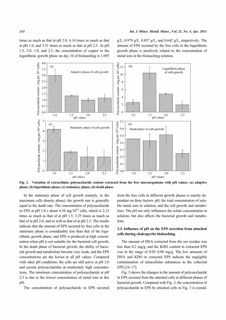
times as much as that at pH 2.0, 4.14 times as much as that at pH 1.0, and 5.51 times as much as that at pH 2.5. At pH 1.5, 2.0, 1.0, and 2.5, the concentration of copper in the logarithmic growth phase on day 10 of bioleaching is 1.095
g/L, 0.979 g/L, 0.857 g/L, and 0.642 g/L, respectively. The amount of EPS secreted by the free cells in the logarithmic growth phase is positively related to the concentration of metal ions in the bioleaching solution.
Fig. 2. Variation of extracellular polysaccharide content extracted from the free microorganisms with pH values: (a) adaptive phase; (b) logarithmic phase; (c) stationary phase; (d) death phase.
In the stationary phase of cell growth (namely, in the maximum cells density phase), the growth rate is generally equal to the death rate. The concentration of polysaccharide in EPS at pH 1.0 s about 4.10 mg/1010 cells, which is 2.15 times as much as that of at pH 1.5, 3.25 times as much as that of at pH 2.0, and as well as that of at pH 2.5. The results indicate that the amount of EPS secreted by free cells in the stationary phase is considerably less than that of the loga-rithmic growth phase, and EPS is produced at high concen-tration when pH is not suitable for the bacterial cell growth. In the death phase of bacterial growth, the ability of bacte-rial growth and metabolism become very weak, and the EPS concentrations are the lowest at all pH values. Compared with other pH conditions, the cells are still active at pH 2.0 and secrete polysaccharides at moderately high concentra-tions. The minimum concentration of polysaccharide at pH 2.5 is due to the lowest concentration of metal ions at this pH.
The concentration of polysaccharide in EPS secreted
from the free cells in different growth phases is mainly de-pendent on three factors: pH, the total concentration of solu-ble metal ions in solution, and the cell growth and metabo-lism. The pH not only influences the solute concentration in solution, but also affects the bacterial growth and metabo-lism.
3.3. Influence of pH on the EPS secretion from attached cells during chalcopyrite bioleaching
The amount of DNA extracted from the ore residue was less than 0.2 mg/g, and the KDO content in extracted EPS was in the range of 0.03–0.08 mg/g. The low amounts of DNA and KDO in extracted EPS indicate the negligible contamination of intracellular substances in the collected EPS [16–17].
Fig. 3 shows the changes in the amount of polysaccharide in EPS secreted from the attached cells at different phases of bacterial growth. Compared with Fig. 2, the concentration of polysaccharide in EPS by attached cells in Fig. 3 is consid-

R.L. Yu et al., Effect of pH values on the extracellular polysaccharide secreted by Acidithiobacillus ferrooxidans… 315
erably higher than that of free cells at different pH condi-tions. The concentration of polysaccharide in EPS of at-tached cells in various growth phases decreases with the in-crease in pH value as the following order: pH 1.0 > 1.5 > 2.0 > 2.5, which is positively related to the solution acidity. However, there is an obvious difference, the amount of polysaccharide in EPS of free cells in the leaching solution is closely related to the total concentration of solution in the logarithmic phase, whereas, the concentration of polysac-charide in EPS of the attached cells on ore is only related to the pH value of the bioleaching solution in the logarithmic phase. It indicates that the attached cells on the ore surface are moderately affected by the concentration of soluble
metal ions in the bioleaching solution, or in other word, there are only a few free metal ions, such as Fe3+, Fe2+, and Cu2+ on the ore surface due to EPS bound action. It was re-ported that Fe2+
can shift from EPS phase into solution phase, but it is difficult for Fe3+
to shift from EPS phase into solu-tion phase due to its hydroxylation and EPS complex action. At the same time, small molecule H+ ions can move freely in the EPS biofilm layer. The result is in agreement with Ref. [19]. Zeng et al. [17] reported that the EPS concentration gradually increased with the bioleaching time and reached a stable value toward the end. This was because the solution pH value decreased gradually with the bioleaching time and reached a stable value toward the end in this study.
Fig. 3. Variation of extracellular polysaccharide content extracted from attached microorganisms with pH values: (a) adaptive phase; (b) logarithmic phase; (c) stationary phase; (d) death phase.
4. Conclusions
(1) The influence of pH values on extracellular polysac-charides secreted by free cells in the bioleaching solution is closely related to three factors: the solution pH value, the total concentration of soluble metal ions, and the ability of growth and metabolism. The optimal bacterial growth con-ditions are less suitable for EPS production.
(2) The polysaccharide concentration in EPS by attached cells is higher than that by free cells, and decreases with the
increase in pH values, but is not related to the total concen-tration of soluble metal ions in the solution due to the EPS bound action in the biofilm on the chalcopyrite surface.
Acknowledgements
This work was financially supported by the National Ba-sic Research Priorities Program of China (No. 2010CB630903) and the National Nature Science Founda-tion of China (No. 31200382).

316 Int. J. Miner. Metall. Mater., Vol. 21, No. 4, Apr. 2014
References
[1] H.R. Watling, The bioleaching of sulphide minerals with emphasis on copper sulphides: a review, Hydrometallurgy, 84(2006), No. 1−2, p. 81.
[2] N. Pradhan, K.C. Nathsaram, K. Srinivasa Rao, L.B. Sukla, and B.K. Mishra, Heap bioleaching of chalcopyrite: a review, Miner. Eng., 21(2008), No. 5, p. 355.
[3] C. Klauber, A critical review of the surface chemistry of acidic ferric sulphate dissolution of chalcopyrite with regards to hindered dissolution, Int. J. Miner. Process., 86(2008), No. 1−4, p. 1.
[4] J. Vilcáez, R. Yamada, and C. Inoue, Effect of pH reduction and ferric ion addition on the leaching of chalcopyrite at thermophilic temperatures, Hydrometallurgy, 96(2009), No. 1−2, p. 62.
[5] N. Hiroyoshi, H. Kitagawa, and M. Tsunekawa, Effect of so-lution composition on the optimum redox potential for chal-copyrite leaching in sulfuric acid solutions, Hydrometallurgy, 91(2008), No. 1−4, p. 144.
[6] Y.G. Wang, L.J. Su, L.J. Zhang, W.M. Zeng, J.Z. Wu, L.L. Wan, G.Z. Qiu, X.H. Chen, and H.B. Zhou, Bioleaching of chalcopyrite by defined mixed moderately thermophilic con-sortium including a marine acidophilic halotolerant bacterium, Bioresour. Technol., 121(2012), p. 348.
[7] J.A. Brierley, Acidophilic thermophilic archaebacteria: po-tential application for metals recovery, FEMS Microbiol. Lett., 75(1990), No. 2−3, p. 287.
[8] E. Gómez, A. Ballester, M.L. Blázquez, and F. González, Silver-catalysed bioleaching of a chalcopyrite concentrate with mixed cultures of moderately thermophilic microorgan-isms, Hydrometallurgy, 51(1999), No. 1, p. 37.
[9] W. Sand and T. Gehrke, Extracellular polymeric substances mediate bioleaching/biocorrosion via interfacial processes involving iron (III) ions and acidophilic bacteria, Res. Micro-biol., 157(2006), No. 1, p. 49.
[10] R.L. Yu, D.L. Zhong, L. Miao, F.D. Wu, G.Z. Qiu, and G.H. Gu, Relationship and effect of redox potential, jarosites and extracellular polymeric substances in bioleaching chalcopy-rite by acidithiobacillus ferrooxidans, Trans. Nonferrous Met.
Soc. China, 21(2011), No. 7, p. 1634. [11] C. Pogliani and E. Donati, The role of exopolymers in the
bioleaching of a non-ferrous metal sulphide, J. Ind. Microbiol. Biotechnol., 22(1999), p. 88.
[12] T. Gehrke, R. Hallmann, and W. Sand, Importance of exopolymers from Thiobacillus ferrooxidans and Lepto-spirillum ferrooxidans for bioleaching. [in] Biohydrometal-lurgical Processing, C.A. Jerez, T. Vargas, H. Toledo, and J.V. Wiertz, eds., University of Chile, Santiago, 1(1995), p. 2.
[13] T. Gehrke, J. Telegdi, D. Thierry, and W. Sand, Importance of extracellular polymeric substances from Thiobacillus fer-rooxidans for bioleaching, Appl. Environ. Microbiol., 64(1998), No. 7, p. 2743.
[14] K. Kinzler, T. Gehrke, J. Telegdi, and W. Sand, Bioleaching: a result of interfacial processes caused by extracellular poly-meric substances (EPS), Hydrometallurgy, 71(2003), No. 1−2, p. 83.
[15] R.L. Yu, J. Liu, A. Chen, D.L. Zhong, Q. Li, W.Q. Qin, G.Z. Qiu, and G.H. Gu, Interaction mechanism of Cu2+, Fe3+ ions and extracellular polymeric substances during bioleaching chalcopyrite by Acidithiobacillus ferrooxidans ATCC2370, Trans. Nonferrous Met. Soc. China, 23(2013), p. 231.
[16] R.L. Yu, Y. Ou, J.X. Tan, F.D. Wu, J. Sun, L. Miao, and D.L. Zhong, Effect of EPS on adhesion of Acidithiobacillus fer-rooxidans on chalcopyrite and pyrite mineral surfaces, Trans. Nonferrous Met. Soc. China, 21(2011), No. 2, p. 407.
[17] W.M. Zeng, G.Z. Qiu, H.B. Zhou, X.D. Liu, M. Chen, W.L. Chao, C.G. Zhang, and J.H. Peng, Characterization of ex-tracellular polymeric substances extracted during the bioleaching of chalcopyrite concentrate, Hydrometallurgy, 100(2010), No. 3−4, p. 177.
[18] Y.D. Karkhanis, J.Y. Zeltner, J.J. Jackson, and D.J. Carlo, A new and improved microassay to determine 2-keto-3-de-oxyoctonate in lipopolysaccharide of gram-negative bacteria, Anal. Biochem., 85(1978), No. 2, p. 595.
[19] R.L. Yu, J.X. Tan, G.L. Gu, Y.H. Hu, and G.Z. Qiu, Mecha-nism of bioleaching chalcopyrite by Acidithiobacillus fer-rooxidans in agar-simulated extracellular polymeric sub-stances media, J. Cent. South Univ. Technol., 17(2010), No. 1, p. 56.