effect of origanum syriacum thymus vulgaris pelargonium...
TRANSCRIPT
رارـــــإق
:أنا الموقع أدناه مقدم الرسالة التي تحمل العنوان
Effect of Origanum syriacum, Hibiscus sabdariffa, Thymus vulgaris and Pelargonium graveolens Extracts on
Human Leukemia THP-1 Cells in Vitro
هدي الخاص، باستثناء ما تمت اإلشارة إلیه أقر بأن ما اشتملت علیه هذه الرسالة إنما هي نتاج ج
حیثما ورد، وٕان هذه الرسالة ككل، أو أي جزء منها لم یقدم من قبل لنیل درجة أو لقب علمي أو .بحثي لدى أیة مؤسسة تعلیمیة أو بحثیة أخرى
DECLARATION
The work provided in this thesis, unless otherwise referenced, is the researcher's own work, and has not been submitted elsewhere for any other degree or qualification
Doa'a Mohamad Faris دعاء محمد فارس :اسم الطالب
:Signature :التوقیع
:Date م 11/1/2014 :التاریخ
Student's name:
The Islamic University of Gaza Deanship of Postgraduate Studies
Faculty of Science Biological Sciences Master Program
Effect of Origanum syriacum, Hibiscus sabdariffa, Thymus vulgaris and Pelargonium graveolens Extracts on Human Leukemia THP-1 Cells in Vitro
Prepared by:
Doa'a Faris
Supervisors:
Dr. Abdalla Abed, PhD Human Genetics The Islamic University of Gaza, Faculty of Science, Biology Department
Dr. Basim Ayesh, PhD Molecular Biochemistry Al Aqsa University, Faculty of Science, Medical Technology Department
A Thesis Submitted in Partial Fulfillment of the Requirements for the Degree of Master in Biological Sciences/ Medical Technology
2013 A.D/ 1433 A.H.
Gaza, Palestine

II
Abstract
Introduction: Leukemia is an aggressive disease that if untreated it leads to death.
Because the conventional leukemia treatment protocols are not effective in all cases and
result in serious side effects, discovering new safer and more effective anticancer agents
from natural materials is the aim of many medical researches nowadays. Within this
perspective, the present study was constructed to assess the possible anticancer effects
of Origanum syriacum, Hibiscus sabdariffa, Thymus vulgaris, and Pelargonium
graveolens, extracts on human leukemic THP-1 cells and to evaluate the effect of these
extracts toward normal human peripheral blood mononuclear cells (PBMCs).
Methodology: The 3-[4,5-dimethylthiazol-2-yl]-2,5-diphenyl tetrazolium bromide
(MTT) assay was used to assess the antiproliferative effects while cytotoxicity was
evaluated using the lactate dehydrogenase (LDH) assay. Ethanol extracts of the four
plants were prepared at various concentrations then were investigated against both THP-
1 cells and PBMCs by MTT assay. After that, three of the extracts were tested with
LDH assay against THP-1 cells.
Results: All extracts, except P. graveolens extract, exhibited a concentration dependent
antiproliferative effect against THP-1 leukemic cells with IC50 values of 15.47 mg/mL
for H. sabdariffa, 2.126 mg/mL for O. syriacum, and 0.1569 mg/mL for T. vulgaris.
Also they showed inhibition against PBMCs with IC50 values of 3.514 mg/mL for H.
sabdariffa, 0.4247 mg/mL for O. syriacum, and 0.3345 mg/mL for T. vulgaris. Only O.
syriacum extract exerted a cytotoxic effect against leukemic cells, as evaluated by the
LDH assay, with LC50 value of 9.646 mg/mL while T. vulgaris and P. graveolens
extracts did not show any cytotoxicity.
Conclusion: It can be concluded that only T. vulgaris extract, which showed a relative
selective inhibition toward leukemic cells, may be useful in developing successful
treating agents for leukemia.
Key words: Leukemia, THP-1 Cells, Origanum syriacum, Hibiscus sabdariffa, Thymus
vulgaris, Pelargonium graveolens, antiproliferative, cytotoxic.
III
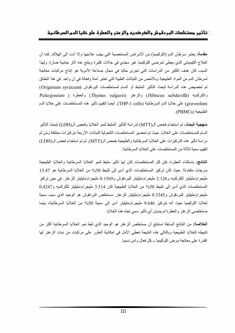
كما أن . الھالكیجب عالجھا وإال أدت الى من األمراض المستعصیة التي ) اللوكیمیا(یعتبر سرطان الدم :مقدمة
ولھذا . ثار جانبیة ضارةمجدي في حاالت كثیرة وینتج عنھ آ العالج الكیمیائي الذي یعطى لمرضى اللوكیمیا غیر
في مجال صناعة األدویة ھو إنتاج مركبات معالجة التي تجرى حالیا ھدف الكثیر من الدراساتالسبب كان
في ھذا النطاق . اتات الطبیة التي تعتبر آمنة وفعالة في آن واحدلسرطان الدم من المواد الطبیعیة وباألخص من النب
)Origanum syriacum( تم تخصیص ھذه الدراسة لبحث التأثیر المثبط أو السام لمستخلصات البردقوش
Pelargonium( والعطرة )Thymus vulgaris( والزعتر )Hibiscus sabdariffa( والكركدیھ
graveolens( على خالیا الدم السرطانیة)THP-1 cells .(ھذه المستخلصات على خالیا الدم یضا لتقییم تأثیرأ
).PBMCs(الطبیعیة
لبحث التأثیر (LDH)لدراسة التأثیر المثبط لنمو الخالیا وفحص ال MTT)(فحص التم استخدام :منھجیة البحث
ومن ثمات الكحولیة للنباتات األربعة بتركیزات مختلفة تم تحضیر المستخلصحیث . السام للمستخلصات على الخالیا
(LDH)فحص التم استخدام ثم .)MTT(بفحص ال لسرطانیة والطبیعیةدراسة تأثیر ھذه التركیزات على الخالیا ا
.مستخلصات على الخالیا السرطانیةال ة منتقییم سمیة ثالثل
تأثیر مثبط لنمو الخالیا السرطانیة والخالیا الطبیعیة فإن كل المستخلصات كان لھا، باستثناء العطرة :جئالنتا
15.47من الخالیا السرطانیة ھو % 50تثبیط أدى إلىیز المستخلصات الذي حیث كان ترك. بدرجات متفاوتة
یز في حین ترك. ملیلیتر للزعتر/ملیجرام 0.1569ملیلیتر للبردقوش و/ملیجرام 2.126ملیلیتر للكركدیھ و/ملیجرام
0.4247ملیلیتر للكركدیھ و/ملیجرام 3.514من الخالیا الطبیعیة كان % 50تثبیط أدى إلىصات الذي المستخل
الذي سبب سمیة مستخلص البردقوش ھو الوحید. ملیلیتر للزعتر/ملیجرام 0.3345ملیلیتر للبردقوش و/ملیجرام
بینما ، من الخالیا السرطانیة% 50ملیلیتر أدى إلى سمیة /ملیجرام 9.646بتركیز حیث أنھ لخالیا اللوكیمیا
.مستخلصي الزعتر والعطرة لم یبدیان أي تأثیر سمي تجاه ھذه الخالیا
نستنتج أن مستخلص الزعتر ھو الوحید الذي ثبط نمو الخالیا السرطانیة أكثر من السابقة النتائج من :الخالصة
لھا من نبات الزعتر إمكانیة العثور على مركباتة وبالتالي ھذه النتیجة تعطي األمل في یتثبیطھ للخالیا الطبیع
.ل فعال وآمن نسبیامرض اللوكیمیا ب–ك القدرة على معالجة

IV
Dedication
To my mother and father.
To my brothers and sister.
To everyone likes science
and works hard to obtain it.
Doa'a M. Faris

V
Acknowledgement
Firstly, all praises and thanks are to Allah, the lord of the worlds. Without his grace and
assistance, nothing can be possible.
I would like to express my profound gratitude and deep regards to my teachers Dr.
Abdalla Abed and Dr. Basim Ayesh, the supervisors of this thesis, for their exemplary
guidance, monitoring, support, patience and constant encouragement throughout the
course of this thesis.
I would like to thank all members of the Faculty of Science in the Islamic University
of Gaza, especially, the biology department's laboratory technicians, as well as the
medical technology department's laboratory technicians, for their help during the
practical stage of the thesis.
My sincere gratitude is to the Scientific Research Affairs in the Islamic University of
Gaza for their financial support.
I would like to thank Eng. Mahmoud Abd El-Al, the area manager of the Arcomed
Medical Supplies Company, as well as all company employees for the ordering of
supplies.
Thank to Eng. Amjad El-Agha and Eng. Raed El-Sakka for their help in plants
collection. In addition, special thanks to Dr. Mohammed Abo Ouda for his kind
assistance in the identification and authentication of the plants used in this work.
I would like to thank all my colleagues in the European Gaza Hospital Laboratory, with
special thanks to the laboratory managers Mr. Rushdi Rasras and Miss Sofia Zourob
for their support, patience, and encouragement during all study stages of master.
Lastly, the biggest thank and the deepest regards are for my parents due to their love,
support, patience and supplications without which the completion of this work would
not be possible. Also, a lot of thanks to my brothers, sister and friends for their
continuous help, and constant encouragement.

VI
Table of Contents
Abstract ………………………………………………………………………….… II
Abstract in Arabic ………………………………………………….……………... III
Dedication ……………………………………………………………….………… IV
Acknowledgement …………………………………………………….………..…. V
List of Figures ………………………………………………………..………….… VIII
List of Tables ……………………………………………………….…………...… IX
List of Abbreviations ……………………………………………………………… X
1. Introduction …………………………………………………… 1 1.1 Overview………………………………………………………………….… 1
1.2 General Objective …………………………………..…………………….… 3
1.3 Specific Objectives ………..…………………………..……………………. 3
1.4 Study Significance …………...………………………..……………………. 3
2. Literature Review ……………………………………………... 4 2.1 Leukemia ………………………………………………………………….... 4
2.1.1 Acute Myeloid Leukemia …..………………………………………………. 5
2.1.2 Management of Acute Myeloid Leukemias …….…………………………. 7
2.2 Medicinal Plants ……………………………..…………………………….. 8
2.2.1 Origanum syriacum ………………..……………………………………….. 9
2.2.2 Hibiscus sabdariffa …………..…………………………………………….. 12
2.2.3 Thymus vulgaris……………………………………………………..……… 16
2.2.4 Pelargonium graveolens …..……………………………………………….. 19
2.2.5 Mechanisms of Medicinal Plants Anticancer Effects ……………………… 21
3. Materials and Methods ……………………………………….. 24 3.1 Materials ……………………………...……………………..……………… 24
3.2 Collection and Preparation of Plant Materials ……………………………… 25
3.3 Extraction of Plant Materials …………………………………..…………… 26
3.4 THP-1 Cell Line Processing and Maintenance …………………..…….....… 27
3.4.1 Cells Receiving and Culturing ……………………………………..……..… 27
3.4.2 Cells Freezing in Stocks ……………………………………………..…...… 28
VII
3.4.3 Determination of THP-1 Cells Growth Characteristics ………...……..….… 28
3.4.4 Routine Maintenance of THP-1 Cells in Culture …...………………..…..… 29
3.5 Determination of MTT and LDH Assays Sensitivity to THP-1 Cells …...…. 29
3.6 Assessment of Plant Extracts Effects on THP-1 Cells by MTT Assay ......… 31
3.7 Assessment of Plant Extracts Effects on Peripheral Blood Mononuclear
Cells (PBMCs) by MTT Assay …………….………………………….……
33
3.8 Determination of Plant Extracts Toxicity on THP-1 Cells by LDH Assay … 34
3.9 Statistical Analysis ……………………………………………………….… 36
4. Results ……………………………………………………….… 37 4.1 Plants Crude Extracts Percentage Yields …………………………………… 37
4.2 Morphology and Growth Characteristics of THP-1 Cells ………………..… 37
4.3 MTT and LDH Assays Sensitivity Studies ………………………………… 39
4.4 Plant Extracts Effects on THP-1 Cells by MTT Assay ……………..……… 39
4.5 Plant Extracts Effects on Peripheral Blood Mononuclear Cells (PBMCs) by
MTT Assay …………………………………………………………….……
43
4.6 Plant Extracts Toxicity on THP-1 Cells by LDH Assay …………………… 47
5. Discussion …………………………………………………….... 50
6. Conclusion and Recommendations …………………………... 57 6.1 Conclusion ………………………………………………………………..… 57
6.2 Recommendations ……………………………………………………......… 57
7. References …….………………………….…………………… 59
VIII
List of Figures
Figure Page
2.1 Origanum syriacum ……………..………………………………….... 10 2.2 Hibiscus sabdariffa ………………...……………………………….... 12 2.3 Thymus vulgaris …………………………………………………….... 16 2.4 Pelargonium graveolens …………………...………………………… 19 3.1 Rotary evaporator ……..……………………..……………………..... 26 3.2 ELISA reader device ………………………………………………..... 30 4.1 THP-1 cells under the inverted microscope ………………………..… 38 4.2 Growth curves of THP-1 cells …………………………………..….... 38 4.3 Sensitivity curves of MTT and LDH assays to THP-1 cells …...…….. 39 4.4 Dose response curves of H. sabdariffa and O. syriacum extracts
effects on THP-1 cells …………………….………..…………………
41 4.5 Dose response curves of T. vulgaris and P. graveolens extracts as
well as DMSO effects on THP-1 cells ……….………..……………...
41 4.6 Dose response curves of H. sabdariffa and O. syriacum extracts
effects on PBMCs …………………………………………..………...
43 4.7 Dose response curves of T. vulgaris and P. graveolens extracts as
well as DMSO effects on PBMCs …………………………................
43 4.8 Comparison between the effects of H. sabdariffa extract on THP-1
cells and on PBMCs ………………………………………………...
46 4.9 Comparison between the effects of O. syriacum extract on THP-1
cells and on PBMCs ……………………………………………..….
46 4.10 Comparison between the effects of T. vulgaris extract on THP-1 cells
and on PBMCs ………………………………………………..…….
47 4.11 LDH toxicity dose response curves of O. syriacum, T. vulgaris, and
P. graveolens extracts as well as DMSO on THP-1 cells ……..……...
48
IX
List of Tables
Table Page 4.1 Percentage yields (%) of all dried extracts obtained after solvent
evaporation …………………………………………………………. 37
4.2 THP-1 cells viability results by MTT test ………………………….. 40 4.3 IC50 values of the extracts on THP-1 cells ………………………….. 42 4.4 Peripheral blood mononuclear cells viability results by MTT test …. 44 4.5 IC50 values of the extracts on PBMCs ……………………………… 45 4.6 THP-1 cells viability results by LDH test ……………………...…… 48

X
List of Abbreviations
ALL Acute Lymphoblastic Leukemia
AML Acute Myeloid Leukemia
AMoL Acute Monocytic Leukemia
ANLL Acute Non-Lymphocytic Leukemia
BM Bone Marrow
C3b the large element of complement 3
DMSO Dimethylsulfoxide
DNA Deoxyribonucleic acid
ECACC European Collection of Cell Cultures
ELISA Enzyme Linked Immuno-Sorbent Assay
FAB French–American–British
FBS Fetal Bovine Serum
Fc Fragment, crystallizable
H. sabdariffa Hibiscus sabdariffa Linn.
HDL High Density Lipoprotein
HEPES Hydroxyethyl Piperazineethanesulfonic Acid
IC50 Median Inhibition Concentration
LC50 Median Lethal Concentration
LDH Lactate Dehydrogenase
LDL Low Density Lipoprotein
MDS Myelodysplastic Syndromes
MTT 3-(4,5-dimethylthiazol-2-yl)-2,5-diphenyltetrazolium bromide
NAD Nicotinamide Adenine Dinucleotide
O. syriacum Origanum syriacum L.
P. graveolens Pelargonium graveolens
PBMCs Peripheral Blood Mononuclear Cells
PBS Phosphate Buffered Saline
PHA Phytohemagglutinin
R2 Coefficient of correlation
ROS Reactive Oxygen Species
RPMI Roswell Park Memorial Institute

XI
SD Standard Deviation
T. vulgaris Thymus vulgaris L.
UV Ultraviolet
WHO World Health Organization
x g gravity at the Earth’s surface (relative centrifugal force unit)

1
Chapter 1 Introduction
1.1 Overview
Cancer is one of the most severe health problems in both developing and
developed countries. It is a general term applied to malignant diseases characterized by
rapid and uncontrolled proliferation of abnormal cells which may mass together to form
a solid tumor or proliferate throughout the body, and when progress it causes death.
Cancer cells are able to grow, invade neighboring tissues and may also affect other
organs through the lymphatic system or bloodstream. Carcinogens such as viruses, some
chemicals, tobacco use and radiation cause aberration of the genetic material of cells.
This leads to the up-regulation of oncogenes which promote cell growth and/or the
down-regulation of tumor suppressor genes which inhibit cell division. Changes in
many genes are required to transform a normal cell into a cancer cell (Knudson, 2001).
In Palestine, the cancer incidence rate was 74.0 per 100,000 of population in
2012, while in the previous year, 2011, it was 64.2 per 100,000 of population. In these
two years, cancer was considered as the second leading cause of death among
Palestinians after cardiovascular diseases (Ministry of Health, 2011, 2013). Also there
was a remarkable increase in cancer mortality compared with 2007 and 2010, from
10.3% in 2007 to 10.8% in 2010 then increases to reach 13.6% from the total deaths in
2012 (Ministry of Health, 2013).
There are many types of cancer and each type is named according to the tissue
or organ in which it was originated. Leukemia is cancer of the blood or bone marrow
that was first recognized by the German pathologist Rudolf Virchow in 1845 (Virchow,
1845). Leukemia refers to a group of malignant diseases characterized by an abnormal
proliferation and differentiation of blood cells and their precursors in the bone marrow.
Leukemias cases in 2012 comprised 6.1% of cancer cases in Palestine (Ministry of
Health, 2013). Anemia, neutropenia and thrombocytopenia are important consequences
of infiltration of the bone marrow, which in turn can lead to infection and hemorrhage
(Bain, 2003). There are both acute and chronic forms of leukemia, each with many
subtypes that vary in their response to treatment. Leukemia is treated mainly with

2
chemotherapy in addition to medical radiation therapy, hormone treatments, or bone
marrow transplantation.
The use of chemotherapeutic drugs in leukemia treatment although is effective
in some cases, it has diverse side effects. These agents are highly toxic to all body cells,
and they destroy normal cells as well as cancerous cells. This leads to disturbances in
normal cells function where fatigue, infection, anemia, hair loss and bleeding problems
result after each chemotherapy dose, also heart problems and lung tissue damage may
appear later after treatment has ended (Limper, 2004; De et al., 2012). In addition, in
some cases chemotherapy resistance develops against the used chemotherapeutic agents
and other drugs must be used in the treatment (Marie & Legrand, 2003).
These severe side effects of chemotherapy have highlighted the importance of
natural products, especially medicinal plants, and its usage as alternative agents in
cancer treatment. Plants are natural reservoir of medicinal agents almost free from the
side effects normally caused by synthetic chemicals (Fennel et al., 2004). The World
Health Organization estimates that herbal medicine is still the main stay of about 75-
80% of the world population, mainly in the developing countries for primary health care
because of better cultural acceptability, better compatibility with the human body, and
lesser side effects (Kamboj, 2000; Yadav & Dixit, 2008).
Plants have been used for treating various diseases of human beings and animals
since time immemorial. More than 50% of all modern drugs in clinical use are of
natural products, many of which have the ability to control cancer cells (Rosangkima
and Prasad, 2004). Medicinal plants possess immunomodulatory and antioxidant
properties, leading to anticancer activities. They are known to have versatile
immunomodulatory activity by stimulating both non-specific and specific immunity
(Agrawala et al., 2001; Pandey and Madhuri, 2006). The antioxidants may prevent and
cure cancer and other diseases by protecting the cells from damage caused by free
radicals. Many plant-derived products have been reported to exhibit potent antitumor
activity against several rodent and human cancer cell lines (Rao et al., 2004).
In the Mediterranean region there are about 2,600 species of plants, many of
which are considered to have medicinal effects (Saad et al., 2005). However, there is
relatively limited research on the medicinal plants and herbs in this region. Therefore,
any researches in this field are encouraged to discover the medicinal effects of these
3
plants and to develop new effective drugs from these plants to prevent and treat the
various diseases including cancer.
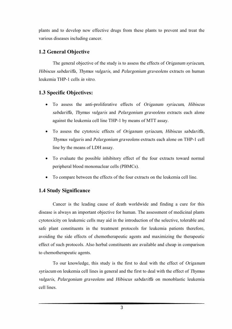
1.2 General Objective
The general objective of the study is to assess the effects of Origanum syriacum,
Hibiscus sabdariffa, Thymus vulgaris, and Pelargonium graveolens extracts on human
leukemia THP-1 cells in vitro.
1.3 Specific Objectives:
• To assess the anti-proliferative effects of Origanum syriacum, Hibiscus
sabdariffa, Thymus vulgaris and Pelargonium graveolens extracts each alone
against the leukemia cell line THP-1 by means of MTT assay.
• To assess the cytotoxic effects of Origanum syriacum, Hibiscus sabdariffa,
Thymus vulgaris and Pelargonium graveolens extracts each alone on THP-1 cell
line by the means of LDH assay.
• To evaluate the possible inhibitory effect of the four extracts toward normal
peripheral blood mononuclear cells (PBMCs).
• To compare between the effects of the four extracts on the leukemia cell line.
1.4 Study Significance
Cancer is the leading cause of death worldwide and finding a cure for this
disease is always an important objective for human. The assessment of medicinal plants
cytotoxicity on leukemic cells may aid in the introduction of the selective, tolerable and
safe plant constituents in the treatment protocols for leukemia patients therefore,
avoiding the side effects of chemotherapeutic agents and maximizing the therapeutic
effect of such protocols. Also herbal constituents are available and cheap in comparison
to chemotherapeutic agents.
To our knowledge, this study is the first to deal with the effect of Origanum
syriacum on leukemia cell lines in general and the first to deal with the effect of Thymus
vulgaris, Pelargonium graveolens and Hibiscus sabdariffa on monoblastic leukemia
cell lines.

4
Chapter 2 Literature Review
2.1 Leukemia
Leukemia is a type of hematopoietic neoplasms that characterized by abnormal
proliferation and increase of immature blood cells in bone marrow and peripheral blood.
The cause of leukemia is a mutation in a single stem cell, the progeny of which form a
clone of leukemic cells. Often there is a series of genetic alterations rather than a single
event. Genetic events contributing to malignant transformation include inappropriate
expression of oncogenes and/or loss of function of tumor suppressor genes. The cell in
which the leukemic transformation occurs may be a lymphoid precursor, a myeloid
precursor or a pluripotent stem cell capable of differentiating into both myeloid and
lymphoid cells. Myeloid leukemias can arise in a lineage-restricted cell or in a
multipotent stem cell capable of differentiating into cells of erythroid, granulocytic,
monocytic and megakaryocytic lineages.
Leukemias are divided into acute leukemias, which, if untreated, lead to death in
weeks or months; and chronic leukemias, which, if untreated, lead to death in months or
years. They are further subdivided into lymphoid, myeloid and biphenotypic leukemias,
the latter showing both lymphoid and myeloid differentiation. In acute leukemias there
is a defect in maturation, leading to an imbalance between proliferation and maturation;
since cells of the leukemic clone continue to proliferate without maturing to end cells,
as a result, there is continued expansion of the leukemic clone and immature cells
predominate. Chronic leukemias are characterized by an expanded pool of proliferating
cells that keep their capacity to differentiate to end cells.
The lone detailed Palestinian cancer report published by the Palestinian Ministry
of Health was covering the period 1995-2000. In that period, acute lymphoid leukemia
accounted for 36.5% of leukemia cases in Gaza Strip, acute myeloid leukemia
accounted for 20.7%, plasma cell multiple myeloma accounted for 15.2% of cases,
chronic lymphocytic leukemia accounted for 12.5% of cases and chronic myelocytic
leukemia accounted for 15.2% of cases (Al Najar et al., 2002).
5
2.1.1 Acute Myeloid Leukemia
The terms acute myeloid leukemia (AML), acute myelogenous leukemia, and
acute nonlymphocytic leukemia (ANLL) refer to a group of marrow-based neoplasms
that have clinical similarities but distinct morphologic, immunophenotypic, and
cytogenetic features (Greer et al., 2003). AML is one of the acute leukemias' major
types in which there is a clonal growth of any one of several non-lymphoid
hematopoietic progenitors which preserve the capacity of self-renewal, but are severely
limited in their ability to differentiate into functional mature cells. These various
progenitors include cells of granulocytic, monocyte/macrophage, erythroid, and
megakaryocytic lineages. The other major type of acute leukemias is called acute
lymphoblastic leukemia (ALL), in which the abnormal proliferation is in the lymphoid
progenitor cells (that is, immature lymphocytes). The distinction between the two types
is based on morphological, cytochemical, immunological and cytogenetic differences
and is of paramount importance as the treatment and prognosis are usually different
(Provan, 2003).
In AML, a myeloid stem cell (or an early progenitor cell) is transformed. This
stem cell expands and proliferates in the blood and bone marrow and suppresses normal
hematopoiesis. A characteristic finding in AML is the leukemic bulge (the appearance
of immature leukemic cells in blood and bone marrow without terminal maturation).
Blasts of myeloid leukemias proliferate under the influence of myeloid growth factors,
some of which are produced by the leukemic cells themselves (autocrine growth of
tumor cells) (Munker et al., 2007).
According to French–American–British (FAB) classification (Bennett et al.,
1976), AML is categorized as acute myeloblastic leukemia without (M1) and with (M2)
maturation, acute hypergranular promyelocytic leukemia and its variant (M3 and M3V),
acute myelomonocytic leukemia (M4), acute monoblastic (M5a) and monocytic (M5b)
leukemia, acute erythroleukemia (M6) and acute megakaryoblastic leukemia (M7)
(Bennett et al., 1985). M0 is AML without maturation and with minimal evidence of
myeloid differentiation. In addition to the above categories there are several very rare
types of AML, which are not included in the FAB classification. These include mast
cell leukemia and Langerhans’ cell leukemia. In addition, the diagnosis of hypoplastic

6
AML requires consideration. Transient abnormal myelopoiesis of Down’s syndrome
may also be regarded as a variant of AML (Bain, 2003).
The WHO classification of acute leukemias and myelodysplastic syndromes
(MDS) has evolved away from the FAB classification, which is based on morphology,
to include not only morphology, but also clinical, immunophenotypic, and cytogenetic
features (Brunning et al., 2001; Vardiman et al., 2002). According to this classification,
AMLs are recognized as one of the three main categories of myeloid neoplasms, along
with MDS and myeloproliferative disorders. There are five major categories recognized
by the WHO: (a) AML with recurrent genetic abnormalities; (b) AML with multilineage
dysplasia; (c) AML and MDS, therapy related; (d) AML not otherwise categorized; and
(e) acute leukemia of ambiguous lineage.
Acute myeloid leukemia accounts for 10-15% of childhood leukemia, but it is
the commonest leukemia of adulthood, particularly as chronic myeloproliferative
disorders and preleukemic conditions such as myelodysplasia usually progress to acute
myeloid leukemia rather than acute lymphoblastic leukemia. The incidence increases
with age, and the median age at presentation is 60 years (Provan, 2003).
The acute monocytic/monoblastic leukemia (AMoL, FAB-M5) is an AML
subtype where the monocytic component of bone marrow (BM) non-erythroid cells is
greater than 80%. In acute monoblastic leukemia (FAB-M5a), the majority of
monocytic cells are monoblasts (in the BM and/or peripheral blood), while in acute
monocytic leukemia (FAB-M5b), the majority of monocytic cells are promonocytes
(≥80%) (Jaffe et al., 2001). AMoL is considered a distinct type of AML with
characteristic biological and clinical features; it is associated with hyperleukocytosis
(Cuttner et al., 1980), extramedullary involvement (Peterson et al., 1981), and
coagulation abnormalities including disseminated intravascular coagulation (Mangal et
al., 1984).
AMoL is frequently associated with specific chromosomal translocations
including t(8;16)(p11;p13) (Heim et al., 1987) and various translocations involving the
MLL locus at 11q23 such as t(9;11)(p22;q23), t(11;19)(q23;p13.1), and others (Baer et
al., 1998). Many patients with AMoL have mutations in the Flt3 gene (approximately
40%) either internal tandem duplications of the juxtamembrane region or point
7
mutations in the second tyrosine kinase domain, and such mutations are associated with
an unfavorable outcome (Rombouts et al., 2000; Thiede et al., 2002).
2.1.2 Management of Acute Myeloid Leukemias
The common therapeutic strategy for most patients with AML has been divided
into two general phases: remission induction and postremission (or consolidation)
therapy.
The remission induction therapy in leukemia is designed to produce the rapid
restoration of normal bone marrow function. The term complete remission is reserved
for patients who have full recovery of normal peripheral blood counts with recovery of
normal bone marrow cellularity; less than 5% blast cells are present in the bone marrow,
and none can have a leukemic phenotype or cytogenetic abnormality (Rathnasabapathy
& Lancet, 2003). The standard induction protocol is the combination of an
anthracycline with cytosine arabinoside (3 + 7 protocol). The common regimen
combines 3 days of daunorubicin with 7 days of continuous infusion of cytosine
arabinoside. With minor variations, this protocol has been the basis for the treatment of
AML for the last 25 years and has been able to induce remission in 60–80% of patients
with newly diagnosed AML. After the induction treatment, the patients remain
granulocytopenic for at least 15–20 days and are susceptible to bacterial, fungal, and
viral infections. The disturbances of coagulation and the thrombocytopenia predispose
the patients to bleeding and need support with platelet concentrates and other blood
products (Munker et al., 2007).
A consolidation or postremission treatment is administered to patients who have
reached complete remission. Two (to four) courses of consolidation are considered as
standard. Depending on the risk factors for AML, 20–25% of the patients will survive
for more than 4 year (Munker et al., 2007). Postremission therapy is directed toward
further reduction in the residual leukemic cell number, which may be as high as 108 to
109 cells at initial complete remission. The elimination of these residual leukemic cells
may be accomplished by either cytotoxic chemotherapy, causing significant
myelosuppression and even myeloablation (e.g. requiring autologous stem cell rescue)
or by replacement of a patient’s stem cells through allogeneic transplantation, a
procedure combining myeloablation and immunotherapy (Rathnasabapathy & Lancet,
2003).

8
Despite the hopeful steps in leukemias treatment researches, considerable
challenges remain (Lucas et al., 2010). Successful treatment of patients with
hematologic malignancies varies widely not just by disease category but by subset
(genetic or otherwise) within each disease, and reasons for this are generally unclear.
Second, scientific advances made in recent years have a long trek to clinical application,
and improving testing strategies to streamline the process and eliminate poor drug
candidates early will be invaluable. Third, the dissemination of tumor cells in
hematologic malignancies precludes localized surgery and radiation approaches, and the
sheltered microenvironment of bone marrow limits the efficacy of many treatments.
Advances in understanding the protective role of the microenvironment will be essential
for successful treatment of leukemias and lymphomas. Finally, novel and potent agents
must be identified that target tumor cell survival pathways while sparing normal cells.
This is a significant challenge, as there are undoubtedly targetable survival mechanisms
yet to be discovered. To make advances toward curative therapy, identification of new
tumor survival pathways and protective mechanisms must be continued; we cannot rely
only on what is currently known.
This last challenge highlights a key strength of natural product investigation.
Using cytotoxic activity-guided screening strategies, agents with potent anti-tumor
activity can be identified in an equitable way, increasing the probability of revealing
novel tumoricidal pathways and survival mechanisms. Once an effective molecule is
identified, the mode of action can be investigated and additional or alternative agents
that impact the target can be synthesized. Thus even when an active natural product
does not reach clinical use, its investigation can provide critical clues for the
development of new targeted cancer drugs (Lucas et al., 2010).
2.2 Medicinal Plants
Natural products, obtained to date mainly from fungi, higher plants, and soil
microorganisms, have a long history of helpful use by human for the treatment of
various diseases. These substances may be useful in their structurally original form or
may be derivatized by chemical synthesis to enhance potency or pharmacologic
properties such as water solubility or thermostability (Kinghorn, 2008). The term
“natural product” is generally taken to mean a compound that has no known primary
biochemical role in an organism. Such compounds are also called “secondary

9
metabolites”, and apparently are produced by the organism for ecological or defensive
purposes, thus promoting its survival (Williams et al., 1989).
Terrestrial plants have been used as medicines in Egypt, China, India and
Greece from ancient time and a significant number of modern drugs have been
developed from them (Shoeb, 2006). The first written records on the medicinal uses of
plants appeared in about 2600 BC from the Sumerians and Akkaidians (Samuelsson,
1999). The “Ebers Papyrus”, the best known Egyptian pharmaceutical record, which
documented over 700 drugs, represents the history of Egyptian medicine dated from
1500 BC (Shoeb, 2006). The Chinese Materia Medica, which describes more than 600
medicinal plants, has been well documented with the first record dating from about
1100 BC (Cragg et al., 1997). Documentation of the Ayurvedic system recorded in
Susruta and Charaka dates from about 1000 BC (Kappor, 1990). The Greeks also
contributed significantly to the logical development of the herbal drugs. Dioscorides,
the Greek physician (100 AD), described in his work “De Materia Medica” more than
600 medicinal plants (Samuelsson, 1999).
The National Cancer Institute collected about 35,000 plant samples from 20
countries and has screened around 114,000 extracts for anticancer activity (Shoeb,
2005). There are some plant derived natural products which currently used in the
treatment of hematological malignancies such as vinca alkaloids, which used in
lymphomas and acute lymphoblastic leukemias treatments (Karon et al., 1966; Dancey
& Steward, 1995), and podophyllotoxin derivatives, which have a potential usefulness
in the treatment of acute myeloid leukemias, Hodgkin’s and non-Hodgkin’s lymphomas
(Hande, 1998). Other plant natural products have been under clinical trials where they
showed promise for treating several hematological malignancies as flavopiridol (Byrd et
al., 2007) and meisoindigo (Wang et al., 2005; Weng et al., 2005), while there are plant
natural products which are yet under preclinical investigations for hematological
malignancies treatment as combretastatins (Fang et al., 2007; Petit et al., 2008) and
honokiol (Battle et al., 2005).
2.2.1 Origanum syriacum (Syrian Oregano)
The genus Origanum (oregano) is significant in the family Lamiaceae and
comprises of around 900 species of annual, perennial and shrubby herbs, widespread

throughout the world (Bayder et al., 2004; Kordali et al., 2008
arranged in groups and sections
Majorana, together with Origanum majorana
1980).
O. syriacum inhabits a large area in the eastern Mediterranean. It can be found in
southern Turkey, on Cyprus, in Syria,
Peninsula and grows from nearly sea level up to at least 2000 m in rocky soils,
limestone (Ietswaart, 1980).
traditional medicine, flavor and fragrance, and for aromatherapy in the form of bath,
massage, steam inhalation and vaporization. It is used in teas and cooked or baked foods
(Alma et al., 2003).
2.2.1.1 Botanic Description
Origanum syriacum L. is a perennial
herb, 60–90 cm high, with creeping woody
roots, branched woody, hairy stems. Leaves are
opposite, shortly or subsessile (petiolate to 8
mm), ovate, 5–35 mm × 4–23 mm and hairy,
margins are entire or remotely serrate, the apex
is obtuse. The upper leaf surface is darker; the
lower leaf surface is brighter with secretory
glands. Flowers are shortly petiolate and hairy.
Bracts are obovate or elliptic, 2
3.5 mm, acute or obtuse, entire or denticulate.
A two lipped pale purple corolla 4.5
and a five toothed tubular campanulas calyx (
2.2.1.2 Chemical Composition
The green leaves of the
characteristic and fragrance. The extraction product can vary in quality, quantity and
composition according to climate, soil composition, geographical location, seasonal
variation, plant organ, age and
al., 2007, 2008). Origanum essential oils were found to contain mainly thymol and
10
Bayder et al., 2004; Kordali et al., 2008). These species are
s and sections where Origanum syriacum L. is placed in the section
Origanum majorana L. and Origanum onites L (
inhabits a large area in the eastern Mediterranean. It can be found in
southern Turkey, on Cyprus, in Syria, Lebanon, Palestine, Jordan, and on the Sinai
Peninsula and grows from nearly sea level up to at least 2000 m in rocky soils,
The leaves of O. syriacum have been used
traditional medicine, flavor and fragrance, and for aromatherapy in the form of bath,
massage, steam inhalation and vaporization. It is used in teas and cooked or baked foods
L. is a perennial
with creeping woody
roots, branched woody, hairy stems. Leaves are
opposite, shortly or subsessile (petiolate to 8
23 mm and hairy,
margins are entire or remotely serrate, the apex
is obtuse. The upper leaf surface is darker; the
wer leaf surface is brighter with secretory
glands. Flowers are shortly petiolate and hairy.
Bracts are obovate or elliptic, 2–5 mm × 1.5–
3.5 mm, acute or obtuse, entire or denticulate.
A two lipped pale purple corolla 4.5–7.5 mm
campanulas calyx (Alma et al., 2003; Kintzios, 2004
2.2.1.2 Chemical Composition
The green leaves of the Origanum herb are rich in essential oil which confers its
characteristic and fragrance. The extraction product can vary in quality, quantity and
composition according to climate, soil composition, geographical location, seasonal
and vegetative cycle stage, and harvesting time (
essential oils were found to contain mainly thymol and
Figure 2.1: Origanum syriacum(http://www.wildflowers.co.il/english/plant.asp?ID=129
These species are
L. is placed in the section
L (Ietswaart,
inhabits a large area in the eastern Mediterranean. It can be found in
, Jordan, and on the Sinai
Peninsula and grows from nearly sea level up to at least 2000 m in rocky soils, often on
have been used in herbal
traditional medicine, flavor and fragrance, and for aromatherapy in the form of bath,
massage, steam inhalation and vaporization. It is used in teas and cooked or baked foods
Alma et al., 2003; Kintzios, 2004).
herb are rich in essential oil which confers its
characteristic and fragrance. The extraction product can vary in quality, quantity and
composition according to climate, soil composition, geographical location, seasonal
harvesting time (Abu Lafi et
essential oils were found to contain mainly thymol and
Origanum syriacum http://www.wildflowers.co.il/english/plant.asp?ID=129).

11
carvacrol, monoterpene glycosides, phenols including gallic acid, rosmarinic acid,
caffeic acid, apigenin, naringenin and luteolin-7-O-glucoside (Zein et al., 2011).
The detailed investigation of the methanol extract of the aerial parts of O.
syriacum led to the isolation of eleven flavonoid compounds: acacetin-7-O-[2"-O-α-L-
rhamnopyranosyl-6"-O-β -D-glucopyranosyl]-β-D-glucopyranoside luteolin, apigenin,
luteolin-6-C-glucoside, luteolin-3'-methylether-6-C-glucoside, luteolin-7,4'-
dimethylether-6-C-glucoside, apigenin-7-methylether-6-C-glucoside, apigenin-7-O-
glucoside, diosmetin-7-O-glucoside, acacetin-7-O-glucoside, and acacetin-7-O-
rutinoside (El-Desouky et al., 2009).
2.2.1.3 Medicinal Uses
In folk medicine Origanum syriacum is used for treating gastrointestinal
problems and respiratory diseases (Ali-Shtayeh et al., 2000; Aburjai et al., 2007). In
Jordan the plant is reported to be additionally used as a carminative, pectoral,
antitussive, aperitif, and anti-stomachache and against arthritis (Lev & Amar, 2002;
Aburjai et al., 2007). In different historical records from the area of Bilad Al-Sham,
Origanum sp. was used against internal diseases, hemorrhoids, sexual diseases, pains,
animals' bites, and poison (Lev, 2002).
In recent years, O. syriacum has drawn attention for its antioxidant activity and
acetylcholinesterase inhibition (Alzheimer’s disease) (Loizzo et al., 2009; Zein et al.,
2011), antifungal and antibacterial activity (Kintzios, 2004), analgesic activity,
antiflogistic activity (atherosclerosis, Alzheimer’s disease), antirheumatic, expectorant,
sedative, antiparasitic and antihelminthic activities (Zein et al., 2011).
2.2.1.4 Anticancer Studies
Several studies were carried on the composition, antioxidant and antibacterial
effects of Origanum syriacum but few researches which were concerned with the
antiproliferative and antitumor activities of this plant.
El-Desouky et al. in 2009 have investigated the cytotoxicity of the methanol
extract of O. syriacum against human cervical adenocarcinoma, Hela cells, by the MTT
assay and the results indicated that the extract induced growth inhibition activity in a
time and dose dependent manner on the Hela cells. The IC
extract on Hela cells was 474.92
Another study by Al-Kalaldeh et al.
have investigated O. syriacum
breast cell line (MCF7). The antiproliferative activities of the hydrodistilled volatile oils
and the crude ethanol and water extracts were evaluated using the sulphorhodamine B
assay. The ethanol crude extracts of
toward MCF7 with IC50 value of
essential oils nor aqueous extracts demonstrated cytotoxic activity.
2.2.2 Hibiscus sabdariffa (Roselle
Hibiscus belongs to the plant family Malvaceae and it is one of the most flower
plants grown worldwide. There are more than 300 species of
(Ismail et al., 2008). One of them is roselle (
commonly used to make jellies, jams and beverages. The brilliant red color of its calyx
makes it a valuable food product, apart from its multitude of traditional medicinal uses.
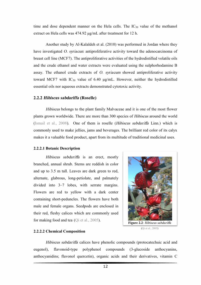
2.2.2.1 Botanic Description
Hibiscus sabdariffa
branched, annual shrub. Stems are
and up to 3.5 m tall. Leaves are dark
alternate, glabrous, long-petiolate,
divided into 3–7 lobes, with serrate margins.
Flowers are red to yellow with a dark center
containing short-peduncles. The flowers have both
male and female organs. Seedpods are enclosed in
their red, fleshy calices which
for making food and tea (Qi et al., 2005
2.2.2.2 Chemical Composition
Hibiscus sabdariffa calices
eugenol), flavonoid-type polyphenol
anthocyanidins; flavonol quercetin), organic acids and their derivatives, vitamin
12
manner on the Hela cells. The IC50 value of the
extract on Hela cells was 474.92 μg/mL after treatment for 12 h.
Kalaldeh et al. (2010) was performed in Jordan where they
O. syriacum antiproliferative activity toward the adenocarcinoma of
The antiproliferative activities of the hydrodistilled volatile oils
thanol and water extracts were evaluated using the sulphorhodamine B
assay. The ethanol crude extracts of O. syriacum showed antiproliferative activity
value of 6.40 μg/mL. However, neither the hydrodistilled
aqueous extracts demonstrated cytotoxic activity.
Roselle)
belongs to the plant family Malvaceae and it is one of the most flower
plants grown worldwide. There are more than 300 species of Hibiscus around the world
One of them is roselle (Hibiscus sabdariffa Linn.
commonly used to make jellies, jams and beverages. The brilliant red color of its calyx
makes it a valuable food product, apart from its multitude of traditional medicinal uses.
Hibiscus sabdariffa is an erect, mostly
annual shrub. Stems are reddish in color
m tall. Leaves are dark green to red,
petiolate, and palmately
lobes, with serrate margins.
yellow with a dark center
The flowers have both
male and female organs. Seedpods are enclosed in
ces which are commonly used
Qi et al., 2005).
2.2.2.2 Chemical Composition
calices have phenolic compounds (protocatechuic
type polyphenol compounds (3-glucoside anthocyanins,
flavonol quercetin), organic acids and their derivatives, vitamin
Figure 2.2: Hibiscus sabdariffa(Qi et al., 2005)
of the methanol
was performed in Jordan where they
the adenocarcinoma of
The antiproliferative activities of the hydrodistilled volatile oils
thanol and water extracts were evaluated using the sulphorhodamine B
showed antiproliferative activity
the hydrodistilled
belongs to the plant family Malvaceae and it is one of the most flower
around the world
Linn.) which is
commonly used to make jellies, jams and beverages. The brilliant red color of its calyx
makes it a valuable food product, apart from its multitude of traditional medicinal uses.
rotocatechuic acid and
glucoside anthocyanins,
flavonol quercetin), organic acids and their derivatives, vitamin C
Hibiscus sabdariffa )

13
(ascorbic acid), B1 (thiamin), B2 (riboflavin), and a carotenoid (β-carotene) (Carvajal-
Zarrabal et al., 2012).
The composition of the essential oil includes benzoic acid, nonanoic acid,
citrnellic acid, benzyl benzoate, eugenol, and eicosane (Zhang & Wang, 2007).
2.2.2.3 Medicinal Uses
Hibiscus sabdariffa has several pharmacological effects as antihypertensive,
hepatoprotective, antihyperlipidemic, antioxidant, anticancer, anticonvulsant,
anxiogenic, analgesic, central nervous system depressant, antipyretic, anti-
inflammatory, and antimicrobial activities (Mahadevan et al., 2009; Khatun et al.,
2011).
Tea of H. sabdariffa calices showed 11.2 % reduction in the systolic blood
pressure and 10.7 % decrease in diastolic pressure (Haji Faraji & Haji Tarkhani, 1999).
Effectiveness and tolerability of a standardized extract was studied in patients with mild
to moderate hypertension which revealed a reduction in systolic and diastolic blood
pressure by more than 10% (Herrera-Arellano et al., 2004).
Protocatechuic acid, a simple phenolic compound isolated from H. sabdariffa
showed protective effects against cytotoxicity and genotoxicity of hepatocytes induced
by tert-butylhydroperoxide. One of mechanisms may be associated with its property of
scavenging free radicals (Tseng et al., 1996). H. sabdariffa extract offers
hepatoprotection by influencing the levels of lipid peroxidation products and liver
marker enzymes in experimental hyperammonemia and this could be due to the free
radical scavenging property of natural antioxidants present in the plant (Essa et al.,
2006).
Inhibitory effects of the plant extract on low density lipoprotein (LDL) oxidation
and anti-hyperlipidemia in fructose and cholesterol fed rats was demonstrated (Chen et
al., 2004). It revealed that the extract reduced the level of LDL and the ratio of LDL-
cholesterol to HDL-cholesterol (Carvajal-Zarrabal et al., 2005). Antioxidant effects of
the aqueous extracts of dried calyx using rat low density lipoprotein was investigated
and the study demonstrated protective effect of roselle on LDL oxidation (Hirunpanich
et al., 2005).

14
The antioxidant and free radical scavenging effects of two fractions of the
ethanol extract (chloroform soluble fraction and ethylacetate soluble fraction) obtained
from its dried flowers were investigated and found that both fractions scavenge
hydrogen peroxide and inhibit superoxide anions radicals (Farombi & Fakoya, 2005).
2.2.2.4 Anticancer Studies
Several studies were performed on the apoptotic and antitumor activities of
Hibiscus sabdariffa extracts and its constituents as anthocyanins and protocatechuic
acid. Also the apoptotic pathway was examined in most of these studies to assess the
mechanism which is followed by H. sabdariffa to act as antiproliferative and anticancer
plant.
Treatment with Hibiscus protocatechuic acid (PCA) (0.2 to 2 mm) isolated from
H. sabdariffa was carried out on HL-60 and Bcl-2 overexpressed human leukemia cells
(HL60/Bcl-2-350) (Tseng et al., 2000). Inhibition of HL-60 cell survival was dependant
on PCA concentration and time (2 mM; 9 hours). Further studies showed that Hibiscus
PCA application reduced Bcl-2 protein expression to 47%, and increased Bax protein
expression to 181% after 1.5 hr as compared with time 0. Overexpression of Bcl-2 in
HL-60 cells delayed the occurrence of Hibiscus PCA-induced apoptosis. These data
suggest that Hibiscus PCA is an apoptosis inducer in human leukemia cells, and that RB
phosphorylation and Bcl-2 protein may play a crucial role in the early stage.
In 2005, a study performed in Japan by Hou et al., where Delphinidin 3-
sambubioside (Dp3-Sam), an anthocyanin which was isolated from the dried calices of
Hibiscus sabdariffa L. induced a dose-dependent apoptosis in human leukemia cells
(HL-60) as characterized by cell morphology, DNA fragmentation, activation of
caspase-3, -8, and -9, and inactivation of poly(ADP)ribose polymerase (PARP).
Molecular data showed that Dp3-Sam induced Bid truncation, mitochondrial membrane
potential loss, and cytochrome c release from mitochondria to cytosol. Moreover, Dp3-
Sam caused a time- and dose-dependent elevation of intracellular reactive oxygen
species (ROS) level in HL-60 cells, and antioxidants such as N-acetyl-L-cysteine
(NAC) and catalase could effectively block Dp3-Sam-induced ROS generation,
caspase-3 activity, and DNA fragmentation. These data indicate that Dp3-Sam might
15
induce apoptosis in HL-60 cells through a ROS-mediated mitochondrial dysfunction
pathway.
Another study in 2005 was performed in Taiwan by Chang et al. where they
explored the effect of Hibiscus anthocyanins (HAs) on human cancer cells. The results
showed that HAs could cause cancer cell apoptosis, especially in HL-60 cells. Using
flow cytometry, they found that HAs treatment (0–4 mg/ml) markedly induced
apoptosis in HL-60 cells in a dose- and time-dependent manner.
In the same year, a study by Lin et al. was published. Treatment with methanolic
H. sabdariffa extract was carried out on seven cancer cell lines: human gastric
adenocarcinoma (AGS), human promyelocytic leukemia (HL-60), hepatocellular
carcinoma (Hep3B), colorectal adenocarcinoma (Caco-2), hepatoblastoma (HepG2),
adenocarcinoma (MCF-7), human oral epidermoid carcinoma (KB), and one control
cellular line: mouse fibroblast cells. Evaluation of implied apoptotic pathway(s) was
made. Methanolic H. sabdariffa extract induced apoptosis in all cellular lines in a
concentration-dependent form. The AGS cells were the most susceptible with a drug
concentration resulting in 50% in vitro inhibition (IC50) of 0.95 mg/mL compared to
control cells (IC50 of 2.98 mg/mL). Evidence that methanolic H. sabdariffa extract
promoted both apoptotic routes (intrinsic: phosphorylation by kinase p53; extrinsic:
phosphorylation by kinase p-38) was observed.
Akim et al. (2011) in Malaysia investigated Hibiscus sabdariffa antiproliferative
effect on breast (MCF-7 and MDA-MB-231), ovarian (Caov-3) and cervical (HeLa)
cancer cell lines. Commercialized roselle juice (RJ) at three storage periods, 1 week
(WRJ), 1 month (MRJ) and 1 year (YRJ), were tested using the MTT (3-[4,5-
dimethylthiazol-2-yl]- 2,5-diphenyl tetrazolium bromide) assay on the four cell lines to
obtain the percentage viability of the cells. The cells were incubated for 72 h after
inoculation with RJ and the control group was without treatment. The IC50 was found to
be highest for Caov-3 cells (2.267±1.193% (v/v)) whereas MCF-7 cells exhibited the
lowest (0.432±0.278% (v/v)) IC50 value after treatment with MRJ. Increasing
concentrations of sample corresponded to lower percentage viability of cells for all
samples, however the interaction within and between cell type and storage period was
not significant (p>0.05).
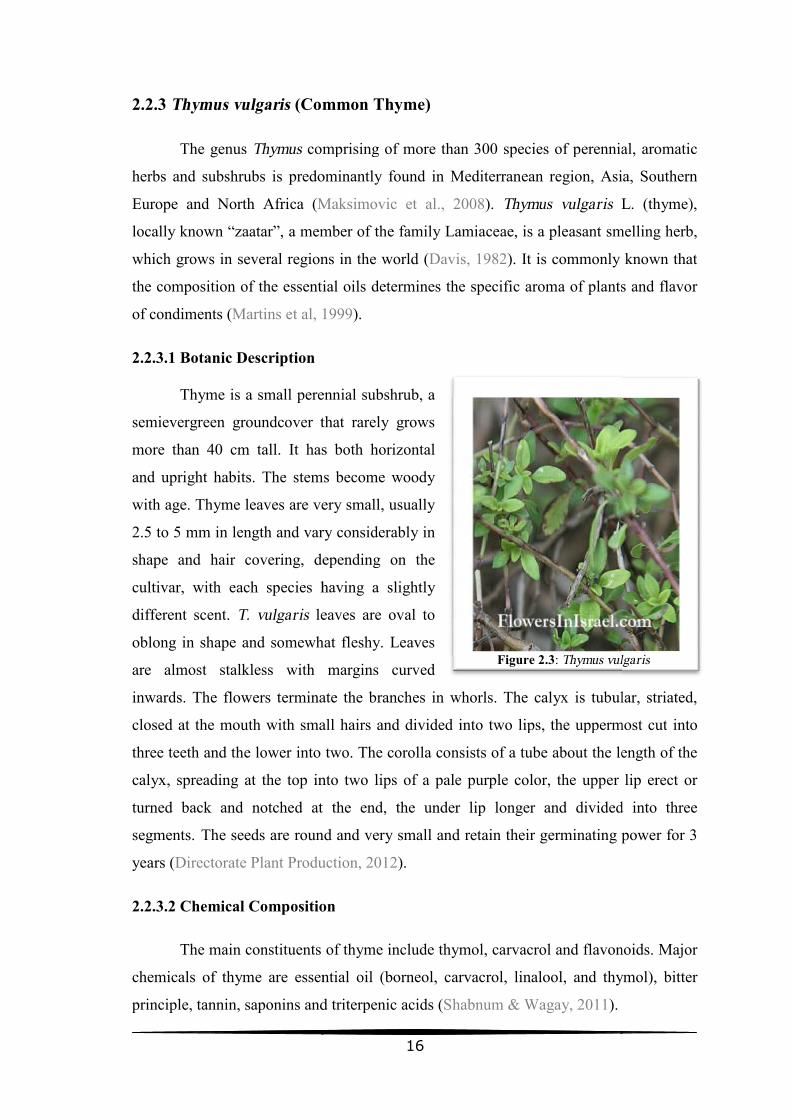
2.2.3 Thymus vulgaris (Common
The genus Thymus comprising of
herbs and subshrubs is predominantly
Europe and North Africa (Maksimovic et al., 2008
locally known “zaatar”, a member of the family Lamiaceae,
which grows in several regions in the world (
the composition of the essential oils determines the specific a
of condiments (Martins et al, 1999
2.2.3.1 Botanic Description
Thyme is a small perennial subshrub, a
semievergreen groundcover that rarely grows
more than 40 cm tall. It has both horizontal
and upright habits. The stems become
with age. Thyme leaves are very small, usually
2.5 to 5 mm in length and vary considerably in
shape and hair covering, depending on the
cultivar, with each species having a slightly
different scent. T. vulgaris leaves are
oblong in shape and somewhat fleshy. Leaves
are almost stalkless with
inwards. The flowers terminate the branches in whorls. The calyx is tubular, striated,
closed at the mouth with small hairs and divided into two lips, the uppermost cut
three teeth and the lower into two. The corolla consists of a tube about the
calyx, spreading at the top into
turned back and notched at the end, the under lip longer and
segments. The seeds are round and very small and retain their germinating power for 3
years (Directorate Plant Production
2.2.3.2 Chemical Composition
The main constituents of thyme include thymol, carvacrol and flavonoids.
chemicals of thyme are essential oil
principle, tannin, saponins and triterpenic acids
16
Common Thyme)
comprising of more than 300 species of perennial, aromatic
herbs and subshrubs is predominantly found in Mediterranean region, Asia, Southern
Maksimovic et al., 2008). Thymus vulgaris L.
“zaatar”, a member of the family Lamiaceae, is a pleasant smelling
which grows in several regions in the world (Davis, 1982). It is commonly known that
essential oils determines the specific aroma of plants and flavor
Martins et al, 1999).
Thyme is a small perennial subshrub, a
groundcover that rarely grows
than 40 cm tall. It has both horizontal
habits. The stems become woody
eaves are very small, usually
length and vary considerably in
depending on the
having a slightly
leaves are oval to
somewhat fleshy. Leaves
margins curved
inwards. The flowers terminate the branches in whorls. The calyx is tubular, striated,
small hairs and divided into two lips, the uppermost cut
the lower into two. The corolla consists of a tube about the length of the
calyx, spreading at the top into two lips of a pale purple color, the upper lip erect or
turned back and notched at the end, the under lip longer and divided into three
e seeds are round and very small and retain their germinating power for 3
Directorate Plant Production, 2012).
Composition
The main constituents of thyme include thymol, carvacrol and flavonoids.
essential oil (borneol, carvacrol, linalool, and thymol
principle, tannin, saponins and triterpenic acids (Shabnum & Wagay, 2011).
Figure 2.3: Thymus vulgaris
of perennial, aromatic
found in Mediterranean region, Asia, Southern
L. (thyme),
is a pleasant smelling herb,
). It is commonly known that
roma of plants and flavor
inwards. The flowers terminate the branches in whorls. The calyx is tubular, striated,
small hairs and divided into two lips, the uppermost cut into
length of the
upper lip erect or
divided into three
e seeds are round and very small and retain their germinating power for 3
The main constituents of thyme include thymol, carvacrol and flavonoids. Major
and thymol), bitter
Thymus vulgaris

17
Thymus vulgaris shows a polymorphic variation in monoterpene production, the
presence of intraspecific chemotype variation being common in the genus Thymus. Each
of the six chemotypes, geraniol (G), α-terpineol (A), thuyanol-4 (U), linalool (L),
carvacrol (C), and thymol (T), is named after its dominant monoterpene (Thompson et
al., 2003).
2.2.3.3 Medicinal Uses
Extracts of T. vulgaris are useful in traditional medicine because of their anti-
asthmatic, bronchodilator, antiseptic, antispasmodic, antitussive, antibacterial,
antifungal and antiviral activities (Marino et al., 1999; Pina-Vaz et al., 2004). Also,
these extracts have shown immunomodulating properties (Bukovska et al., 2007; Ocaña
& Reglero, 2012).
T. vulgaris is also quoted by various authors for its polyphenol and flavonoid
contents and its potential antioxidant and free radical scavenging, anti-inflammatory,
vasorelaxant, anti-platelet, antithrombin, anti-hyperlipidemic and anti-diabetic
properties (Miura et al., 2002; Vigo et al., 2004; El-Nekeety et al., 2011). A recent study
(Kensara1 et al., 2013) also shows that supplementation with T. vulgaris as an herbal
remedy has shown remarkable antihypertensive effect and marked improvement on
hypertension-related biochemical changes and aortic vascular damage in rats.
In aromatherapy, the distinct types, thymol, ‘red thyme oil’, linalol type are used
for its very gentle soft action and thuyanol for antiviral properties. A rectified product,
‘white thyme oil’ is also used, and it is milder on the skin. Applied to the skin, thyme
relieves bites and stings, and relieves sciatica and rheumatic aches and pains. Thyme is
useful for ringworm, athlete’s foot, thrush, and other fungal infections, as well as
scabies and lice (Directorate Plant Production, 2012).
2.2.3.4 Anticancer Studies
Some recent studies that concerned with the cytotoxic effect of Thymus vulgaris
are mentioned here.
In Moroco, Jaafari et al. (2007) published a paper where T. vulgaris essential
oils as well as two pure products (carvacrol and thymol) were tested for their antitumor

18
activity against P815 mastocytoma cell line using colorimetric MTT assay. While all
these products showed a dose dependent cytotoxic effect, the carvacrol (with IC50 less
than 0.004% (v/v)) was the most cytotoxic one compared to the others. The IC50 of
thymol was 0.015% (v/v). Interestingly, when these products were tested against the
normal human peripheral blood mononuclear cells (PBMC), they (except thymol)
showed a proliferative effect instead of a cytotoxic one. Thymol had cytotoxic effect on
the PBMC.
Zu et al. (2010) in China, tested the essential oil of T. vulgaris for its in vitro
toxicology against three human cancer cell lines, PC-3, A-549 and MCF-7 cancer cells.
T. vulgaris essential oil exhibited cytotoxicity towards three human cancer cells. Its IC50
values on PC-3, A549 and MCF-7 tumor cell lines were 0.010% (v/v), 0.011% (v/v) and
0.030% (v/v), respectively.
Another study in Germany was performed by Sertel et al. (2011), where
cytotoxicity of thyme essential oil was investigated on the head and neck squamous cell
carcinoma (HNSCC) cell line. They found that the IC50 of thyme essential oil extract
was 369 μg/ mL.
Berrington and Lall (2012) in South Africa published a paper where acetone
extract of T. vulgaris L. was evaluated for its in vitro cytotoxicity against a
noncancerous African green monkey kidney (Vero) cell line and an adenocarcinoma
cervical cancer (HeLa) cell line. Cytotoxicity was measured using XTT (Sodium 3'-[1-
(phenyl amino-carbonyl)-3,4-tetrazolium]-bis-[4-methoxy-6-nitro] benzene sulfonic
acid hydrate) colorimetric assay, and low cytotoxic effect was exhibited by the extract
on the studied cell lines. IC50 on Hela cell line was >200 μg/mL and on Vero cell line
was 138.4±2.60 μg/mL.
In Poland, Berdowska et al. (2013) evaluated the cytotoxicity of dried aqueous
extracts from T. vulgaris on two human breast cancer cell lines: Adriamycin-resistant
MCF-7/Adr and wild-type MCF-7/wt by the MTT assay, and found that T. vulgaris
exhibited cytotoxicity against both cell lines with higher toxicity against MCF-7/Adr.

2.2.4 Pelargonium graveolens
Pelargonium graveolens
belongs to the Geraniaceae family (
Africa (Lis-Balchin, 2004) as well as reunion Madagascar, Egypt and Moroco (
al., 2006). There are over 700
grown for ornamental purposes (
2.2.4.1 Botanic Description
Pelargonium graveolens
much-branched shrub that can reach a height of
up to 1.3 m and a spread of 1 m. The hairy stems
are herbaceous when young, becoming woody
with age. The deeply incised leaves are velvety
and soft to the touch due to the presence of
numerous glandular hairs. The leaves are
strongly rose-scented. The showy white to
pinkish flowers are borne in an umbel
inflorescence and are present from
summer peaking in spring (http://www.plantzafrica.com/plantnop/
2.2.4.2 Chemical Composition
The P. graveolens whole plant extract yield
oils (Kolodziej, 2000; Butles, 2004
geranium essential oil have been identified. The major components were citronellol
(29.90%), trans-geraniol (18.03%), 10
linalool (5.13%), geranyl acetate (4.52%),
(2.53%), geranyl tiglate (2.50%) and gemacrene D (2.05%).
Geranium essential oil is rich in oxygenated components and commercial
rhodinol (linalool + citronellol + geraniol) fraction (
2009). Galloyl C-glycosidic flavones, non galloyl flavones, phenolics (flavonoids and
tannins), benzoic and cinnamic acid derivatives were reported in aerial parts (
et al., 2005; Kolodziej & Kiderlen, 2007
19
Pelargonium graveolens (Rose-Scented Geranium)
Pelargonium graveolens is a perennial aromatic and medicinal herb/shrub that
belongs to the Geraniaceae family (Verma et al., 2011). It was originated in
as well as reunion Madagascar, Egypt and Moroco (
). There are over 700 varieties of cultivated geraniums; however, most are
for ornamental purposes (Lis-Balchin, 2002; Shawl et al., 2006).
Pelargonium graveolens is an erect,
can reach a height of
3 m and a spread of 1 m. The hairy stems
are herbaceous when young, becoming woody
with age. The deeply incised leaves are velvety
and soft to the touch due to the presence of
numerous glandular hairs. The leaves are
scented. The showy white to
pinkish flowers are borne in an umbel-like
inflorescence and are present from late winter to
http://www.plantzafrica.com/plantnop/pelarggrav.htm
2.2.4.2 Chemical Composition
whole plant extract yield contains high quantity
Butles, 2004). Thirty two compounds constituting 99.23% of
geranium essential oil have been identified. The major components were citronellol
geraniol (18.03%), 10-epi-γ-eudesmol (8.27%), isomenthone (5.44%),
linalool (5.13%), geranyl acetate (4.52%), γ- cadinene (2.89%), geranyl butyrate
(2.53%), geranyl tiglate (2.50%) and gemacrene D (2.05%).
Geranium essential oil is rich in oxygenated components and commercial
rhodinol (linalool + citronellol + geraniol) fraction (Rajeswara Rao et al., 2002; Fayed
glycosidic flavones, non galloyl flavones, phenolics (flavonoids and
tannins), benzoic and cinnamic acid derivatives were reported in aerial parts (
Kiderlen, 2007).
Figure 2.4: Pelargonium graveolens
is a perennial aromatic and medicinal herb/shrub that
). It was originated in South
as well as reunion Madagascar, Egypt and Moroco (Shawl et
of cultivated geraniums; however, most are
pelarggrav.htm).
of essential
). Thirty two compounds constituting 99.23% of
geranium essential oil have been identified. The major components were citronellol
eudesmol (8.27%), isomenthone (5.44%),
adinene (2.89%), geranyl butyrate
Geranium essential oil is rich in oxygenated components and commercial
et al., 2002; Fayed,
glycosidic flavones, non galloyl flavones, phenolics (flavonoids and
tannins), benzoic and cinnamic acid derivatives were reported in aerial parts (Goedecke
Pelargonium graveolens
20
2.2.4.3 Medicinal Uses
The geranium essential oil has historically been used in the treatment of
dysentery, hemorrhoids, inflammation, heavy menstrual flows and even cancer (Kang et
al., 2010). The French medicinal community currently treats diabetes, diarrhea,
gallbladder problems, gastric ulcers, liver problems, sterility and urinary stones with
this oil (Amabeoku, 2009; Elmann et al., 2010). In Chinese homeopathy, the geranium
essential oil is known to open up the liver charka and promote the expulsion of toxins
that prohibit the achievement of balance within the body (Higley & Higley, 2001).
Geranium essential oil is also utilized in the perfumery, cosmetic and
aromatherapy industries all over the world. It has since become indispensable
aromatherapy oil. It is one of the best skincare oils because it is good in opening skin
pores and cleaning oily complexions (Weiss, 1997; Miller, 2002; Peterson et al., 2005).
The geranium leaves are used as a form of herbal tea to de-stress, fight anxiety, ease
tension, improve circulation and cure tonsillitis (Peterson et al., 2005).
2.2.4.4 Anticancer Studies
Some of the major chemical compounds (citronellol, citronellyl formate,
geraniol, and citronellyl acetate) of P. graveolens oil possess marginal antitumor
activities (Fang et al., 1989). ). Geraniol inhibited the growth of leukemia and
melanoma cells (Shoff et al., 1991), hepatoma cells (Yu et al., 1995), and pancreatic
cancer cells (Burke et al., 1997).
Analysis of geranium essential oil showed citronellol and transgeraniol as the
major constituents which are known to possess antioxidant and anticancer properties
(Haag et al., 1992). The antiproliferative effects of geraniol on hepatoma and melanoma
cell growth have been ascribed to inhibition of 3-hydroxy-3-methylglutaryl-CoA
(HMG-CoA) reductase, a key enzyme of mevalonate biosynthesis (Elson, 1995).
Burke et al. (1997) assessed the anticancer activity of geraniol and found that it
has significant (60-90%) inhibition of the anchorage-independent growth of human
MIA PaCa-2 pancreatic tumor cells. Also, many studies showed that P. graveolens has
potential antitumor activity against uterine cervical neoplasia (Duke & Ayensu, 1985;
De Moura et al., 2002).

21
Geraniol sensitizes colonic cancer cells to 5-FU treatment, by increasing the
cytotoxicity of the drug, resulting from the facilitated transport of 5-FU and the
blockade of the morphological and functional differentiation of the cancer cells
(Carnesecchi et al., 2002). This essential oil caused a 50% decrease of ornithine
decarboxylase activity, a key enzyme of polyamine biosynthesis, which is enhanced in
cancer growth. Geraniol also activated the intracellular catabolism of polyamines,
indicating that polyamine metabolism is presumably a target in the antiproliferative
properties of geraniol. Geraniol has no cytotoxic effect, is mainly cytostatic, and inhibits
DNA synthesis, leading to the accumulation of Caco-2 cells in the S phase (Carnesecchi
et al., 2004).
The anticancer activity of the geranium essential oil on two human
promyelocytic leukemia cell lines (HL-60 and NB4) using trypan blue assay showed the
anticancer activity with the LC50: 62.50 and 86.5 μg/ml in NB4 and HL-60 cell lines
respectively, demonstrating the potential of the essential oils for cancer treatment
(Fayed, 2009). Also, Zhuang et al. (2009) reported that citronellol, an oil soluble
compound derived from the geranium, has anticancer and anti-inflammatory properties.
2.2.5 Mechanisms of Medicinal Plants' Anticancer Effects
The medicinal plants experiments on cell lines as well as experiments in animals
demonstrated that these plants may display an anticancer effect by different
mechanisms, including: suppressing the initiation or reversing the promotion stage in
multistep carcinogenesis, blocking the progression of precancerous cells into malignant
ones, inducing apoptosis and differentiation of cancer cells, enhancing the immune
system of the body, inhibiting angiogenesis and reversing multidrug resistance of
chemotherapeutic agents (Surh, 2003; Romero-Jimenez et al., 2005).
The initiation of carcinogenesis can be prevented by natural products which act
as potent antioxidants and free radical scavengers and which are supposed to minimize
DNA damage by reacting with free radicals. Some of the phytochemical antioxidants of
folk medicine are inhibitors of lipoxygenase and urokinase. Inhibition of these enzymes
by folk medicines could prevent or reduce cancer growth and in this way, their
mechanism of action can be established (Rao et al., 2008).
22
Various medicinal plants, which are called cytotoxic, have been documented in
the literature to induce apoptosis in cancer cells. Apoptosis (programmed cell death) is
the principal mechanism through which unwanted or injured cells are safely eliminated
from the body. This programmed cell death is mediated via either an extrinsic apoptotic
pathway or an intrinsic apoptotic pathway (Elmore, 2007). These two apoptosis
signaling pathways differ in the origin of their apoptosis signal, but converge upon a
common pathway (Elmorea et al., 2005). The extrinsic pathway is initiated by the
stimulation of the cell surface ‘death receptor’ due to the binding of death ligand and the
intrinsic pathway is also known as the mitochondrial pathway in which an intracellular
apoptotic signal initiates the process (Deep et al., 2010).
Cytostasis, at the cellular level, may be defined as the inhibition of cell growth
and/or proliferation, and this initial event can be followed by cell death if the cytostasis
is prolonged and profound; alternatively, it could also be followed by cellular escape
and regrowth (Rixe & Fojo, 2007). Inhibition of cells proliferation is tightly linked to
mechanisms that regulate cell cycle progression or mechanisms that induce metabolic
suppression in the cells. Cell cycle can be arrested while cells are metabolically active,
but when metabolism is suppressed, the cell cycle and consequently the proliferation are
stopped.
During the cell cycle, mammalian cells coordinate cell growth, genome
replication, and division. Two irreversible events subdivide the cell cycle into distinct
phases: the onset of DNA replication defines S phase; and cell division defines M
phase. Cells grow and carry out additional functions during the gap phases G1 and G2.
The changing activity states of cyclin-dependent kinases (Cdks) regulate the transition
between different stages of the cell cycle (Murray, 2004). Prolonged cell cycle arrest in
a phase other than G0 is intolerable to a cell and must be resolved by either initiating a
path to cell death or escaping the block, a decision that may depend in part on the
cellular context in which the arrest occurs. It is well known that the propensity to
apoptosis varies among cell types, with lymphoid cells most prone to undergo
apoptosis. In cells more prone to apoptosis, one can often see cytostatic agents inducing
apoptosis. In an apoptosis-prone cellular context, the cellular arrest, weak though it may
be, is sufficient to trigger an apoptotic response (Rixe & Fojo, 2007).

23
Metabolic suppression in cancer cells which can be induced by some anticancer
agents leads to proliferation arrest. Autophagy is a central metabolic stress response
conserved throughout evolution from yeast to man and represents a key pathway in
metabolic stress adaptation. Macroautophagy involves the sequestration of internal
components and organelles into double-membrane structures known as autophagic
vesicles, and subsequent degradation by the lysosome. Autophagy performs an
important physiological role as a waste disposal system for the removal of aged or
damaged organelles, particularly mitochondria. However, when faced with metabolic
stress such as nutrient depletion (amino acids, glucose) or hypoxia, cells activate the
autophagic pathway (Lum et al. 2005).
In this study, two kinds of in vitro anticancer effects of the extracts were studied
by two assays, MTT (3- (4,5-dimethylthiazol-2-yl)-2,5-diphenyl tetrazolium bromide)
assay and LDH (lactate dehydrogenase) assay, to discriminate between cytostatic and
cytotoxic effects of the studied medicinal plants.

24
Chapter 3 Materials and Methods
All procedures of this study were performed in the laboratories of Biology
Department in the Islamic University of Gaza in the period from September 2012 to
June 2013.
3.1 Materials
Items Company
Cells
THP 1 cell line (human leukemic monocyte) ECACC
Kits
In vitro toxicology assay kit MTT based Sigma
In vitro toxicology assay kit, lactic dehydrogenase based Sigma-Aldrich
Reagents and Consumables
RPMI 1640 medium modified with 2.05 mM L-glutamine and
25 mM HEPES
Sigma-Aldrich
Fetal bovine serum Sigma
Histopaque-1077 Sigma-Aldrich
Penicillin-Streptomycin solution stabilized, with 10,000 units
penicillin and 10 mg streptomycin/mL.
Sigma
Trypan blue solution cell culture tested Sigma-Aldrich
Phosphate buffered saline tablets Sigma-Aldrich
Dimethylsulfoxide (DMSO) Sigma-Aldrich
Triton X-100 solution
Phytohemagglutinin PHA-P, lyophilized powder (contains
buffer salts and NaCl)
Sigma-Aldrich
Disposables
Millex-GP syringe filter units, pore size 0.22 μm, filter diam.
33 mm, sterile; γ-irradiated
Sigma-Aldrich
Suspension culture flask with filter cap (75cm2) Greiner Bio- One
24 well suspension plate with lid, sterile Greiner Bio- One

25
96 well suspension culture plate with lid, clear, sterile (flat
bottom)
Greiner Bio- One
Sterile Tube Test PP Conical Graduated 15 ml, 50 ml Greiner Bio- One
Sterile serological pipettes/ Sterile pipette tips with barrier/
Sterile Petri dishes
Labcon
Equipments
Rotary Evaporator Hahn shin Scientific
Bio Safety Cabinet N-Biotek
CO2 Incubator N-Biotek
Centrifuge Hanil BioMed Inc.
ELISA reader Thermo Scientific
Autoclave Cristofoli Biosseguranca
37°C Water Bath N-Biotek
4 °C Refrigerator and -20°C, -80°C Freezers Selecta
Oven Boxun
Balances Adam
Compound Light Microscopes: Upright and Inverted LW Scientific
Pipettes 10, 50, 200, and 1000 µL, Multichannel Pipette, and
Motorized Pipetting Device
Labmed, Boeco
3.2 Collection and Preparation of Plant Materials
Thymus vulgaris seeds were purchased from PlantiCo.(Poland) and implanted in
Al-Breem plantation (Khanyounus) in January 2012.
In July 2012, aerial parts of both Origanum syriacum and Thymus vulgaris and
leaves of Pelargonium graveolens plants were collected from Al-Breem plantation after
kindly performing taxonomy authentication of the plants' samples by Dr. Mohammed
Abo Ouda (PhD plant taxonomy, Alaqsa University, Gaza). The collected plants parts
were washed with water in order to remove dust, soil and insects then dried by tissue
paper, after that they were air dried under shade for 3 weeks, and then they were
completely dried by oven at 40°C for 1 to 1.5 hour.

Hibiscus sabdariffa plant calices were collected in September 2012 from
Ministry of Agriculture plantations and after washing with water, they were ai
under shade for 25 day then were completely dried by oven at 50°C for 1 hour.
The dried parts for each plant were gri
stored in nylon bags until extraction
3.3 Extraction of Plant M
Origanum syriacum extract was prepared by soaking the plant
parts in 40% ethanol in a weight/solvent volume ratio of 1/10 at room temperature. The
plant powder was soaked for 7 days with occasional mixing through this period.
Hibiscus sabdariffa extract was prepared by adding
plant dried and grinded calices
percentage in the solvent. Plant
plant was allowed to soak in the
Thymus vulgaris and Pelargonium graveolens
the plant dried and grinded
weight/solvent volume ratio of 1/10 at room temperature. The plant powder was soaked
for 4 days with occasional mixing
After the soaking periods, the extracts were filtered
ensure removing any remaining debris, then all extracts were dried by rotary evaporator
(see figure 3.1) at 40°C for
Figure 3.1: Rotary evaporator
26
plant calices were collected in September 2012 from
Ministry of Agriculture plantations and after washing with water, they were ai
then were completely dried by oven at 50°C for 1 hour.
The dried parts for each plant were grinded by a blender mixer and
s until extraction has been made.
Materials
extract was prepared by soaking the plant dried and grinded
in 40% ethanol in a weight/solvent volume ratio of 1/10 at room temperature. The
soaked for 7 days with occasional mixing through this period.
extract was prepared by adding boiling distilled water on
calices and after cooling, ethanol was added to reach 10%
. Plant weight/solvent volume ratio was 1/10, and then
the 10% ethanol solvent for 6 hours at room temperature
Pelargonium graveolens extracts were prepared by
dried and grinded parts in 90% ethanol (Bayoub et al., 2010
weight/solvent volume ratio of 1/10 at room temperature. The plant powder was soaked
days with occasional mixing through this period.
After the soaking periods, the extracts were filtered under vacuum three times to
ensure removing any remaining debris, then all extracts were dried by rotary evaporator
(see figure 3.1) at 40°C for T. vulgaris and P. graveolens extracts and 45°C for
Rotary evaporator
plant calices were collected in September 2012 from
Ministry of Agriculture plantations and after washing with water, they were air dried
then were completely dried by oven at 50°C for 1 hour.
and directly
dried and grinded
in 40% ethanol in a weight/solvent volume ratio of 1/10 at room temperature. The
soaked for 7 days with occasional mixing through this period.
boiling distilled water on the
ethanol was added to reach 10%
and then the
at room temperature.
extracts were prepared by soaking
Bayoub et al., 2010) in a
weight/solvent volume ratio of 1/10 at room temperature. The plant powder was soaked
three times to
ensure removing any remaining debris, then all extracts were dried by rotary evaporator
extracts and 45°C for O.

27
syriacum extract while H. sabdariffa extract was dried at 50°C. Evaporating flask
rotation speed was 100 rpm through drying of all extracts.
The dried extracts were either stored directly at -20 °C freezer or dissolved in
appropriate solvent at concentration of 200 mg/ml (20% extract dry weight/volume)
(Freidberg, 2009) to prepare stock solutions which also were stored at -20 °C freezer
until they were used. T. vulgaris and P. graveolens extracts stock solutions were
prepared in Dimethyl Sulfoxide (DMSO) solvent while O. syriacum and H. sabdariffa
extracts stock solutions were prepared in RPMI 1640 cell culture medium with 10%
fetal bovine serum (FBS).
Throughout cell culture experiments, stock solutions were diluted with RPMI
1640 medium with 10% fetal bovine serum to achieve the working concentrations. The
first extracts working solutions were sterilized by filtration with 0.22 µm syringe filters
and the next working solutions have been prepared aseptically from these sterile
concentrations.
3.4 THP-1 Cell Line Processing and Maintenance
All cell culture procedures and cells handling processes were performed under
strict aseptic technique guidelines to avoid any contamination may be occur. Safety
cabinet was sterilized by 70% ethanol and UV radiation before each use and all
equipments which were used in cells handling either were already purchased sterile or
sterilized by autoclaving before use. Anything or equipment had been entered to the
hood was sprayed with 70% ethanol before entering.
3.4.1 Cells Receiving and Culturing
THP-1 cell line (human monocytic leukemia cell line) was received in March
2013 from European Collection of Cell Cultures (ECACC, UK) via Sigma-Aldrich.
THP-1 cell line was derived from the peripheral blood of a 1 year old male with acute
monocytic leukemia. THP-1 cells have Fc and C3b receptors and lack surface and
cytoplasmic immunoglobulins. These cells also stain positive for alpha-napthhyl
butyrate esterase, produce lysozymes and are phagocytic (both latex beads and
sensitized erythrocytes). THP-1 cells can also restore the response of purified T
28
lymphocytes to Concanavlin A, show increased CO2 production on phagocytosis and
can be differentiated into macrophage-like cells using for example DMSO.
The THP-1 cell line was shipped in frozen ampoule surrounded with dry ice
templates and upon receiving, the cells were immediately processed as recommended
by ECACC to avoid any delay that may cause cell stress and death. The media used
firstly for culturing cells was RPMI 1640 cell culture media with 2.05 mM L-glutamine,
100 µg/mL streptomycin, 100 U/mL penicillin and 20 % fetal bovine serum (FBS). FBS
was previously heat inactivated in water bath at 56°C for 30 minutes; this high FBS
concentration was used to recover cells' growth by providing growth factors. Cells were
cultured in Greiner Bio- One, DNase- and RNase- free tissue culture flasks, incubated in
the CO2 incubator with 5% CO2 at 37°C and were checked daily until cells growth was
established.
3.4.2 Cells Freezing in Stocks
As soon as cell line propagation was initiated and cell number had been
increasing there was necessity for making cell line stocks. Freezing of cells at -85°C
was performed in cryovials at high cell concentration (6-8X10⁶ cells/mL) in freezing
medium which was heat inactivated fetal bovine serum with 10-15% glycerol. Freezing
was made while cells were in the log phase of growth (Freshney, 2005). Cell stocks
provide a reservoir if any unexpected sudden cell loss in culture has occurred, also cells
in culture must be substituted periodically from stocks because it is not recommended to
maintain cells in continuous culture for long periods of time. Through the progress of
this study, every three months of continuous cell culture, cells were substituted from
new cell line stock to conserve cells characteristics.
3.4.3 Determination of THP-1 Cells Growth Characteristics
Growth characteristics were defined by following cells growth for nine days.
Three cell concentrations, 1X10⁵, 3X10⁴ and 1X10⁴ cells/mL, were prepared in
modified RPMI 1640 culture medium with 2.05 mM L-glutamine and 25 mM HEPES,
supplemented with 20% heat inactivated FBS. Nine 24-well plates' wells were seeded
from each concentration in triplicates and the plates were incubated in the CO2
incubator at 5% CO2 and 37°C. Every day (except the fifth day), one plate was taken
out from the incubator and cells in all seeded wells were counted by hemocytometer and

29
cells number in each milliliter was calculated. The cells/mL concentration was plotted
against days to construct a curve representing cell growth through this period.
3.4.4 Routine Maintenance of THP-1 Cells in Culture
Cells which were used in MTT and LDH assays, were routinely maintained by
subculturing in Greiner Bio- One, DNase- and Rnase- free tissue culture flasks in
Modified RPMI-1640 complete medium, with 2.05 mM L-glutamine and 25 mM
HEPES and without antibiotics, the media was supplemented with 10% heat inactivated
FBS. The flasks were incubated in CO2 incubator with 5% CO2 at 37°C and were
regularly screened by naked eye as well as by inverted microscope to observe any
morphology change or any contamination have been occurred. When, rarely,
contamination has been seen, the contaminated flasks were immediately discarded to
avoid the spread of this contamination to other flasks.
Media addition was performed periodically (often every 2-4 days), firstly, to
feed cells and secondly, to conserve cells at most times in the log phase of growth.
Media and flasks were changed usually at intervals to remove any remained toxic
metabolic byproducts. Media change was made by centrifuging cells at 150 x g for 5
minutes in sterile centrifuge tubes (Falcon tubes) then discarding the supernatant and
resuspending in appropriate volume of fresh warm cell culture media.
Cells' counts were performed regularly to determine the media dilution factor
needed to preserve cells concentration between 105-106 cells/mL. Also cells viability
estimation by trypan blue exclusion test was performed concurrently with cells count to
confirm cells health and wellness. Before each MTT or LDH assay, cells count and
viability estimation were performed and only the cells which were in the exponential
phase of growth and with viability of at least 95% were used in the assays.
3.5 Determination of MTT and LDH Assays Sensitivity to THP-1 Cells
To determine the range of THP-1 cells number that MTT and LDH assays can
detect in a linear manner, different concentrations of THP-1 cells (cells/mL medium)
were prepared and assayed by both MTT and LDH kits. Sensitivity curves were
constructed by plotting the kit absorbance against cell concentrations.

Serial dilutions of cell concentrations rang
prepared in RPMI 1640 medium supple
wells of 96-well plates were seeded with 100 µL from each concentration
in addition to blank wells which were filled with media alone.
The MTT plate was incubated in 5% CO
from MTT reagent were added to each well. After incubation of the plate in the CO
incubator for 4 hours, 100 µL from MTT solubilization solution were added to each
well and mixing was performed by Pipetting up and down t
crystals. The plate absorbance
(figure 3.2) at 550 nm and the reference absorbance
absorbance) at 620 nm. (The procedure was performed following the
provided by MTT kit from Sigma
wavelengths).
After seeding the various cell concentrations to the LDH plate, 10 µL from the
cell lysis solution provided in
incubated in the CO2 incubator for 45 minutes. After incubation, the plate was
centrifuged at 250 x g for 4 minutes, and 50 µL from each well supernatant were
transferred to new plate wells. Reaction m
of LDH assay substrate solution, LDH
preparation. From this mixture,
incubation was made at room temperature for 20 minute
with aluminum foil). Ten microliters from 1N HCl solution were added to each well to
Figure 3.2:
30
ell concentrations ranging from 103 to 107 cells/mL
prepared in RPMI 1640 medium supplemented with 10% heat inactivated FBS, and
well plates were seeded with 100 µL from each concentration in triplicates,
in addition to blank wells which were filled with media alone.
The MTT plate was incubated in 5% CO2 at 37°C for 30 minutes then 10 µL
from MTT reagent were added to each well. After incubation of the plate in the CO
incubator for 4 hours, 100 µL from MTT solubilization solution were added to each
well and mixing was performed by Pipetting up and down to dissolve any formazan
absorbance was read by microplate reader device (or ELISA reader
at 550 nm and the reference absorbance (non specific background
(The procedure was performed following the instructions sheet
MTT kit from Sigma-Aldrich with slight alteration of the reading
After seeding the various cell concentrations to the LDH plate, 10 µL from the
cell lysis solution provided in the LDH kit were added to each well and the plate was
incubator for 45 minutes. After incubation, the plate was
centrifuged at 250 x g for 4 minutes, and 50 µL from each well supernatant were
transferred to new plate wells. Reaction mixture was prepared by mixing equal volumes
olution, LDH assay dye solution, and LDH assay
From this mixture, 100 µL were added to the 50 µL supernatant and
incubation was made at room temperature for 20 minutes in dark (the plate was foiled
with aluminum foil). Ten microliters from 1N HCl solution were added to each well to
Figure 3.2: ELISA reader device
cells/mL were
mented with 10% heat inactivated FBS, and
in triplicates,
at 37°C for 30 minutes then 10 µL
from MTT reagent were added to each well. After incubation of the plate in the CO2
incubator for 4 hours, 100 µL from MTT solubilization solution were added to each
o dissolve any formazan
ELISA reader)
non specific background
tructions sheet
with slight alteration of the reading
After seeding the various cell concentrations to the LDH plate, 10 µL from the
LDH kit were added to each well and the plate was
incubator for 45 minutes. After incubation, the plate was
centrifuged at 250 x g for 4 minutes, and 50 µL from each well supernatant were
mixing equal volumes
ssay cofactor
100 µL were added to the 50 µL supernatant and
s in dark (the plate was foiled
with aluminum foil). Ten microliters from 1N HCl solution were added to each well to

31
stop the reaction, and then the absorbance of the plates was read by ELISA reader at
450 nm and the reference absorbance was read at 620 nm (The procedure was
performed following the instructions sheet provided by LDH kit from Sigma-Aldrich
with slight alteration of the reading wavelengths).
The MTT and LDH absorbance was plotted against cell concentration and the
linear part of both sensitivity curves was determined.
3.6 Assessment of Plant Extracts Effects on THP-1 Cells by MTT
Assay
The THP-1 cells at constant initial number were exposed to various
concentrations prepared from the four tested plant extracts for 48±1 hours in the CO2
incubator and then were assayed by MTT assay to determine cells' activity after treating
with each extract concentration as well as non treated cells' activity (control). The
results were expressed as % of control.
Various extracts concentrations were prepared from stock extracts solutions in
RPMI 1640 cell culture medium supplemented with 10% heat inactivated FBS in sterile
centrifuge or microcentrifuge tubes. The concentrations range for O. syriacum extract
was from 16 to 0.2 mg/mL, for H. sabdariffa extract was from 40 to 0.15625 mg/mL,
for T. vulgaris extract was from 4.0 to 0.04 mg/mL, and for P. graveolens extract was
from 10 to 0.15625 mg/mL. DMSO concentrations were also prepared to eliminate any
DMSO toxicity in T. vulgaris and P. graveolens extract solutions because their stock
solutions were prepared in DMSO. Experimented DMSO concentrations were in the
range from 2.5 to 0.2% (v/v).
The working extracts concentrations (in the above) were determined by initial
extracts toxicity experiments with 5-fold or 3-fold serial dilution primary concentrations
(the highest experimented concentration was 40 mg/mL for all extracts), then according
to the resulting curves, the concentrations were widen in the range of extract
effectiveness.
THP-1 cell suspension of 4X10⁵ cells/mL was prepared in RPMI 1640 cell
culture medium supplemented with 10% heat inactivated FBS and 100 µL from this
suspension were seeded by a multichannel pipette to the test and control wells of sterile
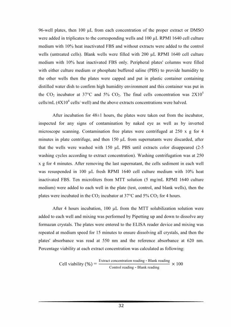
32
96-well plates, then 100 µL from each concentration of the proper extract or DMSO
were added in triplicates to the corresponding wells and 100 µL RPMI 1640 cell culture
medium with 10% heat inactivated FBS and without extracts were added to the control
wells (untreated cells). Blank wells were filled with 200 µL RPMI 1640 cell culture
medium with 10% heat inactivated FBS only. Peripheral plates' columns were filled
with either culture medium or phosphate buffered saline (PBS) to provide humidity to
the other wells then the plates were capped and put in plastic container containing
distilled water dish to confirm high humidity environment and this container was put in
the CO2 incubator at 37°C and 5% CO2. The final cells concentration was 2X10⁵
cells/mL (4X10⁴ cells/ well) and the above extracts concentrations were halved.
After incubation for 48±1 hours, the plates were taken out from the incubator,
inspected for any signs of contamination by naked eye as well as by inverted
microscope scanning. Contamination free plates were centrifuged at 250 x g for 4
minutes in plate centrifuge, and then 150 µL from supernatants were discarded, after
that the wells were washed with 150 µL PBS until extracts color disappeared (2-5
washing cycles according to extract concentration). Washing centrifugation was at 250
x g for 4 minutes. After removing the last supernatant, the cells sediment in each well
was resuspended in 100 µL fresh RPMI 1640 cell culture medium with 10% heat
inactivated FBS. Ten microliters from MTT solution (5 mg/mL RPMI 1640 culture
medium) were added to each well in the plate (test, control, and blank wells), then the
plates were incubated in the CO2 incubator at 37°C and 5% CO2 for 4 hours.
After 4 hours incubation, 100 µL from the MTT solubilization solution were
added to each well and mixing was performed by Pipetting up and down to dissolve any
formazan crystals. The plates were entered to the ELISA reader device and mixing was
repeated at medium speed for 15 minutes to ensure dissolving all crystals, and then the
plates' absorbance was read at 550 nm and the reference absorbance at 620 nm.
Percentage viability at each extract concentration was calculated as following:
Cell viability (%) = Extract concentration reading - Blank reading
Control reading - Blank reading × 100

33
3.7 Assessment of Plant Extracts Effects on Peripheral Blood
Mononuclear Cells (PBMCs) by MTT Assay
The aim of modern cancer chemotherapy is to selectively destroy cancerous
cells while having as little effect as possible on the patient’s normal cell populations and
general homeostatic balance. A plant extract or compound isolated from such an extract
exhibiting anticancer activity is not only required to induce apoptosis in malignant cells;
it also needs to do so selectively in malignant cells without affecting normal healthy
cells.
The degree of toxicity expressed by extracts of O. syriacum, H. sabdariffa, T.
vulgaris and P. graveolens on THP-1 cells by means of MTT assay have been
investigated as seen in the previous section. Investigation of the possible expression of
toxicity of the extracts in normal, healthy human cells was judged as the next logical
step.
In order to obtain PBMCs, venous blood was collected from a healthy,
consenting adult in tubes containing sodium heparin as anticoagulant and processed
within 2 hours of collection. 5.0 ± 0.5 mL from this blood were layered on
approximately equal volume of Histopaque-1077 previously prepared in two or three 15
mL conical centrifuge tubes (according to the total blood volume; roughly 10-15 mL in
each experiment). The layered blood was centrifuged at 400 x g for exactly 30 minutes
at room temperature, after that the upper layer (plasma) was carefully aspirated and
discarded except for approximately 0.5 mL and the opaque interface which contains
PBMCs was transferred with a pipette to clean 15 mL conical centrifuge tube. The cells
were washed with 10 mL PBS then washed twice with 5 mL PBS or culture medium.
Washing centrifugation was at 250 x g for 10 minutes. PBMCs were resuspended in
modified RPMI 1640 complete medium, with 2.05 mM L-glutamine and 25 mM
HEPES supplemented with 10% FBS, 5 µg/ mL phytohemagglutinin (PHA), 100
µg/mL streptomycin, and 100 U/mL penicillin. The cells were firstly resuspended in 5
mL medium, counted by hemocytometer and diluted by medium to reach concentration
of 1X10⁶ cells/mL (The procedure was performed following the instructions sheet
provided by Histopaque-1077 from Sigma-Aldrich with slight alteration of the starting
volumes). Concurrently with cell counting and by trypan blue exclusion test, viability
was estimated and in all experiments, PBMCs viability was at least 98%.

34
Extracts concentrations ranges tested on PBMCs were as following: from 10 to
0.15625 mg/mL for O. syriacum; from 20 to 0.3125 mg/mL for H. sabdariffa; from 2.5
to 0.078125 mg/mL for T. vulgaris and from 5.0 to 0.15625 mg/mL for P. graveolens.
DMSO concentrations were from 5.0 to 0.15625% (v/v).
From 1X10⁶ cells/mL PBMCs suspension, 100 µL were seeded by a
multichannel pipette to the test and control wells of sterile 96-well plates, then 100 µL
from each extract or DMSO concentration were added in triplicates (except otherwise
indicated) to the corresponding wells and 100 µL RPMI 1640 cell culture medium with
10% heat inactivated FBS and without extracts were added to the control wells. The
final cells concentration was 5X10⁵ cells/mL (1X10⁵ cells/ well) and the above extracts
concentrations were halved also PHA and antibiotics concentrations were halved. Blank
wells were filled with 200 µL RPMI 1640 cell culture medium with 10% heat
inactivated FBS only. As in leukemia cells experiments, peripheral plates' columns were
filled with either culture medium or phosphate buffered saline (PBS) then the plates
were put in plastic container containing distilled water dish and this container was put in
the CO2 incubator at 37°C and 5% CO2.
After 48 ±1 hours of incubation, contamination free plates were processed by
MTT assay which was performed typically as with THP-1 cells. Percentage viability at
each extract concentration was calculated and plotted against extract concentrations.
3.8 Determination of Plant Extracts Toxicity on THP-1 Cells by LDH
Assay
To discriminate between the cytotoxic effect of the plant extract and the growth
inhibitory effect, LDH assay was proposed because it investigates the lysis degree of the
cells under study, while MTT assay investigates the degree of metabolic activity and
proliferation of the cells under study.
Three plants' extracts only (O. syriacum, T. vulgaris, and P. graveolens) which
their cytotoxic effects were investigated by LDH toxicity assay. H. sabdariffa effective
concentrations presented by MTT assay were very high (~10-20 mg/mL) and to process
LDH assay on this extract, higher concentrations (>20 mg/mL) than that used in MTT
assay is required (as happened with the other extracts) and this is difficult because the
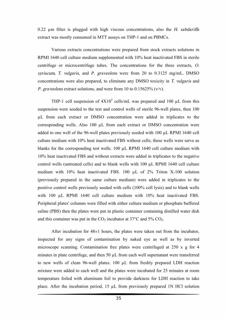
35
0.22 µm filter is plugged with high viscous concentrations, also the H. sabdariffa
extract was mostly consumed in MTT assays on THP-1 and on PBMCs.
Various extracts concentrations were prepared from stock extracts solutions in
RPMI 1640 cell culture medium supplemented with 10% heat inactivated FBS in sterile
centrifuge or microcentrifuge tubes. The concentrations for the three extracts, O.
syriacum, T. vulgaris, and P. graveolens were from 20 to 0.3125 mg/mL. DMSO
concentrations were also prepared, to eliminate any DMSO toxicity in T. vulgaris and
P. graveolens extract solutions, and were from 10 to 0.15625% (v/v).
THP-1 cell suspension of 4X10⁵ cells/mL was prepared and 100 µL from this
suspension were seeded to the test and control wells of sterile 96-well plates, then 100
µL from each extract or DMSO concentration were added in triplicates to the
corresponding wells. Also 100 µL from each extract or DMSO concentration were
added to one well of the 96-well plates previously seeded with 100 µL RPMI 1640 cell
culture medium with 10% heat inactivated FBS without cells; these wells were serve as
blanks for the corresponding test wells. 100 µL RPMI 1640 cell culture medium with
10% heat inactivated FBS and without extracts were added in triplicates to the negative
control wells (untreated cells) and to blank wells with 100 µL RPMI 1640 cell culture
medium with 10% heat inactivated FBS. 100 µL of 2% Triton X-100 solution
(previously prepared in the same culture medium) were added in triplicates to the
positive control wells previously seeded with cells (100% cell lysis) and to blank wells
with 100 µL RPMI 1640 cell culture medium with 10% heat inactivated FBS.
Peripheral plates' columns were filled with either culture medium or phosphate buffered
saline (PBS) then the plates were put in plastic container containing distilled water dish
and this container was put in the CO2 incubator at 37°C and 5% CO2.
After incubation for 48±1 hours, the plates were taken out from the incubator,
inspected for any signs of contamination by naked eye as well as by inverted
microscope scanning. Contamination free plates were centrifuged at 250 x g for 4
minutes in plate centrifuge, and then 50 µL from each well supernatant were transferred
to new wells of clean 96-well plates. 100 µL from freshly prepared LDH reaction
mixture were added to each well and the plates were incubated for 25 minutes at room
temperature foiled with aluminum foil to provide darkness for LDH reaction to take
place. After the incubation period, 15 µL from previously prepared 1N HCl solution
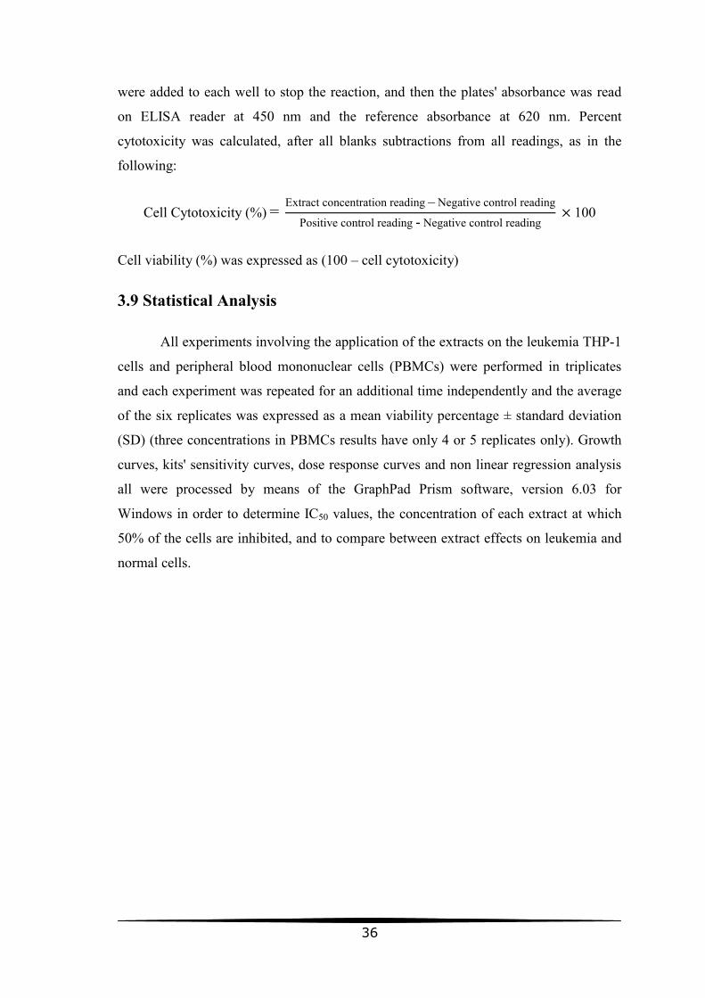
36
were added to each well to stop the reaction, and then the plates' absorbance was read
on ELISA reader at 450 nm and the reference absorbance at 620 nm. Percent
cytotoxicity was calculated, after all blanks subtractions from all readings, as in the
following:
Cell Cytotoxicity (%) = Extract concentration reading – Negative control reading
Positive control reading - Negative control reading × 100
Cell viability (%) was expressed as (100 – cell cytotoxicity)
3.9 Statistical Analysis
All experiments involving the application of the extracts on the leukemia THP-1
cells and peripheral blood mononuclear cells (PBMCs) were performed in triplicates
and each experiment was repeated for an additional time independently and the average
of the six replicates was expressed as a mean viability percentage ± standard deviation
(SD) (three concentrations in PBMCs results have only 4 or 5 replicates only). Growth
curves, kits' sensitivity curves, dose response curves and non linear regression analysis
all were processed by means of the GraphPad Prism software, version 6.03 for
Windows in order to determine IC50 values, the concentration of each extract at which
50% of the cells are inhibited, and to compare between extract effects on leukemia and
normal cells.

37
Chapter 4 Results
4.1 Plants Crude Extracts Percentage Yields
After evaporation of the extracts solvent by rotary evaporator, the pre-weighed
evaporating flasks containing the crude extracts of H. sabdariffa, O. syriacum, T.
vulgaris and P. graveolens were weighed again in order to determine the weight of
crude extracts obtained. Table 4.1 shows the percentage yields of the dry extracts
obtained (w/w).
Table 4.1: Percentage yields (%) of all dried extracts obtained after solvent evaporation.
Plant Weight of dried plant material
(gram)
Weight of extracts after solvent
evaporation (gram)
Percentage yields (%) w/w
H. sabdariffa 17.7 2.8 15.8
O. syriacum 20.0 3.4 17.0
T. vulgaris 20.0 2.4 12.0
P. graveolens 20.0 1.3 6.5
The firstly prepared O. syriacum and P. graveolens extracts have been
consumed during this study and a second round of extraction was needed. The
percentage yield of the dried extracts in the second round was different where O.
syriacum extract percentage yield was reduced to 9.5% and P. graveolens extract
percentage yield was reduced to 5.5%.
4.2 Morphology and Growth Characteristics of THP-1 Cells
THP-1 cells are monoblastic, leukemic cells and when culturing them, they grow
in suspension either as single cells or in small aggregates floating in the medium
without attaching to the container surface. These cells were spherical in shape and have
shown size variation. Some cells had protrusions and this is a common feature for
monocytic lineage. Figure 4.1 shows the morphology of THP-1 cells in culture as seen
under the inverted microscope.
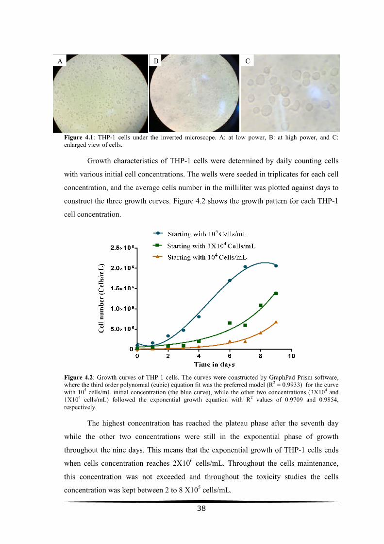
38
Figure 4.1: THP-1 cells under the inverted microscope. A: at low power, B: at high power, and C: enlarged view of cells.
Growth characteristics of THP-1 cells were determined by daily counting cells
with various initial cell concentrations. The wells were seeded in triplicates for each cell
concentration, and the average cells number in the milliliter was plotted against days to
construct the three growth curves. Figure 4.2 shows the growth pattern for each THP-1
cell concentration.
Figure 4.2: Growth curves of THP-1 cells. The curves were constructed by GraphPad Prism software, where the third order polynomial (cubic) equation fit was the preferred model (R2 = 0.9933) for the curve with 105 cells/mL initial concentration (the blue curve), while the other two concentrations (3X10⁴ and 1X10⁴ cells/mL) followed the exponential growth equation with R2 values of 0.9709 and 0.9854, respectively.
The highest concentration has reached the plateau phase after the seventh day
while the other two concentrations were still in the exponential phase of growth
throughout the nine days. This means that the exponential growth of THP-1 cells ends
when cells concentration reaches 2X10⁶ cells/mL. Throughout the cells maintenance,
this concentration was not exceeded and throughout the toxicity studies the cells
concentration was kept between 2 to 8 X10⁵ cells/mL.
C A B

39
4.3 MTT and LDH Assays Sensitivity Studies
Various concentrations of THP-1 cells ranging from 103 to 107 cells/mL were
assayed by both MTT and LDH kits to determine the range of THP-1 cells
concentrations that these assays identify them in linear manner. Sensitivity curves were
constructed by plotting kit absorbance against cell concentration. Figure 4.3 shows the
sensitivity curves for MTT and LDH kits.
Figure 4.3: Sensitivity curves of MTT and LDH assays to THP-1 cells. The curves were constructed by GraphPad Prism software and for both MTT (the red curve) and LDH readings (green curve), hyperbola model was the chosen fit model where R2 was 0.9995 for MTT curve and 0.9902 for LDH curve.
It was clear that the linear part of MTT and LDH curves has resulted by the cell
concentration range of 105 to 106 cells/mL. So, the initial cell concentration used in
these assays was 2X10⁵ cells/mL.
4.4 Plant Extracts Effects on THP-1 Cells by MTT Assay
The THP-1 cells were incubated with various concentrations of the four tested
plant extracts as well as DMSO respective concentrations for 48±1 hours and then were
assayed by MTT assay to determine the extracts and DMSO toxicity to the cells as
indicated in the materials and methods chapter. The results were expressed as a mean
percentage ± standard deviation (SD) of the mean negative control (untreated cells)
absorbance (see table 4.2). These data were analyzed using GraphPad Prism software
which used to construct dose response curves of the various extracts on the cells.

40
Table 4.2: THP-1 cells viability results by MTT test
H. sabdariffa O. syriacum T. vulgaris P. graveolens DMSO
Ext. Con. Cell Viability ± SD Ext. Con. Cell Viability ± SD Ext. Con. Cell Viability ± SD Ext. Con. Cell Viability ± SD Conc. Cell Viability ± SD
20 4.855837 ± 2.144999 8 2.736752 ± 1.074961 2 1.8547 ± 0.907174 5 3.088908 ± 0.677149 1.25 49.73694 ± 13.19307
15 55.26018 ± 4.429925 4 5.4888 ± 1.256352 1 1.77851 ± 0.828674 2.5 56.0178 ± 8.515269 0.5 81.98459 ± 12.5284
10 87.58607 ± 3.889591 2 53.69078 ± 5.803497 0.6 3.512489 ± 0.369931 1.5 100.8334 ± 11.00955 0.375 96.83851 ± 13.81734
5 82.98313 ± 6.372505 1.5 78.74228 ± 6.008436 0.4 11.77499 ± 2.022615 1.25 102.6689 ± 14.45633 0.3125 98.86212 ± 7.99779
2.5 100.0763 ± 13.11831 0.9 91.95539 ± 17.80762 0.2 48.26649 ± 7.536668 0.625 102.1776 ± 7.772026 0.25 112.3425 ± 17.32835
1.25 95.52168 ± 10.46768 0.7 92.25451 ± 18.00967 0.1 83.81131 ± 14.24472 0.3125 106.6482 ± 21.08935 0.15625 119.737 ± 6.406995
0.625 96.69471 ± 6.036152 0.5 93.27058 ± 13.71432 0.07 106.663 ± 20.01096 0.078125 116.401 ± 14.18109 0.1 107.6259 ± 20.48886
0.3125 100.8167 ± 9.708884 0.3 96.64085 ± 12.18187 0.02 119.5942 ± 28.42441
Ext. Conc. = Extract concentration in mg/mL Conc. = Concentration of DMSO (%)
SD = Standard deviation Cell Viability unit is percentage (%) 0.078125 96.32957 ± 5.106631 0.1 98.97904 ± 18.73132
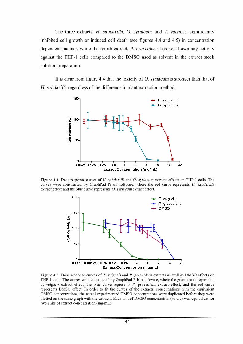
41
The three extracts, H. sabdariffa, O. syriacum, and T. vulgaris, significantly
inhibited cell growth or induced cell death (see figures 4.4 and 4.5) in concentration
dependent manner, while the fourth extract, P. graveolens, has not shown any activity
against the THP-1 cells compared to the DMSO used as solvent in the extract stock
solution preparation.
It is clear from figure 4.4 that the toxicity of O. syriacum is stronger than that of
H. sabdariffa regardless of the difference in plant extraction method.
Figure 4.4: Dose response curves of H. sabdariffa and O. syriacum extracts effects on THP-1 cells. The curves were constructed by GraphPad Prism software, where the red curve represents H. sabdariffa extract effect and the blue curve represents O. syriacum extract effect.
Figure 4.5: Dose response curves of T. vulgaris and P. graveolens extracts as well as DMSO effects on THP-1 cells. The curves were constructed by GraphPad Prism software, where the green curve represents T. vulgaris extract effect, the blue curve represents P. graveolens extract effect, and the red curve represents DMSO effect. In order to fit the curves of the extracts' concentrations with the equivalent DMSO concentrations, the actual experimented DMSO concentrations were duplicated before they were blotted on the same graph with the extracts. Each unit of DMSO concentration (% v/v) was equivalent for two units of extract concentration (mg/mL).

42
The T. vulgaris and P. graveolens extracts have been dissolved in DMSO
solvent and by comparison to the DMSO effect (see figure 4.5), it is clear that in the
range of the inhibitory concentrations of T. vulgaris extract, the DMSO had no activity
on the cells, so the dose dependent inhibition seen was attributed to the T. vulgaris
extract alone. In contrast, P. graveolens extract cell inhibition which appeared in figure
4.5 was attributed to the DMSO solvent effect because the two curves behaved roughly
similar.
GraphPad Prism software was used to perform non linear regression analysis for
each of the extracts as well as DMSO control on the THP-1 cells in order to determine
IC50 values, the concentration of each extract as well as DMSO, at which 50% of the
cells were inhibited. Table 4.3 shows the IC50 values for all extracts and DMSO.
Table 4.3: IC50 values of the extracts on THP-1 cells.
Plant Extract IC50 value (mg/mL) R2 value 95% Confidence Interval
H. sabdariffa 15.47 0.9187 14.99 to 15.96
O. syriacum 2.126 0.9094 1.934 to 2.339
T. vulgaris 0.1569 0.9312 0.1326 to 0.1857
P. graveolens 2.535 0.9041 2.334 to 2.753
DMSO 1.919 0.7018 1.470 to 2.505
All values were determined by GraphPad Prism nonlinear regression analysis. Log (inhibitor) vs. response - Variable slope (four parameters) equation was the best fit model for all the extracts and DMSO curves when compared by the software to the other dose response inhibition models. The bottom of all curves was constrained to zero value. R2 values and 95% confidence intervals for each IC50 are also indicated.
T. vulgaris extract had the highest cell growth inhibitory effect or toxicity on
THP-1 cells with lowest IC50 value followed by O. syriacum then H. sabdariffa extracts.
IC50 value for DMSO was less than that of P. graveolens which considered having no
toxic effect on THP-1 cells.

43
4.5 Plant Extracts Effects on Peripheral Blood Mononuclear Cells
(PBMCs) by MTT Assay
PBMCs were separated by Histopaque-1077 and the effect of the plant extracts
on them was assessed by MTT assay after exposing the cells to various extract
concentrations as with THP-1 cells (PBMCs MTT viability results are detailed in table
4.4). Dose response curves of the extracts effects on PBMCs are shown in figures 4.6
and 4.7.
Figure 4.6: Dose response curves of H. sabdariffa and O. syriacum extracts effects on PBMCs. The curves were constructed by GraphPad Prism software, where the red curve represents H. sabdariffa extract effect and the blue curve represents O. syriacum extract effect.
As in toxicity studies on THP-1 cells, the toxicity of O. syriacum on PBMCs is
stronger than that of H. sabdariffa.
Figure 4.7: Dose response curves of T. vulgaris and P. graveolens extracts as well as DMSO effects on PBMCs. The curves were constructed by GraphPad Prism software, where the green curve represents T. vulgaris extract effect, the blue curve represents P. graveolens extract effect, and the red curve represents DMSO effect. In order to fit the curves of the extracts' concentrations with the equivalent DMSO concentrations, the actual experimented DMSO concentrations were duplicated before they were blotted on the same graph with the extracts. Each unit of DMSO concentration (% v/v) was equivalent for two units of extract concentration (mg/mL).

44
Table 4.4: Peripheral blood mononuclear cells viability results by MTT test
H. sabdariffa O. syriacum T. vulgaris P. graveolens DMSO
Ext. Con. Cell Viability ± SD Ext. Con. Cell Viability ± SD Ext. Con. Cell Viability ± SD Ext. Con. Cell Viability ± SD Con. Cell Viability ± SD
20 20.4263 ± 7.783533 10 19.18277 ± 4.965453 2.5 6.211406 ± 2.103669 5 5.080394 ± 4.728847 5 16.65396 ± 3.636308
10 14.13379 ± 2.146318 5 8.580562 ± 2.505742 1.25 3.735115 ± 1.682673 2.5 9.028989 ± 3.42996 2.5 58.1178 ± 4.478077
5 16.86796 ± 4.816498 2.5 7.429661 ± 4.544233 0.625 9.731462 ± 2.897807 1.25 19.33511 ± 4.078954 1.25 82.17661 ± 5.649995
2.5 62.82931 ± 7.711574 1.25 10.23557 ± 5.335506 0.3125 49.67798 ± 4.579114 0.625 47.33645 ± 5.625008 0.625 97.45749 ± 7.848894
1.25 71.9416 ± 8.730852 0.625 18.32953 ± 7.645902 0.15625 78.28277 ± 4.515461 0.3125 69.78986 ± 4.080817 0.3125 91.04869 ± 10.13889
0.625 81.74278 ± 5.671631 0.3125 63.85361 ± 7.829562 0.078125 88.10691 ± 6.401121 0.15625 88.17717 ± 12.14388 0.15625 102.3971 ± 5.811382
0.3125 84.10846 ± 4.269885 0.15625 81.1585 ± 6.287436
Ext. Conc. = Extract concentration in mg/mL Conc. = Concentration of DMSO (%) SD = Standard deviation Cell Viability unit is percentage (%)

45
Visually (figure 4.7), it appears that in the range of the inhibitory concentrations
of T. vulgaris and P. graveolens extracts, the DMSO had little activity on the PBMCs,
so the dose dependent inhibition of these cells was mainly attributed to the T. vulgaris
and P. graveolens extracts. P. graveolens extract has inhibitory effect on PBMCs while
on THP-1 cells it has not had any inhibitory activity.
The IC50 values for the four extracts and DMSO on PBMCs are listed in table
4.5. Figures from 4.8 to 4.10 show comparison between the effect of each extract on
both THP-1 cells and PBMCs.
Table 4.5: IC50 values of the extracts on PBMCs.
Plant Extract IC50 value (mg/mL) R2 value 95% Confidence Interval
H. sabdariffa 3.514 0.8975 2.908 to 4.246
O. syriacum 0.4247 0.8663 0.3385 to 0.5327
T. vulgaris 0.3345 0.9820 0.3114 to 0.3593
P. graveolens 0.5534 0.9635 0.4323 to 0.7085
DMSO 5.676 0.9386 5.140 to 6.268
All values were determined by GraphPad Prism nonlinear regression analysis. Log (inhibitor) vs. response - Variable slope (four parameters) equation was the best fit model for all extracts, except H. sabdariffa, curves. Also it was the best fit for DMSO curve. H. sabdariffa extract curve was fitted by asymmetric sigmoidal, 5 PL, equation. In all of these extracts curves the bottom was constrained to zero value. R2 values and 95% confidence intervals for each IC50 are also indicated.
T. vulgaris extract had the highest toxicity on PBMCs with lowest IC50 value
followed by O. syriacum then P. graveolens and H. sabdariffa extracts. IC50 value for
DMSO was high in comparison to T. vulgaris and P. graveolens extracts.
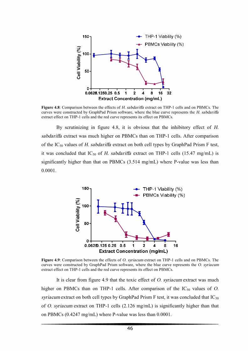
46
Cel
l Via
bilit
y (%
)
Figure 4.8: Comparison between the effects of H. sabdariffa extract on THP-1 cells and on PBMCs. The curves were constructed by GraphPad Prism software, where the blue curve represents the H. sabdariffa extract effect on THP-1 cells and the red curve represents its effect on PBMCs.
By scrutinizing in figure 4.8, it is obvious that the inhibitory effect of H.
sabdariffa extract was much higher on PBMCs than on THP-1 cells. After comparison
of the IC50 values of H. sabdariffa extract on both cell types by GraphPad Prism F test,
it was concluded that IC50 of H. sabdariffa extract on THP-1 cells (15.47 mg/mL) is
significantly higher than that on PBMCs (3.514 mg/mL) where P-value was less than
0.0001.
Figure 4.9: Comparison between the effects of O. syriacum extract on THP-1 cells and on PBMCs. The curves were constructed by GraphPad Prism software, where the blue curve represents the O. syriacum extract effect on THP-1 cells and the red curve represents its effect on PBMCs.
It is clear from figure 4.9 that the toxic effect of O. syriacum extract was much
higher on PBMCs than on THP-1 cells. After comparison of the IC50 values of O.
syriacum extract on both cell types by GraphPad Prism F test, it was concluded that IC50
of O. syriacum extract on THP-1 cells (2.126 mg/mL) is significantly higher than that
on PBMCs (0.4247 mg/mL) where P-value was less than 0.0001.
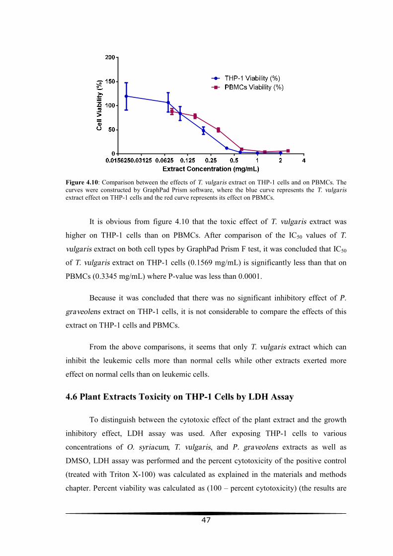
47
Figure 4.10: Comparison between the effects of T. vulgaris extract on THP-1 cells and on PBMCs. The curves were constructed by GraphPad Prism software, where the blue curve represents the T. vulgaris extract effect on THP-1 cells and the red curve represents its effect on PBMCs.
It is obvious from figure 4.10 that the toxic effect of T. vulgaris extract was
higher on THP-1 cells than on PBMCs. After comparison of the IC50 values of T.
vulgaris extract on both cell types by GraphPad Prism F test, it was concluded that IC50
of T. vulgaris extract on THP-1 cells (0.1569 mg/mL) is significantly less than that on
PBMCs (0.3345 mg/mL) where P-value was less than 0.0001.
Because it was concluded that there was no significant inhibitory effect of P.
graveolens extract on THP-1 cells, it is not considerable to compare the effects of this
extract on THP-1 cells and PBMCs.
From the above comparisons, it seems that only T. vulgaris extract which can
inhibit the leukemic cells more than normal cells while other extracts exerted more
effect on normal cells than on leukemic cells.
4.6 Plant Extracts Toxicity on THP-1 Cells by LDH Assay
To distinguish between the cytotoxic effect of the plant extract and the growth
inhibitory effect, LDH assay was used. After exposing THP-1 cells to various
concentrations of O. syriacum, T. vulgaris, and P. graveolens extracts as well as
DMSO, LDH assay was performed and the percent cytotoxicity of the positive control
(treated with Triton X-100) was calculated as explained in the materials and methods
chapter. Percent viability was calculated as (100 – percent cytotoxicity) (the results are

48
listed in table 4.6) then data analysis and dose response curves were processed by
GraphPad Prism software.
Table 4.6: THP-1 cells viability results by LDH test Ext.
Conc. Cell Viability ± Standard Deviation (%)
O. syriacum T. vulgaris P. graveolens DMSO
20 -5.11004 ± 33.28134 69.27668 ± 11.2473 64.47616 ± 6.810537 69.50853 ± 6.001513
10 49.73109 ± 6.13207 73.98201 ± 8.411463 72.82904 ± 3.853286 72.42552 ± 4.153989
5 77.34154 ± 3.874169 82.31145 ± 5.650866 77.26892 ± 8.014075 82.7447 ± 1.265742
2.5 93.27701 ± 2.722283 87.86831 ± 10.33276 92.48398 ± 6.372939 94.08492 ± 4.1521
0.625 96.55623 ± 3.565946 95.42127 ± 4.289322 94.49315 ± 3.338085 96.71546 ± 3.253288
0.3125 100.1726 ± 3.416478 95.75146 ± 5.786234 97.01363 ± 2.562171 97.63185 ± 3.140129
Ext. Conc. = Extract concentration in mg/mL
5 10 15 20 25
-50
0
50
100
150
Extract Concentration (mg/mL)
Cel
l Via
bilit
y (%
)
O. syriacumT. vulgarisP. graveolensDMSO Effect
Figure 4.11: LDH toxicity dose response curves of O. syriacum, T. vulgaris, and P. graveolens extracts as well as DMSO on THP-1 cells. These curves were constructed by GraphPad Prism software. In order to fit the curves of the T. vulgaris and P graveolens extracts' concentrations with the equivalent DMSO concentrations, the actual experimented DMSO concentrations were duplicated before they were blotted on the same graph with the extracts. Each unit of DMSO concentration (% v/v) was equivalent for two units of extract concentration (mg/mL).
As seen in figure 4.11, both T. vulgaris and P. graveolens had no cytotoxic
activity on THP-1 cells and the slight inhibition which appears in the figure was not
different from their solvent (DMSO) inhibition. By comparing the three curves (T.
vulgaris, P. graveolens and DMSO curves) with GraphPad Prism F test through non

49
linear regression analysis, the P-value was 0.5649 and this indicates that there was no
significant difference between the three curves. These findings means that while P.
graveolens extract has not had any inhibitory effect on THP-1 cells, the T. vulgaris
extract expressed only growth and proliferation inhibition activity and not cytotoxic
activity against THP-1 cells at the applied concentrations and exposure time.
O. syriacum extract exerted cytotoxic effect against THP-1 cells, as it appears in
figure 4.11, by relatively high concentrations. LC50 value for O. syriacum extract was
9.646 mg/mL (R2 = 0.8798) as calculated by non linear regression analysis using
asymmetric sigmoidal, 5 PL equation model with constraining the bottom to zero value.
50
Chapter 5 Discussion
In the recent years, a remarkable interest in the investigation of anticancer
properties of natural products has been seen. This interest is due to the fact that
conventional cancer treatment procedures (such as surgery, radiotherapy and
chemotherapy) are not effective in all cases and result in serious side effects, in addition
to the drug resistance associated with chemotherapeutic drugs. Furthermore, the limited
availability and the expensiveness of most chemotherapy drugs, also the trust that
natural products are safer, all have contributed to the popularity of traditional medicine.
This study was originated to fulfill the aim of discovering new anticancer agents from
natural materials.
Cultured cancerous cell lines with comparison to normal healthy cell lines are
commonly used to assess the anticancer activities of isolated phytochemicals and
extracts of medicinal plants. Some herbs may be harmful to the human body if they are
used improperly, and some herbs may cause serious toxicity when taken excessively or
under inappropriate circumstances. The successful treating material is that which
exhibits its therapeutic effect without causing serious side effects or exerting toxicity to
the body cells and organs. Therefore, it is not enough to investigate the therapeutic
activities of a given material without assessing its cytotoxicity against normal cells. If
this cytotoxicity was not excluded or at least reduced, then this material is not
considered a successful therapeutic drug.
The present study objectives were: assessing the antileukemic effects of the
Origanum syriacum, Hibiscus sabdariffa, Thymus vulgaris, and Pelargonium
graveolens extracts on THP-1 cells, also investigation of their inhibitory effect against
normal PBMCs to assess their selectivity. In addition, the detection of cytotoxicity of
the extracts on leukemic cells by LDH assay was a third objective.
Anticancer materials which induce cancer cell death and apoptosis are called
cytotoxic and those which do not kill cancer cells but instead stop the proliferation are
called cytostatic. Whether a drug seems cytostatic or cytotoxic may be drug independent
and instead depend on the dose used, the schedule of administration, the phase of the

51
cell cycle in which the drug acts and in which the cell resides, and the cellular context
(Rixe & Fojo, 2007).
MTT cell proliferation assessment kit was used to evaluate the growth inhibition
activity of the four plants extracts on both leukemic and normal cells. MTT system
developed by Mosmann (1983) is a means of measuring the activity of living cells via
mitochondrial dehydrogenases. The cleavage of the tetrazolium salt MTT (3- (4,5-
dimethylthiazol-2-yl)-2,5-diphenyl tetrazolium bromide) into a blue colored product
(formazan) by mitochondrial dehydrogenases is potentially very useful for assaying cell
survival and proliferation. The conversion takes place only in living cells and the
amount of intracellular formazan produced, which can be measured
spectrophotometrically after dissolving it, is proportional to the number of viable cells
present.
Because both cultures with died cells and those with cells that have stopped
proliferation will result in a decreased MTT absorbance values, there was a need for
another assay that discriminates between cytotoxic and cytostatic effects of the extracts.
Lactate dehydrogenase (LDH) assay was used for this purpose. The LDH assay
(Korzeniewski & Callewaert, 1983) is a means of measuring membrane integrity as a
function of the amount of cytoplasmic LDH released into the medium. The assay is
based on the reduction of NAD by LDH. The resulting reduced NAD (NADH) is
utilized in the reduction of a tetrazolium dye to colored formazan product that is
measured spectrophotometrically. When cell death takes place in culture either by
necrosis or by apoptosis, the end of died cells is lysis and therefore, the intracellular
materials including LDH enzyme are released in to the extracellular medium. Cell
cytotoxicity can be estimated by assaying the cell free culture medium after exposing
cells to the test material for a period of time.
In this study, the only extract that resulted in both reduced viability and
increased cytotoxicity is the O. syriacum extract that is most likely to have apoptotic or
necrotic activity. On the other hand, based on above discussion, the P. graveolens and
T. vulgaris extracts have only exhibited cytostatic and not cytotoxic activities at the
applied concentrations and exposure times. These extracts had likely halted the cells
proliferation at one level or the other and did not kill the cells. H. sabdariffa extract was
not investigated by LDH assay in this study.
52
The extract of H. sabdariffa has showed a dose dependent inhibitory effect on
THP-1 leukemic cells with an IC50 value of 15.47 mg/mL, but its inhibitory effect on
normal cells was roughly five-fold stronger (IC50= 3.514 mg/mL). This finding means
that H. sabdariffa extract at certain concentrations was significantly more potent to
normal cells than leukemic cells and at the concentrations that did not inhibit PBMCs
(figure 4.8), it had not any inhibitory effect on the THP-1 leukemic cells.
Concurred with these results, one previous study showed that H. sabdariffa
crude aqueous extract did not exhibit significant inhibitory effect on some cancer cell
lines as Hep G2/C3A, Hep 3B, and SK-HEP-1 hepatoma cells (Lin et al., 2002). On the
other hand, most previous studies showed that H. sabdariffa has anticancer and/or
antileukemic activities with little cytotoxicity toward normal cells. For example,
Hibiscus anthocyanins (HAs) extracted from the roselle calyx as well as Hibiscus
polyphenol-rich extracts (HPE) were found to have a concentration-dependent
inhibitory effect on the growth of the cell lines: NIH3T3, Hep G2, MCF-7, KB, Caco-2,
Hep 3B, HL-60, and AGS cells (Chang et al., 2005; Hou et al., 2005; Lin et al., 2005).
The least cytotoxicity of both HAs and HPE was toward the normal mouse fibroblast
cell line. Another study by Akim et al. (2011) concluded that H. sabdariffa juice
exhibits anticancer activities on human breast cancer, human cervical cancer and human
ovarian cancer cell lines without investigating the cytotoxic effect against normal cells.
However, the apparent contrary of the above studies with the present study may
has resulted from the fact that in all of these studies, if the normal cells were used to
assess the cytotoxicity of the H. sabdariffa, the selected cells were the normal mouse
fibroblast cells (NIH3T3), and none has used the peripheral blood mononuclear cells
(PBMCs) as a normal cells model as was done in this study. It is known that NIH3T3
cell line grows better in culture conditions than the primary PBMCs cells and this may
lower their sensitivity to the extract in comparison to the PBMCs sensitivity. In a study
of H. sabdariffa extract effective protection of cultured PBMCs from the cellular death
induced by H2O2, the PBMCs were exposed to incremental doses of H. sabdariffa
extract (up to 2000 µg/mL) for 24 h and the viability of PBMCs was more than 95% but
when the extract concentration exceeded this limit (2 mg/mL), it exhibited a
cytotoxicity toward PBMCs (Beltrán-Debón et al., 2010). This finding is more or less
similar to the present study results where IC50 of H. sabdariffa extract on PBMCs was
3.514 mg/mL.
53
The effect of O. syriacum extract was similar to that of H. sabdariffa extract but
the IC50 value on THP-1 cells was 2.126 mg/mL, and its inhibitory effect on normal
cells (PBMCs) was extremely stronger (IC50= 0.4247 mg/mL). Not only the inhibition
of O. syriacum extract was not selective to leukemic cells, but also it does no longer
exist toward THP-1 leukemic cells at the non cytotoxic concentrations (figure 4.9).
With LDH assay, the effect of O. syriacum extract against THP-1 cells was
distinguished to be cytotoxic or apoptotic rather than cytostatic and LC50 value was
9.646 mg/mL. This would be a much more valuable finding if the effect of O. syriacum
extract was selective to the leukemic cells only.
The present study is the first to investigate the anticancer effect of O. syriacum
against leukemic cells. No previous studies were found in the literature that have
assessed the antileukemic effect of O. syriacum, but there are few studies which
investigated its anticancer activity against cancer cell lines other than leukemia cells. El-
Desouky et al. (2009) and Al-Kalaldeh et al. (2010) found that the ethanol crude
extracts of O. syriacum showed antiproliferative activity to adenocarcinoma of breast
cell line (MCF7) and human cervical adenocarcinoma (HeLa) cells with IC50 values of
6.40 μg/mL and 474.2 μg/mL, respectively. However, none of these studies investigated
the cytotoxicity of the O. syriacum extracts against PBMCs. There is only one study
(Al-Ali et al., 2013) found in the literature that deals with the O. syriacum cytotoxicity
against normal cells. And however the normal cells used by that study researchers were
the diploid human embryonic fibroblasts (MRC-5 cell line) and not PBMCs, their
results are consistent with the present study results where the crude methanolic extract
of O. syriacum has showed cytotoxic activity against the normal MRC-5 cells and IC50
was >64 µg/mL.
Not as other extracts, T. vulgaris extract has showed different results. It has
exhibited an inhibitory effect toward THP-1 leukemic cells (IC50= 0.1569 mg/mL) in a
dose dependent manner and this inhibition was twofold more potent than the extract
inhibition on normal mononuclear cells (IC50= 0.3345 mg/mL). This finding suggests
that T. vulgaris extract inhibition is more selective to leukemic cells than normal cells,
and at certain concentrations, T. vulgaris may be used to inhibit leukemia without
causing significant cytotoxicity toward normal body cells.

54
Consistent with these results, Amir and Karimi (2001) in their study found that
the ethanolic extract of T. vulgaris showed a dose dependent inhibition of proliferation
of human breast cancer (SK-Br-3, MDA-MB-435) and leukemia (U937 and K562) cell
lines as well as cervical epithelial carcinoma (HeLa) cell line which was the least
sensitive cell line to the extract. The most sensitive cells were U937 and MDA (50%
inhibition at 10 µg/mL). Also they found that the effect of T. vulgaris on normal Vero
cell line and PBMCs was stimulatory at low concentrations (10-100 µg/mL) and
inhibitory when extract concentration increased (at 400 µg/mL, the inhibition of Vero
cells was 27.6%). In contrary, Berrington and Lall (2012) in another study have
different results and found that acetone extract of T. vulgaris inhibits the normal Vero
cell line (IC50= 138.4 μg/mL) more than the HeLa cells (IC50> 200 μg/mL).
Furthermore the essential oil of T. vulgaris was tested on head and neck squamous cell
carcinoma (HNSCC) by Sertel et al. (2011) and also was found to have a relatively low
toxicity with an IC50 value of 369 μg/mL.
The IC50 value of T. vulgaris extract on THP-1 cells was low in comparison to
other extracts values. However, the effect of this extract on THP-1 cells was only
cytostatic, and at the concentrations used in this study with LDH assay there was no
detected cytotoxic or apoptotic effect of this extract. Whether the T. vulgaris extract
effect was cytotoxic or cytostatic, it is a hopeful finding that this plant contains cancer
therapeutic agents that are more selective to leukemic cells than normal cells at
relatively low concentrations.
The carvacrol and thymol may be the most constituents of T. vulgaris extract
which are responsible for its anticancer activities. Many studies have reported the
anticancer effect of these essential oils. Carvacrol and thymol significantly reduced the
level of DNA damage induced in K-562 cells by the strong oxidant H2O2 (Horvathova
et al., 2007). Furthermore, carvacrol has an important in vitro antitumor effect against
tumor cell lines like Hep-2 (Stammati et al., 1999), B16 (He et al., 1997) and A-549
(Tansu Koparal & Zeytinoglu, 2003; Zeytinoglu et al., 2003). The effect of carvacrol
and thymol on cell cycle progression in K-562 tumor cells was examined by Jaafari et
al. (2012) with flow cytometry after DNA staining in order to investigate the molecular
mechanism of their cytotoxic activity. The results revealed that carvacrol stopped the
cell cycle progression in S phase; however, thymol stopped it in G0/G1 phase.

55
P. graveolens extract has not exerted any effect, neither cytotoxic nor growth
inhibitory, against the THP-1 leukemic cells at the studied concentrations. However, it
has exhibited inhibitory activity against PBMCs (figure 4.7). There are no studies found
in the literature that have assessed the P. graveolens anticancer activities using the plant
crude extracts while many studies have detected the plant's essential oils (citronellol,
geraniol, and transgeraniol) anticancer and antileukemic behaviors (subsection 2.2.4).
The contrary of this study finding with those studies' results may be due to the use of the
plant crude extract in the present study where the concentrations of the essential oils
with detected anticancer activities are low in comparison to the separated and purified
essential oils used in those studies.
It is also expected that the cytotoxicity of the used extracts in this study against
the normal PBMCs was relatively high due to the application of the crude extract and
not purified compounds. The crude extract contains many unpurified and unspecified
materials that may exert non specific inhibition against the already difficult to grow
PBMCs and not necessarily that the compounds which posses anticancer or
antileukemic activities are the same compounds that exhibit non specific inhibition
against the normal cells. This thesis is a screening study and many subsequent more
detailed studies can be performed to study the antiproliferative and cytotoxic effect of
the purified compounds of the extracts which have showed antileukemic activities in
this preliminary study.
Furthermore, it is expected that the studied extracts if investigated in vivo, they
will exhibit less cytotoxicity against normal cells than that exhibited when the
investigation occurs in vitro. The in vitro cytotoxic effect exerted by the extracts against
the normal cells may be due to the fact that the primary blood mononuclear cells
behavior outside the body is different from that inside the body because the culture
conditions even when are optimal are not as the physiological environment in vivo. This
difference may affect normal cells more than leukemic cancerous cells that have gained
some growth advantages during their multistage progress towards cancer.
There is a study by Lakis et al. (2012) in which the T. vulgaris and O. syriacum
essential oils cytotoxic effect on normal cells was investigated in vivo. The
hematological and biochemical routine parameters of Wistar rats were assayed in order
to determine a possible secondary effect after the oral administration of T. vulgaris and
56
O. syriacum essential oils. The oral dose used in the study corresponded to 0.03g
carvacrol/Kg bw/day in the case of T. vulgaris essential oil and 0.015g thymol/Kg body
weight/day in case of O. syriacum essential oil. No significant variations of the
hematological or biochemical parameters were noticed after the oral treatment with T.
vulgaris essential oil (29.74% thymol) and O. syriacum essential oil (66.64% carvacrol)
compared to the control group treated with physiological solution.

57
Chapter 6 Conclusion and Recommendations
6.1 Conclusion
In this study the in vitro antiproliferative and cytotoxic effects of Origanum
syriacum, Hibiscus sabdariffa, Thymus vulgaris, and Pelargonium graveolens extracts
on human leukemic THP-1 cells were assessed. Also the inhibitory effects of these
extracts on normal peripheral blood mononuclear cells were evaluated.
It was found that both extracts Origanum syriacum and Hibiscus sabdariffa
exert concentration dependent antiproliferative effect on leukemia as well as on normal
cells. Origanum syriacum extract also exhibited cytotoxic activity against THP-1
leukemic cells which was a significant finding in this study. These extracts need to be
more selective toward leukemic cells than normal cells before they are considered
successful antileukemic agents.
Pelargonium graveolens extract did not exhibit any inhibitory effect neither
antiproliferative nor cytotoxic toward leukemic cells at the applied concentrations and
exposure time. In addition, it has exerted antiproliferative effect against normal cells. As
a result, it is unpredicted that this extract may possess significant therapeutic
characteristics for leukemia.
The fourth extract, Thymus vulgaris, has had more selective antiproliferative
effect against leukemic cells and its effect was only cytostatic and not cytotoxic at the
applied concentrations and exposure time. At relatively low concentrations, this extract
was able to inhibit THP-1 cells proliferation more than normal cells proliferation and
this inhibition was in a dose dependent manner. This important finding gives a hope that
Thymus vulgaris active materials can be useful in leukemia treatment development.
6.2 Recommendations
It is recommended that further research should be undertaken in the following areas:
• Investigation of the Origanum syriacum, Hibiscus sabdariffa, and Thymus
vulgaris extracts toxicity in vivo at the effective antileukemic concentrations.
58
• Purification of Origanum syriacum, Hibiscus sabdariffa, and Thymus vulgaris
extracts active materials and assessment of the antiproliferative and cytotoxic
effects of these purified materials against leukemic cells as well as normal cells
both in vitro and in vivo.
• Following the mode of action of these extracts against leukemia cells at the
molecular level to understand their antileukemic mechanisms.
• Assessment of these extracts anticancer effect against malignant cells of cancer
types other than leukemia.
• Assessment of other medicinal plants extracts on both leukemia cells and other
cancer cells.
59
Chapter 7 References
• Abu Lafi S, Odeh I, Dewik H, et al. (2007) Natural compounds of Palestine flora.
Comparison analysis by static headspace and steam distillation GC-MS of
semivolatile secondary metabolites from leaves of cultivated Palestinian Majorana
syriaca. Biomedical papers of the Medical Faculty of the University Palacký,
Olomouc, Czechoslovakia, 151: 21-29.
• Abu Lafi S, Odeh I, Dewik H, et al. (2008) Thymol and carvacrol production from
leaves of wild Palestinian Majorana syriaca. Bioresource Technology, 99: 3914-
3918.
• Aburjai T, Hudaib M, Tayyem R, et al. (2007) Ethnopharmacological survey of
medicinal herbs in Jordan, the Ajloun Heights region. Journal of
Ethnopharmacology, 110: 294–304.
• Agrawala S, Chatterjee S. and Misra S (2001) Immune-potentiation activity of a
poly-herbal formulation ‘IMMU-21’ (research name). Phytomedica, 2: 1–22.
• Akim A, Ling L, Rahmat A, et al. (2011) Antioxidant and anti-proliferative
activities of roselle juice on Caov-3, MCF-7, MDA-MB-231 and HeLa cancer cell
lines. African Journal of Pharmacy and Pharmacology, 5: 957-965.
• Al Najar K., Awad R, and Thabet K (2002) Cancer 1995-2000 - Palestine. Ministry
of Health, Palestine.
• Al-Ali K, El-Beshbishy H, Alghaithy A, et al. (2013) In vitro Antioxidant Potential
and Antiprotozoal Activity of Methanolic Extract of Mentha longifolia and
Origanum syriacum. Journal of Biological Sciences, 13: 207-216.
• Ali-Shtayeh M, Yaniv Z, and Mahajna J (2000) Ethnobotanical survey in the
Palestinian area: a classification of the healing potential of medicinal plants. Journal
of Ethnopharmacology, 73: 221–232.
• Al-Kalaldeh J, Abu-Dahab R, and Afifi F (2010) Volatile oil composition and
antiproliferative activity of Laurus nobilis, Origanum syriacum, Origanum vulgare,
and Salvia triloba against human breast adenocarcinoma cells. Nutrition Research,
30: 271-278.
60
• Alma M, Mavi A, Yildirim A, et al. (2003) Screening of chemical composition and
in vitro antioxidant and antimicrobial activities of the essential oils from Origanum
syriacum L. growing in Turkey. Biological and Pharmaceutical Bulletin, 26: 1725–
1729.
• Amabeoku G (2009) Antidiarrhoeal activity of Geranium incanum Burm of
(Geraniaceae) leaf aqueous extract in mice. Journal of Ethnopharmacology, 123:
190–193.
• Amir G and Karimi M (2001) Cytotoxic activity of Thymus vulgaris, Achillea
millefolium and Thuja orientalis on different growing cell lines. Medical Journal of
the Islamic Republic of Iran, 15: 149-154.
• Baer M, Stewart C, Lawrence D, et al. (1998) Acute myeloid leukemia with 11q23
translocations: Myelomonocytic immunophenotype by multiparameter low
cytometry. Leukemia, 12: 317-325.
• Bain B (2003) Leukemia Diagnosis 3rd Edition. Blackwell Publishing Ltd, Oxford,
UK.
• Battle T, Arbiser J, and Frank D (2005) The natural product honokiol induces
caspase-dependent apoptosis in B-cell chronic lymphocytic leukemia (B-CLL) cells.
Blood, 106: 690–697.
• Baydar H, Sagdic O, Ozkan G, et al. (2004) Antibacterial activity and composition
of essential oils from Origanum, Thymbra and Satureja species with commercial
importance in Turkey. Food Control, 15: 169–172.
• Bayoub K, Baibai T, Mountassif D, et al. (2010) Antibacterial activities of the crude
ethanol extracts of medicinal plants against Listeria monocytogenes and some other
pathogenic strains. African Journal of Biotechnology, 9: 4251-4258.
• Beltrán-Debón R, Alonso-Villaverde C, Aragonès G, et al. (2010) The aqueous
extract of Hibiscus sabdariffa calices modulates the production of monocyte
chemoattractant protein-1 in humans. Phytomedicine, 17: 186-191.
• Bennett J, Catovsky D, Daniel M, et al. (1976) Proposals for the classification of the
acute leukaemias. French–American–British (FAB) Cooperative Group. British
Journal of Haematology, 33: 451–458.

61
• Bennett J, Catovsky D, Daniel M, et al. (1985) Criteria for the diagnosis of acute
leukemia of megakaryocyte lineage (M7). A report of the French–American–British
Cooperative Group. Annals of Internal Medicine 103: 460–462.
• Berdowska I, Zieliński B, Fecka I, et al. (2013) Cytotoxic impact of phenolics from
Lamiaceae species on human breast cancer cells. Food Chemistry, 15: 1313-1321.
• Berrington D and Lall N (2012) Anticancer activity of certain herbs and spices on
the cervical epithelial carcinoma (HeLa) cell line. Evidence-Based Complementary
and Alternative Medicine, 2012: 1-11.
• Brunning R, Matutes E, Harris N, et al. (2001) Acute myeloid leukaemia:
Introduction. In Jaffe E, Harris N, Stein H, et al., World Health Organization
Classification of Tumours. Pathology and Genetics of Tumours of Haematopoietic
and Lymphoid Tissues, International Agency for Research on Cancer Press, Lyon,
France.
• Bukovska A, Cikos S, Juhas S, et al. (2007) Effects of a combination of thyme and
oregano essential oils on TNBS-induced colitis in mice. Mediators Inflammation,
2007: 1-9.
• Burke Y, Stark M, Roac S, et al. (1997) Inhibition of pancreatic cancer growth by
the dietary isopreniods farnesol and geraniol. Lipids, 32: 151-156.
• Butles M (2004) The role of natural product chemistry in drug discovery. Journal of
Natural Products, 67: 2141-2153.
• Byrd J, Lin T, Dalton J, et al. (2007) Flavopiridol administered using a
pharmacologically derived schedule is associated with marked clinical efficacy in
refractory, genetically high-risk chronic lymphocytic leukemia. Blood, 109: 399–
404.
• Carnesecchi S, Klangley F, Exinger F, et al. (2002) Geraniol, a component of plant
essential oils sensitize human colonic cancer to 5-fluorouracil treatment. Journal of
Pharmacology and Experimental Therapeutics, 301: 625-630.
• Carnesecchi S, RuiBras G, Amyaouch B, et al. (2004) Geraniol a component of
plant essential oil, modulates DNA synthesis and potentiates 5-fluorouracile
efficacy on human colon Tumor xenograft. Cancer letters, 215: 53-55
62
• Carvajal-Zarrabal O, María D, Barradas-Dermitz D, et al. (2012) Hibiscus
sabdariffa L., roselle calyx, from ethnobotany to pharmacology. Journal of
Experimental Pharmacology, 4: 25–39.
• Carvajal-Zarrabal O, Waliszewski S, Barradas-Dermitz D, et al. (2005) The
consumption of Hibiscus sabdariffa dried calyx ethanolic extract reduced lipid
profile in rats. Plant Foods for Human Nutrition, 60: 153–159.
• Chang Y, Huang H, Hsu J, et al. (2005) Hibiscus anthocyanins rich extract-induced
apoptotic cell death in human promyelocytic leukemia cells. Toxicology and
Applied Pharmacology, 205: 201– 212.
• Chen C, Chou F, Ho Y, et al. (2004) Inhibitory effects of Hibiscus sabdariffa L.,
extract on low density lipoprotein oxidation and anti-hyperlipidemia in fructose-fed
and cholesterol fed rats. Journal of the Science of Food and Agriculture, 84: 1989-
1996.
• Cuttner J, Conjalka M, Reilly M, et al. (1980) Association of monocytic leukemia in
patients with extreme leukocytosis. The American Journal of Medicine, 69: 555-
558.
• Dancey J and Steward W (1995) The role of vindesine in oncology--
recommendations after 10 years' experience. Anticancer Drugs, 6: 625–636.
• Davis P (1982) Flora of Turkey and the East Aegean Islands (Volume 7). Edinburgh
University Press, Edinburgh, Scotland.
• De C, Phookan J, Parikh V, et al. (2012) Decitabine Induced Transient
Cardiomyopathy: A Case Report. Clinical Medicine Insights: Oncology, 6: 325–
329.
• De Moura M, E Silva J, De Oliveira R, et al. (2002) Natural products reported as
potential inhibitors of uterine cervical neoplasia. Acta Farmaceutica Bonaerense, 21:
67-74.
• Deep G, Gangar S, Oberlies N, et al. (2010) Isosilybin A induces apoptosis in
human prostate cancer cells via targeting Akt, NF-kB, and androgen receptor
signaling. Molecular Carcinogenesis, 49: 902–912.

63
• Directorate Plant Production (2012) Thyme production, 2nd Edition. Agriculture,
Forestry & Fisheries, Pretoria, South Africa.
• Duke J and Ayensu E (1985) Medicinal Plants of China (Volume 2). Reference
Publications Inc, Michigan, USA.
• El-Desouky S, Ibrahima L, Kawashtya S, et al. (2009) Phytochemical constituents
and biological activities of Origanum syriacum. Zeitschrift für Naturforschung, 64:
447 – 451.
• Elmann A, Mordechay S, Rindner M, et al. (2010) Anti- neuroinflammatory effects
of geranium oil in microglial cells. Journal of Functional Foods, 2: 17–22.
• Elmore S (2007) Apoptosis: a review of programmed cell death. Toxicologic
Pathology, 35: 495–516.
• Elmorea S, Oostb T, and Park C (2005) Inhibitors of anti-apoptotic proteins for
cancer therapy. Annual Reports in Medicinal Chemistry, 40: 255-270.
• El-Nekeety A, Mohamed S, Hathout A, et al. (2011) Antioxidant properties of
Thymus vulgaris oil against aflatoxin-induce oxidative stress in male rats. Toxicon,
57: 984–991.
• Elson C (1995) Suppression of mevalonate pathway activities by dietary
isoprenoids: protective roles in cancer and cardiovascular disease. Journal of
Nutrition, 125: 1666-1672.
• Essa M, Subramanian P, Suthakar G, et al. (2006) Influence of Hibiscus sabdariffa
(gongura) on the levels of circulatory lipid peroxidation products and liver marker
enzymes in experimental hyperammonemia. Journal of Applied Biomedicine, 4: 53-
58.
• Fang H, Su X, Liu H, et al. (1989) Studies on the chemical components and anti-
tumour action of the volatile oils from Pelargonium graveolens. Acta Pharmaceutica
Sinica, 24: 366-371.
• Fang L, He Q, Hu Y, et al. (2007) MZ3 induces apoptosis in human leukemia cells.
Cancer Chemotherapy and Pharmacology, 59: 397-405.

64
• Farombi E and Fakoya A (2005) Free radical scavenging and antigenotoxic
activities of natural phenolic compounds in dried flowers of Hibiscus sabdariffa L.
Molecular Nutrition and Food Research, 49: 1120-1128.
• Fayed S (2009) Antioxidant and anticancer activities of Citrus reticulate (Petitgrain
Mandarin) and Pelargonium graveolens (Geranium) essential oils. Research Journal
of Agriculture and Biological Sciences, 5: 740-747.
• Fennell C, Lindsey K, McGaw L, et al. (2004) Assessing African medicinal plants
for efficacy and safety: Pharmacological screening and toxicology. Journal of
Ethnopharmacoly, 94: 205-217.
• Freidberg R (2009) An investigation in to the antimicrobial and anticancer activities
of Geranium incanum, Artemisia afra and Artemisia absinthium. A Master thesis,
Nelson Mandela Metropolitan University, Port Elizabeth, South Africa.
• Freshney R (2005) Culture of Animal Cells (A Manual of Basic Technique) 5th
Edition. John Wiley and Sons, Inc., New Jersey, USA.
• Goedecke T, Kaloga M, Kolodziej H (2005) A phenol glucoside, uncommon
coumarins and flavonoids from Pelargonium sidoides. Zeitschrift für
Naturforschung, 60: 677-682.
• Greer J, Foerster J, Lukens J, et al. (2003) Wintrobe's Clinical Hematology, 11th
Edition. Lippincott Williams & Wilkins, Philadelphia, USA.
• Haag J, Lindstrom M, and Gould M (1992) Limonene-induced regression of
mammary carcinomas. Cancer Research, 52: 4021-4026.
• Haji Faraji M and Haji Tarkhani A. (1999) The effect of sour tea (Hibiscus
sabdariffa) on essential hypertension. Journal of Ethnopharmacology, 65: 231-236.
• Hande K (1998) Etoposide: four decades of development of a topoisomerase II
inhibitor. European Journal of Cancer, 34: 1514–1521.
• He L, Mo H, Hadisusilo S, et al. (1997) Isoprenoids suppress the growth of murine
B16 melanomas in vitro and in vivo. Journal of Nutrition, 127: 668- 674.
• Heim S, Avanzi G, Billström R, et al. (1987) A new specific chromosomal
rearrangement, t(8;16)(p11;p13), in acute monocytic leukaemia. British Journal of
Haematology, 66: 323-326.
65
• Herrera-Arellano A, Flores-Romero S, Chavez-Soto M, et al. (2004) Effectiveness
and tolerability of a standardized extract from Hibiscus sabdariffa in patients with
mild to moderate hypertension: a controlled and randomized clinical trial.
Phytomedicine, 11: 375- 382.
• Higley C and Higley A (2001) Reference Guide for Essential Oils. Abundant
Health, London, UK.
• Hirunpanich V, Utaipat A, Morales N, et al. (2005) Antioxidant effects of aqueous
extracts from dried calyx of Hibiscus sabdariffa Linn. (Roselle) in vitro using rat
low-density lipoprotein (LDL). Biological and Pharmaceutical Bulletin, 28: 481–
484.
• Horvathova E, Turcaniova V, and Slamenova D (2007) Comparative study of DNA-
damaging and DNA protective effects of selected components of essential plant oils
in human leukemic cells K562. Neoplasma, 54: 478-483.
• Hou D, Tong X, Terahara N, et al. (2005) Delphinidin 3-sambubioside, a Hibiscus
anthocyanin, induces apoptosis in human leukemia cells through reactive oxygen
species-mediated mitochondrial pathway. Archives of Biochemistry and Biophysics,
440: 101–109.
• http://www.flowersinisrael.com/Thymusvulgaris_page.htm accessed in 7/9/2013.
• http://www.henriettesherbal.com/pictures/p10/pages/pelargonium-graveolens-2.htm
accessed in 14/9/2013.
• http://www.plantzafrica.com/plantnop/pelarggrav.htm accessed in 14/9/2013
• http://www.wildflowers.co.il/english/plant.asp?ID=129 accessed in 31/8/2013.
• Ietswaart J (1980) A Taxonomic Re Vision of the Genus Origanum (Labiatae).
Leiden University Press, Leiden, The Netherlands.
• Ismail A, Ikram E, and Nazri H (2008) Roselle (Hibiscus sabdariffa L.) seeds-
nutritional composition, protein quality and health benefits. Food, 2: 1-16.
• Jaafari A, Mouse H, Rakib E, et al. (2007) Chemical composition and antitumor
activity of different wild varieties of Moroccan thyme. Brazilian Journal of
Pharmacognosy, 17: 477-491.

66
• Jaafari A, Tilaoui M, Mouse H, et al. (2012) Comparative study of the antitumor
effect of natural monoterpenes: relationship to cell cycle analysis. Brazilian Journal
of Pharmacognosy, 22: 534-540.
• Jaffe E, Harris N, Stein H, et al. (2001) World health organization classification of
tumours; pathology and genetics of tumours of the haemopoietic and lymphoid
tissues. International Agency for Research on Cancer Press, Lyon, France.
• Kamboj V (2000) Herbal medicine. Current Science, 78: 35-39.
• Kang H, Na S, and Kim Y (2010) Effects of oral care with essential oil on
improvement in oral health status of hospice patients. Journal of Korean Academy
of Nursing, 40: 473–481.
• Kappor L (1990) CRC Handbook of ayurvedic medicinal plants. CRC Press,
Florida, USA.
• Karon M, Freireich E, Frei E, et al. (1966) The role of vincristine in the treatment of
childhood acute leukemia. Clinical Pharmacology and Therapeutics, 7: 332–339.
• Kensara1 O, ElSawy N, El-Shemi A, et al. (2013) Thymus vulgaris supplementation
attenuates blood pressure and aorta damage in hypertensive rats. Journal of
Medicinal Plants Research, 7: 669-676.
• Khatun M, Islam M, Mamun A, et al. (2011) In vivo evaluation of CNS depressant
and antinociceptive activities of methanol extract of Hibiscus sabdariffa fruits.
Journal of Applied Sciences Research, 7: 798-804.
• Kinghorn A (2008) Drug discovery from natural products; In Lemke T and
Williams D, Foye’s Principles of Medicinal Chemistry, 6th Edition. Walters
Kluwer/Lippincott Willliams & Wilkins, Baltimore, MD.
• Kintzios S (2004) Oregano: The Genera Origanum and Lippia. 1st Edition. Taylor
and Francis, London, UK.
• Knudson A (2001) Two genetic hits (more or less) to cancer. Nature Reviews
Cancer, 1: 157–62.
• Kolodziej H (2000) Traditionally used Pelargonium species: Chemistry and
biological activity of umckaloabo extracts and their constituents. Phytotherapy
Research, 3: 77-93.
67
• Kolodziej H and Kiderlen A (2007) In vitro evaluation of antibacterial and immuno
modulatory activities of Pelargonium reniforme, Pelargonium sidoides and the
related herbal drug preparation EPs® 7630. Phytomedicine, 14: 18-26.
• Kordali S, Cakir A, Ozer H, et al. (2008) Antifungal, phytotoxic and insecticidal
properties of essential oil isolated from Turkish Origanum acutidens and its three
components, carvacrol, thymol and p-cymene. Bioresource Technology, 99: 8788-
8795.
• Korzeniewski C and Callewaert D (1983) An enzyme-release assay for natural
cytotoxicity. Journal of Immunological Methods, 64: 313-320.
• Lakis Z, Mihele D, Nicorescu I, et al. (2012) The antimicrobial activity of Thymus
vulgaris and Origanum syriacum essential oils on Staphylococcus aureus,
Streptococcus pneumoniae and Candida albicans. Farmacia, 60: 857-865.
• Lev E (2002) Reconstructed materia medica of the Medieval and Ottoman al-Sham.
Journal of Ethnopharmacology, 80: 167–179.
• Lev E and Amar Z (2002) Ethnopharmacological survey of traditional drugs sold in
the Kingdom of Jordan. Journal of Ethnopharmacology, 82: 131–145.
• Limper A (2004) Chemotherapy-induced lung disease. Clinics in Chest Medicine,
25: 53– 64.
• Lin H, Huang H, Huang C, et al. (2005) Hibiscus polyphenol-rich extract induces
apoptosis in human gastric carcinoma cells via p53 phosphorylation and p38
MAPK/FasL cascade pathway. Molecular Carcinogenesis, 43: 86–99.
• Lin L, Liu L, Chiang L, et al. (2002) In vitro anti-hepatoma activity of fifteen
natural medicines from Canada. Phytotherapy Research, 16: 440–444.
• Lis-Balchin M (2002) Geranium and Pelargonium: The genera Geranium and
Pelargonium. Taylor and Francis, London, UK.
• Lis-Balchin M (2004) Geranium, In Peter K, Handbook of herbs and spices
(Volume 2). Woodhead Publishing Series in Food Science, Oxford, UK.
• Loizzo M, Menichini F, Conforti F, et al. (2009) Chemical analysis, antioxidant,
antiinflammatory and anticholinesterase activities of Origanum ehrenbergii Boiss
and Origanum syriacum L. essential oils. Food Chemistry, 117: 174–180.

68
• Lucas D, Still P, Pérez L, et al. (2010) Potential of plant-derived natural products in
the treatment of leukemia and lymphoma. Current Drug Targets, 11: 812–822.
• Lum J, DeBerardinis R, and Thompson C (2005) Autophagy in metazoans: Cell
survival in the land of plenty. Nature Reviews Molecular Cell Biology, 6: 439–448.
• Mahadevan N, Shivali A, and Kamboj P (2009) Hibiscus sabdariffa Linn. An
overview. Natural Product Radiance, 8: 77-83.
• Maksimovic Z, Stojanovic D, Sostaric I, et al. (2008) Composition and radical-
scavenging activity of Thymus glabrescens Willd. (Lamiaceae) essential oil. Journal
of the Science of Food and Agriculture, 88: 2036-2041.
• Mangal A, Grossman L, and Vickars L (1984) Disseminated intravascular
coagulation in acute monoblastic leukemia: Response to heparin therapy. Canadian
Medical Association Journal, 130: 731-733.
• Marie J and Legrand O (2003) Drug Resistance in Acute Leukaemia and Reversion.
Turkish Journal of Medical Sciences, 33: 271-279.
• Marino M, Bersani C, and Comi G (1999) Antimicrobial activity of the essential oils
of Thymus vulgaris L. measured using a bioimpedometric method. Journal of Food
Protection, 62: 1017–1023.
• Martins A, Salgueiro L, Vila R, et al. (1999) Composition of the essential oils of
Ocimum canum, O. gratissimum and O. minimum. Planta Medica, 65: 187-189.
• Miller D (2002) The taxonomy of Pelargonium species and cultivars, their origins
and growth in the wild. Geranium and Pelargoniums: The genera Geranium and
Pelargonium. In Lis-Balchin M, Medicinal and Aromatic Plants-Industrial Profiles.
Taylor and Francis, London, UK.
• Ministry of Health (2011) Health Report, Palestine First Quarter 2011. Palestinian
Health Information Center, Palestine.
• Ministry of Health (2013) Health Annual Report, Palestine 2012. Palestinian Health
Information Center, Palestine.
• Miura K, Kikuzaki H, and Nakatani N (2002) Antioxidant activity of chemical
components from sage (Salvia officinalis L.) and thyme (Thymus vulgaris L.)

69
measured by the oil stability index method. Journal of Agricultural and Food
Chemistry, 50: 1845–1851.
• Mossman T (1983) Rapid colorimetric assay for cellular growth and survival:
application to proliferation and cytotoxicity assays. Journal of Immunological
Methods, 65: 55-63.
• Munker R, Hiller E, Glass J, et al. (2007) Modern hematology: biology and clinical
management, 2nd Edition. Humana Press Inc., Totowa, New Jersey.
• Murray A (2004) Recycling the cell cycle: Cyclins revisited. Cell, 116: 221–234.
• Ocaña A, and Reglero G (2012) Effects of thyme extract oils (from Thymus
vulgaris, Thymus zygis, and Thymus hyemalis) on cytokine production and gene
expression of oxLDL-stimulated THP-1- Macrophages. Journal of Obesity, 2012: 1-
11.
• Pandey G and Madhuri S (2006) Medicinal plants: better remedy for neoplasm.
Indian Drugs, 43: 869–874.
• Peterson A, Goto M, Machmuah S, et al. (2005) Extraction of essential oil from
geranium (Pelargonium graveolens) with supercritical carbon dioxide. Journal of
Chemical Technology and Biotechnology, 81: 167-172.
• Peterson L, Dehner L, and Brunning R (1981) Extramedullary masses as presenting
features of acute monoblastic leukemia. American Journal of Clinical Pathology, 75:
140-148.
• Petit I, Karajannis M, Vincent L, et al. (2008) The microtubule-targeting agent
CA4P regresses leukemic xenografts by disrupting interaction with vascular cells
and mitochondrial-dependent cell death. Blood 111: 1951–1961.
• Pina-Vaz C, Gonçalves C, Rodrigues E, et al. (2004) Antifungal activity of Thymus
oils and their major compounds. Journal of the European Academy of Dermatology
and Venereology, 18: 73–78.
• Provan D (2003) ABC of Clinical Haematology 2nd Edition. BMJ Books, London,
UK.
• Qi Y, Chin K, Malekian F, et al. (2005) Biological characteristics, nutritional and
medicinal value of roselle, Hibiscus sabdariffa. Southern University and A&M
70
College System, Agriculture Research and Extension Center, Circular Urban
Forestry Natural Resources and Environment, 604.
• Rajeswara Rao B (2002) Biomass yield, essential oil yield and oil composition of
rose-scented geranium (Pelargonium species) as influenced by row spacing and
intercropping with cornmint (Manthaavensis L.f piperascens Malinv. ex Holmes)
Industrial Crops and Products 16: 133–144.
• Rao G, Kumar S, Islam M, et al. (2008) Folk medicines for anticancer therapy-a
current status (Review Article). Cancer Therapy, 6: 913-922.
• Rao K, Schwartz S, Nair H, et al. (2004) Plant derived products as a source of
cellular growth inhibitory phytochemicals on PC-3M, DU-145 and LNCaP prostate
cancer cell lines. Current Science, 87: 1585- 1588.
• Rathnasabapathy R and Lancet J (2003) Management of Acute Myelogenous
Leukemia in the Elderly. Cancer Control, 10: 469-477.
• Rixe O and Fojo T (2007) Is cell death a critical end point for anticancer therapies
or is cytostasis sufficient. Clinical Cancer Research, 13: 7280-7287.
• Rombouts W, Blokland I, Lowenberg B, et al. (2000) Biological characteristics and
prognosis of adult acute myeloid leukemia with internal tandem duplications in the
Flt3 gene. Leukemia 14: 675-683.
• Romero-Jimenez M, Campos-Sanchez J, Analla M, et al. (2005) Genotoxicity and
anti-genotoxicity of some traditional medicinal herbs. Mutation Research, 585: 147–
155.
• Rosangkima G and Prasad S (2004) Antitumour activity of some plants from
Meghalaya and Mizoram against murine ascites Dolton’s lymphoma. Indian Journal
of Experimental Biology, 42: 981–988.
• Saad B, Azaizeh H, and Said O (2005) Tradition and perspectives of Arab herbal
medicine: a review. Evidence-Based Complementary and Alternative Medicine 2:
475–479.
• Samuelsson G (1999) Drugs of natural origin: a textbook of pharmacognosy. 4th
Edition. Swedish Pharmaceutical Press, Stockholm, Sweden.
71
• Sertel S, Eichhorn T, Plinkert P, et al. (2011) Cytotoxicity of Thymus vulgaris
essential oil towards human oral cavity squamous cell carcinoma. Anticancer
Research, 31: 81–87.
• Shabnum S and Wagay M (2011) Essential oil composition of Thymus vulgaris L.
and their uses. Journal of Research and Development, 11: 83-94.
• Shawl A, Kumar T, Chishti N, et al. (2006) Cultivation of rose scented geranium
(Pelargonium sp.) as a cash crop in Kashmir valley. Asian Journal of Plant
Sciences, 5: 673–675.
• Shoeb M (2005) Cytotoxic compounds from the Genus Centaurea, PhD Thesis. The
Robert Gordon University, Aberdeen, UK.
• Shoeb M (2006) Anticancer agents from medicinal plants (Minireview). Bangladesh
Journal of Pharmacology, 1: 35-41.
• Shoff M, Grummer M, Yatvin M, et al. (1991) Concentration dependent increase of
murine P388 and B16 population doubling time by the acyclic monoterpene
geraniol. Cancer Research, 51: 37–42.
• Stammati A, Bonsi P, Zucco F, et al. (1999) Toxicity of selected plant volatils in
microbial and mammalian short-term. Food and Chemical Toxicology, 37: 813-823.
• Surh Y (2003) Cancer chemoprevention with dietary phytochemicals. Nature
Reviews Cancer, 3: 768– 780.
• Tansu Koparal A, and Zeytinoglu M (2003) Effects of carvacrol on a human non-
small cell lung cancer (NSCLC) cell line, A549. Cytotechnology, 43: 149-154.
• Thiede C, Steudel C, Mohr B, et al. (2002) Flt3-activating mutations in 979 patients
with acute myelogenous leukemia: Association with FAB subtypes and
identification of subgroups with poor prognosis. Blood 99: 4326-4335.
• Thompson J, Chalcha J, Michet A, et al. (2003) Qualitative and quantitative
variation in monoterpene co-occurrence and composition in the essential oil of
Thymus vulgaris chemotypes. Journal of Chemical Ecology, 29: 859-880.
• Tseng T, Kao T, Chu C, et al. (2000) Induction of apoptosis by Hibiscus
protocatechuic acid in human leukemia cells via reduction of retinoblastoma (RB)
phosphorylation and Bcl-2 expression. Biochemical Pharmacology, 60: 307–315.
72
• Tseng T, Wang C, Kao E, et al. (1996) Hibiscus protocatechuic acid protects against
oxidative damage induced by tert-butylhydroperoxide in rat primary hepatocytes.
Chemico-Biological Interactions, 101: 137–148.
• Vardiman J, Harris N, and Burnning R (2002) The World Health Organization
(WHO) classification of the myeloid neoplasms. Blood 100: 2292–2302.
• Verma R, Rahman L, Verma R, et al. (2011) Assessing N-use efficiency, planting
time and economics of fertilizer N in rose-scented geranium (Pelargonium
graveolens L’ Herit) in Western Himalayan Region of India. African Journal of
Agricultural Research, 6: 553-559.
• Vigo E, Cepeda A, Gualillo O, et al. (2004) In-vitro anti-inflammatory effect of
Eucalyptus globulus and Thymus vulgaris: nitric oxide inhibition in J77RA.1 murine
macrophages. Journal of Pharmacy and Pharmacology, 56: 257–263.
• Virchow R (1845) Weisses blut. Frorieps Notizen, 36: 151-156.
• Wang Y, Zhu X, Xiao Z, et al. (2005) Inducing effect of mesindigo on apoptosis of
leukemia cell line HL-60 and its mechanisms. Aizheng, 24: 1464–1468.
• Weiss E (1997) Essential oil crops. Centre for Agriculture and Biosciences (CAB)
International, New York and UK.
• Weng Y, Liu B, Peng Z, et al. (2005) Effects of mesindigo on acute promyelocytic
leukemia cell line NB4 cells in vitro. Baixuebing Linbaliu, 14: 136–139.
• Williams D, Stone M, Hauck P, et al. (1989) Why are secondary metabolites
(natural products) biosynthesized. Journal of Natural Products, 52: 1189–1208.
• Yadav N and Dixit V (2008) Recent approaches in herbal drug standardization.
International Journal of Intergrative Biology, 2: 195-203.
• Yu S, Hildebrandt L, and Elson C (1995) Geraniol, an inhibitor of mevolonatz
biosynthesis, suppression of the growth of hepatoma melanomas transplanted to rat
and mice. Journal of Nutrition, 125: 2763-2767.
• Zein S, Awada S, Rachidi S, et al. (2011) Chemical analysis of essential oil from
Lebanese wild and cultivated Origanum syriacum L. (Lamiaceae) before and after
flowering. Journal of Medicinal Plant Research, 5: 379–387.
73
• Zeytinoglu H, Incesu Z, and Baser K (2003) Inhibition of DNA synthesis by
carvacrol in mouse myoblast cells bearing a human N-RAS oncogene.
Phytomedicine, 10: 292-299.
• Zhang Y and Wang Z (2007) Essential oil composition of Hibiscus sabdariffa from
Yunnan, China. Chemistry of Natural Compounds, 43: 714-715.
• Zhuang S , Chen S, Tsai J, et al. (2009) Effect of citronellol and the Chinese
medical herb complex on cellular immunity of cancer patients receiving
chemotherapy/radiotherapy. Phytotherapy Research, 23: 785-790.
• Zu Y, Yu H, Liang L, et al. (2010) Activities of ten essential oils towards
Propionibacterium acnes and PC-3, A-549 and MCF-7 cancer cells. Molecules, 15:
3200-3210.