effect of nano- and micro-hydroxyapatite/chitosan-gelatin network film on human gastric cancer cells
TRANSCRIPT
Materials Letters 62 (2008) 3220–3223
Contents lists available at ScienceDirect
Materials Letters
j ourna l homepage: www.e lsev ie r.com/ locate /mat le t
Effect of nano- and micro-hydroxyapatite/chitosan-gelatin network film on humangastric cancer cells
Junjie Li a,b, Yuji Yin b, Fanglian Yao a,b,⁎, Lili Zhang b, Kangde Yao b,⁎a School of Chemical Engineering and Technology, Tianjin University, Tianjin, 300072, Chinab Research Institute of Polymeric Materials, Tianjin University, Tianjin, 300072, China
a r t i c l e i n f o
⁎ Corresponding author. Research Institute of PolymerTianjin, 300072, China. Tel.: +86 22 27404983; fax: +86
E-mail addresses: [email protected] (F. Yao), rip
0167-577X/$ – see front matter © 2008 Published by Edoi:10.1016/j.matlet.2008.02.072
a b s t r a c t
Article history:Received 22 September 2007Accepted 15 February 2008Available online 4 March 2008
After a tumorotomy operation, desirable biomaterials should be able to inhibit the cancer cell growth aroundthe defects. In this article, a comparative study of human gastric cancer SGC-7901 cells behavior on the micro-hydorxyapatite (mHA) and nano-hydroxyapatite (nHA) surface layer was carried out. nHA crystalline wasbiomineralized on the chitosan-gelatin network films and the mean size of the nHA crystalline was 17–25 nmevaluated by X-ray diffraction (XRD) and transmission electron microscope (TEM), while the sintered mHAwith average size 5 μm was deposited on the films directly. The surface characteristic observed by scanningelectron microscope (SEM). MTT assay of SGC-7901 cells cultured on nHA and mHA surface suggested thatnHA crystalline can effectively inhibit the proliferation performance of SGC-7901 cells. The size of HAcrystalline has important influence on the adhesion performance and morphology of SGC-7901 cells.
© 2008 Published by Elsevier B.V.
Keywords:Nano-hydroxyapatiteMicro-hydroxyapatiteChitosanGelationAnticancer
1. Introduction
Cancer diseases are the leading causes for deaths. Recently, moreand more researches focus on novel biomaterials to get the anticancereffect. In the past few years it was reported that inorganicnanoparticles could kill the cancer cells [1], such as, TiO2 nanoparticles[2]. Hydroxyapatite (HA, Ca10(PO4)6(OH)10) is the most widelyaccepted bioactive material [3] and could inhibit the proliferation ofseveral kinds of cancer cells, such as liver cancer cells and osteo-sarcoma U2-OS cells [4,5], moreover, HA nanoparticles has strongeranticancer effect than HA microparticles. Fu et al suggested thatnanoscale HA suspension had a greater inhibition effect on U2-OS cellproliferation than micron HA suspension in vitro, and nano HA (nHA)particles had excellent inhibition effect for hepatocellular carcinomaBel-7402 cells in vitro [6,7]. Liu et al [8] also found that nHA not onlyexpressed obvious antitumor effect in vivo of rabbit body, but also hadlow side-effect.
In our previous research, the nHA crystalline on the surface ofchitosan–gelatin network films (nHCG) were successfully formatted[9]. Here, chitosan–gelatin network films modified surface with nano-hydroxyapatite (nHCG) or micro-hydroxyapatite (mHCG) were pre-pared and characterized. Using human gastric cancer SGC-7901 cellsas a model cell, the effects of nHCG and mHCG surface on cancer cells'behaviors were investigated.
ic Materials, Tianjin University,22 [email protected] (K. Yao).
lsevier B.V.
2. Methods
2.1. Sample preparation
nHCG network films were prepared as previously reported [9].Briefly, first, chitosan–gelatin (CG) network films were prepared bycasting their 2wt% acetic aqueous solution, which contains gultar-aldehyd. Then, the CG network films were immersed in Ca(NO3)2. Trisbuffer solution for 12 hours. After being washed with deionized water,the CG network films were soaked for another 12 h in Na3PO4 Trisbuffer solution with a pH to 11–14 that was adjusted using NaOHsolution. The above nHA formation step was repeated several timesaccording to the sediment amount of nHA. The nHCG composites wereformatted. In this article, the samples were denoted by using nHCG(cycle X), where X is the repeated times. The CG network films wereimmersed in a suspension composed of hydroxyapatite with a meansize of 5 μm for 12 h. The mHCG was formed after drying.
2.2. Characterization
HA crystalline of samples were investigated by XRD analysis underthe operating conditions of 40 kV and 200 mA and TEM operated at200 kV. The surface morphology of the samples was examined bySEM.
2.3. Cell experiments
1640 culture medium containing 10% fetal bovine serum (FBS)(500 μl) and 6.0×10³ SGC-7901 cells were added to 48 wells platescovered with different films and tissue culture plates (TCPS). The cells
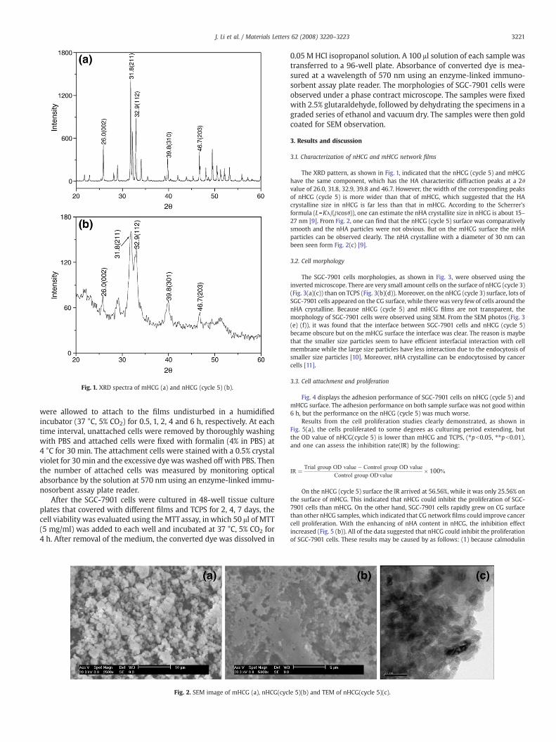
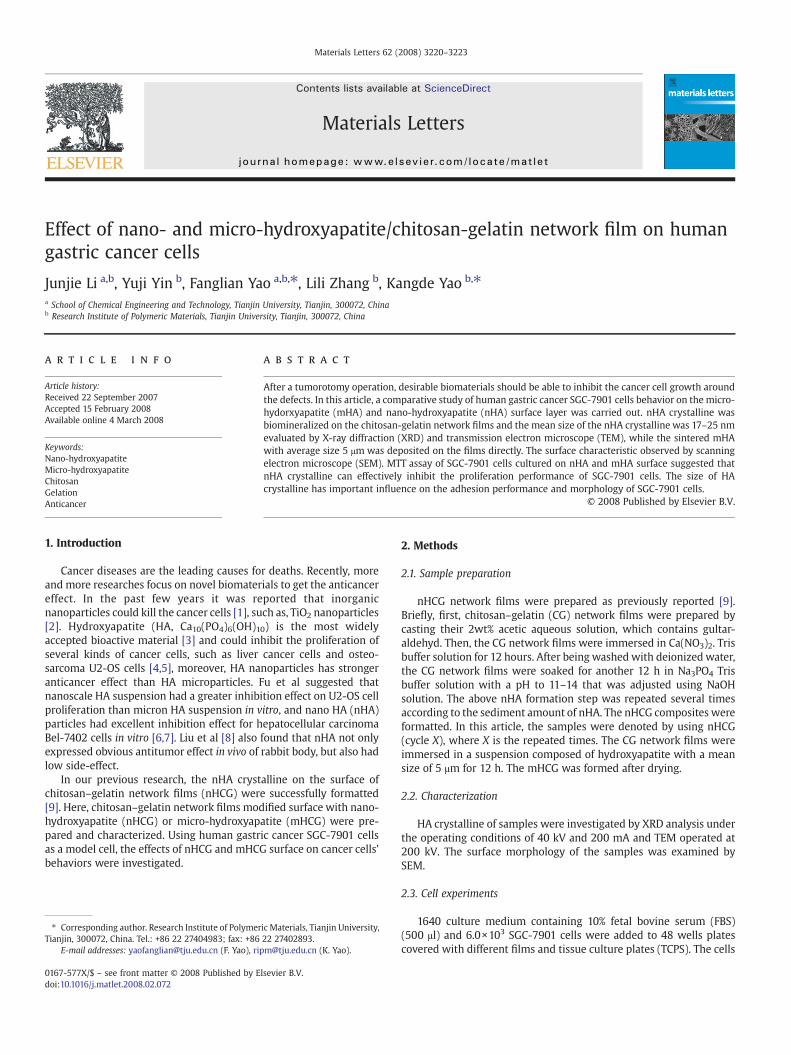
Fig. 1. XRD spectra of mHCG (a) and nHCG (cycle 5) (b).
3221J. Li et al. / Materials Letters 62 (2008) 3220–3223
were allowed to attach to the films undisturbed in a humidifiedincubator (37 °C, 5% CO2) for 0.5, 1, 2, 4 and 6 h, respectively. At eachtime interval, unattached cells were removed by thoroughly washingwith PBS and attached cells were fixed with formalin (4% in PBS) at4 °C for 30 min. The attachment cells were stained with a 0.5% crystalviolet for 30 min and the excessive dye was washed off with PBS. Thenthe number of attached cells was measured by monitoring opticalabsorbance by the solution at 570 nm using an enzyme-linked immu-nosorbent assay plate reader.
After the SGC-7901 cells were cultured in 48-well tissue cultureplates that covered with different films and TCPS for 2, 4, 7 days, thecell viability was evaluated using the MTT assay, in which 50 μl of MTT(5 mg/ml) was added to each well and incubated at 37 °C, 5% CO2 for4 h. After removal of the medium, the converted dye was dissolved in
Fig. 2. SEM image of mHCG (a), nHCG(cyc
0.05 M HCl isopropanol solution. A 100 μl solution of each sample wastransferred to a 96-well plate. Absorbance of converted dye is mea-sured at a wavelength of 570 nm using an enzyme-linked immuno-sorbent assay plate reader. The morphologies of SGC-7901 cells wereobserved under a phase contract microscope. The samples were fixedwith 2.5% glutaraldehyde, followed by dehydrating the specimens in agraded series of ethanol and vacuum dry. The samples were then goldcoated for SEM observation.
3. Results and discussion
3.1. Characterization of nHCG and mHCG network films
The XRD pattern, as shown in Fig. 1, indicated that the nHCG (cycle 5) and mHCGhave the same component, which has the HA characteritic diffraction peaks at a 2θvalue of 26.0, 31.8, 32.9, 39.8 and 46.7. However, the width of the corresponding peaksof nHCG (cycle 5) is more wider than that of mHCG, which suggested that the HAcrystalline size in nHCG is far less than that in mHCG. According to the Scherrer'sformula (L=Kλ/(βcosθ)), one can estimate the nHA crystallite size in nHCG is about 15–27 nm [9]. From Fig. 2, one can find that the nHCG (cycle 5) surface was comparativelysmooth and the nHA particles were not obvious. But on the mHCG surface the mHAparticles can be observed clearly. The nHA crystalline with a diameter of 30 nm canbeen seen form Fig. 2(c) [9].
3.2. Cell morphology
The SGC-7901 cells morphologies, as shown in Fig. 3, were observed using theinvertedmicroscope. There are very small amount cells on the surface of nHCG (cycle 3)(Fig. 3(a)(c)) than on TCPS (Fig. 3(b)(d)). Moreover, on the nHCG (cycle 3) surface, lots ofSGC-7901 cells appeared on the CG surface, while therewas very few of cells around thenHA crystalline. Because nHCG (cycle 5) and mHCG films are not transparent, themorphology of SGC-7901 cells were observed using SEM. From the SEM photos (Fig. 3(e) (f)), it was found that the interface between SGC-7901 cells and nHCG (cycle 5)became obscure but on the mHCG surface the interface was clear. The reason is maybethat the smaller size particles seem to have efficient interfacial interaction with cellmembrane while the large size particles have less interaction due to the endocytosis ofsmaller size particles [10]. Moreover, nHA crystalline can be endocytosised by cancercells [11].
3.3. Cell attachment and proliferation
Fig. 4 displays the adhesion performance of SGC-7901 cells on nHCG (cycle 5) andmHCG surface. The adhesion performance on both sample surface was not good within6 h, but the performance on the nHCG (cycle 5) was much worse.
Results from the cell proliferation studies clearly demonstrated, as shown inFig. 5(a), the cells proliferated to some degrees as culturing period extending, butthe OD value of nHCG(cycle 5) is lower than mHCG and TCPS, (⁎pb0.05, ⁎⁎pb0.01),and one can assess the inhibition rate(IR) by the following:
IR ¼ Trial group OD value� Control group OD valueControl group ODvalue
� 100k
On the nHCG (cycle 5) surface the IR arrived at 56.56%, while it was only 25.56% onthe surface of mHCG. This indicated that nHCG could inhibit the proliferation of SGC-7901 cells than mHCG. On the other hand, SGC-7901 cells rapidly grew on CG surfacethan other nHCG samples, which indicated that CG network films could improve cancercell proliferation. With the enhancing of nHA content in nHCG, the inhibition effectincreased (Fig. 5 (b)). All of the data suggested that nHCG could inhibit the proliferationof SGC-7901 cells. These results may be caused by as follows: (1) because calmodulin
le 5)(b) and TEM of nHCG(cycle 5)(c).
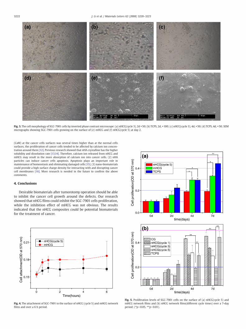
Fig. 3. The cell morphology of SGC-7901 cells by inverted phase contrast microscope: (a) nHCG(cycle 3), 2d ×50; (b) TCPS, 2d, ×100; (c) nHCG(cycle 3), 4d, ×50; (d) TCPS, 4d, ×50; SEMmicrographs showing SGC-7901 cells growing on the surface of (e) mHCG and (f) nHCG(cycle 5) at day 2.
3222 J. Li et al. / Materials Letters 62 (2008) 3220–3223
(CaM) at the cancer cells surfaces was several times higher than at the normal cellssurfaces, the proliferation of cancer cells tended to be affected by calcium ion concen-tration around them [12]. Previous research showed that nHA crystalline has the highersolubility and dissolution rate [13,14]. Therefore, calcium ion released from nHCG andmHCG may result in the more absorption of calcium ion into cancer cells. (2) nHAparticles can induce cancer cells apoptosis. Apoptosis plays an important role inmaintenance of homeostasis and eliminating damaged cells [15]. (3) nano-biomaterialscould provide a high surface charge density for interacting with and disrupting cancercell membranes [16]. More research is needed in the future to confirm the abovecomments.
4. Conclusions
Desirable biomaterials after tumorotomy operation should be ableto inhibit the cancer cell growth around the defects. Our researchshowed that nHCG films could inhibit the SGC-7901 cells proliferation,while the inhibition effect of mHCG was not obvious. The resultsindicated that the nHCG composites could be potential biomaterialsfor the treatment of cancer.
Fig. 4. The attachment of SGC-7901 to the surface of nHCG (cycle 5) and mHCG networkfilms and over a 6 h period.
Fig. 5. Proliferation levels of SGC-7901 cells on the surface of (a) nHCG(cycle 5) andmHCG network films and (b) nHCG network films(different cycle times) over a 7-dayperiod; (⁎pb0.05, ⁎⁎pb0.01).

3223J. Li et al. / Materials Letters 62 (2008) 3220–3223
Acknowledgements
The authors would like to thank Dr. Yun Bai of University of Albertafor advice of English writing. This work has been supported in part byNational Nature Science Foundation of China 30470482, 50773050,30670572, 50233020 and 50200300; Tianjin Municipal NationalScience Foundation Key Project 043803211; Key Projects in the TianjinScience & Technology Pillar Program via grant 06YFSZSF01000.
Reference
[1] J. Couzin, Science 96 (2002) 2314.[2] L.Y. Feng, S.P. Li, Y.H. Yan, et al., Key Eng. Mater. 192–1 (2000) 325.[3] G.S. Sailaja, P. Ramesh, T.V. Kumary, et al., Acta Biomater. 2 (2006) 651.
[4] M.Z. Yin, Y.C. Han, H.L. Dai, et al., J. Wuhan Univ. Technol. 21 (2006) 102.[5] Q. Fu, N. Zhou, W.H. Huang, et al., J. Biomed. Mater. Res. A. 74 (2005) 156.[6] M.Z. Yin, Y.C. Han, I.W. Bauer, et al., Biomed. Mater. 1 (2006) 38.[7] Z.S. Liu, S.L. Tang, Z.L. Ai, World J. Gastroenterol. 9 (2003) 1968.[8] J. Hu, Z.S. Liu, S.L. Tang, et al., World J. Gastroenterol. 13 (2007) 2798.[9] J.J. Li, Y.P. Chen, Y.J. Yin, et al., Biomaterials 28 (2007) 781.[10] L.F. Qi, Z.R. Xu, Bioorg. Med. Chem. Lett. 16 (2006) 4243.[11] S.P. Li, S. Hu, Y.H. Yan, et al., J. Wuhan Univ. Technol. 22 (2007) 288.[12] Q.N. Fu, N. Zhou, W.H. Huang, et al., J. Mater. Sci., Mater. M. 15 (2004) 1333.[13] F.C.M. Driessens, M.G. Boltong, E.A.P. Maeyer, et al., Biomaterials 23 (2002) 4011.[14] I. Papageorgiou, C. Brown, R. Schins, et al., Biomaterials 28 (2007) 2946.[15] Y.C. Dong, S.S. Feng, Biomaterials 25 (2004) 2843.[16] Z. Shi, K.G. Neoh, E.T. Kang, et al., Biomaterials 27 (2006) 2440.