effect of low-dose tungsten on human thyroid stem
TRANSCRIPT

https://doi.org/10.1530/ERC-19-0176https://erc.bioscientifica.com © 2019 Society for Endocrinology
Printed in Great BritainPublished by Bioscientifica Ltd.
26:8Endocrine-Related Cancer
F Gianì et al. Tungsten and thyroid stem/progenitor cells
713–725
-19-0176
RESEARCH
Effect of low-dose tungsten on human thyroid stem/precursor cells and their progeny
Fiorenza Gianì1, Giuseppe Pandini1, Nunzio Massimo Scalisi1, Paolo Vigneri2, Carmine Fazzari3, Pasqualino Malandrino1, Marco Russo1, Romilda Masucci4, Antonino Belfiore1, Gwabriella Pellegriti1 and Riccardo Vigneri1,5
1Endocrinology, Department of Clinical and Experimental Medicine, University of Catania, Garibaldi-Nesima Medical Center, Catania, Italy2Medical Oncology and Center of Experimental Oncology and Hematology, Department of Clinical and Experimental Medicine, University of Catania, A.O.U Policlinico Vittorio Emanuele, Catania, Italy3Humanitas, Catania Oncology Center, Catania, Italy4Surgical Oncology, Garibaldi-Nesima Medical Center, Catania, Italy5IC Crystallography Institute, National Research Council, CNR, Catania, Italy
Correspondence should be addressed to R Vigneri: [email protected]
Abstract
Thyroid cancer incidence is increased in volcanic areas where environment pollution biocontaminates residents. Tungsten (W) is the most increased heavy metal in drinking water of Mount Etna volcanic area where it exceeds the normal range in the urine of 27% inhabitants. The possible connection between increased tungsten and thyroid cancer has never been studied. We investigated in vitro the effect tungsten on both human thyrocytes in primary culture, thyrospheres (aggregates of stem/precursor thyroid cells) and thyrocytes differentiated from tungsten-exposed thyrospheres. Chronic exposure to low-dose (nanomolar range, as in the urines of volcanic area residents) soluble tungsten had major biological effects on thyroid stem/precursor cells, promoting growth with a biphasic (hormetic) dose-response and reducing apoptosis. No such effects were observed in mature thyrocytes. In addition, tungsten-exposed thyrospheres had abnormal expression of genes commonly altered also in thyroid cancer and increased activation of the DNA-repair proteins H2AX and 53BP1. Moreover, exposure to tungsten decreased thyrosphere differentiation, as indicated by the reduced expression of thyroid-specific genes in derived thyrocytes that also showed preneoplastic changes such as increased anchorage-independent growth, clonogenic growth and migration capacity. The mechanism of action of tungsten on thyroid stem/precursor cells is unclear but involves membrane G-proteins and activation of the ERK signaling pathway. These data indicate that chronic exposure to slightly increased tungsten, harmless for mature thyrocytes, importantly affects the biology of stem/precursor thyroid cells and of their progeny, inducing characteristics of preneoplastic transformation.
Introduction
Thyroid cancer (TC) is increasing the most among cancer incidences worldwide (Davies & Welch 2006, Simard et al. 2012). Much evidence, including increased mortality
despite of earlier diagnosis and better treatment, indicates that increased scrutiny and overdiagnosis are not the only causes and that a true increase in TC is also occurring
Endocrine-Related Cancer (2019) 26, 713–725
8
Key Words
f tungsten
f thyroid stem cells
f thyrospheres
f thyroid cancer
f thyroid carcinogenesis
26
Downloaded from Bioscientifica.com at 11/30/2021 05:07:00AMvia free access

Printed in Great BritainPublished by Bioscientifica Ltd.https://doi.org/10.1530/ERC-19-0176
https://erc.bioscientifica.com © 2019 Society for Endocrinology
714F Gianì et al. Tungsten and thyroid stem/progenitor cells
26:8Endocrine-Related Cancer
(Vigneri et al. 2015, Lim et al. 2017). The causes of the increasing incidence of TC are unclear but, because of the rapid change in the last decades, they are most likely environmental rather than genetic.
TC incidence is increased in volcanic areas suggesting a possible effect of carcinogens related to the volcanic environment (Vigneri et al. 2017). In the volcanic area of Mount Etna in Sicily, the increased incidence of TC is associated with a relevant non-anthropogenic environmental pollution and human biocontamination with a variety of trace elements and heavy metals, as documented by the increased concentration of these elements in the urines and hair of residents (Varrica et al. 2014, Malandrino et al. 2016). In particular, four elements (boron, molybdenum, palladium and tungsten) exceed the urine reference values in more than 20% of the volcanic area inhabitants (Malandrino et al. 2016). Among these elements tungsten (or wolfram; W) is the most increased in water of the volcanic area (approximately 50-fold higher than that in the water of adjacent non-volcanic areas) (Malandrino et al. 2016).
Tungsten use in the industrialized world has steadily expanded in the recent decades from 32,200 tons in 1998 to 88,100 tons in 2016 (https://www.usgs.gov/centers/nmic/tungsten-statistics-and-information). Cement carbides (hardmetals, superalloys) and military applications are the most important tungsten usages (International Tungsten Industry Association; http://www.itia.info/tungsten-primary-uses.html) and, unavoidably, the increased use has spawned greater contamination in the natural and human environment (Koutsospyros et al. 2006). Therefore, tungsten must be considered an emerging toxicant, acting either alone or by augmenting the effect of other toxicants and cell stressors (Witten et al. 2012, Bolt & Mann 2016). Additionally, tungsten has been reported to have also potential carcinogenic effects both in vitro (Harris et al. 2015, Laulicht et al. 2015) and in vivo (Kelly et al. 2013). However, there is no definitive information on the carcinogenic potential of this heavy metal in soluble form, particularly after chronic exposure. This major gap in knowledge and evidence is absolute for the thyroid although this gland is among the organs where soluble tungsten is rapidly distributed after absorption (Lemus & Venezia 2015). Therefore, a role of increased tungsten in the environment and increased TC incidence can be hypothesized.
Our previous observations indicate that (a) TC incidence is more than doubled in the Mount Etna volcanic area relative to that in the adjacent non-volcanic areas (Pellegriti et al. 2009) and (b) in the urines
of residents of the Mount Etna volcanic area tungsten levels are more than doubled relative to values found in residents of adjacent areas and exceed the Italian reference values (95th percentile = 0.25 μg/L) in 27% of cases (Malandrino et al. 2016) and the US reference values (95th percentile = 0.40 μg/L) in 14.3% of cases (https://www.cdc.gov/exposurereport/index.html). Based on the consideration that in the Mount Etna volcanic area residents’ exposure to increased tungsten is a chronic, life-long condition starting in prenatal life because the fetus and the newborn are exposed by transplacental and breast-feeding routes (Wide et al. 1986, Pitt et al. 1991), we hypothesized that early exposure to increased tungsten might predispose individuals to TC risk later in life. Stem/progenitor cells, in fact, may be more susceptible to the detrimental effect of increased toxicant and transmit the damage to their differentiated progeny, as previously observed in other human and animal models (Waalkes et al. 2008, McInturf et al. 2011, Xu et al. 2013, Kopras et al. 2014, Howe et al. 2018).
We explored this hypothesis in vitro using human stem/progenitor thyroid cells (thyrospheres) exposed to the soluble salt sodium tungstate at concentrations in the range of the tungsten levels measured in the urine of residents in the Sicilian volcanic area (nanomolar range, much lower than that in most previous studies on W biological effects in vitro and in vivo).
Our study indicates that low-dose tungsten has complex effects on human thyroid progenitor cells, compatible with an increased carcinogenic susceptibility persisting in differentiated thyrocytes derived from exposed stem/precursor cells. In contrast, low-dose tungsten has no significant effect in differentiated human thyroid cells.
Materials and methods
Tissue specimens
Thirty-six patients (all females, ages ranging from 30 to 65 years) admitted to surgery for a solitary thyroid nodule classified TIR 3 at cytology (Fadda et al. 2010) and resulted benign at pathology examination, gave written informed consent to donate a small aliquot of their excised thyroid tissue. Approximately 0.5–1.0 g of normal appearing thyroid tissue, at least 5 mm distant from the nodule, was collected in the surgery room, pathologically examined for normal histological morphology and used for the following in vitro studies.
Downloaded from Bioscientifica.com at 11/30/2021 05:07:00AMvia free access

https://erc.bioscientifica.com © 2019 Society for Endocrinology
Printed in Great BritainPublished by Bioscientifica Ltd.https://doi.org/10.1530/ERC-19-0176
715F Gianì et al. Tungsten and thyroid stem/progenitor cells
26:8Endocrine-Related Cancer
The tissue sampling was approved by the Local Ethics Committee (Catania 2, N. 12/2015/CECT2).
Human thyroid cells
Three different human thyroid cell models were generated from the collected thyroid tissue: (1) thyrocytes in primary culture; (2) stem/progenitor thyroid cells (thyrospheres); and (3) thyrocytes differentiated from thyrospheres, indicated as ‘secondary’ thyrocytes.
Nonadherent thyrospheres and monolayers of normal thyrocytes in primary culture were established as previously described (Giani et al. 2015). Briefly, after meticulous removal of fibrous tissue, the fresh thyroid tissue was first minced with sterile scissors and then digested with collagenase IV (1 mg/mL, Sigma-Aldrich) for 2 h at 37°C. The resulting cell suspension containing intact and fragmented thyroid follicles was centrifuged (400 g for 10 min), and the pellet was suspended in DMEM/F12 culture medium (Sigma) supplemented with both 2 mM glutamine (Sigma) and 2% heat-inactivated fetal bovine serum (FBS; Invitrogen) and incubated at 37°C in a 5% CO2 atmosphere. After 12–24 h, viable thyroid cells and fibroblasts attached to the flasks and supernatants containing unattached cells were transferred into fresh flasks, and the cells were cultured at 100% confluence. Residual fibroblasts, when present, were depleted using magnetic anti-fibroblast beads (Miltenyi Biotec) according to the manufacturer’s instructions.
These thyrocytes were used to obtain the following cell cultures:
– ‘Primary’ mature thyrocytes were grown in the previously described DMEM/F12 medium supplemented with 1 mU/mL bovine thyroid-stimulating hormone (TSH) (Sigma) and a 1% insulin-transferrin-sodium selenite liquid medium supplement (ITS; Thermo).
– Stem/progenitor thyroid cells (forming spheroid aggregates called ‘thyrospheres’) were obtained by trypsinization and seeding individual cells at density 1–5 × 10⁴ cells/mL and cultured in ultralow attachment plastic flasks (Eppendorf) in the standard stem cell medium, that is the previously described DMEM/F-12 supplemented with 2% B-27 (Thermo) and enriched with human recombinant epidermal growth factor (EGF, 20 mg/mL; Sigma). Under these culture conditions, stem/progenitor thyroid cells form free-floating thyrospheres.
– ‘Secondary’ thyrocytes were obtained by differentiation from thyrospheres. Briefly, thyrospheres were collected
by centrifugation at 1100 g for 5 min, supernatants were discarded and disaggregated cells were transferred into adherent tissue culture dishes and cultured in differentiation medium (DMEM/F-12 supplemented with 2% FBS, 1 mU/mL bovine TSH and 1% ITS). Fully mature ‘secondary’ thyrocytes were obtained as monolayers after 7 days.
Cell exposure to tungsten
Sodium tungstate dihydrate (Na2WO4.2H2O; Sigma) was dissolved in deionized water and added to the culture media at concentrations in the range 1 nM–1 μM (W = 0.18–180.0 μg/L) and for the times indicated. Culture media were replaced every 2 or 3 days to limit Na2WO4.2H2O conversion to polytungstates (Kelly et al. 2013).
5-bromo-2'-deoxyuridine (BrdU) incorporation
Cell proliferation was measured by BrdU incorporation: thyrocytes and cells disaggregated from thyrospheres were seeded at a cell density of 8000–10,000 cells/well in a 96-well microtiter plate precoated with poly-l-lysine in phenol red-free DMEM/F12 medium. After adhesion and starvation for 24 h, Na2WO4 was added, and the cells were incubated for additional 72 h when BrdU incorporation was measured using the DELFIA cell proliferation kit (PerkinElmer Life Sciences) according to the manufacturer’s instructions.
Cell apoptosis measurement
Thyrospheres and differentiated thyrocytes cultured in the presence or the absence of Na2WO4 for 15 days were exposed for 4 h to the apoptosis-inducing agents staurosporine (1 μM, Sigma) or raptinal (10 μM, Sigma). Caspase 3/7 activity was measured using the Caspase-Glo 3/7 assay (Promega) according to the manufacturer’s protocol.
Gene transcript analysis
The effect of Na2WO4 on stemness and thyroid-specific genes was evaluated as follows in thyrospheres chronically exposed to tungstate 30 nM: (a) total RNA (1 μg) was reverse transcribed using a High-Capacity cDNA Reverse Transcription Kit (Thermo) and subjected to RT-PCR analysis. Synthesized cDNA was combined in a PCR using primers for the genes of interest (Tables 1 and 2) and (b) genes contained in the human stem cell RT2 Profiler
Downloaded from Bioscientifica.com at 11/30/2021 05:07:00AMvia free access
Printed in Great BritainPublished by Bioscientifica Ltd.https://doi.org/10.1530/ERC-19-0176
https://erc.bioscientifica.com © 2019 Society for Endocrinology
716F Gianì et al. Tungsten and thyroid stem/progenitor cells
26:8Endocrine-Related Cancer
PCR array were measured according to the user’s manual (Qiagen). Cycling conditions were as follows: 10 min at 95°C followed by 40 cycles at 95°C for 15 sec and 60°C for 1 min. Cut-off values for gene expression fold changes were established at +1.3 for gene upregulation and −1.3 for downregulation. Relative quantification of target genes was performed using the comparative CT method (DDCt).
Measurement of DNA-repair proteins
Histone H2AX phosphorylation and 53BP1 expression were measured by immunofluorescence and WB. Cells disaggregated from thyrospheres were seeded onto poly-l-lysine-coated glass coverslips and exposed to Na2WO4 (30-3000 nM, 30-180 min). Cells were then washed, preextracted with Triton buffer (20 mM Hepes, pH 7.4, 100 mM NaCl, 5 mM MgCl2, 300 mM sucrose and
0.5% Triton X-100) for 5 min at RT and then fixed in 4% para-formaldehyde for 10 min at RT and blocked with 3% BSA in PBS. Then the polyclonal anti-53BPI antibody (Santa Cruz Biotechnology) or the anti-phospho-H2AX antibody (Cell Signaling, 9718), diluted in blocking buffer, was added (1 h at RT). Samples were then washed with PBS and incubated with Alexa Fluor-594-conjugated goat anti-rabbit (Thermo). DNA was counterstained with DAPI in Vectashield mounting agent (Santa Cruz). Images were acquired with an Olympus BH-2 microscope.
Western blot analysis
Thyrospheres and differentiated thyrocytes were lysed and subjected to Western blot analysis, as previously described (Vella et al. 2009). The following antibodies were purchased from Cell Signaling Technology (Cell Signaling Technology): anti-ERK1/2, anti-P-ERK1/2 (T202/Y204),
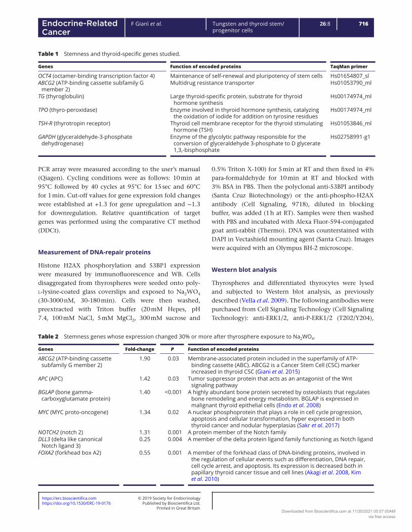
Table 1 Stemness and thyroid-specific genes studied.
Genes Function of encoded proteins TaqMan primer
OCT4 (octamer-binding transcription factor 4) Maintenance of self-renewal and pluripotency of stem cells Hs01654807_slABCG2 (ATP-binding cassette subfamily G
member 2)Multidrug resistance transporter Hs01053790_ml
TG (thyroglobulin) Large thyroid-specific protein, substrate for thyroid hormone synthesis
Hs00174974_ml
TPO (thyro-peroxidase) Enzyme involved in thyroid hormone synthesis, catalyzing the oxidation of iodide for addition on tyrosine residues
Hs00174974_ml
TSH-R (thyrotropin receptor) Thyroid cell membrane receptor for the thyroid stimulating hormone (TSH)
Hs01053846_ml
GAPDH (glyceraldehyde-3-phosphate dehydrogenase)
Enzyme of the glycolytic pathway responsible for the conversion of glyceraldehyde 3-phosphate to D glycerate 1,3,-bisphosphate
Hs02758991-g1
Table 2 Stemness genes whose expression changed 30% or more after thyrosphere exposure to Na2WO4.
Genes Fold-change P Function of encoded proteins
ABCG2 (ATP-binding cassette subfamily G member 2)
1.90 0.03 Membrane-associated protein included in the superfamily of ATP-binding cassette (ABC). ABCG2 is a Cancer Stem Cell (CSC) marker increased in thyroid CSC (Giani et al. 2015)
APC (APC) 1.42 0.03 Tumor suppressor protein that acts as an antagonist of the Wnt signaling pathway
BGLAP (bone gamma-carboxyglutamate protein)
1.40 <0.001 A highly abundant bone protein secreted by osteoblasts that regulates bone remodeling and energy metabolism. BGLAP is expressed in malignant thyroid epithelial cells (Endo et al. 2008)
MYC (MYC proto-oncogene) 1.34 0.02 A nuclear phosphoprotein that plays a role in cell cycle progression, apoptosis and cellular transformation, hyper expressed in both thyroid cancer and nodular hyperplasias (Sakr et al. 2017)
NOTCH2 (notch 2) 1.31 0.001 A protein member of the Notch familyDLL3 (delta like canonical
Notch ligand 3)0.25 0.004 A member of the delta protein ligand family functioning as Notch ligand
FOXA2 (forkhead box A2) 0.55 0.001 A member of the forkhead class of DNA-binding proteins, involved in the regulation of cellular events such as differentiation, DNA repair, cell cycle arrest, and apoptosis. Its expression is decreased both in papillary thyroid cancer tissue and cell lines (Akagi et al. 2008, Kim et al. 2010)
Downloaded from Bioscientifica.com at 11/30/2021 05:07:00AMvia free access

https://erc.bioscientifica.com © 2019 Society for Endocrinology
Printed in Great BritainPublished by Bioscientifica Ltd.https://doi.org/10.1530/ERC-19-0176
717F Gianì et al. Tungsten and thyroid stem/progenitor cells
26:8Endocrine-Related Cancer
anti-AKT, anti-P-AKT, anti-P-H2AX(S139); anti-GRB2 was obtained from Santa Cruz. ERK phosphorylation in cells exposed to Na2WO4 was also measured after preincubation for 24 h with either iberiotoxin (IbTX 300 nM, a specific BKαβ1 channel blocker, Sigma) or pertussis toxin (PTX, 50 ng/ml, a Gi/o protein inhibitor, Sigma).
Measurements of in vitro cell transformation
To investigate tungsten induced cell transformation, mature thyrocytes differentiated from thyrospheres exposed or not exposed for 15 days to Na2WO4 were comparatively tested with three independent procedures: (a) anchorage-independent growth in soft agar: 3 × 10⁴ cells/well were plated into 12-well plates using a two-layer soft agar system (bottom layer of 0.5% and top layer of 0.35% agar). Cells were incubated for 3 weeks and then colonies (diameter >75 μm) were visualized with 0.5 mg/mL MTT, photographed and analyzed with NIH ImageJ; (b) clonogenic assay (plate colony forming assay): 500 cells/well seeded in 12-well plates were incubated with complete medium at 37°C in 5% CO2 for 3 weeks. Grown colonies (containing >50 cells) were stained with 0.1% crystal violet and photographed and the number recorded; (c) migration assay (scratch wound-healing assay): cells were seeded in 12-well plates precoated with poly-l-lysine and then incubated in 5% CO2 at 37°C until a confluent monolayer was formed. Then a scratch was made manually by scraping the cell monolayers with a 200 μL pipette tip. After washing twice with PBS to remove floating cells, the cells were cultured in serum-free medium and images captured at 0, 24 and 48 h time points to evaluate cell migration over the scratch.
Statistical analysis
All data were expressed as mean ± s.e.m. Statistical analyses were performed using the GraphPad Prism 5.0 Software. All differences between mean values were evaluated by the Student’s t-test. A two-sided P < 0.05 was accepted as significant.
Results
Na2WO4 stimulates proliferation and inhibits apoptosis in thyrospheres but not in mature thyrocytes
Experiments carried out in both thyrospheres and mature thyrocytes in primary culture indicated that
chronic exposure to tungstate increased growth only in thyrospheres. Three-day incubation in the presence of Na2WO4 significantly increased BrdU incorporation relative to control thyrospheres already at 1 nM Na2WO4 (= 0.18 μg/L), further increased at 10–100 nM and then declined, showing no difference from non-exposed thyrospheres at 1 μM Na2WO4 (Fig. 1A). No effect, at any concentration tested, was observed for mature thyrocytes.
In parallel experiments exposure to Na2WO4 decreased apoptosis. Staurosporine (1 μM) increased caspase 3/7 activity in both thyrospheres and thyrocytes (289 ± 13.3% and 182 ± 15.0%, respectively). Concomitant chronic exposure to tungstate significantly (P < 0.05) reduced caspase 3/7 activity in thyrospheres (172 ± 7.6%) but not in thyrocytes (222 ± 9.6%) (Fig. 1B). Similar results were obtained using raptinal 10 μM which rapidly initiated the
Figure 1Chronic exposure to Na2WO4 affects thyrosphere, but not differentiated thyrocyte, growth and apoptosis. (A) BrdU incorporation in thyrospheres and thyrocytes after exposure to increasing concentrations of Na2WO4 (n = 6 separate experiments). The vertical dotted line indicates the average W concentration in urines of residents of the volcanic area in Sicily (0.2 µg/L of W). (B) Effect of pro-apoptotic staurosporine (STS 1 µM, 4 h) on caspase 3/7 activity in thyrospheres and thyrocytes incubated with or without Na2WO4 30 nM for 14 days (n = 4 separate experiments). (C) DNA content measured in thyrospheres and thyrocytes after chronic exposure to increasing concentrations of Na2WO4 (n = 4 separate experiments). (D) Representative phase-contrast microscopy images of morphological changes in thyrospheres and thyrocytes incubated with or without tungstate (30 nM, 14 days, n = 4 independent experiments). For all data: mean values are indicated with s.e.m.; paired samples are compared by two-tailed Student t-test (NS, non-significant; *P < 0.05, **P < 0.01).
Downloaded from Bioscientifica.com at 11/30/2021 05:07:00AMvia free access

Printed in Great BritainPublished by Bioscientifica Ltd.https://doi.org/10.1530/ERC-19-0176
https://erc.bioscientifica.com © 2019 Society for Endocrinology
718F Gianì et al. Tungsten and thyroid stem/progenitor cells
26:8Endocrine-Related Cancer
intrinsic pathway of caspase-dependent apoptosis (data not shown).
As a result of increased proliferation and decreased apoptosis, thyrosphere growth, as evaluated by DNA content, significantly increased in Na2WO4-treated thyrospheres but not in thyrocytes (Fig. 1C). These data were confirmed by phase-contrast microscopy imaging: thyrospheres exposed to Na2WO4 (30 nM for 14 days) were larger (average surface increase in three separate experiments = +130%, P = 0.02) and morphologically irregular compared to control thyrospheres. In contrast, no morphological change was observed in mature thyrocytes (Fig. 1D).
Chronic exposure to Na2WO4 affects thyroid stem/precursor cell differentiation
The analysis of gene transcripts indicates that exposure to tungstate affects thyroid cell differentiation. In control thyrospheres, the stemness genes octamer-binding transcription factor 4 OCT-4 and ATP-binding cassette subfamily G member 2 (ABCG-2) were highly expressed, while the thyroid-specific genes thyroglobulin (TG), thyroperoxidase (TPO) and TSH-receptor (TSH-R) were expressed at low levels. As expected, the opposite occurred in differentiated thyrocytes (Fig. 2, panel A).
When chronically exposed to Na2WO4, an increase in stemness gene expression and a decrease in thyroid-specific gene expression occurred in both thyrospheres and in mature thyrocytes derived from exposed thyrospheres. Chronic exposure to tungstate, therefore, decreases the expression of thyroid differentiation genes not only in thyroid progenitor cells (P < 0.01 for TG and P < 0.05 for TPO and TSH-R) but also in mature thyrocytes derived from the exposed progenitors (P < 0.01 for TG and <0.05 for the other transcripts relative to thyrocytes differentiated from non-exposed thyrospheres). The opposite occurred for stemness gene expression: exposure to tungstate increased OCT-4 and ABCG-2 expression in both stem/precursor and differentiated cells (Fig. 2).
When ‘secondary’ thyrocytes differentiated from non-exposed thyrospheres were treated with tungstate no effect was observed on gene expression, mirroring the behavior of ‘primary’ thyrocytes (Fig. 2B). However, when tungstate was added during the differentiation process of the control (non-exposed to Na2WO4) thyrospheres, the differentiated thyrocytes showed a reduced expression of all thyroid-specific genes (−25, −30 and −18% for TG, TPO and TSH-R, respectively, P < 0.05) relative to thyrocytes differentiated from the same control thyrospheres but in the absence of Na2WO4 (Fig. 2C).
These data indicate that chronic exposure to low concentrations of tungstate reduces the differentiation potential in thyroid stem/precursor cells and induces gene expression abnormalities also in derived thyrocytes.
Figure 2Effect of chronic exposure to Na2WO4 on the expression of stemness genes (OCT-4 and ABCG-2) and thyroid-specific genes (TG, TPO and TSH-R) in thyrospheres and differentiated thyrocytes. (A) Changes of gene expression in thyrospheres and derived thyrocytes induced by exposure of stem/precursor cells (TS, thyrospheres) to Na2WO4 (30 nM for 14 days). Derived (‘secondary’) thyrocytes (TC) were evaluated 7 days after differentiation, when showing morphological features of mature thyrocytes. Changes in stemness and thyroid-specific gene expression caused by tungstate in progenitor cells were present also in their progeny. For all data, mRNA expression was determined by real-time quantitative PCR as described in Materials and methods and normalized relative to GAPDH mRNA expression level. The results are presented as fold-change relative to control (not Na2WO4 exposed) thyrospheres. Data indicate the mean value ± s.e.m. of four independent experiments. mRNA content in paired samples was compared by two-tailed Student t-test (*P < 0.05; **P < 0.01). (B) Mature thyrocytes in primary culture (‘primary’) or derived from normal thyrospheres not exposed to tungstate (‘secondary’) have a similar expression of thyroid-specific genes. In both cell types, gene expression is not affected by exposure to Na2WO4 for seven days. (C) The expression of thyroid-specific genes is reduced in thyrocytes differentiated from control thyrospheres (not exposed to Na2WO4) when tungstate is added to the medium during the 7 days of the differentiation procedure. For both (B) and (C), the results are presented as the fold-change relative to control thyrocytes. Data (mean of three independent experiments ± s.e.m.) are compared by two-tailed Student t-test (*P < 0.05).
Downloaded from Bioscientifica.com at 11/30/2021 05:07:00AMvia free access

https://erc.bioscientifica.com © 2019 Society for Endocrinology
Printed in Great BritainPublished by Bioscientifica Ltd.https://doi.org/10.1530/ERC-19-0176
719F Gianì et al. Tungsten and thyroid stem/progenitor cells
26:8Endocrine-Related Cancer
Similar effects were also observed when Na2WO4 was added during the differentiation process of thyrospheres that were not preexposed to tungstate but did not occur when tungstate was added after cell differentiation.
Na2WO4 alters the thyroid stem/precursor cell gene profile
In paired thyrosphere specimens either chronically exposed or non-exposed to 30 nM Na2WO4 the gene expression pattern was different. Compared with control thyrospheres used as a reference, increased or decreased expression of several genes was observed after exposure to Na2WO4.
Of the 84 genes characterizing the stem cell gene profile a 30% or greater expression change was observed for ABCG-2, APC, BGLAP, MYC and NOTCH-2, all increased, while the expression of DLL3 and FOXA2 was decreased (Tables 1 and 2). Some of these genes play a general role in carcinogenesis and most, including ABCG-2 (Fadda et al. 2010), cMYC (Sakr et al. 2017), BGLAP (Endo et al. 2008) and FOXA2 (Akagi et al. 2008, Kim et al. 2010) may be abnormally expressed in TC.
Thyrocytes derived from Na2WO4-exposed thyrospheres present biological characteristics typical of transformed cells
To evaluate whether stem/precursor cell exposure to tungstate affected the biological characteristics in their progeny of mature thyrocytes, we compared thyrocytes derived from thyrospheres either exposed or not exposed to Na2WO4 in three different transformation assays.
In all three assays, thyrocytes derived from Na2WO4-exposed stem/precursor cells showed an abnormal behavior: they formed more numerous and larger colonies in the anchorage-independent growth assay (Fig. 3A), more and larger colonies in the clonogenic assay (Fig. 3B) and a greater migration capacity in the scratch wound-healing assay (Fig. 3C).
In thyrocytes differentiated from Na2WO4-treated thyrospheres, statistically significant increases relative to the control thyrocytes were observed for both soft agar (18.9 ± 4.0 vs 4.4 ± 2.3 colonies/well; n = 3, P < 0.05) and plate colony formation (9.4 ± 0.9 vs 3.9 ± 1.0 colonies/well; n = 3, P < 0.01) assays. For the migration assay microphotographs taken 24 and 48 h after making a scratch in confluent cultures showed that thyrocytes derived from Na2WO4-treated thyrospheres had an
Figure 3In vitro transformation assays in thyrocytes differentiated from thyrospheres either chronically exposed or not exposed (controls) to Na2WO4. Both the adherence-independent growth in soft agar (A) and the clonogenic potential in plate formation assay (B) show that thyrocytes differentiated from thyrospheres chronically exposed to Na2WO4 (30 nM) form more and larger colonies than thyrocytes differentiated from control, not Na2WO4-exposed thyrospheres. Representative images and the average values (number of colonies/well, mean ± s.e.m.) of three separate experiments are shown (*P < 0.05; **P < 0.01 vs control thyrocytes). (C) Microphotographs of the scratch wound-healing assay show that thyrocytes from W-exposed thyrospheres partially healed the scratch progressively at 24 and 48 h indicating an increased migration activity compared to controls (n = 3).
Downloaded from Bioscientifica.com at 11/30/2021 05:07:00AMvia free access

Printed in Great BritainPublished by Bioscientifica Ltd.https://doi.org/10.1530/ERC-19-0176
https://erc.bioscientifica.com © 2019 Society for Endocrinology
720F Gianì et al. Tungsten and thyroid stem/progenitor cells
26:8Endocrine-Related Cancer
increased migration activity from the wound scratch edge relative to thyrocytes derived from control thyrospheres (Fig. 3C).
These data indicate that the effects of Na2WO4 on thyrospheres also cause biological abnormalities in their differentiated progeny that, in three independent in vitro transformation assays, show a significant growth advantage and biological characteristics typical of neoplastic status.
Exposure to Na2WO4 affects DNA-repair protein activity
At the concentrations 30, 300 and 3000 nM altered the expression and function of DNA-repair proteins H2AX and 53BP1, both markers of DNA double-strand breaks (DSBs) and, therefore, early signs of DNA damage. In thyrospheres exposed to Na2WO4, H2AX histone phosphorylation on serine 139 (γH2AX) was already evident after 30 min and peaked at 90–180 min (P < 0.01, Fig. 4C). This effect of Na2WO4 was dose dependent, with a significant increase relative to baseline at 30 nM Na2WO4, further increasing at 300 nM (P < 0.01) and then decreasing at higher concentrations (3000 nM) (Fig. 4B).
In parallel experiments a marked increase in p53-binding protein 1 (53BP1) expression was also observed in thyrospheres exposed to Na2WO4 (Fig. 4A), with p53BP1
relocation in the nucleus forming nuclear foci after 90 min of exposure to Na2WO4 30 nM.
These abnormalities of DNA-repair proteins are indicative of low-dose tungsten genotoxicity in stem/precursor thyroid cells. Noteworthy, increased 53BP1 nuclear foci formation has been reported in papillary TC, closely associated with BRAFV600E mutation (Nakashima et al. 2008, Mussazhanova et al. 2013).
Effects of Na2WO4 on intracellular signaling
To investigate the cellular components and pathways involved in Na2WO4 effect on exposed thyrospheres and their progeny, we first examined two major components of intracellular signaling.
The phosphorylation of the extracellular signal-regulated protein kinase (ERK 1/2) and of protein kinase B (PKB or AKT) was measured in thyrospheres and thyrocytes exposed to Na2WO4 30 nM for 5–30 min (Fig. 5). No effect of tungstate was observed in differentiated thyrocytes for both AKT and ERK 1/2 phosphorylation. In contrast, 5 min after exposure to Na2WO4 a significant increase (P < 0.01) in ERK 1/2 phosphorylation was observed in thyrospheres but not in thyrocytes. This effect slightly increased after 15 min of exposure and then decreased at 30 min when, however, was still significantly increased (P < 0.05) relative to baseline (Fig. 5A). No change in AKT phosphorylation
Figure 4Low-dose Na2WO4 alters the expression and function of DNA-repair proteins H2AX and 53BP1. (A) Representative immunofluorescence photographs show that endogenous γ-H2AX and 53BP1 (detected using anti-γ-H2AX and anti-53BP1 antibodies followed by Alexa Fluor-594-conjugated second antibody) are induced and localized in the nucleus (visualized with DAPI) after stem/precursor thyroid cell exposure to Na2WO4 30 nM for 90 min. (B and C) Western blot analyses show that H2AX phosphorylation induced by Na2WO4 is dose dependent (significant increase at 30 nM, peak at 300 nM and decreasing at 3000 nM) and time dependent. Histograms indicate mean values ± s.e.m. of densitometric readings of three separate Western blots after thyrosphere exposure to Na2WO4 at increasing doses (B) and for different times (C, Na2WO4 30 nM).
Downloaded from Bioscientifica.com at 11/30/2021 05:07:00AMvia free access

https://erc.bioscientifica.com © 2019 Society for Endocrinology
Printed in Great BritainPublished by Bioscientifica Ltd.https://doi.org/10.1530/ERC-19-0176
721F Gianì et al. Tungsten and thyroid stem/progenitor cells
26:8Endocrine-Related Cancer
was observed in Na2WO4-exposed thyrospheres at the times studied.
We than analyzed the cell membrane components involved in the intracellular transmission of W biological effects. Since tungstate has been reported to activate voltage- and Ca++-dependent K+ (BK) channels (Fernandez-Marino et al. 2012) and G-protein stimulation of the Ras/ERK cascade (Zafra et al. 2013), we investigated whether the inhibition of these membrane proteins blocked the effect of tungstate on the ERK phosphorylation. Thyrospheres were preincubated with iberiotoxin (IbTX 300 nM), a specific BK channel blocker, and then ERK phosphorylation was measured and found only mildly reduced (P = NS) relative to the control (Fig. 5B). In contrast, preincubation with the pertussis toxin (PTX, 50 ng/mL), a G-protein inhibitor, completely abolished the effect of tungstate on ERK phosphorylation (P < 0.001, Fig. 5B).
To evaluate the relevance of this cellular pathway (activated ERK signaling via G-protein) on the tungstate-dependent thyrosphere growth, we measured the growth of thyrospheres exposed for three days to 30 nM Na2WO4 in the presence or absence of PTX. The presence of PTX reduced Na2WO4-stimulated BrdU incorporation by approximately 50% (P = 0.012, Fig. 5C).
Altogether, these results indicate that thyroid stem/precursor cell proliferation after chronic exposure to Na2WO4 occurs via a G-protein and ERK pathway activation although additional mechanisms cannot be excluded.
Discussion
In the present study, we first observed that chronic exposure to sodium tungstate at very low (nM)
Figure 5Effect of Na2WO4 on intracellular signaling in thyroid cells. (A) Thyrospheres and differentiated thyrocytes, treated or not treated with 30 nM Na2WO4, were lysed at the indicated times and analyzed by Western blot for both AKT and ERK 1/2 phosphorylation. No effect was observed on AKT phosphorylation. In contrast, ERK 1/2 phosphorylation was stimulated in thyrospheres but not in thyrocytes. The maximum increase occurred 5–15 min after exposure to Na2WO4. The effect of EGF (5 ng/mL) is shown as positive control. A representative immunoblot from three independent experiments is shown. Histograms represent the mean ± s.e.m. of densitometric values normalized to β-actin and expressed as percent of control (untreated thyrospheres). (B) PTX but not IbTx inhibits ERK 1/2 phosphorylation induced by Na2WO4 in thyrospheres. Thyrospheres pretreated or not pretreated with either IbTX (300 nM for 45 min) or PTX (50 ng/mL for 28 h) were exposed to Na2WO4 30 nM for 10 min. Western blot analysis from whole lysates was performed to evaluate ERK 1/2 phosphorylation, and a representative immunoblot from three independent experiments is shown. Histograms represent the mean ± s.e.m. of densitometric values normalized to GRB2 and are expressed as percent of control (not treated) thyrospheres. (C) Thyrosphere proliferation induced by Na2WO4 and evaluated by BrdU incorporation is blunted by the presence of the ERK 1/2 inhibitor PTX. Thyrospheres were exposed to 30 nM Na2WO4 for 3 days in the absence (control) or the presence of PTX (50 ng/mL) and BrdU incorporation was measured. Histograms represent the mean values ± s.e.m. of four separate experiments. Data are presented as percent of control (untreated) thyrospheres. For all data comparisons are made by two-tailed Student t-test (*P < 0.05; **P < 0.01; ***P < 0.001).
Downloaded from Bioscientifica.com at 11/30/2021 05:07:00AMvia free access

Printed in Great BritainPublished by Bioscientifica Ltd.https://doi.org/10.1530/ERC-19-0176
https://erc.bioscientifica.com © 2019 Society for Endocrinology
722F Gianì et al. Tungsten and thyroid stem/progenitor cells
26:8Endocrine-Related Cancer
concentrations, although harmless for mature thyrocytes both in primary culture and ‘secondary’ (derived from thyrospheres), has major biological effects on stem/precursor thyroid cells. The W concentrations having these in vitro effects on human thyrospheres are in the same range of values found in the urine of individuals living in the volcanic area of Mount Etna where W is released by the non-anthropogenic pollution of volcanic activity. The effect of W on stem/precursor cell growth does not follow a linear dose-response but rather an inverted U-shaped hormetic model (Calabrese 2004) (Fig. 1), as previously described for other metals such as As, Cd, Cu and Hg (Damelin et al. 2000, Hao et al. 2009) and specifically reported in embryonic cells of other tissues: human embryo lung fibroblasts for As (Yang et al. 2017) and human embryonic kidney 293 cells (HEK 293) for Cd and Hg (Jiang et al. 2018). In parallel experiments, we observed that staurosporine-induced apoptosis was more than 30% reduced in thyrospheres exposed to Na2WO4 but again, no effect was observed on mature thyrocyte apoptosis (Fig. 1). Additionally, the tungstate inhibition of apoptosis has already been reported in pancreatic β-cells (Oliveira et al. 2014) but not in other models (Osterburg et al. 2010). In all those studies, however, the W concentrations used were much higher than those used in our study.
The effect of W on thyroid cells, therefore, occurs after chronic (days/weeks) exposure to very low doses and involves only undifferentiated or partially differentiated thyroid cells.
Exposure to tungstate also reduced thyroid progenitor cell differentiation, as indicated by the increased expression of stemness genes (ABCG-2 and OCT-4) and decreased differentiation markers (TG, TPO and TSH-R) in W-exposed thyrospheres (Fig. 2). More importantly, similar abnormalities were also observed in mature thyrocytes derived from W-exposed progenitors, indicating that W-induced alterations in thyrospheres are transferred to the derived progeny. The decreased expression of thyroid-specific markers also occurs when tungstate is added during the differentiation phase of previously unexposed thyrospheres (Fig. 2, panels B and C). These data indicate that both undifferentiated and differentiating, but not differentiated human thyroid cells are sensitive to the effect of low-dose tungstate.
Increased proliferation with decreased apoptosis and reduced differentiation are changes compatible with a preneoplastic state. These changes, persisting in the progeny of exposed progenitors, indicate that stem/precursor cell exposure to W may produce a population
of mature thyroid cells prone to transformation. This possibility is confirmed by the abnormal biological characteristics of thyrocytes derived from Na2WO4-exposed thyrospheres: in three independent in vitro transformation assays these thyrocytes showed increased migration, increased clonogenicity and increased adherence-independent growth. These observations confirm the potential carcinogenic effect of low-dose tungstate on thyroid progenitor cells.
The molecular and cellular mechanisms of heavy metal carcinogenicity are complex and not well understood (Beyersmann & Hartwig 2008, Koedrith et al. 2013). These mechanisms may be tissue specific and may involve metal uptake through the cell membrane, intracellular transport and distribution, interaction with intracellular enzymes and macromolecules, genotoxic activity with DNA damage, altered repair protein activity and epigenetic changes.
Despite the concern raised by its use in the military industry (Miller et al. 2004) and by the occurrence of leukemia clusters in areas of heavy W pollution (Sheppard et al. 2012), the tungsten mechanisms of carcinogenicity have been studied only in a limited manner (Lemus & Venezia 2015) and never at the thyroid level.
In the present study, we investigated only a few sites and pathways possibly connected with the tungstate mechanism of action in undifferentiated or partially differentiated human thyroid cells. We found that G proteins and consequently activated ERK signaling pathway play an important role on Na2WO4-induced thyrosphere proliferation. We also found that at the low dose used, Na2WO4 altered the expression profile of stemness genes such as ABCG2, cMYC, BGLAP and FOXA2 that may be abnormally expressed also in TC (Tables 1 and 2). Finally, we found that DNA-repair proteins H2AX and 53BP1 were activated in a time- and dose-dependent manner in thyrospheres exposed to W, suggesting a direct or indirect DNA damage (double-strand breaks) induced by W.
Tungsten is not classified as carcinogenic to humans, such as IARC class I metals (As, Cd, Cr, Hg, Ni, Pb and others), mainly because insufficient data have been collected in this regard (https://monographs.iarc.fr/agents-classified-by-the-iarc). However, much evidence suggests that tungsten and its compounds may have carcinogenic effects (Sheppard et al. 2012, Witten et al. 2012, Laulicht et al. 2015). In particular, chronic exposure to W caused altered development of B lymphocytes both in vitro and in vivo (Guilbert et al. 2011, Wu et al. 2019), an observation reminiscent of the increased incidence of
Downloaded from Bioscientifica.com at 11/30/2021 05:07:00AMvia free access

https://erc.bioscientifica.com © 2019 Society for Endocrinology
Printed in Great BritainPublished by Bioscientifica Ltd.https://doi.org/10.1530/ERC-19-0176
723F Gianì et al. Tungsten and thyroid stem/progenitor cells
26:8Endocrine-Related Cancer
lymphatic malignances in the Mount Etna area (Russo et al. 2015), similar to the marked increase in TC incidence in the same area.
A limitation of the present study is the lack of investigation of thyroid biology in vivo after chronic exposure to low-dose tungsten. Since W effects are observed only in thyroid stem/precursor cells, the in vivo model should include the offspring of exposed mothers (McInturf et al. 2011), mimicking what occurs in newborns of residents of the volcanic area and coherent with present in vitro data in thyroid stem/precursor cells. This in vivo study entails the difficulty of establishing the required number of individuals to be studied in order to obtain a significant number of events. Also the unknown W metabolism and concentration in different species, considering that W effects may be species dependent (Johnson et al. 2010) is a problem in planning studies in experimental animals.
A second limitation concerns the uninvestigated specificity of the low-dose W effects on thyroid cells. Specificity may be the consequence of increased metal accumulation in a tissue but also of peculiar biological characteristics of that tissue. For instance, enzyme xanthine oxidase, an important source of hydrogen peroxide, is almost completely inhibited by tungstate in the rat thyroid (Kawada et al. 1982) and additional different thyroid-specific mechanism might be involved. Bioinorganic chemistry studies are needed to identify why and how chronic exposure to low-dose W affects thyroid stem/precursor cells.
In conclusion, the present study provides novel information on the effects of chronic exposure to low-dose tungsten on thyroid biology. First, W at nanomolar concentration has no toxic effect in human mature thyrocytes but causes mitogenic and genotoxic effects in stem/precursor thyroid cells with a hormetic pattern that might require the reassessment of toxic W concentrations for humans (Frisbie et al. 2015), considering stem cells as a critical target for its detrimental effects. Second, the abnormalities observed in human stem/precursor thyroid cells after chronic exposure to low-dose W levels include increased proliferation, reduced apoptosis, reduced differentiation, DNA damage and multiple abnormalities in gene expression and signs of transformation in derived differentiated thyrocytes. In fact, mature thyrocytes differentiated from stem/precursor cells chronically exposed to low-dose levels of W present biological characteristics compatible with a condition of cancer-prone cells, possibly representing a latent cell population needing only additional carcinogenic stimulation later in
cell life to develop cancer (Waalkes et al. 2008, McInturf et al. 2011). Finally, the molecular and cellular mechanisms involved in such effects of W on thyroid progenitor cells are unclear but include G-protein membrane receptors and ERK transduction pathway activation, impaired differentiation, DNA damage and altered DNA expression. Whether these effects are independent or consequential is not known.
The volcanic area of Mount Etna is a natural model of metal pollution in the environment and increased incidence of TC (Vigneri et al. 2017); W is the most increased metal in drinking water in that area, 50-fold higher than in the adjacent non-volcanic areas (Malandrino et al. 2016) although its level is still within the normal range in the urines of most residents. It is premature to extrapolate our in vitro findings to human health. However, since tungsten use has sharply and continuously increased in recent decades in the industrialized world, its possible role as a risk factor for the increasing TC incidence worldwide should be considered.
Declaration of interestThe authors declare that there is no conflict of interest that could be perceived as prejudicing the impartiality of the research reported.
FundingThis study was supported by a grant from the AIRC Foundation (Associazione Italiana Ricerca Cancro, Milan, Italy) grant no. 19897 to R V.
Author contribution statementF G, G Pa and R V conceived this study and planned the experimental design. F G, G Pa, and M S performed in vitro experiments. C F, P M, M R and R M selected patients and provided tissues for in vitro experiments. A B, P V, G Pe and R V critically revised the manuscript for important scientific issues and interpretation of results. R V wrote the manuscript and coordinated the study. All authors contributed significantly to data analysis and reviewed and approved the final manuscript.
AcknowledgmentsRita Pennisi is gratefully acknowledged for her skillfull assistance during manuscript and figure preparation.
ReferencesAkagi T, Luong QT, Gui D, Said J, Selektar J, Yung A, Bunce CM,
Braunstein GD & Koeffler HP 2008 Induction of sodium iodide symporter gene and molecular characterisation of HNF3 beta/FoxA2, TTF-1 and C/EBP beta in thyroid carcinoma cells. British Journal of Cancer 99 781–788. (https://doi.org/10.1038/sj.bjc.6604544)
Downloaded from Bioscientifica.com at 11/30/2021 05:07:00AMvia free access

Printed in Great BritainPublished by Bioscientifica Ltd.https://doi.org/10.1530/ERC-19-0176
https://erc.bioscientifica.com © 2019 Society for Endocrinology
724F Gianì et al. Tungsten and thyroid stem/progenitor cells
26:8Endocrine-Related Cancer
Beyersmann D & Hartwig A 2008 Carcinogenic metal compounds: recent insight into molecular and cellular mechanisms. Archives of Toxicology 82 493–512. (https://doi.org/10.1007/s00204- 008-0313-y)
Bolt AM & Mann KK 2016 Tungsten: an emerging toxicant, alone or in combination. Current Environmental Health Reports 3 405–415. (https://doi.org/10.1007/s40572-016-0106-z)
Calabrese EJ 2004 Hormesis: a revolution in toxicology, risk assessment and medicine. EMBO Reports 5 S37–S40. (https://doi.org/10.1038/sj.embor.7400222)
Damelin LH, Vokes S, Whitcutt JM, Damelin SB & Alexander JJ 2000 Hormesis: a stress response in cells exposed to low levels of heavy metals. Human and Experimental Toxicology 19 420–430. (https://doi.org/10.1191/096032700678816133)
Davies L & Welch HG 2006 Increasing incidence of thyroid cancer in the United States, 1973–2002. JAMA 295 2164–2167. (https://doi.org/10.1001/jama.295.18.2164)
Endo T, Ohta K & Kobayashi T 2008 Expression and function of Cbfa-1/Runx2 in thyroid papillary carcinoma cells. Journal of Clinical Endocrinology and Metabolism 93 2409–2412. (https://doi.org/10.1210/jc.2007-2805)
Fadda G, Basolo F, Bondi A, Bussolati G, Crescenzi A, Nappi O, Nardi F, Papotti M, Taddei G, Palombini L, et al. 2010 Cytological classification of thyroid nodules. Proposal of the SIAPEC-IAP Italian Consensus Working Group. Pathologica 102 405–408.
Fernandez-Marino AI, Porras-Gonzalez C, Gonzalez-Rodriguez P, Selent J, Pastor M, Urena J, Castellano A, Valverde MA & Fernandez-Fernandez JM 2012 Tungstate activates BK channels in a beta subunit- and Mg2+-dependent manner: relevance for arterial vasodilatation. Cardiovascular Research 95 29–38. (https://doi.org/10.1093/cvr/cvs139)
Frisbie SH, Mitchell EJ & Sarkar B 2015 Urgent need to reevaluate the latest World Health Organization guidelines for toxic inorganic substances in drinking water. Environmental Health 14 63. (https://doi.org/10.1186/s12940-015-0050-7)
Giani F, Vella V, Nicolosi ML, Fierabracci A, Lotta S, Malaguarnera R, Belfiore A, Vigneri R & Frasca F 2015 Thyrospheres from normal or malignant thyroid tissue have different biological, functional, and genetic features. Journal of Clinical Endocrinology and Metabolism 100 E1168–E1178. (https://doi.org/10.1210/JC.2014-4163)
Guilbert C, Kelly AD, Petruccelli LA, Lemaire M & Mann KK 2011 Exposure to tungsten induces DNA damage and apoptosis in developing B lymphocytes. Leukemia 25 1900–1904. (https://doi.org/10.1038/leu.2011.160)
Hao C, Hao W, Wei X, Xing L, Jiang J & Shang L 2009 The role of MAPK in the biphasic dose-response phenomenon induced by cadmium and mercury in HEK293 cells. Toxicology in Vitro 23 660–666. (https://doi.org/10.1016/j.tiv.2009.03.005)
Harris RM, Williams TD, Waring RH & Hodges NJ 2015 Molecular basis of carcinogenicity of tungsten alloy particles. Toxicology and Applied Pharmacology 283 223–233. (https://doi.org/10.1016/j.taap.2015.01.013)
Howe CG, Eckel SP, Habre R, Girguis MS, Gao L, Lurmann FW, Gilliland FD & Breton CV 2018 Association of prenatal exposure to ambient and traffic-related air pollution with newborn thyroid function: findings from the children’s health study. JAMA Network Open 1 e182172. (https://doi.org/10.1001/jamanetworkopen.2018.2172)
Jiang H, Zhao X, Fang J & Xiao Y 2018 Physiological responses and metal uptake of Miscanthus under cadmium/arsenic stress. Environmental Science and Pollution Research International 25 28275–28284. (https://doi.org/10.1007/s11356-018-2835-z)
Johnson DR, Ang C, Bednar AJ & Inouye LS 2010 Tungsten effects on phosphate-dependent biochemical pathways are species and liver cell line dependent. Toxicological Sciences 116 523–532. (https://doi.org/10.1093/toxsci/kfq124)
Kawada J, Shirakawa Y, Yoshimura Y & Nishida M 1982 Thyroid xanthine oxidase and its role in thyroid iodine metabolism in the rat: difference between effects of allopurinol and tungstate. Journal of Endocrinology 95 117–124. (https://doi.org/10.1677/joe.0.0950117)
Kelly AD, Lemaire M, Young YK, Eustache JH, Guilbert C, Molina MF & Mann KK 2013 In vivo tungsten exposure alters B-cell development and increases DNA damage in murine bone marrow. Toxicological Sciences 131 434–446. (https://doi.org/10.1093/toxsci/kfs324)
Kim HS, Kim DH, Kim JY, Jeoung NH, Lee IK, Bong JG & Jung ED 2010 Microarray analysis of papillary thyroid cancers in Korean. Korean Journal of Internal Medicine 25 399–407. (https://doi.org/10.3904/kjim.2010.25.4.399)
Koedrith P, Kim H, Weon JI & Seo YR 2013 Toxicogenomic approaches for understanding molecular mechanisms of heavy metal mutagenicity and carcinogenicity. International Journal of Hygiene and Environmental Health 216 587–598. (https://doi.org/10.1016/j.ijheh.2013.02.010)
Kopras E, Potluri V, Bermudez ML, Williams K, Belcher S & Kasper S 2014 Actions of endocrine-disrupting chemicals on stem/progenitor cells during development and disease. Endocrine-Related Cancer 21 T1–T12. (https://doi.org/10.1530/ERC-13-0360)
Koutsospyros A, Braida W, Christodoulatos C, Dermatas D & Strigul N 2006 A review of tungsten: from environmental obscurity to scrutiny. Journal of Hazardous Materials 136 1–19. (https://doi.org/10.1016/j.jhazmat.2005.11.007)
Laulicht F, Brocato J, Cartularo L, Vaughan J, Wu F, Kluz T, Sun H, Oksuz BA, Shen S, Peana M, et al. 2015 Tungsten-induced carcinogenesis in human bronchial epithelial cells. Toxicology and Applied Pharmacology 288 33–39. (https://doi.org/10.1016/j.taap.2015.07.003)
Lemus R & Venezia CF 2015 An update to the toxicological profile for water-soluble and sparingly soluble tungsten substances. Critical Reviews in Toxicology 45 388–411. (https://doi.org/10.3109/10408444.2014.1003422)
Lim H, Devesa SS, Sosa JA, Check D & Kitahara CM 2017 Trends in thyroid cancer incidence and mortality in the United States, 1974–2013. JAMA 317 1338–1348. (https://doi.org/10.1001/jama.2017.2719)
Malandrino P, Russo M, Ronchi A, Minoia C, Cataldo D, Regalbuto C, Giordano C, Attard M, Squatrito S, Trimarchi F, et al. 2016 Increased thyroid cancer incidence in a basaltic volcanic area is associated with non-anthropogenic pollution and biocontamination. Endocrine 53 471–479. (https://doi.org/10.1007/s12020-015-0761-0)
McInturf SM, Bekkedal MY, Wilfong E, Arfsten D, Chapman G & Gunasekar PG 2011 The potential reproductive, neurobehavioral and systemic effects of soluble sodium tungstate exposure in Sprague-Dawley rats. Toxicology and Applied Pharmacology 254 133–137. (https://doi.org/10.1016/j.taap.2010.04.021)
Miller AC, Brooks K, Smith J & Page N 2004 Effect of the militarily-relevant heavy metals, depleted uranium and heavy metal tungsten-alloy on gene expression in human liver carcinoma cells (HepG2). Molecular and Cellular Biochemistry 255 247–256. (https://doi.org/10.1023/B:MCBI.0000007280.72510.96)
Mussazhanova Z, Matsuda K, Naruke Y, Mitsutake N, Stanojevic B, Rougounovitch T, Saenko V, Suzuki K, Nishihara E, Hirokawa M, et al. 2013 Significance of p53-binding protein 1 (53BP1) expression in thyroid papillary microcarcinoma: association with BRAFV600E mutation status. Histopathology 63 726–734. (https://doi.org/10.1111/his.12233)
Nakashima M, Suzuki K, Meirmanov S, Naruke Y, Matsuu‐Matsuyama M, Shichijo K, Saenko V, Kondo H, Hayashi T, Ito M, et al. 2008 Foci formation of P53‐binding protein 1 in thyroid tumors: activation of genomic instability during thyroid carcinogenesis. International Journal of Cancer 122 1082–1088. (https://doi.org/10.1002/ijc.23223)
Oliveira JM, Rebuffat SA, Gasa R, Burks DJ, Garcia A, Kalko SG, Zafra D, Guinovart JJ & Gomis R 2014 Tungstate promotes beta-cell survival
Downloaded from Bioscientifica.com at 11/30/2021 05:07:00AMvia free access

https://erc.bioscientifica.com © 2019 Society for Endocrinology
Printed in Great BritainPublished by Bioscientifica Ltd.https://doi.org/10.1530/ERC-19-0176
725F Gianì et al. Tungsten and thyroid stem/progenitor cells
26:8Endocrine-Related Cancer
in Irs2-/- mice. American Journal of Physiology: Endocrinology and Metabolism 306 E36–E47. (https://doi.org/10.1152/ajpendo.00409.2013)
Osterburg AR, Robinson CT, Schwemberger S, Mokashi V, Stockelman M & Babcock GF 2010 Sodium tungstate (Na2WO4) exposure increases apoptosis in human peripheral blood lymphocytes. Journal of Immunotoxicology 7 174–182. (https://doi.org/10.3109/15476911003631617)
Pellegriti G, De Vathaire F, Scollo C, Attard M, Giordano C, Arena S, Dardanoni G, Frasca F, Malandrino P, Vermiglio F, et al. 2009 Papillary thyroid cancer incidence in the volcanic area of Sicily. Journal of the National Cancer Institute 101 1575–1583. (https://doi.org/10.1093/jnci/djp354)
Pitt RM, McKelvey TG, Saenger JS, Shah AK, Jones HP, Manci EA & Powell RW 1991 A tungsten-supplemented diet delivered by transplacental and breast-feeding routes lowers intestinal xanthine oxidase activity and affords cytoprotection in ischemia-reperfusion injury to the small intestine. Journal of Pediatric Surgery 26 930–935. (https://doi.org/10.1016/0022-3468(91)90839-L)
Russo M, Malandrino P, Addario WP, Dardanoni G, Vigneri P, Pellegriti G, Squatrito S & Vigneri R 2015 Several site-specific cancers are increased in the volcanic area in Sicily. Anticancer Research 35 3995–4001.
Sakr HI, Chute DJ, Nasr C & Sturgis CD 2017 cMYC expression in thyroid follicular cell-derived carcinomas: a role in thyroid tumorigenesis. Diagnostic Pathology 12 71. (https://doi.org/10.1186/s13000-017-0661-0)
Sheppard PR, Bierman BJ, Rhodes K, Ridenour G & Witten ML 2012 Comparison of size and geography of airborne tungsten particles in Fallon, Nevada, and Sweet Home, Oregon, with implications for public health. Journal of Environmental and Public Health 2012 509458. (https://doi.org/10.1155/2012/509458)
Simard EP, Ward EM, Siegel R & Jemal A 2012 Cancers with increasing incidence trends in the United States: 1999 through 2008. CA: A Cancer Journal for Clinicians 62 118–128. (https://doi.org/10.3322/caac.20141)
Varrica D, Tamburo E, Dongarra G & Sposito F 2014 Trace elements in scalp hair of children chronically exposed to volcanic activity (Mt. Etna, Italy). Science of the Total Environment 470–471 117–126. (https://doi.org/10.1016/j.scitotenv.2013.09.058)
Vella V, Puppin C, Damante G, Vigneri R, Sanfilippo M, Vigneri P, Tell G & Frasca F 2009 DeltaNp73alpha inhibits PTEN expression in thyroid cancer cells. International Journal of Cancer 124 2539–2548. (https://doi.org/10.1002/ijc.24221)
Vigneri R, Malandrino P & Vigneri P 2015 The changing epidemiology of thyroid cancer: why is incidence increasing? Current Opinion in Oncology 27 1–7. (https://doi.org/10.1097/CCO.0000000000000148)
Vigneri R, Malandrino P, Giani F, Russo M & Vigneri P 2017 Heavy metals in the volcanic environment and thyroid cancer. Molecular and Cellular Endocrinology 457 73–80. (https://doi.org/10.1016/j.mce.2016.10.027)
Waalkes MP, Liu J, Germolec DR, Trempus CS, Cannon RE, Tokar EJ, Tennant RW, Ward JM & Diwan BA 2008 Arsenic exposure in utero exacerbates skin cancer response in adulthood with contemporaneous distortion of tumor stem cell dynamics. Cancer Research 68 8278–8285. (https://doi.org/10.1158/0008-5472.CAN-08-2099)
Wide M, Danielsson BR & Dencker L 1986 Distribution of tungstate in pregnant mice and effects on embryonic cells in vitro. Environmental Research 40 487–498. (https://doi.org/10.1016/S0013-9351(86)80124-4)
Witten ML, Sheppard PR & Witten BL 2012 Tungsten toxicity. Chemico-Biological Interactions 196 87–88. (https://doi.org/10.1016/j.cbi.2011.12.002)
Wu T, Bolt AM, Chou H, Plourde D, De Jay N, Guilbert C, Young YK, Kleinman CL & Mann KK 2019 Tungsten blocks murine B lymphocyte differentiation and proliferation Through downregulation of IL-7 receptor/Pax5 signaling. Toxicological Sciences [epub]. (https://doi.org/10.1093/toxsci/kfz080)
Xu S, Chen G, Peng W, Renko K & Derwahl M 2013 Oestrogen action on thyroid progenitor cells: relevant for the pathogenesis of thyroid nodules? Journal of Endocrinology 218 125–133. (https://doi.org/10.1530/JOE-13-0029)
Yang MH, Chang KJ, Zheng JC, Huang H, Sun GY, Zhao XW, Li B & Xiu QY 2017 Anti-angiogenic effect of arsenic trioxide in lung cancer via inhibition of endothelial cell migration, proliferation and tube formation. Oncology Letters 14 3103–3109. (https://doi.org/10.3892/ol.2017.6518)
Zafra D, Nocito L, Dominguez J & Guinovart JJ 2013 Sodium tungstate activates glycogen synthesis through a non-canonical mechanism involving G-proteins. FEBS Letters 587 291–296. (https://doi.org/10.1016/j.febslet.2012.11.034)
Received in final form 14 May 2019Accepted 30 May 2019Accepted Preprint published online 30 May 2019
Downloaded from Bioscientifica.com at 11/30/2021 05:07:00AMvia free access