effect of l-ascorbic acid and alpha
TRANSCRIPT

32
Exploratory Animal and Medical Research, Vol.10, Issue 1, June, 2020
Research Article
Explor Anim Med Res,Vol.10, Issue - 1, 2020, p. 32-41
ISSN 2277- 470X (Print), ISSN 2319-247X (Online)Website: www.animalmedicalresearch.org
EFFECT OF L-ASCORBIC ACID AND ALPHA-TOCOPHEROL ON OVARIAN
REGRESSION, HORMONAL CHANGES AND GENE EXPRESSION
IN JAPANESE QUAIL DURING STRESS
Nonigopal Shit1*, Kochiganti Venkata2, Hanumat Sastry2, Gyanendra Singh3, Jag Mohan4
Received 15 May 2020, revised 17 June 2020
ABSTRACT: L-ascorbic acid (L-AA) and á-tocopherol (ααααα-TP) facilitate the first line of defence and regulate neuro-endocrine mechanism to optimize performance during stress but how molecular mechanism controls ovarian functions inbirds are still unclear. In view of this fact, the study was aimed to appraise the effect of natural antioxidants on ovarianfunctions of Japanese quail during stress. One hundred and forty four Japanese quails (10weeks) were equally dividedinto four groups i.e. Gr I (control), Gr II (positive control), Gr III and IV (feed withdrawal). Birds from Gr II and IVreceived the L-AA and ααααα-TP@250ppm each through drinking water and studied for a period of 10 days. Six birds weresacrificed each on 1, 2, 4, 6, 8 and 10 days and morphological changes were evaluated. Serum concentration of estrogenand progesterone were estimated using RIA protocol. The expression study of luteinizing hormone receptor (LHR) andprogesterone hormone receptor (PHR) gene were carried out in ovary and follicles (F
1, F
2, F
3) by Quantitative RT-PCR.
The significant reduction in body weight and reproductive tracts’ weight were observed in Gr III and IV though lowseverity was recorded in the later group. The concentration of both estrogen and progesterone hormones were significantly(P<0.05) lowered with the study period. The expression study revealed a significant (P<0.05) down regulation of LHR andPHR gene in hierarchial follicles and the magnitude of fold expression was moderate in Gr IV. This study concludes thatsupplementation of L-AA and á-TP may counteract the negative impact of stress in ovarian functions and long termtreatment would synchronize better neuro-endocrine and molecular mechanism in Japanese quail.
Key words: Ovarian regression, Hormones, Gene expression, Induced stress, Japanese quail.
1Animal Science Unit, RRS-TZ, Uttar Banga Krishi Viswavidyalaya, Pundibari, Cooch Behar, West Bengal. Tel:08016163138, 2ICAR-CARI, Regional Centre, Bhubaneswar, Odisha, 3Division of Veterinary Physiology and Climatology,ICAR-IVRI, Izatnagar, UP, 4Division of Physiology and Reproduction, ICAR-CARI, Izatnagar, UP.Corresponding author, e-mail: [email protected]
INTRODUCTIONStress induced corticosterone level through the
activation of HPA axis causes reduction in feed intakeand subsequently lowers the plasma level of LH, estradioland progesterone (Sundareson et al. 2007, Agarwal et al.2013) in dose dependent manner (Moudgal et al. 1991).It is also established that vitamin C and E in plasma andminerals are declined which subsequently increasesoxidative damage and imbalance the antioxidant status(Klasing 1998, Sahin et al. 2002). Many efforts have beenmade with L-ascorbic acid (L-AA) and á-tocopherol in-order to improve reproductive performance uponalleviation of stress impact in domestic chicken.
Literally, L-ascorbic acid (L-AA) is a 6-carbon lactoneis synthesised from glucose in the kidney of birds andreptiles and liver in some mammals. It plays an importantrole in the biosynthesis of corticosterone and serves asco-factor for the bio-conversion of vitamin D
3 to 1,25-
(OH)2D
3 to compensate homeostasis mechanism during
stress. However, this biological process becomesinsufficient under stress at temperature and relativehumidity fluctuation, high productive rate and parasiteinfestation (Gursu 2004). Vitamin C has evidentlysuppressed the adrenalin mediating alpha-receptorsinduced follicular atresia in chicken during in-vitrocontrolled study (Moudgal et al.1985). Since poultry

33
cannot synthesise vitamin E (α-TP), it must be met fromdietary source to stabilize the cellular function duringstress. It acts as a natural antioxidant and facilitates thefirst line of defence against per-oxidation of unsaturatedfatty acids and free radicals in the cell membrane. Dietarysupplementation of vitamin E improves generalperformance, immunity and reproductive efficiency invarious poultry species including Japanese quail (Biswas2003). Biological trials of α-tocopherol @ 250ppm andin combination with L-ascorbic acid (L-AA) aresynergistically beneficial in combating heat stress inpullets (Sinkalu and Ayo 2008) and in Japanese quails(Ciftci et al. 2005).
Considering the anatomy and physiological homologyto chicken, Japanese quail (Coturnix coturnix japonica)has now been introduced as a model animal in heavyexpensive bio-medical research. Similar to chicken, theyup-hold a uniform reproductive sequence, ovarianfollicular growth and clutch pattern based on theinteractions between endogenous and exogenous rhythmcontrolled by granulosa and theca cell layers of thefollicles (Onagbesan and Peddie 1988). Induced stressby feed withdrawal (FW) in White Leghorn hens(Agarwal et al. 2013) and Japanese quail (Shit et al. 2016)has documented significant negative impact on tissueregression, expression of luteinizing hormone receptor(LHR) and progesterone hormone receptor (PHR)mRNA. Sahin and Kucuk (2001) reported vitamin C iseffective in increasing feed efficiency and amelioratingdetrimental effects of stress on reproduction in Japanesequail. In our previous study, dietary L-ascorbic acid (L-AA) showed positive effect in ameliorating cold stresson production performance and fertility percentageinlaying Japanese quail (Shit et al. 2012). Most studiesrelated to the adverse effect of stress and its ameliorationby dietary supplementation of L-AA and α-TP have beencarried out in domestic chicken. However, the role ofthese anti-stressors on ovarian function and subsequentcellular changes during feed withdrawal in Japanese quailis still unknown.
Therefore, in the present study the expression patternof Luteinizing hormone receptor (LHR) and Progesteronehormone receptor (PHR) gene in the ovary and hierarchialfollicles were investigated using real-time PCR. Thereproductive tissue regression and hormonal changeswere also evaluated to correlate the cellular changesduring feed withdrawal and subsequent ameliorationconsidering the synergistic effect of L-AA and α-TP, thenatural anti-oxidants.
MATERIALS AND METHODSBirds and experimental design
The protocols involving the care and use of animalsfor these experiments were in accordance with the rulesof the ‘Animal Ethics Monitoring Committee of theCentral Avian Research Institute, Izatnagar. A total ofone hundred and forty four (144) Japanese quail hens(10 weeks) from the same hatch were randomly selectedfrom the institute quail farm and used for this study. Theywere equally divided into four groups i.e. Group I (Gr I),Group II (Gr II), Group III (Gr III) and Group IV (Gr IV)with an account of thirty six birds /group. All were cagedindividually (20x20x20 cm3) under 14:10h light:darkcycle.
Induction of stressBirds from Gr I served as un-treated control while Gr
II was considered as positive control who received quaillayer ration (ME-2716 Kcal/kg, CP-20.03 %, Ca-3.06%,P-0.33%, Lysin-1.09 and Methionine-0.45%). There wasa complete feed withdrawal (FW) for those birds fromGr III and Gr IV for a period of 10days according to Bell(2003). However, L-ascorbic acid (L-AA) and α-tokopherol (α-TP) @ 250ppm each were supplementedto Gr-II and IV through drinking water for this study.Birds from control group were supplied ad-libitum feedand water throughout the study period.
Sample collection and processingSix birds from each group (I-IV) were sacrificed by
cervical dislocation on day 1, 2, 4, 6, 8 and 10 ofexperiment. Immediately after slaughter, reproductiveorgans of individual bird were accounted (0.01gspecificity) to specify the percent reduction. Ovarianfollicles were separated from the ovary immediately andweighed in order to categories the largest (F
1), second-
largest (F2) and third-largest (F
3) follicles. Follicles were
cut open transversely along the stigma to drain the yolkmaterial completely. The follicular membrane waswashed repeatedly with ice-cold sterile saline to devoidof yolk material. Follicles were collected withoutseparating granulosa and theca layers. All tissue sampleswere incubated separately in RNA stabilization solution(RNAlater, Ambion Inc., USA) at 4 oC overnight. Then,samples were removed from RNAlater and stored at -80oC for 1–2 weeks as per the manufacturer’s instructions,till the RNA isolation was performed.
Hormones assaySerum was extracted following standard protocol and
Effect of L-ascorbic acid and alpha-tocopherol on ovarian regression...

34
Exploratory Animal and Medical Research, Vol.10, Issue 1, June, 2020
subjected to estimation of estradiol and progesteroneconcentration. The sex steroids were assessed usingcommercial RIA kit (Immunotech, SAS) following themanufacturer’s instructions. Inter- and intra-assaycoefficients of variation were 8.5% and 7.5%, 6.8% and4.2%, and 6.8% and 4.4%, respectively.
Expression study by Quantitative RT-PCRTotal RNA was purified from follicular tissues,
homogenised in Trizol reagent according tomanufacturer’s protocol (Invitrogen, USA). The integrityof the RNA samples was verified at A
260 vs A
280 by Nano-
Drop system (Thermo, 2000). Each RNA sample (5mg)was treated with 5U of RNase-free DNase (Biogene,USA) at 37 °C till 1h to make free from genomic DNAcontamination and subsequently inactivated by incubationat 65 °C for 10min. Complementary DNA (cDNA) forthe Quantitative RT-PCR reactions were generated from1ìg total RNA from all samples using ‘Revert Aid Firststrand cDNA synthesis kit’ (MBI Fermentas, USA)according to the manufacturer’s instructions. Theresultant cDNA was stored at -20 °C for further used.
The expressions of individual gene targets wereanalysed using Syber Green master mix in IQ5 cyclerreal-time PCR system (Bio-Rad, USA). The specificprimer pairs were designed from the coding region ofchicken luteinizing hormone and progesterone hormonereceptors mRNA sequences available in Gene Bankconsidering the close relationship between chicken andquail in phylogeny and sequences. The sequence offorward and reverse primer for Luteinizing hormonereceptor (LHR), Progesterone Hormone Receptor (PHR)and â-actin (control gene) are shown in Table 1.
The amplification was carried out in 25µl volume(triplicate) containing 1x QuantiTect SYBR Green PCRmaster mix (QIAGEN GmBH), a 0.2-mM concentrationof each gene-specific primer and 1µl of cDNA template.The three-step real-time PCR program included anenzyme activation step set to 10min at 95 °C (firstsegment, one cycle), 10s at 95 °C and 30s at T
m of
a specific primer pair (second segment, 40 cycles)followed by 10s at 95 oC and 72 oC for 45s(dissociation curve segment). Controls lacking cDNAtemplate were included to determine the specificity oftarget cDNA amplification. The melting curve generatedfor each sample upon completion of amplificationwas used to determine the specificity of polymerasechain reaction. To generate gene-specific standardcurves, plasmids containing each of the different geneswere serially diluted from 10-1 to 10-5. Each RT-qPCRexperiment contained triplicates of test samples, one no-template control (NTC) and a log
10 dilution series.
Statistical analysisThe data collected on physical parameters were
analyzed using two-way analysis of variance and meanscompared using Duncan’s multiple range tests (Duncan1955). The data received on serum hormones wasanalyzed using Graph pad prism, version 4.0 (Graph pad,La Jolla, USA) software. The relative mRNA expressionlevel was normalized against ß-actin and the foldexpression of the gene of interest was calculatedaccording to Pfaffl et al. (2002).
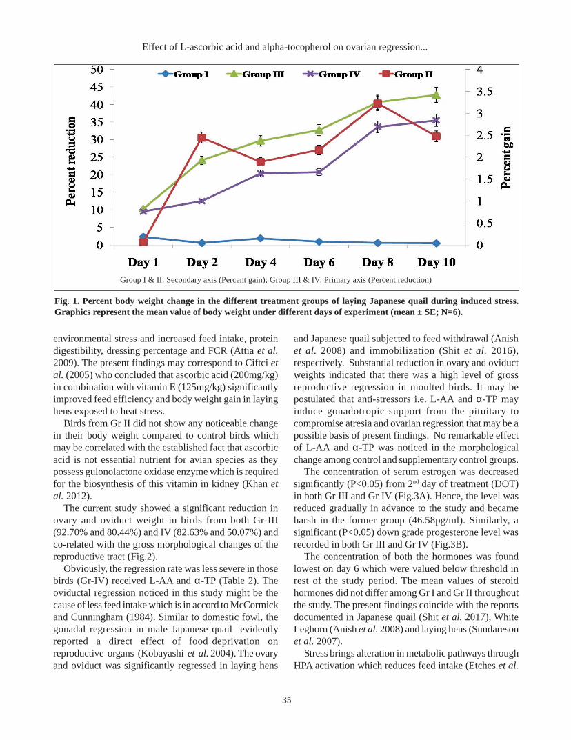
RESULTS AND DISCUSSIONThe percent body weight reduction was found 42.68
and 35.49 in Gr III and Gr IV respectively (Figure 1).Though a similar trend of reduction was noticed in bothFW treatments but the drastic change was recorded inthe former group. The birds in Gr II gained 2.47 percentmore body weight compared to those of control birds(Gr I).
In spite of the same treatment, presence of anti-oxidantvitamins could be the cause of stress alleviation resultingGr IV confirmed less severe change in their body weight.The result agreed with Sahin and Kucuk (2001) whostated that dietary ascorbic acid (250ppm) significantlystimulate growth and improved feed efficiency in stressedchicken and quail respectively. Ascorbic acid (250mg/kg) has relieved the birds from the negative effects of
LHR ATTGTGCTCCTCGTCCTC GTCTATGGCGTGGTTGTAG 162 56
PHR GGAAGGGCAGCACAACTATT GACACGCTGGACAGTTCTTC 83 56
B-actin GGAAGTTACTCGCCTCTG AAAGACACTTGTTGGGTTAC 127 58
Forward Reverse
Primer sequence* (5'-3' order) Ampliconsize (bp)
Annealingtemp. ( oC)
Target gene
Table 1. Primer pair sequences with their amplicon size and annealing temperature used for Quantitative RT-PCR.
LHR, luteinizing hormone receptor, PHR, progesterone hormone receptor
35
environmental stress and increased feed intake, proteindigestibility, dressing percentage and FCR (Attia et al.2009). The present findings may correspond to Ciftci etal. (2005) who concluded that ascorbic acid (200mg/kg)in combination with vitamin E (125mg/kg) significantlyimproved feed efficiency and body weight gain in layinghens exposed to heat stress.
Birds from Gr II did not show any noticeable changein their body weight compared to control birds whichmay be correlated with the established fact that ascorbicacid is not essential nutrient for avian species as theypossess gulonolactone oxidase enzyme which is requiredfor the biosynthesis of this vitamin in kidney (Khan etal. 2012).
The current study showed a significant reduction inovary and oviduct weight in birds from both Gr-III(92.70% and 80.44%) and IV (82.63% and 50.07%) andco-related with the gross morphological changes of thereproductive tract (Fig.2).
Obviously, the regression rate was less severe in thosebirds (Gr-IV) received L-AA and α-TP (Table 2). Theoviductal regression noticed in this study might be thecause of less feed intake which is in accord to McCormickand Cunningham (1984). Similar to domestic fowl, thegonadal regression in male Japanese quail evidentlyreported a direct effect of food deprivation onreproductive organs (Kobayashi et al. 2004). The ovaryand oviduct was significantly regressed in laying hens
and Japanese quail subjected to feed withdrawal (Anishet al. 2008) and immobilization (Shit et al. 2016),respectively. Substantial reduction in ovary and oviductweights indicated that there was a high level of grossreproductive regression in moulted birds. It may bepostulated that anti-stressors i.e. L-AA and α-TP mayinduce gonadotropic support from the pituitary tocompromise atresia and ovarian regression that may be apossible basis of present findings. No remarkable effectof L-AA and α-TP was noticed in the morphologicalchange among control and supplementary control groups.
The concentration of serum estrogen was decreasedsignificantly (P<0.05) from 2nd day of treatment (DOT)in both Gr III and Gr IV (Fig.3A). Hence, the level wasreduced gradually in advance to the study and becameharsh in the former group (46.58pg/ml). Similarly, asignificant (P<0.05) down grade progesterone level wasrecorded in both Gr III and Gr IV (Fig.3B).
The concentration of both the hormones was foundlowest on day 6 which were valued below threshold inrest of the study period. The mean values of steroidhormones did not differ among Gr I and Gr II throughoutthe study. The present findings coincide with the reportsdocumented in Japanese quail (Shit et al. 2017), WhiteLeghorn (Anish et al. 2008) and laying hens (Sundaresonet al. 2007).
Stress brings alteration in metabolic pathways throughHPA activation which reduces feed intake (Etches et al.
Group I & II: Secondary axis (Percent gain); Group III & IV: Primary axis (Percent reduction)
Fig. 1. Percent body weight change in the different treatment groups of laying Japanese quail during induced stress.Graphics represent the mean value of body weight under different days of experiment (mean ± SE; N=6).
Effect of L-ascorbic acid and alpha-tocopherol on ovarian regression...

36
Exploratory Animal and Medical Research, Vol.10, Issue 1, June, 2020
1984) and subsequently lowers the plasma level of sexsteroids through the ovarian regression in dose dependentmanner (Moudgal et al. 1991). The tocoferol is the majorchain-breaking antioxidant in lipid phases such as cellularmembranes or low density lipoproteins (LDL), and theoxidising free radical chain reactions are terminated inaqueous compartments with ascorbic acid as terminalreductant and their synergistic effect could be the resultof stress alleviation in Gr IV. Supplementation of naturalanti-oxidant did not exhibit any role on steroid hormonesas observed in control groups.
The expression profile of LHR and PHR genessupposed to be involved in the physiological andmolecular events of ovarian functions during inducedstress by feed withdrawal. As there were no hierarchicalfollicles detected after 6thday of the experiment,expression study was carried out in the whole ovary.However, gene expression analysis during the rest of thedays was performed in three larger hierarchical follicles(F
1, F
2 and F
3).
The luteinizing hormone receptor (LHR) has beenclassically described as a receptor present in gonads(ovary and testis) controlling various reproductive
processes. However, recent evidences suggest thepresence of LHR in the extragonadal tissue, particularlyin the female reproductive tract of human, bovine,porcine, rat, mouse, rabbit and turkey (You et al. 2000,Mukherjee et al. 1994). The LHR gene expression wassignificantly (P<0.05) down regulated in the follicles fromGr-III and Gr-IV on day 4 and 6 respectively of this study(Fig.4). However, the magnitude of expression was morerigorous in GR-III at any point of this study compared toits counterparts. This finding is in agreement to Agarwalet al. (2013) and Anish et al. (2008) who hypothesizedthat the pituitary response to luteinizing hormone–releasing hormone (LHRH) for the secretion of LH duringmoulting is reduced which may result in inhibition ofgonadotrophin-sensitive steroidogenesis in hens. Nosignificant improvement was recorded in the relative foldexpression of LHR gene in Gr-IV which implied longerdietary inclusion of L-AA and α-TP may optimize anti-stressors effect to assuage the negative blow of stress.
Activation of the G-protein coupled LH-R on muralgranulosa cells in response to the LH surge stimulatesadenylate cyclase, increases intracellular cAMP, therebyactivating protein kinase A and inducing expression of
DA
Y -
1D
AY
-6
DA
Y -
10Group I Group II Group III Group IV
Fig.2. Changes in the gross morphology of reproductive system under different experimental groups of Japanese quailduring induced stress.

37
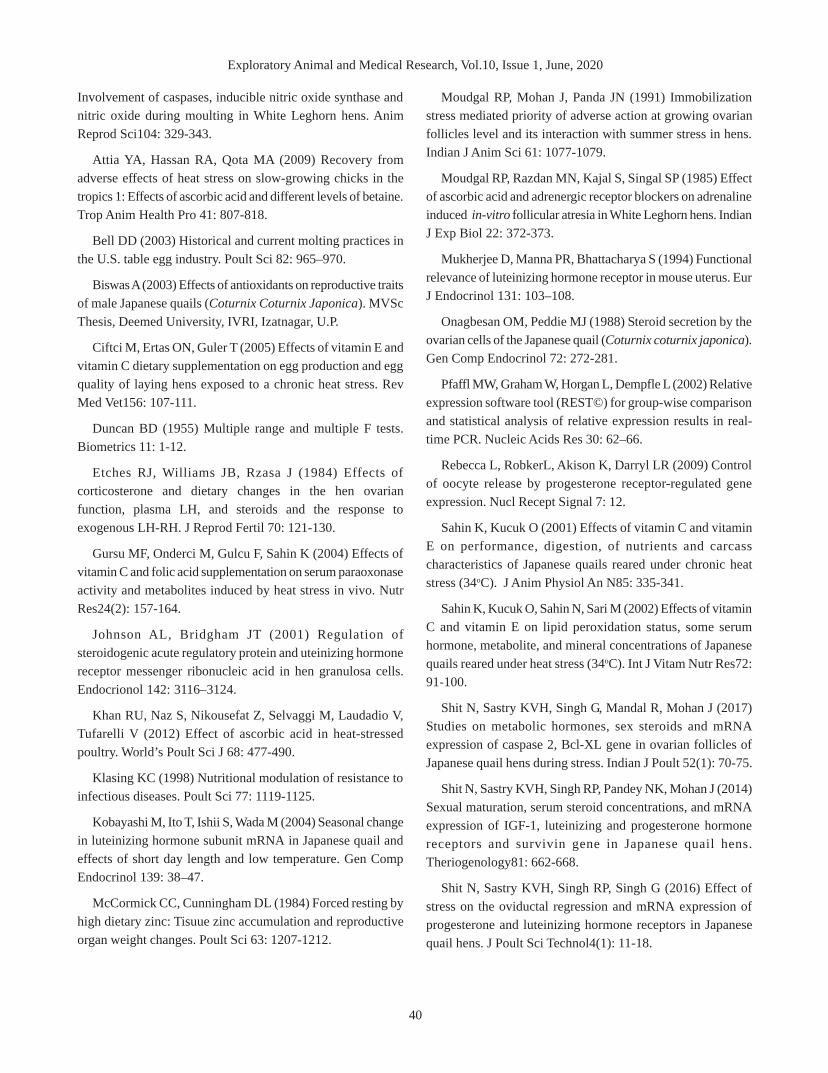
PHR (Rebecca et al. 2009). Subsequent to selection,follicle undergoes a transition from largely FSHdependence to LH-dependence which elicits progesteroneproduction in preovulatory follicles (Johnson andBridgham 2001). The expression profile of PHR gene inthe follicles and ovarian tissue is shown in Fig.5. Themean expression level of PHR was down-regulated inthe yellow follicles from Gr-III on 4th day of theexperiment. The sensitivity of ovarian tissues towardsFW stress was evidently more and confirmed significant(P<0.05) down regulation throughout this study. Theexpression pattern in Gr-IV varied inconsistentlythroughout the study. No statistical disparity was observedin the mRNA expression of PHR gene among controlgroups. This result could be co-related with serumconcentration of progesterone as declined in progress ofthis study. Though the severity of fold expression wasless in Gr-IV but did not vary significantly and could be
the effect of L-AA and α-TP to induce gonadotrophin-sensitive steroidogenesis. The expression level of PHRwas found to be up-regulated in the hierarchial follicleson sexual maturation in Japanese quail (Shit et al. 2014)but no literature is available so far to correlate thesepresent findings. However, further study of longerduration is required to correlate their effects in mitigatingthe detrimental consequence of stress on the expressionpattern of PHR.
CONCLUSIONThe results of the study suggested that Japanese quail
is as sensitive as domestic chicken to stress induced byFW. The gross morphological regression and profile ofsteroid hormones were found well correlated with downregulated mRNA expression of IGF-1, LHR and PHR. Itappears that supplementation of vitamin C and E mayalleviates negative effect of induced stress and improves
Day Group I Group II Group III Group IV Significance
Day 1 6.44±0.41 7.39±1.18 6.08±0.99p 6.45±0.77p NS
Day 2 6.66±0.87a 6.16±0.91a 3.82±0.79bq 4.11±0.80bq *
Day 4 6.37±0.94a 6.75±1.16a 3.58±1.25bq 3.90±0.33bq *
Day 6 7.36±0.56a 6.93±0.43a 0.76±0.06cr 3.37±0.22bq **
Day 8 7.13±0.99a 7.41±0.68a 0.56±0.18cr 1.68±0.26cr **
Day 10 6.66±0.26a 7.05±0.92a 0.40±0.05cr 1.12±0.02cr **
Significance NS NS ** **
A. Ovary weight (g)
Table 2. Changes in the ovary weight (A) and oviduct weight (B) of control and different treatment groups of Japanesequail during induced stress (mean ± SE; N=6).
Group I: Control, Group II: Control with dietary vitamins, Group III: Feed withdrawal, Group IV: Feed withdrawal with vitamins. Meansbearing different superscript in column pqrs and row abc differ significantly.* P<0.05; ** P<0.01, NS non-significant.
Day Group I Group II Group III Group IV Significance
Day 1 8.16±0.60 9.10±1.02 7.29±0.51p 8.18±0.82p NS
Day 2 8.28±0.33a 8.17±0.26a 5.87±0.96bq 7.28±0.71abp *
Day 4 7.46±0.43a 7.85±0.85a 5.71±0.94bq 6.28±1.56abpq *
Day 6 7.83±0.33a 8.37±1.28a 2.81±0.23cr 5.70±0.79bq **
Day 8 8.46±0.35a 7.08±0.80a 2.26±0.26crs 5.08±1.52bq **
Day 10 7.54±0.26a 8.73±1.22a 1.23±0.30cs 3.92±1.53br **
Significance NS NS ** *
B. Oviduct weight (g)
Group I: Control, Group II: Control with dietary vitamins, Group III: Feed withdrawal, Group IV: Feed withdrawal with vitamins. Meansbearing different superscript in column pqrs and row abc differ significantly. * P<0.05; ** P<0.01, NS non-significant.
Effect of L-ascorbic acid and alpha-tocopherol on ovarian regression...

38
Exploratory Animal and Medical Research, Vol.10, Issue 1, June, 2020
Panel -A Panel -B
Fig. 3. The serum hormone profile of different treatment groups of Japanese quail during induced stress. Graphics representthe mean level of estrogen (panel A) and progesterone (panel B) at different days of experiment (mean ± SE, N=6).
A B
C D
E F
* indicates a significant difference between control and treatment groups, P<0.05.
Fig. 4. mRNA expression of Luteinizing hormone receptor (LHR) in the ovary and ovarian follicles (F1, F
2 & F
3) from
different dietary treatment groups of Japanese quail. The graphics represent the mean ± SE; N=6 and the panel A,B,C,D,Eand F denotes days of treatment.
39
the morphological, bio-chemical and cellular function inJapanese quail. Further it is to mention that vitamin Cand E is found dietary non-essential when the species ismaintained under standard management without stress.However, the exact mechanism of FW inducedreproductive regression and sequence of events inalleviation of stress by vitamin C and E supplementationin Japanese quail is yet to be unravelled.
REFERENCES
Agarwal R, Sastry KVH, Tripathi V, Singh R, Saxena R,Mohan J, Singh RP (2013) Expression profile of Luteinizinghormone receptor gene in hierarchal follicles and regressingoviduct tissues of White Leghorn hens during moulting. ReprodDomest Anim 48: 278–283.
Anish D, Sastry KVH, Sundaresan NR, Saxena VK, SinghR, Mohan J (2008) Reproductive tissue regression:
A B
C D
E F
* indicates a significant difference between control and treatment groups, P<0.05.
Fig. 5. mRNA expression of Progesterone hormone receptor (PHR) in the ovary and ovarian follicles (F1, F
2 & F
3) from
different dietary treatment groups of laying Japanese quail. The graphics represent the mean ± SE; N=6 and the panelA,B,C,D,E and F denotes days of treatment.
Effect of L-ascorbic acid and alpha-tocopherol on ovarian regression...
40
Exploratory Animal and Medical Research, Vol.10, Issue 1, June, 2020
Involvement of caspases, inducible nitric oxide synthase andnitric oxide during moulting in White Leghorn hens. AnimReprod Sci104: 329-343.
Attia YA, Hassan RA, Qota MA (2009) Recovery fromadverse effects of heat stress on slow-growing chicks in thetropics 1: Effects of ascorbic acid and different levels of betaine.Trop Anim Health Pro 41: 807-818.
Bell DD (2003) Historical and current molting practices inthe U.S. table egg industry. Poult Sci 82: 965–970.
Biswas A (2003) Effects of antioxidants on reproductive traitsof male Japanese quails (Coturnix Coturnix Japonica). MVScThesis, Deemed University, IVRI, Izatnagar, U.P.
Ciftci M, Ertas ON, Guler T (2005) Effects of vitamin E andvitamin C dietary supplementation on egg production and eggquality of laying hens exposed to a chronic heat stress. RevMed Vet156: 107-111.
Duncan BD (1955) Multiple range and multiple F tests.Biometrics 11: 1-12.
Etches RJ, Williams JB, Rzasa J (1984) Effects ofcorticosterone and dietary changes in the hen ovarianfunction, plasma LH, and steroids and the response toexogenous LH-RH. J Reprod Fertil 70: 121-130.
Gursu MF, Onderci M, Gulcu F, Sahin K (2004) Effects ofvitamin C and folic acid supplementation on serum paraoxonaseactivity and metabolites induced by heat stress in vivo. NutrRes24(2): 157-164.
Johnson AL, Bridgham JT (2001) Regulation ofsteroidogenic acute regulatory protein and uteinizing hormonereceptor messenger ribonucleic acid in hen granulosa cells.Endocrionol 142: 3116–3124.
Khan RU, Naz S, Nikousefat Z, Selvaggi M, Laudadio V,Tufarelli V (2012) Effect of ascorbic acid in heat-stressedpoultry. World’s Poult Sci J 68: 477-490.
Klasing KC (1998) Nutritional modulation of resistance toinfectious diseases. Poult Sci 77: 1119-1125.
Kobayashi M, Ito T, Ishii S, Wada M (2004) Seasonal changein luteinizing hormone subunit mRNA in Japanese quail andeffects of short day length and low temperature. Gen CompEndocrinol 139: 38–47.
McCormick CC, Cunningham DL (1984) Forced resting byhigh dietary zinc: Tisuue zinc accumulation and reproductiveorgan weight changes. Poult Sci 63: 1207-1212.
Moudgal RP, Mohan J, Panda JN (1991) Immobilizationstress mediated priority of adverse action at growing ovarianfollicles level and its interaction with summer stress in hens.Indian J Anim Sci 61: 1077-1079.
Moudgal RP, Razdan MN, Kajal S, Singal SP (1985) Effectof ascorbic acid and adrenergic receptor blockers on adrenalineinduced in-vitro follicular atresia in White Leghorn hens. IndianJ Exp Biol 22: 372-373.
Mukherjee D, Manna PR, Bhattacharya S (1994) Functionalrelevance of luteinizing hormone receptor in mouse uterus. EurJ Endocrinol 131: 103–108.
Onagbesan OM, Peddie MJ (1988) Steroid secretion by theovarian cells of the Japanese quail (Coturnix coturnix japonica).Gen Comp Endocrinol 72: 272-281.
Pfaffl MW, Graham W, Horgan L, Dempfle L (2002) Relativeexpression software tool (REST©) for group-wise comparisonand statistical analysis of relative expression results in real-time PCR. Nucleic Acids Res 30: 62–66.
Rebecca L, RobkerL, Akison K, Darryl LR (2009) Controlof oocyte release by progesterone receptor-regulated geneexpression. Nucl Recept Signal 7: 12.
Sahin K, Kucuk O (2001) Effects of vitamin C and vitaminE on performance, digestion, of nutrients and carcasscharacteristics of Japanese quails reared under chronic heatstress (34oC). J Anim Physiol An N85: 335-341.
Sahin K, Kucuk O, Sahin N, Sari M (2002) Effects of vitaminC and vitamin E on lipid peroxidation status, some serumhormone, metabolite, and mineral concentrations of Japanesequails reared under heat stress (34oC). Int J Vitam Nutr Res72:91-100.
Shit N, Sastry KVH, Singh G, Mandal R, Mohan J (2017)Studies on metabolic hormones, sex steroids and mRNAexpression of caspase 2, Bcl-XL gene in ovarian follicles ofJapanese quail hens during stress. Indian J Poult 52(1): 70-75.
Shit N, Sastry KVH, Singh RP, Pandey NK, Mohan J (2014)Sexual maturation, serum steroid concentrations, and mRNAexpression of IGF-1, luteinizing and progesterone hormonereceptors and survivin gene in Japanese quail hens.Theriogenology81: 662-668.
Shit N, Sastry KVH, Singh RP, Singh G (2016) Effect ofstress on the oviductal regression and mRNA expression ofprogesterone and luteinizing hormone receptors in Japanesequail hens. J Poult Sci Technol4(1): 11-18.
41
Shit N, Singh RP, Sastry KVH, Agarwal R, Singh R, PandeyNK, Mohan J (2012) Effect of dietary L-ascorbic acid (L-AA)on production performance, egg quality traits and fertility inJapanese quail (Coturnix japonica) at low ambient temperature.Asian Australas J Anim Sci 25(7): 1009-1014.
Sinkalu VO, Ayo JO (2008) Diurnal fluctuations in rectaltemperature of black Harco pullets administered with vitaminsA and C during the hot-dry season. Int J Poult Sci 7: 1065-1070.
*Cite this article as: Shit N, Sastry KVH, Singh G, Mohan J (2020) Effect of L-ascorbic acid and alpha-tocopherolon ovarian regression, hormonal changes and gene expression in Japanese quail during stress. Explor Anim Med Res10(1): 32-41.
Sundareson NR, Anish D, Sastry KVH, Saxen VK, MohanJ, Ahmed KA (2007) Cytokines in reproductive remodeling ofmolting White Leghorn hens. J Reprod Immunol 73: 39–50.
You S, Kim H, Hsu CC, Halawani ME, Foster DN (2000)Three different turkey luteinizing hormone receptor (tLH-R)isoforms I: characterization of alternative spliced tLH-Risoforms and their regulated expression in diverse tissues. BiolReprod62: 108–116.
Effect of L-ascorbic acid and alpha-tocopherol on ovarian regression...