effect of felodipine on myocardial and renal injury ... · effect of felodipine on myocardial and...
TRANSCRIPT
www.bjournal.com.brwww.bjournal.com.br
Faculdade de Medicina de Ribeirão Preto
CampusRibeirão Preto
Institutional Sponsors
The Brazilian Journal of Medical and Biological Research is partially financed by
Volume 43 (5) 409-521 May 2010
Braz J Med Biol Res, May 2010, Volume 43(5) 506-514
Effect of felodipine on myocardial and renal injury induced by aldosterone-high salt hypertension in uninephrectomized rats
B.B. Matsubara, M. Franco, J.S. Janicki and L.S. Matsubara
Brazilian Journal of Medical and Biological Research (2010) 43: 506-514ISSN 0100-879X
Effect of felodipine on myocardial and renal injury induced by aldosterone-high salt
hypertension in uninephrectomized rats
B.B. Matsubara1, M. Franco2, J.S. Janicki3 and L.S. Matsubara1
1Departamento de Clínica Médica, Faculdade de Medicina de Botucatu, Universidade Estadual Paulista, Botucatu, SP, Brasil
2Departamento de Patologia, Escola Paulista de Medicina, Universidade Federal de São Paulo, São Paulo, SP, Brasil
3Department of Cell and Developmental Biology and Anatomy, University of South Carolina, Columbia, SC, USA
Abstract
It has been recently shown that calcium channel blockers might have a protective effect on cardiac fibrogenesis induced by aldosterone. The objective of this study was to evaluate the protective effect of felodipine, a dihydropyridine calcium channel blocker, against heart and kidney damage caused by aldosterone-high sodium intake in uninephrectomized rats. Wistar rats were divided into three groups: CNEP (uninephrectomized + 1% NaCl in the drinking water, N = 9); ALDO (same as CNEP group plus continuous infusion of 0.75 µg/h aldosterone, N = 12); ALDOF (same as ALDO group plus 30 mg·kg-1·day-1 felodipine in the drinking water, N = 10). All results were compared with those of age-matched, untreated rats (CTL group, N = 10). After 6 weeks, tail cuff blood pressure was recorded and the rats were killed for histological analysis. Blood pressure (mmHg) was significantly elevated (P < 0.05) in ALDO (180 ± 20) and ALDOF (168 ± 13) compared to CTL (123 ± 12) and CNEP (134 ± 13). Heart damage (lesion scores - median and interquartile range) was 7.0 (5.5-8.0) in ALDO and was fully prevented in ALDOF (1.5; 1.0-2.0). Also, left ventricular collagen volume fraction (%) in ALDOF (2.9 ± 0.5) was similar to CTL (2.9 ± 0.5) and CNEP (3.4 ± 0.4) and decreased compared to ALDO (5.1 ± 1.6). Felodipine partially prevented kidney injury since the damage score for ALDOF (2.0; 2.0-3.0) was significantly decreased compared to ALDO (7.5; 4.0-10.5), although higher than CTL (null score). Felodipine has a protective effect on the myocardium and kidney as evidenced by decreased perivascular inflammation, myo-cardial necrosis and fibrosis.
Key words: Aldosterone; Dihydropyridine; Fibrinoid necrosis; Vascular remodeling; Hypertension; Myocardial remodeling
Introduction
www.bjournal.com.brBraz J Med Biol Res 43(5) 2010
Correspondence: B.B. Matsubara, Departamento de Clínica Médica, Faculdade de Medicina de Botucatu, UNESP, Distrito Rubião Júnior, s/n, 18618-000 Botucatu, SP, Brasil. Fax: +55-14-3882-2238. E-mail: [email protected]
Received September 15, 2009. Accepted April 14, 2010. Available online April 30, 2010. Published May 14, 2010.
Aldosterone-high salt intake produces a variety of deleteri-ous actions on the cardiovascular system of uninephrectomized rats. Previous studies have shown inflammation and oxidative stress within the arteriolar wall of the heart and kidney, and inflammatory tissue remodeling (1,2). The histological features of malignant or accelerated hypertension have also been described in this model (1,3).
The mechanisms by which aldosterone induces cardiac tissue damage are not fully understood and seem to involve genomic and non-genomic effects (4). Genomic action is the classical mechanism by which aldosterone binds to miner-alocorticoid receptors (MR) and promotes protein synthesis. Non-genomic effects of aldosterone occur within minutes, inducing vasoconstriction (5). Also, vascular smooth muscle
cells from spontaneously hypertensive rats show an increased aldosterone-induced non-genomic signaling mediated by c-Src. This signaling appears to be important in the profibrotic and proinflammatory actions of aldosterone in this genetic model of hypertension (6).
Recently, it has been demonstrated that cardiomyocytes have putative aldosterone-regulated MR genes (7). Accord-ingly, the first event leading to cardiovascular inflammation and remodeling may be associated with inappropriate MR activation. In addition, Dietz et al. (8) have recently reported that dihydro-pyridine calcium channel blockers (CCBs) have MR antagonist activity and that a number of widely used dihydropyridine CCBs, including felodipine, compete for aldosterone binding to MR and have the effect of MR antagonists on cardiomyocytes. L-

Felodipine and aldosterone-high salt hypertension 507
www.bjournal.com.br Braz J Med Biol Res 43(5) 2010
type calcium channel antagonists are effective in decreasing blood pressure and have been shown to be renoprotective. However, whether these protective effects are due to blood pressure reduction or to a specific pharmacologic blockade is still a matter of debate.
We, therefore, hypothesized that felodipine may protect the heart and kidney from damage by decreasing the perivas-cular inflammation induced by aldosterone-high salt intake in uninephrectomized rats. The objective of this study was to determine whether felodipine, a dihydropyridine calcium channel blocker, is effective in preventing heart and kidney damage in uninephrectomized rats subjected to aldosterone-high sodium intake.
Material and Methods
AnimalsTwelve-week-old male Wistar rats (300 ± 20 g) were ob-
tained from the Central Animal Facility of the Botucatu Medical School, São Paulo State University, SP, Brazil. The animals were housed under standard environmental conditions and maintained on a commercial rat laboratory diet (Purina Labina, Brazil) and tap water ad libitum. The experimental protocol was approved by the Animal Care and Use Committee of our Institution and conformed to the NIH Guidelines on the Care and Use of Laboratory Animals (Institute of Laboratory Animal Research, Commission on Life Sciences, National Research Council, 1996).
Experimental protocolLaparotomy and unilateral nephrectomy were performed
under sodium thiopental anesthesia (50 mg/kg, intraperitone-ally). Immediately after surgery and recovery from anesthesia, the rats were randomly divided into three groups as follows: CNEP (N = 9) - rats maintained on high NaCl (1%) in the drink-ing water. ALDO (N = 12) - rats maintained on high NaCl as the CNEP group and continuously infused with aldosterone (Sigma, USA; 0.75 µg/h), via minipumps implanted subcutane-ously (Alzet minipumps mod. 2002, Alza Corp., USA). ALDOF (N = 10) - same as the ALDO group plus felodipine (30 mg· kg-1·day-1) in the drinking water. All results were compared to an additional control group of animals that were not unilaterally nephrectomized and that did not receive high NaCl, aldosterone or felodipine (N = 10).
Tail cuff blood pressure and body weight were recorded before and 6 weeks after surgery. At the end of the 6-week period, all rats were anesthetized with sodium thiopental (50 mg/kg, ip) and killed by thoracotomy and blood samples were taken from the heart for electrolyte and creatinine measure-ments. The heart and the remaining kidney were excised and weighed. Left and right ventricles were weighed separately.
Histopathological analysisThe morphometric analysis of the myocardium was per-
formed as described previously (9). Transverse sections of the
left (LV) and right (RV) ventricles and a sample of the kidney were fixed in 10% buffered formalin and embedded in paraffin. Five-micron sections of myocardium and kidney were stained with hematoxylin-eosin. Also, ventricular sections were stained with the collagen-specific stain picrosirius red (sirius red F3BA in aqueous saturated picric acid).
Interstitial myocardial collagen volume fraction (CVF). The CVF of the entire section was determined using a Leica microscope (objective lens 40X) attached to a video camera and connected to a personal computer equipped with image analyzer software (Image-Pro Plus 3.0, Media Cybernetics, USA). The components of cardiac tissue were identified ac-cording to their color (i.e., red for collagen fibers, yellow for myocytes, and white for interstitial space). The digitized profiles were transferred to the computer and CVF was calculated as the sum of all connective tissue areas divided by the sum of the connective tissue and myocyte areas. On the average, 35 microscopic fields were analyzed. Perivascular collagen was excluded from this analysis.
Scores of myocardial damage Two pathologists blind as to the source of the hematoxylin-
eosin-stained tissue section, independently examined the slides and rated the damage using the criteria shown in Table 1. This approach emphasized cellular necrosis and inflammatory phenomena. Arteriole remodeling was analyzed by calculat-ing the relative wall thickness (RWT) and relative perivascular fibrosis (RPF) as follows: RWT = (WA - LA) / LA, where WA is the area of vascular wall, and LA is the lumen area, and RPF = (FA - WA) / WA, where FA is the perivascular fibrosis area.
All analyses were performed using two separate sets of vessels as follows: vessels with luminal diameters less than 50 μm and vessels with lumen diameters between 50-100 μm. At least four vessels of each set were measured in all sections and only vessels with a circular cross-section were analyzed.
Statistical analysisDifferences among groups were analyzed by one-way
analysis of variance (ANOVA) followed by pairwise multiple comparison (Bonferroni t-test). ANOVA on ranks followed by the Dunn test was used to compare scores among groups. Spearman’s correlation was used to compare LV and RV damage scores and to analyze the association between LV or kidney score damage with systolic blood pressure. Differ-ences were considered to be statistically significant when P < 0.05/k, where k is the number of statistical comparisons (Bonferroni correction).
Results
Morphometric data and blood pressure results are present-ed in Table 2. Systolic blood pressure increased in both the ALDO and ALDOF groups indicating a non-antihypertensive effect of felodipine. Accordingly, both hypertensive groups presented increased LV and RV weight. The increase in

508 B.B. Matsubara et al.
www.bjournal.com.brBraz J Med Biol Res 43(5) 2010
Table 1. Criteria for histological damage score.
Score
Myocardial scores1. Myocyte lesions
1.1. Necrosis of few cells +11.2. Microinfarction +31.3. Infarction (area >0.05 cm2) +5Note: Frequent lesions (>3 foci) +1
2. Benign vascular damage2.1. Media hypertrophy +12.2. Wall hyalinization (subendothelial) +12.3. Arteriolar fibroelastosis +1Note: Frequent damage (>3 vessels) +1
3. Severe vascular damage3.1. Transmural fibrinoid necrosis +43.2. Myointimal proliferation +4Note: Frequent damage (>3 vessels) +1
4. Perivascular lesion4.1. Limited inflammatory infiltrate +14.2. Inflammatory infiltrate with perivascular myocyte damage +3Note: Frequent damage (>3 vessels) +1
Kidney scores1. Benign vascular damage
1.1. Media layer hypertrophy +11.2. Wall hyalinization (subendothelial) +11.3. Arteriolar fibroelastosis +1Note: Frequent damage (>3 vessels) +1
2. Severe arterial damage2.1. Glomerulonecrosis +42.2. Transmural fibrinoid necrosis +42.3. Myointimal proliferation +4Note: Frequent damage (>3 vessels) +1
3. Other lesions3.1. Acute tubular necrosis +1
Table 2. Morphometric and blood pressure data.
Group BW (g) SBP (mmHg) LVW/BW (mg/g) RVW/BW (mg/g) CVFLV (%) CVFRV (%)
CTL 389 ± 21a 123 ± 12a 1.82 ± 0.12a 0.61 ± 0.05a 2.9 ± 0.67a 4.0 ± 0.34a
CNEP 414 ± 34a 134 ± 13a 1.85 ± 0.08a 0.60 ± 0.09a 3.4 ± 0.44a 3.6 ± 0.94a
ALDO 399 ± 32a 180 ± 20b 2.51 ± 0.29b 0.69 ± 0.08b 5.1 ± 1.61b 5.6 ± 1.42b
ALDOF 416 ± 29a 168 ± 13b 2.22 ± 0.13c 0.69 ± 0.06b 2.9 ± 0.48a 3.6 ± 0.44a
Data are reported as means ± SD. CTL = control; CNEP = uninephrectomy plus 1% salt in the drinking water; ALDO = same as CNEP plus 6-week subcutaneously infused aldosterone; ALDOF = same as ALDO plus 6-week oral felodipine; BW = body weight; SBP = systolic blood pressure; LVW = left ven-tricle weight; RVW = right ventricle weight; CVFLV = collagen volume fraction of left ventricle; CVFRV = collagen volume fraction of right ventricle. Means with different superscript letters are significantly different (P < 0.05) in all pairwise multiple comparison procedures (ANOVA and Bonferroni t-test).
Felodipine and aldosterone-high salt hypertension 509
www.bjournal.com.br Braz J Med Biol Res 43(5) 2010
CVF that occurred in both ventricles of the ALDO group was prevented in the felodipine-treated group.
Serum electrolytes and creatinine data are shown in Table 3. Aldosterone infusion increased serum sodium level and decreased potassium level, effects that were not prevented by felodipine treatment. Conversely, felodipine prevented the increase in serum creatinine observed in the CNEP and ALDO groups.
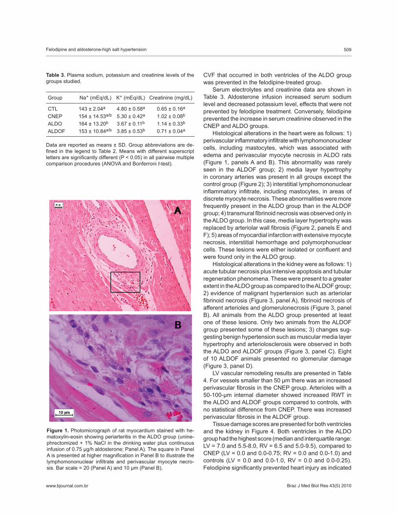
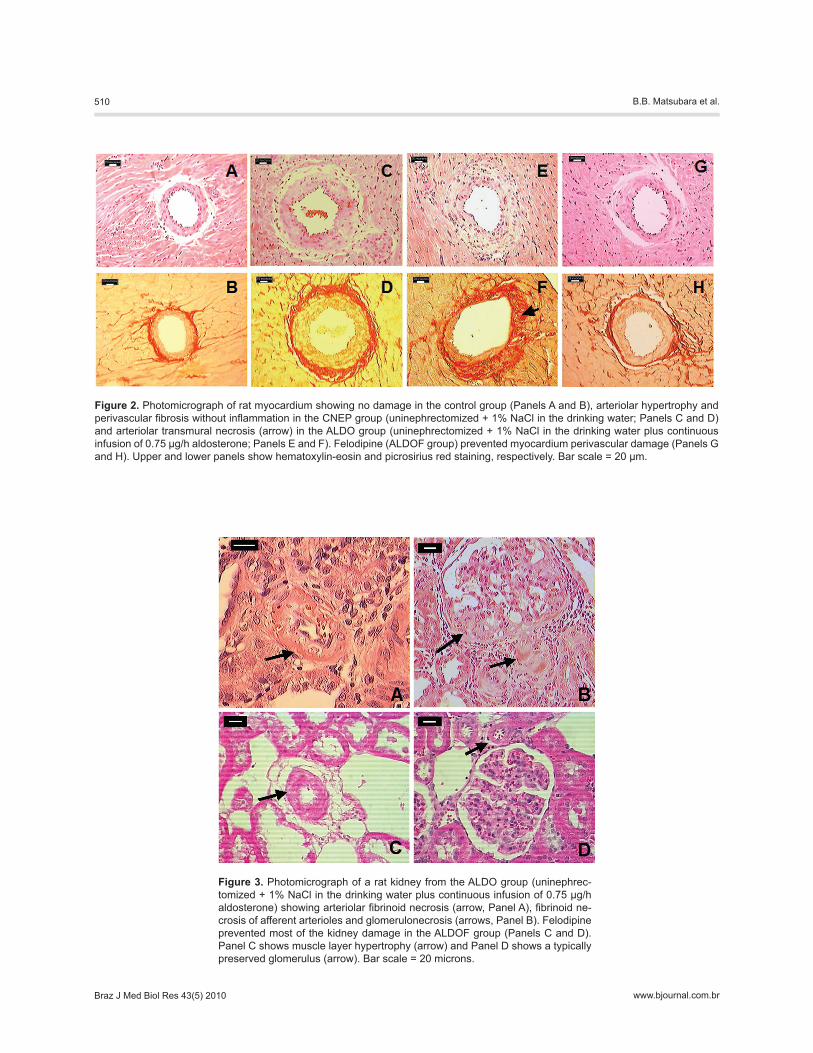
Histological alterations in the heart were as follows: 1) perivascular inflammatory infiltrate with lymphomononuclear cells, including mastocytes, which was associated with edema and perivascular myocyte necrosis in ALDO rats (Figure 1, panels A and B). This abnormality was rarely seen in the ALDOF group; 2) media layer hypertrophy in coronary arteries was present in all groups except the control group (Figure 2); 3) interstitial lymphomononuclear inflammatory infiltrate, including mastocytes, in areas of discrete myocyte necrosis. These abnormalities were more frequently present in the ALDO group than in the ALDOF group; 4) transmural fibrinoid necrosis was observed only in the ALDO group. In this case, media layer hypertrophy was replaced by arteriolar wall fibrosis (Figure 2, panels E and F); 5) areas of myocardial infarction with extensive myocyte necrosis, interstitial hemorrhage and polymorphonuclear cells. These lesions were either isolated or confluent and were found only in the ALDO group.
Histological alterations in the kidney were as follows: 1) acute tubular necrosis plus intensive apoptosis and tubular regeneration phenomena. These were present to a greater extent in the ALDO group as compared to the ALDOF group; 2) evidence of malignant hypertension such as arteriolar fibrinoid necrosis (Figure 3, panel A), fibrinoid necrosis of afferent arterioles and glomerulonecrosis (Figure 3, panel B). All animals from the ALDO group presented at least one of these lesions. Only two animals from the ALDOF group presented some of these lesions; 3) changes sug-gesting benign hypertension such as muscular media layer hypertrophy and arteriolosclerosis were observed in both the ALDO and ALDOF groups (Figure 3, panel C). Eight of 10 ALDOF animals presented no glomerular damage (Figure 3, panel D).
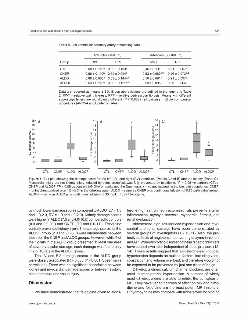
LV vascular remodeling results are presented in Table 4. For vessels smaller than 50 μm there was an increased perivascular fibrosis in the CNEP group. Arterioles with a 50-100-μm internal diameter showed increased RWT in the ALDO and ALDOF groups compared to controls, with no statistical difference from CNEP. There was increased perivascular fibrosis in the ALDOF group.
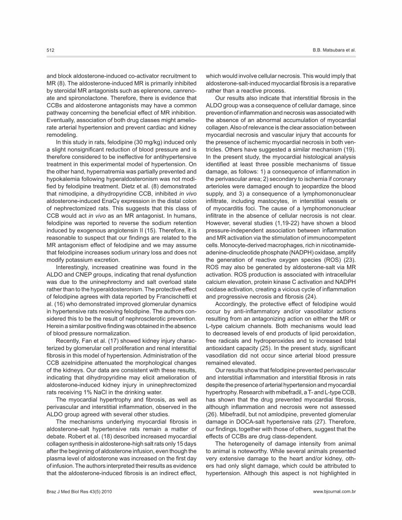
Tissue damage scores are presented for both ventricles and the kidney in Figure 4. Both ventricles in the ALDO group had the highest score (median and interquartile range: LV = 7.0 and 5.5-8.0, RV = 6.5 and 5.0-9.5), compared to CNEP (LV = 0.0 and 0.0-0.75; RV = 0.0 and 0.0-1.0) and controls (LV = 0.0 and 0.0-1.0, RV = 0.0 and 0.0-0.25). Felodipine significantly prevented heart injury as indicated
Figure 1. Photomicrograph of rat myocardium stained with he-matoxylin-eosin showing periarteritis in the ALDO group (unine-phrectomized + 1% NaCl in the drinking water plus continuous infusion of 0.75 µg/h aldosterone; Panel A). The square in Panel A is presented at higher magnification in Panel B to illustrate the lymphomononuclear infiltrate and perivascular myocyte necro-sis. Bar scale = 20 (Panel A) and 10 μm (Panel B).
Table 3. Plasma sodium, potassium and creatinine levels of the groups studied.
Group Na+ (mEq/dL) K+ (mEq/dL) Creatinine (mg/dL)
CTL 143 ± 2.04a 4.80 ± 0.58a 0.65 ± 0.16a
CNEP 154 ± 14.53a/b 5.30 ± 0.42a 1.02 ± 0.08b
ALDO 164 ± 13.20b 3.67 ± 0.11b 1.14 ± 0.33b
ALDOF 153 ± 10.84a/b 3.85 ± 0.53b 0.71 ± 0.04a
Data are reported as means ± SD. Group abbreviations are de-fined in the legend to Table 2. Means with different superscript letters are significantly different (P < 0.05) in all pairwise multiple comparison procedures (ANOVA and Bonferroni t-test).
510 B.B. Matsubara et al.
www.bjournal.com.brBraz J Med Biol Res 43(5) 2010
Figure 2. Photomicrograph of rat myocardium showing no damage in the control group (Panels A and B), arteriolar hypertrophy and perivascular fibrosis without inflammation in the CNEP group (uninephrectomized + 1% NaCl in the drinking water; Panels C and D) and arteriolar transmural necrosis (arrow) in the ALDO group (uninephrectomized + 1% NaCl in the drinking water plus continuous infusion of 0.75 µg/h aldosterone; Panels E and F). Felodipine (ALDOF group) prevented myocardium perivascular damage (Panels G and H). Upper and lower panels show hematoxylin-eosin and picrosirius red staining, respectively. Bar scale = 20 μm.
Figure 3. Photomicrograph of a rat kidney from the ALDO group (uninephrec-tomized + 1% NaCl in the drinking water plus continuous infusion of 0.75 µg/h aldosterone) showing arteriolar fibrinoid necrosis (arrow, Panel A), fibrinoid ne-crosis of afferent arterioles and glomerulonecrosis (arrows, Panel B). Felodipine prevented most of the kidney damage in the ALDOF group (Panels C and D). Panel C shows muscle layer hypertrophy (arrow) and Panel D shows a typically preserved glomerulus (arrow). Bar scale = 20 microns.
Felodipine and aldosterone-high salt hypertension 511
www.bjournal.com.br Braz J Med Biol Res 43(5) 2010
by much lower damage scores compared to ALDO (LV = 1.5 and 1.0-2.0, RV = 1.0 and 1.0-2.0). Kidney damage scores were higher in ALDO (7.5 and 4.0-10.5) compared to controls (0.0 and 0.0-0.0) and CNEP (0.0 and 0.0-1.0). Felodipine partially prevented kidney injury. The damage scores for the ALDOF group (2.0 and 2.0-3.0) were intermediate between those for the CNEP and ALDO groups. However, while 9 of the 12 rats in the ALDO group presented at least one area of severe vascular damage, such damage was found only in 2 of 10 rats in the ALDOF group.
The LV and RV damage scores in the ALDO group were closely associated (R = 0.838, P < 0.001; Spearman’s correlation). There was no significant association between kidney and myocardial damage scores or between systolic blood pressure and tissue injury.
Discussion
We have demonstrated that felodipine given to aldos-
terone-high salt uninephrectomized rats prevents arterial inflammation, myocyte necrosis, myocardial fibrosis, and renal dysfunction.
Aldosterone-high salt-induced hypertension and myo-cardial and renal damage have been demonstrated by several groups of investigators (1,2,10,11). Also, the pro-tective effects of angiotensin-converting enzyme inhibitors and AT1, mineralocorticoid and endothelin receptor blockers have been shown to be independent of blood pressure (12-14). These results suggest that aldosterone-salt-induced hypertension depends on multiple factors, including vaso-constriction and volume overload, and therefore would not be expected to be prevented by just one class of drugs.
Dihydropyridines, calcium channel blockers, are often used to treat arterial hypertension. A number of widely used dihydropyridine are able to inhibit the activation of MR. They have varied degrees of effect on MR and nimo-dipine and felodipine are the most potent MR inhibitors. Dihydropyridine may compete with aldosterone for binding
Figure 4. Box-plot showing the damage score for the left (LV) and right (RV) ventricles (Panels A and B) and the kidney (Panel C). Myocardial injury but not kidney injury induced by aldosterone/salt was fully prevented by felodipine. *P < 0.05 vs controls (CTL), CNEP and ALDOF; #P < 0.05 vs controls (ANOVA on ranks and the Dunn test). + = values exceeding the box-plot boundaries; CNEP = uninephrectomized plus 1% NaCl in the drinking water; ALDO = same as CNEP plus continuous infusion of 0.75 µg/h aldosterone; ALDOF = same as ALDO plus continuous infusion of 30 mg·kg-1·day-1 felodipine.
Table 4. Left ventricular coronary artery remodeling data.
Arterioles (<50 µm) Arterioles (50-100 µm)
Group RWT RPF RWT RPF
CTL 0.68 ± 0.110a 0.32 ± 0.103a 0.46 ± 0.13a 0.21 ± 0.067a
CNEP 0.66 ± 0.110a 0.39 ± 0.094b 0.53 ± 0.089a/b 0.26 ± 0.073a/b
ALDO 0.69 ± 0.085a 0.39 ± 0.144a/b 0.58 ± 0.091b 0.21 ± 0.097a
ALDOF 0.69 ± 0.110a 0.36 ± 0.121a/b 0.59 ± 0.095b 0.29 ± 0.064b
Data are reported as means ± SD. Group abbreviations are defined in the legend to Table 2. RWT = relative wall thickness; RPF = relative perivascular fibrosis. Means with different superscript letters are significantly different (P < 0.05) in all pairwise multiple comparison procedures (ANOVA and Bonferroni t-test).
512 B.B. Matsubara et al.
www.bjournal.com.brBraz J Med Biol Res 43(5) 2010
and block aldosterone-induced co-activator recruitment to MR (8). The aldosterone-induced MR is primarily inhibited by steroidal MR antagonists such as eplerenone, canreno-ate and spironolactone. Therefore, there is evidence that CCBs and aldosterone antagonists may have a common pathway concerning the beneficial effect of MR inhibition. Eventually, association of both drug classes might amelio-rate arterial hypertension and prevent cardiac and kidney remodeling.
In this study in rats, felodipine (30 mg/kg) induced only a slight nonsignificant reduction of blood pressure and is therefore considered to be ineffective for antihypertensive treatment in this experimental model of hypertension. On the other hand, hypernatremia was partially prevented and hypokalemia following hyperaldosteronism was not modi-fied by felodipine treatment. Dietz et al. (8) demonstrated that nimodipine, a dihydropyridine CCB, inhibited in vivo aldosterone-induced EnaCγ expression in the distal colon of nephrectomized rats. This suggests that this class of CCB would act in vivo as an MR antagonist. In humans, felodipine was reported to reverse the sodium retention induced by exogenous angiotensin II (15). Therefore, it is reasonable to suspect that our findings are related to the MR antagonism effect of felodipine and we may assume that felodipine increases sodium urinary loss and does not modify potassium excretion.
Interestingly, increased creatinine was found in the ALDO and CNEP groups, indicating that renal dysfunction was due to the uninephrectomy and salt overload state rather than to the hyperaldosteronism. The protective effect of felodipine agrees with data reported by Francischetti et al. (16) who demonstrated improved glomerular dynamics in hypertensive rats receiving felodipine. The authors con-sidered this to be the result of nephrosclerotic prevention. Herein a similar positive finding was obtained in the absence of blood pressure normalization.
Recently, Fan et al. (17) showed kidney injury charac-terized by glomerular cell proliferation and renal interstitial fibrosis in this model of hypertension. Administration of the CCB azelnidipine attenuated the morphological changes of the kidneys. Our data are consistent with these results, indicating that dihydropyridine may elicit amelioration of aldosterone-induced kidney injury in uninephrectomized rats receiving 1% NaCl in the drinking water.
The myocardial hypertrophy and fibrosis, as well as perivascular and interstitial inflammation, observed in the ALDO group agreed with several other studies.
The mechanisms underlying myocardial fibrosis in aldosterone-salt hypertensive rats remain a matter of debate. Robert et al. (18) described increased myocardial collagen synthesis in aldosterone-high salt rats only 15 days after the beginning of aldosterone infusion, even though the plasma level of aldosterone was increased on the first day of infusion. The authors interpreted their results as evidence that the aldosterone-induced fibrosis is an indirect effect,
which would involve cellular necrosis. This would imply that aldosterone-salt-induced myocardial fibrosis is a reparative rather than a reactive process.
Our results also indicate that interstitial fibrosis in the ALDO group was a consequence of cellular damage, since prevention of inflammation and necrosis was associated with the absence of an abnormal accumulation of myocardial collagen. Also of relevance is the clear association between myocardial necrosis and vascular injury that accounts for the presence of ischemic myocardial necrosis in both ven-tricles. Others have suggested a similar mechanism (19). In the present study, the myocardial histological analysis identified at least three possible mechanisms of tissue damage, as follows: 1) a consequence of inflammation in the perivascular area; 2) secondary to ischemia if coronary arterioles were damaged enough to jeopardize the blood supply, and 3) a consequence of a lymphomononuclear infiltrate, including mastocytes, in interstitial vessels or of myocarditis foci. The cause of a lymphomononuclear infiltrate in the absence of cellular necrosis is not clear. However, several studies (1,19-22) have shown a blood pressure-independent association between inflammation and MR activation via the stimulation of immunocompetent cells. Monocyte-derived macrophages, rich in nicotinamide-adenine-dinucleotide phosphate (NADPH) oxidase, amplify the generation of reactive oxygen species (ROS) (23). ROS may also be generated by aldosterone-salt via MR activation. ROS production is associated with intracellular calcium elevation, protein kinase C activation and NADPH oxidase activation, creating a vicious cycle of inflammation and progressive necrosis and fibrosis (24).
Accordingly, the protective effect of felodipine would occur by anti-inflammatory and/or vasodilator actions resulting from an antagonizing action on either the MR or L-type calcium channels. Both mechanisms would lead to decreased levels of end products of lipid peroxidation, free radicals and hydroperoxides and to increased total antioxidant capacity (25). In the present study, significant vasodilation did not occur since arterial blood pressure remained elevated.
Our results show that felodipine prevented perivascular and interstitial inflammation and interstitial fibrosis in rats despite the presence of arterial hypertension and myocardial hypertrophy. Research with mibefradil, a T- and L-type CCB, has shown that the drug prevented myocardial fibrosis, although inflammation and necrosis were not assessed (26). Mibefradil, but not amlodipine, prevented glomerular damage in DOCA-salt hypertensive rats (27). Therefore, our findings, together with those of others, suggest that the effects of CCBs are drug class-dependent.
The heterogeneity of damage intensity from animal to animal is noteworthy. While several animals presented very extensive damage to the heart and/or kidney, oth-ers had only slight damage, which could be attributed to hypertension. Although this aspect is not highlighted in
Felodipine and aldosterone-high salt hypertension 513
www.bjournal.com.br Braz J Med Biol Res 43(5) 2010
the literature, it suggests that aldosterone-induced tissue damage clearly depends on the individual’s response. We did not find a significant correlation between systolic blood pressure and either the kidney or the myocardial damage score in agreement with previous findings (28), suggesting a hypertension-independent mechanism for perivascular inflammation and myocardial necrosis.
In the present study, kidney and heart damage was analyzed in the same rat receiving an aldosterone-high salt diet. The results indicated that the presence of severe injury to the heart did not always indicate damage to the kidney or vice versa. This observation suggests that the physiological control and response to injury may differ depending on the target organ.
The distinct patterns of remodeling in large and small arterioles suggest a coronary vascular selectivity associ-ated with this experimental model. Interestingly, coronary arteriole remodeling was not prevented by felodipine and occurred in the absence of hypertension and inflammation, indicating a reactive process induced by high salt intake in the perivascular area. This remodeling may not require a high level of circulating aldosterone and may involve a local
synthesis induced by high sodium (29). In normotensive WKY rats, a high salt diet alone has been shown to cause myocardial perivascular fibrosis (30). Normotensive rats fed a high salt diet underwent renal vascular and glomerular remodeling (31) and showed depressed reactivity to dilator agonists (32). Therefore, it is reasonable to assume that reactive perivascular fibrosis induced by high salt intake would cause a decreased coronary flow reserve and sec-ondary myocardial remodeling.
The lack of increased perivascular fibrosis in the ALDO group could be explained by the marked exudative process still taking place at the end of the experiment. More than likely, a longer study period would have revealed a repara-tive process with increased fibrosis.
We have shown that felodipine has a protective effect on the myocardium and kidney as indicated by decreased perivascular inflammation and myocardial necrosis and fibrosis in aldosterone-high salt intake-hypertensive rats.
Acknowledgments
Research supported by FAPESP and CNPq.
References
1. Blasi ER, Rocha R, Rudolph AE, Blomme EA, Polly ML, McMahon EG. Aldosterone/salt induces renal inflammation and fibrosis in hypertensive rats. Kidney Int 2003; 63: 1791-1800.
2. Robert V, Silvestre JS, Charlemagne D, Sabri A, Trouve P, Wassef M, et al. Biological determinants of aldosterone-induced cardiac fibrosis in rats. Hypertension 1995; 26: 971-978.
3. Gavras H, Brunner HR, Laragh JH, Vaughan ED Jr, Koss M, Cote LJ, et al. Malignant hypertension resulting from de-oxycorticosterone acetate and salt excess: role of renin and sodium in vascular changes. Circ Res 1975; 36: 300-309.
4. Wehling M, Neylon CB, Fullerton M, Bobik A, Funder JW. Nongenomic effects of aldosterone on intracellular Ca2+ in vascular smooth muscle cells. Circ Res 1995; 76: 973-979.
5. Mihailidou AS. Nongenomic actions of aldosterone: physi-ological or pathophysiological role? Steroids 2006; 71: 277-280.
6. Callera GE, Montezano AC, Yogi A, Tostes RC, He Y, Schif-frin EL, et al. c-Src-dependent nongenomic signaling re-sponses to aldosterone are increased in vascular myocytes from spontaneously hypertensive rats. Hypertension 2005; 46: 1032-1038.
7. Fejes-Toth G, Naray-Fejes-Toth A. Early aldosterone-regu-lated genes in cardiomyocytes: clues to cardiac remodeling? Endocrinology 2007; 148: 1502-1510.
8. Dietz JD, Du S, Bolten CW, Payne MA, Xia C, Blinn JR, et al. A number of marketed dihydropyridine calcium channel blockers have mineralocorticoid receptor antagonist activity. Hypertension 2008; 51: 742-748.
9. Matsubara LS, Matsubara BB, Okoshi MP, Cicogna AC, Jan-icki JS. Alterations in myocardial collagen content affect rat
papillary muscle function. Am J Physiol Heart Circ Physiol 2000; 279: H1534-H1539.
10. Brilla CG, Weber KT. Mineralocorticoid excess, dietary sodium, and myocardial fibrosis. J Lab Clin Med 1992; 120: 893-901.
11. Campbell SE, Janicki JS, Matsubara BB, Weber KT. Myo-cardial fibrosis in the rat with mineralocorticoid excess. Prevention of scarring by amiloride. Am J Hypertens 1993; 6: 487-495.
12. Brown L, Duce B, Miric G, Sernia C. Reversal of cardiac fibrosis in deoxycorticosterone acetate-salt hypertensive rats by inhibition of the renin-angiotensin system. J Am Soc Nephrol 1999; 10 (Suppl 11): S143-S148.
13. Robert V, Heymes C, Silvestre JS, Sabri A, Swynghedauw B, Delcayre C. Angiotensin AT1 receptor subtype as a car-diac target of aldosterone: role in aldosterone-salt-induced fibrosis. Hypertension 1999; 33: 981-986.
14. Ammarguellat F, Larouche I, Schiffrin EL. Myocardial fibrosis in DOCA-salt hypertensive rats: effect of endothelin ET(A) receptor antagonism. Circulation 2001; 103: 319-324.
15. van Hamersvelt HW, Sluiter HE, Wetzels JF, Koene RA, Huysmans FT. Is natriuresis on felodipine due to reversal of the renal effects of angiotensin II? Kidney Int Suppl 1992; 36: S73-S77.
16. Francischetti A, Ono H, Frohlich ED. Renoprotective effects of felodipine and/or enalapril in spontaneously hypertensive rats with and without L-NAME. Hypertension 1998; 31: 795-801.
17. Fan YY, Kohno M, Nakano D, Hitomi H, Nagai Y, Fujisawa Y, et al. Inhibitory effects of a dihydropyridine calcium channel blocker on renal injury in aldosterone-infused rats. J Hyper-tens 2009; 27: 1855-1862.
514 B.B. Matsubara et al.
www.bjournal.com.brBraz J Med Biol Res 43(5) 2010
18. Robert V, Van Thiem N, Cheav SL, Mouas C, Swynghedauw B, Delcayre C. Increased cardiac types I and III collagen mRNAs in aldosterone-salt hypertension. Hypertension 1994; 24: 30-36.
19. Rocha R, Rudolph AE, Frierdich GE, Nachowiak DA, Kekec BK, Blomme EA, et al. Aldosterone induces a vascular in-flammatory phenotype in the rat heart. Am J Physiol Heart Circ Physiol 2002; 283: H1802-H1810.
20. Rocha R, Stier CT Jr, Kifor I, Ochoa-Maya MR, Rennke HG, Williams GH, et al. Aldosterone: a mediator of myocardial necrosis and renal arteriopathy. Endocrinology 2000; 141: 3871-3878.
21. Joffe HV, Adler GK. Effect of aldosterone and mineralocorti-coid receptor blockade on vascular inflammation. Heart Fail Rev 2005; 10: 31-37.
22. Nicoletti A, Michel JB. Cardiac fibrosis and inflammation: interaction with hemodynamic and hormonal factors. Car-diovasc Res 1999; 41: 532-543.
23. Brown NJ. Aldosterone and vascular inflammation. Hyper-tension 2008; 51: 161-167.
24. Felder RB. Mineralocorticoid receptors, inflammation and sympathetic drive in a rat model of systolic heart failure. Exp Physiol 2010; 95: 19-25.
25. Digiesi V, Fiorillo C, Cosmi L, Rossetti M, Lenuzza M, Guidi D, et al. Reactive oxygen species and antioxidant status in essential arterial hypertension during therapy with dihydro-pyridine calcium channel antagonists. Clin Ter 2000; 151: 15-18.
26. Ramires FJ, Sun Y, Weber KT. Myocardial fibrosis associ-ated with aldosterone or angiotensin II administration: at-tenuation by calcium channel blockade. J Mol Cell Cardiol 1998; 30: 475-483.
27. Karam H, Clozel JP, Bruneval P, Gonzalez MF, Menard J. Contrasting effects of selective T- and L-type calcium chan-nel blockade on glomerular damage in DOCA hypertensive rats. Hypertension 1999; 34: 673-678.
28. Matsubara BB, Matsubara LS, Franco M, Padovani JC, Janicki JS. The effect of non-antihypertensive doses of angiotensin converting enzyme inhibitor on myocardial necrosis and hypertrophy in young rats with renovascular hypertension. Int J Exp Pathol 1999; 80: 97-104.
29. Bonvalet JP, Alfaidy N, Farman N, Lombes M. Aldosterone: intracellular receptors in human heart. Eur Heart J 1995; 16 (Suppl N): 92-97.
30. Yu HC, Burrell LM, Black MJ, Wu LL, Dilley RJ, Cooper ME, et al. Salt induces myocardial and renal fibrosis in normotensive and hypertensive rats. Circulation 1998; 98: 2621-2628.
31. Zhu A, Yoneda T, Demura M, Karashima S, Usukura M, Yam-agishi M, et al. Effect of mineralocorticoid receptor blockade on the renal renin-angiotensin system in Dahl salt-sensitive hypertensive rats. J Hypertens 2009; 27: 800-805.
32. Frisbee JC, Lombard JH. Chronic elevations in salt intake and reduced renal mass hypertension compromise mecha-nisms of arteriolar dilation. Microvasc Res 1998; 56: 218-227.