effect of combined mycoplasma pneumoniae and pneumococcal infections in hamsters
TRANSCRIPT
EFFECT OF COMBINED MYCOPLASMA PNEUMONXAE AND PNEUMOCOCCAL INFECTIONS IN HAMSTERS*
Chien Liu, Panida Jayanetra,? and Douglas W. Voth Departments of Medicine and Pediatrics University of Kansas School of Medicine
Kansas City, Kan.
The role of Mycoplasma pneumoniae as an etiologic agent for primary atypical pneumonia in humans is well established.' The most common bacterial agent in acute lobar pneumonia is pneumococcus, and the same organism is also frequently recovered from purulent sputums in patients with chronic bronchitis.
Intranasal (IN) inoculation of M . pneumoniae in cotton rats or hamsters results in multiplication of the microorganism in respiratory tissues. Some of the inoculated animals may show pneumonic consolidation but no clinical illness.2.9 In our laboratory, when hamsters were inoculated intranasally with 200 M . pneumoniae (F.H. strain) organisms, mycoplasmal multiplication was demon- strable in the nasal turbinates, trachea and lungs on the fourth day after inocula- tion with an average infectivity titers between 4-6 logs per gram of tissue. (FIGURE 1 ) Larger inoculums may show mycoplasmal colonization in hamster respiratory tissues as early as the first day after inoculation.
A strain of type 1 pneumococcus (Pn-1), which was made virulent after serial passages in mice by intraperitoneal inoculations, was used for these experiments. This Pn-1 regularly produced a fatal disease in mice in about 12-14 hours after intraperitoneal inoculation. When 20 organisms of Pn-1 were inoculated intra- nasally in hamsters, active multiplication of pneumococci was demonstrable in the upper respiratory tract (nasal turbinates and trachea) one day after inoculation and in the lungs from the second day on. (FIGURE 2). These inoculated hamsters did not appear sick clinically, although varying degrees of pneumonic consolida- tions were seen during autopsy.
To investigate the effect of combined mycoplasmal and pneumococcal infection in hamsters, five groups of animals were inoculated as follows:
Group A: Pneumococci in a dosage of 2 organisms per hamsters were given intranasally .
GroupB: Same as Group A, but 20 pneumococci per hamster were given intranasally ,
Group C: M. pneumoniae in a dosage of 200,000 organisms per hamster were given intranasally.
Group D: M . pneumoniae given intranasally, as in Group C, followed on the fourth day by 2 pneumococci per hamster intranasally.
Group E: Same as in Group D, but 20 pneumococci per hamster were given. At one or two-day intervals, two hamsters from each group were killed by
intraperitoneal injection of 10 mg pentobarbital sodium. The axillary artery was severed and blood was collected aseptically for pneumococcal bacteremia titra- tions. An aliquot of blood was also inoculated in PPLO broth for M. pneumoniae growth. The lungs were examined closely for pneumonia and a 10% suspension was made and titrated in sheep blood agar plates or PPLO broths,4 for quantitative determinations of pneumococci and mycoplasma, respectively.
Results on pneumococcal bacteremia in this experiment are summarized in
* Supported by NIH grant HD-02567 and a grant from the Upjohn Company. t Research Fellow in Pediatrics, sponsored by the Rockefeller Foundation.
828

Liu el a2. : Pneumococcal Infections in Hamsters 829
DAYS AFTER INOCULATION
FIGURE 1. Multiplication of M . pneurnoniae in respiratory tract of hamsters following intra- nasal inoculation.
L . ‘.
o ’ ; , , , , , , , l , l l y , I 3 5 7 9 II 13
DAYS AFTER INOCULATION
FrouaE 2. Multiplication of pneumococci in respiratory tract of hamsters following intra- nasal inoculation.
FIGURE 3. It can be seen that when a small inoculum of 2 organisms of pneumo- cocci was given intranasally to each hamster (Group A), 2/ 11 ( 18% ) of inocu- lated animals showed pneumococcal bacteremia. In combined infection, when M. pneumoniae was given first, followed four days later by 2 organisms of pneu- mococci, 5 / 1 1 (41 % ) developed bacteremia (Group D) . If a larger inoculum of 20 pneumococci was given intranasally to hamsters as a single infection, 7/ 12 (58% ) of inoculated animals had bacteremia. The same inoculum of 20 pneumo- cocci following M. pneumoniae inoculation in combined infection produced pneumococcal bacteremia in 1 O/ 15 animals (67 % ) .
From a statistical point of view, the difference between the incidence of

830 Annals New York Academy of Sciences 67%
14
12 v1
I- g 10
9 8 I
b 6 2 4
2 *5 hamsters died
3 moribund at sacrifice
A D B E Pn-l ALONE MptPn-l Pn-l ALONE Mp t Pn-l
(2 Pn/HAMSTER) (20 Pn/HAMSTER)
FIGURE 3. Incidence of pneumococcal bacteremia in hamsters inoculated intranasally with pneumococci alone or in combination with M. pneumonhc.
pneumococcal bacteremia in single infection and in combined infection was not significant. However, in the combined infection group the animals were much sicker; five out of ten infected hamsters were dead at the time of sacrifice, and three additional ones were moribund and would probably have died if they were not sacrificed for autopsy immediately. The significance of combined infection over single infection can be brought out more clearly by comparing the titers of pneumococci in the blood of inoculated hamsters, FIGURE 4 shows that when pneumococci were given alone, at a dosage of 2 organisms per hamster, bacte- remia came late on the sixth and the eighth day after inoculation, as titrated from the two positive hamsters. On the other hand, in the combined infection group, five hamsters out of eleven had bacteremia. The bacteremia developed earlier, since one hamster showed 5 logs of pneumococci in the blood on the third day
10- -
= 8 - E a - 8 d 6 - U I W K 4- W
- -
5 - 2 - m
0 I 3 5 7 9
DAYS AFTER INOCULATION FIGURE 4. Bacteremia. in hamsters following intranasal inoculation of Mycoplasma and
pneumococci (dosage: 2 Pn-lhamster).

Liu er al. : Pneumococcal Infections in Hamsters 83 1
DAYS AFTER INOCULATION FIOURE 5. Bacteremia in hamsters following intranasal inoculation of Mycoplasma and
pneumococci (dosage: 20 Pn-l/hamster).
after pneumococcus inoculation. On the fourth day there were two positive hamsters, one had 7 logs and the other had greater than 9 logs of pneumococci in his blood culture, with an average of 8 logs plotted on the curve. On the sixth day, there were also two hamsters with bacteremia, one had 9 logs and the other had 5 logs of pneumococci in the blood, with an average of 7 logs plotted on the curve.
The above observation on the difference in the degree and time of appearance of pneumococcal bacteremia between single and combined infection groups of hamsters is further substantiated by the data from animals receiving 20 pneu- mococcal organisms (FIGURE 5 ) . It can be seen that when pneumococci were given alone, bacteremia began on the second day and lasted for about eight days, with a highest titer of 7 logs. No animals in this group looked moribund or died spontaneously. On the other hand, in the combined infection group, one hamster showed bacteremia on the first postinoculation day, with more than 7 logs of pneumococci in the blood. On the second day, both hamsters had bacteremia, with titers of 7 and 4 logs respectively. On the third day, both hamsters again showed bacteremia, one had more than 10 logs and the other had 6 logs of pneumococci per ml. On the fourth day, both hamsters showed 6 logs of organisms in the blood. On the sixth day, both hamsters had more than 10 logs of pneumo- cocci in their blood cultures and were dead. On the seventh day, another hamster was dead, with more than 10 logs of bacteria recovered from the blood. The average bacteremia titers in the combined infection group were much greater than those in hamsters infected with Pn-1 alone.
From these data it can be concluded that in combined infection, when hamsters were first inoculated intranasally with pneumoniue, followed by a virulent strain of pneumococci four days later, a very severe systemic infection with profound pneumococcal bacteremia was the result.
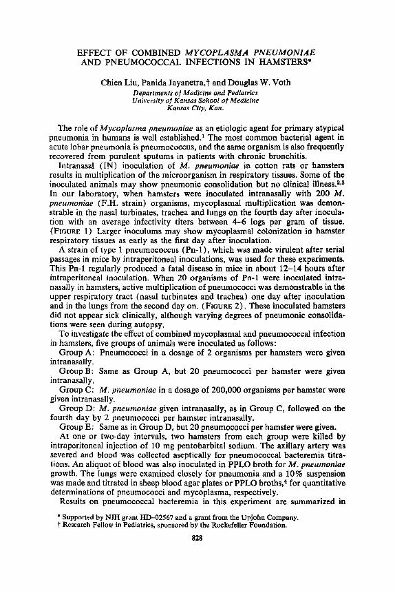
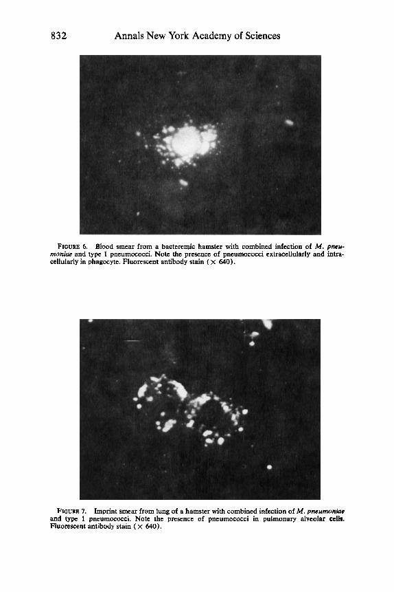
In moribund hamsters, when the titers of pneumococci were greater than 7-8 logs per ml, a blood smear showed that many pneumococcal organisms seemed both extracellularly and intracellularly in phagocytes when stained by specific type 1 pneumococcal fluorescent conjugate. Similarly, imprint smears from the
832 Annals New York Academy of Sciences
FIGURE 6. Blood smear from a bacteremic hamster with combined infection of M. pneu- monia and type 1 pneumococci. Note the presence of pneumococci extracellularly and intra- cellularly in phagocyte. Fluorescent antibody stain ( X 640).
FIGURE 7. Imprint smear from lung of a hamster with combined infection of M. pneumonhe and type 1 pneumococci. Note the presence of pneumococci in pulmonary alveolar cellm. Fluorescent antibody stain ( x 640).

Liu et a!. : Pneumococcal Infections in Hamsters TABLE 1
Group A: M . pneumoniae Group B: Pneumococcus type 1 Group C: No immunization
833
6/10 0/14 1/10 nd.
ll/l6t 5/18
PNEUMOCOCCAL BACTEREMU IN IMMUNIZED HAMSTERS
Pneumococcal Bacteremia Immunization Status
Challenged with 2000 Pn-1 Challenged with 200 Pn-1
lungs of these hamsters also showed fluorescent pneumococci in pulmonary alveoli or in phagocytes (FIGURES 6,7).
At this point we would like to ask: what is the mechanism by which a pneumo- coccal infection superimposed on a mycoplasmal infection can result in a serious systemic septicemia? One working hypothesis would be that perhaps in myco- plasmal infection the multiplication of organisms may damage the respiratory mucosa or the pulmonary alveolar phagocytes. This mechanism may then f a d - tate the entrance of pneumococci from the respiratory tract to the blood, covert- ing a local infection in the lung to a fulminating systemic septicemia. If this hypothesis has merit, then immunization with mycoplasma should modify the disease favorably.
To test this hypothesis, three groups of hamsters were used for the following experiment:
Group A: Immunized with live M. pneumoniae by giving 200,000 organisms intranasally .
Group B: Immunized with live type 1 pneumococcus by giving 20 organisms intranasally .
Group C: No immunization, control animals. Four weeks after immunization with live organisms, hamsters in all three
groups were challenged, with doses of 200,000 M. pneumoniae administered intranasally, followed on the fourth day by 2,000 or 200 homologous type 1 pneumococci. As can be seen in TABLE 1, with a very large dose of 2,000 pneumo- cocci, 11 out of 16 control hamsters developed bacteremia including 5 who died or appeared moribund at the time of sacrifice. In the group immunized with pneumococci previously, only 1 animal out of 10 showed a low grade bacteremia with 100 pneumococci per ml of blood. In the group immunized with M . pneumo- niae, 6 out of 10 animals developed pneumococcal bacteremia, but none of them appeared moribund or died. When a smaller inoculum of 200 pneumococcal or- ganisms was given to these animals, no bacteremia was seen in the M . pneumoniae immunized hamsters, but 5 out of 18 control unimmunized animals developed pneumococcal bacteremia.
The data presented can be summarized in the following way. From the clinical point of view, pneumonia caused by Mycoplasma pneumoniae is usually a benign disease in human beings. The pneumonitis is self-limiting. Mortality and morbidity of the illness are extremely low. On the other hand, pneumococcal lobar pneu- monia is a severe infection particularly in patients with septicemia. Using hamsters as an experimental model, under the experimental conditions described, intra- nasal inoculation of hamsters with M . pneumoniae or pneumococcus alone did not produce a serious infection. However, when mycoplasma was inoculated first and given sufficient time for the organisms to colonize and multiply in the hamster
834 Annals New York Academy of Sciences
lungs, in this instance about 4 days after mycoplasma inoculation, further inocula- tion with a mouse virulent type 1 pneumococcus, in relatively small numbers, can produce a fulminating disease. It has been reported that a synergistic effect with enhanced mortality occurred in mice with dual infection of the respiratory tract with parainfluenza virus and H . influenzae type b bacilli.5 We hypothesize that the mycoplasmal infection may have damaged the epithelial cells lining the respiratory tract or the phagocytic functions of pulmonary alveolar cells. This makes it easier for the pneumococci to gain entrance from the respiratory tract to the blood and to convert a local infection in the lungs to a systemic septicemia. This hypothesis is supported by the fact that immunization of hamsters with live mycoplasma did confer a partially protective effect on animals subjected to a rechallenge with combined infections of M . pneumoniae and pneumococci. Although animal ex- perimental data should not be extrapolated to clinical medicine, a search should be undertaken for possible interactions between mycoplasmal, bacterial and viral infections in potentiating the severity of human respiratory diseases.
Acknowledgments
The able technical assistance of Mrs. Linda Lohoefener and Miss Sharon Hempler are gratefully acknowledged.
References 1. DINGLE, J. H. & W. S. JORDAN. 1965. Mycoplasma pneumoniae infections. I n Bacterial
and Mycotic Infections of Man. 4th edit. R. J. Dubos & J. G. Hirsch, Eds. J. P. Lippincott Co. Philadelphia, Pa.
2. EATON, M. D., G. MEIKLUOHN & W. VAN HERICK. 1944. Studies on the etiology of primary atypical pneumonia. 1. A filterable agent transmissible to cotton rats, hamster, and chick embryos. J. Exp. Med. 79: 649-668.
3. DAJANI, A. S., W. A. CLYDE, JR. & F. W. DENNY. 1965. Experimental infection with Mycoplasma pneumoniae (Eaton agent). J. Exp. Med. 121: 1071-1086.
4. CHANOCK, R. M., L. HAYPLICK & M. F. BARILE. Growth on artificial medium of an agent associated with atypical pneumonia and its identification as a PPLO. Proc. Nat. Acad. Sci. 48: 41-49.
5. DEGR~, M. & L. A. GLASGOW. 1968. Synergistic effect in viral bacterial infection. 1. Com- bined infection of the respiratory tract in mice with parainfluenza virus and hemophilus influenza. J. Infect. Dis. 118: 449-462.
1962.