effect of cinnamaldehyde and citral combination on ...penicillium expansum, as a main postharvest...
TRANSCRIPT
fmicb-09-00597 March 27, 2018 Time: 17:12 # 1
ORIGINAL RESEARCHpublished: 29 March 2018
doi: 10.3389/fmicb.2018.00597
Edited by:Juan Aguirre,
Universidad de Chile, Chile
Reviewed by:Boqiang Li,
Institute of Botany (CAS), ChinaMassimo Reverberi,
Sapienza Università di Roma, Italy
*Correspondence:Tianli Yue
†These authors have contributedequally to this work.
Specialty section:This article was submitted to
Food Microbiology,a section of the journal
Frontiers in Microbiology
Received: 18 November 2017Accepted: 15 March 2018Published: 29 March 2018
Citation:Wang Y, Feng K, Yang H, Zhang Z,
Yuan Y and Yue T (2018) Effectof Cinnamaldehyde and Citral
Combination on TranscriptionalProfile, Growth, Oxidative Damage
and Patulin Biosynthesis of Penicilliumexpansum. Front. Microbiol. 9:597.
doi: 10.3389/fmicb.2018.00597
Effect of Cinnamaldehyde and CitralCombination on TranscriptionalProfile, Growth, Oxidative Damageand Patulin Biosynthesis ofPenicillium expansumYuan Wang1,2,3,4†, Kewei Feng5†, Haihua Yang2,3,4, Zhiwei Zhang6, Yahong Yuan2,3,4 andTianli Yue1,2,3,4*
1 College of Food Science and Engineering, Northwest University, Xi’an, China, 2 College of Food Science and Engineering,Northwest A&F University, Yangling, China, 3 Laboratory of Quality and Safety Risk Assessment for Agro-products (Yangling),Ministry of Agriculture, Beijing, China, 4 National Engineering Research Center of Agriculture Integration Test (Yangling),Yangling, China, 5 State Key Laboratory of Crop Stress Biology in Arid Areas, College of Agronomy, Northwest A&FUniversity, Yangling, China, 6 College of Food Science and Engineering, Qingdao Agricultural University, Qingdao, China
Penicillium expansum, as a main postharvest pathogen of fruits, can secrete patulin(PAT), causing fruit decay and health problems. In this study, the antifungal test, SEM(scanning electron microscope) observation, transcriptional profile, PAT biosynthesis,and physiological characters of P. expansum exposed to cinnamaldehyde and citralcombination (Cin/Cit) were evaluated. Cin/Cit could inhibit the mycelial growth and sporegermination of P. expansum in a dose-dependent manner. Besides, Cin/Cit causedspores and mycelia wrinkled and depressed by SEM observation. Gene expressionprofiles of P. expansum were conducted by RNA sequencing (RNA-seq) in the presenceor absence of Cin/Cit treatment. A total of 1713 differentially expressed genes (DEGs)were obtained, including 793 down-regulated and 920 up-regulated genes. Mostof the DEGs participated in the biosynthesis of secondary metabolites, amino acidmetabolism, and oxidation-reduction process, etc. Cin/Cit induced the dysfunction ofthe mitochondrial membrane, causing the potential influence on energy metabolismand reactive oxidative species production. The changes of superoxide dismutase (SOD)and catalase (CAT) activities combing with the increase of hydrogen peroxide contentindicated the oxidative stress on P. expansum induced by Cin/Cit, which correspondedwell with the transcriptional results. Moreover, both the RNA-seq data and the qRT-PCR showed the remarkable down-regulation of genes included in the PAT biosyntheticpathway under the Cin/Cit treatment. These findings provided more useful informationabout the antifungal mechanism of Cin/Cit against P. expansum at molecular and genelevels and suggested that Cin/Cit is a potential candidate to control P. expansum.
Keywords: cinnamaldehyde, citral, P. expansum, RNA-seq, PAT biosynthesis
Frontiers in Microbiology | www.frontiersin.org 1 March 2018 | Volume 9 | Article 597

fmicb-09-00597 March 27, 2018 Time: 17:12 # 2
Wang et al. Response of P. expansum to Cin/Cit
INTRODUCTION
Fungal and mycotoxins contaminations are the major problemsin agricultural products safety and human health. Penicilliumexpansum is one of the most devastating pathogens, whichcauses blue mold decay on many types of fruit. P. expansumcan spread rapidly under the suitable condition and secretesPAT, a mycotoxin with potential mutagenic, carcinogenic,teratogenic and embryotoxic effects on humans (Moake et al.,2005). Currently, besides certain prophylactic measures appliedto prevent the development of pathogen in the storageenvironments, the control of postharvest pathogens mainlydepends on the synthetic fungicides (Lai et al., 2017). However,losses of the fungicides efficiency due to the emergence offungicide-resistant pathogens, and public concern over chemicalresidues in food and environment have promoted investigationsof alternative strategies to combat fungal decay and to increasefood safety and public health (Tian et al., 2015).
Essential oils (EOs) are volatile liquid extracted from thenatural raw material of plant, containing various individualconstituents (ICs) such as terpenes, terpenoids, or aromatic andaliphatic constituents (de Souza et al., 2016). Most EOs andtheir ICs are cited as “generally recognized as safe” (GRAS) infood by the USFDA, and have proved to possess antimicrobialactivities (Neri et al., 2006; Zhang et al., 2016). Cinnamaldehydeand citral are the major components of Cinnamon bark EO andCymbopogon citratus EO, respectively, and have been reportedto inhibit microbial growth as a botanical fungicide in fruitand its products. Cinnamaldehyde or cinnamaldehyde-emulsionwas effectively applied to inhibit Salmonella typhimurium andStaphylococcus aureus in watermelon juice, Escherichia coliO157: H7 and Salmonella enterica in apple juice (Friedmanet al., 2004; Jo et al., 2015). Additionally, the dual combinationof the trans-cinnamaldehyde-citral emulsion at 100 µg/mlwas proved to inhibit Zygosaccharomyces bailii in apple juicestored at 20◦C for 27 days (Loeffler et al., 2014). Bothcinnamaldehyde and citral exhibit direct inhibition to thepathogen, as well as indirectly control the pathogen growth byimproving the host’s defense system (Fan et al., 2014; Jiang et al.,2015).
Researchers have investigated the complex action modes ofEOs from various perspectives. The main mechanism is basedon their lipophilic character, which facilitates the access of thehydrophobic compounds to the cytoplasmic membrane, causingmembrane damage, loss of intracellular substances, and finallymicrobial death (de Souza et al., 2016). Cinnamaldehyde wasreported to inhibit the growth of Aspergillus flavus, Fusariumverticillioides, A. ochraceus, and E. coli, S. aureus by cellulardamage (Hua et al., 2014; Xing et al., 2014; Sun et al., 2016; Zhanget al., 2016). Citral exhibited its antimicrobial activity againstCronobacter sakazakii, P. digitatum, and P. italicum by affectingmembrane function or integrity (Tao et al., 2014; Zheng et al.,2015; Shi et al., 2016). Although cinnamaldehyde or citral hasbeen reported in control of P. expansum, the previous works wereinvolved in antifungal activities and the application efficacy infood packaging (Neri et al., 2006; Xing et al., 2010; Camele et al.,2012; Balaguer et al., 2013; Manso et al., 2015). The knowledge of
their action modes on P. expansum at the molecular level is ratherlimited, and thus requires further investigations.
Recently, RNA-seq technology has been used in many studiesof gene expression, including the investigation of fungal responsemechanism to EOs or ICs. Ouyang et al. (2016) demonstrated thatcitral inhibited P. digitatum by the down-regulation of ergosterolbiosynthesis through transcriptional profiling analysis (Ouyanget al., 2016). In another report, the transcriptional profile ofParacoccidioides lutzii exposed to argentilactone, a constituentof the EO of Hyptis ovalifolia, was evaluated to investigate theresponse mechanism (Araujo et al., 2016). Additionally, RNA-seqtechnology has been used to analyze the molecular mechanismof fungi–host interaction, or fungal drug-resistance (Liu et al.,2015; Barad et al., 2016; Wang H. et al., 2016). Therefore, thepresent study is to evaluate the effect of Cin/Cit on growth,micromorphology, oxidative damage, and PAT biosynthesisof P. expansum. And the whole gene expression profile inP. expansum response to Cin/Cit was further analyzed based onthe data from RNA-seq, in an effort to explore the molecularmechanism and key pathways or genes involved in.
MATERIALS AND METHODS
Chemicals, Strain, and GrowthConditionsAnalytical-grade cinnamaldehyde (95%) and citral (96%) werepurchased from Jiangxi Xuesong Natural Medicinal Oil Co., Ltd.,China. P. expansum F-WY-12-02 was isolated from droppedkiwifruits in our laboratory (Wang Y. et al., 2016) and weremaintained in potato dextrose agar (PDA) and stored at 4◦C.This isolate has previously shown to produce 146.58 µg/g of PATin PDA plates after 10 days at 25◦C. The minimum inhibitoryconcentration (MIC) was determined by dilution method in PDB(potato dextrose broth) medium as a previous work with minormodifications (Zhang et al., 2016). The EOs was dissolved inTween 80 and added to the 10 ml sterile PDB medium to besaved as the stock solution. Then the EOs stock was dilutedto obtain final concentrations ranging from 20 to 200 mg/L.Finally, 50 µl of 106 spores/ml P. expansum conidia were added toeach PDB medium (5 ml) containing different concentrations ofEOs. Tubes were incubated at 25◦C for 3 days under continuousshaking. Controls were performed in PDB medium with only thespore suspension. MIC was defined as the lowest concentrationof EO with no visible fungal growth. The minimum fungicidalconcentration (MFC) was measured by a subculture of 100 µlfrom each tube with no visible fungal growth on a PDA platefollowed by incubation at 25◦C for 3–5 days. MFC was defined asthe lowest concentration of EO with initial inoculum fungi killed.Each test was performed in triplicate. The Fractional InhibitoryConcentration Index (FICI) of EOs was determined accordingto a previous study to explore the interaction of two EOs (Itenet al., 2009). The result showed that FICI of cinnamaldehydeand citral was 1, indicating their addition effect. Therefore, inthe present study, a combined application of Cin/Cit was chosenagainst P. expansum F-WY-12-02. The MIC, MFC, and FICI ofCin/Cit were shown in Supplementary Table S1.
Frontiers in Microbiology | www.frontiersin.org 2 March 2018 | Volume 9 | Article 597

fmicb-09-00597 March 27, 2018 Time: 17:12 # 3
Wang et al. Response of P. expansum to Cin/Cit
Antifungal Effects of Cin/Cit onP. expansumSpores GerminationConidia from P. expansum, cultivated on PDA medium at 25◦Cfor 7 days, was harvested using sterile distilled water containing0.05% (v/v) Tween 80. Then 500 µl spore suspension (106
spores/ml) were added to each sterile tube containing 1.5 mlCin/Cit solution, providing the final concentrations of 1/4 MIC,1/2 MIC, MIC, 2 MIC. Tubes were incubated at 25◦C for 12 hwith 120 rpm. At the end of the incubation period, germinatedspores were observed using a light microscope (Olympus, Tokyo,Japan). Germination was defined as the length of the germtube was greater than or equal to the diameter of the swollenspore. The percentage of germinated spores was calculated asP(%)= (Ngerminated spores/Ntotal spores) 100%.
Mycelial GrowthInfluence of Cin/Cit on the growth of P. expansum was evaluatedby colony diameter and mycelial dry weight. For colony diameter,50 µl of 106 spores/ml was spotted on the center of PDA platecontaining 1/4 MIC-2 MIC of Cin/Cit. Plates were sealed usingParafilm and incubated for 120 h at 25◦C. Colony diameter wasmeasured every 24 h using the cross method. PDA plates withoutCin/Cit were set as control. The radial growth inhibition after120 h was evaluated. The inhibitory effect of Cin/Cit on themycelial weight was determined as described previously (Sunet al., 2016). The concentration of Cin/Cit ranged from 1/4MIC-2 MIC. Incubation was conducted at 25◦C for 120 h withagitation (120 rpm). After filtering and washing, the inhibitionof mycelial weight at the end of incubation was measured.Three replicates were performed for each treatment. Growthinhibition was calculated by the equation: I (%)= (Dc (Wc)−Dt(Wt))× 100/Dc (Wc). I: inhibition of mycelial growth; Dc (Wc):colony diameter or mycelial weight in control group; Dt (Wt):colony diameter, or mycelial weight in the treatment group.
Scanning Electron Microscopy (SEM)AnalysisMycelia from 72 h incubation of P. expansum in PDB werecollected and washed with PBS (pH 7.4). Then 0.5 g of wetmycelia were added in tubes containing 5 ml PDB and Cin/Cit at0, MIC, 2 MIC. For spores, 100 µl of Cin/Cit was added to tubescontaining 4.9 ml of spores suspension (107 spores/ml) , reachingthe final concentration of 0, MIC, 2 MIC. After incubation at25◦C, 120 rpm for 12 h, both mycelia and spores were harvestedby centrifugation for 5 min at 10, 000 × g and washed twicewith PBS, followed by a series of pretreatments based on Sunet al. (2016). At last, the micromorphology was observed using ascanning electron microscope (S-3400N, Hitachi, Tokyo, Japan).
Transcriptome AnalysisSpores of P. expansum were inoculated in PDB mediumsupplemented with 0 or 1/2 MIC of Cin/Cit and cultured at25◦C, 120 rpm for 5 days. Each group has two biologicalreplicates. The harvested mycelia were quickly frozen withliquid nitrogen for total RNAs preparation, RNA quality
detection, cDNA libraries construction and RNA-seq, which wasperformed at Novogene Bioinformatics Technology Co., Ltd.(Beijing, China). Total RNA was extracted using TRIzol reagent(Invitrogen, United States) according to the manufacturer’sinstruction and treated with RNase-free DNase I. The agarose gelelectrophoresis, a NanoDrop R©2000 spectrophotometer (ThermoScientific, Wilmington, DE, United States), a Qubit R©Fluorometer2.0 (Life Technologies, Carlsbad, CA, United States) and anAgilent 2100 bioanalyzer (Agilent Technologies, Santa Clara,CA, United States) were used to test the concentrationand integrity of RNA samples (Wang H. et al., 2016).The cDNA libraries were constructed according to previousreports and were sequenced on the Illumina HiSeq2000platform (HiSeq2000, Illumina, San Diego, CA, United States,2010) (Lin et al., 2013; Lai et al., 2017). The resultingRNA-seq reads were mapped onto the reference genome ofP. expansum Link isolate d1 from Israel (denoted as PEXP)using Tophat v2.0.12 (Kim et al., 2013; Ballester et al.,2015).
Additionally, the followed analysis, including quantificationof gene expression (HTSeq v0.6.1), identification of differentiallyexpressed genes (DEGs) (DESeq v1.10.1), enrichment analysisof DEGs by GO enrichment analysis (GOSeq Release2.12) andKEGG pathways analysis (KOBAS v2.0), were carried out basedon a previous report (Wang H. et al., 2016). The RNA-Seqdata have been deposited in the Genome Sequence Archive inBIG Data Center, Beijing Institute of Genomics (BIG), ChineseAcademy of Sciences, with accession code CRA000582.
Mitochondrial Membrane Potential(MMP) MeasurementThe effect of Cin/Cit on MMP of P. expansum was evaluatedusing 1 µg/ml of rhodamine 123 (RH123, Sigma-Aldrich),respectively. A conidial suspension (107 spores/ml) was preparedin PBS with 2% (w/v) D-glucose. The cells were incubated withvarious concentrations of Cin/Cit (0, 1/2 MIC, 2 MIC) at 25◦C,120 rpm for 10 h (Tian et al., 2015). Finally, the spores werecollected, washed with PBS and stained for 15 min at roomtemperature in the dark. After that, the spores were washed twicewith PBS and then observed by flow cytometer (BD Biosciences,United States) with the excitation, and emission wavelength as488 and 525 nm, respectively. Unstained cell suspensions wereincluded as autofluorescence controls. Each treatment includedthree replicates.
Determination of Glucan, Chitin, andProtein of Cell WallAfter Cin/Cit treatment (0, 1/2 MIC) for 5 days, mycelia werefiltered and washed with PBS, then cell walls were isolated andanalyzed using the acid (H2SO4) treatment method (Francois,2006). Cell wall monosaccharide concentrations were normalizedusing the total dry cell wall mass for individual samples. Hotalkaline (NaOH) method was used to extract cell wall proteins.The amounts of proteins in cell wall extracts were measured byBradford method (Bradford, 1976) using bovine serum albuminas a standard.
Frontiers in Microbiology | www.frontiersin.org 3 March 2018 | Volume 9 | Article 597
fmicb-09-00597 March 27, 2018 Time: 17:12 # 4
Wang et al. Response of P. expansum to Cin/Cit
Hydrogen Peroxide (H2O2) MeasurementMycelia from 5 days incubation in PDB in presence or absence ofCin/Cit (0, 1/2 MIC) were collected by filtration and washed withPBS. After that, the mycelia were subjected to the determinationof H2O2 based on the previous method (Jiang et al., 2015). TheH2O2 content was calculated using H2O2 as a standard and theresult was expressed as µmol/g mycelia.
Assay of SOD and CAT ActivityMycelia were homogenized in cold PBS (10 mM, pH 7.0) byquartz sand. After that, the homogenates were centrifuged at 13,000 × g for 30 min at 4◦C. The proteins and enzymes wereanalyzed in the supernatants (Sun et al., 2016). The determinationof proteins contents in extract fluids was based on Bradford’smethod (Bradford, 1976). superoxide dismutase (SOD) activitywas assayed by its ability to inhibit the photochemical reductionof nitrotetrazolium blue chloride (NBT) at 560 nm (Beauchampand Fridovich, 1971). The SOD activity was defined as units permilligrams of protein (U/mg pro). CAT activity was determinedaccording to a previous method (Candan and Tarhan, 2003). Thespecific CAT activity was expressed as U/mg protein/min.
Detection of PAT Production andRelated-Genes ExpressionSpores of P. expansum were cultured statically in PDB medium(105 spores/ml) supplemented with 0, 1/2 MIC, MIC of Cin/Citat 25◦C, 120 rpm for 5 days. The culture solutions were filteredto separate mycelium and filtrate. Mycelia were washed, driedand weighted. The filtrate was collected for PAT determination.PAT was extracted with ethyl acetate by AOAC Official Method2000.02 (MacDonald et al., 2000). Analysis of PAT was performedon the HPLC (LC-2010A; Shimadzu, Kyoto, Japan) system. Theanalytical column used was Eclipse Plus C18 (250 mm× 4.6 mmi.d., 5 mm, Agilent Technologies, United States). The eluent usedwas acetonitrile-water (10: 90, v/v) at a flow rate of 1.0 ml/minand the sample injecting volume was 20 µl. The UV detectorwavelength was set at 276 nm.
Under 0 or 1/2 MIC Cin/Cit stress, conidia of P. expansumwere cultured in PDB medium at 25◦C, 120 rpm for 5 days. Afterthat, mycelia were harvested and subsequently flash-frozen withliquid nitrogen, ground to fine powder, and stored at −80◦Cfor RNA isolation. The total RNA was isolated using the FungalRNA Kit (Omega Bio-Tek, Inc. Norcross, Ga., United States)according to the manufacturer’s instructions. Genomic DNAelimination and first strand cDNA synthesis were carried outby PrimeScript RT reagent Kit with gDNA Eraser (Takara BioInc., Japan) according to the protocol of producer. The qRT-PCR (quantitative real-time polymerase chain reaction) wasperformed using SYBR Premix Ex TaqTM II (Takara Bio Inc.,Japan) in a Thermal Cycler DiceTM Real-Time System (Thermal,United States). The primer pairs for the specific 15 genes involvedin PAT biosynthesis were synthesized based on a previous study(Zong et al., 2015). The PCR conditions were as follows: 95◦Cfor 30 s, followed by 40 cycles of 95◦C for 5 s, 60◦C for30 s. The change in fluorescence of SYBR Green in every cyclewas monitored by the system software, and the threshold cycle
(Ct) over the background was calculated for each reaction.The sample was normalized using β-tubulin and the relativeexpression levels were measured using the 2(−11Ct) analysismethod.
Statistical AnalysisExcept for the transcriptome analysis, all the data were expressedas the mean ± SD by measuring three independent replicatesand were performed using SPSS version 22.0 (SPSS Inc.,IBM, Armonk, NY, United States) and Origin 9.0 (Origin LabCorporation, United States). Significance was evaluated by one-way analysis of variance (ANOVA). Tukey’s multiple-range testwas performed following a significant (∗p < 0.05, ∗∗p < 0.01) test.
RESULTS AND DISCUSSION
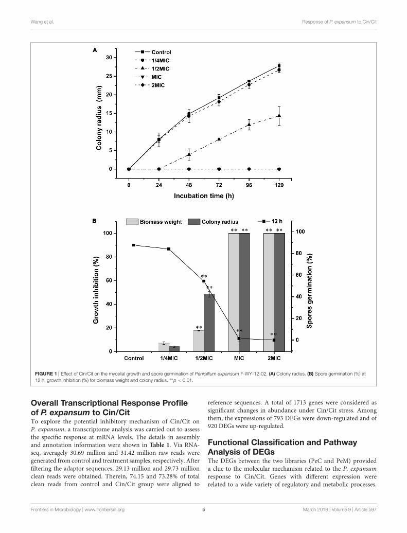
Antifungal Effects of Cin/Cit onP. expansum F-WY-12-02Inhibitory effects of Cin/Cit on the colony growth, biomass,and spore production of P. expansum were presented inFigure 1. Cin/Cit exhibited the capacity to delay or inhibitfungal growth in a dose-dependent manner. The extension ofthe colony radius and the accumulation of the biomass weightof P. expansum were completely inhibited by Cin/Cit at MICand 2 MIC during the whole incubation period (Figures 1A,B).After 5 days incubation, the lowest concentration of Cin/Cit(1/4 MIC) inhibited the extension of colony radius and theincrease in biomass weight by 4.19 and 7.12%, respectively.With regard to the spore germination rate of P. expansum at12 h, spores treated with 1/4 MIC (84.1%) showed no significantdifference compared with controls (87.6%). Additionally, therewere no conidia formed in PDA plates filled with MIC and2 MIC of Cin/Cit. Overall, it is obvious that Cin/Cit couldslow down or totally inhibit the colony extension, myceliabiomass accumulation and spore germination of P. expansum.The inhibitory effects are related to the dose and treatmentduration.
SEM ObservationTo understand the action mode of Cin/Cit against P. expansum,we started with evaluating the morphological alteration inP. expansum spores and mycelia by SEM (Figure 2). Therewere significant morphological differences between controlsand treated P. expansum cells. The controls appeared to benormal with intact and plump spheroidal or tubular structure.After exposure to Cin/Cit at MIC, the spores and hyphaebecame wrinkled, thinner and exhibited clear depressions on thesurface. The degrees of morphological modifications increasedas Cin/Cit concentration increased. The morphological changesof P. expansum under Cin/Cit treatment were similar to somefungi and bacteria, such as A. flavus, F. verticillioides, E. coli, andS. aureus treated by cinnamon essential oil (Xing et al., 2014;Sun et al., 2016; Zhang et al., 2016). The wrinkled and thinnersurface of spores or mycelia may be due to the release of cellularcontent.
Frontiers in Microbiology | www.frontiersin.org 4 March 2018 | Volume 9 | Article 597
fmicb-09-00597 March 27, 2018 Time: 17:12 # 5
Wang et al. Response of P. expansum to Cin/Cit
FIGURE 1 | Effect of Cin/Cit on the mycelial growth and spore germination of Penicillium expansum F-WY-12-02. (A) Colony radius. (B) Spore germination (%) at12 h, growth inhibition (%) for biomass weight and colony radius. ∗∗p < 0.01.
Overall Transcriptional Response Profileof P. expansum to Cin/CitTo explore the potential inhibitory mechanism of Cin/Cit onP. expansum, a transcriptome analysis was carried out to assessthe specific response at mRNA levels. The details in assemblyand annotation information were shown in Table 1. Via RNA-seq, averagely 30.69 million and 31.42 million raw reads weregenerated from control and treatment samples, respectively. Afterfiltering the adaptor sequences, 29.13 million and 29.73 millionclean reads were obtained. Therein, 74.15 and 73.28% of totalclean reads from control and Cin/Cit group were aligned to
reference sequences. A total of 1713 genes were considered assignificant changes in abundance under Cin/Cit stress. Amongthem, the expressions of 793 DEGs were down-regulated and of920 DEGs were up-regulated.
Functional Classification and PathwayAnalysis of DEGsThe DEGs between the two libraries (PeC and PeM) provideda clue to the molecular mechanism related to the P. expansumresponse to Cin/Cit. Genes with different expression wererelated to a wide variety of regulatory and metabolic processes.
Frontiers in Microbiology | www.frontiersin.org 5 March 2018 | Volume 9 | Article 597

fmicb-09-00597 March 27, 2018 Time: 17:12 # 6
Wang et al. Response of P. expansum to Cin/Cit
FIGURE 2 | Scanning electron microphotographs of P. expansum F-WY-12-02. (A) Untreated spores. (B) Cells treated with Cin/Cit at 1/2 MIC. (C) Cells treated withCin/Cit at 2MIC. (D) Untreated mycelia. (E) Mycelia treated with Cin/Cit at 1/2 MIC. (F) Mycelia treated with Cin/Cit at 2 MIC.
TABLE 1 | Summary of RNA-seq reads in control (PeC) and treatment (PeM)groups of Penicillium expansum F-WY-12-02.
Parameter PeC PeM PeC and PeM
Raw reads 30694374 31424213
Clean reads 29133442 29730617
Total mapped 74.15% 73.28%
Clean bases 4.37 G 4.46 G
Error rate (%) 0.02 0.02
Q20 (%) 95.45 95.81
Q30 (%) 90.04 89.71
GC content (%) 52.07 52.48
The number of all genes 11468
Genes annotation against GO 8055
DEGs annotation against GO 1261
Genes annotation against KEGG 4029
DEGs annotation against KEGG 842
Up-regulated genes 920
Down-regulated genes 793
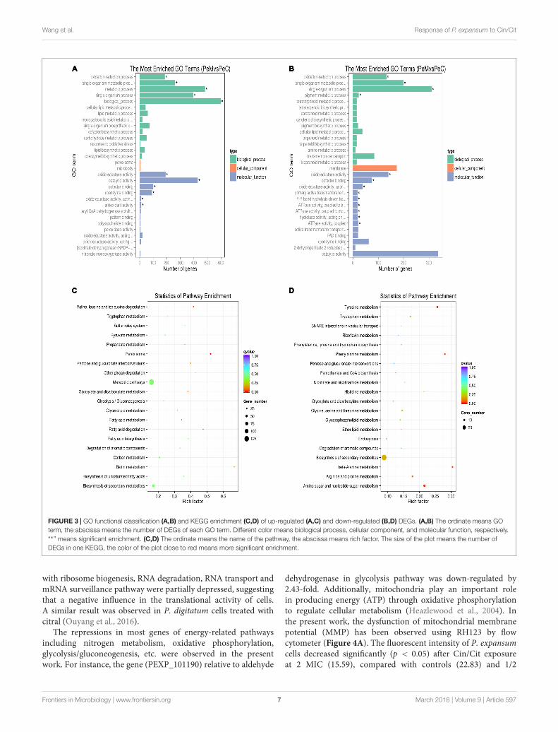
To better analyze the functions, metabolic pathways andinteractions of the 1713 DEGs, GO, and KEGG enrichmentanalyses were performed. A total of 1261 DEGs were mappedto 2289 GO terms. Among of which, 1361, 658, and 270GO terms belonged to biological process, molecular functionand cellular component, respectively. Figure 3A showed thetop 30 enriched functional categories of 920 up-regulatedDEGs. Therein, oxidation-reduction process, single-organismmetabolic process, and metabolic process, etc. were significantenrichment terms in the biological process. Oxidoreductaseactivity, catalytic activity, cofactor binding, etc. were the main
functional terms in molecular function. For the down-regulatedDEGs (Figure 3B), oxidation-reduction process, single-organismmetabolic process, and pigment metabolic process, etc. were themost abundant belonging to the biological process. Additionally,most of the significant enrichment terms in the molecularfunction were related to oxidoreductase activity, ATPase activity,transmembrane transporter activity, etc. DEGs were mappedto 90 KEGG pathways. Therein, the most abundant DEGs(171) were enriched in the metabolic pathway (pcs01100), 85DEGs were enriched in Biosynthesis of secondary metabolites(pcs01110), 28 and 27 DEGs were enriched in Biosynthesis ofamino acids (pcs01230), and Carbon metabolism (pcs01200),respectively. Moreover, the pathways affected in the presentwork may be related to two processes, including sporesgermination and mycelial growth. Figures 3C,D showed thescatter plots of the top 20 KEGG enrichment of up-regulatedand down-regulated DEGs, respectively. The KEGG enrichmentresults showed that the pathways which were sensitive to theCin/Cit stress mainly belonged to the peroxisome, amino acidsmetabolism or degradation, fatty acid metabolism, biosynthesisor degradation, biosynthesis of secondary metabolites etc. Thosesensitive pathways associated with genetic information, energymetabolism, cell integrity, cell membrane, oxidation-reductionprocess, etc. will be further analyzed.
Genes Involved in Stress ResponseTo alleviate the unfavorable growth situations caused by Cin/Cittreatment, P. expansum cells will develop relative responsesby adjusting gene expression pattern. In the present study,considerable alterations in gene expression levels related tostress response were found in P. expansum. Genes associated
Frontiers in Microbiology | www.frontiersin.org 6 March 2018 | Volume 9 | Article 597
fmicb-09-00597 March 27, 2018 Time: 17:12 # 7
Wang et al. Response of P. expansum to Cin/Cit
FIGURE 3 | GO functional classification (A,B) and KEGG enrichment (C,D) of up-regulated (A,C) and down-regulated (B,D) DEGs. (A,B) The ordinate means GOterm, the abscissa means the number of DEGs of each GO term. Different color means biological process, cellular component, and molecular function, respectively.“∗” means significant enrichment. (C,D) The ordinate means the name of the pathway, the abscissa means rich factor. The size of the plot means the number ofDEGs in one KEGG, the color of the plot close to red means more significant enrichment.
with ribosome biogenesis, RNA degradation, RNA transport andmRNA surveillance pathway were partially depressed, suggestingthat a negative influence in the translational activity of cells.A similar result was observed in P. digitatum cells treated withcitral (Ouyang et al., 2016).
The repressions in most genes of energy-related pathwaysincluding nitrogen metabolism, oxidative phosphorylation,glycolysis/gluconeogenesis, etc. were observed in the presentwork. For instance, the gene (PEXP_101190) relative to aldehyde
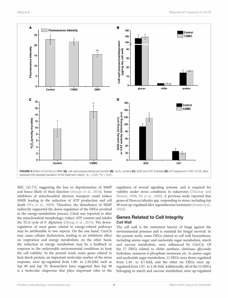
dehydrogenase in glycolysis pathway was down-regulated by2.43-fold. Additionally, mitochondria play an important rolein producing energy (ATP) through oxidative phosphorylationto regulate cellular metabolism (Heazlewood et al., 2004). Inthe present work, the dysfunction of mitochondrial membranepotential (MMP) has been observed using RH123 by flowcytometer (Figure 4A). The fluorescent intensity of P. expansumcells decreased significantly (p < 0.05) after Cin/Cit exposureat 2 MIC (15.59), compared with controls (22.83) and 1/2
Frontiers in Microbiology | www.frontiersin.org 7 March 2018 | Volume 9 | Article 597
fmicb-09-00597 March 27, 2018 Time: 17:12 # 8
Wang et al. Response of P. expansum to Cin/Cit
FIGURE 4 | Effect of Cin/Cit on MMP (A), cell wall polysaccharide and protein (B), H2O2 content (C), SOD and CAT activities (D) of P. expansum F-WY-12-02. Barsrepresent the standard deviation of the treatment means. ∗p < 0.05, ∗∗p < 0.01.
MIC (21.77), suggesting the loss or depolarization of MMPand hence likely of their function (Araujo et al., 2016). Someinhibitors of mitochondrial electron transport could reduceMMP, leading to the reduction of ATP production and celldeath (Wu et al., 2009). Therefore, the disturbance of MMPindirectly supported the down-regulation of the DEGs involvedin the energy-metabolism process. Citral was reported to alterthe mitochondrial morphology, reduce ATP content and inhibitthe TCA cycle of P. digitatum (Zheng et al., 2015). The down-regulation of most genes related to energy-related pathwaysmay be attributable to two aspects. On the one hand, Cin/Citmay cause cellular dysfunction, leading to an inhibitory effecton respiration and energy metabolism; on the other hand,the reduction in energy metabolism may be a feedback orresponse to the unfavorable environmental conditions to keepthe cell viability. In the present work, some genes related toheat shock protein, an important molecular marker of the stressresponse, were up-regulated from 1.86- to 2.28-fold, such ashsp 90 and hsp 70. Researchers have suggested that hsp 90is a molecular chaperone that plays important roles in the
regulation of several signaling systems, and is required forviability under stress conditions in eukaryotes (Thomas andBaneyx, 1998; Ali et al., 2006). A previous study reported thatgenes of Paracoccidioides spp. responding to stress, including hsp90 were up-regulated after argentilactone treatment (Araujo et al.,2016).
Genes Related to Cell IntegrityCell WallThe cell wall is the outermost barrier of fungi against theenvironmental pressure and is essential for fungal survival. Inthe present work, some DEGs related to cell wall biosynthesis,including amino sugar and nucleotide sugar metabolism, starchand sucrose metabolism, were influenced by Cin/Cit. Ofthe 17 DEGs related to chitin synthase, chitinase, glycosidehydrolase, mannose-6-phosphate isomerase etc. in amino sugarand nucleotide sugar metabolism, 11 DEGs were down-regulatedfrom 1.45- to 4.7-fold, and the other six DEGs were up-regulated from 1.07- to 2.38-fold. Additionally, all of the 12 DEGsbelonging to starch and sucrose metabolism were up-regulated
Frontiers in Microbiology | www.frontiersin.org 8 March 2018 | Volume 9 | Article 597
fmicb-09-00597 March 27, 2018 Time: 17:12 # 9
Wang et al. Response of P. expansum to Cin/Cit
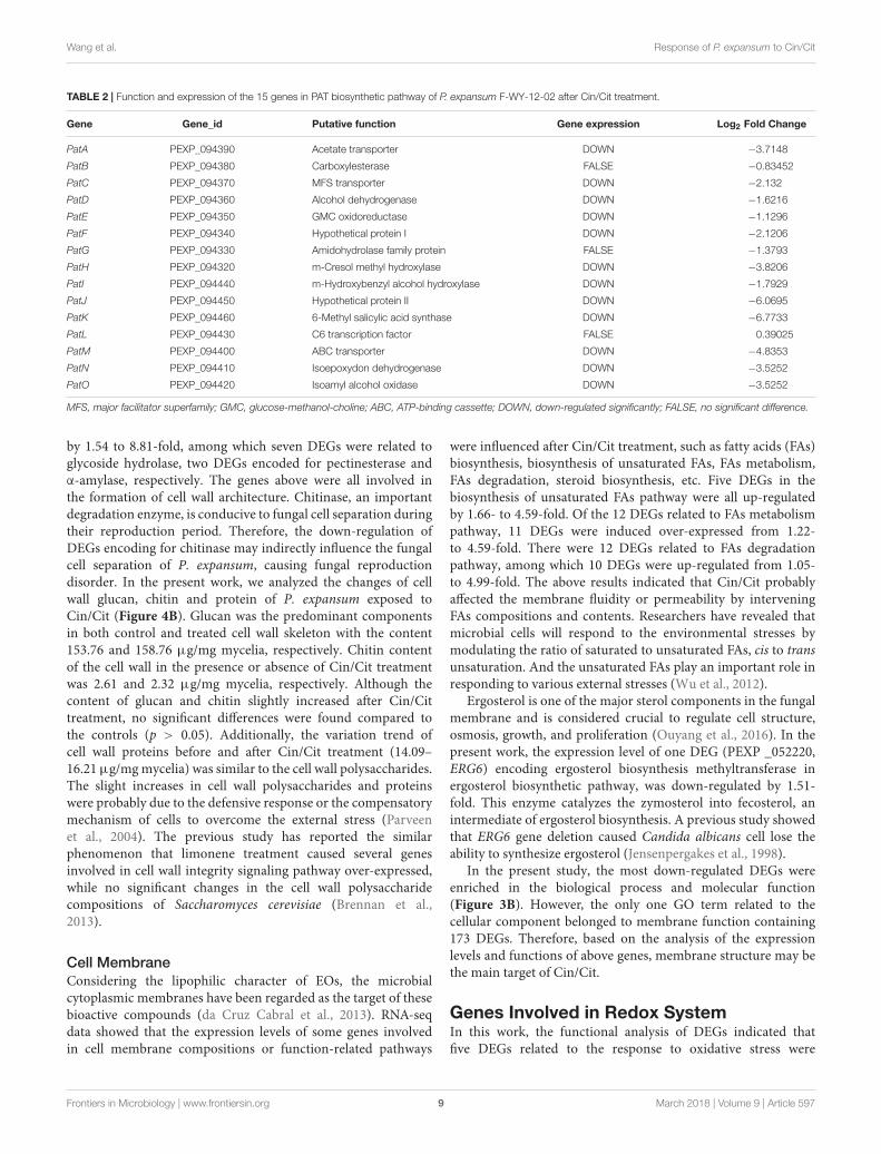
TABLE 2 | Function and expression of the 15 genes in PAT biosynthetic pathway of P. expansum F-WY-12-02 after Cin/Cit treatment.
Gene Gene_id Putative function Gene expression Log2 Fold Change
PatA PEXP_094390 Acetate transporter DOWN −3.7148
PatB PEXP_094380 Carboxylesterase FALSE −0.83452
PatC PEXP_094370 MFS transporter DOWN −2.132
PatD PEXP_094360 Alcohol dehydrogenase DOWN −1.6216
PatE PEXP_094350 GMC oxidoreductase DOWN −1.1296
PatF PEXP_094340 Hypothetical protein I DOWN −2.1206
PatG PEXP_094330 Amidohydrolase family protein FALSE −1.3793
PatH PEXP_094320 m-Cresol methyl hydroxylase DOWN −3.8206
PatI PEXP_094440 m-Hydroxybenzyl alcohol hydroxylase DOWN −1.7929
PatJ PEXP_094450 Hypothetical protein II DOWN −6.0695
PatK PEXP_094460 6-Methyl salicylic acid synthase DOWN −6.7733
PatL PEXP_094430 C6 transcription factor FALSE 0.39025
PatM PEXP_094400 ABC transporter DOWN −4.8353
PatN PEXP_094410 Isoepoxydon dehydrogenase DOWN −3.5252
PatO PEXP_094420 Isoamyl alcohol oxidase DOWN −3.5252
MFS, major facilitator superfamily; GMC, glucose-methanol-choline; ABC, ATP-binding cassette; DOWN, down-regulated significantly; FALSE, no significant difference.
by 1.54 to 8.81-fold, among which seven DEGs were related toglycoside hydrolase, two DEGs encoded for pectinesterase andα-amylase, respectively. The genes above were all involved inthe formation of cell wall architecture. Chitinase, an importantdegradation enzyme, is conducive to fungal cell separation duringtheir reproduction period. Therefore, the down-regulation ofDEGs encoding for chitinase may indirectly influence the fungalcell separation of P. expansum, causing fungal reproductiondisorder. In the present work, we analyzed the changes of cellwall glucan, chitin and protein of P. expansum exposed toCin/Cit (Figure 4B). Glucan was the predominant componentsin both control and treated cell wall skeleton with the content153.76 and 158.76 µg/mg mycelia, respectively. Chitin contentof the cell wall in the presence or absence of Cin/Cit treatmentwas 2.61 and 2.32 µg/mg mycelia, respectively. Although thecontent of glucan and chitin slightly increased after Cin/Cittreatment, no significant differences were found compared tothe controls (p > 0.05). Additionally, the variation trend ofcell wall proteins before and after Cin/Cit treatment (14.09–16.21 µg/mg mycelia) was similar to the cell wall polysaccharides.The slight increases in cell wall polysaccharides and proteinswere probably due to the defensive response or the compensatorymechanism of cells to overcome the external stress (Parveenet al., 2004). The previous study has reported the similarphenomenon that limonene treatment caused several genesinvolved in cell wall integrity signaling pathway over-expressed,while no significant changes in the cell wall polysaccharidecompositions of Saccharomyces cerevisiae (Brennan et al.,2013).
Cell MembraneConsidering the lipophilic character of EOs, the microbialcytoplasmic membranes have been regarded as the target of thesebioactive compounds (da Cruz Cabral et al., 2013). RNA-seqdata showed that the expression levels of some genes involvedin cell membrane compositions or function-related pathways
were influenced after Cin/Cit treatment, such as fatty acids (FAs)biosynthesis, biosynthesis of unsaturated FAs, FAs metabolism,FAs degradation, steroid biosynthesis, etc. Five DEGs in thebiosynthesis of unsaturated FAs pathway were all up-regulatedby 1.66- to 4.59-fold. Of the 12 DEGs related to FAs metabolismpathway, 11 DEGs were induced over-expressed from 1.22-to 4.59-fold. There were 12 DEGs related to FAs degradationpathway, among which 10 DEGs were up-regulated from 1.05-to 4.99-fold. The above results indicated that Cin/Cit probablyaffected the membrane fluidity or permeability by interveningFAs compositions and contents. Researchers have revealed thatmicrobial cells will respond to the environmental stresses bymodulating the ratio of saturated to unsaturated FAs, cis to transunsaturation. And the unsaturated FAs play an important role inresponding to various external stresses (Wu et al., 2012).
Ergosterol is one of the major sterol components in the fungalmembrane and is considered crucial to regulate cell structure,osmosis, growth, and proliferation (Ouyang et al., 2016). In thepresent work, the expression level of one DEG (PEXP _052220,ERG6) encoding ergosterol biosynthesis methyltransferase inergosterol biosynthetic pathway, was down-regulated by 1.51-fold. This enzyme catalyzes the zymosterol into fecosterol, anintermediate of ergosterol biosynthesis. A previous study showedthat ERG6 gene deletion caused Candida albicans cell lose theability to synthesize ergosterol (Jensenpergakes et al., 1998).
In the present study, the most down-regulated DEGs wereenriched in the biological process and molecular function(Figure 3B). However, the only one GO term related to thecellular component belonged to membrane function containing173 DEGs. Therefore, based on the analysis of the expressionlevels and functions of above genes, membrane structure may bethe main target of Cin/Cit.
Genes Involved in Redox SystemIn this work, the functional analysis of DEGs indicated thatfive DEGs related to the response to oxidative stress were
Frontiers in Microbiology | www.frontiersin.org 9 March 2018 | Volume 9 | Article 597
fmicb-09-00597 March 27, 2018 Time: 17:12 # 10
Wang et al. Response of P. expansum to Cin/Cit
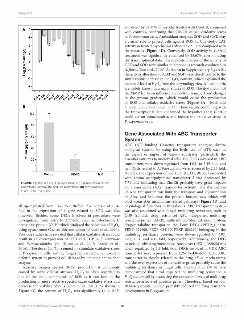
FIGURE 5 | Effect of Cin/Cit on expressions of 15 genes involved in PATbiosynthetic pathway (A), and PAT biosynthesis (B) of P. expansumF-WY-12-02. ∗∗p < 0.01.
all up-regulated from 1.47- to 3.78-fold. An increase of 1.54-fold in the expression of a gene related to SOD was alsoobserved. Besides, some DEGs involved in peroxidase wereup-regulated from 1.47- to 3.77-fold, such as cytochrome Cperoxidase protein (CCP) which catalyzed the reduction of H2O2using cytochrome C as an electron donor (Araujo et al., 2016).Previous studies have revealed that cellular oxidative stress couldresult in an overexpression of SOD and CCP in S. cerevisiaeand Paracoccidioides spp. (Kwon et al., 2003; Araujo et al.,2016). Therefore, Cin/Cit seemed to stimulate oxidative stressin P. expansum cells, and the fungus represented an antioxidantdefense system to prevent cell damage by inducing antioxidantenzyme.
Reactive oxygen species (ROS) production is commonlycaused by some cellular stresses. H2O2 is often regarded asone of the main compounds of ROS as it can lead to theproduction of more reactive species, cause oxidative stress anddecrease the viability of cells (Chen et al., 2015). As shown inFigure 4C, the content of H2O2 was significantly (p < 0.05)
enhanced by 54.37% in mycelia treated with Cin/Cit, comparedwith controls, confirming that Cin/Cit caused oxidative stressin P. expansum cells. Antioxidant enzymes SOD and CAT, playa crucial role to protect cells against ROS. In this study, CATactivity in treated mycelia was reduced by 21.08% compared withthe controls (Figure 4D). Conversely, SOD activity in Cin/Cittreatment was significantly enhanced by 25.67%, corroboratingthe transcriptional data. The opposite changes of the activity ofCAT and SOD were similar to a previous research conducted inA. flavus (Sun et al., 2016). As shown in Supplementary Figure S1,the activity alterations of CAT and SOD were closely related to thesimultaneous increase in the H2O2 content, which explained theincreased level of H2O2 from the enzymology view. Mitochondriaare widely known as a major source of ROS. The dysfunction ofthe MMP led to an influence on electron transport and changesin the proton gradient, which would cause the productionof ROS and cellular oxidative stress (Figure 4A) (Jezek andHlavatá, 2005; Suski et al., 2012). These results combining withthe transcriptional data confirmed the hypothesis that Cin/Citcould act on mitochondria, and induce the oxidative stress inP. expansum cells.
Gene Associated With ABC TransporterSystemABC (ATP-Binding Cassette) transporters energize diversebiological systems by using the hydrolysis of ATP, such asthe export or import of various substrates, particularly theessential nutrients to microbial cells. Ten DEGs involved in ABCtransporters were down-regulated from 1.03- to 3.45-fold, andtwo DEGs related to ATPase activity were repressed by 2.25-fold.Notably, the expression of one DEG (PEXP_101560) associatedwith amino acid/polyamine transporter I was decreased by7.71-fold, indicating that Cin/Cit probably have great impactson amino acids (AAs) transporter activity. The dysfunctionof AAs transporter can limit the transport and consumptionof AAs, and influence the protein biosynthesis, which willblock some AAs metabolism-related pathways (Figure 3D) andphysiological functions in fungal cells. ABC transporter systemwas also associated with fungal multidrug resistance, such asCDR (candida drug resistance) ABC transporter, multidrugresistance protein (MRP)/multi-antimicrobial extrusion protein,drug/metabolite transporter, etc. Four DEGs (PEXP_105370,PEXP_024900, PEXP_034130, PEXP_086290) belonging to themultidrug resistance protein, were down-regulated by 3.07,2.81, 1.51, and 4.10-fold, respectively. Additionally, the DEGassociated with drug/metabolite transporter (PEXP_068020) wasdown-regulated by 1.2-fold. Four DEGs involved in CDR ABCtransporter were repressed from 2.26- to 4.84-fold. CDR ABCtransporter is closely related to the drug efflux mechanisms,and the over-expression of its relative genes probably cause themultidrug resistance in fungal cells. Ouyang et al. (2016) havedemonstrated that citral impaired the multidrug resistance inP. digitatum cell by decreasing the expression levels of multidrugresistance-associated protein genes. Therefore, based on ourRNA-seq results, Cin/Cit probably reduced the drug resistancedevelopment in P. expansum.
Frontiers in Microbiology | www.frontiersin.org 10 March 2018 | Volume 9 | Article 597
fmicb-09-00597 March 27, 2018 Time: 17:12 # 11
Wang et al. Response of P. expansum to Cin/Cit
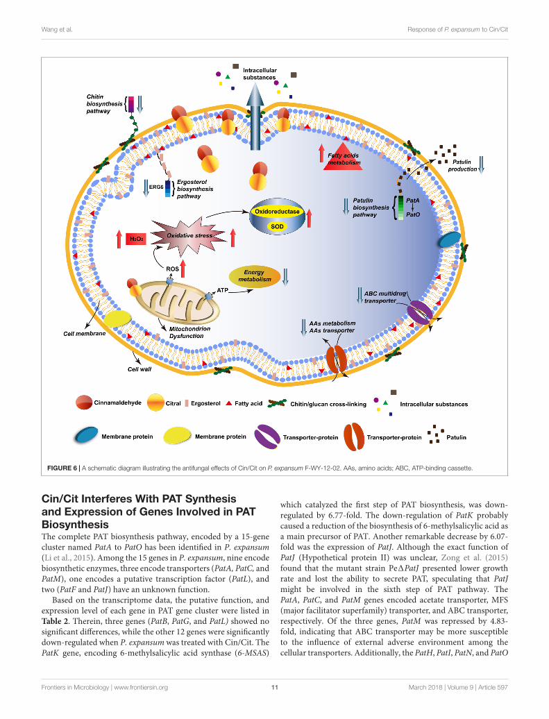
FIGURE 6 | A schematic diagram illustrating the antifungal effects of Cin/Cit on P. expansum F-WY-12-02. AAs, amino acids; ABC, ATP-binding cassette.
Cin/Cit Interferes With PAT Synthesisand Expression of Genes Involved in PATBiosynthesisThe complete PAT biosynthesis pathway, encoded by a 15-genecluster named PatA to PatO has been identified in P. expansum(Li et al., 2015). Among the 15 genes in P. expansum, nine encodebiosynthetic enzymes, three encode transporters (PatA, PatC, andPatM), one encodes a putative transcription factor (PatL), andtwo (PatF and PatJ) have an unknown function.
Based on the transcriptome data, the putative function, andexpression level of each gene in PAT gene cluster were listed inTable 2. Therein, three genes (PatB, PatG, and PatL) showed nosignificant differences, while the other 12 genes were significantlydown-regulated when P. expansum was treated with Cin/Cit. ThePatK gene, encoding 6-methylsalicylic acid synthase (6-MSAS)
which catalyzed the first step of PAT biosynthesis, was down-regulated by 6.77-fold. The down-regulation of PatK probablycaused a reduction of the biosynthesis of 6-methylsalicylic acid asa main precursor of PAT. Another remarkable decrease by 6.07-fold was the expression of PatJ. Although the exact function ofPatJ (Hypothetical protein II) was unclear, Zong et al. (2015)found that the mutant strain Pe1PatJ presented lower growthrate and lost the ability to secrete PAT, speculating that PatJmight be involved in the sixth step of PAT pathway. ThePatA, PatC, and PatM genes encoded acetate transporter, MFS(major facilitator superfamily) transporter, and ABC transporter,respectively. Of the three genes, PatM was repressed by 4.83-fold, indicating that ABC transporter may be more susceptibleto the influence of external adverse environment among thecellular transporters. Additionally, the PatH, PatI, PatN, and PatO
Frontiers in Microbiology | www.frontiersin.org 11 March 2018 | Volume 9 | Article 597
fmicb-09-00597 March 27, 2018 Time: 17:12 # 12
Wang et al. Response of P. expansum to Cin/Cit
genes were down-regulated by 3.82, 1.79, 3.525, and 3.525-fold,respectively. PatH and PatI, involved in the third and fourthsteps of PAT biosynthesis pathway, respectively (White et al.,2006; Artigot et al., 2009). PatN gene encoded isoepoxydondehydrogenase that catalyzed the conversion of isoepoxydonto phyllostine. A previous study showed that the deletion ofthe PatN gene in a P. expansum strain resulted in an 87.5%decrease in PAT production (Barad et al., 2013). According to theRNA-seq results in this study, Cin/Cit created a PAT restrictivecondition to P. expansum, so that the expressions of most genesinvolved in PAT biosynthetic pathway, particularly the earlysteps, were significantly down-regulated, which were similar tothe previous observations (Tannous et al., 2014; Lai et al., 2017).Furthermore, the qRT-PCR was performed and demonstratedthat the expressions of all the 15 genes were down-regulatedunder Cin/Cit stress (1/2 MIC) (Figure 5A). Meanwhile, PATproduction was also significantly (p < 0.01) decreased by 75.69%and 100% in Cin/Cit group at 1/2 MIC and MIC, respectively(Figure 5B).
CONCLUSION
As summarized in Figure 6, the lipophilic characters of EOsfacilitated their access to the cytoplasmic membrane, thencaused changes in membrane permeability, loss of intracellularsubstances, and wrinkles and depressions on cell surfaces. RNA-seq indicated that genes involved in amino sugar and nucleotidesugar metabolism (chitin biosynthesis), ergosterol biosynthesis,energy metabolism, AAs metabolism and ABC transporters (suchas AAs transporter, ABC multidrug transporters) in P. expansumwere mostly down-regulated, which probably affected the cellularprimary structure and limited the nutrition transport, whilereduced the drug resistance development. Particularly, PATbiosynthetic gene cluster was significantly down-regulated,directly causing lower levels of PAT production, which wasverified by qRT-PCR and PAT measurement assays. Additionally,most genes associated with FAs metabolism were up-regulated,inducing more unsaturated FAs accumulation, which was acritical response mechanism of the cell to environmental stress.
Besides, mitochondrial function and oxidation-reduction processwere remarkably influenced, the dysfunction of MMP resultedin ROS accumulation, such as H2O2, then induced the activityof oxidoreductase. The physiological and biochemical characterassays corresponded well with the RNA-seq result, which verifiedthat Cin/Cit caused oxidative stress on P. expansum. Thesefindings provide useful information to better understand theaction mode of Cin/Cit on P. expansum at the molecular leveland are conducive to develop more effective ways to prevent orcontrol P. expansum contaminations.
ETHICS STATEMENT
This article does not contain any studies with human participantsor animals performed by any of the authors.
AUTHOR CONTRIBUTIONS
YW, YY, and TY conceived and designed the experiments. YWand KF performed the experiments. YW and KF analyzed thedata. YW, ZZ, and HY drafted the manuscript. All authors readand approved the final manuscript.
FUNDING
This research was supported by Scientific and TechnologyCooperation Project in Hong Kong, Macao, and Taiwan of China(2015DFT30130), the National Basic Research Program of China(2013FY113400), the National Natural Science Foundation ofChina (31671866), National Agricultural Products Quality andSafety Risk Assessment Program (GJFP201701302).
SUPPLEMENTARY MATERIAL
The Supplementary Material for this article can be foundonline at: https://www.frontiersin.org/articles/10.3389/fmicb.2018.00597/full#supplementary-material
REFERENCESAli, M. M., Roe, S. M., Vaughan, C. K., Meyer, P., Panaretou, B., Piper,
P. W., et al. (2006). Crystal structure of an Hsp90-nucleotide-p23/Sba1closed chaperone complex. Nature 440, 1013–1017. doi: 10.1038/nature04716
Araujo, F. S., Coelho, L. M., Silva, L. D., Neto, B. R. D., Parente-Rocha, J. A., Bailao,A. M., et al. (2016). Effects of argentilactone on the transcriptional profile,cell wall and oxidative stress of Paracoccidioides spp. PLoS Negl. Trop. Dis.10:e0004309. doi: 10.1371/journal.pntd.0004309
Artigot, M. P., Loiseau, N., Laffitte, J., Mas-Reguieg, L., Tadrist, S., Oswald,I. P., et al. (2009). Molecular cloning and functional characterizationof two CYP619 cytochrome P450s involved in biosynthesis of patulinin Aspergillus clavatus. Microbiology 155, 1738–1747. doi: 10.1099/mic.0.024836-0
Balaguer, M. P., Lopez-Carballo, G., Catala, R., Gavara, R., and Hernandez-Munoz, P. (2013). Antifungal properties of gliadin films incorporating
cinnamaldehyde and application in active food packaging of bread andcheese spread foodstuffs. Int. J. Food Microbiol. 166, 369–377. doi: 10.1016/j.ijfoodmicro.2013.08.012
Ballester, A. R., Marcet-Houben, M., Levin, E., Sela, N., Selma-Lazaro, C.,Carmona, L., et al. (2015). Genome, transcriptome, and functional analysesof Penicillium expansum provide new insights into secondary metabolism andpathogenicity. Mol. Plant Microbe Interact. 28, 232–248. doi: 10.1094/mpmi-09-14-0261-fi
Barad, S., Horowitz, S. B., Kobiler, I., Sherman, A., and Prusky, D.(2013). Accumulation of the mycotoxin patulin in the presence ofgluconic acid contributes to pathogenicity of Penicillium expansum.Mol. Plant Microbe Interact. 27, 66–77. doi: 10.1094/MPMI-05-13-0138-R
Barad, S., Sela, N., Kumar, D., Kumar-Dubey, A., Glam-Matana, N., Sherman, A.,et al. (2016). Fungal and host transcriptome analysis of pH-regulated genesduring colonization of apple fruits by Penicillium expansum. BMC Genomics17:330. doi: 10.1186/s12864-016-2665-7
Frontiers in Microbiology | www.frontiersin.org 12 March 2018 | Volume 9 | Article 597

fmicb-09-00597 March 27, 2018 Time: 17:12 # 13
Wang et al. Response of P. expansum to Cin/Cit
Beauchamp, C., and Fridovich, I. (1971). Superoxide dismutase: improved assaysand an assay applicable to acrylamide gels. Anal. Biochem. 44, 276–287.doi: 10.1016/0003-2697(71)90370-8
Bradford, M. M. (1976). A rapid and sensitive method for the quantitationof microgram quantities of protein utilizing the principle of protein-dye binding. Anal. Biochem. 72, 248–254. doi: 10.1016/0003-2697(76)90527-3
Brennan, T. C., Kromer, J. O., and Nielsen, L. K. (2013). Physiological andtranscriptional responses of Saccharomyces cerevisiae to d-limonene showchanges to the cell wall but not to the plasma membrane. Appl. Environ.Microbiol. 79, 3590–3600. doi: 10.1128/AEM.00463-13
Camele, I., Altieri, L., De Martino, L., De Feo, V., Mancini, E., and Rana, G. L.(2012). In vitro control of post-harvest fruit rot fungi by some plant essential oilcomponents. Int. J. Mol. Sci. 13, 2290–2300. doi: 10.3390/ijms13022290
Candan, N., and Tarhan, L. (2003). Relationship among chlorophyll-carotenoidcontent, antioxidant enzyme activities and lipid peroxidation levels by Mg2+
deficiency in the Mentha pulegium leaves. Plant Physiol. Biochem. 41, 35–40.doi: 10.1016/S0981-9428(02)00006-2
Chen, J., Li, B., Qin, G., and Tian, S. (2015). Mechanism of H2O2-induced oxidativestress regulating viability and biocontrol ability of Rhodotorula glutinis. Int. J.Food Microbiol. 193, 152–158. doi: 10.1016/j.ijfoodmicro.2014.10.025
da Cruz Cabral, L., Fernández Pinto, V., and Patriarca, A. (2013). Application ofplant derived compounds to control fungal spoilage and mycotoxin productionin foods. Int. J. Food Microbiol. 166, 1–14. doi: 10.1016/j.ijfoodmicro.2013.05.026
de Souza, E. L., da Cruz Almeida, E. T., and de Sousa Guedes, J. P. (2016). Thepotential of the incorporation of essential oils and their individual constituentsto improve microbial safety in juices: a review. Compr. Rev. Food Sci. Food Saf.15, 753–772. doi: 10.1111/1541-4337.12208
Fan, F., Tao, N., Jia, L., and He, X. (2014). Use of citral incorporated in postharvestwax of citrus fruit as a botanical fungicide against Penicillium digitatum.Postharvest Biol. Tecnol. 90, 52–55. doi: 10.1016/j.postharvbio.2013.12.005
Francois, J. M. (2006). A simple method for quantitative determination ofpolysaccharides in fungal cell walls. Nat. Protoc. 1, 2995–3000. doi: 10.1038/nprot.2006.457
Friedman, M., Henika, P. R., Levin, C. E., and Mandrell, R. E. (2004). Antibacterialactivities of plant essential oils and their components against Escherichia coliO157:H7 and Salmonella enterica in apple juice. J. Agric. Food Chem. 52,6042–6048. doi: 10.1021/jf0495340
Heazlewood, J. L., Tontifilippini, J., Gout, A. M., Day, D. A., Whelan, J., andMillar, A. H. (2004). Experimental analysis of the arabidopsis mitochondrialproteome highlights signaling and regulatory components, provides assessmentof targeting prediction programs, and indicates plant-specific mitochondrialproteins. Plant Cell 16, 241–256. doi: 10.1105/tpc.016055
Hua, H., Xing, F., Selvaraj, J. N., Wang, Y., Zhao, Y., Zhou, L., et al. (2014).Inhibitory effect of essential oils on Aspergillus ochraceus Growth andochratoxin a production. PLoS One 9:e108285. doi: 10.1371/journal.pone.0108285
Iten, F., Saller, R., Abel, G., and Reichling, J. (2009). Additive antimicrobial effectsof the active components of the essential oil of Thymus vulgaris — chemotypecarvacrol. Planta Med. 75, 1231–1236. doi: 10.1055/s-0029-1185541
Jensenpergakes, K. L., Kennedy, M. A., Lees, N. D., Barbuch, R. J., Koegel, C., andBard, M. (1998). Sequencing, disruption, and characterization of the Candidaalbicans sterol methyltransferase (ERG6) gene: drug susceptibility studies inerg6 mutants. Antimicrob. Agents Chemother. 42, 1160–1167.
Jezek, P., and Hlavatá, L. (2005). Mitochondria in homeostasis of reactive oxygenspecies in cell, tissues, and organism. Int. J. Biochem. Cell Biol. 37, 2478.doi: 10.1016/j.biocel.2005.05.013
Jiang, T. J., Luo, Z. S., and Ying, T. J. (2015). Fumigation with essentialoils improves sensory quality and enhanced antioxidant ability of shiitakemushroom (Lentinus edodes). Food Chem. 172, 692–698. doi: 10.1016/j.foodchem.2014.09.130
Jo, Y., Chun, J., Kwon, Y., Min, S., Hong, G., and Choi, M. (2015). Physicaland antimicrobial properties of trans-cinnamaldehyde nanoemulsions in watermelon juice. Lwt Food Sci. Technol. 60, 444–451. doi: 10.1016/j.lwt.2014.09.041
Kim, D., Pertea, G., Trapnell, C., Pimentel, H., Kelley, R., and Salzberg, S. L. (2013).TopHat2: accurate alignment of transcriptomes in the presence of insertions,
deletions and gene fusions. Genome Biol. 14, 1–13. doi: 10.1186/gb-2013-14-4-r36
Kwon, M., Chong, S., Han, S., and Kim, K. (2003). Oxidative stresses elevate theexpression of cytochrome c peroxidase in Saccharomyces cerevisiae. Biochim.Biophys. Acta 1623, 1–5. doi: 10.1016/S0304-4165(03)00151-X
Lai, T., Wang, Y., Fan, Y., Zhou, Y., Bao, Y., and Zhou, T. (2017). The response ofgrowth and patulin production of postharvest pathogen Penicillium expansumto exogenous potassium phosphite treatment. Int. J. Food Microbiol. 244, 1–10.doi: 10.1016/j.ijfoodmicro.2016.12.017
Li, B., Zong, Y., Du, Z., Chen, Y., Zhang, Z., Qin, G., et al. (2015). Genomiccharacterization reveals insights into patulin biosynthesis and pathogenicityin Penicillium species. Mol. Plant Microbe Interact. 28, 635–647. doi: 10.1094/mpmi-12-14-0398-fi
Lin, J. Q., Zhao, X. X., Zhi, Q. Q., Zhao, M., and He, Z. M. (2013). Transcriptomicprofiling of Aspergillus flavus in response to 5-azacytidine. Fungal Genet. Biol.56, 78–86. doi: 10.1016/j.fgb.2013.04.007
Liu, J., Wang, S., Qin, T., Li, N., Niu, Y. H., Li, D. D., et al. (2015). Wholetranscriptome analysis of Penicillium digitatum strains treatmented withprochloraz reveals their drug-resistant mechanisms. BMC Genomics 16:855.doi: 10.1186/s12864-015-2043-x
Loeffler, M., Beiser, S., Suriyarak, S., Gibis, M., and Weiss, J. (2014). Antimicrobialefficacy of emulsified essential oil components against weak acid-adaptedspoilage yeasts in clear and cloudy apple juice. J. Food Prot. 77, 1325–1335.doi: 10.4315/0362-028X.JFP-13-393
MacDonald, S., Long, M., Gilbert, J., and Felgueiras, I. (2000). Liquidchromatographic method for determination of patulin in clear and cloudy applejuices and apple puree: collaborative study. J. AOAC Int. 83, 1387–1394.
Manso, S., Becerril, R., Nerin, C., and Gomez-Lus, R. (2015). Influence of pH andtemperature variations on vapor phase action of an antifungal food packagingagainst five mold strains. Food Control 47, 20–26. doi: 10.1016/j.foodcont.2014.06.014
Moake, M., Padillazakour, O. I., and Worobo, R. W. (2005). Comprehensive reviewof patulin control methods in foods. Compr. Rev. Food Sci. Food Saf. 4, 8–21.doi: 10.1111/j.1541-4337.2005.tb00068.x
Neri, F., Mari, M., and Brigati, S. (2006). Control of Penicillium expansum by plantvolatile compounds. Plant Pathol. 55, 100–105. doi: 10.1111/j.1365-3059.2005.01312.x
Ouyang, Q., Tao, N., and Jing, G. (2016). Transcriptional profiling analysis ofPenicillium digitatum, the causal agent of citrus green mold, unravels aninhibited ergosterol biosynthesis pathway in response to citral. BMC Genomics17:599. doi: 10.1186/s12864-016-2943-4
Parveen, M., Hasan, M. K., Takahashi, J., Murata, Y., Kitagawa, E., Kodama, O.,et al. (2004). Response of Saccharomyces cerevisiae to a monoterpene:evaluation of antifungal potential by DNA microarray analysis. J. Antimicrob.Chemother. 54, 46–55. doi: 10.1093/jac/dkh245
Shi, C., Song, K., Zhang, X., Sun, Y., Sui, Y., Chen, Y., et al. (2016). Antimicrobialactivity and possible mechanism of action of citral against Cronobactersakazakii. PLoS One 11:e0159006. doi: 10.1371/journal.pone.0159006
Sun, Q., Shang, B., Wang, L., Lu, Z., and Liu, Y. (2016). Cinnamaldehyde inhibitsfungal growth and aflatoxin B1 biosynthesis by modulating the oxidative stressresponse of Aspergillus flavus. Appl. Microbiol. Biotechnol. 100, 1355–1364.doi: 10.1007/s00253-015-7159-z
Suski, J. M., Lebiedzinska, M., Bonora, M., Pinton, P., Duszynski, J., andWieckowski, M. R. (2012). Relation between mitochondrial membranepotential and ROS formation. Methods Mol. Biol. 810, 183–205. doi: 10.1007/978-1-61779-382-0_12
Tannous, J., El Khoury, R., Snini, S. P., Lippi, Y., Khoury, A., Atoui, A., et al.(2014). Sequencing, physical organization and kinetic expression of the patulinbiosynthetic gene cluster from Penicillium expansum. Int. J. Food Microbiol. 189,51–60. doi: 10.1016/j.ijfoodmicro.2014.07.028
Tao, N., OuYang, Q., and Jia, L. (2014). Citral inhibits mycelial growth ofPenicillium italicum by a membrane damage mechanism. Food Control 41,116–121. doi: 10.1016/j.foodcont.2014.01.010
Thomas, J. G., and Baneyx, F. (1998). Roles of the Escherichia coli small heat shockproteins IbpA and IbpB in thermal stress management: comparison with ClpA.ClpB, and HtpG in vivo. J. Bacteriol. 180, 5165–5172.
Tian, J., Wang, Y., Zeng, H., Li, Z., Zhang, P., Tessema, A., et al. (2015). Efficacy andpossible mechanisms of perillaldehyde in control of Aspergillus niger causing
Frontiers in Microbiology | www.frontiersin.org 13 March 2018 | Volume 9 | Article 597

fmicb-09-00597 March 27, 2018 Time: 17:12 # 14
Wang et al. Response of P. expansum to Cin/Cit
grape decay. Int. J. Food Microbiol. 202, 27–34. doi: 10.1016/j.ijfoodmicro.2015.02.022
Wang, H., Lei, Y., Yan, L., Wan, L., Ren, X., Chen, S., et al. (2016).Functional genomic analysis of Aspergillus flavus interacting withresistant and susceptible peanut. Toxins 8:46. doi: 10.3390/toxins8020046
Wang, Y., Yuan, Y., Liu, B., Zhang, Z., and Yue, T. (2016). Biocontrol activityand patulin-removal effects of Bacillus subtilis, Rhodobacter sphaeroides andAgrobacterium tumefaciens against Penicillium expansum. J. Appl. Microbiol.121, 1384–1393. doi: 10.1111/jam.13208
White, S., O’Callaghan, J., and Dobson, A. D. W. (2006). Cloning and molecularcharacterization of Penicillium expansum genes upregulated under conditionspermissive for patulin biosynthesis. FEMS Microbiol. Lett. 255, 17–26.doi: 10.1111/j.1574-6968.2005.00051.x
Wu, C., Zhang, J., Wang, M., Du, G., and Chen, J. (2012). Lactobacilluscasei combats acid stress by maintaining cell membrane functionality.J. Ind. Microbiol. Biotechnol. 39, 1031–1039. doi: 10.1007/s10295-012-1104-2
Wu, X., Cheng, A., Sun, L., Sun, S., and Lou, H. (2009). Plagiochin E, anantifungal bis (bibenzyl), exerts its antifungal activity through mitochondrialdysfunction-induced reactive oxygen species accumulation in Candidaalbicans. Biochim. Biophys. Acta 1790, 770–777. doi: 10.1016/j.bbagen.2009.05.002
Xing, F., Hua, H., Selvaraj, J. N., Zhao, Y., Zhou, L., Liu, X., et al. (2014).Growth inhibition and morphological alterations of Fusarium verticillioides bycinnamon oil and cinnamaldehyde. Food Control 46, 343–350. doi: 10.1016/j.foodcont.2014.04.037
Xing, Y. G., Li, X. H., Xu, Q. L., Yun, J. A., and Lu, Y. Q. (2010). Antifungal activitiesof cinnamon oil against Rhizopus nigricans, Aspergillus flavus and Penicilliumexpansum in vitro and in vivo fruit test. Int. J. Food Sci. Technol. 45, 1837–1842.doi: 10.1111/j.1365-2621.2010.02342.x
Zhang, Y. B., Liu, X. Y., Wang, Y. F., Jiang, P. P., and Quek, S. (2016). Antibacterialactivity and mechanism of cinnamon essential oil against Escherichia coli andStaphylococcus aureus. Food Control 59, 282–289. doi: 10.1016/j.foodcont.2015.05.032
Zheng, S., Jing, G., Wang, X., Ouyang, Q., Jia, L., and Tao, N. (2015). Citral exerts itsantifungal activity against Penicillium digitatum by affecting the mitochondrialmorphology and function. Food Chem. 178, 76–81. doi: 10.1016/j.foodchem.2015.01.077
Zong, Y., Li, B., and Tian, S. (2015). Effects of carbon, nitrogen and ambient pH onpatulin production and related gene expression in Penicillium expansum. Int. J.Food Microbiol. 206, 102–108. doi: 10.1016/j.ijfoodmicro.2015.05.007
Conflict of Interest Statement: The authors declare that the research wasconducted in the absence of any commercial or financial relationships that couldbe construed as a potential conflict of interest.
Copyright © 2018 Wang, Feng, Yang, Zhang, Yuan and Yue. This is an open-accessarticle distributed under the terms of the Creative Commons Attribution License(CC BY). The use, distribution or reproduction in other forums is permitted, providedthe original author(s) and the copyright owner are credited and that the originalpublication in this journal is cited, in accordance with accepted academic practice.No use, distribution or reproduction is permitted which does not comply with theseterms.
Frontiers in Microbiology | www.frontiersin.org 14 March 2018 | Volume 9 | Article 597