effect of anthracycline antibiotics on oxygen …...tathione peroxidase (glutathione:hydrogen...
TRANSCRIPT

[CANCER RESEARCH 43,460-472, February 1983] 0008-5472/83/0043-0000502.00
Effect of Anthracycl ine Antibiot ics on Oxygen Radical Formation
in Rat H e a r t 1
J a m e s H. D o r o s h o w 2
Pharmacology Section, Department of Medical Oncology, City of Hope National Medical Center, Duarte, California 9101 O, and Department of Medicine, University of Southern California School of Medicine, Los Angeles, California 90033
A B S T R A C T
This investigation examined the effect of the anthracycline antitumor agents on reactive oxygen metabolism in rat heart. Oxygen radical production by doxorubicin, daunorubicin, and various anthracycline analogues was determined in heart ho- mogenate, sarcoplasmic reticulum, mitochondria, and cytosol, the major sites of cardiac damage by the anthracycline drugs. Superoxide production in heart sarcosomes was significantly increased by anthracycline treatment; for doxorubicin, the re- action appeared to follow saturation kinetics with an apparent Kr~ of 1 12.62 #M, required NADPH as cofactor, was accom- panied by the accumulation of hydrogen peroxide, and proba- bly resulted from the transfer of electrons to molecular oxygen by the doxorubicin semiquinone after reduction of the drug by sarcosomal NADPH:cytochrome P-450 reductase (NADPH:ferricytochrome oxidoreductase, EC 1.6.2.4). Super- oxide formation was also significantly enhanced by the anthra- cycline antibiotics in the mitochondrial fraction. Doxorubicin stimulated mitochondrial superoxide formation in a dose-de- pendent manner that also appeared to follow saturation kinetics (apparent Km of 454.55/~M); however, drug-related superoxide production by mitochondria required NADH rather than NADPH and was significantly increased in the presence of rotenone, which suggested that the proximal portion of the mitochondrial NADH dehydrogenase complex [NADH:(acceptor) oxidoreduc- tase, EC 1.6.99.3] was responsible for the reduction of doxo- rubicin at this site. In heart cytosol, anthracycline-induced superoxide formation and oxygen consumption required NADH and were significantly reduced by allopurinol, a potent inhibitor of xanthine oxidase (xanthine:oxygen oxidoreductase, EC 1.2.3.2). Reactive oxygen production was detected in all of our studies despite the presence of both superoxide dismutase (superoxide:superoxide oxidoreductase, EC 1.1 5.1.1 ) and glu- tathione peroxidase (glutathione:hydrogen peroxide oxidore- ductase, EC 1.1 1.1.9) in each cardiac fraction.
These results suggest that free radical formation by the anthracycline antitumor agents, which occurs in the same myocardial compartments that are subject to drug-induced tissue injury, may damage the heart by exceeding the oxygen radical detoxifying capacity of cardiac mitochondria and sar- coplasmic reticulum.
I N T R O D U C T I O N
The anthracycline antitumor agents doxorubicin, daunorub-
1 This study was supported by USPHS Grant 31788-02 from the National Cancer Institute, Department of Health and Human Services. Presented in part on May 28, 1980, at the Annual Meeting of the American Association for Cancer Research, Inc., in San Diego, Calif. (13).
2 Recipient of a special fellowship from the Leukemia Society of America. To whom requests for reprints should be addressed, at the Pharmacology Section, Department of Medical Oncology, City of Hope National Medical Center, 1500 East Duarte Road, Duarte, Calif. 91010.
Received March 11, 1982; accepted October 28, 1982.
icin, rubidazone, and others are among the most effective antineoplastic drugs currently available for the treatment of several different human cancers (7). Unfortunately, the clinical usefulness of these drugs has been diminished by their asso- ciation with a potentially life-threatening form of cardiac toxicity (28) that in most circumstances limits cumulative drug dosage to the equivalent of 500 to 550 mg of doxorubicin per sq m (28).
Although the mechanism of anthracycline cardiac toxicity remains incompletely understood, recent studies have sug- gested that the cytotoxic effects of these agents may be related to the formation of semiquinone free radical drug intermediates in v ivo (45). Handa and Sato (18), Goodman and Hochstein (17), and Bachur et al. (3-5) have established that hepatic microsomes support the transfer of electrons from NADPH to the quinone moiety of doxorubicin. This is probably due to an interaction between the anthracycline ring and the flavin com- ponent of microsomal NADPH:cytochrome P-450 reductase (24). Under anaerobic conditions, this interaction leads to the enzymatic cleavage of the anthracycline glycosidic bond (26). Aerobically, the anthracycline semiquinone may donate un- paired electrons to molecular oxygen, forming reactive oxygen radicals (such as superoxide anion, hydrogen peroxide, or others) that can critically disrupt a wide range of essential intracellular macromolecules (1 6, 49).
The significance of these observations is underscored by recent investigations suggesting that myocardial cells have a limited capacity to detoxify oxygen radicals enzymatically (12, 44, 49); hence, the heart may be particularly susceptible to injury from reactive oxygen species generated as a result of anthracycline administration. Conversely, enhancement of the endogenous defenses of the heart against oxygen radicals with free radical scavenging agents (such as N-acetylcysteine) may reduce the cardiac membrane damage that follows treatment with anthracycline drugs (1 1,37, 41, 51 ).
To evaluate the role of oxygen radical formation in anthra- cycline cardiac toxicity further, we have attempted to quantitate the effect of these drugs on reactive oxygen metabolism in various subcellular fractions from rat heart. Our results indicate that drugs of the anthracycline class significantly increase both superoxide anion and hydrogen peroxide production in cardiac mitochondria, cytosol, and sarcoplasmic reticulum. Thus, the pathological picture of anthracycline cardiac toxicity charac- terized by disruption of heart mitochondrial and sarcoplasmic reticular membranes (15) may be explained by drug-induced free radical formation in specific myocardial compartments.
M A T E R I A L S A N D M E T H O D S
Experimental Animals. Male Sprague-Dawley rats weighing 180 to 200 g were obtained from Mission Laboratory Supply Co., Rosemead, Calif. From the time of weaning, these animals were maintained on a
460 CANCER RESEARCH VOL. 43
Research. on April 17, 2020. © 1983 American Association for Cancercancerres.aacrjournals.org Downloaded from

diet of Wayne Lab-Blox rat pellets with water available ad libitum. Rat heart was chosen for study because the rat appears to develop both acute and chronic cardiac toxici ty after anthracycline treatment that is similar in many respects to anthracycline cardiomyopathy in humans (35).
Materials. Doxorubicin hydrochloride of clinical grade, as well as chromatographically pure drug, was obtained from Adria Laboratories, Inc., Wilmington, Del. Daunorubicin, rubidazone, and 5-iminodaunorub- icin were supplied by the Drug Synthesis and Chemistry Branch, Division of Cancer Treatment, National Cancer Institute, Bethesda, Md. Aclacinomycin A was generously supplied by Dr. W. T. Bradner, Bristol Laboratories, Syracuse, N. Y. Actinomycin D was obtained from Merck Sharp and Dohm, West Point, Pa. The drugs were reconstituted in sterile water unless indicated otherwise and protected from light until used. Glutathione (reduced form), glutathione reductase type III, so- dium azide, ATP, MgCI2, bovine erythrocyte SOD, 3 [2900 uni ts/mg as assayed by the method of McCord and Fridovich (33)], bovine albumin Fraction V, xanthine, xanthine oxidase (Grade 1), cytochrome c (type VI from horse heart), EDTA, NADPH type III, NADH Grade III, NADP § NAD + Grade V, flavin adenine dinucleotide Grade III, flavin mononu- cleotide, sodium succinate, sucrose, D-mannitol, dimethyl sulfoxide Grade I, L-histidine, EGTA, sodium HEPES, DTNB, rotenone, dicu- marol, D-o~-tocopherol acid succinate type IV, D-~x-tocopherol type I, AMP type III, and adenosine were purchased from Sigma Chemical Co., St. Louis, Mo. Deferoxamine mesylate was purchased from Ciba Pharmaceutical Co., Summit, N. J. Methanol (spectral grade), hydrogen peroxide (30% solution), ethyl alcohol (99% pure), potassium cyanide, sodium acetate, and acetic anhydride were obtained from Fisher Sci- entific Co., Fair Lawn, N. J. Chelex 100 resin (100 to 200 mesh, sodium salt) was purchased from Bio-Rad Laboratories, Richmond, Calif. Catalase (EC 1.11.1.6) of analytical grade (65,000 uni ts/mg protein) was obtained from Boehringer Mannheim Biochemicals, Indi- anapolis, Ind., and was devoid of SOD activity when assayed by the method of McCord and Fridovich (33). Only glass-distilled, deionized water was used in these studies.
Preparation of Rat Heart Subcellular Fractions. Experimental ani- mals were killed by cervical dislocation; the cardiac ventricles were excised, blotted dry, trimmed of connective tissue, and then minced into 20 to 30-mg replicates while being kept on melted ice. To prepare rat heart homogenate, the minced cardiac ventricles were vigorously washed free of erythrocytes with an iced solution of 225 mM mannitol and 75 mM sucrose, pH 7.4, containing 1 mM EGTA, 100 /~M D-~- tocopherol, and 1 /~g SOD per ml. The mannitol:sucrose:EGTA was treated with Chelex 100 resin before use to remove trace quantities of iron salts in the buffer. The ventricles were homogenized at 4 ~ for 10 sec in 4 volumes of the mannitol:sucrose:EGTA buffer that contained tocopherol and SOD with a Brinkmann Model PCU-2-110 Polytron (Brinkmann Instruments, Inc., Westbury, N. Y.). We have shown pre- viously that this procedure leads to minimal contamination of the homogenate by erythrocyte-derived proteins (12). The rat heart ho- mogenate was centrifuged at 4 ~ and 480 x g for 5 min to remove nuclear and myofibrillar debris, and the resulting supernatant was used without further modification as the crude homogenate fraction.
To prepare heart sarcosomes, the minced cardiac ventricles were vigorously washed with an iced solution of 100 mM KCI containing 5 mM histidine, pH 7.3, and were then homogenized with the Polytron at 4 ~ for 2 min in 4 volumes of the KCl:histidine buffer. For certain studies, the homogenization was performed in KCl:histidine buffer that con- tained 100 #M D-~-tocopherol and 1 /~g SOD per ml and that had been treated with Chelex to decrease the potential for membrane peroxida- tion during the homogenization process. The sarcosomal fraction was obtained by differential ultracentrifugation of the tissue homogenate as described by Martonosi (32) and was resuspended before use in 150
3 The abbreviations used are: SOD, superoxide dismutase; EGTA, ethylene- glycolbis(tg-arninoethylether)-N,N'-tetraacetic acid; HEPES, N-2-hydroxyethylpi- perazine-N'-2-ethanesulfonic acid; DTNB, 5,5'-dithiobis(2-nitrobenzoic acid).
Cardiac Oxygen Radical Formation by Doxorubicin
mM potassium phosphate buffer, pH 7.4, containing 100 /~M EDTA. Where indicated, heart sarcoplasmic reticulum was also prepared by the method described by Harigaya and Schwartz (19).
To produce the rat cardiac mitochondrial fraction, the minced heart muscle was vigorously washed 4 times with an iced solution of 225 mM mannitol and 75 mM sucrose containing 1 mM EGTA and then homog- enized for 10 sec with the Polytron on ice in 10 volumes of iced mannitol:sucrose:EGTA. Where specified, the cardiac tissue was ho- mogenized in iced mannitol:sucrose:EGTA that had been treated with Chelex and that contained 100 /J,M n-~-tocopherol and 1 /~g SOD per ml. The heart mitochondrial fraction was prepared from the homogenate by a technique described previously (34) and was resuspended before use in 250 mM sucrose:20 mM HEPES buffer, pH 7.4. Where indicated, the mitochondrial fraction was frozen and thawed 3 times in a dry ice- methanol mixture to ensure membrane disruption prior to assays for oxygen radical formation.
To prepare rat heart cytosol, the minced ventricles were washed 4 times in 250 mM sucrose containing 1 mM EDTA and then homogenized on ice with the Polytron for 2 rain in 4 volumes of iced sucrose:EDTA. The homogenate was centrifuged at 4 ~ and 1000 x g for 20 min to eliminate nuclei and membranous debris; the resulting supernatant was centrifuged subsequently at 8000 x g for 20 min to remove cardiac mitochondria. The postmitochondrial supernatant was then centrifuged for 1 hr at 105,000 x g and 4 ~ in a Beckman Model L5-50 ultracen- trifuge (Beckman Instruments, Inc., Palo Alto, Calif). The final super- natant was decanted at the end of ultracentrifugation and was used directly as the cardiac cytosol fraction.
The experimental heart homogenate, as well as the sarcosomal, mitochondrial, or cytosolic fractions were used on the day of prepara- tion.
Superoxide Assay. Superoxide anion production in experimental samples was determined by the rate of SOD-inhibitable acetylated cytochrome c reduction. The cytochrome c utilized was acetylated before use, as described by Azzi et al. (2), to eliminate interfering reactions by cytochrome c oxidases or reductases in the cardiac sarcosomal, mitochondrial, or cytosolic fractions. The initial, linear rate of acetylated cytochrome c reduction was determined spectrophoto- metr ical ly at 550 nm and 37 ~ in a Gilford Model 250 recording spectrophotometer (Gilford Instrument Laboratories, Inc., Oberlin, Ohio) equipped with a circulating water bath. Superoxide formation was calculated from the rate of acetylated cytochrome c reduction that was inhibited by SOD using an extinction coefficient for cytochrome c (reduced minus oxidized) of 19.6 mM -1 cm -1 (55). For experiments assessing the effect of DTNB on superoxide production, the sulfhydryl reagent was added to the paired reaction mixtures which were then preincubated for 2 min at 37 ~ before the addition of NADPH. Preincu- bation was not performed in experiments examining the effect of other agents on the rate of superoxide formation.
Superoxide formation in the rat heart mitochondrial fraction was examined in a fashion similar to that for heart sarcosomes as described in Table 7. Chemotherapeutic agents were added, where indicated, before initiation with NADH. Superoxide production in rat cardiac cytosol was determined as described in Table 11.
Measurement of NADPH Consumption. The oxidation of NADPH by heart sarcosomes was determined in triplicate at 37 ~ by the initial, linear change in optical density at 340 nm using the Gilford spectro- photometer. The 1-ml reaction mixture contained 150 #mol potassium phosphate buffer, pH 7.4, 1 O0 nmol EDTA, 200/~g sarcosomal protein, 1 O0 nmol NADPH, and the indicated amount of anthracycline. NADPH consumption was initiated by the addition of the sarcosomal protein and was calculated using an extinction coefficient of 6.22 mM -1 cm -~ (20).
Oxygen Consumption Measurements in Cardiac Subcellular Frac- t ions. Oxygen consumption was measured at 37 ~ with a YSI Model 53 oxygen-monitoring system (Yellow Springs Instrument Co., Yellow Springs, Ohio). Oxygen consumption by the crude rat heart homoge- nate was determined in a 3-ml reaction mixture that contained 750
FEBRUARY 1983 461
Research. on April 17, 2020. © 1983 American Association for Cancercancerres.aacrjournals.org Downloaded from
J. H. Do roshow
/~mol sucrose, 60/zmol HEPES, pH 7.4,300 nmol EDTA, 15 Fmol KCN, 10 mg homogenate protein, 3/Lmol either NADH or NADPH, and, where indicated, 600 nmol doxorubicin. All reactants were bubbled with air for 30 rain at 37 ~ before use.
For the determination of oxygen consumption by cardiac sarco- somes, the 3-ml reaction system contained 450/~mol potassium phos- phate buffer (pH 7.4), 300 nmol EDTA, and 1.5 mg sarcosomal protein. After equilibration of buffers and sarcosomes for 4 min in the reaction vessel, 3/~mol NADPH were added to initiate the reaction; the electrode was then inserted, and the linear control rate of oxygen consumption was determined for 10 rain thereafter. In experiments with chemother- apeutic agents, 405 nmol of drug were added with the sarcosomes. In some studies, small volumes of specific reagents (typically 5 to 10/~1) were added to the reaction vessel using a Hamilton syringe introduced into the test chamber through the access slot of the oxygen electrode plunger. The rate of oxygen consumption was based on a value of 597 nmol for the total dissolved oxygen content of the reaction mixture (8).
Oxygen consumption in the rat heart mitochondrial fraction and rat heart cytosol was determined in a fashion similar to that for heart sarcosomes as described in Tables 10 and 12.
Enzyme Assays. The NADPH:cytochrome P-450 reductase activity of rat heart sarcosomes was measured by a technique described previously (48) using nonacetylated cytochrome c as the electron acceptor. To assess the effect of DTNB (100/~M) and NADP § (1 mM) on NADPH:cytochrome P-450 reductase activity, these reagents were preincubated with the sarcosomes for 2 min before the addition of NADPH.
Glutathione peroxidase activity was determined in rat heart subcel- lular fractions as described previously (1 2, 42), except that enzyme assays were initiated by 440 rather than 220 nmol hydrogen peroxide in these experiments. Glutathione peroxidase activity in heart cytosol was assayed using a protein concentration of 80 /~g/ml in the final reaction mixture. The heart sarcosomal fraction was resuspended after preparation in 250 mM sucrose:l mM EDTA, rather than potassium phosphate buffer, for the determination of glutathione peroxidase ac- tivity. Glutathione peroxidase activity was determined using 400 to 500 /~g sarcosomal protein per ml. Glutathione peroxidase activity in the mitochondrial fraction was measured using 200 /~g protein per ml; before the enzyme assay, rat heart mitochondria were disrupted by sonication at 90 watts with a Biosonik IV Sonifier equipped with a microtip. Sonication for 1 min was carried out in 4 cycles of 15 sec each with cooling of the mitochondria on melted ice between each cycle. Where indicated, the sonicated mitochondrial fraction was cen- trifuged for 60 min at 105,000 x g and 4~ the resulting supernatant at a concentration of 80/~g protein per ml and the pellet containing mitochondrial membranes (200 #g protein per ml) were also assayed for glutathione peroxidase activity. The data have been expressed as nmol NADPH oxidized to NADP + per min per mg protein using the extinction coefficient for NADPH of 6.22 mM -1 cm -1 (20).
Cardiac SOD levels were determined in the sarcosomal, mitochon- drial, and cytosolic fractions using the xanthine:xanthine oxidase: cytochrome c assay as reported previously (12). In these experiments, acetylated cytochrome c (11.2 #M) was utilized in addition to KCN (10 #M) to eliminate interference from cytochrome oxidases in the experi- mental samples. The protein concentrations of the heart fractions assayed for SOD activity were adjusted to obtain approximately 50% inhibition of the rate of cytochrome c reduction produced by the xanthine:xanthine oxidase system. For heart cytosol, this required 15 to 20/~g protein per ml. We used 200/~g sarcosomal protein per ml and 40 to 50 #g mitochondrial protein per ml to produce approximately half-maximal inhibition of the rate of cytochrome c reduction in this system. SOD activity in heart mitochondria was determined after soni- cation, as described for glutathione peroxidase, to remove permeability barriers for appropriate substrates in the assay. The SOD activity of sonicated mitochondria fractionated by ultracentrifugation was also examined. The effect of KCN (1 mM) on mitochondrial SOD levels was investigated after incubating mitochondrial preparations with this agent
for 15 min at 25~ following incubation, these samples were appropri- ately diluted in buffer and assayed as described above.
Protein Determination. Protein concentrations in subcellular heart fractions were determined by the method of Lowry et al. (31) using crystalline bovine albumin as the standard.
Statistical Methods. Data were analyzed with the 2-tailed t test for independent means [not significant, p > 0.05 (1)].
RESULTS
Rat Heart Crude Homogenate
Effect of Doxorubicin on Oxygen Consumption by the Rat Heart Homogenate. To examine the impact of doxorubic in on rat heart oxygen radical metabolism, we investigated the effect of the drug on oxygen consumpt ion by a crude heart homog- enate. In the presence of KCN to inhibit cy tochrome oxidase, the control rate of oxygen consumpt ion by the heart homoge- nate was 0 .99 _+ 0 .13 (S.E.) n m o l / m i n / m g protein with NADH as cofactor (n = 4) and 1.65 _+ 0.01 n m o l / m i n / m g using NADPH (n = 3). In the presence of chromatographica l ly ho- mogeneous doxorubic in (200/~M), oxygen consumpt ion by the homogenate fract ion was signif icantly increased with either cofactor; the rate of oxygen consumpt ion using NADH was 4.51 +_ 0.71 n m o l / m i n / m g (n -- 4, p < 0.01), and the rate with NADPH as cofactor was 5.64 _+ 0.44 n m o l / m i n / m g (n = 4, p < 0.01). Because doxorubic in enhanced oxygen con- sumption by rat heart homogenate prepared with tocopherol and SOD, it is unl ikely that membrane peroxidat ion during the homogenat ion process itself could account for these findings. We also found that denaturat ion of the homogenate by heating in a boiling water bath for 30 min decreased the rate of oxygen consumpt ion in the doxorubic in- t reated samples to control levels; the rate with NADH was 1.41 ___ 0.11 n m o l / m i n / m g (n = 3, p < 0.01 compared to unheated samples), and the rate with NADPH was 1.01 +_ 0.03 n m o l / m i n / m g (n = 3, p < 0.01 compared to unheated samples).
Rat Heart Sarcoplasmic Reticulum
Effect of Anthracycline Antibiotics on Superoxide Forma- tion in Heart Sarcosomes. To examine the hypothesis that free radical formation may play a role in anthracycl ine cardiac toxicity, we investigated the effect of anthracycl ine ant i tumor agents on oxygen radical formation by rat heart sarcoplasmic reticulum, a major intracel lular site of drug- induced cardiac injury (15). In these experiments, we used a range of drug concentrat ions that approximated both the peak level of dox- orubicin in rat plasma after i.v. drug administrat ion (54) and the apparent Km for the activation of doxorubic in to its free radical by hepatic microsomes (3). One of several representa- tive exper iments is shown in Chart 1. The addit ion of NADPH to rat cardiac sarcosomes produced a limited degree of ace- tylated cy tochrome c reduct ion that was part ial ly inhibited by SOD (Chart 1A). In the presence of doxorubic in, however, SOD-inhibi table acetylated cy tochrome c reduct ion was sub- stantial ly increased (Chart 1B). Doxorubic in enhanced super- oxide product ion by heart sarcosomes in a concentrat ion-re- lated fashion that appeared to fol low saturation kinetics (Chart 2). Kinetic constants were determined for doxorubic in from a L ineweaver-Burk plot of the data (Chart 2, inset); and the apparent Km and Vmax were found to be 112.62/~M and 13.25
462 CANCER RESEARCH VOL. 43
Research. on April 17, 2020. © 1983 American Association for Cancercancerres.aacrjournals.org Downloaded from

A. B,
L S O D
,_ 3.70 17.32 /
o , - T i ,
Chart 1. Effect of doxorubicin on acetylated cytochrome c reduction by cardiac sarcosomes. The sequential additions of NADPH (1 #mol), doxorubicin (135 nmol), or SOD (10/~g) have each been indicated by arrows. The numbers above the tracings represent the rate of acetylated cytochrome c reduction in nmol/min/mg protein. A, control reaction; B, cytochrome c reduction in the presence of doxorubicin.
-~ 12 Lu ~d -I-i
\
.E E
\
"6 8 E
(3
a: 4 o la.
ua c~
uj
~ 0
-o.o,o o.o off, .o o.ooo.o 5 150 225
DOXORUBICIN CONCENTRATION (p.M)
300
Chart 2. Effect of doxorubicin dose on superoxide anion production in rat heart sarcoplasmic reticulum and a Lineweaver-Burk plot for doxorubicin (inset). Points, mean of at least 3 experiments for each dose of doxorubicin; bars, S.E.
n m o l / m i n / m g , r espec t i ve l y . In t hese e x p e r i m e n t s , s u p e r o x i d e
p r o d u c t i o n by d o x o r u b i c i n va r i ed w i th the a m o u n t of s a r c o s o -
mal p ro te in used ; at a d o x o r u b i c i n c o n c e n t r a t i o n of 1 3 5 /~M,
s u p e r o x i d e f o r m a t i o n i n c r e a s e d f rom (n = 3) 1 . 0 3 _+ 0 .21
n m o l / m i n to 1 . 4 9 +_ 0 . 2 6 , 1 . 7 0 +_ 0 . 2 9 , and 2 . 3 5 +__ 0 . 3 9
n m o l / m i n w h e n 100 , 2 0 0 , 3 0 0 , or 5 0 0 /zg of s a r c o s o m a l
p ro te in per ml w e r e used in the assay . S u p e r o x i d e p r o d u c t i o n
w a s a b o l i s h e d a f te r the hea r t s a r c o s o m e s w e r e d e n a t u r e d by
heat (Tab le 1 ); and as s h o w n in Tab le 1, all c o m p o n e n t s of t h i s
e x p e r i m e n t a l s ys tem, i n c l u d i n g a c e t y l a t e d c y t o c h r o m e c,
NADPH, hea r t s a r c o s o m e s , and a n t h r a c y c l i n e d rug , w e r e n e c -
e s s a r y to d e m o n s t r a t e a s i g n i f i c a n t e n h a n c e m e n t of s u p e r o x i d e
f o rma t i on by d o x o r u b i c i n . As d e m o n s t r a t e d in Tab le 1, w e
f ound tha t s a r c o s o m a l s u p e r o x i d e p r o d u c t i o n w a s no t d im in -
i shed by the po ten t i ron c h e l a t i n g a g e n t d e f e r o x a m i n e or by
the use of c h r o m a t o g r a p h i c a l l y p u r e d o x o r u b i c i n . T h e s e resu l t s
s t r o n g l y s u g g e s t tha t the s u p e r o x i d e p r o d u c t i o n m e a s u r e d in
th is s ys tem is no t a b y p r o d u c t of i ron c o n t a m i n a t i o n or of
h o m o g e n i z a t i o n - r e l a t e d m e m b r a n e d a m a g e . F u r t h e r m o r e , su -
p e r o x i d e p r o d u c t i o n w a s no t e n h a n c e d by an e n e r g y s o u r c e in
the form of ATP and MgCI2 (Tab le 1 ). T h e c o f a c t o r r e q u i r e m e n t
for o x y g e n rad ica l p r o d u c t i o n in ou r s a r c o s o m a l p r e p a r a t i o n
C a r d i a c O x y g e n R a d i c a l F o r m a t i o n b y D o x o r u b i c i n
w a s spec i f i c ; o n l y N A D P H c o u l d s u p p o r t d r u g - r e l a t e d c y t o -
c h r o m e c r e d u c t i o n in t h e s e s t u d i e s (Tab le 2).
We a lso p e r f o r m e d e x p e r i m e n t s to a s s e s s the s p e c i f i c i t y of
ou r a c e t y l a t e d c y t o c h r o m e c a s s a y fo r t he q u a n t i t a t i o n of d r u g -
i n d u c e d s u p e r o x i d e f o rma t i on . We f o u n d tha t S O D - i n h i b i t a b l e
c y t o c h r o m e c r e d u c t i o n w a s no t a f f ec ted by d i m e t h y l s u l f o x i d e ,
a po ten t h y d r o x y l rad i ca l s c a v e n g e r , o r by ca ta l ase , u s e d in a
c o n c e n t r a t i o n su f f i c i en t to e l i m i n a t e h y d r o g e n p e r o x i d e f rom
the r eac t i on m i x t u r e (Tab le 1). F u r t h e r m o r e , the a d d i t i o n of
h e a t - d e n a t u r e d S O D to the e x p e r i m e n t a l s y s t e m p r o d u c e d no
s i g n i f i c a n t c h a n g e in the ra te of d r u g - s t i m u l a t e d s u p e r o x i d e
p r o d u c t i o n (Tab le 1 ), s t r o n g l y s u g g e s t i n g tha t s u p e r o x i d e w a s
m e a s u r e d in o u r s tud ies . We f o u n d tha t at a c o n s t a n t d r u g
dose , the a p p a r e n t ra te of s u p e r o x i d e p r o d u c t i o n i n c r e a s e d
w i th the a m o u n t of c y t o c h r o m e c u s e d in the a s s a y s y s t e m ;
h o w e v e r , the ra te of d r u g - r e l a t e d s u p e r o x i d e p r o d u c t i o n by
hea r t s a r c o s o m e s w a s m a x i m a l at an a c e t y l a t e d c y t o c h r o m e c
c o n c e n t r a t i o n of 56 /zM and d id no t i n c r e a s e f u r t h e r w h e n
Table 1 Requirements for doxorubicin-stimulated superoxide formation in the rat heart
sarcosomal fraction Superoxide production in heart sarcosomes was determined in paired, 1-ml
reaction mixtures containing 150 #mol of potassium phosphate buffer, pH 7.4, 100 nmol of EDTA, 56 nmol of acetylated cytochrome c, 200/zg of sarcosomal protein, and either 0 or 10 #g of SOD. The chemotherapeutic agent was added to the paired reaction mixtures, where specified, before the initiation of the reaction by addition of 1 /~mol of NADPH.
Superoxide produc- tion (nmol cyto-
chrome c reduced/ Experimental system min/mg) n a
Control 1.39 _ 0.15 b 12 - cytochrome c 0.00 ___ 0.00 3 - NADPH 0.00 ___ 0.00 3 - sarcosomes 0.22 + 0.04 3 Using heat-denatured sarcosomes c 0.00 + 0.00 3 Using heat-denatured SOD c 1.07 4- 0.25 3 + catalase (3000 units) 1.53 4- 0.51 3 + dimethyl sulfoxide (13 #mol) 1.66 4- 0.08 3 Using sarcosomes prepared with to- 1.48 4- 0.31 4
copherol and SOD
Doxorubicin (135 nmol) 9.03 -*- 0.68 d 8 -- cytochrome c 0.00 4- 0.00 3 - NADPH 0.00 4- 0.00 3 - sarcosomes 0.50 4- 0.04 3 Using heat-denatured sarcosomes 0.00 4- 0.00 3 Using heat-denatured SOD 7.60 4- 0.33 e 3 + catalase (3000 units) 8.26 4- 0.41e 3 + dimethyl sulfoxide (13/~mol) 7.85 _ 0.69 e 3
Doxorubicin (135 nmol) f 13.97 4- 0.97 g 6 + deferoxamine (100 nmol) h 12.62 4- 0.13 g 3 -- EDTA 13.77 4- 0.26 g 3 - EDTA + deferoxamine (100 nmol) 13.18 4- 0.33 g 3 - EDTA + deferoxamine (100 nmol) 13.01 4- 0.26 g 3
+ MgCI2 (4 Fmol) + ATP (1 /~mol)
a Number of experiments. b Mean + S.E. of the rate of superoxide production in the rat heart sarcosomal
fraction. c Sarcosomes or SOD heated for 60 min in a boiling water bath; samples
containing heat-denatured SOD were paired against identical mixtures with native dismutase.
d Significantly different from control (p < 0.001 ). e No significant difference between results from samples containing heat-
denatured SOD, catalase, or dimethyl sulfoxide and those obtained using doxo- rubicin alone.
t Doxorubicin used in these experiments was preservative free and chromato- graphically homogeneous.
g Sarcosomes for these experiments were prepared with tocopherol and SOD; and the potassium phosphate buffer used had undergone ion-exchange chromatography to remove trace amounts of iron.
h Deferoxamine was added before the EDTA.
FEBRUARY 1983 4 6 3
Research. on April 17, 2020. © 1983 American Association for Cancercancerres.aacrjournals.org Downloaded from

J. H. D o r o s h o w
Table 2 Cofactor requirements for doxorubicin-stimulated superoxide production in rat
cardiac sarcosomes
Superoxide production in the rat heart sarcosomal fraction was determined as described in "Materials and Methods." For these experiments, doxorubicin was included in the reaction mixtures (135 nmol), and superoxide formation was initiated by the addition of the cofactor. For these studies, the cofactor concen- tration was 1 mM.
Superoxide formation (nmol cytochrome c reduced/min/
Cofactor mg) n a
NADPH 8.85 • 0.82 b 3 NADP § 0.43 +_ 0.05 3 NADH 0.82 • 0.36 8 NAD + 0.33 +_ 0.09 3 FMN c 0.69 • 0.13 3 FAD 0.26 • 0.11 3 Adenosine 0.49 • 0.08 3
a Number of experiments. b Mean _ S.E. of the rate of doxorubicin-enhanced superoxide formation in
the sarcosomal fraction. c FMN, flavin mononucleotide; FAD, flavin adenine dinucleotide.
h igher c y t o c h r o m e c c o n c e n t r a t i o n s were exam ined (data not
shown) . Fur thermore , we de te rm ined that the inh ib i t ion of cy -
t och rome c reduc t ion by SOD did not i nc rease above a d is-
mutase concen t ra t i on of 1 0 / ~ g / m l ; thus, th is enzyme c o n c e n -
t ra t ion was chosen for all expe r imen ta l s tud ies (da ta not
shown) . Final ly, we e x a m i n e d the rate of d r u g - i n d u c e d supe r -
ox ide p roduc t i on in rat hear t sa rcop lasm ic re t icu lum p repared
by an a l ternate t e c h n i q u e desc r i bed p rev ious ly (19). Us ing
sa rcosomes p repared by th is method, the add i t ion of d o x o r u b -
icin (1 35/~M) s ign i f i can t l y inc reased s u p e r o x i d e fo rmat ion f rom
the cont ro l rate (n = 4) of 1 .56 _+ 0 .38 n m o l / m i n / m g to 5 . 2 0
+_ 0 .33 n m o l / m i n / m g (n = 4, p < 0 .001 ) . These s tud ies
sugges ted that rat hear t sa rcosomes , wh i ch are k n o w n to
conta in sa rcop lasm ic re t i cu la r membranes (19), are capab le of
suppor t i ng o x y g e n rad ica l fo rmat ion that is s ign i f i can t l y in-
c reased in the p resence of doxo rub i c i n .
Because var ious an th racyc l i ne an t ib io t i cs in add i t i on to dox -
o rub ic in are known to p roduce ca rd iac t ox i c i t y in humans (28)
and in expe r imen ta l an imals (9), s u p e r o x i d e p roduc t i on by a
ser ies of an th racyc l i ne d rugs was inves t iga ted in hear t sa rco -
somes (Table 3). At equ imo la r concen t ra t i ons d a u n o r u b i c i n ,
rub idazone, and a c l a c i n o m y c i n A inc reased s u p e r o x i d e pro-
duc t ion 5 to 9 .5 t imes above cont ro l levels, p < 0 .01 . On the
o ther hand, 5 - im inodauno rub i c i n , a dauno rub i c i n ana logue in
wh ich a n i t rogen has been subs t i tu ted for one of the q u i n o n e -
r ing o x y g e n atoms, d id not s t imulate s u p e r o x i d e p r o d u c t i o n in
heart sa rcosomes . Final ly, ac t i nomyc in D, a n o n a n t h r a c y c l i n e
an t i tumor an t ib io t i c tha t may enhance the ca rd iac t o x i c i t y of
d o x o r u b i c i n (27), p r o d u c e d a small but s ta t i s t i ca l l y s ign i f i can t
inc rease in the rate of ca rd iac s u p e r o x i d e fo rmat ion .
In an a t tempt to de te rm ine the mechan ism of d o x o r u b i c i n -
enhanced o x y g e n rad ica l fo rmat ion in the heart , we measu red
the rate of NADPH c o n s u m p t i o n in ca rd iac s a r c o s o m e s af ter
t rea tment wi th the an th racyc l i ne drug. Doxo rub i c i n s t imu la ted
NADPH ox ida t i on in a dose- re la ted manner ; NADPH c o n s u m p -
t ion (n -- 4) i nc reased f rom the 2 .29 _ 0 . 2 8 n m o l / m i n / m g
cont ro l level to 4 . 2 8 _+ 0 .10 , 7 .02 ___ 0 .31 , 9 . 2 6 ___ 0 .63 , and
13 .02 _+ 0.71 n m o l / m i n / m g af ter t rea tmen t of the s a r c o s o m e s
wi th 20, 45, 90, or 135 /~M doxo rub i c i n , respec t i ve ly . At each
drug concen t ra t i on tes ted , the rate of NADPH c o n s u m p t i o n
w a s s ign i f i can t ly g r e a t e r than cont ro l levels ( p < 0..001). For
c o m p a r i s o n , w e a lso e x a m i n e d the ab i l i ty of 5 - i m i n o d a u n o r u b -
icin to e n h a n c e s a r c o s o m a l NADPH consump t i on . At c o n c e n -
t ra t ions of 45, 90, and 135/~M, th is drug p roduced no s ign i f -
icant inc rease over cont ro l levels in the rate of NADPH ox ida t i on
by heart sa rcosomes . We also found that rat hear t sa rcosomes
con ta ined substant ia l N A D P H : c y t o c h r o m e P - 4 5 0 reduc tase ac-
t iv i ty (Table 4). When the sarcosomal p repara t ion was t reated
wi th agen ts known to inh ib i t the ac t i v i t y of N A D P H : c y t o c h r o m e
P -450 reduc tase [NADP § and DTNB (48)] , enzyme ac t i v i t y
dec reased to 3 8 . 8 % of cont ro l levels wi th DTNB, p < 0 .001 ,
and to 4 6 . 2 % of cont ro l ac t i v i t y in p resence of excess NADP §
( p < 0 . 0 0 1 ) (Table 4). The ef fect of the c y t o c h r o m e P - 4 5 0
reduc tase inh ib i to rs on s u p e r o x i d e format ion paral le led the i r
act ion on sarcosomal P -450 reduc tase ac t iv i t y ; d o x o r u b i c i n -
enhanced s u p e r o x i d e p roduc t i on was 3 9 % of contro l levels
af ter t rea tment of the s a r c o s o m e s wi th the su l fhydry l reagen t
DTNB, p < 0 .05 , and 3 8 . 4 % of cont ro l levels when e x c e s s
NADP + was added to the exper imen ta l sys tem, p < 0.01 (Table
4). Taken together , these expe r imen t s sugges ted that d o x o -
rub ic in s ign i f i can t l y inc reased the t rans fe r of e lec t rons f rom
NADPH to molecu lar o x y g e n by the c y t o c h r o m e P-450 reduc-
tase of ca rd iac sa rcop lasm ic ret icu lum.
It has been repor ted recent ly that 2 chemica ls , adenos ine
Table 3 Effect of anticancer agents on superoxide production by the rat cardiac
sarcosomal fraction
Superoxide production in rat heart sarcosomes was determined as described in "Materials and Methods." For these studies, all drugs were present at a concentration of 135/~M.
Superoxide formation (nmol cytochrome c reduced/
Drug min/mg) n a
Daunorubicin 9.06 • 0.39 b' c 3 Rubidazone 7.22 • 0.94 c 3 Aclacinomycin A 13.18 • 0.46 c 3 5-1minodaunorubicin 1.96 • 0.43 d 3 Actinomycin D 2.47 _+ 0.31c 3
a Number of experiments. b Mean • S.E. c Significantly higher than control rate of superoxide formation in heart
sarcosomes (p < 0.01; Table 1). d Significantly different from doxorubicin-enhanced rate of superoxide for-
mation in heart sarcosomes (p < 0.001; Table 1).
Table 4 Effect of inhibitors of NADPH-cytochrome P-450 reductase on superoxide
formation in heart sarcosomes
NADPH'cytochrome P-450 reductase was assayed at 30 ~ as described in "Materials and Methods" using nonacetylated cytochrome c and 200/~g sarco- somal protein per ml. Reactions were initiated with 100 nmol NADPH. Where indicated, the reaction mixtures were preincubated with DTNB or NADP § for 2 min prior to the initiation of cytochrome c reduction. In these experiments, superoxide production was assessed as described in "Materials and Methods" except that 100 nmol rather than 1 /~mol of NADPH were used.
Experimental system
NADPH:cytochrome P- 450 reductase activity (nmol cytochrome c re-
duced/min/mg)
Superoxide forma- tion (nmol SOD-in- hibitable acetylated cytochrome c re- duced/min/mg)
Control + DTNB (100 nmol) + NADP + (1 /~mol)
Doxorubicin (135 nmol) + DTNB (100 nmol) + NADP + (1 #mol)
15.12 • 0.61a (4) b 0.56___0.13(4) 5.87 _+ 0.31 (4) c 0.38 + 0.13 (3) 6.99 _ 0.66 (4) c 0.26 + 0.08 (3)
5.23 _ 0.56 (4) 2.04 + 0.76 (3) d 2.01 ___ 0.01 (3) e
a Mean + S.E. b Numbers in parentheses, number of experiments. c Significantly different from control (p < 0.001). d Significantly different from experiments using doxorubicin alone (p < 0.05). e Significantly different from experiments using doxorubicin alone (p < 0.01 ).
4 6 4 CANCER RESEARCH VOL. 43
Research. on April 17, 2020. © 1983 American Association for Cancercancerres.aacrjournals.org Downloaded from

Cardiac Oxygen Radical Formation by Doxorubicin
Table 5
Effect of tocopherol on superoxide formation and NADPH oxidation in the rat heart sarcosomal fraction
Superox ide format ion and NADPH oxidat ion were determined spect rophotometr ica l ly at 37 ~ as descr ibed in "Mater ia ls and Methods . "
Superox ide for- mation (nmol cy- tochrome c re- NADPH ox idat ion
Tocophero l used React ion system d u c e d / m i n / m g ) n a ( n m o l / m i n / m g ) n
D-e-Tocopherol acid succinate b
D-(x-Tocopherol f
Control 1.15 +_ 0.13 c 6 4 .29 • 0 .23 d + tocophero l 3 .42 • 0 .31d
(1 O0 ~,M) + tocophero l 0 .66 +_ 0 .46 c 3
(200 ,u,M) + SOD (10 p,g/ml) 5.91 • 0 .39 d
Doxorubic in (135/~M) 9 .26 +_ 0 .28 c 6 10 .16 ___ 0 .47 d + tocophero l 4 .23 • 0.31d' e
(1 O0 ,u,M) + tocopherol 4 .36 • 0 .66 c' e 6
(200 #M) + SOD (10 #g /m l ) 8 .79 • 0.71 d
Control g 1.73 • 0 .69 3 + tocophero l g 1.56 _+ 0.36 3
(200 ,u,M) Doxorubic in (1 35 ~,M) g 7.32 +_ 0.31 3
+ tocophero l g 3.88 +_ 0.41e 7 (200/~M)
a Number of exper iments. b D-(x-TocopheroI succinate in absolute ethanol hydrolyzed with 2 molar equivalents of KOH. c Mean +_ S.E. of the rate of superox ide formation; absolute ethanol concentrat ion 2%. d Mean _+ S.E. of the rate of NADPH oxidat ion; absolute ethanol concent ra t ion 1%. e Signif icant ly di f ferent from the rate of drug-treated samples wi thout tocophero l ( p < 0.01), f o-(x-Tocopherol prepared in dimethyl sulfoxide. g Dimethyl su l fox ide concentrat ion, 280 mM in all exper iments.
and ~x-tocopherol, are capable of reducing the cardiac toxicity of doxorubicin under certain experimental conditions (39, 51 ). Thus, we investigated the effect of these agents on superoxide production in heart sarcosomes. Sarcosomal superoxide pro- duction in the presence of 135 #M doxorubicin and 100 /~M NADPH was 5.71 +_ 0.23 nmol /min/mg, n = 3. After the addition of 1 or 10 /~mol AMP (the intracellular product of exogenous adenosine administration) to this experimental sys- tem, superoxide production was 6.38 ___ 0.18 nmol /m in /mg (n = 5, not significant) or 1.63 +_ 0.31 nmol /min /mg (n = 3, p < 0.001), respectively. Thus, the cardioprotective role of adenosine might be mediated by the enhancement of intracel- lular AMP content, perhaps because AMP is a known inhibitor of NADPH:cytochrome P-450 reductase (53).
As shown in Table 5, ~x-tocopherol also significantly reduced drug-related superoxide formation in heart sarcoplasmic retic- ulum. Using either natural D-(x-tocopherol or D-o~-tocopherol succinate, doxorubicin-stimulated superoxide formation in heart sarcosomes was reduced by 50% in the presence of 200 /~M tocopherol. A reduction in anthracycline-enhanced oxygen radical formation by tocopherol has been suggested to underlie the mechanism of cardioprotective activity of tocopherol in experimental animal models of doxorubicin cardiac toxicity (37, 51). However, in our sarcosomal system, o~-tocopherol also reduced the rate of NADPH oxidation by more than 58%, p < 0.01, whereas SOD, the most potent scavenger of superoxide anion known (16), had no significant effect on the rate of drug- induced NADPH consumption (Table 5). Finally, tocopherol (1 mM) had no effect on the base-line absorbance of the experi- mental concentrations of cytochrome c or NADPH used in these studies when examined at 550 and 340 nm, respectively. Thus, the direct oxygen radical scavenging actions of tocoph- erol which can be demonstrated under specific experimental conditions (40) may not explain the inhibitory effect of tocoph-
erol on sarcosomal superoxide formation that we found in these investigations.
Effect of Anthracycline Antibiotics on Oxygen Consump- tion in Heart Sarcosomes. To confirm that the anthracycline antibiotics stimulated reactive oxygen metabolism by cardiac sarcoplasmic reticulum, we examined the effect of these drugs on the rate of oxygen consumption by heart sarcosomes. We also investigated the effect of a variety of chemical modifiers on drug-enhanced free radical formation. The addition of dox- orubicin (135 /~M) to heart sarcosomes increased the rate of oxygen consumption by more than 5 nmol /m in /mg (p < 0.01 ) (Table 6). It is probable that a substantial part of this increase in sarcosomal oxygen consumption was due to the stimulation of superoxide formation demonstrated previously. We found that treatment of the sarcosomes with sodium azide (a potent inhibitor of catalase) or potassium cyanide (which inhibits both catalase and SOD) significantly enhanced the apparent rate of drug-related oxygen consumption in the sarcosomal prepara- tion (Table 6). This suggested that oxygen radical metabolism in heart sarcosomes may, at least partially, be diminished by the antioxidant enzyme activities associated with the subcel- lular fraction used for study. Conversely, it would also appear that the cardiac free radical formation that is stimulated by doxorubicin can exceed the detoxifying capacity of the sarco- somal antioxidant enzyme system.
As shown in Table 6, dicumarol had no effect on sarcosomal oxygen consumption; hence, the enzyme DT diaphorase [NAD(P)H:(quinone-acceptor) oxidoreductase, EC 1.6.99.2] which is exquisitely sensitive to inhibition by dicumarol (14) may not be involved in the activation of anthracycline drugs to free radicals in rat heart sarcosomes. On the other hand, when cytochrome c was present in the reaction mixture before initi- ation with NADPH, the apparent rate of sarcosomal oxygen consumption was significantly reduced (Table 6); this may be
FEBRUARY 1983 465
Research. on April 17, 2020. © 1983 American Association for Cancercancerres.aacrjournals.org Downloaded from

J. H. D o r o s h o w
Table 6 Effect of doxorubicin on oxygen consumption by the rat heart sarcosomal
fraction
Oxygen consumption in rat heart sarcosomes was examined as described in "Materials and Methods." For these experiments, doxorubicin and the other reactants were equilibrated in the 3-ml reaction vessel for 4 min, and then oxygen consumption was initiated by the addition of 3 #mol of NADPH.
Oxygen consumption Reaction system (nmol 02/min/mg) n a
Control 10.31 _ 0.48 b 19 + sodium azide (3/~mol) 9.75 __. 0.20 3 + KCN (3 #moo 7.56 _ 0.40 3
Doxorubicin (405 nmol) 15.40 _4- 0.60 c 25 + sodium azide (3 #moo 17.23 + 0.27 d 3 + KCN (3 #mob 20.98 ___ 0.28 e 3 + dicumarol (30 nmol) 16.91 + 0.60 3 + adenosine (3/~mol) 16.95 ___ 0.48 3 + acetylated cytochrome c 12.61 + 0.67 f 3
(168 nmol) + tocopherol g
600 nmol 11.94 + 0.24 f 3 3 #mol 8.76 + 1.59 f 3
5-1minodaunorubicin (405 nmol) 10.43 _+ 0.44 h 9
a Number of experiments. b Mean ___ S.E. of the rate of oxygen consumption in the rat heart sarcosomal
fraction. c Significantly higher than control, p < 0.01. d Significantly higher than samples treated with doxorubicin alone (p < 0.02). e Significantly higher than samples treated with doxorubicin alone (p < 0.01 ). f Significantly lower than samples treated with doxorubicin alone (p < 0.01 ). g ~x-Tocopherol succinate prepared as described in Table 5. h Significantly different from samples treated with doxorubicin (p < 0.01).
exp la ined by the d i rec t r e d u c t i o n of f e r r i c y t o c h r o m e c by the
s u p e r o x i d e an ion to y ie ld f e r r o c y t o c h r o m e c and m o l e c u l a r o x y g e n w h i c h is c o n t i n u o u s l y r e g e n e r a t e d in the reac t i on ves-
sel.
in fu r the r expe r imen t s , w e f ound that a d e n o s i n e had no e f fec t on s a r c o s o m a l o x y g e n c o n s u m p t i o n bu t tha t t o c o p h e r o l w a s s t r o n g l y inh ib i to ry (Tab le 6). In o r d e r to e x a m i n e the poss ib i l i t y that bu f fe r c o n d i t i o n s m igh t have c o n t r i b u t e d to ou r
resu l t s w i th t ocophe ro l , w e r e p e a t e d t hese e x p e r i m e n t s e x a c t l y as d e s c r i b e d in " M a t e r i a l s and M e t h o d s " and in Tab le 6,
e x c e p t that o x y g e n c o n s u m p t i o n w a s m e a s u r e d us ing a 0 . 2 5
M s u c r o s e : 2 0 mM H E P E S buf fer , pH 7 . 4 , ra the r than the O. 1 5 M p o t a s s i u m p h o s p h a t e buf fer . U n d e r t hese cond i t i ons , the add i t i on of t o c o p h e r o l (1 mM) r e d u c e d d o x o r u b i c i n - e n h a n c e d o x y g e n c o n s u m p t i o n f rom 2 6 . 0 7 ___ 1 .39 n m o l / m i n / m g (n = 3) to 5 . 3 7 + 0 . 3 6 n m o l / m i n / m g (n = 3, p < 0 .01 ). Final ly, w e
have con f i rmed tha t the a l te ra t ion of the a n t h r a c y c l i n e q u i n o n e
r ing in 5 - i m i n o d a u n o r u b i c i n p r o d u c e s a d r u g w i th a s ign i f i can t l y d im in i shed c a p a c i t y to s t imu la te reac t i ve o x y g e n me tabo l i sm (Tab le 6).
B e c a u s e severa l r e c e n t s t u d i e s have s u g g e s t e d tha t o x y g e n rad ica l cy to tox i c i t y may resu l t f rom in t race l lu la r h y d r o g e n pe r -
ox ide a c c u m u l a t i o n (12 , 44 , 49) , the d r u g - s t i m u l a t e d hear t
s a r c o s o m e s w e r e e x a m i n e d for e v i d e n c e of h y d r o g e n p e r o x i d e fo rmat ion . As seen in Cha r t 3A, o x y g e n w a s re l eased by the
add i t i on of e x c e s s c a t a l a s e to rat hear t s a r c o s o m e s t r ea ted w i th d o x o r u b i c i n , i nd i ca t i ng that h y d r o g e n p e r o x i d e as wel l as s u p e r o x i d e an ion had been p r o d u c e d in t hese e x p e r i m e n t s . C a t a l a s e - i n d u c e d o x y g e n re lease i n c r e a s e d f rom 1 . 5 3 _+
0 . 3 0 % (n = 3) o f the tota l s a r c o s o m a l o x y g e n c o n s u m p t i o n in con t ro l p r e p a r a t i o n s to 6 . 3 6 +_ 0 . 9 4 % (n -- 4) in s a r c o s o m e s e x p o s e d to 1 3 5 /~M d o x o r u b i c i n ( p < 0 .01 ) . W h e n e x c e s s ace ty la ted c y t o c h r o m e c w a s a d d e d to the d r u g - t r e a t e d c a r d i a c
s a r c o s o m e s af ter f ree radical f o rma t i on had c o m m e n c e d , s u b -
s tant ia l o x y g e n re lease w a s d e t e c t e d (Cha r t 3B). Th is may
have been d u e to the reduc t i on of f e r r i c y t o c h r o m e c by s u p e r - ox ide but cou ld a lso re f lec t the pe rox i da t i c act iv i ty of c y t o -
c h r o m e c that has been s h o w n to o c c u r in the p r e s e n c e of NAD(P)H u n d e r we l l - de f i ned expe r imen ta l c o n d i t i o n s (36) .
Rat H e a r t M i t o c h o n d r i a l F r a c t i o n
E f f e c t of A n t h r a c y c l i n e A n t i b i o t i c s on S u p e r o x i d e F o r m a - t ion in t h e R a t H e a r t M i t o c h o n d r i a l F r a c t i o n . W e e x a m i n e d the e f fec t of a n t h r a c y c l i n e an t ib io t i cs on s u p e r o x i d e fo rma t ion by a rat hear t m i t ochond r i a l f rac t ion b e c a u s e u l t ras t ruc tu ra l d a m a g e to the hear t a f ter a n t h r a c y c l i n e t r e a t m e n t is c h a r a c t e r -
ized by d i s rup t i on of c a r d i a c m i t o c h o n d r i a as wel l as s a r c o -
p lasm ic re t i cu la r m e m b r a n e s (15) , A r ep resen ta t i ve expe r i - ment , s h o w n in Cha r t 4, revea led tha t ace t y l a t ed c y t o c h r o m e c r educ t i on by hear t m i t o c h o n d r i a w a s in i t ia ted by the add i t i on of N A D H and w a s par t ia l ly inh ib i ted by SOD. In the p r e s e n c e of d o x o r u b i c i n (135/~M), m i t ochond r i a l c y t o c h r o m e c r educ t i on
w a s m a r k e d l y i nc reased (Char t 4B) . For the m i tochond r i a l
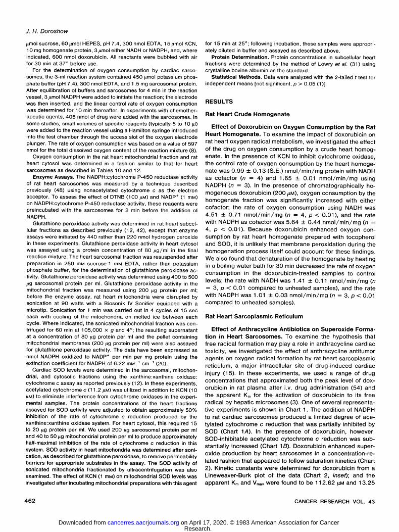
f rac t ion , d o x o r u b i c i n s t imu la ted s u p e r o x i d e p r o d u c t i o n in a c o n c e n t r a t i o n - d e p e n d e n t m a n n e r that a p p e a r e d to fo l low sat - u ra t ion k ine t i cs (Char t 5). The k ine t ic c o n s t a n t s fo r d o x o r u b i c i n in th is m i t ochond r i a l s ys tem w e r e d e t e r m i n e d f rom a L ine- w e a v e r - B u r k p lo t of the da ta (Char t 5, i n s e t ) ; the a p p a r e n t Km
0.32
. . . . . 1~14.95
MIN.
PH B
DOXORUBICtN ~18,71 59.7 nmol 2 oz v
CYTOCHROME cf MIN.
i i I i i i i i i i i i i i i i i J ~ i i i
Chart 3. Stimulation of the rate of sarcosomal oxygen consumption by doxo- rubicin. Oxygen consumption by heart sarcosomes is shown in representative examples from multiple experiments. The addition of NADPH (3 #moO, doxorub- icin (405 nmol), catalase (4500 units), or acetylated cytochrome c (168 nmol) to the 3-ml reaction vessel was accomplished through the access slot of the oxygen electrode plunger and has been indicated by an arrow. The numbers above each experiment indicate the rate of oxygen consumption in nmol/min/mg protein. A, doxorubicin-enhanced oxygen consumption and the effect of catalase; B, effect of cytochrome c on doxorubicin-stimulated oxygen consumption.
g . A t, 5o t,5 o
7.48
A A=O.050
" 2 MIN ,'- I I I I I I I I
Chart 4. Effect of doxorubicin on acetylated cytochrome c reduction by the heart mitochondrial fraction. The sequential addition of NADH (1 #mol) orSOD (10 #g) to the 1-ml reaction system has been indicated by an a r r o w . Experiment A (A) is the mitochondrial control sample; the sample in Experiment B (B) contained doxorubicin (135 #M). The numbers above each experiment represent the rate of acetylated cytochrome c reduction in nmol/min/mg protein.
4 6 6 CANCER RESEARCH VOL. 43
Research. on April 17, 2020. © 1983 American Association for Cancercancerres.aacrjournals.org Downloaded from
3O (A §
~ 25
[1..~ 20
~ ' ~ 15
o 5o loo 15o 2oo 25o
DOXORUBICIN CONCENTRATION (H-M)
Chart 5. Effect of doxorubicin concentration on superoxide anion production in the rat heart mitochondrial fraction and a Lineweaver-Burk plot for doxorubicin (inset). Points, mean of at least 3 experiments for each dose of doxorubicin; bars, S.E.
Table 7 Requirements for doxorubicin-enhanced superoxide production in rat heart
mitochondria Superoxide formation in the mitochondrial fraction was examined using paired,
1-ml reaction mixtures containing 250 Fmol of sucrose, 20/~mol of HEPES, pH 7.4, 100 nmol of EDTA, 56 nmol of acetylated cytochrome c, 50/~g of mitochon- drial protein, and either 0 or 10/~g of SOD. The reaction mixture was preincubated for 4 rain at 37 ~ with 4 nmol of rotenone before initiation with 1 /xmol of NADH.
Superoxide formation (nmol cytochrome c
Reaction mixture reduced/min/mg) n a
Control 2.04 + 0.41 b 16 -- rotenone 3.57 +_ 0.51 6 -- NADH 0.00 +_ 0.00 6 - NADH plus NADPH (1 #moo 0.51 • 0.20 3 Using freeze-thawed mitochondria 2.86 +_ 0.71 3 Using mitochondria prepared with 1.02 • 0.71 4
tocopherol and SOD
Doxorubicin (:135 nmol) 12.75 +__ 0.82 c 24 - NADH 0.51 +_ 0.20 6 - rotenone 8.57 • 0.82 d 14 Using heat-denatured mitochon- 0.00 • 0.00 3
dria e Using heat-denatured SOD e 8.87 • 1.12 3 + NAD* (10/~mol) 3.32 • 0.26 f 3 Using freeze-thawed mitochondria 15.40 + 1.53 5
Doxorubicin (135 nmol) g 18.77 _+ 0.92 h 4 + deferoxamine (:100 nmol) i ~ q8 • 1.02 h 3 - EDTA 17.95 +_ 1.84 h 3 -- EDTA plus deferoxamine (100 13.46 • 1.33 h 3
nmol) - EDTA + deferoxamine (100 22.34 +_ 2.45 h 3
nmol) + MgCI2 (4/~mol) + ATP (1 /~mol)
a Number of experiments. b Mean • S.E. of the rate of superoxide production in the rat heart mitochon-
drial fraction. c Significantly different from control (p < 0.001 ). d Rate of superoxide formation significantly lower than in reaction mixture
containing rotenone (p < 0.01). e Mitochondria or SOD was heated for 60 rain in a boiling water bath; samples
containing heat-denatured SOD were paired against identical mixtures with native dismutase.
f Rate of superoxide formation significantly lower than in reaction mixture containing NADH alone (p < 0.001 ).
u Doxorubicin used in these experiments was preservative free and chro- matographically homogeneous.
n Mitochondria for these experiments were prepared with tocopherol and SOD; experiments were performed in buffer that had undergone ion-exchange chromatography with Chetex to remove trace amounts of iron.
i Deferoxamine was added before the EDTA.
C a r d i a c O x y g e n R a d i c a l F o r m a t i o n b y D o x o r u b i c i n
and Vmax w e r e f o u n d to be 4 5 4 . 5 5 /~M and 5 2 . 6 3 n m o l / m i n /
mg, r e s p e c t i v e l y . In t h e s e e x p e r i m e n t s , d r u g - s t i m u l a t e d s u p e r -
o x i d e f o r m a t i o n w a s N A D H d e p e n d e n t , w a s a b o l i s h e d a f te r the
hear t m i t o c h o n d r i a w e r e d e n a t u r e d by heat , and w a s no t s ig -
n i f i can t l y a l t e red by d i s r u p t i o n of m i t o c h o n d r i a l m e m b r a n e
i n teg r i t y w i th 3 c y c l e s of f r e e z i n g and t h a w i n g ( T a b l e 7).
F u r t h e r m o r e , d r u g - i n d u c e d o x y g e n rad i ca l p r o d u c t i o n w a s no t
d e c r e a s e d by the i r o n - c h e l a t i n g a g e n t d e f e r o x a m i n e o r by
p r e p a r a t i o n of the m i t o c h o n d r i a in a t o c o p h e r o l - and S O D -
c o n t a i n i n g bu f f e r ( T a b l e 7). T h e c o f a c t o r r e q u i r e m e n t fo r mi-
t o c h o n d r i a l f ree rad i ca l p r o d u c t i o n w a s spec i f i c ; on l y N A D H
w a s c a p a b l e of s u p p o r t i n g d r u g - i n d u c e d s u p e r o x i d e f o r m a t i o n
in bo th in tac t m i t o c h o n d r i a and t h o s e e x p o s e d to 3 c y c l e s of
f r e e z i n g and t h a w i n g ( T a b l e 8). F u r t h e r m o r e , d o x o r u b i c i n - e n -
h a n c e d s u p e r o x i d e f o r m a t i o n w a s s i g n i f i c a n t l y r e d u c e d w h e n
m i t o c h o n d r i a l e l e c t r o n t r a n s p o r t w a s no t b l o c k e d at the N A D H
d e h y d r o g e n a s e s i te by r o t e n o n e o r w h e n e x c e s s N A D § w a s
a d d e d to the r e a c t i o n s y s t e m (Tab le 7). Th i s s u g g e s t e d tha t
the ac t i v i t y o f N A D H d e h y d r o g e n a s e tha t can be i nh ib i t ed by
e x c e s s N A D § ( 25 ) m a y be r e s p o n s i b l e for the a c t i v a t i o n of
a n t h r a c y c l i n e a n t i b i o t i c s to f ree r a d i c a l s in hea r t m i t o c h o n d r i a .
F ina l ly , we f o u n d tha t the add i t i on of S O D tha t had been
d e n a t u r e d by hea t to t he m i t o c h o n d r i a l s y s t e m p r o d u c e d no
s i gn i f i can t c h a n g e in the ra te of d r u g - e n h a n c e d s u p e r o x i d e
f o rma t i on , s u g g e s t i n g that , as in the e x p e r i m e n t s w i th hea r t
s a r c o p l a s m i c r e t i c u l u m , s u p e r o x i d e an ion p r o d u c t i o n had b e e n
m e a s u r e d in t h e s e s t u d i e s w i th m i t o c h o n d r i a ( T a b l e 7).
W e a lso i n v e s t i g a t e d the ab i l i t y o f seve ra l o t h e r a n t h r a c y c l i n e
an t i b i o t i c s to i n c r e a s e s u p e r o x i d e f o r m a t i o n in the hea r t m i to -
c h o n d r i a l f r ac t i on . A t e q u i m o l a r c o n c e n t r a t i o n s (1 35 /~M) , d a u -
n o r u b i c i n , r u b i d a z o n e , and a c l a c i n o m y c i n A all s i g n i f i c a n t l y
e n h a n c e d the ra te of m i t o c h o n d r i a l s u p e r o x i d e p r o d u c t i o n o v e r
con t ro l leve ls , p < 0 .001 ( T a b l e 9). F u r t h e r m o r e , a c o m p a r i s o n
of T a b l e 3 w i th T a b l e 9 revea l s tha t fo r bo th the hea r t m i to -
c h o n d r i a l f r ac t i on and hea r t s a r c o s o m e s , a c l a c i n o m y c i n A in-
c r e a s e d s u p e r o x i d e f o r m a t i o n m o s t and r u b i d a z o n e least . In
the m i t o c h o n d r i a l s y s t e m , 5 - i m i n o d a u n o r u b i c i n d id no t s t i m u -
Table 8 Cofactor requirements for doxorubicin-enhanced superoxide formation in rat
heart mitochondria Superoxide production in the rat heart mitochondrial fraction was determined
as described in "Materials and Methods." For these experiments, doxorubicin was included in the reaction mixtures (135 nmol), and superoxide production was initiated by the addition of the cofactor. The cofactor concentration was 1 mM except for sodium succinate which was used at a concentration of 5 mM in the reaction system.
Superoxide formation (nmol/min/mg)
Intact mitochon- Freeze-thawed mi- Cofactor dria n a tochondria b n
NADH 12.85 ___ 1.84 c 4 20.40 _ 3.34 d 3 NAD ~ 0.24 _+ 0.15 3 0.00 +__ 0.00 3 NADPH 3.16 ___ 0.51 3 0.00 ___ 0.00 3 NADP § 0.00 _+ 0.00 3 0.00 • 0.00 3 FMN e 0.68 • 0.36 3 0.00 • 0.00 3 FAD 0.00 +_ 0.00 3 0.00 _+ 0.00 3 Succinate 0.68 +_ 0.34 3 0.00 • 0.00 3
a Number of experiments. b Mitochondria used for these experiments were prepared with tocopherol
and SOD and were exposed to 3 cycles of freezing and thawing before use as described in "Materials and Methods."
c Mean 4- S.E. of the rate of doxorubicin-enhanced superoxide formation in the mitochondrial fraction.
d Doxorubicin used in these experiments was preservative free and chro- matographically homogeneous.
e FMN, flavin mononucleotide; FAD, flavin adenine dinucleotide.
FEBRUARY 1983 4 6 7
Research. on April 17, 2020. © 1983 American Association for Cancercancerres.aacrjournals.org Downloaded from

J. H. D o r o s h o w
Table 9 Effect of chemotherapeutic agents on superoxide formation by rat heart
mitochondria
Superoxide production in the rat heart mitochondrial fraction was determined as described in "Materials and Methods." For these experiments, all drugs were present at a concentration of 135 #M.
Superoxide formation (nmol cytochrome c reduced/
Drug min/mg) n a
Daunorubicin 17.03 ___ 2.35 b' c 3 Rubidazone 9.38 _ 2.14 c 6 Aclacinomycin A 29.89 _+ 3.16 c 3 5-1minodaunorubicin 0.00 --- 0.00 3 Actinomycin D 3.26 +_ 0.41d 3
a Number of experiments. b Mean +_ S.E. of the rate of drug-enhanced superoxide formation in the rat
heart mitochondrial fraction. c Significantly higher than control rate of superoxJde formation in the heart
mitochondrial fraction (p < 0.001, Table 7). d Significantly different from control rate of superoxide formation in the heart
mitochondrial fraction (p < 0.01, Table 7).
Table 10 Effect of doxorubicin on oxygen consumption by rat heart mitochondria
Oxygen consumption in the rat heart mitochondrial fraction was determined in a 3-ml reaction vessel containing 750/Lmol of sucrose, 60 #mol of HEPES, pH 7.4, 300 nmol of EDTA, 12 nmol of rotenone, 600/~g of mitochondrial protein, 3 /~mol of NADH, and, where specified, doxorubicin. For these experiments, dox- orubicin and the other reagents were equilibrated in the reaction vessel for 4 min; oxygen consumption was then initiated by the addition of NADH.
Oxygen consumption Reaction system (nmol 02/min/mg) n a
Control 4.28 + 0.30 b 15 + sodium azide (3/~mol) 4.48 + 0.40 5
Doxorubicin (405 nmol) 7.32 + 0.34 c 19 + sodium azide (3/~mol) 9.45 + 0.30 d 8 + KCN (3 Fmol) 8.76 ___ 0.30 e 7 + dicumarol (f30 nmol) 7.73 +_ 0.43 3 + tocopherol
300 nmol 6.66 +_ 0.19 3 3 #mol 7.26 _+ 1.29 3
Doxorubicin (750 nmol) 9.65 • 0.60 g 3
a Number of experiments. b Mean _4- S.E. of the rate of oxygen consumption in the rat heart mitochondrial
fraction. c Significantly higher than control, p < 0.001. d Significantly higher than samples treated with doxorubicin alone, p < 0.001. e Significantly higher than samples treated with doxorubicin alone, p < 0.05. f ~x-Tocopherol succinate hydrolyzed with 2 molar equivalents of KOH as
described previously (3). g Significantly higher than samples treated with the lower doxorubicin dose,
p < 0.01.
la te f ree rad ica l p r o d u c t i o n , and a c t i n o m y c i n D t r e a t m e n t led
to a sma l l , bu t s i gn i f i can t , i n c r e a s e in the ra te o f s u p e r o x i d e
f o r m a t i o n (Tab le 9).
Effect of Anthracycl ine Antibiot ics on Oxygen Consump- tion b y t h e Rat Heart Mitochondrial Fraction. To i n v e s t i g a t e
f u r t h e r t he e f fec t of the a n t h r a c y c l i n e a n t i b i o t i c s on m i t o c h o n -
d r ia l r e a c t i v e o x y g e n m e t a b o l i s m , the ra te of o x y g e n c o n s u m p -
t ion by rat hea r t m i t o c h o n d r i a e x p o s e d to d o x o r u b i c i n w a s
e x a m i n e d . D o x o r u b i c i n s i g n i f i c a n t l y i n c r e a s e d m i t o c h o n d r i a l
o x y g e n c o n s u m p t i o n o v e r c o n t r o l l eve ls in a d o s e - d e p e n d e n t
m a n n e r (Tab le 10). In t h e s e e x p e r i m e n t s , s o d i u m az ide and
K C N [ w h i c h inh ib i t c a t a l a s e bu t no t m i t o c h o n d r i a l S O D (52 ) ]
b o t h s i g n i f i c a n t l y e n h a n c e d m i t o c h o n d r i a l o x y g e n c o n s u m p t i o n
in t he p r e s e n c e of d o x o r u b i c i n (Tab le 10) ; h o w e v e r , n e i t h e r
d i c u m a r o l no r ~x- tocophero l p r o d u c e d a n y s t a t i s t i ca l l y s ign i f i -
c a n t e f f ec t on the rate of o x y g e n c o n s u m p t i o n by th i s p r e p a -
ra t i on (Tab le 1 0). O x y g e n c o n s u m p t i o n in bo th the c o n t r o l and
d r u g - t r e a t e d s a m p l e s va r i ed w i th the c o n c e n t r a t i o n of m i to -
c h o n d r i a l p ro te i n used ; at m i t o c h o n d r i a l p r o t e i n c o n c e n t r a t i o n s
of 5 0 and 2 0 0 /~g/ml , o x y g e n c o n s u m p t i o n i n c r e a s e d f rom
0 . 3 8 _+ 0 . 0 3 to 0 . 8 2 _+ 0 . 1 2 n m o l / m i n (n = 4, p < 0 . 0 1 ) i n the
con t ro l s a m p l e s and f rom 0 . 5 6 _+ 0 . 0 6 n m o l / m i n to 1 .67 ___
0 . 0 8 n m o l / m i n (n = 4, p < 0 .001 ) in s a m p l e s t r ea ted w i th 1 35
/LM d o x o r u b i c i n . M u c h of th is i n c r e a s e in o x y g e n c o n s u m p t i o n
p r o d u c e d by d o x o r u b i c i n p r o b a b l y re f l ec t s the d r u g - i n d u c e d
s u p e r o x i d e f o r m a t i o n d e m o n s t r a t e d p r e v i o u s l y .
In a d d i t i o n to s u p e r o x i d e an ion p r o d u c t i o n , d o x o r u b i c i n a lso
s t imu la ted h y d r o g e n p e r o x i d e f o r m a t i o n by the rat hear t m i to -
c h o n d r i a l f r ac t i on . As s h o w n in Cha r t 6, A and C, o x y g e n
re lease o b s e r v e d a f te r the a d d i t i o n of e x c e s s ca ta l ase to the
m i t o c h o n d r i a l f r ac t i on w a s more s u b s t a n t i a l in the d r u g - t r e a t e d
samp le . H o w e v e r , o x y g e n re l ease w a s m u c h m o r e a p p a r e n t in
m i t o c h o n d r i a p r e i n c u b a t e d w i th s o d i u m az ide (Cha r t 6, B and
D); in t h e s e p r e p a r a t i o n s , c a t a l a s e - i n d u c e d o x y g e n re lease
i n c r e a s e d f rom 3 .9 +_ 1.1 % (n = 3) of the to ta l m i t o c h o n d r i a l
o x y g e n c o n s u m p t i o n in the c o n t r o l s a m p l e s to 1 5 . 9 +_ 3 . 1 %
(n = 3) in m i t o c h o n d r i a e x p o s e d to d o x o r u b i c i n ( 1 3 5 /~M) ( p
< 0 .02 ) .
Rat Heart Cytosol
Effect of Doxorubicin on Reactive Oxygen Metabol ism in Rat Heart C y t o s o l . To c o m p l e t e o u r i n v e s t i g a t i o n of the s i te(s)
of c a r d i a c f ree rad ica l f o r m a t i o n by the a n t h r a c y c l i n e an t i b io t -
ics, hea r t c y t o s o l w a s e x a m i n e d for i ts ab i l i t y to s u p p o r t d r u g -
i n d u c e d s u p e r o x i d e an ion f o rma t i on . We f o u n d tha t d o x o r u b i -
c in i n c r e a s e d s u p e r o x i d e p r o d u c t i o n more t h a n 10 - fo ld ove r
con t ro l l eve ls in c a r d i a c c y t o s o l ( p < 0 . 0 0 1 , Tab le 1 1 ). Cy to -
so l i c r eac t i ve o x y g e n m e t a b o l i s m requ i r ed N A D H ra the r t han
NADPH, w a s ab la ted w h e n the hea r t c y t o s o l w a s d e n a t u r e d by
heat , and w a s r e d u c e d by more than 3 5 % a f te r t r e a t m e n t w i th
A B
CATALASE CATALASE
C D
CATALASE CATALASE r
i--4 MIN "l L t i-,4 MIN ,~ i t J
Chart 6. Effect of sodium azide on the rate of oxygen consumption in the rat heart mitochondrial fraction. Oxygen consumption by the rat heart mitochondrial fraction is shown in representative examples from multiple experiments. In A,' catalase (4500 units) has been added to the 3-ml control mitochondrial fraction after oxygen consumption was begun with NADH (3 #mol); in B, sodium azide (3 /~mol) had been added to the mitochondrial sample before the addition of NADH. For the experiment shown in C, the mitochondrial fraction was treated with doxorubicin (405 nmol) before initiation with NADH; in D, the 3-ml doxorubicin- treated mitochondrial sample contained sodium azide (3 /~mol). Catalase was added with a Hamilton syringe through the access slot of the oxygen electrode plunger. The numbers above each experiment represent the rate of oxygen consumption in nmol/min/mg protein.
4 6 8 CANCER RESEARCH VOL. 43
Research. on April 17, 2020. © 1983 American Association for Cancercancerres.aacrjournals.org Downloaded from

the x a n t h i n e o x i d a s e i nh ib i t o r a l l opu r i no l (1 0 0 / ~ M ) ( p < 0 . 0 2 ;
T a b l e 1 1 ).
F ree rad ica l p r o d u c t i o n by d o x o r u b i c i n in the c y t o s o l f r a c t i o n
w a s c o n f i r m e d by m e a s u r e m e n t of d r u g - s t i m u l a t e d o x y g e n
c o n s u m p t i o n . H o w e v e r , b e c a u s e the s p e c i f i c ac t i v i t i es o f the
a n t i o x i d a n t e n z y m e s in c a r d i a c c y t o s o l a re the h i ghes t of a n y
ce l l u la r c o m p a r t m e n t in the hear t (see be low) , it w a s n e c e s s a r y
to inh ib i t c y t o s o l i c S O D and c a t a l a s e w i th K C N to m e a s u r e the
e f fec t of d o x o r u b i c i n on c y t o s o l i c o x y g e n rad ica l m e t a b o l i s m
a c c u r a t e l y . As s h o w n in Tab le 12, d o x o r u b i c i n s i g n i f i c a n t l y
i n c r e a s e d the ra te of o x y g e n c o n s u m p t i o n in hear t c y t o s o l by
a p r o c e s s tha t r e q u i r e d N A D H . Th i s d r u g - r e l a t e d i n c r e a s e w a s
no t a l t e red by d i c u m a r o l bu t w a s s i g n i f i c a n t l y r e d u c e d by bo th
a c e t y l a t e d c y t o c h r o m e c and a l l opu r i no l (Tab le 12) . T a k e n
t o g e t h e r , the resu l t s o f t h e s e e x p e r i m e n t s s u g g e s t e d tha t d o x -
o r u b i c i n s t imu la ted reac t i ve o x y g e n m e t a b o l i s m ( p r i n c i p a l l y
s u p e r o x i d e an ion f o r m a t i o n ) in hear t c y t o s o l as wel l as c a r d i a c
s a r c o s o m e s and m i t o c h o n d r i a .
Antioxidant Enzyme Levels
Glutathione Peroxidase. W e have e x a m i n e d p r e v i o u s l y the
e n z y m a t i c d e f e n s e s of m o u s e hear t a g a i n s t o x i d a n t c h a l l e n g e ;
the resu l t s o f tha t i n v e s t i g a t i o n i n d i c a t e d tha t S O D and se le -
n i u m - d e p e n d e n t g l u t a t h i o n e p e r o x i d a s e p l a y e d a m a j o r ro le in
the d e t o x i f i c a t i o n of c a r d i a c r eac t i ve o x y g e n m e t a b o l i t e s (12) .
In the p r e s e n t w o r k , we have e x t e n d e d t h o s e s t u d i e s by de -
t e r m i n g the spec i f i c ac t i v i t i es of S O D and g l u t a t h i o n e p e r o x i -
d a s e in each of the ce l l u l a r c o m p a r t m e n t s of rat hea r t e x a m i n e d
fo r the ab i l i t y to s u p p o r t f ree rad ica l p r o d u c t i o n by d o x o r u b i c i n .
As seen in T a b l e 13, the ac t i v i t y of c a r d i a c g l u t a t h i o n e p e r o x -
i dase w a s g r e a t e s t in ra t hear t c y toso l , p < 0 . 0 0 1 . T h e g lu ta -
t h i one p e r o x i d a s e level o f the p r e p a r a t i o n of hea r t s a r c o p l a s m i c
r e t i c u l u m used fo r o u r f ree rad ica l e x p e r i m e n t s w a s less t han
1 5 % of tha t in the c y t o s o l f r ac t i on ( T a b l e 1 3). T h e g l u t a t h i o n e
p e r o x i d a s e ac t i v i t y of hea r t m i t o c h o n d r i a e x p o s e d to u l t r a s o n i c
d i s r u p t i o n w a s less than 1 0 % of the c o r r e s p o n d i n g level in
c a r d i a c cy toso l . H o w e v e r , u l t r a c e n t r i f u g a t i o n of the s o n i c a t e d
Table 11 Effect of doxorubicin on superoxide production in rat heart cytosol
Superoxide production in rat cardiac cytosol was determined with paired, 1- ml reaction mixtures that contained 150 #mol potassium phosphate buffer, pH 7.4, 100 nmol EDTA, 56 nmol acetylated cytochrome c, 1 O0/~g cytosolic protein, and either 0 or 10 #g SOD. Reactions were begun by the addition of 1 /~mol NAD(P)H in the presence or absence of doxorubicin.
Superoxide production (nmol cytochrome c re-
Reaction system duced/min/mg) n a
Control Using NADPH 0.71 4- 0.31 b 6 Using NADH 0.56 4- 0.20 6
Doxorubicin (135 nmol) Using NADPH 0.84 + 0.36 6 Using NADH 6.22 + 0.67 c 15 - NADH 0.00 • 0.00 d 3 Using NADH with heat-denatured 0.56 4- 0.51d 3
cytosoI e Using NADH plus allopurinol (100 4.03 4- 0.26 f 4
nmol)
a Number of experiments. b Mean • S.E. of the rate of superoxide formation in rat heart cytosol. c Significantly different from control levels (p < 0.001 ). d Significantly different from complete drug-treated sample utilizing NADH
(p < 0.001). e Cytosol heated for 60 min in a boiling water bath. f Significantly different from sample treated with doxorubicin alone ( p < 0.02).
C a r d i a c O x y g e n R a d i c a l F o r m a t i o n b y D o x o r u b i c i n
Table 12 Effect of doxorubicin on oxygen consumption by rat heart cytosol
Oxygen consumption in the cytosol fraction was examined in a 3-ml volume that contained 750 #mol sucrose, 60 #tool HEPES, pH 7.4, 300 nmol EDTA, 300 #g cytosolic protein, 3 #mol either NADH or NADPH, and 405 nmol doxorubicin where specified. For these experiments, doxorubicin and the other reactants were equilibrated in the 3-ml reaction vessel for 4 min; oxygen consumption was then initiated by the addition of 3 #mol NAD(P)H through the access slot of the electrode plunger. Sodium azide and KCN were used in a concentration of 1 mM for all experiments.
Oxygen consumption Experimental system (nmol O2/min/mg) n a
Control + sodium azide and NADPH 1.89 • 0.16 b 3 + sodium azide and NADH 2.19 4- 0.20 6 + KCN and NADH 1.79 _+ 0.40 3 + KCN, NADH, and allopurinol 0.00 _+ 0.00 3
(300 nmol)
Doxorubicin (405 nmol) + sodium azide and NADPH 3.18 _+ 0.40 3 + sodium azide and NADH 10.94 4- 0.99 c 5 + KCN and NADH 11.74 4- 0.60 d 5 + KCN, NADH, and dicumarol 10.35 +_ 1.39 3
(30 nmol) + KCN, NADH, and acetylated 8.56 -4,- 0 . 4 0 e 3
cytochrome c (168 nmol) + KCN, NADH, and allopurinol 7.16 • 1.39 f 3
(300 nmol)
a Number of experiments. b Mean 4- S.E. of the rate of oxygen consumption in heart cytosol. c Significantly different from azide-treated control (p < 0.001). d Significantly different from KCN-treated control (p < 0.001 ). e Significantly different from KCN-treated sample containing doxorubicin
alone (p < 0.01). f Significantly different from KCN-treated sample containing doxorubicin alone
(p < 0.02).
Table 13 Antioxidant enzyme levels in rat heart subcellular fractions
Rat heart cytosol, sarcosomes, and mitochondria were prepared and assayed for glutathione peroxidase and SOD activity as described in "Materials and Methods." Before determination of enzyme activity, the mitochondrial fraction was exposed to ultrasonic disruption as detailed in "Materials and Methods" in order to eliminate permeability barriers to appropriate substrates in these assays; the supernatant and pellet resulting from centrifugation of the sonicated mito- chondria at 105,000 x g and 4 ~ for 60 min were also assayed for glutathione peroxidase and SOD activity.
Glutathione peroxi- dase (nmol NADPH
Heart fraction oxidized/min/mg) SOD (/~g SOD/mg)
Cytosol 704.2 _ 33.4 a" b 13.20 +_ 0.88 c Sarcosomes 100.8 +_ 0.27 2.78 • 0.10 Mitochondria 64.3 4- 11.3 7.60 • 0.10
7.95 4- 0.90 d' e 105,000 x g supernatant 307.0 +_ 5.8 9.96 4- 0.62
8.80 • 0.24 d' e 105,000 X g pellet 53.6 4- 2.2 7.30 4- 1.60
6.40 • 0.30 d" e
a Mean 4- S.E. of triplicate determinations of enzyme activity from at least 3 exp.erimental samples.
o Glutathione peroxidase activity in cytosol was significantly higher than enzyme levels in sarcosomes or sonicated mitochondria (p < 0.001).
c SOD activity in cytosol was significantly higher than enzyme levels in sarcosomes or sonicated mitochondria (p < 0.01 ).
d SOD activity in these samples was assayed after treatment with 1 mM KCN for15 min at 25 ~ .
e No significant difference in the SOD activity of corresponding mitochondrial fractions with or without KCN treatment.
m i t o c h o n d r i a p r o d u c e d a s u p e r n a t a n t w i th n e a r l y 4 5 % of t he
spec i f i c ac t i v i t y of the hea r t c y t o s o l f r ac t i on ( T a b l e 1 3). T h u s ,
it s e e m e d p r o b a b l e tha t the g l u t a t h i o n e p e r o x i d a s e ac t i v i t y o f
rat hear t m i t o c h o n d r i a [ in c o n t r a s t to tha t in the m o u s e (23 ) ]
w a s l oca ted p r ima r i l y in t he m i t o c h o n d r i a l ma t r i x .
S u p e r o x i d e D i s m u t a s e . T h e d i s t r i b u t i o n of S O D w i th in t he
rat hear t w a s s im i la r to tha t d e s c r i b e d for g l u t a t h i o n e p e r o x i -
FEBRUARY 1983 4 6 9
Research. on April 17, 2020. © 1983 American Association for Cancercancerres.aacrjournals.org Downloaded from

J. H. Doroshow
dase. SOD activity in cytosol was significantly greater than the level measured in either heart sarcosomes or mitochondria, p < 0.01 (Table 13). Furthermore, a major part of the SOD activity of the mitochondrial fraction was contained in the mitochondrial matrix (Table 13). Preincubation with KCN did not significantly change the specific activity of SOD measured in any mitochondrial preparation; this suggested that the man- ganese-dependent enzyme comprised a major portion of the SOD in rat heart mitochondria (52). However, the presence of a SOD activity from the intermembranous space and its cyanide sensitivity were not addressed in these experiments; and thus, the possibility that rat heart mitochondria also contain a copper and zinc-dependent dismutase similar to that in liver cannot be excluded (52).
DISCUSSION
If drug-induced oxygen radical formation is a major deter- minant of the cardiac toxicity of the anthracycline antibiotics, it should help to explain specific morphological features of the heart damage that characteristically accompanies treatment with these drugs. Furthermore, it should also provide some insight into the apparently selective nature of this particular form of myocardial insult. In this study, we have attempted to evaluate the role of reactive oxygen metabolism in the cardiac toxicity of the anthracycline antitumor agents along these lines by investigating both the extent and the mechanism(s) of an- thracycline-related free radical production in rat heart.
We have shown that treatment of heart sarcosomes, mito- chondria, and cytosol with anthracycline drugs produces a concentration-related increase in superoxide anion and hydro- gen peroxide production. This occurs despite the presence of both superoxide dismutase and glutathione peroxidase (2 ma- jor cardiac enzymatic defenses against oxygen radicals) in each heart fraction. Although drug-stimulated oxygen radical metabolism was enhanced further when these enzymes, as well as cardiac catalase, were inhibited by treatment with cyanide or azide, our findings suggest that oxygen radical production by the anthracycline antibiotics may exceed the detoxifying capacity of the individual heart fractions. Because anthracycline cardiac toxicity is characterized by degeneration of heart mitochondria and sarcoplasmic reticulum (1 5), reactive oxygen accumulation may explain drug-induced membrane damage at these specific intracellular sites.
Enhancement of oxygen radical metabolism by the anthra- cycline drugs was demonstrated for each myocardial fraction examined; however, the mechanism(s) of free radical formation in these compartments differed. Superoxide production by rat heart sarcosomes required NADPH, was not inhibited by cya- nide or dicumarol, and was accompanied by the oxidation of NADPH. Furthermore, inhibition of sarcosomal cytochrome P- 450 reductase activity by treatment with excess NADP + or a sulfhydryl reagent produced a parallel decrease in superoxide formation by doxorubicin. These experiments strongly suggest that the NADPH: cytochrome P-450 reductase activity of heart sarcosomes was responsible for the reduction of the anthra- cycline antibiotics to their respective semiquinones.
On the other hand, superoxide formation in the mitochondrial fraction was dependent on NADH, and not succinate, was rotenone and dicumarol insensitive, and was significantly in- hibited by addition of excess NAD +. These experiments indi-
cate that the NADH dehydrogenase complex was the site of anthracycline reduction in our experiments with intact mito- chondria. Thayer (49) obtained similar results recently for doxorubicin using a preparation of cardiac submitochondrial particles. However, in the absence of specific mitochondrial fractionation studies, one cannot exclude the possibility that the NADH:cytochrome bs reductase (NADH:cytochrome b5 ox- idoreductase, EC 1.6.2.2) of the outer mitochondrial membrane (6), in addition to a component of the electron transport chain, contributed to these findings.
Finally, we found that reactive oxygen metabolism in heart cytosol was heat labile, dicumarol insensitive, and dependent on NADH as cofactor; treatment of the cytosol fraction with allopurinol resulted in a significant reduction in drug-related superoxide formation and oxygen consumption. Thus, part of the free radical formation observed in heart cytosol might be related to the activation of doxorubicin to its semiquinone intermediate by xanthine oxidase, a reaction demonstrated previously with a purified preparation of this enzyme (43). Although our heart cytosol was carefully prepared in an attempt to eliminate any substantial contamination by the mitochondrial fraction, it is still possible that mitochondrial components could have interfered with these experiments and might be respon- sible for supporting at least part of the drug-related superoxide formation in the cytosol fraction.
From these studies, it may be suggested that 3 different flavin-containing enzymes (NADPH:cytochrome P-450 reduc- tase, NADH dehydrogenase, and xanthine oxidase), which have been shown previously to be capable of catalyzing the one-electron reduction of a wide variety of quinone drugs (21, 38), are responsible for the activation of the anthracycline antibiotics to free radical species in heart sarcoplasmic retic- ulum, mitochondria, and cytosol. Furthermore, because inhi- bition of cardiac DT diaphorase by dicumarol had no effect on free radical production in any heart fraction, it appears that this enzyme has no significant impact on the activation of anthra- cycline drugs in rat heart.
In an aerobic environment, such as the myocardial cell, anthracycline semiquinone radicals rapidly donate their un- paired electrons to molecular oxygen, forming superoxide an- ion and/or hydrogen peroxide (22). We have shown that treat- ment of heart sarcosomes with anthracycline drugs leads to the production of both of these reactive oxygen metabolites. A comparison of the rates of oxygen consumption and superoxide formation in sarcosomes treated with doxorubicin suggests that under identical experimental conditions, the production of superoxide anion, at least initially, accounts for most of the oxygen consumed. In that case, the hydrogen peroxide formed must result from the spontaneous or SOD-catalyzed dismuta- tion of superoxide anion rather than from the direct, 2-electron reduction of oxygen.
If the anthracycline antitumor agents augment the production of reactive oxygen metabolites in vivo, the limited capacity of cardiac muscle to detoxify free radicals could be overwhelmed (10), resulting in an extensive alteration in myocardial function at the sites of oxygen radical accumulation. Drug-induced peroxidation of sarcoplasmic reticular membrane could inter- fere with tubular calcium transport and muscular contractility (30) and might explain the sarcoplasmic vacuolation that ap- pears to be the earliest morphological concomitant of anthra- cycline cardiac toxicity in humans (50). Reduction of the an-
470 CANCER RESEARCH VOL. 43
Research. on April 17, 2020. © 1983 American Association for Cancercancerres.aacrjournals.org Downloaded from

t h r a c y c l i n e q u i n o n e at a p r o x i m a l po r t i on o f the m i t o c h o n d r i a l
e l e c t r o n t r a n s p o r t c h a i n c o u l d d i ve r t e l e c t r o n f l ow f rom e n e r g y
p r o d u c t i o n , t h e r e b y r e d u c i n g m y o c a r d i a l A T P c o n t e n t ; f u r t h e r -
more , m i t o c h o n d r i a l m e m b r a n e i n t eg r i t y and the r e g u l a t i o n o f
the level o f i n t r a c e l l u l a r r e d u c i n g e q u i v a l e n t s m igh t be a l t e red
in th is m a n n e r (46) . F ree rad ica l p r o d u c t i o n in the s o l u b l e
c o m p a r t m e n t c o u l d lead to the o x i d a t i o n of c r i t i ca l s u l f h y d r y l -
c o n t a i n i n g e n z y m e s ( s u c h as g l y c e r a l d e h y d e : 3 - p h o s p h a t e de -
h y d r o g e n a s e ) w i th an add i t i ona l , f u r t h e r d e c r e a s e in c a r d i a c
e n e r g y me tabo l i sm . As w e have s u g g e s t e d r e c e n t l y (10) , be -
c a u s e hea r t ce l l s a re so r i ch in h e m e - c o n t a i n i n g p r o t e i n s tha t
may u n d e r g o a u t o x i d a t i o n , it is p r o b a b l e tha t t he p r e s e n c e of
e x c e s s i n t r a c e l l u l a r p e r o x i d e in the a n t h r a c y c l i n e - t r e a t e d hea r t
w o u l d e s t a b l i s h an e x t r a o r d i n a r i l y f a v o r a b l e m i l i eu for the fo r -
ma t i on of h i g h l y r eac t i ve h y d r o x y l and p e r o x y r a d i c a l s w i th a
p r o v e n c a p a c i t y to p r o d u c e m e m b r a n e p e r o x i d a t i o n .
F ina l l y , we e v a l u a t e d the e f fec t o f seve ra l a n t h r a c y c l i n e and
n o n a n t h r a c y c l i n e a n a l o g u e s on reac t i ve o x y g e n m e t a b o l i s m in
the rat hea r t f r ac t i ons . D a u n o r u b i c i n , r u b i d a z o n e , and a c l a c i -
n o m y c i n A all e f f ec t i ve l y s t imu la ted s u p e r o x i d e p r o d u c t i o n in
c a r d i a c s a r c o s o m e s and m i t o c h o n d r i a . A c t i n o m y c i n D, w h i c h
has been r e p o r t e d to i n c r e a s e the c a r d i a c t o x i c i t y of d o x o r u b -
ic in C27), p r o d u c e d a smal l , bu t s i gn i f i can t , i n c r e a s e in s u p e r -
o x i d e p r o d u c t i o n in the hea r t f r ac t i ons ; th i s is in a g r e e m e n t
w i th a r e c e n t r epo r t i n d i c a t i n g tha t a c t i n o m y c i n D a u g m e n t s
s u p e r o x i d e f o r m a t i o n by h e p a t i c m i c r o s o m e s (47) . H o w e v e r ,
the d a u n o r u b i c i n a n a l o g u e 5 - i m i n o d a u n o r u b i c i n d id no t p r o -
d u c e any s i g n i f i c a n t e f fec t on e i t he r s a r c o s o m a l or m i t o c h o n -
dr ia l f ree rad ica l f o rma t i on , w h i c h s u p p o r t s a s t u d y by L o w n e t
al. (29) , s u g g e s t i n g tha t o x i d a t i o n - r e d u c t i o n c y c l i n g a f te r c h e m -
ical r e d u c t i o n of d a u n o r u b i c i n is e s s e n t i a l l y e l i m i n a t e d by the
im ino s u b s t i t u t i o n on the q u i n o n e r ing. T h e s e resu l t s may
e x p l a i n r e p o r t s of l esse r c a r d i a c t o x i c i t y for th i s a g e n t (29) .
In s u m m a r y , we p r o p o s e tha t the a n t h r a c y c l i n e a n t i b i o t i c s
se l ec t i ve l y in ju re the hear t b e c a u s e the i r a d m i n i s t r a t i o n may
lead to o x y g e n rad ica l a c c u m u l a t i o n in the s p e c i f i c m y o c a r d i a l
c o m p a r t m e n t s c a p a b l e of r e d u c t i v e l y a c t i v a t i n g the d r u g s to
s e m i q u i n o n e f ree rad i ca l i n t e r m e d i a t e s .
ACKNOWLEDGMENTS
I wish to thank Jill Reeves for her excellent technical assistance. I also wish to express my sincere thanks to Dr. Paul Hochstein of the Institute for Toxicology, University of Southern California Schools of Medicine and Pharmacy, for his ongoing encouragement and thoughtful discussion of this work.
REFERENCES 1. Armitage, P. Statistical Methods in Medical Research, pp. 104-126. Oxford:
Blackwell Scientific Publications, Ltd., 1971. 2. Azzi, A., Montecucco, C., and Richter, C. The use of acetylated ferricyto-
chrome c for the detection of superoxide radicals produced in biological membranes. Biochem. Biophys. Res. Commun., 65: 597-603, 1975.
3. Bachur, N. R., Gordon, S. L., and Gee, M. V. Anthracycline antibiotic augmentation of microsomal electron transport and free radical formation. Mol. Pharmacol., 13: 901-910, 1977.
4. Bachur, N. R., Gordon, S. L., and Gee, M. V. A general mechanism for microsomal activation of quinone anticancer agents to free radicals. Cancer Res., 38:1745-1750, 1978.
5. Bachur, N. R., Gordon, S. L., Gee, M. V., and Kon, H. NADPH cytochrome P-450 reductase activation of quinone anticancer agents to free radicals. Proc. Natl. Acad. Sci. U. S. A., 76: 954-957, 1979.
6. Bernardi, P., and Azzone, G. F. Cytochrome c as an electron shuttle between the outer and inner mitochondrial membranes. J. Biol. Chem., 256: 7189-7192, 1981.
7. Carter, S. K. Adriamycin--a review. J. Natl. Cancer Inst., 55:1265-1274, 1975.
8. Chappel, J. B. The oxidation of citrate, isocitrate, and cis-aconitate by
C a r d i a c O x y g e n R a d i c a l F o r m a t i o n b y D o x o r u b i c i n
isolated mitochondria. Biochem. J., 90: 225-237, 1964. 9. Dantchev, D., Slioussartchouk, V., Paintrand, M., Hayat, M., Bourut, C., and
Mathe, G. Electron microscopic studies of the heart and light microscopic studies of the skin after treatment of golden hamsters with Adriamycin, detorubicin, AD-32, and aclacinomycin. Cancer Treat. Rep., 63: 875-888, 1979.
10. Doroshow, J. H., and Locker, G. Y. Role of oxygen radical formation in anthracycline cardiac toxicity. In: F. Muggia, C. W. Young, and S. K. Carter (eds.), Anthracycline Antibiotics in Cancer Therapy, pp. 31 7-330. Boston: Martinus Nijhoff Publishers, 1982.
11. Doroshow, J. H., Locker, G. Y., Ifrim, I., and Myers, C. E. Prevention of doxorubicin cardiac toxicity in the mouse by n-acetylcysteine. J. Clin. Invest., 68: 1053-1064, 1981.
12. Doroshow, J. H., Locker, G. Y., and Myers, C. E. Enzymatic defenses of the mouse heart against reactive oxygen metabolites: alterations produced by doxorubicin. J. Clin. Invest., 65:128-135, 1980.
13. Doroshow, J. H., and Reeves, J. Anthracycline-enhanced oxygen radical formation in the heart. Proc. Am. Assoc. Cancer Res., 21: 266, 1980.
14. Ernster, L. DT diaphorase. Methods Enzymol., 10: 309-317, 1967. 15. Ferrans, V. J. Overview of cardiac pathology in relation to anthracycline
cardiotoxicity. Cancer Treat. Rep., 62: 955-961, 1978. 16. Fridovich, I. The biology of oxygen radicals. Science (Wash. D. C.), 201:
875-880, 1978. 17. Goodman, J., and Hochstein, P. Generation of free radicals and lipid per-
oxidation by redox cycling of Adriamycin and daunomycin. Biochem. Bio- phys. Res. Commun., 77: 797-803, 1977.
18. Handa, K., and Sato, S. Stimulation of microsomal NADPH oxidation by quinone group-containing anticancer chemicals. Gann, 67: 523-528, 1976.
19. Harigaya, S., and Schwartz, A. Rate of calcium binding and uptake in normal animal and failing human cardiac muscle: membrane vesicles (relaxing system) and mitochondria. Circ. Res., 25:781-794, 1969.
20. Horecker, B. L., and Kornberg, A. The extinction coefficients of the reduced band of pyridine nucleotides. J. Biol. Chem., 1 75: 385-390, 1948.
21. lyanagi, T., and Yamazaki, I. One-electron-transfer reactions in biochemical systems. V. Difference in the mechanism of quinone reduction by the NADH dehydrogenase and the NAD(P)H dehydrogenase (DT-diaphorase). Biochim. Biophys. Acta, 216: 282-294, 1970.
22. Kalyanaraman, B., Perez-Reyes, E., and Mason, R. P. Spin-trapping and direct electron spin resonance investigations of the redox metabolism of quinone anticancer drugs. Biochim. Biophys. Acta, 630:119-130, 1980.
23. Katki, A., and Myers, C. E. Membrane-bound glutathione peroxidase-like activity in mitochondria. Biochem. Biophys. Res. Commun., 96: 85-91, 1980.
24. Kharasch, E. D., and Novak, R. F. Ring current effects in Adriamycin-flavin mononucleotide comptexation as observed by 1H FT NMR spectroscopy. Biochem. Biophys. Res. Commun., 92: 1320-1326, 1980.
25. King, T. E., and Howard, R. L. Preparations and properties of soluble NADH dehydrogenases from cardiac muscle. Methods Enzymol., 10: 275-294, 1967.
26. Komiyama, T., Oki, T., and Inui, T. A proposed reaction mechanism for the enzymatic reductive cleavage of glycosidic bond in anthracycline antibiotics. J. Antibiot. (Tokyo), 32:1219-1222, 1979.
27. Kushner, J. P., Hansen, V. L., and Hammar, S. P. Cardiomyopathy after widely separated courses of Adriamycin exacerbated by actinomycin-D and mithramycin. Cancer (Phila.), 36: 1577-1584, 1975.
28. Lenaz, L., and Page, J. Cardiotoxicity of Adriamycin and related anthracy- clines. Cancer Treat. Rev., 3:111-120, 1976.
29. Lown, J. W., Chen, H.-H., Plambeck, J. A., and Acton, E. M. Diminished superoxide anion generation by reduced 5-iminodaunorubicin relative to daunorubicin and the relationship to cardiotoxicity of the anthracycline antitumor agents. Biochem. Pharmacol., 28: 2563-2568, 1979.
30. Lowrey, K., Glende, E. A., and Recknagel, R. O. Destruction of liver micro- somal calcium pump activity by carbon tetrachloride and bromotrichloro- methane. Biochem. Pharmacol., 30: 135-140, 1981.
31. Lowry, O., Rosebrough, N. J., Farr, A. L., and Randall, R. J. Protein measurement with the Folin phenol reagent. J. Biol. Chem., 193: 265-275, 1951.
32. Martonosi, A. Sarcoplasmic reticulum. IV. Solubilization of microsomal aden- osine triphosphatase. J. Biol. Chem., 243:71-81, 1968.
33. McCord, J. M., and Fridovich, I. Superoxide dismutase--an enzymatic function of erythrocuprein (hemocuprein). J. Biol. Chem., 244: 6049-6055, 1969.
34. Mela, L., and Seitz, S. Isolation of mitochondria with emphasis on heart mitochondria from small amounts of tissue. Methods Enzymol., 55: 39-46, 1979.
35. Mettler, F. P., Young, D. M., and Ward, J. M. Adriamycin-induced cardiotox- icity (cardiomyopathy and congestive heart failure) in rats. Cancer Res., 37: 2705-2713, 1977.
36. Misra, H. P., and Fridovich, I. A peroxide-dependent reduction of cytochrome c by NADH. Biochim. Biophys. Acta, 292:815-824, 1973.
37. Myers, C. E., McGuire, W. P., Liss, R. H., Ifrim, I., Grotzinger, K., and Young, R. C. Adriamycin: the role of lipid peroxidation in cardiac toxicity and tumor response. Science (Wash. D. C.), 197:165-1 67, 1977.
FEBRUARY 1983 471
Research. on April 17, 2020. © 1983 American Association for Cancercancerres.aacrjournals.org Downloaded from

J. H. D o r o s h o w
38. Nakamura, S., and Yamazaki, I. One-electron transfer reactions in biochem- ical systems. IV. A mixed mechanism in the reaction of milk xanthine oxidase with electron acceptors. Biochim. Biophys. Acta, 189: 29-37, 1969.
39. Newman, R. Ao, Hacker, M. P., and Krakoff, I. H. Amelioration of Adriamycin and daunorubicin myocardial toxicity by adenosine. Cancer Res., 41: 3483-3488, 1981.
40. Nishikimi, M., Yamada, H., and Yagi, K. Oxidation by superoxide of tocoph- erols dispersed in aqueous media with deoxycholate. Biochim. Biophys. Acta, 627: 101-108, 1980.
41. Olson, R. D., MacDonald, J. S., Van Boxtel, C. J., Boerth, R. C., Harbison, R. D., Slonim, A. E., Freeman, R. W., and Oates, J. A. Regulatory role of glutathione and soluble sulfhydryl groups in the toxicity of Adriamycin. J. Pharmacol. Exp. Ther., 215: 450-454, 1980.
42. Paglia, D. E., and Valentine, W. N. Studies on the quantitative and qualitative characterization of erythrocyte glutathione peroxidase. J. Lab. Clin. Med., 70:158-169, 1967.
43. Pan, S.-S., and Bachur, N. R. Xanthine oxidase catalyzed reductive cleavage of anthracycline antibiotics and free radical formation. Mol. Pharmacol., 1 7: 95-99, 1980.
44. Revis, N. W., and Marusic, N. Glutathione peroxidase activity and selenium concentration in the hearts of doxorubicin-treated rabbits. J. Mol. Cell. Cardiol., 10: 945-951, 1978.
45. Sato, S., Iwaizumi, M., Handa, K., and Tamura, Y. Electron spin resonance study on the mode of generation of free radicals of daunomycin, Adriamycin, and carboquone in NAD(P)H-microsome system. Gann, 68: 603-608, 1977.
46. Sies, H., Graf, P., and Estrela, J. Hepatic calcium efflux during cytochrome
P-450-dependent drug oxidations at the endoplasmic reticulum in intact liver. Proc. Natl. Acad. Sci. U. S. A., 78: 3358-3362, 1981.
47. Sinha, B. K., and Cox, M. G. Stimulation of superoxide formation by acti- nomycin D and its N2-substituted spin-labeled derivatives. Mol. Pharmacol., 17: 432-434, 1980.
48. Strobel, H. W., and Dignam, J. D. Purification and properties of NADPH- cytochrome P-450 reductase. Methods Enzymol., 52: 89-96, 1978.
49. Thayer, W. S. Adriamycin stimulated superoxide formation in submitochon- drial particles. Chem.-Biol. Interact., 19: 265-278, 1977.
50. Unverferth, D. V., Magorien, R. D., Unverferth, B. P., Talley, R. L., Balcerzak, S. P., and Baba, N. Human myocardial morphologic and functional changes in the first 24 hours after doxorubicin administration. Cancer Treat. Rep., 65: 1093-1097, 1981.
51. Wang, Y.-M., Madanat, F. F., Kimball, J. C., Gleiser, C. A., Ali, M. K., Kaufman, M. W., and Van Eys, J. Effect of vitamin e against Adriamycin- induced toxicity in rabbits. Cancer Res., 40:1022-1027, 1980.
52. Weisiger, R. A., and Fridovich, I. Mitochondrial superoxide dismutase: site of synthesis and intramitochondrial localization. J. Biol. Chem., 248: 4793-4796, 1973.
53. Williams, C. H. Flavin-containing dehydrogenases. In: P. D. Boyer (ed.), The Enzymes, Vol. 13, pp. 165-173. New York: Academic Press, Inc., 1976.
54. Yesair, D. W., Schwartzbach, E., Shuck, D., Denine, E. P., and Asbell, M. A. Comparative pharmacokinetics of daunomycin and Adriamycin in several animal species. Cancer Res., 32:1177-1183, 1972.
55. Yonetani, T., and Ray, G. S. Studies on cytochrome c peroxidase: purifica- tion and some properties. J. Biol. Chem., 240: 4503-4508, 1965.
4 7 2 CANCER RESEARCH VOL. 43
Research. on April 17, 2020. © 1983 American Association for Cancercancerres.aacrjournals.org Downloaded from

1983;43:460-472. Cancer Res James H. Doroshow Formation in Rat HeartEffect of Anthracycline Antibiotics on Oxygen Radical
Updated version
http://cancerres.aacrjournals.org/content/43/2/460
Access the most recent version of this article at:
E-mail alerts related to this article or journal.Sign up to receive free email-alerts
Subscriptions
Reprints and
To order reprints of this article or to subscribe to the journal, contact the AACR Publications
Permissions
Rightslink site. Click on "Request Permissions" which will take you to the Copyright Clearance Center's (CCC)
.http://cancerres.aacrjournals.org/content/43/2/460To request permission to re-use all or part of this article, use this link
Research. on April 17, 2020. © 1983 American Association for Cancercancerres.aacrjournals.org Downloaded from