eeg experiment for extra credit sign up on the sheet
Post on 18-Dec-2015
227 views
TRANSCRIPT

EEG Experiment for Extra Credit
Sign up on the sheet

Structural and Functional Imaging
• Cortical Flattening– Software such as
BrainVoyager can “inflate” the cortex like a balloon so that sulci and gyri are “flattened”
– functional data can be transformed with the same complex function
– functional and structural data can be overlaid so that distribution on cortical sheet can be visualized
QuickTime™ and a decompressor
are needed to see this picture.

Principles of MRI

Principles of MRI
• Some terms:– Nuclear Magnetic Resonance (NMR)
• quantum property of protons
• energy absorbed when precession frequency matches radio frequency
– Magnetic Resonance Imaging (MRI)• uses spatial differences in resonance frequencies to form an
image
• basis of anatomical MRI
– functional Magnetic Resonance Imaging (fMRI)• exploits magnetic properties of hemaglobin to create images
changes in cortical blood flow

Principles of NMR
• Protons are like little magnets– they orient in magnetic fields like
compass needles
– what way do they normally point?

QuickTime™ and a decompressor
are needed to see this picture.
Principles of NMR
• Protons are like little magnets– they orient in magnetic fields like
compass needles
– what way do they normally point?
– normally aligned with Earth’s magnetic field

Principles of NMR
• Protons are like little magnets– they orient in magnetic fields like
compass needles
– what way do they normally point?
– normally aligned with Earth’s magnetic field
– NMR uses a big magnet to align all the protons in a sample (e.g. brain tissue)

Principles of NMR
• Protons are like little magnets– Radio Frequency pulse will knock
protons at an angle relative to the magnetic field

Principles of NMR
• Protons are like little magnets– Radio Frequency pulse will knock
protons at an angle relative to the magnetic field
– once out of alignment, the protons begin to precess

Principles of NMR
• Protons are like little magnets– Radio Frequency pulse will knock
protons at an angle relative to the magnetic field
– once out of alignment, the protons begin to precess
– protons gradually realign with field (relaxation)

Principles of NMR
• Protons are like little magnets– Radio Frequency pulse will knock
protons at an angle relative to the magnetic field
– once out of alignment, the protons begin to precess
– protons gradually realign with field (relaxation)
– protons “echo” back the radio frequency that originally tipped them over
– That radio “echo” forms the basis of the MRI image

Principles of NMR
• Protons are like little magnets– The following simple equation
explains MRI image formation

MRI Image Formation
• First you need a scanner:
– The first MRI scanner

MRI Image Formation
• Modern Scanners

MRI Image Formation
• Our Scanner

MRI Image Formation
• Our Scanner

MRI Image Formation
• Our Scanner

MRI Image Formation
• Our Scanner

MRI Image Formation
• MRI Image formation– resonance frequency
depends on field strength
– gradient coils alter resonance frequency over distance
– slight differences in the “echo” frequency indicate the location of each proton
– second-dimension of a slice is coded by the phase of the protons
field gradient = frequency gradient

Functional Imaging
• Functional Imaging must provide a spatial depiction of some process that is at least indirectly related to neural activity
• in most imaging (i.e. PET, fMRI) that process is change in blood oxygenation related to changes in regional cerebral blood flow
• Why should we measure blood oxygenation?

Functional Imaging
• Why should we measure blood oxygenation?
• Onset of a stimulus (or cognitive task) changes local blood oxygenation– first with a decrease
– then with an “overshoot”
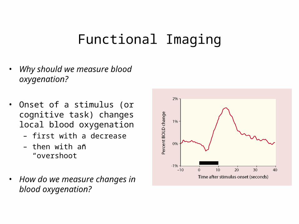
Functional Imaging
• Why should we measure blood oxygenation?
• Onset of a stimulus (or cognitive task) changes local blood oxygenation– first with a decrease
– then with an “overshoot”
• How do we measure changes in blood oxygenation?