editors'choice a new bernissartiid crocodyliform from the
TRANSCRIPT

Acta Palaeontol. Pol. 60 (2): 257–268, 2015 http://dx.doi.org/10.4202/app.00038.2013
A new bernissartiid crocodyliform from the Lower Cretaceous Wessex Formation (Wealden Group, Barremian) of the Isle of Wight, southern England
STEVEN C. SWEETMAN, ULYSSE PEDREIRA-SEGADE, and STEVEN U. VIDOVIC
Sweetman, S.C., Pedreira-Segade, U., and Vidovic, S.U. 2015. A new bernissartiid crocodyliform from the Lower Cre-taceous Wessex Formation (Wealden Group, Barremian) of the Isle of Wight, southern England. Acta Palaeontologica Polonica 60 (2): 257–268.
A substantially complete skull of a small crocodyliform recently found on the foreshore near Yaverland on the south-east coast of the Isle of Wight, southern England is described. The locality, mode of preservation and associated matrix indicate that it is derived from one of the plant debris beds of the Lower Cretaceous Wessex Formation (Barremian, Wealden Group). The dentition, unique among crocodyliforms, serves to confirm that the specimen is referable to the, until now, monotypic family Bernissartiidae. Apomorphies, including placement of the choana entirely within the pterygoids and disposition of cranial sutures demonstrate that the Isle of Wight skull cannot be referred to Bernissartia fagesii, known from contemporaneous strata. Furthermore, these characters indicate that the specimen should not be referred to a new species of Bernissartia. It is therefore placed in a new genus and species, Koumpiodontosuchus apros-dokiti. The systematic position of Bernissartiidae, and characters used to diagnose Eusuchia, including placement of the choana(e) within the prerygoids, are discussed. Until recently this condition was thought to be restricted to Eusuchia with all non-eusuchian neosuchian crorocdiliforms possessing choanae bounded posteriorly by the pterygoids and an-teriorly by the palatines. While the choana of Koumpiodontosuchus aprosdokiti gen. et sp. nov. is entirely bounded by the pterygoids it differs from the choanae of eusuchians in lacking a median septum, being anteroposteriorly elongate and in its anterior placement.
Key words: Crocodyliformes, Eusuchia, Neosuchia, Bernissartiidae, Cretaceous, Barremian, Wealden, England.
Steven C. Sweetman [[email protected]] and Steven U. Vidovic [[email protected]], Palaeobiolo-gy Research Group, School of Earth and Environmental Sciences, University of Portsmouth, Burnaby Building, Burna-by Road, Portsmouth, PO1 3QL, United Kingdom.Ulysse Pedreira-Segade [[email protected]], Département des Sciences de la Terre, Université Claude Bernard Lyon 1 et Ecole Normale Supérieure de Lyon, 2 rue Dubois, 69622 Villeurbanne cedex, France.
Received 4 April 2013, accepted 28 February 2014, available online 11 March 2014.
Copyright © 2015 S.C. Sweetman et al. This is an open-access article distributed under the terms of the Creative Com-mons Attribution License, which permits unrestricted use, distribution, and reproduction in any medium, provided the original author and source are credited.
IntroductionThe crocodyliform assemblages of the Wealden Supergroup of southern Britain have recently been the subject of a com-prehensive review which also involved the erection of a number of new taxa (Salisbury and Naish 2011). The au-thors were aware of the skull described here, but it had not been accessioned in a registered collection. Furthermore, no preparation of the specimen had been undertaken and most of the ventral surface was obscured by matrix. Subsequently, the skull was accessioned in a museum, and the ventral sur-
face has been prepared to reveal the palatal bones, the sutures between them and the location of the choana.Institutional abbreviations.—IRSNB, Institut Royal des Sci-ences Naturelles de Belgique, Brussels; IWCMS, the Isle of Wight County Museum Service (incorporating Dinosaur Isle museum and visitor attraction); NHMUK, the Natural History Museum, London.Other abbreviations.—MPTs, most parsimonious trees.
Nomenclatural acts.—The electronic edition of this article conforms to the requirements of the amended Internation-
Editors' choice

258 ACTA PALAEONTOLOGICA POLONICA 60 (2), 2015
al Code of Zoological Nomenclature, and hence the new names contained herein are available under that Code from the electronic edition of this article. This published work and the nomenclatural acts it contains have been registered in ZooBank. The ZooBank LSID (Life Science Identifier) for this publication is: urn: lsid: zoobank.org: pub: 7C6C43AF-B045-4AEC-A22E-29349F13A507.
Locality, geological and palaeontological settingsThe skull was found in two parts loose on the beach north-east of Yaverland coastal car park at UK National Grid Reference SZ 615851 on the south-east coast of the Isle of Wight (Fig. 1). The posterior part was found in March 2011 and by remarkable serendipity the gracile rostral section, which had suffered little post-exhumation abrasion, was recovered some three months later. Coastal erosion is continuous along this part of the coast and cliff falls commonly occur, especially during the winter months. In the vicinity of the sites of collection the upper part of the Wessex Formation is exposed and is late Barremian in age (Kerth and Hailwood 1988; Hughes and McDougall 1990; Allen and Wimbledon 1991; Robinson and Hesselbo 2004). At this locality it incorporates both isolated, and a stacked sequence of plant debris beds (Sweetman and Insole 2010). These are the main source of plant, invertebrate and vertebrate fossils. The state of preservation of the skull and the associated matrix suggest that it is derived from one of the plant debris beds between beds 26 and 38 (Radley 1994; Fig. 2).
The Wessex Formation represents primarily fluvial depo-sition on a near coast floodplain with subordinate lacustrine and terrestrial deposits. The majority of the succession is de-
void of organic remains, reflecting unfavourable conditions for their preservation. However, the plant debris beds, which comprise localized debris flow deposits and which make up only a small proportion of the succession, contain a highly diverse fauna and flora derived from a variety of terrestrial and freshwater environments (see Sweetman and Insole 2010 for a detailed discussion; Sweetman 2011a for a general re-view of the Wealden Group of the Isle of Wight; and Batten 2011 for an account of the flora and fauna as a whole). The vertebrate fauna, which is best known for its high diversity of non-avian dinosaurs, also includes a diverse assemblage of crocodyliforms, comprising six named taxa and at least three more known from fragmentary, isolated remains (Salisbury and Naish 2011; Sweetman 2011b; SCS personal observa-tions). Among the former are isolated teeth referred by Buf-fetaut and Ford (1979) to Bernissartia sp. indet., which may in fact belong to the taxon described below.
Material and methodsThe skull is substantially complete, including both the cra-nium and mandibles in occlusion. It is broken, the break ev-idently having occurred during natural exhumation, into two parts diagonally from the anterior edge of the orbit on the right side to approximately one quarter the length of the rostrum from its posterior end on the left side. The posterior part has the accession number IWCMS 2012.203 and the rostral part
Fig. 1. Outline map of southern Britain showing the location of the Isle of Wight (A). Outline geological map of the Isle of Wight showing the locality from which the holotype specimen IWCMS 2012.203–204 was obtained (B).
Fig. 2. Schematic lithological log of the upper part of the Wessex Forma-tion exposed in the cliff and on the foreshore in the vicinity of Yaverland on the south east coast of the Isle of Wight. Modified from Radley (1994) with additional data.
Sandown
Ventnor
YarmouthNewport
Cowes
Ryde
5 km
N
Paleogene (undifferentiated)
Chalk Group
Gault/Upper Greensand
Lower Greensand Group
Wealden Group
Yaverland
Brook Bay
Brighstone Bay
London
BA
200 km
Cretaceous
TheNeedles
locality
Vectis Formation
p p
p
**
** *
40
39
38
37
36
35
34
33
29
3130
28
27
26
25
32
p p
p p
p p
p p
p
mudstone
plant debris bed
sandstone / sand
sideritic concretions
pyriteintraclasts
calcareous concretions
limonitic concretions
irregular lamination
contorted lamination
slickensides
mudcracks
burrows
rootlet structures
plant debris
comminuted plant debrisextrabasinal clasts
barite veins
bivalves
gastropods
o andstracods charophytes*vertebrate remains
dinosaur footprints
irregular surface
cross lamination
1
5m
Bed(Radley1994)

SWEETMAN ET AL.—EARLY CRETACEOUS BERNISSARTIID CROCODYLIFORM FROM SOUTHERN ENGLAND 259
IWCMS 2012.204. The latter lacks small fragments adjacent to the fracture on the dorsal surface and a small section of the mandible posteriorly on the left side. The premaxillae are missing their extreme anterior ends where the somewhat procumbent first maxillary and procumbent dentary teeth (see below) have also lost their anterior ends, apparently to abra-sion from wave action on the beach prior to collection. How-ever, post-exhumation abrasion is minimal for both parts of the skull, indicating that both were collected soon after release from the enclosing matrix, despite the time separating their discovery. Other parts of the skull have also been lost, partic-ularly posteriorly on the right side as described below, but this appears to have been the result of processes prior to burial. A small amount of matrix adhering to the dorsal surface was removed mechanically using standard techniques, but when the specimen was first examined large concretionary masses of pyrite and siderite almost completely obscured the palatal surface. These have now been removed from the pterygoids and palatines but not laterally between the mandibles and the cranium in order to protect the structural integrity of the two parts of the specimen. For the same reason, no attempt has yet been made to remove matrix from other parts of the skull, so a small number of cranial characters remain undeterminable. Photographs were taken using a Canon G12 digital camera. Examination of the specimen was facilitated by the use of a Nikon SMZ800 stereo binocular microscope, without which the identification of a number of sutures would have been ex-tremely difficult. Measurements were taken using a precision, plastic vernier calliper to avoid damaging the specimen. In the description that follows, teeth are counted sequentially from the anterior ends of the mandibular and maxillary dentitions. A phylogenetic analysis was conducted using TNT (Goloboff et al. 2008). This employed 242 mostly cranial characters scored for 36 taxa as further discussed below.
Preservation.—Except as described above, bones compris-ing the skull and mandible are well preserved. Slight crush-ing has occurred and bones of the skull are very slightly displaced to the left. This has resulted in numerous fractures and slight opening of some sutures. Fractures render deter-mination of some suture lines, particularly those immediately anterior to the anterior margins of the orbits, problematic. Pyrite and siderite obscure parts of the dentition on both the left and right sides, but an accurate determination of the up-per dentition can be made, although some of the teeth have been broken post mortem. As preserved, with the jaws in occlusion, dentary teeth, with the exception of those occur-ring at the anterior end, are obscured by the upper dentition, as further discussed below. Enamel is missing from some of the teeth, but where this occurs tooth ornamentation can be determined from the underlying dentine.
Ontogenetic status.—Although small (anteroposterior length as defined below ca. 112 mm), the skull exhibits no juvenile characteristics sensu Joffe (1967): the cranial table is ful-ly ossified; the margins of the supratemporal fenestrae are well-defined; interdental septa between posterior teeth, if
present, are obscured by matrix but posterior teeth are robust and equivalent in size to the largest of the substantial number of isolated teeth of similar morphology recovered from mi-crovertebrate remains; dermal sculpture is well developed; and sutures are fully closed, rendering some of them difficult to determine. These features serve to confirm that the skull represents a mature individual which, based on an extrapola-tion of linear regression for extant crocodilians (Sereno et al. 2001) would have had a total length of ca. 660 mm.
Systematic palaeontologySuperorder Crocodylomorpha Hay, 1930Order Crocodyliformes Hay, 1930Suborder Mesoeucrocodylia Whetstone and Whybrow, 1983Infraorder Neosuchia Benton and Clark, 1988Family Bernissartiidae Dollo, 1883Type genus: Bernissartia Dollo, 1883; Barremian fissure fill at the Sainte- Barbe coal mine, Bernissart, Belgium.Genera included: Bernissartia Dollo, 1883; Koumpiodontosuchus gen. nov.
Diagnosis (based on cranial characters).—Small crocodyli-forms with estimated total length ca. 600–660 mm, differing from all others by possession of heterodont, tribodont denti-tion with low crowned, bulbous, mesiodistally elongate, sin-gle cusped posterior teeth lacking mesial and distal carinae; brevirostrine skull with preorbital/cranial length ratio ca. 0.55; orbits with an anterolaterally projecting notch bounded by the lachrymals; horizontally orientated premaxillae with slight expansion anterior to the undivided, anterodorsally placed naris; presence of a lateral notch at the contact be-tween the premaxilla and maxilla; posterior dentary teeth that occlude medial to the maxillary teeth; enlarged third and fourth dentary teeth that occlude inside the lateral edge of the rostrum in a depression, rather than lateral to the rostrum as in extant crocodilians; robust postorbital bars; frontals with no interorbital transverse ridge; absence of external mandib-ular fossae; frontals between the orbits narrower than their anterior and posterior ends; exoccipitals in the paroccipital process with a curved outline above the quadrates; jugal with convex dorsal margin below the orbit; wide separation of the posterior margin of the orbits from the anterior mar-gins of the supratemporal fenestrae; absence of antorbital fenestrae; profuse dorsal ornamentation of the quadratojugal; anteroposteriorly elongate, elliptical choana lacking a medi-an septum; lack of preorbital foramen; mandibular dentition with two adjacently placed caniniform teeth; presence of lac-rimo-nasal suture; orbits larger than supratemporal fenestrae; infratemporal fenestrae triangular in outline, quadratojugal spine present; long posterolateral projections of the squamo-sals pointed; and frontals slightly concave between the orbits giving rise to a low supraorbital rim medially.

260 ACTA PALAEONTOLOGICA POLONICA 60 (2), 2015
Remarks.—In the following differential diagnosis, compari-sons are made with Norell and Clark’s (1990) description of Bernissartia fagesii. This is based on their examination of the lectotype IRSNB R46. Their interpretation differs in some respects from earlier interpretations of the type material and of a specimen from the Barremian of Spain referred to B. fa-gesii by Buscalioni and Sanz (1990). Furthermore, Andrade et al. (2011) provide a list of character states scored for 418 cranial characters based on their examination of IRSNB R46. Their interpretation of some characters is also at variance with those of other authors, although in most cases this re-sults from their substantially more complex assessment.
Genus Koumpiodontosuchus nov.Etymology: From Greek κουμπί, button, οδοντωτός, toothed, in refer-ence to the characteristic button-shaped posterior teeth, ancient Greek σοΰχος, for the Egyptian crocodile-headed god Sobek.Type species: Koumpiodontosuchus aprosdokiti sp. nov.; monotypic; see below.
Diagnosis.—As for species; see below.Stratigraphic and geographic range.—As for the type spe-cies; see below.
Koumpiodontosuchus aprosdokiti sp. nov.Fig. 3.
Etymology: From Greek απροσδόκητη, unexpected, in reference to the unexpected fortuity of recovery of the rostral part of the specimen and the unexpected conclusions with regard to its taxonomic status.Holotype: Combined specimens IWCMS 2012.203 and 204, the poste-rior and rostral parts, respectively, of a substantially complete, isolated skull comprising the cranium and mandibles in occlusion with it.Type locality: The foreshore near the village of Yaverland on the south-east coast of the Isle of Wight, UK; British National Grid Reference SZ 615851.Type horizon: Wessex Formation, Lower Cretaceous, Barremian, exact horizon uncertain but from one of the plant debris beds occurring be-tween beds 26 and 38 (Radley 1994).
Diagnosis.—Small, brevirostrine crocodyliform, estimated total length ca. 660 mm and preorbital/cranial length ratio ca. 0.54, with heterodont, tribodont dentition unique to the only other known member of the family, but differing from it in dental formula: third and fourth mandibular teeth caniniform and placed in alveoli lacking an interalveolar septum; five globose posterior maxillary teeth. Anteroposteriorly elon-gate choana with no median septum, entirely bounded by the pterygoids but in an extreme anterior position. Premaxil-lary median suture with nasals not extending to the posterior border of the naris. Splenial participating in the mandibular symphysis for a little more than one fifth of its anteroposte-rior length. Orbits with an anterolateral notch.Differential diagnosis.—Differs from Bernissartia fagesii in: nasals that do not extend anteriorly to form part of the posterior margin of the naris; premaxillae with a long me-dian suture which bifurcates posteriorly at contact with the nasals; rostrum less laterally expanded anteriorly; long lac-rimo-jugal suture; jugal height in the infratemporal region
“stepped” with respect to the anterior part; posterior part of the jugal long and terminating in a sharp apophysis; splenial participates in the mandibular symphysis for approximately one fifth of its length; mandibular symphysis proportionately longer than that of B. fagesii; anterior frontal process in dor-sal contact with the nasals quadrangular and wide; frontals comprise a substantial part of the anterior margin of the su-pratemporal fenestrae; lachrymals much longer than broad; pterygoids fused posteriorly, with a median suture anteriorly and entirely enclosing the choana; postorbitals rather small and smaller than the squamosals; palatines more anteriorly expanded; dental formula; and lateral outline of the skull seen in dorsal view as described below.Description.—Skull: Skull length measured from a line join-ing the posterior extremities of the parietals to the tip of the rostrum is ca. 112 mm. The exact length cannot be determined in view of the fracture between the anterior and posterior parts and erosion of the anterior margins of the premaxillae but would have been slightly greater. This compares with a simi-larly measured length of ca. 111 mm for Bernissartia fagesii taken from the photograph provided by Buffetaut (1975: pl. 1). The preorbital/cranial length ratio is ca. 0.54. In dorsal aspect (Fig. 3A) the lateral margins are gently convex from the posterior end to approximately one quarter of the distance anteriorly from the posterior margin of the rostrum. The latter tapers anteriorly with a slight lateral expansion in the region of the fourth and fifth maxillary teeth. A notch occurs at the anterolateral junction between the maxillae and premaxillae, which are laterally expanded anteriorly and surround the an-terodorsally placed, undivided naris.
Orbits, supratemporal and infratemporal fenestrae: Orbits are large and on the left side most of the orbital margin is pre-served. Unusually, but in common with Bernissartia fagesii, there is a pronounced anterolateral notch in the orbit. This was considered by Norell and Clark (1990) to be an artefact of crushing of the skull of the lectotype but is also seen in an apparently juvenile specimen, also assigned to B. fagesii by Buscalioni and Sanz (1990), from the Barremian of Spain. This serves to confirm that the notch is a synapomorphy of the Bernissartiidae. The supratemporal fenestrae are small, occu-pying about one third of the dorsal area occupied by the orbits and are oval in dorsal outline with their long axes anteroposte-riorly orientated. The infratemporal fenestrae are triangular in outline, tapering posteriorly. Most of the external surfaces of bones comprising the cranium are profusely sculpted with pits whereas the dentary (here and below, see Fig. 3) is sculpted with grooves.
Premaxilla: The premaxillae are horizontally orientated and surround the subcircular naris which opens anterodor-sally. The premaxillae are separated anteriorly by an antero-posteriorly elongate median suture which bifurcates poste-riorly at contact with the nasals. In dorsal view the bones are paddle- shaped being expanded laterally anterior to the notch accommodating caniniform teeth of the lower denti-tion. The premaxillae participate for a little more than half of the anteroposterior width of the notch. Each premaxilla

SWEETMAN ET AL.—EARLY CRETACEOUS BERNISSARTIID CROCODYLIFORM FROM SOUTHERN ENGLAND 261
Fig. 3. Bernissartiid crocodyliform Koumpiodontosuchus aprosdokiti gen. et sp. nov. from the Barremian Wessex Formation of the Isle of Wight, UK. Holotype specimen IWCMS 2012.203–204, in dorsal (A), occipital (B), ventral (C), and left lateral (D) views. Photographs (A1–D1), schematic repre-sentations to show anatomical features including sutures between bones (A2–D2). “?” in B2 denotes an indeterminate bone fragment adhering to the left lateral margin of the foramen magnum.
50 mm
2AA1
2DD1
2B
B1
2CC1
naris
premaxilla
maxilla
nasal
lachrymal
prefrontal
jugal
frontal
orbit
infratemporalfenestra
postorbital supratemporalfenestrasquamosal
parietal
quadrate
quadratojugal
postorbitalbar
pterygoidcranioquadrate
passage
quadratojugal
quadrate
squamosal
carotidforamen
osteoderm
foramen magnum
supraoccipital
parietal
occipital condyle
basioccipital
post-temporalforamen
exoccipital
?
maxilla
dentary
dentary
maxilla
palatine
splenial
angularangular
surangular
suborbitalfenestra
ectopterygoid
basioccipitalchoana pterygoid infratemporal fenestra e
xoccip
ital-squam
osal
sutu
re
jugal
orbit
post-orbital
posto
rbital-squam
osal
sutu
re
premaxilla
osteoderm

262 ACTA PALAEONTOLOGICA POLONICA 60 (2), 2015
accommodates four acuminate teeth. Except at their antero-lateral margins the premaxillae cannot be seen in ventral view because they are obscured by the symphyseal part of the mandibles.
Maxilla: The maxillae comprise most of the rostrum and both their dorsal and lateral surfaces are evenly sculpted with sub-circular pits of somewhat variable diameter. There is a pronounced lateral convexity over the fourth and fifth maxillary teeth. Each maxilla accommodates 19 teeth but on the left side, where the dentition is not obscured by matrix, the alveoli at the fifth and seventh loci are vacant. However, traces of the lateral margins of an apparently partially re-sorbed root can be seen in the seventh alveolus. The poste-riormost five teeth (at loci 15–19) are bulbous. That at locus 14 is acuminate but still somewhat globose. On the left side the apex of the first of the globose teeth (at the 15th locus) appears unworn but the two teeth posterior to it are worn api-cally and bear horizontal facets. Ornamentation of posterior teeth is similar to that described for B. fagesii in comprising numerous fine apicobasally orientated ridges which do not bifurcate and which become very faint towards the base of the crown. The 17th tooth on both sides of the dentition also has an apicobasally orientated labial groove which would render the teeth kidney shaped in occlusal view, as pre-viously reported for isolated teeth from the Isle of Wight referred to Bernissartia sp. indet. and to Bernissartia sp., cf. Bernissartia and Bernissartiidae indet. from elsewhere in Europe and southern Scandinavia (Buffetaut and Ford 1979; Schwarz-Wings et al. 2009 and references therein). Antorbital fenestrae are absent.
In ventral view the maxillae are separated by a median suture which extends posteriorly as far as the 8th–9th max-illary teeth. The suture between the maxillae and palatines is broadly U-shaped, projecting anteriorly. On either side of the median suture each maxilla is somewhat ventrally convex. On the left side, where a portion of the mandible is missing from IWCMS 2012.204, the maxilla bears an anteroposteriorly-orientated groove immediately labial to the maxillary teeth to accommodate those of the dentary. Where visible, the maxillae do not participate in the anterior margin of the suborbital fenestrae. However, some anterior or anterolateral participation is suggested for areas obscured by the mandibles.
Nasal: The suture separating the nasals from the lachry-mals, maxillae and premaxillae is sigmoidal (Fig. 3A2). The nasals are separated from each other by a median suture, and anteriorly meet to form a sharply pointed apophysis that separates the premaxillae. They do not extend to the naris but posteriorly they contact the lachrymals in a well-defined su-ture. In contrast, that separating the nasals from the prefron-tals is indistinct and has not been ascertained with certainty.
Lachrymal: The lachrymals are anteroposteriorly longer than mediolaterally wide and posteriorly comprise the an-terolateral margin of the orbit.
Prefrontal: The prefrontals are anterolaterally-postero-medially wide, being approximately twice as wide as they
are anteromedially-posterolaterally long, they form a large part of the anteromedial border of the orbits.
Frontal: The frontals are fused posteriorly but are separat-ed by a median suture anteriorly. They are medially constrict-ed and narrow between the orbits as reported for B. fagesii. In this region they are also somewhat ventrally concave, the raised lateral edges forming low supraorbital rims around the medial margins of the orbits. Posteriorly they are somewhat laterally expanded and on the undamaged left side can be seen to form approximately half of the anterior margin of the supratemporal fenestra. Medially the suture between the frontals and the parietals is transverse but laterally it is some-what posteriorly deflected as it approaches the supratemporal fenestra as seen on the undamaged left side. Anteriorly the frontals are separated from the nasals by a mediolaterally- orientated suture.
Parietal: The parietals are completely fused and com-prise a ventrally concave platform posterior and medial to the supratemporal fenestrae, of which they form most of their medial margins. The parietals are parallel-sided later-ally along their contact with the squamosals and posteriorly form a posteriorly convex process above the supraoccipital. This has a laterally narrow and shallow median excavation unlike the deeper and wider excavation seen in the same position in B. fagesii, which renders the posterior margin of its parietals bilobate. Small posterodorsal articular processes are developed at the lateral extremities of the bones possibly for articulation with nuchal osteoderms. In occipital view, the suture between supraoccipital and parietals is placed at the ventral extremity of the dorsal process of the parietals, but close to the suture with the squamosals.
Squamosal: In contrast to the parietals, the squamosals are somewhat dorsally convex. They extend and narrow posterolaterally becoming pointed at their extreme posterior ends. They are gently convex laterally above the quadrates and the dorsal margins of the infratemporal fenestrae. An-teromedially as seen on the left side, the squamosal forms a large part of the posterior and lateral margin of the su-pratemporal fenestra. The squamosal is separated from the postorbital by a suture, the lateral extremity of which is somewhat more anteriorly placed than the medial extremity. On the occipital surface of the squamosals foramina occur posteriorly at their junction with the parietals. The suture between the squamosals and exoccipitals descends from the lateral margin of the foramen on each side to lie on the dor-sal surface of the exoccipital at its posterolateral extremity. While dorsal convexity of the squamosals is indicative of immaturity in some crocodyliforms, for the reasons stated above it is not considered to represent immaturity of the individual described here.
Postorbital: As seen on the left side, the suture between the postorbital and frontal is sinuous and anteroposteriorly orientated. Posteromedially the bone forms the anterolater-al margin of the supratemporal fenestra. Anterolaterally the bone is broken and it is not therefore possible to ascertain the shape of its external margin. The unornamented postorbital

SWEETMAN ET AL.—EARLY CRETACEOUS BERNISSARTIID CROCODYLIFORM FROM SOUTHERN ENGLAND 263
bar is massive being comprised of elements of the postorbital and the jugal. Medial to the suture between those bones it is substantially raised anterodorsally and it is vertical or very slightly anteriorly inclined in lateral view. In anterior view the anterodorsal margin of the postorbital bar is somewhat medially inclined.
Jugal: The jugal is anteroposteriorly long and terminates posteriorly in a sharp apophysis. In lateral view the bone broadens progressively anterior to this. Below the anterior margin of the infratemporal fenestra, at its contact with the posterior extremity of the maxilla, the jugal is three times deeper than it is anteriorly below the lateral margin of the or-bits and is triangular in lateral view, the vertex being ventral-ly directed. Either side of this the ventral margins are dorsally convex. The anterior suture with the lachrymal is long and that with the maxilla occupies half its anteroposterior length. The jugal is entirely sculpted laterally with sub-circular pits, in contrast to the lateral surface of the maxilla underlying it, which is sculpted with larger, somewhat shallower, antero-posteriorly elongated pits
Quadratojugal: Laterodorsally the quadratojugal is or-namented with pits, but dorsomedially these are absent. A long, anteriorly projecting spine extends into the infratem-poral fenestra, but this does not participate in its medial mar-gin, which comprises the lateral margin of the postorbital and the squamosal. On the right side, the internal surface of the quadratojugal is exposed to reveal the suture with the quadrate, which lies parallel to the lateral margin of the quadratojugal spine.
Quadrate: The unornamented dorsal surface of the quad-rate is visible on the right side. Anteriorly it is pierced by two foramina. The larger of these is located lateral and somewhat anterior to the other. Both foramina lie in a depression, the anterolateral margin of which is bounded by the suture be-tween the quadratojugal and the quadrate. The depression represents the lateral floor of the middle ear cavity. Medi-ally the cranioquadrate canal opens broadly anterolaterally comprising a dorsolaterally-ventromedially inclined opening between the exoccipital and the quadrate.
Palatine: Posterior to the suture with the maxillae the palatines are separated by an anteroposteriorly complete median suture. Anteriorly the bones are laterally expanded particularly in the areas anterior to the anterior extremities of the suborbital fenestrae. Posterior to this they are very slightly medially concave towards the mid-point of the sub-orbital fenestrae but broaden slightly at the posterior ends as they approach the pterygoids. The area of contact with the underlying anterior margins of the pterygoids (see below) is somewhat posterodorsally inclined to create in combination a broad V-shape posterior process (Fig. 3C2) which becomes very thin posteriorly. In addition, each of the palatine bones has a small posteriorly projecting spinal process at its pos-terolateral extremity, which overlaps the anteroventrolateral extremities of the pterygoids.
Pterygoid: The pterygoids are very wide and comprise the entire posterior margin of visible parts of the suborbital
fenestrae. They also appear to completely surround the cho-ana (internal naris) the anterior margin of which lies ca. 2.3 mm posterior to the anterior margins of the bones. Anterior to the anterior margin of the internal naris the pterygoids are separated along a median suture but they are fused posterior to its posterior margin. Anteriorly, as indicated above, the pterygoids appear to lie dorsal to the posterior margins of the palatines in an overlapping relationship rather than meeting them in vermiform sutures as normally observed. This rela-tionship and placement of the choana within the pterygoids is unique and raises questions concerning our interpretation. However, very careful examination in all available orien-tations failed to reveal any trace of sutures posterior to the anterior margin of the choana, particularly on the left side where there are few fractures and there is little crushing. This together with matched, posteriorly projecting lateral spinal processes at what are perceived to be the posterolateral margins of the palatines supports this interpretation. In the broad areas posterior to the posterior margins of the subor-bital fenestrae the pterygoids are dorsally convex forming a domed roof to the posterior part of the palate.
Choana: The choana or internal naris lacks a dividing sep-tum and comprises a single opening. It is elliptical in outline but broader anteriorly than posteriorly with an anteroposterior length of ca. 16 mm and a maximum width of ca. 7 mm.
Ectopterygoid: The ectopterygoids are largely obscured by the mandibles but their medial suture with the pterygoids is posteromedially convex. Their ventral surfaces are conflu-ent with those of the pterygoids, ventromedially declined and contribute to doming of the palate in this region.
Supraoccipital: In occipital view the supraoccipital is tri-angular in outline, its vertex directed ventrally. It does not extend to the dorsal margin of the foramen magnum which is bounded dorsally and laterally by the exoccipitals. Ventrally, a posteriorly projecting midline ridge is present but the dorsal part is broken away.
Exoccipital: The occipital surfaces of the exoccipitals are penetrated by a large number of minute, randomly distributed foramina. A vertically orientated suture separates the left and right exoccipital bones and meets the supraoccipital a short distance below the ventral extremity of the distal supraoccip-ital ridge. As seen on the right side, the exoccipital forms a large part of the lateral margin of the foramen magnum. As also seen on the right side, laterally it forms the posterodorsal surface of the cranioquadrate passage and contacts the squa-mosal posterior to the posterior margin of the quadrate. On the left side the suture between the exoccipital and quadrate is obscured and on the right side the suture line could not be determined ventrally. A large foramen is present lateral to the lateral extremity of the occipital condyle possibly for accommodation of the carotid artery. Dorsal to this there are two further closely associated foramina. Medial to these and close to the lateral margin of the foramen magnum there is another substantial foramen, the internal opening of which cannot be seen. In ventral view there is a line of denticulation extending from the medial extremity of the cranioquadrate

264 ACTA PALAEONTOLOGICA POLONICA 60 (2), 2015
canal to the lateral border of this foramen. A number of min-ute foramina are also present in the same area and as in other crocodyliforms these foramina are assumed to lie within the exoccipital rather than the quadrate.
Quadrate: Only the dorsal part of the quadrate is visible on the right side and it is entirely obscured on the left. As described above the suture between the quadrate and exo-ccipital cannot be determined with certainty and the bones may be almost entirely fused.
Basioccipital: In contrast to the suture between the quad-rate and the exoccipital, the suture delineating the basioc-cipital is well-defined extending in ventral view from the dorsolateral margin of the occipital condyle anterolaterally. Commencing approximately one quarter of the distance to the ventral extremity of the basioccipital a strongly laterally compressed, posteriorly projecting keel is present which may have extended to the ventral extremity of the bone. However, the base of the crest was broken away prior to final burial and fossilization. Lateral to the keel, the bone is excavated to produce well-defined, dorsolaterally orientated concavities. A number of randomly distributed, minute foramina are pres-ent. Ventrally the basioccipital contacts the pterygoids where it extends as a V-shaped process on the palatal surface. The basisphenoid is not visible, being obscured by the basioccip-ital-pterygoid contact.
Occipital condyle: The occipital condyle is dorsoventral-ly flattened and elliptical in occipital view. The posterior surface is concave comprising two laterally placed condyles separated by a shallow anteriorly U-shaped valley.
Foramen magnum: The left side of the foramen magnum is obscured by matrix and an adhering indeterminate frag-ment of bone. However, its outline can be determined from the unobscured right side. It is triangular with a shallow dorsally convex dorsal surface and strongly concave ventral surface bounded laterally by shallow ventrolaterally convex surfaces. The dorsal and lateral margins are bounded exclu-sively by the exoccipitals. The basioccipital appears to form a small part of the extreme ventral margin of the foramen magnum and a suture between it and the exoccipital can be seen on the ventral surface of the foramen on its right side.
Dentary: Only the anterior part of the mandibular denti-tion is visible, the posterior part being obscured both by ma-trix and the maxilla, which occludes labial to it. Posterior to the acuminate anterior teeth described below, dentary teeth are placed lingual to, between, and closely adjacent to those of the maxilla in an imbricate relationship. This can be seen on the left side where part of the mandible is missing. How-ever, very few of the large number of isolated, low-crowned, Wessex Formation bernissartiid teeth examined show lateral facets attributable to contact between teeth in the upper and lower dentitions (SCS personal observation). Teeth in the anterior part of the mandibular dentition are mostly broken, but those occurring posteriorly as far as that lying between the 9th and 10th maxillary teeth are evidently acuminate. The two closely juxtaposed dentary teeth occupying alveoli lacking a dividing septum and which occlude into the notch
occurring at the lateral junction between the premaxilla and maxilla are enlarged and caniniform. The anteriormost den-tary teeth are procumbent and their apices extend anterodor-sally beyond the labial extremity of the premaxillary teeth. Morphology of the more posterior teeth cannot currently be determined as the lingual surfaces are covered by matrix which it was considered undesirable to remove. The ventral margin of the dentary is straight in lateral view. Posterior to the posterior margin of the mandibular symphysis the dentary is sculpted with anteroposteriorly elongate pits and grooves and anterior to this the surface bears many deep pits of small diameter at the base of which small foramina can be seen in many. The dorsolateral surface bears an an-teroposteriorly extensive groove into which the maxillary teeth occlude. The bone tapers anteriorly from its posterior end, but is laterally expanded as it approaches the posterior margin of the mandibular symphysis. In the mid part of the symphysial region the bone is laterally expanded giving the anterior part a paddle-shaped profile. Posteroventrally as seen in lateral view, the dentary contacts the angular as a posteriorly extending apophysis and as a blunter posteriorly projecting apophysis with less regular outline dorsolateral-ly. The latter extends as a thin sheet of bone over both the angular and surangular.
Splenial: As seen in the transverse sections revealed at the break between the anterior and posterior parts of the skull, the splenial does not participate in mandibular alveoli. The bone is lateromedially narrow and of uniform thickness except at its extreme anterior and posterior ends where it becomes extremely thin and acuminate in ventral view. Anteriorly, the splenial participates in ca. 22% of the anteroposterior length of the mandibular symphysis (for 5 mm of the 23 mm of its total length).
Angular: A large part of the posterior part of the angular is missing on the right side and the posteroventral part is missing on the left side. Direct comparison with the shape of the ventral border of the angular of B. fagesii cannot therefore be made. The bone is, however, robust with an anteroventral surface ornamentation similar to that observed on the dentary. Posteriorly this ornamentation is lacking. The suture between the angular and surangular is dorsoven-trally declined.
Surangular: Anteriorly the surangular becomes very thin, terminating in an anteriorly projecting, posteriorly V-shaped apophysis which overlies the dentary. Lateral ornamentation comprises a number of pits more closely resembling those seen on the dorsal surface of the skull (e.g., those on the squamosal) than those on other parts of the mandible.
Articular: The articular is entirely broken away on the right side but a fragment bounded by the posterior margin (as preserved) of the surangular can be seen in occipital view.
Indeterminate fragments: A number of osteoderm frag-ments are present on the left side in the area adjacent to the basioccipital and exoccipital. These are of a size consistent with assignment to the specimen under discussion, their sur-face ornament is similar to that described for Bernissartia
SWEETMAN ET AL.—EARLY CRETACEOUS BERNISSARTIID CROCODYLIFORM FROM SOUTHERN ENGLAND 265
but none preserve diagnostic features. A bone fragment of uncertain affinities adheres to the left lateral margin of the foramen magnum.Stratigraphic and geographic range.—Known with certain-ty only from the upper part of the Wealden Group Wessex Formation (late Barremian) at the type locality. However, isolated teeth identical to those seen in the holotype specimen occur throughout the Wealden Group (Barremian–early Ap-tian) of the Isle of Wight (Buffetaut and Ford 1979) and have been recovered from Lower Cretaceous strata elsewhere in southern Britain.
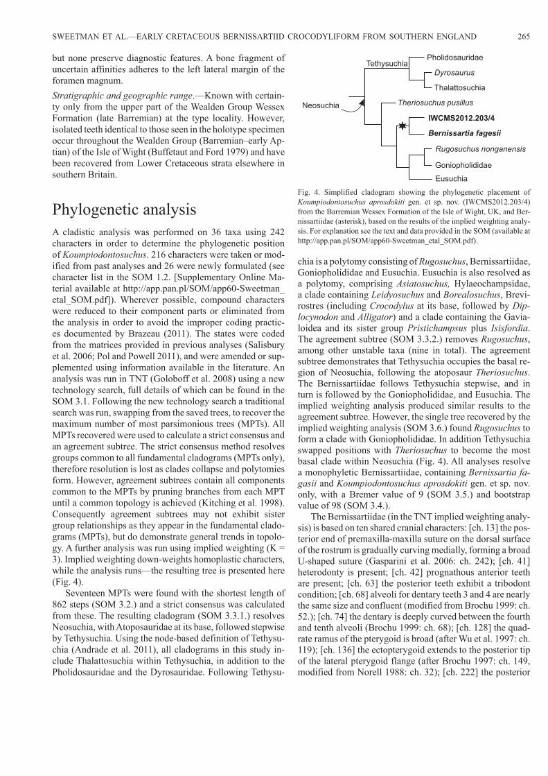
Phylogenetic analysisA cladistic analysis was performed on 36 taxa using 242 characters in order to determine the phylogenetic position of Koumpiodontosuchus. 216 characters were taken or mod-ified from past analyses and 26 were newly formulated (see character list in the SOM 1.2. [Supplementary Online Ma-terial available at http://app.pan.pl/SOM/app60-Sweetman_etal_SOM.pdf]). Wherever possible, compound characters were reduced to their component parts or eliminated from the analysis in order to avoid the improper coding practic-es documented by Brazeau (2011). The states were coded from the matrices provided in previous analyses (Salisbury et al. 2006; Pol and Powell 2011), and were amended or sup-plemented using information available in the literature. An analysis was run in TNT (Goloboff et al. 2008) using a new technology search, full details of which can be found in the SOM 3.1. Following the new technology search a traditional search was run, swapping from the saved trees, to recover the maximum number of most parsimonious trees (MPTs). All MPTs recovered were used to calculate a strict consensus and an agreement subtree. The strict consensus method resolves groups common to all fundamental cladograms (MPTs only), therefore resolution is lost as clades collapse and polytomies form. However, agreement subtrees contain all components common to the MPTs by pruning branches from each MPT until a common topology is achieved (Kitching et al. 1998). Consequently agreement subtrees may not exhibit sister group relationships as they appear in the fundamental clado-grams (MPTs), but do demonstrate general trends in topolo-gy. A further analysis was run using implied weighting (K = 3). Implied weighting down-weights homoplastic characters, while the analysis runs—the resulting tree is presented here (Fig. 4).
Seventeen MPTs were found with the shortest length of 862 steps (SOM 3.2.) and a strict consensus was calculated from these. The resulting cladogram (SOM 3.3.1.) resolves Neosuchia, with Atoposauridae at its base, followed stepwise by Tethysuchia. Using the node-based definition of Tethysu-chia (Andrade et al. 2011), all cladograms in this study in-clude Thalattosuchia within Tethysuchia, in addition to the Pholidosauridae and the Dyrosauridae. Following Tethysu-
chia is a polytomy consisting of Rugosuchus, Bernissartiidae, Goniopholididae and Eusuchia. Eusuchia is also resolved as a polytomy, comprising Asiatosuchus, Hylaeochampsidae, a clade containing Leidyosuchus and Borealosuchus, Brevi-rostres (including Crocodylus at its base, followed by Dip-locynodon and Alligator) and a clade containing the Gavia-loidea and its sister group Pristichampsus plus Isisfordia. The agreement subtree (SOM 3.3.2.) removes Rugosuchus, among other unstable taxa (nine in total). The agreement subtree demonstrates that Tethysuchia occupies the basal re-gion of Neosuchia, following the atoposaur Theriosuchus. The Bernissartiidae follows Tethysuchia stepwise, and in turn is followed by the Goniopholididae, and Eusuchia. The implied weighting analysis produced similar results to the agreement subtree. However, the single tree recovered by the implied weighting analysis (SOM 3.6.) found Rugosuchus to form a clade with Goniopholididae. In addition Tethysuchia swapped positions with Theriosuchus to become the most basal clade within Neosuchia (Fig. 4). All analyses resolve a monophyletic Bernissartiidae, containing Bernissartia fa-gasii and Koumpiodontosuchus aprosdokiti gen. et sp. nov. only, with a Bremer value of 9 (SOM 3.5.) and bootstrap value of 98 (SOM 3.4.).
The Bernissartiidae (in the TNT implied weighting analy-sis) is based on ten shared cranial characters: [ch. 13] the pos-terior end of premaxilla-maxilla suture on the dorsal surface of the rostrum is gradually curving medially, forming a broad U-shaped suture (Gasparini et al. 2006: ch. 242); [ch. 41] heterodonty is present; [ch. 42] prognathous anterior teeth are present; [ch. 63] the posterior teeth exhibit a tribodont condition; [ch. 68] alveoli for dentary teeth 3 and 4 are nearly the same size and confluent (modified from Brochu 1999: ch. 52.); [ch. 74] the dentary is deeply curved between the fourth and tenth alveoli (Brochu 1999: ch. 68); [ch. 128] the quad-rate ramus of the pterygoid is broad (after Wu et al. 1997: ch. 119); [ch. 136] the ectopterygoid extends to the posterior tip of the lateral pterygoid flange (after Brochu 1997: ch. 149, modified from Norell 1988: ch. 32); [ch. 222] the posterior
Fig. 4. Simplified cladogram showing the phylogenetic placement of Koumpiodontosuchus aprosdokiti gen. et sp. nov. (IWCMS2012.203/4) from the Barremian Wessex Formation of the Isle of Wight, UK, and Ber-nissartiidae (asterisk), based on the results of the implied weighting analy-sis. For explanation see the text and data provided in the SOM (available at http://app.pan.pl/SOM/app60-Sweetman_etal_SOM.pdf).
IWCMS2012.203/4
Theriosuchus pusillus
Rugosuchus nonganensis
Pholidosauridae
Goniopholididae
Thalattosuchia
Dyrosaurus
Bernissartia fagesii
Eusuchia
Neosuchia
Tethysuchia

266 ACTA PALAEONTOLOGICA POLONICA 60 (2), 2015
surface of supraoccipital has bilateral posterior prominenc-es (Clark 1994: ch. 64); and [ch. 223] the supraoccipital is exposed on the dorsal surface of the skull roof (Ortega et al. 2000: ch. 62).
This analysis demonstrates that Bernissartiidae is a strongly supported monophyletic clade within Neosuchia, close to Tethysuchia and that it is the sister group to Rugo-suchus plus Goniopholididae and Eusuchia. Complete inclu-sion of the choana in the pterygoids of Koumpiodontosuchus suggests that the condition may have evolved more than once in Neosuchia, and therefore is not an apomorphy of Eusuchia, contrary to the findings of Salisbury et al. (2006). Salisbury et al. (2006) found Isisfordia to be the most basal eusuchian, but this analysis consistently recovers Isisford-ia within Eusuchia as the sister taxon to Pristichampsus or close to the crown. Prior to Salisbury et al.’s (2006) analysis, Brochu (1999) proposed a node-based definition of Eusu-chia that relies on the placement of Hylaeochampsa as the sister taxon to Crocodylia but in all analyses the relationship of Hylaeochampsidae to Crocodylia is unstable and poorly supported, so we cannot concur with Brochu’s (1999) node-based definition. The complete inclusion of the choana in the pterygoids of Koumpiodontosuchus is at odds with the cur-rent definition of Eusuchia, but redefining it requires more in depth analysis beyond the scope of this study.
Discussion and conclusionsAll extant crocodyliforms are placed within Eusuchia and for many years two of the key characters defining this clade were placement of the choana(e) within the pterygoids and the presence of procoelous vertebrae. Furthermore, all non-eu-suchian neosuchian crocodyliforms were assumed to have choanae anteriorly placed and bounded posteriorly by the pterygoids and anteriorly by the palatines. However, recently skull material for the Late Cretaceous, Madagascan mesoeu-crocodylian Mahajangasuchus insignis has become avail-able and uniquely, until now, the internal nares are bounded entirely by the pterygoids in this non-eusuchian taxon (see Turner and Buckley 2008 for a description, phylogenetic analysis and discussion). While placement of the choana in Bernissartia fagesii is difficult to determine owing to the state of preservation of the skull and a covering of lacquer on the lectotype specimen (the paralectotype lacks the skull), Buffetaut (1975) interpreted it as being bounded by both the pterygoids and palatines. This interpretation was followed in a later study (Norell and Clark 1990). Another specimen thought to represent a juvenile of B. fagesii (Buscalioni et al. 1984; Buscalioni and Sanz 1990), but which may represent another species of Bernissartia (Norell and Clark 1990), has also been reported from the Barremian of Spain and in this the choana is unequivocally bounded by both the pterygoids and palatines. While Buffetaut (1975) considered the verte-brae of the lectotype of B. fagessi to be amphicoelous, one of the cervical vertebrae preserved in the Spanish specimen
was described as “incipiently procoelous” by Buscalioni and Sanz (1990). Furthermore, Norell and Clark (1990), while agreeing that nearly all the vertebrae visible in the type Bel-gian material are amphicoelous, including those in the trunk and cervical series, they also report a first caudal that is bi-convex and a second that is procoelous. Small, isolated croc-odylomorph vertebrae are occasionally encountered while surface prospecting the Wessex Formation and are also oc-casionally found among vertebrate material recovered while bulk screening for microvertebrate remains. However, we are unaware of any procoelous crocodylomorph vertebrae of a size that would render them attributable to the new taxon among material in IWCMS collections. Furthermore, SCS has not found any, despite having processed some four tonnes of matrix for microvertebrate remains, although a number of small amphicoelous crocodylomorph vertebrae have been recovered. However, a number of procoelous croc-odylomorph vertebrae too large to be referred to bernissartiid crocodyliforms are known from the Wealden Supergroup of the Weald Sub-basin and from the Wessex Formation of the Isle of Wight. Among the former is an articulated series of 12 from the Valanginian Hastings Group, probably the Ashdown Formation, at Hastings, East Sussex. These comprise the type material of Heterosuchus valdensis Seeley, 1887, now considered to be undiagnostic, so rendering H. valdensis a nomen dubium (Clark and Norell 1992; Salisbury and Naish 2011). On the Isle of Wight two isolated procoelous vertebrae and an agglomeration of postcranial remains including pro-coelous thoracic vertebrae have been recorded from Brook Bay on the south west coast. This locality has also yielded the type material of the eusuchian Hylaeochampsa vectiana Owen, 1874, which comprises the posterior part of a skull.
The discovery of the choana within the pterygoids was unexpected. It also raised questions concerning the system-atic placement of Bernissartiidae and current understanding of characters used to diagnose Eusuchia. Our analysis indi-cates that the Bernissartiidae (now comprising Bernissartia and Koumpiodontosuchus gen. nov.) is a family of advanced neosuchians that can be distinguished from members of Eu-suchia on the basis of (Andrade et al. 2006): pterygoids that completely (or very nearly completely) comprise the poste-rior border of the subtemporal fenestrae. In eusuchians the pterygoid may or may not contact the dorsal margin of the subtemporal fenestra and in taxa where it does its participa-tion always comprises a minor component of the posterior border; the extreme anterior location of the choana within the pterygoids (they are posteriorly placed in eusuchians); the anteroposteriorly elongate choanae in members of Bernis-sartiidae (they are anteroposteriorly short and mediolaterally wide in eusuchians); lack of a bony ridge at the external mar-gin of the choana; lack of an interchoanal septum invariably present in eusuchians; and in choanae that are proportionate-ly large (in area of the palatal opening) compared to those of eusuchians. As also demonstrated by Mahajangasuchus, in isolation, placement of the choana(e) within the pterygoids is not sufficient to include taxa in Eusuchia.

SWEETMAN ET AL.—EARLY CRETACEOUS BERNISSARTIID CROCODYLIFORM FROM SOUTHERN ENGLAND 267
Buffetaut and Ford (1979) discuss the functional and etho-logical significance of the tribodont dentition of bernissartiid crocodyliforms and conclude that it represents adaptation to a durophagus and probably conchifragous diet. The pro-cumbent first dentary teeth of Koumpiodontosuchus appear to support this conclusion being suited to dislodging hard-shelled prey from soft substrates. However, the acuminate, heterodont anterior teeth also form a prehensile apparatus as in other crocodyliforms and it seems likely that both Bernis-sartia and Koumpiodontosuchus while being primarily con-chifragous were also generalists when suitable feeding op-portunities arose. A reconstruction of Koumpiodontosuchus feeding on the largest of the viviparid gastropods occurring in the Wessex Formation, Viviparous cariniferous Sowerby, 1826 (in Sowerby 1826–29), is provided in Fig. 5.
Notes added in proofBefore agreeing upon the name Koumpiodontosuchus apros-dokiti we took advice from a Greek speaker who saw nothing wrong with the binomial chosen. Since then another has ad-vised that koumpi is a contemporary word for button originat-ing from the more formal word komvion (κομβιον). Addition-ally, as the generic name is masculine and the specific name feminine (something not pointed out by our original advisor) “Komviodontosuchus aprosdoketos” would have been more appropriate. However, the name was registered with ZooBank at the time of electronic publication of this article and has been cited by others since then. In view of this we do not feel that emendation of the name is now appropriate.
At the time proofs were being reviewed the first author received news of the death of Zofia Kielan-Jaworowska. Her enormous contribution to palaeontology and to the success of this journal require no introduction. As an undergraduate, as long ago as the 1970s, she was an inspiration and latterly Zofia provided unhesitating and always intellectually stim-ulating support and advice whenever required. She will be sorely missed.
AcknowledgementsWe are very grateful to the collectors for generously donating their finds to IWCMS thereby permitting scientific study of the skull as a whole. Diane Trevarthen (Hayle, Cornwall, UK) found and donated the posterior part and Austin and Finley Nathan (Chelmsford, Essex, UK) found and donated the roastal part. We thank IWCMS for access to the specimen, lab space for its study and particularly Gary Blackwell for his careful preparation work. James M. Clark (George Washington University, Washington D.C., USA) is thanked for his comprehensive review and Michael J. Benton (University of Bristol, UK) is thanked for editorial assistance, both of which facilitated substantial improve-ments to the manuscript. We also thank Mark Witton (University of Portsmouth, UK) for his meticulous reconstruction. UP-S thanks the University of Portsmouth for hosting an internship during which this study was undertaken.
ReferencesAllen, P. and Wimbledon, W.A. 1991. Correlation of NW European Pur-
beck-Wealden (non-marine Lower Cretaceous) as seen from the En-glish type-areas. Cretaceous Research 12: 511–526.
Fig. 5. A reconstruction of Koumpiodontosuchus aprosdokiti gen. et sp. nov. feeding on the viviparid gastropod Viviparous cariniferous Sowerby, 1826 (in Sowerby 1826–29). Smaller gastropods shown include Prophysa sp. Illustration by Mark Witton.

268 ACTA PALAEONTOLOGICA POLONICA 60 (2), 2015
Andrade, M.B. de, Bertini, R.J., and Pinheiro, A.E.P. 2006. Observations on the palate and choanae structures in Mesoeucrocodylia (Archosau-ria, Crocodylomorpha): phylogenetic implications. Revista Brasileira de Paleontologia 9: 323–332.
Andrade, M.B. de, Edmonds, R., Benton, M.J., and Schouten, R. 2011. A new Berriasian species of Goniopholis (Mesoeucrocodylia, Neosu-chia) from England, and a review of the genus. Zoological Journal of the Linnean Society 163: S66–S108.
Batten, D.J. (ed.). 2011. English Wealden Fossils. Palaeontological Asso-ciation Field Guide to Fossils, 14. 769 pp. The Palaeontological As-sociation, London.
Benton, M.J. and Clark, J.M. 1988. Archosaur phylogeny and the relation-ships of the Crocodilia. In: M.J. Benton (ed.), The Phylogeny and Classification of Tetrapods. 1: Amphibians, Reptiles, Birds. Systemat-ics Association, Special Volume 35A: 295–338.
Brazeau, M.D. 2011. Problematic character coding methods in morphol-ogy and their effects. Biological Journal of the Linnean Society 104: 489–498.
Brochu, C.A. 1997 Morphology, fossils, divergence timing, and the phy-logenetic relationships of Gavialis. Systematic Biology 46: 479–522.
Brochu, C.A. 1999. Phylogenetics, taxonomy, and historical biogeography of Alligatoroidea. Journal of Vertebrate Paleontology 19 (Supplement 2): 9–100.
Buffetaut, E. 1975. Sur l’anatomie et la position systematique de Bernis-sartia fagesii Dollo, L., 1883, crocodilien Wealdien de Bernissart, Belque. Bulletin de l’Institute Royal des Sciences Naturelles de Bel-gique, Sciences de la Terre 51 (2): 1–20.
Buffetaut, E. and Ford, R.L.E. 1979. The crocodilian Bernissartia in the Wealden of the Isle of Wight. Palaeontology 22: 905–912.
Buscalioni, A.D. and Sanz, J.L. 1990 The small crocodile Bernissartia fa-gesii from the Lower Cretaceous of Galve (Teruel, Spain). Bulletin de l’Institut Royal des Sciences Naturelles de Belgique, Sciences de la Terre 60: 129–150.
Buscalioni, A.D., Buffetaut, E., and Sanz, J.L. 1984. An immature spec-imen of the crocodilian Bernissartia from the Lower Cretaceous of Galve (Province of Teruel, Spain). Palaeontology 27: 809–813.
Clark, J.M. 1994. Patterns of evolution in Mesozoic Crocodyliformes. In: N.C. Fraser and H.-D. Sues (eds.), In the Shadow of the Dinosaurs, 84–97. Cambridge University Press, New York.
Clark, J.M. and Norell, M.A. 1992. The Early Cretaceous crocodylomorph Hylaeochampsa vectiana from the Wealden of the Isle of Wight. Amer-ican Museum Novitates 3032: 1–19.
Dollo, L. 1883. Premiere note sur les crocodiliens de Bernissart. Bulletin de l’Institut Royal des Sciences Naturelles de Belgique 2: 309–338.
Gasparini, Z., Pol. D., and Spalletti, L.A. 2006. An unusual marine croco-dyliform from the Jurassic–Cretaceous boundary of Patagonia. Science 311: 70–73.
Goloboff, P.A., Farris, J.S. and Nixon, K.C. 2008. TNT, a free program for phylogenetic analysis. Cladistics 24: 774–786.
Hay, O.P. 1930. Second Bibliography and Catalogue of the Fossil Vertebrata of North America. xiv + 1074 pp. Carnegie Institute, Washington DC.
Hughes, N.F. and McDougall, A.B. 1990. New Wealden correlation for the Wessex Basin. Proceedings of the Geologists’ Association 100: 85–90.
Joffe, J. 1967. The “dwarf” crocodiles of the Purbeck Formation, Dorset: a reappraisal. Palaeontology 10: 629–639.
Kerth, M. and Hailwood, E.A. 1988. Magnetostratigraphy of the Lower Cre-taceous Vectis Formation (Wealden Group) on the Isle of Wight, south-ern England. Journal of the Geological Society, London 145: 351–360.
Kitching, I.J., Forey, P.L., Humphries, C.J., and Williams, D.M. 1998. Cla-distics: The Theory and Practice of Parsimony Analysis. Second edition. 228 pp. Oxford University Press, Oxford.
Norell, M.A. 1988. Cladistic Approaches to Evolution and Paleobiolo-gy as Applied to the Phylogeny of Alligatorids. 544 pp. Unpublished Ph.D. Thesis, Yale University, New Haven.
Norell, M.A. and Clark, J.M. 1990. A reanalysis of Bernissartia fagesii, with comments on its phylogenetic position and its bearing on the origin and diagnosis of the Eusuchia. Bulletin de l’Institut Royal des Sciences Naturelles de Belgique, Sciences de la Terre 60: 115–128.
Ortega, F., Gasparina, Z., Buscalioni, A., and Calvo, J.O. 2000. A new spe-cies of Araripesuchus (Crocodylomorpha, Mesoeucrocodylia) from the Lower Cretaceous of Patagonia (Argentina). Journal of Vertebrate Paleontology 20: 57–76.
Owen, R. 1874. Monograph of the fossil Reptilia of the Wealden and Pur-beck formations. Supplement No. IV (Hylaeochampsa). Monograph of the Palaeontolographical Society 27 (No. 125 for 1873): 1–7.
Pol, D. and Powell, J.E. 2011. A new sebecid mesoeucrocodylian from the Rio Loro Formation (Palaeocene) of north-western Argentina. Zoolog-ical Journal of the Linnean Society 163: S7–S36.
Radley, J.D. 1994. Stratigraphy, palaeontology and palaeoenvironment of the Wessex Formation (Wealden Group, Lower Cretaceous) at Yaver-land, Isle of Wight, southern England. Proceedings of the Geologists’ Association 105: 199–208.
Robinson, S.A. and Hesselbo, S.P. 2004. Fossil-wood carbon-isotope strati graphy of the non-marine Wealden Group (Lower Cretaceous, southern England). Journal of the Geological Society of London 161: 133–145.
Salisbury, S.W., Molnar, R.E., Frey, E., and Willis, P.M.A. 2006. The ori-gin of modern crocodyliforms: new evidence from the Cretaceous of Australia. Proceedings of the Royal Society B 273: 2439–2448.
Salisbury, S.W. and Naish, D. 2011. Crocodilians. In: D.J. Batten (ed.), English Wealden Fossils. Palaeontological Association Field Guide to Fossils, 14, 305–369. The Palaeontological Association, London.
Schwarz-Wings, D., Rees, J., and Lindgren, J. 2009. Lower Cretaceous mesoeucrocodylians from Scandinavia (Denmark and Sweden). Cre-taceous Research 30: 1345–1355.
Seeley, H.G. 1887. On Heterosuchus valdensis, Seeley, a procoelian croc-odile from from the Hastings Sand of Hastings. Quarterly Journal of the Geological Society 43: 212–215.
Sereno, P.C., Larsson, H.C.E., Sidor, C.A., and Gado, B. 2001. The giant crocodyliform Sarcosuchus from the Cretaceous of Africa. Science 294: 1516–1519.
Sowerby, J. de C. 1826–29. The Mineral Conchology of Great Britain; or Coloured Figures and Descriptions of Those Remains of Testaceous Animals or Shells, Which Have Been Preserved at Various Times and Depths in the Earth. Volume 6. 230 pp. Richard Taylor, London (series begin by father, J. Sowerby).
Sweetman, S.C. 2011a. The Wealden of the Isle of Wight. In: D.J. Bat-ten (ed.), English Wealden Fossils. Palaeontological Association Field Guide to Fossils, 14, 52–77. The Palaeontological Association, London.
Sweetman, S.C. 2011b. Vertebrate microfossils. In: D.J. Batten (ed.), English Wealden Fossils. Palaeontological Association Field Guide to Fossils, 14, 192–204. The Palaeontological Association, London.
Sweetman, S.C. and Insole, A.H. 2010.The plant debris beds of the Early Cretaceous (Barremian) Wessex Formation of the Isle of Wight, south-ern England: their genesis and palaeontological significance. Palaeo-geography, Palaeoclimatology, Palaeoecology 292: 409–424.
Turner, A.H. and Buckley, G.A. 2008. Mahajangasuchus insignis (Crocodyli-formes: Mesoeucrocodylia) cranial anatomy and new data on the origin of the eusuchian-style palate. Journal of Vertebrate Paleontology 28: 382–408.
Whetstone, K.N. and Whybrow, P.J. 1983. A “cursorial” crocodilian from the Triassic of Lesotho (Basutoland), southern Africa. Occasional Papers of the Museum of Natural History, University of Kansas 106: 1–37.
Wu, X.C., Sues, H.D., and Dong, Z.M. 1997. Sichuanosuchus shuhanen-sis, a new? Early Cretaceous protosuchian (Archosauria: Crocodyl-iformes) from Sichuan (China), and the monophyly of Protosuchia. Journal of Vertebrate Paleontology 17: 89–103.