editorialjmaj, march 2006 — vol. 49, no. 3 93 editorial percutaneous endoscopic gastrostomy (peg),...
TRANSCRIPT

Vol.49, No.3 March 2006
Editorial
Is Percutaneous Endoscopic Gastrostomy Really a Safe Procedurefor High Aged Patients?Kazuichi Okazaki ................................................................................................................................................... 93
Original Articles
A Survey of Percutaneous Endoscopic Gastrostomy in 202 JapaneseMedical InstitutionsYutaka Suzuki, Mitsuyoshi Urashima, Hideharu Ninomiya, Michio Sowa,Yoshiki Hiki, Hiroaki Suzuki, Yoshio Ishibashi, Toshirou Kura,Naruo Kawasaki, Katsuhiko Yanaga .................................................................................................................. 94
How Elderly People Die of Nonmalignant Pulmonary Disease at HomeYoshihisa Hirakawa, Yuichiro Masuda, Masafumi Kuzuya, Akihisa Iguchi,Kazumasa Uemura ................................................................................................................................................ 106
Review Articles
The History and the Present of Minamata Disease—Entering the second half a century—Noriyuki Hachiya ................................................................................................................................................... 112
Nonvalvular Atrial Fibrillation and StrokeShinichi Takahashi ................................................................................................................................................. 119
Short Communication
Professional AutonomyNobuya Hashimoto ................................................................................................................................................ 125
Case Report
Hemosuccus Pancreaticus: Clearly identified by timely duodenoscopy,multiplanar volume reformation of CT image and celiac angiographyShotaro Enomoto, Naohisa Yahagi, Mitsuhiro Fujishiro, Masashi Oka,Naomi Kakushima, Mikitaka Iguchi, Hiroyuki Isayama, Kimihiko Yanaoka,Kenji Arii, Hideyuki Tamai, Yasuhito Shimizu, Masao Ichinose, Masao Omata ........................................ 128
Current Activities of JMA
Measures Against Lifestyle Related DiseasesTakashi Tsuchiya .................................................................................................................................................... 132
Clinical Topics in Japan
Recent Topics in Myasthenia GravisTetsuro Konishi ...................................................................................................................................................... 135

93JMAJ, March 2006 — Vol. 49, No. 3
Editorial
Percutaneous endoscopic gastrostomy (PEG),which was first performed in 12 children and19 adults by Gauderer,1 is nowadays widely usedfor providing enteral nutrition to patients whocannot take meals orally. The procedure isbelieved to be particularly useful in high riskpatients because general anesthesia is not usuallyrequired. PEG is mainly performed on high-agedpatients rather than on children. Complicationsare rare (6.6% major, 6.6% minor); PEG-relatedmortality and morbidity are 1–2% and 3–12%,2,3
respectively, and are not influenced by patients’age.4 However, little has been known about theincidence of complications and the outcomes oflong-term PEG in Japan.
In this issue, Suzuki et al. report surveillancedata on PEG in Japan they collected by asking760 hospitals nationwide to complete question-naires. Their results indicate that major compli-cations such as death and peritonitis occurredin 24.7% of hospitals. Minor complications includ-ing wound infections (25.5%) and diarrhea(20.6%) occurred more frequently than pneumo-nia (12.3%), vomiting (9.9%), dermatitis (9.0%),granulation (8.8%), accidental self-exertion(7.0%), or constipation (4.5%). Approximately25% of hospitals where more than one erroneousinsertion occurred had higher 30-day mortalityrates. The rate of negative outcomes seems to behigher than anticipated, with an unexpectedly
low 14.3% of PEG patients unable to be dis-charged from hospital. In Japan, people aged 65years or older comprised 20% of the populationin 2005 and their number is rapidly increasing.This age group is predicted to comprise 25% ofJapan’s population in 2015, which indicates thata super-senile era is coming soon and with ita rapid increase in the number of high-agedpatients requiring PEG.
Finally, as the number of lawsuits associatedwith medical trouble is increasing in Japan, doc-tors should take care when performing PEG toreceive informed consent, ensuring that patientsare aware of the possible negative outcomes.
References
1. Gauderer MW, Ponsky JL, Izant RJ Jr. Gastrostomy withoutlaparotomy: a percutaneous endoscopic technique. J PediatrSurg. 1980;15(6):872–875.
2. Larson DE, Burton DD, Schroeder KW, DiMagno EP. Percutane-ous endoscopic gastrostomy. Indications, success, complications,and mortality in 314 consecutive patients. Gastroenterology.1987;93:48–52
3. Hull MA, Rawlings J, Murray FE, et al. Audit of outcome of long-term enteral nutrition by percutaneous endoscopic gastrostomy.Lancet. 1993;341:869–872
4. Shellito PC, Malt RA. Tube gastrostomy. Techniques and com-plications. Ann Surg. 1985;201(2):180–185.
Is Percutaneous Endoscopic GastrostomyReally a Safe Procedure for High Aged Patients?
Kazuichi Okazaki*1
*1 The Third Department of Internal Medicine, Division of Gastroen-terology and Hepatology, Kansai Medical University, OsakaCorrespondence to: Kazuichi Okazaki MD, PhD, Division ofGastroenterology and Hepatology, Kansai Medical UniversityHirakata Hospital, 2-3-1 Shinmachi, Hirakata, 573-1191 Osaka,Japan. Tel: 81-72-804-0101, Fax: 81-72-804-2061,E-mail: [email protected]

94 JMAJ, March 2006 — Vol. 49, No. 3
*1 Department of Surgery, Jikei University School of Medicine, Tokyo; *2 Division of Clinical Research and Development, Jikei University Schoolof Medicine, Tokyo; *3 PEG doctor’s network, Tokyo; *4 Working Group of Home Health Care, Endoscopic Therapy and Quality of Life, Japan;*5 Naganuma Municipal Hospital, HokkaidoCorrespondence to: Mitsuyoshi Urashima MD, PhD, MPH, Division of Clinical Research & Development, The Jikei University School of Medicine,3-25-8 Nishi-shimbashi, Minato-ku, Tokyo 105-8461, Japan. Tel: 81-3-3433-1111, Fax: 81-3-5400-1250, E-mail: [email protected]
Original Article
Introduction
For patients with a functioning gastrointestinaltract but the inability to take food by mouth,early enteral nutrition reduces morbidity andmortality of several conditions compared withtotal parenteral nutrition.1–5 Nasogastric tubesare simple enough to insert but are often intoler-able for the patients. Moreover, these tubes aredifficult to maintain in position and are associ-
ated with a significant risk of aspiration.6 Naso-jejunal tubes are more tolerable to patients, butthey are easily blocked and also difficult keep inposition.7 Therefore, gastrostomy or occasionallyjejunostomy may be used for patients who can-not take food orally. Although gastrostomy orjejunostomy may be easily performed with opensurgery under general anesthesia, the risks ofsurgery may outweigh the benefits, because mostpatients with indications for gastrostomy areolder, malnourished, and have other morbidities.8
A Survey of Percutaneous EndoscopicGastrostomy in 202 Japanese MedicalInstitutions
JMAJ 49(3): 94–105, 2006
Yutaka Suzuki,*1,3 Mitsuyoshi Urashima,*2,3 Hideharu Ninomiya,*3 Michio Sowa,*3,4
Yoshiki Hiki,*3,4 Hiroaki Suzuki,*3,4 Yoshio Ishibashi,*1 Toshirou Kura,*3,5
Naruo Kawasaki,*1 Katsuhiko Yanaga*1
AbstractBackground Percutaneous endoscopic gastrostomy (PEG) is widely used for enteral nutrition of patientsunable to take food by mouth. In this study, we surveyed usefulness of PEG and determined the approximaterates of complications associated with PEG replacement in Japan.
Methods Questionnaires were sent to 760 hospitals; 202 hospitals returned their questionnaires (27% responserate).
Results 5,291 patients underwent PEG in 2004 among these Japanese hospitals. PEG was mainly performedin general hospitals by physicians and surgeons. Most patients were elderly (mean age: 79.8�33.4 SD years old)with cerebral infarction/hemorrhage and dementia and required nutritional support. Major complications such asdeath and peritonitis were experienced by 24.7% of hospitals. Approximately 25% of hospitals experienced morethan one erroneous insertion of the tube into the extra-gastrointestinal tract. Hospitals that experienced erroneousinsertions had higher 30-day mortality rates, whether or not mortality was associated with PEG placement. Onthe other hand, the erroneous insertion rates as well as 30-day mortality were inversely associated with numberof PEG procedures performed during year 2004 at each hospital.
Conclusion These surveillance data imply that a lower patient volume treated with PEG at an institute may beassociated with negative outcomes, although a longitudinal study is necessary to confirm this conclusion.
Kew words Quality of life, Enteral nutrition, Patient care, Morbidity and mortality, Home care
95JMAJ, March 2006 — Vol. 49, No. 3
Percutaneous endoscopic gastrostomy (PEG)as well as percutaneous endoscopic jejunostomy/percutaneous endoscopic duodenostomy are fast,safe, and effective methods for long-term enteraltube feeding as long as no contraindicationsto enteral feeding exist.9–13 Moreover, there maybe less reflux and food aspiration with thesemethods.14 Therefore, PEG is now the preferredtreatment for patients with dysphagia, and its usehas been growing in the United States and theUnited Kingdom.15
Although the primary indication for PEG isthe inability to take food by mouth, indicationsvary widely depending on the physician’s policy.Although PEG insertion and tube exchanges arenot complicated, they can lead to serious andpotentially lethal complications. However, suchnegative findings seem to be outweighed by thepositive results16–20 at least in Japan. We surveyedPEG usage in Japan by sending questionnaires to760 medical institutions and received answersfrom 202 (27% response rate). The results aresummarized in this paper.
Methods
SurveillanceWe selected the 760 hospitals where one or moredoctors are involved in performing PEG forpatients and participate in the PEG doctor’snetwork in Japan (http://www.peg.ne.jp/news/index.html). We sent questionnaires to these 760hospitals and received answers to the question-naires from 202 (27% response rate). A copy of
the 83 questions on the questionnaire appears inAppendix I.
Statistical analysisFactors associated with erroneous insertion ofthe tube into the extra-gastrointestinal tractwere evaluated using either Chi-square test orStudent’s t-test. Kruskal-Wallis equality of popu-lations rank tests adjusted for trend tests, asdescribed by Cuzick,21 were used to determineassociations among three and more groups. Allstatistical analyses were performed using STATA8.0 (STATA Corporation, College Station, TX).
Results
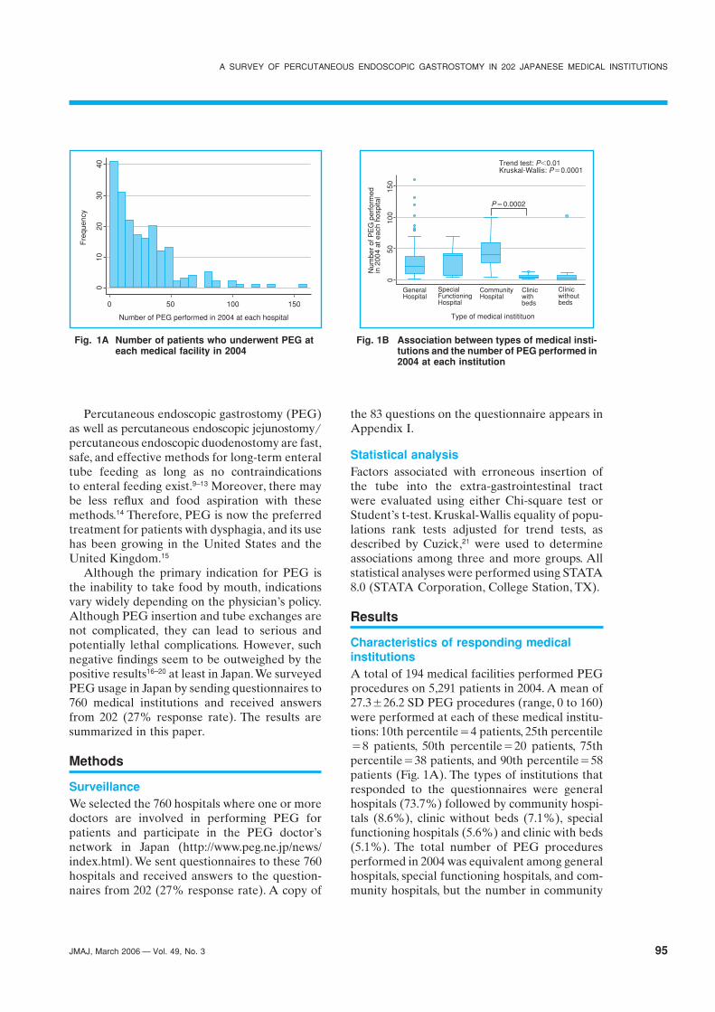
Characteristics of responding medicalinstitutionsA total of 194 medical facilities performed PEGprocedures on 5,291 patients in 2004. A mean of27.3�26.2 SD PEG procedures (range, 0 to 160)were performed at each of these medical institu-tions: 10th percentile�4 patients, 25th percentile�8 patients, 50th percentile�20 patients, 75thpercentile�38 patients, and 90th percentile�58patients (Fig. 1A). The types of institutions thatresponded to the questionnaires were generalhospitals (73.7%) followed by community hospi-tals (8.6%), clinic without beds (7.1%), specialfunctioning hospitals (5.6%) and clinic with beds(5.1%). The total number of PEG proceduresperformed in 2004 was equivalent among generalhospitals, special functioning hospitals, and com-munity hospitals, but the number in community
Fig. 1A Number of patients who underwent PEG ateach medical facility in 2004
Fig. 1B Association between types of medical insti-tutions and the number of PEG performed in2004 at each institution
0
010
2030
Fre
quen
cy
40
50 100 150
Number of PEG performed in 2004 at each hospital
050
100
150
GeneralHospital
Type of medical institituon
Num
ber
of P
EG
per
form
edin
200
4 at
eac
h ho
spita
l
SpecialFunctioningHospital
CommunityHospital
Clinicwithbeds
Trend test: P�0.01Kruskal-Wallis: P�0.0001
P�0.0002
Clinicwithoutbeds
A SURVEY OF PERCUTANEOUS ENDOSCOPIC GASTROSTOMY IN 202 JAPANESE MEDICAL INSTITUTIONS

96 JMAJ, March 2006 — Vol. 49, No. 3
hospitals was greater than in clinic with beds(Fig. 1B). Hospitals with more than 100 bedswere most common in this survey: more than 200beds: 40.7%; 100–200 beds: 25.6%; 20–99 beds:22.1%; less than 19 beds: 5.0%; no beds: 6.5%.The number of PEG procedures per institutionincreased depending on the number of beds(Fig. 1C). The number of hospitals adopting PEGhas grown steadily: PEG started more than 10years ago: 19.8%; 5–10 years ago: 41.7%; 2–5years ago: 22.9%; 1–2 years ago: 15.6%. In particu-lar, medical institutes that began performing PEGprocedures more than 5 years ago treated morepatients than hospitals that began performingthese procedures less than 5 years ago (Fig. 1D).PEG was mainly performed by physicians(44.7%) or surgeons (39.3%); few endoscopists(9.1%) reported performing this surgery. Fewerthan three physicians at each institution per-formed PEG procedures among 73.1% of sur-veyed hospitals. As the number of physicianswho performed PEG at each institute increased,the number of procedures per institution increasedas well (data not shown).
Characteristics of patients who underwentPEGAs stated, 5,291 patients underwent PEG in 2004.The ratio between men and women was 13:14.The mean age was 79.8�33.4 SD years, and mostpatients (95.1%) were aged 60 to 100 years.Enteral nutrition was the primary reason (98%)for undergoing PEG. Most of the underlyingdiseases were neurological dysfunction as fol-
lows: cerebral infarction: 46.2%; hemorrhage:18.0%; dementia: 12.3%; Alzheimer disease:4.0%; trauma: 2.4%; cancer: 3.8%. The underly-ing clinical condition of patients who underwentPEG was dysphagia in about 50% of patients;repeated aspiration pneumonia (19.9%) andreduced oral intake (18.9%) were the next mostcommon conditions.
Methods of performing PEG and status ofteam supportPEG was typically performed in the endoscopyroom (71.9%) and also in the operation room(12.4%) as well as in clinical wards (3.8%), eitherwith conscious sedation (55.8%) or local anes-thesia of the pharynx (21.6%), with (52.4%) orwithout (47.6%) the use of an anticholinergicagent, and using the pull method (69.7%). How-ever, in some medical institutions, the procedurevaried.
Approximately 25% of medical institutionsperformed a throat culture before the procedure.However, most institutions (91.9%) ignored thepossible existence of pathogenic microorganismsin patients’ throats and inserted the tube via theoral route. Bumper (75.1%) and balloon (17.6%)procedures were most common during the initialtube insertion, although neither way was fixed inother hospitals. Of the type of catheter to fix tothe skin at initial construction, the tube type waspreferred (66.8%) followed by the button type(18.7%). Whether or not the gastric wall wasfixed to the abdominal wall varied by institution.Most institutions did not use fluoroscopy to
Fig. 1C Association between the number of beds perinstitute and the number of PEG performedin 2004 at each institution
Fig. 1D Association between the timing when theinstitution enrolled PEG and the number ofPEG performed in 2004 at each institution
050
100
150
0
Number of beds per institute
Num
ber
of P
EG
per
form
edin
200
4 at
eac
h ho
spita
l
1–19 20–99 100–199
Trend test: P�0.01Kruskal-Wallis: P�0.0001
200–
050
100
150
1–2 2–5 5–10 10�
Time when the institution enrolled PEG(number of years ago)
Num
ber
of P
EG
per
form
edin
200
4 at
eac
h ho
spita
l
Trend test: P�0.01Kruskal-Wallis: P�0.0001
P�0.0004
Suzuki Y, Urashima M, Ninomiya H, et al.

97JMAJ, March 2006 — Vol. 49, No. 3
confirm the status of the PEG tube (80.3%).During the exchange of the catheter, bumper(49.8%) and tube methods (26.3%) were mostcommon. The duration between the initial tubeinsertion and the first exchange of the tubewas significantly shorter with the balloon proce-dure (mean 2.22�1.84 SD months) than with thebumper procedure (mean 5.23�2.22 SD months)(Student’s t-test: P�0.0001). Approximately 25%of institutions performed the first tube exchangeunder endoscopy, whereas more than 50% per-formed it manually. Institutes varied in themethod in which they confirmed the status of thePEG tube at the first exchange, although anendoscopic procedure was used most frequently:endoscope: 32.5%; fluoroscopy: 19.5%; injectingair through PEG: 6.5%; checking influx of gastricacid: 20.0%. However, at the second exchange,fluoroscopy was used by most institutions(73.2%) to confirm the status of the PEG tube,whereas injecting air through PEG was the next(14.9%) and endoscopy was applied least(11.9%). Most second exchanges took place amean of 5.23�2.22 SD months after the firstexchange. Approximately one-third of patientsexchanged the PEG tube at home.
A preoperative conference for all patientswho needed PEG was held by 16.5% of medicalfacilities. More than half (67.8%) the hospitals
had no nutrition support team, whereas morethan half (53.1%) had a clinical path designed forpatients who were undergoing PEG.
Feasibility of performing PEG and frequencyof erroneous insertion of the tube intothe extra-gastrointestinal tractNearly half of the medical institutes (47%) hadsome patients in whom it was not feasible toinsert the PEG tube. When the analysis wasrestricted to hospitals that had at least onepatient in whom PEG tube insertion was notfeasible, the natural logarithm of unfeasible PEGpatients divided by the number of PEG pro-cedures performed at each hospital in 2004decreased by an increment of the number of
Fig. 2A Association between the feasibility of PEG andthe number of PEG performed in 2004 at eachhospital
The difference and trend among quantile of number of PEGperformed in 2004 at each hospital were evaluated withKruskal-Wallis test and trend test, respectively. Differencebetween two groups was calculated by Student’s t test.Cutoff of P-value for statistical difference was set at 0.0167.
Fig. 2B Association between feasibility of PEG andnumber of beds at each hospital
Fig. 2C Association between feasibility of PEG andnumber of doctors who can construct PEGin the hospital
�6
�4
�2
02
0–7 8–20 21–37 38–160
Number of PEG performed in 2004 at each hospital
Trend test: P�0.01Kruskal-Wallis: P�0.0001
P�0.0015
P�0.0077
P�0.0032
Ln
Num
ber
of u
nfea
sibl
e P
EG
in 2
004
at e
ach
hosp
ital
Num
ber
of P
EG
per
form
edin
200
4 at
eac
h ho
spita
l
�6
�4
�2
02
0 0–19 22–99 100–199 200�
Number of beds at each hospital
Trend test: P�0.01Kruskal-Wallis: P�0.0097
Ln
Num
ber
of u
nfea
sibl
e P
EG
in 2
004
at e
ach
hosp
ital
Num
ber
of P
EG
per
form
edin
200
4 at
eac
h ho
spita
l
�6
�4
�2
02
0 1 2 3 4 5 6 7 8 11
Number of doctors who can perform PEG in the hospital
Trend test: P�0.01Kruskal-Wallis: P�0.021
Ln
Num
ber
of u
nfea
sibl
e P
EG
in 2
004
at e
ach
hosp
ital
Num
ber
of P
EG
per
form
edin
200
4 at
eac
h ho
spita
l
A SURVEY OF PERCUTANEOUS ENDOSCOPIC GASTROSTOMY IN 202 JAPANESE MEDICAL INSTITUTIONS

98 JMAJ, March 2006 — Vol. 49, No. 3
PEG procedures performed at each hospital in2004 (Kruskal-Wallis: P�0.0001: trend test:P�0.01) (Fig. 2A). Similarly, when the number ofbeds or the number of doctors at each instituteincreased, the ratio of unfeasible PEG proce-dures tended to decrease (Fig. 2B and Fig. 2C).
During tube exchange, 23.8% of hospitalsexperienced erroneous insertion of the tube intothe extra-gastrointestinal tract. The number oferroneous insertions ranged from one to five.When the analysis was restricted to hospitals thatexperienced at least one erroneous insertion, thenatural logarithm of the number of erroneousinsertions of PEG tubes divided by the numberof PEG procedures performed at each hospital in2004 decreased by an increment of the number ofPEG procedures performed at each hospital in2004 (Kruskal-Wallis: P�0.0001: trend test:P�0.01) (Fig. 3A). Similarly, when the numberof beds or number of doctors at each instituteincreased, the ratio of erroneous PEG insertionstended to decrease (Fig. 3B and Fig. 3C).
Factors associated with erroneous insertion oftubes into the extra-gastrointestinal tract areshown in Table 1. Institutes with no experiencewith erroneous insertions tended to use balloonand button procedures rather than bumper andtube procedures at exchange. When the PEGwas exchanged under endoscopy, the risk of erro-neous insertion decreased. Changing the tube athome did not significantly increase the risk of anerroneous insertion.
Incidence of major and minor complicationsDeath as an adverse event was experienced by40.7% of the institutes after adopting PEG; thenumber of deaths ranged from 0 to 19 witha mean of 0.94�1.78 SD per hospital. Twoinstitutes indicated that death was caused by thePEG procedure in 3 patients. The percentage ofpatients who died within 1 month and 6 monthswas 5.0% and 12.6%, respectively, althoughsurvival data for 20.1% of patients were not dis-closed. Institutes that experienced erroneouslyinserted tubes into the extra-gastrointestinaltract also tended to have significantly higher risksof death within 1 month, whether or not it wasdue to a complication that occurred during the
Fig. 3A Association between feasibility of PEG andnumber of PEG performed in 2004 at eachhospital
Fig. 3B Association between feasibility of PEG andnumber of beds at each hospital
Fig. 3C Association between feasibility of PEG andnumber of doctors who can construct PEGin the hospital
�5
�4
�3
�2
�1
0
0–7 8–20 21–37 38–160
Number of PEG performed in 2004 at each hospital
Trend test: P�0.01Kruskal-Wallis: P�0.0001
P�0.0001
P�0.0002
P�0.0038
Ln
Num
ber
of e
rron
eous
inse
rtio
ns o
f PE
Gin
200
4 at
eac
h ho
spita
l
Num
ber
of P
EG
per
form
edin
200
4 at
eac
h ho
spita
l
�5
�4
�3
�2
�1
0
0 0–19 22–99 100–199 200�
Number of beds at each hospital
Trend test: P�0.01Kruskal-Wallis: P�0.0041
P�0.012
Ln
Num
ber
of e
rron
eous
inse
rtio
ns o
f PE
Gin
200
4 at
eac
h ho
spita
l
Num
ber
of P
EG
per
form
edin
200
4 at
eac
h ho
spita
l
�5
�4
�3
�2
�1
0
0 1 2 3 4 5 6 7 8 11
Number of doctors who can construct PEG in the hospital
Trend test: P�0.02Kruskal-Wallis: Not significant
Ln
Num
ber
of e
rron
eous
inse
rtio
ns o
f PE
Gin
200
4 at
eac
h ho
spita
l
Num
ber
of P
EG
per
form
edin
200
4 at
eac
h ho
spita
l
Suzuki Y, Urashima M, Ninomiya H, et al.

99JMAJ, March 2006 — Vol. 49, No. 3
Table 1 Factors associated with erroneous insertion of the tube into the extra-gastrointestinal tract
No experience More than oneQuestionnaire of error experience of error P value*
n�47 (%) n�155 (%)
Which method do you use for stomach internalfixation at exchange?
Balloon 43 (28) 10 (15) 0.037Bumper 78 (51) 12 (47)Balloon or bumper 33 (21) 24 (38)
Which method do you use for outside the bodyshape at exchange?
Button 78 (51) 14 (30) 0.006Tube 35 (23) 9 (19)Button or tube 41 (27) 24 (51)
Please select the method used at the first exchange
Exchange under endoscopic observation 44 (29) 7 (15) 0.045Manual exchange 89 (58) 28 (60)Either way 20 (13) 12 (26)
What kind of confirmation method did you useat the first exchange?
Endoscope 52 (34) 13 (28) NSFluoroscopy 28 (18) 11 (23)Injecting air through PEG 11 (7) 2 (4)Check influx of gastric acid 30 (20) 10 (21)Varied by situation 32 (21) 11 (23)
Were patients allowed to change the PEG tube at home?
Yes 49 (32) 21 (46) NSNo 106 (68) 25 (54)
*: Calculated by chi-square test. When the P-value was less than 0.05, the difference was considered statistical significant.
Table 2 Outcomes associated with erroneous insertion of the tube into the extra-gastrointestinal tract
No experience More than oneQuestionnaire Total of error experience of error P value
n�202 (%) n�155 (%) n�47 (%)
Did you experience any deaths within 1 monthafter PEG insertion?
Yes 77 (38) 50 (34) 27 (63)No 125 (62) 96 (66) 16 (37) 0.001*1
How many in 2004? (Mean�SD) 0.94�1.78 0.83�1.86 1.33�1.43 NS*2
Did you experience any deaths during tubeexchange?
Yes 2 (1) 1 (1) 1 (2)No 202 (99) 154 (99) 46 (98) NS*1
How many in 2004? (Mean�SD) 0.015�0.157 0.01�0.16 0.02�0.15 NS*2
Did you experience major complications afterPEG insertion?
Yes 46 (25) 32 (23) 14 (32)No 140 (75) 110 (77) 30 (68) NS*1
Did you experience any deaths related to a majorcomplication within 1 month after PEG insertion?
Yes 17 (9) 8 (6) 9 (20) 0.003*1
No 173 (91) 137 (94) 36 (80) 0.0111*2
How many in 2004? (Mean�SD) 0.13�0.45 0.08�0.36 0.28�0.63
*1: Calculated by chi-square test; *2: Calculated by Student’s t test
A SURVEY OF PERCUTANEOUS ENDOSCOPIC GASTROSTOMY IN 202 JAPANESE MEDICAL INSTITUTIONS

100 JMAJ, March 2006 — Vol. 49, No. 3
PEG procedure (Table 2). The 30-day mortalityrate in each institute was inversely associatedwith the number of PEG procedures performedat each hospital in 2004 (Fig. 4).
Major complications such as death and perito-nitis associated with the procedures of PEG inser-tion or replacement were experienced by 24.7%of hospitals during 2004. Minor complications,including wound infection (25.5%) and diarrhea(20.6%), occurred more frequently than pneumo-nia (12.3%), vomiting (9.9%), dermatitis (9.0%),granulation (8.8%), accidental self-exertion(7.0%), constipation (4.5%), and others amongall patients treated with PEG in year 2004. Theinfection rate (number of infections/number ofPEG procedures) was not associated with theusage patterns of antibiotics or the type of deviceused (data not shown).
After discharge from the medical institutionwhere PEG was performed.
After discharge from the medical institutewhere PEG was performed, only 14.3% of patientswent home. Most patients were transferred toother hospitals.
Issue of insuranceWhen asked “In your institute, are costs forgastric catheter exchange covered under specialinsurance medical care material costs?” 12.6%answered “yes”. When asked “In medical facili-ties using diagnosis related grouping and institu-tions using insurance for the elderly, the costsassociated with gastric catheter exchange cannotbe separately claimed under insurance as a spe-
cial healthcare material cost. Do you think that itshould be paid separately as a special healthcarematerial cost?” most hospitals (96.9%) answered“yes”. To the question “Do you file an insuranceclaim when you use enteral nutrition via PEG?”82.5% answered “yes” and 3.2% answered “yes,but we are limited in the maximum charge forthe claim”. To the question “Excluding ‘Elental’,‘Elental P’, ‘Enterude’, ‘Twinline’ and other medi-cal nutrients of half digested state, do you thinkset dietary food for enteral nutrition should be afee-for-service?” 79.1% of hospitals answered“yes”.
Opinions of doctors who perform PEGWhen asked “Do you think that PEG is thebest way to provide nutritional support?” 33.3%answered “yes, it is the best way”, 57.6% answered“yes, it is a good way”, and the remainder answered“No, it is not the best way”. When asked “PEGenables patients to be themselves, that is experi-ence a ‘recovery of everydayness’, and allowscare to be provided at home. Do you agree withthis opinion?” 95% of doctors agreed.
Discussion
We selected 760 hospitals where one or moredoctors are involved in performing PEG forpatients and participate in the PEG doctor’snetwork in Japan. We sent questionnaires tothese 760 hospitals and received answers to thequestionnaires from 202 (27% response rate).In these 202 institutions, totally 5,291 patientsunderwent PEG in 2004. Data indicate that PEGwas mainly performed in general hospitals byphysicians and surgeons on older patients withcerebral infarction/hemorrhage and dementia.Procedures were performed primarily in theendoscopy room under conscious sedation oranesthesia of the pharynx. These trends weresimilar to those in other countries.22,23
The pull and push method were preferred in70% and 9% of surveyed institutions, respec-tively, which may reflect the evidence that percu-taneous placement of a pull-type gastrostomytube was performed with a minimum risk of tractdisruption and peritonitis.24–27 In contrast, onestudy showed that serious complications leadingto laparotomy, wound infection, or intraperito-neal abscess developed in 17 patients (13%), inall of whom the introducer (ie, push) technique
Fig. 4 Association between 30-day mortality after PEGand number of PEG performed in 2004 at eachhospital
�6
�4
�2
02
0–7 8–20 21–37 38–160
Number of PEG performed in 2004 at each hospital
Trend test: P�0.01Kruskal-Wallis: P�0.0001
P�0.0001
P�0.0001
P�0.0001
Ln
Num
ber
of 3
0-da
y de
ath
in 2
004
at e
ach
hosp
ital
Num
ber
of P
EG
per
form
edin
200
4 at
eac
h ho
spita
l
Suzuki Y, Urashima M, Ninomiya H, et al.

101JMAJ, March 2006 — Vol. 49, No. 3
had been used.28 Bumper and tube methods werepreferred for the initial PEG tube insertion. Incontrast, bumper and button methods were pre-ferred for the first manual exchange of the PEGtube. Endoscopy and fluoroscopy were used mostfrequently at the first and second tube exchange,respectively, to confirm the status of the tubeplacement. However, one study showed thatrepeated endoscopy might not be routinelyrequired to assess the proper positioning of theinternal bumper.29
Nearly half of medical institutes had patientswho were not good candidates for PEG. Simi-larly, approximately 25% of institutes reportedexperiencing erroneous insertion of the tube intothe extra-gastrointestinal tract, which can triggerlethal peritonitis. These ratios of unfeasible casesand erroneous insertions as well as 30-day mor-tality decreased as the annual number of PEGprocedures, the number of beds, and the numberof physicians who performed the surgery perinstitution increased, which is a novel and inter-esting finding. Similarly, neonatal mortality andoutcomes of coronary angioplasty are affected bypatient volume.30,31 In addition, institutes with noexperience of erroneous insertions tended to useballoon and button methods rather than bumperand tube methods during tube exchange. Whenthe PEG was exchanged under endoscopy, therisk of erroneous insertion decreased. However,of interest, changing devices at home did notsignificantly increase the risk of erroneous inser-tions. One-third of patients performed PEG tubeexchanges at home.
PEG procedure-related mortality and mor-
bidity are reported to be 1%–2% and 3%–12%,respectively.32,33 In this survey, two institutionsreported three deaths related to the PEG proce-dure; however, this number may be biased byunder-reporting and cannot be directly com-pared with previous reports. In this survey, 5% ofpatients died within 1 month; however, survivalstatus was unknown for 20.1% of patients. Thus,the 30-day mortality in this study cannot be com-pared with that in other reports. Researchers inEngland reported that 30-day mortality for PEGwas 22% during 2002 as opposed 10% 10 yearsearlier.34–36 They speculate that this increasein mortality may be due to a trend towards lessstrict patient selection in the later years.
Major complications were experienced by24.7% of hospitals. Minor complications, includ-ing wound infection, diarrhea, pneumonia, andothers, were experienced in more than half ofinstitutions. Infection rates were not associatedwith antibiotics usage patterns or type of devicesused (data not shown), although antibiotic pro-phylaxis has been demonstrated to reduce therisk of peristomal wound infection associatedwith PEG insertion.37
In conclusion, PEG procedures are widelyused in Japan. Lower rates of unfeasible casesand erroneous insertion as well as 30-day mortal-ity were associated with a higher number of PEGprocedures performed at each hospital. Theseresults suggest that a lower patient volumetreated with PEG at an institute may be associ-ated with negative outcomes, although a longitu-dinal study is necessary to confirm these findings.
References
1. Bozzetti F, Braga M, Gianotti L, Gavazzi C, Mariani L. Postop-erative enteral versus parenteral nutrition in malnourishedpatients with gastrointestinal cancer: a randomised multicentretrial. Lancet. 2001;358:1487–1492.
2. Carr CS, Ling KD, Boulos P, Singer M. Randomised trial ofsafety and efficacy of immediate postoperative enteral feeding inpatients undergoing gastrointestinal resection. BMJ. 1996;312:869–871.
3. Kudsk KA, Croce MA, Fabian TC, et al. Enteral versus parenteralfeeding—Effects on septic morbidity after blunt and penetratingabdominal trauma. Ann Surg. 1992;5:503–513.
4. Moore E, Jones T. Benefits of immediate jejunostomy feedingafter major abdominal trauma—A prospective, randomizedstudy. J Trauma. 1986;26:874–880.
5. Marik PE, Zaloga GP. Meta-analysis of parenteral nutritionversus enteral nutrition in patients with acute pancreatitis. BMJ.2004;328:1407.
6. Ciocon JO, Silverstone FA, Graver LM, Foley CJ. Tube feedings
in elderly patients. Indications, benefits, and complications. ArchIntern Med. 1988;148:429–433.
7. Patrick PG, Marulendra S, Kirby DF, DeLegge MH. Endoscopicnasogastric-jejunal feeding tube placement in critically illpatients. Gastrointest Endosc. 1997;45:72–76.
8. Shellito PC, Malt RA. Tube gastrostomy. Techniques and com-plications. Ann Surg. 1985;201:180–185.
9. Gauderer MW, Ponsky JL, Izant RJ Jr. Gastrostomy withoutlaparotomy: A percutaneous endoscopic technique. J PediatrSurg. 1980;15:872–875.
10. Ponsky JL, Gauderer MW. Percutaneous endoscopic gastros-tomy: A nonoperative technique for feeding gastrostomy.Gastrointest Endosc. 1981;27:9–11.
11. Gopalan S, Khanna S. Enteral nutrition delivery technique. CurrOpin Clin Nutr Metab Care. 2003;6:313–317.
12. Moran BJ, Taylor MB, Johnson CD. Percutaneous endoscopicgastrostomy. Br J Surg. 1990;77:858–862.
13. Wicks C, Gimson A, Vlavianos P, et al. Assessment of the per-
A SURVEY OF PERCUTANEOUS ENDOSCOPIC GASTROSTOMY IN 202 JAPANESE MEDICAL INSTITUTIONS

102 JMAJ, March 2006 — Vol. 49, No. 3
cutaneous endoscopic gastrostomy feeding tube as part of anintegrated approach to enteral feeding. Gut. 1992;33:613–616.
14. Mellinger JD, Ponsky JL. Percutaneous endoscopic gastros-tomy: State of the art, 1998. Endoscopy. 1998;30:126–132.
15. Grant MD, Rudberg MA, Brody JA. Gastrostomy placement andmortality among hospitalized medicare beneficiaries. JAMA.1998;279:1973–1976.
16. Suzuki Y, Hanyu N, Kashiwagi H, Kubo T, Aoki T. [Enteral ali-mentation at home: Why PEG now?] Gan To Kagaku Ryoho.1996;23(Suppl 3):232–244. (in Japanese)
17. Yokota H, Kobayashi H, Yamasaki T, et al. [Significance of per-cutaneous endoscopic gastrostomy (PEG) in home medical care]Gan To Kagaku Ryoho. 1998;25(Suppl 4):521–526. (in Japanese)
18. Seki H, Kameya T, Kimura I. [A survey of current nutritiontherapy for the ALS patients in Japanese national sanatoriums]Rinsho Shinkeigaku. 2000;40(11):1083–1089. (in Japanese)
19. Kawasaki Y, Nakayoshi T, Kuramochi A, et al. [The effects ofpercutaneous endoscopic gastrostomy feeding in patientswith swallowing disturbance] Gan To Kagaku Ryoho. 2001;28(Suppl 1):148–153. (in Japanese)
20. Kuroki M, Sato K, Inoue J, et al. [Usefulness of percutaneousendoscopic gastrostomy (PEG) in home health care—Investiga-tion from the viewpoint of cost effectiveness] Gan To KagakuRyoho. 2003;30(Suppl 1):161–164. (in Japanese)
21. Cuzick J. A Wilcoxon-type test for trend. Stat Med. 1985;4:87–90.22. Fox MR, Harris AW. An assessment of open access referral for
percutaneous endoscopic gastrostomy in a district general hos-pital. Eur J Gastroenterol Hepatol. 2002;14:1245–1249.
23. Erdil A, Saka M, Ates Y, et al. Enteral nutrition via percutaneousendoscopic gastrostomy and nutritional status of patients: Five-year prospective study. J Gastroenterol Hepatol. 2005;20:1002–1007.
24. Szymski GX, Albazzaz AN, Funaki B, et al. Radiologically guidedplacement of pull-type gastrostomy tubes. Radiology. 1997;205:669–673.
25. Tucker AT, Gourin CG, Ghegan MD, Porubsky ES, MartindaleRG, Terris DJ. ‘Push’ versus ‘pull’ percutaneous endoscopic
gastrostomy tube placement in patients with advanced head andneck cancer. Laryngoscope. 2003;113:1898–1902.
26. Mamel JJ. Percutaneous endoscopic gastrostomy. Am J Gastro-enterol. 1989;84:703–710.
27. Hogan RB, DeMarco DC, Hamilton JK, Walker CO, Polter DE.Percutaneous endoscopic gastrostomy—To push or pull. A pro-spective randomized trial. Gastrointest Endosc. 1986;32:253–258.
28. Petersen TI, Kruse A. Complications of percutaneous endo-scopic gastrostomy. Eur J Surg. 1997;163:351–356.
29. Odelowo OO, Dasaree L, Hamilton Y, et al. Is repeat endoscopynecessary after percutaneous endoscopic gastrostomy? J AssocAcad Minor Phys. 2002;13:57–58.
30. Phibbs CS, Bronstein JM, Buxton E, Phibbs RH. The effects ofpatient volume and level of care at the hospital of birth on neo-natal mortality. JAMA. 1996;276:1054–1059.
31. Hannan EL, Racz M, Ryan TJ, et al. Coronary angioplastyvolume-outcome relationships for hospitals and cardiologists.JAMA. 1997;277:892–898.
32. Larson DE, Burton DD, Schroeder KW, DiMagno EP. Percutane-ous endoscopic gastrostomy. Indications, success, complications,and mortality in 314 consecutive patients. Gastroenterology.1987;93:48–52.
33. Hull MA, Rawlings J, Murray FE, et al. Audit of outcome of long-term enteral nutrition by percutaneous endoscopic gastrostomy.Lancet. 1993;341:869–872.
34. Janes SE, Price CS, Khan S. Percutaneous endoscopic gastros-tomy: 30-day mortality trends and risk factors. J Postgrad Med.2005;51:23–29.
35. Lang A. Percutaneous endoscopic gastrostomy. J PostgradMed. 2005;51:28–29.
36. Leontiadis GI, Moschos J, Cowper T, Kadis S. Mortality of per-cutaneous endoscopic gastrostomy in the UK. J Postgrad Med.2005;51:152.
37. Gossner L, Keymling J, Hahn EG, Ell C. Antibiotic prophylaxis inpercutaneous endoscopic gastrostomy (PEG): A prospectiverandomized clinical trial. Endoscopy. 1999;31:119–124.
Appendix I
The questionnaire
Q1 Which clinical department performs PEG in your hospital? (Select as many responses as apply)1. Surgery, 2. Department of Internal Medicine, 3. Department of Endoscopy, 4. Other
Q2 How many years has your institute been performing PEG?1. 1–2 years, 2. 2–5 years, 3. 5–10 years, 4. more than 10 years
Q3 Which department performs PEG most frequently in your institute?1. Department of Gastroenterology, 2. Department of Endoscopy, 3. Department of Internal Medicine, 4. Other
Q4 How many times did you perform PEG between January and December in 2004?
Q5 Please divide the number that you provided in Q4 into men and women?
Q6 How many patients underwent PEG in your institute in 2004 by each age group?1. �10 years, 2. 10–20 years, 3. 20–30 years, 4. 30–40 years, 5. 40–50 years, 6. 50–60 years, 7. 60–70 years,8. 70–80 years, 9. 80–90 years, 10. 90–100 years, 11. 100–years
Q7 Why did you perform PEG?1. Nutritional support, 2. Decompression, 3. Other
Q8 Please indicate the primary disease of the patient who underwent PEG.1. Cerebral infarction, 2. Cerebral hemorrhage, 3. Dementia, 4. Alzheimer’s disease, 5. Traumatic injury, 6. Carcinoma,7. Decompression treatment, 8. Other
Q9 What kind of disabilities did the patient have? (Select all responses that apply)1. Dysphagia due to cerebrovascular or other neural disease, 2. Repeated aspiration pneumonia, 3. Reduced oral intake,4. Crohn’s disease in which perioral intake of food could worsen inflammation in the gastrointestinal tract,5. Disease or injury that oral intake impossible, 6. Inability to keep nasogastric tuve in place (patient pulled it out),7. Discomfort due to insertion of a nasogastric tube for long periods, 8. Other
Q10 Was there a case in which it was not feasible to perform PEG? If yes, in how many cases did this occur?
Suzuki Y, Urashima M, Ninomiya H, et al.

103JMAJ, March 2006 — Vol. 49, No. 3
Q11 Did you perform PEG after gastrectomy? If yes, how many?
Q12 Do you perform percutaneous transesophageal gastrotubing (PTEG)?
Q13 When did you perform PTEG?1. When PEG could not be performed, 2. We perform PTEG even if PEG is available, 3. We do not perform PTEG
Q14 Do you have a preoperative meeting for risk evaluation/nutritional status evaluation/review of future condition of the patientwho will undergo PEG?
1. For all cases, 2. On a case-by-case basis, 3. No
Q15 Does the NST (Nutrition Support Team) coordinate with doctors who perform PEG insertion?
Q16 Do you use a clinical pass?
Q17 Who is responsible for obtaining informed consent? (Select as many responses as apply)1. Chief physician, 2. PEG performer, 3. Other (please specify)
Q18 What do you tell the patient and a family during informed consent? (Select as many responses as apply)1. Importance of nutrition support therapy, 2. Benefit compared with peroral intake, 3. Reduction in nursing care,4. Improve QOL of the patient, 5. Method of PEG construction, 6. Kind of a catheter and timing of exchange,7. Kit/nutrient preparation, 8. Ways of managing the patient, 9. Medical insurance or cost, 10. Other
Q19 In addition to Q18, what else to you go over when you obtain informed consent?
Q20 Where do you perform PEG?1. Operating room, 2. Endoscopy room, 3. Ward, 4. Other
Q21 What kind of anesthesia do you usually use while performing PEG?1. General anesthesia, 2. Conscious sedation, 3. Anesthesia of the pharynx alone, 4. Determined on a case-by-case basis
Q22 Do you use an anti-cholinergic agent as pre-medication before PEG?
Q23 Which methods do you use during the first PEG construction?1. Pull method, 2. Push method, 3. Introducer method, 4. Pull method or Introducer method,5. Push method or Introducer method
Q24 Do you perform a preoperative throat culture?
Q25 When pathogenic microorganisms such as methicillin-resistant Staphylococcus aureus or Pseudomonas spp. are detected onpreoperative throat culture, do you eradicate the organism from the throat?
1. We perform PEG after confirming that the pathogenic microorganism was eradicated, 2. We perform PEG combined with eradication treatment without confirming that the pathogenic microorganism was eradicated,3. We perform PEG without eradication treatment as a general rule
Q26 When pathogenic microorganisms such as methicillin-resistant Staphylococcus aureus or Pseudomonas spp. are detected onpreoperative throat culture, which method or kit do you use?
1. Introducer method, 2. Special device to prevent wound infection, 3. Ordinal method
Q27 When pathogenic microorganisms such as methicillin-resistant Staphylococcus aureus or Pseudomonas spp. are detected onpreoperative throat culture, do you use postoperative prophylactic antibiotics?
1. Yes, we use antibiotics to which the microorganism is sensitive,2. Yes, we use antibiotics independent of the sensitivity of the microorganism,3. No, we do not use antibiotics
Q28 Which type do you use for stomach internal fixation at the first PEG construction?1. Balloon type, 2. Bumper type, 3. Combination of Balloon type and Bumper type
Q29 Which type do you use outside the body at the first PEG construction?1. Button type, 2. Tube type, 3. Combination of Button type and Tube type
Q30 At the first PEG construction, do you fix the gastric wall?1. Yes, for all cases, 2. Determined on a case-by-case basis, 3. No
Q31 At the first PEG construction, do you perform radioscopy?
Q32 Which type do you use for stomach internal fixation at exchange?1. Balloon type, 2. Bumper type, 3. Balloon type or Bumper type
Q33 Which type do you use outside the body at exchange?1. Button type, 2. Tube type, 3. Combination of Button type and Tube type
Q34 How long is the period from PEG construction to first tube exchange using either balloon type or bumper type?1. Balloon type (months), 2. Bumper type (months)
Q35 Please select the method used at the time of first tube exchange.1. Exchange under endoscopic observation, 2. Manual exchange, 3. Combination of both
A SURVEY OF PERCUTANEOUS ENDOSCOPIC GASTROSTOMY IN 202 JAPANESE MEDICAL INSTITUTIONS

104 JMAJ, March 2006 — Vol. 49, No. 3
Q36 At the time of the first tube exchange, what method do you use to confirm tube placement?1. Endoscopy, 2. Radioscopy, 3. Inject air through the PEG, 4. Check influx of gastric acid, 5. Varies by situation
Q37 When do you exchange the tube the third time? After ( ) months
Q38 Please select the method used at the second exchange to confirm tube placement.1. Endoscopy, 2. Radioscopy, 3. Inject air through the PEG, 4. Check influx of gastric acid, 5. Varies by situation
Q39 After the second exchange, what method do you use to confirm tube placement?1. Endoscopy, 2. Radioscopy, 3. Inject air through PEG, 4. Check influx of gastric acid, 5. Varies by situation
Q40 Do you allow tube exchange to be done at home?1. Yes, 2. No
Q41 Have you experienced an intra-abdominal false insertion?1. Yes, 2. NoIf yes, how many times did you experience this?
Q42 Were any deaths triggered by an exchange error?1. Yes, 2. NoIf yes, how many times did this occur?
Q43 Do you give the patient’s family a PEG diary?
Q44 Do you usually perform a pre-operative blood examination?
Q45 Do you usually order chest X-rays as part of the preoperative examination?
Q46 Do you usually order abdominal X-rays as part of the preoperative examination?
Q47 Do you usually order an abdominal CT as part of the preoperative examination?
Q48 Do you usually perform a throat culture as part of the preoperative examination?
Q49 Do you usually perform a nasal cavity culture test as part of the preoperative examination?
Q50 Do you usually perform mouth care as part of the preoperative treatment? If yes, how many times do you go on 1st?
Q51 Do you usually use an antacid agent as part of the preoperative treatment? If yes, please indicate the name of the medicine.
Q52 Do you usually use antibiotics before and during surgery?
Q53 Do you usually use antibiotics after surgery?If yes, for how many days do you use antibiotics?
Q54 How many days after insertion of the PEG do you begin enteral feeding?
Q55 Please indicate whether or not you use each of the following nutrients (yes or no).1. High-density liquid diet food, 2. Half-digested diet food, 3. Half-digested medical diet, 4. Full-digested medical diet,5. Nutrient components medical diet
Q56 Do you use an antacid agent after surgery?1. Yes, 2. No
Q57 How many days after PEG placement must the patient wait to take a shower?
Q58 Has any patient died within 1 month postoperatively of PEG placement? If yes, how many?1. Yes (number), 2. No
Q59 Has any patient died within 1 month postoperatively due to PEG insertion? If yes, how many?1. Yes (number), 2. No
Q60 Did you experience major complications with PEG placement?1. Yes, 2. No, 3. UnavailableIf yes, what kind of complications?
Q61 Did you experience minor complications less than 2 weeks postoperatively after PEG placement? If yes, please indicate whichcomplications occurred.
1. Yes, 2. No, 3. Accident (by oneself) withdrawal, 4. Pneumonia, 5. Vomiting, 6. Diarrhea, 7. Constipation,8. Dermatitis by a leak of nutrient preparation, 9. A bad granulation tissue, 10. Other
Suzuki Y, Urashima M, Ninomiya H, et al.

105JMAJ, March 2006 — Vol. 49, No. 3
Q63 How long did patients survive after PEG placement?1. Less than 1 month, 2. Less than 6 months, 3. More than 7 months
Q64 After surgery, how many patients did your discharge from your hospital? Where were patients discharged?1. Home, 2. Hale and hearty institution, 3. A medical treatment type sick bed institution, 4. Another hospital
Q65 In your institute, are costs for gastric catheter exchange covered under special insurance medical care material costs?1. Yes, 2. No
Q66 In medical facilities using diagnosis related grouping and institutions using insurance for the elderly, the costs associated withgastric catheter exchange cannot be separately claimed under insurance as a special healthcare material cost. Do you thinkthat it should be paid separately as a special healthcare material cost?
Q67 Do you file an insurance claim when you use enteral nutrition via PEG?1. Yes, 2. No, 3. Yes, but we are limited in the maximum amount of the claim.
Q68 Excluding ‘Elental’, ‘Elental P’, ‘Enterude’ ‘Twinline’ and other medical nutrients of half-digested state, do you think set dietaryfood for enteral nutrition should be a fee-for-service?
1. Yes, 2. No
Q69 Do you have any specific opinions about health insurance for enteral nutrition? If yes, please indicate.
F1 How old is the person filling out this questionnaire?1. 20’s, 2. 30’s, 3. 40’s, 4. 50’s, 5. 60’s, 6. More than 70
F2 At the hospital you belong to, how many doctors perform PEG including you?
F3 At the hospital you belong to, how often is PEG performed annually?
F4 Which clinical department performs PEG in your hospital? (Select as many responses as apply)1. Digestive organ surgery, 2. Digestive organ internal medicine, 3. Neurosurgery, 4. Nervous system internal medicine,5. Respiratory division, 6. Otolaryngology, 7. Endoscopic division, 8. Rehabilitation department, 9. Other
F5 Which clinical department manages PEG in your hospital? (Select as many responses as apply)1. Digestive organ surgery, 2. Digestive organ internal medicine, 3. Neurosurgery, 4. Nervous system internal medicine,5. Respiratory division, 6. Otolaryngology, 7. Endoscopic division, 8. Rehabilitation department, 9. Other
F6 At your hospital, do you try rehabilitation after PEG and do you keep an early discharge of the patient in mind?
F7 At your hospital, do you have learning group such as “the gastrostomy committee?” What types of professionals participate asmembers? (Select as many responses as apply)
1. Doctors, 2. Nurses, 3. Dieticians, 4. Pharmacists, 5. Physical therapists, 6. Language hearing persons,7. Clinical psychologists, 8. Health visitors, 9. Others, 10. There is no committee
F8 At your hospital, do you unify the manuals according to a type of the catheter used and the type of problems that can occur?1. Yes, 2. No, but we will try to unify in the future, 3. No plan to unify for now
F9 Do you think that PEG is the best way to provide nutritional support?1. Yes, it is the best way, 2. No, but it is a good way, 3. No, I do not think it is the best way to provide nutritional support
F10 PEG enables patients to be themselves, that is experience a ‘recovery of everydayness’, and allows care to be provided athome. Do you agree with this opinion?
1. Yes, very much, 2. Yes, somewhat, 3. No
F11 Do you know an HEQ studying group?1. Yes, 2. NoWe are involved in HEQ studying group already.1. Yes, 2. No
F12 Please describe your hospital?1. General hospital, 2. Special functioning hospital, 3. Community hospital, 4. Clinic with beds, 5. Clinic without beds
F13 How many beds do you have?1. No beds, 2. Less than 20 beds, 3. 20–99 beds, 4. 100–199 beds, 5. More than 200 beds
F14 Where is your hospital located?1. Hokkaido, 2. Tohoku, 3. Kanto/Keihin, 4. Shizuoka/Koshinetsu, 5. Hokuriku, 6. Tokai, 7. Kinki/Hanshin,8. Chuugoku, 9. Shikoku, 10. Kyushu/Okinawa
A SURVEY OF PERCUTANEOUS ENDOSCOPIC GASTROSTOMY IN 202 JAPANESE MEDICAL INSTITUTIONS

106 JMAJ, March 2006 — Vol. 49, No. 3
*1 Department of Geriatrics, Nagoya University Graduate School of Medicine, Nagoya*2 Center of Medical Education, Nagoya University School of Medicine, NagoyaCorrespondence to: Yoshihisa Hirakawa MD, PhD, Department of Geriatrics, Nagoya University Graduate School of Medicine,65 Tsuruma-cho, Showa-ku, Nagoya, Aichi 466-8550, Japan. Tel: 81-52-744-2364, Fax: 81-52-744-2371, E-mail: [email protected]
Original Article
How Elderly People Die of NonmalignantPulmonary Disease at Home
JMAJ 49(3): 106–111, 2006
Yoshihisa Hirakawa,*1 Yuichiro Masuda,*1 Masafumi Kuzuya,*1
Akihisa Iguchi,*1 Kazumasa Uemura*2
AbstractBackground Elderly patients with nonmalignant pulmonary disease experience different symptoms and carefrom those of elderly patients with lung cancer in the last days of life; nevertheless, the literature on this issue isremarkably sparse. We studied the characteristics of the symptoms and care of elderly patients with nonmalignantpulmonary disease dying at home.
Methods The present analysis included 28 decedents who died of lung cancer and 29 who died of nonmalig-nant pulmonary disease selected from a database of 240 elderly Japanese subjects aged 65 or older dying athome. We assessed their symptom experience and end-of-life care receipt during the last two days of their livesand evaluated the differences between decedents dying of lung cancer and those dying of nonmalignant pulmo-nary disease.
Results Decedents from the non-malignancy group were more likely to experience cough or sputum and lesslikely to show controlled or uncontrolled pain, or coma. This group was also more likely to receive a lower volumeof intravenous drip injection compared to the malignancy group in the 24–48 hours preceding death (800 ml vs475 ml, respectively); they were also more likely to be given antibiotics and less likely to receive opioids.
Conclusions Adequate control of cough and sputum is important for elderly patients dying at home of nonma-lignant pulmonary disease. Further studies on pain control and dehydration are needed to develop appropriateresponses to the end-of-life needs of this group of patients.
Key words Opioids, Lung cancer, Aspiration pneumonia, Infusion, Sedation
other symptoms. Because it seems reasonable toassume that end-of-life care options at home aredifferent from those offered at hospitals,8,9 weshould use information on how elderly patientsdie at home to assist general practitioners (GPs)in designing quality end-of-life care planning.
Furthermore, the elderly are more vulnerableto chronic medical problems such as dementiaand stroke and are less able to perform activitiesof daily living (ADLs) than younger patients.Therefore, the aged often die of nonspecific ill-nesses that arise as a result of old age.5,10 Pulmo-nary disease such as aspiration pneumonia is a
Introduction
Due to the aging of the population, the prefer-ences of elderly patients, and rising health carecosts, a gradual shift from hospital to home isexpected in the place where elderly people spendtheir last years.1–4 Because this shift is expectedto occur in the near future, end-of-life care forthe elderly at home has become a major nationalconcern in Japan.5
The focus of end-of-life care is quality of life,6,7
which is maintained by controlling pain and

107JMAJ, March 2006 — Vol. 49, No. 3
common cause of death among elderly patientsdying of old age.10 Because we suppose that thesymptoms experienced by elderly patients withadvanced cancer differ from those of non-cancerpatients,10 we assume that the symptoms andcare of elderly patients with a nonmalignantpulmonary disease differ from those of elderlypatients with lung cancer in the last days of life.Nevertheless, the literature on this issue is re-markably sparse.
We assessed the frequency of symptoms andend-of-life care during the last two days of thelives of elderly patients dying at home of lungcancer or nonmalignant pulmonary disease andclarified the characteristics of elderly patientsdying of nonmalignant pulmonary disease.
Methods
Study populationThe Dying Elderly At Home (DEATH) Projectis a prospective study of 240 elderly patients withend-stage illnesses dying at home conducted incollaboration with the Japanese Society of Hos-pice and Home Care, a non-profit organizationconsisting of GPs and other medical and socialprofessionals interested in hospice and home-care. To recruit physicians for the study, we sent aprospectus on our research to clinical physicianswho were principle members of society and whowere experienced in providing end-of-life homecare. Sixteen clinic physicians agreed to partici-pate, representing 16 clinics in Western Japan.The subjects of the study were 240 consecutivedecedents aged 65 or older who had used one ofthe study clinics while diagnosed with any illness,including advanced cancer, and who had diedat home between October 2002 and September2004. Decedents were excluded if they weretransferred to a hospital at death. The followinginformation was collected: sociodemographics,activities of daily living (ADLs) (classified ac-cording to the ranking for the disabled elderlydescribed by the Japanese Ministry of Welfare11:Rank J independent in ADLs Rank A house-bound Rank B chair-bound and Rank C bed-ridden), cognitive impairment, observed symp-toms and end-of-life care provided during thelast 48 hours of life. With the approval of theJapanese Society of Hospice and Home Care, weused a questionnaire that included a list of com-mon symptoms and treatments at end-of-life:
Symptoms: dyspnea, uncontrolled pain, controll-ed pain, coma, acute confusion, anxiety, dizziness,nausea and vomiting, anorexia, diarrhea, consti-pation, fever, urinary and/or fecal incontinence,hematemesis, hemoptysis, bottom blood, othertypes of hemorrhage, cough, sputum, others.End-of-life care: heart massage, intubation, me-chanical ventilation, oxygen inhalation, airwayplacement, sputum suction, hyperalimentation,intravenous drip injection (except hyperalimen-tation), antibiotics, vasopressor, blood transfu-sion, opioids, urinary catheter placement, mentalsupport, religious healing, others.
Data collectionImmediately after the death of each studypatient, the patient’s GP was asked to fill out aquestionnaire based on the patient’s medicalcharts and his or her recollection of the clinicalcourse followed. Family members or visitingnurses who witnessed the last 48 hours of thepatient’s life were asked to provide additionalinformation. The GPs and other informationproviders were blinded to the study hypothesisand to anticipated study results. For ethical rea-sons, all data on eligible participants obtainedfrom the Japanese Society of Hospice and HomeCare remained anonymous. The present researchprotocol was reviewed and approved by theNagoya University Research Ethics Board.
Statistical analysisThe survey data was divided into two groupsfor analysis in order to confirm differences inthe characteristics and clinical course betweenelderly decedents who died of primary or meta-static lung cancer and those who died of non-malignant pulmonary disease. The cause of deathwas determined based on the report of the at-tending GP. If the decedents had lung or othercancer but died of nonmalignant pulmonarydisease, we included them in the nonmalignantpulmonary disease group.
The data was analyzed using Statview-J5.0.Group differences were compared using theunpaired-t test and the chi-square test. P valuesof �0.05 were considered to be significant.
Results
Of the 240 decedents enrolled in the DEATHproject, 28 (11.7%) who died of primary or meta-
HOW ELDERLY PEOPLE DIE OF NONMALIGNANT PULMONARY DISEASE AT HOME

108 JMAJ, March 2006 — Vol. 49, No. 3
static lung cancer (malignancy group) and 29(12.1%) who died of nonmalignant pulmonarydisease (non-malignancy group) aged 65 andolder were included in the present analysis. Thedistribution of the decedents’ characteristics isshown in Table 1. Significantly more decedentsfrom the non-malignancy group were older andshowed lower cognitive function. One third ofthe group had cancer (lung cancer: 4/10) but diedof nonmalignant pulmonary disease.
Table 2 shows the symptom experience ofdecedents of both groups during the last twodays of their lives. Decedents from the non-malignancy group were more likely to experiencecough or sputum and less likely to show con-trolled or uncontrolled pain, or coma. There wereno significant differences in dyspnea betweenthe two groups.
Table 3 shows the end-of-life care receipt ofdecedents of both groups during the last twodays of their lives. Decedents from the non-malignancy group were more likely to receiveantibiotics and a higher volume of intravenousdrip injection as compared to the malignancygroup during the last 24–48 hours before death
(800 ml vs 475 ml, respectively). They were alsoless likely to receive opioids.
Discussion
According to the vital statistics surveyed by theJapanese Ministry of Health, Labor and Welfare(http://www.mhlw.go.jp/toukei/saikin/hw/jinkou/geppo/nengai03/index.html), of the 1,015,034 indi-viduals who died in Japan in 2003, 94,900 (9.3%)died of pneumonia and 56,701 (5.6%) died oflung cancer. Additionally, these statistics showthat the percentage of pneumonia increased withadvancing age, and that the percentage of allcancer decreased with advancing age. Thus, ourstudy population included more decedents withlung cancer than the general population; ourresults are therefore not necessarily representa-tive of all individuals aged 65 and older who diedin Japan during the study period.
Little is known regarding the patterns ofsymptom experience and care receipt amongelderly patients dying at home of nonmalignantpulmonary disease. The present study providesnew insights into this issue. We found a high rate
Table 1 Characteristics of decedents with malignant and nonmalignant pulmonary disease
malignancy (n�28) nonmalignancy (n�29)Variables n/average %/SD n/average %/SD P
Gender Female 14 50.00 15 51.72 0.90
Age 75.21 1.42 84.72 1.86 �0.01
ADL J�independent 1 3.57 1 3.45 0.15
A�house-bound 5 17.86 2 6.90
B�chair-bound 7 25.00 3 10.34
C�bed-bound 12 42.86 22 75.86
unknown 3 10.71 1 3.45
Cognitive impairment Present 6 21.43 25 86.21 �0.01
Complication Liver 2 7.14 0 0.00 —
Cardiovascular 2 7.14 6 20.69
Cerebrovascular 1 3.57 5 17.24
Pulmonary 6 21.43 — —
Gastrointestinal 0 0.00 0 0.00
Cancer — — 10* 34.48
Kidney 0 0.00 2 6.90
Others 3 10.71 12 41.38
Unknown 0 0.00 0 0.00
*: 4 lung cancer includedADL: activity of daily living
Hirakawa Y, Masuda Y, Kuzuya M, et al.

109JMAJ, March 2006 — Vol. 49, No. 3
Table 3 Care receipt of decedents with malignant and nonmalignantpulmonary disease in last two days of life
malignancy (n�28) nonmalignancy (n�29)Care n/average %/SD n/average %/SD P
Heart massage 0 0.00 3 10.34 0.08
Intubation 0 0.00 0 0.00 —
Mechanical ventilation 0 0.00 0 0.00 —
Oxygen inhalation 19 67.86 15 51.72 0.21
Airway placement 3 10.71 0 0.00 0.07
Sputum suction 12 42.86 18 62.07 0.15
Hyperalimentation 5 17.86 1 3.45 0.08
Antibiotics 5 17.86 15 51.72 �0.01
Vasopressor 0 0.00 0 0.00 —
Blood transfusion 0 0.00 0 0.00 —
Intravenous drip injection 9 32.14 10 34.48 0.85volume (average�SD)24–48 hours before death 475.00 61.24 800.00 81.65 �0.010–24 hours before death 418.75 105.71 750.00 94.49 0.08
Opioids 17 60.71 4 13.79 �0.01
Urinary catheter placement 8 28.57 5 17.24 0.31
Mental support 0 0.00 0 0.00 —
Religious healing 0 0.00 0 0.00 —Others 1 3.57 1 3.45 0.98
Table 2 Symptom experience of decedents with malignant and nonmalignantpulmonary disease in last two days of life
malignancy (n�28) nonmalignancy (n�29)Symptom n % n % P
Dyspnea 25 89.29 22 75.86 0.18
Pain (uncontrolled) 7 25.00 0 0.00 �0.01
Pain (controlled) 15 53.57 6 20.69 0.01
Coma 15 53.57 4 13.79 �0.01
Acute confusion 3 10.71 3 10.34 0.96
Anxiety 6 21.43 4 13.79 0.45
Dizziness 1 3.57 1 3.45 0.98
Nausea and Vomiting 3 10.71 1 3.45 0.28
Anorexia 16 57.14 16 55.17 0.88
Diarrhea 1 3.57 1 3.45 0.98
Constipation 1 3.57 3 10.34 0.32
Fever 11 39.29 17 58.62 0.14
Incontinence 2 7.14 4 13.79 0.41
Hematemesis 0 0.00 0 0.00 —
Hemoptysis 1 3.57 1 3.45 0.98
Bottom blood 0 0.00 2 6.90 0.16
Other hemorrhage 1 3.57 1 3.45 0.98
Cough 7 25.00 20 68.97 �0.01
Sputum 15 53.57 23 79.31 0.01
Other symptom 6 21.43 5 17.24 0.69
HOW ELDERLY PEOPLE DIE OF NONMALIGNANT PULMONARY DISEASE AT HOME
110 JMAJ, March 2006 — Vol. 49, No. 3
of prevalence of cough or sputum among elderlypatients dying at home of nonmalignant pulmo-nary disease but a low rate of prevalence of painor coma suggesting that patients with nonmalig-nant pulmonary disease need different types oftreatment and interventions from lung cancerpatients. For example, in the present study, theuse of antibiotics was more prevalent in the non-malignancy group than in the malignancy group.One good explanation for this is that GPs useantibiotics to treat cough and sputum caused byrespiratory infection.12 These common end-of-lifesymptoms often distress patients physically andpsychologically,12 and therefore require appro-priate intervention. Our results suggest that thesesymptoms are being treated appropriately.
However, our results also indicate that sputumsuction was not provided frequently enough inthe non-malignancy group, and may have beeninsufficient. In addition, our database did not al-ways capture the full extent of the treatment andintervention for sputum and cough, especially theuse of anticholinergic measures to reduce saliva,bronchodilators or corticosteroids.12 Therefore,it is not clear whether the intervention and treat-ment for sputum and cough among decedentsdying of nonmalignant pulmonary disease wereappropriate in these cases. Further research isneeded to evaluate differences in the prevalenceof sputum and cough as well as in the referralpatterns for symptom intervention between themalignancy and non-malignancy groups.
Coma was not prevalent among the non-malignancy group. Although coma is widely rec-ognized to be a predictor of early mortality,13 thismay not apply to elderly patients dying of non-malignant pulmonary disease. Terminal sedationis important if distress symptoms are uncontrol-lable.14,15 The present results stress the need forfurther discussion on how elderly patients dyingof nonmalignant pulmonary disease should besedated, as compared to elderly lung cancerpatients.
Although the use of opioids was less commonin the nonmalignant disease group, our resultssuggest that pain control was not a major prob-lem for patients dying of nonmalignant diseaseat home, as compared to lung cancer decedents.One possible explanation for this finding is thatdecedents from the non-malignancy group wereboth more cognitively impaired and older thanthose of the malignancy group, because cogni-
tively impaired and older patients tend to bemore tolerant of pain.2,16 It is also possible thatthe patients in the non-malignancy group mayhave failed to inform nurses and physicians abouttheir pain since age and cognitive impairmentare factors that cause serious communicationdifficulties.2,16,17 Thus, we should interpret theseresults with caution.
As in a previous study in Japan,18 the volumeof drip infusion given to the malignancy groupwas approximately 500 ml/day, which was signifi-cantly less than that given to the non-malignancygroup. Andrews et al19 and Morita et al20 suggestthat dehydration in advanced cancer patientsreduces sputum production and improves qualityof life. The GPs may have deliberately dehy-drated decedents from the lung cancer groupbased on this opinion resulting in reduced spu-tum among cancer decedents. There is currentlylittle information available on appropriate strat-egies for giving drip infusions to non-cancerelderly patients in the last days of their lives.Additional studies are needed to examine theeffect of dehydration on end-of-life symptoms inelderly patients dying of nonmalignant disease.
There are several important limitations to thisstudy. First, we relied in part on family reports ofpatient symptoms because of the community set-ting of the study. This may have biased theassessors’ evaluations and the results must there-fore be interpreted with caution. Second, becauseof the large quantity of settings, we enlistedmany different clinics to perform the evaluations.This may limit the validity of the results becausethe diagnosis procedures of cause of death mayvary depending on the GPs in charge of datacollection. Finally, the small number of patientsand limited number of assisting clinics also limitsgeneralization. The results of this secondaryanalytic study should be confirmed by furtherresearch focusing on the symptoms and end-of-life care of elderly patients dying at home ofnonmalignant disease.
Conclusions
The purpose of this secondary analytic study wasto evaluate differences in the symptoms andend-of-life care experienced in the last two daysof life by elderly patients dying at home oflung cancer or nonmalignant pulmonary disease.We found a high rate of prevalence of cough
Hirakawa Y, Masuda Y, Kuzuya M, et al.
111JMAJ, March 2006 — Vol. 49, No. 3
or sputum and a low rate of prevalence of painor coma among decedents with nonmalignantpulmonary disease. These subjects were morelikely to receive a lesser volume of intravenousdrip injection during the last 24–48 hours beforedeath. They were also more likely to be givenantibiotics and less likely to receive opioids.Controlling cough and sputum is important forelderly patients dying at home of nonmalignantpulmonary disease. Further discussion is neededon pain control and dehydration during end-of-
life care for this group of patients.
Acknowledgements
This study was supported in part by the Ministry ofHealth, Labor and Welfare. We extend our apprecia-tion to all members of the Japanese Society of Hospiceand Home Care, and especially to Mr. NobuyoshiDaito and Mr. Sunchi Ryan. We would also like tothank our research assistants Ms. Noriko Sano andMr. Minoru Nishi.
References
1. Sauvaget C, Tsuji I, Li JH, et al. Factors affecting death at homein Japan. Tohoku J Exp Med. 1996;180:87–98.
2. Tiden VP, Tolle SW, Drach LL, Perrin NA. Out-of-hospital death:advance care planning, decedent symptoms, and caregiverburden. J Am Geriatr Soc. 2004;52:532–539.
3. Conill C, Verger E, Henriquez I, et al. Symptom prevalence in thelast week of life. J Pain Symptom Manage. 1997;14:328–331.
4. Smith AA, Carusone SBC, Willison K, et al. Hospitalization andemergency department visits among seniors receiving home-care: a pilot study. BMC Geriatr. 2005;5:9.
5. Hashimoto H. Establishment of terminal care systems on thebasis of patients’ will. In: Iryokeizaikikou ed. Iryohakusyo. Tokyo:Nihon Iryokikaku; 2001:47–58. (in Japanese)
6. Morrison RS, Siu AL. Survival in end-stage dementia followingacute illness. JAMA. 2004;284:47–52.
7. Pekmezaris R, Breuer L, Zaballero A, et al. Predictors of site ofdeath of end-of-life patients: the importance of specificity inadvance directives. J Palliat Med. 2004;7:9–17.
8. Paice JA, Muir JC, Shott S. Palliative care at the end of life:comparing quality in diverse settings. Am J Hosp Palliat Care.2004;21:19–27.
9. Teno JM, Clarridge BR, Casey V, et al. Family perspectives onend-of-life care at the last place of care. JAMA. 2004;291:88–93.
10. Suzuki Y, Iguchi A. Geriatric comprehensive medicine: an ap-proach to terminal care. J Jpn Med Assoc. 2004;93:2508–2513.(in Japanese)
11. Hirakawa Y, Masuda Y, Kimata T, Uemura K, Kuzuya M,Iguchi A. Effects of home massage-rehabilitation therapy forthe bed-ridden elderly: a pilot trial with a three-month follow-up.
Clin Rehabil. 2004;18:1–8.12. Berry PH. Cough. In: Kuebler KK, Berry PH, Heidrich DE, ed.
End-of-Life Care: Clinical Practice Guidelines. Philadelphia: WBSaunders; 2002:235–241.
13. Berry PH, Griffie J, Heidrich DE. The dying process. In: KueblerKK, Berry PH, Heidrich DE, ed. End-of-Life Care: Clinical Prac-tice Guidelines. Philadelphia: WB Saunders; 2002:39–50.
14. Rietjens JA, van der Heide A, Vrakking AM, Onwuteaka-Philipsen BD, van der Maas PJ, van der Wal G. Physicianreports of terminal sedation without hydration or nutrition forpatients nearing death in the Netherlands. Ann Intern Med.2004;141:178–185.
15. Levy MH, Cohen SD. Sedation for the relief of refractorysymptoms in the imminently dying: a fine intentional line. SeminOncol. 2005;32:237–246.
16. Porter FL, Malhotra KM, Wolf CM, Morris JC, Miller JP, SmithMC. Dementia and response to pain in the elderly. Pain. 1996;68:413–421.
17. Shuster JL. Palliative care for advanced dementia. Clin GeriatrMed. 2000;16:373–386.
18. Morita T, Tsunoda J, Inoue S, Chihara S. Characteristics ofpalliative care for elderly cancer patients —a pilot study at ourhospital. Geriat Med. 1997;35:1505–1511. (in Japanese)
19. Andrews M, Bell ER, Smith SA, Tischler JF, Veglia JM.Dehydration in terminally ill patients. Is it appropriate palliativecare? Post Grad Med 1993;93:201–208.
20. Morita T, Tsunoda J, Inoue S, Chihara S, Ichiki, T. Symptomprevalence and risk factors in terminally ill cancer patients.Jpn J Cancer Clin. 1998;44:879–884. (in Japanese)
HOW ELDERLY PEOPLE DIE OF NONMALIGNANT PULMONARY DISEASE AT HOME

112 JMAJ, March 2006 — Vol. 49, No. 3
*1 Social Science Section, Department of International Affairs and Environmental Sciences, National Institute for Minamata Disease, MinamataCorrespondence to: Noriyuki Hachiya PhD, Minamata Disease Archives, 55-10 Mojin-cho, Minamata, Kumamoto 867-0055, JapanTel: 81-966-69-2406, Fax: 81-966-62-8010, E-mail: [email protected]
Review Article
The History and the Present of Minamata Disease—Entering the second half a century—
JMAJ 49(3): 112–118, 2006
Noriyuki Hachiya*1
AbstractMinamata disease is a methylmercury poisoning with neurological symptoms and caused by the daily consump-tion of large quantities of fish and shellfish that were heavily contaminated with the toxic chemical generated inchemical factories and then discharged into the sea. The first epidemic occurred in the southern costal area ofthe Yatsushiro Sea including Minamata mainly through the 1950s to the 1960s and a second in the basin of theAgano River, in the 1960s. Minamata disease is one of the most significant negative consequences associatedwith environmental pollution caused by industrial activity in the world. These epidemics appeared during an erain which productivity took the highest priority and little consideration was given to the environment. Minamatadisease not only took many lives among residents but also caused conflicts in the local community and has lefta large variety of social and political issues. There are many lessons left to learn from the experience, andpersisting issues are far from abating even half a century after the first identification of the disease.
Key words Methylmercury poisoning, Environmental pollution, Causative agent, Compensation of victims,Public health policy
cases are characterized by Hunter-Russell syn-drome that includes sensory disturbance withpredominance in distal portions of the extremi-ties, cerebellar ataxia, and bilateral concentricconstriction of the visual field.3 Among otherneurological signs and symptoms are dysarthria,hearing impairment, disturbance of ocular move-ment, equilibrium disturbance, tremors, etc. Rela-tively mild cases may be associated also withsome subjective complaints including paresthe-sia, arthralgia and myalgia of the extremities,disability using the fingers, easy stumbling andunsteadiness, ageusia, anosmia, cramp, head-aches, failure of memory, insomnia, etc.
Investigation of Causative Agent andSpread of the Pollution
The company responsible for the Minamata epi-demic was the chemical company Chisso. Chisso
Occurrence of Minamata Disease
Minamata is a small town facing the YatsushiroSea, also called Shiranui Sea, in KumamotoPrefecture on Kyushu Island in southern Japan(Fig. 1) and abundant in fishing resources. Ona spring day in 1956, a girl of five years old inthe town was found to have unusual neurologicalsymptoms. She had convulsions and difficultiesin walking and speaking. She was the first well-documented case of Minamata disease, and wasofficially reported with other three cases includ-ing her sister on May 1, 1956.1
Minamata disease is a methylmercury poison-ing associated with the daily consumption oflarge quantities of fish and shellfish heavily con-taminated with the toxic chemical. The diseaseshows a variety of clinical symptoms dependingon the exposure level to the chemical.2 Severe

113JMAJ, March 2006 — Vol. 49, No. 3
was a predominant company with advancedtechnologies in Japan at the time. In the Mina-mata disease episode, methylmercury was gener-ated as a byproduct in reaction chambers formanufacturing acetaldehyde that was synthesizedby a hydrolysis of acetylene using mercury as acatalyst. Methylmercury, after discharging intothe sea, accumulated in fish and shellfish by theabsorption through the gills or digestive tracts.
Following the official identification of the firstpatient in 1956, research teams were organizedin Kumamoto University School of Medicineand later in the Ministry of Health and Welfare(MHW). The University research group identi-fied the causative agent within the first threeyears. However, the epidemiological findingswere not effectively exploited to prevent thespread of the disease.
Early epidemiological studies in 1956 found55 cases of the disease, which included 17 deaths.These studies indicated that the disease wasclosely associated with the consumption of fishand shellfish and suggested that it might be a kindof heavy metal poisoning. The factory waste wassuspected as the source of the causative agentbut it seemed difficult to prove it. Since the inges-tion of marine products caught in Minamata Bayapparently caused the disease, the KumamotoPrefecture government recommended not eatingfish and shellfish of the bay in 1957, but did notactually prohibit the fishing or eating of them.
As new patients continued to be found duringthe summer of 1958, the Minamata fishermen’scooperative claimed compensation for damages
of fishing operation in the hazardous sea areaand demanded immediate elucidation of thecause of the disease. In this period Chisso in-tended to increase the production of acetalde-hyde, for which there was much demand as araw material of octanol, a chemical used in poly-vinyl chloride manufacturing. Engineers of thecompany assumed that if there were any toxicitycontained in the waste, it could be eliminatedby dilution with a large excess amount of sea-water. Chisso changed the acetaldehyde drainagechannel from the waste outfall of MinamataBay, where water tends to stay, to the mouth ofMinamata River in September 1958. However,the results differed from their expectations andstrengthened suspicions about the associationbetween the factory waste and the cause of thedisease. In March of the following year, patientsbegan to appear around the Minamata Rivermouth area in addition to the Minamata Bay andneighboring areas. The areas in which patientsappeared expanded further around every costalregion of the southern Yatsushiro Sea.
The research group of Kumamoto Universitypresented, in July 1959, the organic mercuryhypothesis for the etiology of Minamata diseasebased on pathological and clinical findings andon the fact that mercury was detected at ex-tremely high concentrations in the sludge ofMinamata Bay with a maximum of 2,000 ppmat the waste outfall. Chisso officially arguedthat 1) the factory had been using inorganic, butnot organic, mercury as a catalyst since the 1930swithout appearance of the disease, 2) Minamatadisease had never been reported elsewhere inneighboring chemical plants using mercury, acommon chemical, 3) although alkylmercurycompounds are soluble in organic solvents, ani-mal experiments using cats indicated that toxicagent could not be extracted from poisonousfish or shellfish with the solvents and werefound to remain in insoluble fraction, and 4) theresearch group was unreliable, because it hadbeen presenting other hypotheses of manganese,selenium, and thallium to that point withoutany success. However, the company had notmentioned at all several important facts, suchas 1) the possible formation of methylmercuryduring the course of the chemical reaction inthe synthesizing chamber containing inorganicmercury had been postulated, 2) the productionof acetaldehyde had increased substantially in
Fig. 1 A view of central Minamata City, the Yatsu-shiro Sea and flow of the Minamata Riveron the right side
THE HISTORY AND THE PRESENT OF MINAMATA DISEASE

114 JMAJ, March 2006 — Vol. 49, No. 3
the Minamata factory during the 1950s, 3) anoxidizer of the synthesizing process, manganesedioxide, was replaced by ferric sulfide in 1951,and 4) neurological signs resembling Minamatadisease had been induced in the cats at the Chissolaboratory after the ingestion of not only fish andshellfish caught in the Minamata Bay but a dietmixed with the waste liquid obtained from theacetaldehyde process of the factory. The oxidizerchange is now considered to affect the yield ofmethylmercury in the reaction chamber. Con-cerning the paradoxical findings on the extrac-tion of methylmercury from biological samples,it has since been found that methylmercury co-valently binds to cysteine residue of polypeptidesin organisms (as mentioned below) and it cannotbe extracted by organic solvents without thehydrolysis of protein.
Becoming a Social Issue
After the announcement of the methylmercuryhypothesis by the research group, Minamata dis-ease became a social problem. Fishermen’s asso-ciations pushed Chisso to compensate the fishingindustry, to establish a waste liquid processingfacility, and to cease waste liquid drainage. TheMinistry of International Trade and Industry(MITI) admonished Chisso in October 1959 torestore the drainage channel to the waste outfallof Minamata Bay and to complete the construc-tion of a waste processing facility by the end ofthe year. In November 1959, more than two thou-sands members of fishing cooperatives gatheredin Minamata from the coastal area of the Yatsu-shiro Sea to appeal to the Diet InvestigationTeam that visited Minamata. A thousand of themembers then invaded the factory and at leasta hundred were injured.
In December 1959, Chisso signed an agree-ment with fishermen’s associations on fisherycompensation and held a ceremony cerebratingthe completion of the waste liquid processingfacility “Cyclator”. Kiichi Yoshioka, the Presi-dent of Chisso, reported in the ceremony that thecompletion of the “Cyclator” had perfected thecompany’s waste liquid management. He showeddrinking a glass of the so-called “treated” wateras the waste passed through the facility in frontof the assembled guests including Prefecture’sGovernor. It was revealed many years later,however, that the main function of “Cyclator”
was to precipitate insoluble suspended materialsin water and dissolved chemicals in the wastecould not be removed by the facility.
Chisso also made an agreement in December1959 with the patients’ association consisting of78 patients and some of their families to pay anannuity of 100,000 yen as consolation moneyto each of adult patients. The contract, however,specified that the payments would cease if Chissowas found not to be involved in the cause of thedisease, and that patients would make no furtherdemand for compensation even if the cause ofthe disease was proven to be the Chisso factorywaste. Chisso emphasized that the paymentswere made as a token of the company’s sympathyfor the patients, not as compensation for anydamage.
The sub-committee of the Food and Sani-tation Investigation Committee of MHW, includ-ing Kumamoto University research group, hadcarried out an investigation to elucidate thecause of Minamata disease. In November 1959,the Committee presented its official conclusion,based on the report of the sub-committee, thatMinamata disease was a kind of organic mercurypoisoning induced by the intake of pollutedfish and shellfish caught in Minamata Bay. TheChairperson of the Committee said “althoughthe factory waste was suspected as the cause, thefurther investigation of the Committee is imprac-ticable and the matter should be entrusted to theministries concerned”. At the Cabinet meetingof November 13 in which the minister of MHWpresented the report of the Committee, HayatoIkeda, the minister of MITI (who promoted therapid economic growth policy from 1960 to 1964as prime minister) argued that it would behasty to conclude that organic mercury had beendischarged from the factory.
During the Silence
It was apparent that the organic mercury hypo-thesis had a significant influence not only onthe chemical industries but also on governmentpolicy for industrial promotion. Further investi-gation on the cause was left to the new councilestablished in February 1960 and comprised ofthe Economic Planning Agency, MITI, MHW,and the Fisheries Agency. The Japan ChemicalIndustry Association, a business community,also organized the so-called “Tamiya Committee”
Hachiya N

115JMAJ, March 2006 — Vol. 49, No. 3
after the name of Chairperson Dr. Takeo Tamiya,the President of the Japanese Association ofMedical Sciences. These committees, however,had drawn no conclusion on the cause of thedisease. They gave the impression that methyl-mercury was only one among several differenthypotheses including the degenerated aminehypothesis and that the cause of the disease hadyet to be elucidated.
By the beginning of 1960s, it seemed as if theMinamata disease problem had terminated with-out having clarified the cause of the epidemic,after the contracts on the consolation money andthe fishery compensations had been agreed,after the waste processing facility had been com-pleted, and after the official comments of thegovernment committee were issued. Meanwhile,Chisso increased the production of acetaldehydein 1960 and 1961, and methylmercury pollutionof the sea continued, resulting in the increase ofpotential patients, who were also discriminatedand oppressed in the local community.
Since the mid 1950s the incidence of cerebralpalsy had been extremely high among neonatesin the area Minamata disease occurred. Inci-dences of abortion and stillbirth were also high,and the birth sex ratio indicated a significantdecline of male birth during the 1950s in thedistrict. Two girl patients who died in 1961 and1962 were found to be fetal cases of Minamatadisease after autopsy,4 and 15 patients of cerebralpalsy were certified as the cases in 1962. Theoccurrence of serious congenital effects by theexposure to toxic chemical in utero was unex-pected, because the placental barrier had been
considered to be effective in excluding mosttoxic chemicals from entering the fetus. It is nowknown that methylmercury covalently binds tocysteine, as mentioned above, to form cysteine-methylmercury, a structural analogue of an es-sential amino acid methionine (Fig. 2), whichcan effectively pass through not only the placentabut the blood-brain barrier via an amino acidtransporter.5
A Chisso engineer successively isolated meth-ylmercury in the waste from the acetaldehydesynthesizing process of the factory in 1961. How-ever the company did not release the findings.In the following year, the isolation of methyl-mercury was published by members of Kuma-moto University Research group, independentlyfrom the Chisso engineer’s findings, from notonly shellfish in the Minamata Bay but also thesediment of the bay. It was 1967 that the by-production of methylmercury was demonstratedin the synthesizing process.
Second Minamata Disease
In the history of public health, diseases cansometimes be eradicated before their causesare properly understood.6 However, this wasnot the case with Minamata disease. The inter-ruption of the exposure to causative agent iseffective to prevent disease. From an epidemio-logical standpoint, it is not a necessary require-ment to demonstrate strictly the causal relation-ship between specific agent and onset of thedisease before removing the possibility of expo-sure. It should be argued that the delay in takingprecautions to eliminate the suspected causativeagent was a failure in political decision making.7
While the causative agent of Minamata diseasehad not been officially accepted in spite of theresearch achievements and no comprehensivemeasure was taken to prevent the pollution fromenlarging, another tragedy happened when pa-tients of methylmercury poisoning were foundalong the basin of the Agano River in NiigataPrefecture in January 1965. It was the secondMinamata disease epidemic. The experience inMinamata was enough for the governmentto take prompt measures including a medicalexamination involving a hair mercury survey ofthe inhabitants of the lower basin of the AganoRiver. Contraception was recommended forwomen who had hair mercury levels of 50 ppm
Fig. 2 Formation of cysteine-methlymercury, ananalogue of methionine (after A Yasutake)
methylmercury
methioninecysteine Cysteine-methylmercury
THE HISTORY AND THE PRESENT OF MINAMATA DISEASE

116 JMAJ, March 2006 — Vol. 49, No. 3
or higher to prevent fetal cases. Showa Denko’sKanose factory located on the upper AganoRiver, which had been synthesizing acetaldehydeuntil January 1965, was suspected as the mostplausible source of methylmercury in the basin.8
By the mid 1960s in Japan, air and waterpollution, known as kogai (public hazard), hadbecome a serious problem as the negative con-sequences of the rapid growth of the heavyand chemical industries.9 The Basic Law forEnvironmental Pollution Control was establish-ed in 1967. The central government had recog-nized that identifying a cause, instead of leavingit ambiguous, might help to settle the problems.The government had to determine whetherMinamata disease was associated with environ-mental pollution that was under the jurisdictionof the law. In September 1968, the governmentpresented a collective view on the two epidemicsof Minamata disease and stated that the causa-tive agent in Kumamoto was methylmercury dis-charged from Chisso Minamata factory and thatthe waste of Showa Denko’s Kanose factory hadsubstantially caused the methylmercury pollu-tion in Niigata. Twelve years had already passedsince the first recognition of the patient in 1956.
Compensation and Relief
Officially certified patients are eligible to receive
compensation from the companies responsible.The total number of certified patients was 111in Yatsushiro Sea area and 32 in Niigata by 1968,year of the government’s announcement. Afterfour years struggles in lawsuits, the Niigata andKumamoto District Courts in 1971 and 1973respectively, ruled that full responsibility forMinamata disease lay with the companies. TheKumamoto District Court also voided, becauseof a violation of public order, the consolationmoney agreement that specified for patients torenounce making claim for further compensa-tion. It had become apparent that damagesshould not be tolerated even among a smallnumber of residents as an acceptable cost forachievements in economic development as awhole. Company compensation was legally justi-fied. Applications for certification of Minamatadisease then increased since the 1970s (Fig. 3).It should be noted that the apparent increase inthe number of patients and applications duringthe period does not indicate an actual increasein the onset of the disease but acceleration ofmaking applications by pre-existing patients.The total number of certified cases was 2,955 by2005 in the Yatsushiro Sea costal areas and thebasin of the Agano River as shown in Table 1.Fig. 4 indicates the change in the number ofliving patients who receive compensation fromChisso, with the decrease in the number over
Fig. 3 Number of application (white bar) for the official certification andof certified patient (blue bar) of Minamata disease (up to 2000)
1958
1956
1960
1962
1964
1966
1968
1970
1972
1974
1976
1978
1980
1982
1984
1986
1988
1990
1992
1994
1996
1998
2000
2,500
2,000
1,500
1,000
500
0
year
num
ber
Hachiya N

117JMAJ, March 2006 — Vol. 49, No. 3
the decade indicating the aging of patients andabout 700 still alive in 2004. The certification ofMinamata disease has been conducted by theofficial Certification Boards on Minamata dis-ease according to the medical criteria laid outby Ministry of the Environment. Only about 15%of applications have been certified as Minamatadisease (c.a. 12% in Yatsushiro Sea costal areas,and c.a. 32% in the basin of the Agano River).Several lawsuits had been filed between 1973and 1988 by plaintiffs including patients whoseapplications had been rejected, seeking to claimcompensation from the companies responsible
and from the governments.There are two main difficulties concerning
the evaluation of adverse health effects inducedby methylmercury exposure at moderate or lowdoses. First, as methylmercury has a relativelyshort biological half life in the human body,70–90 days on average,10 it is difficult to assessthe past exposure dose of the chemical usingbiological samples taken from residents. Second,most complaints of Minamata disease are sub-jective and may not be free from bias, especiallygiven the possibility of linkage with compensa-tion for the patients. Few large-scale epidemio-
Fig. 4 Changes in the number of survived patients (solid line), who were officially certifiedas Minamata disease and receive compensation from ChissoSolid bar indicates number of new certification. Data were taken by courtesy of Chisso Corp.
1973 1975 1977 1979 1981 1983 1985 1987
year
num
ber
of p
atie
nt
1989 1991 1993 1995 1997 1999 2001 2003
0
200
400
600
800
1000
1200
1400
Table 1 Number of Minamata disease patients, recipients of official relief servicesand applicants for the certification as of 2005 (including deceased cases)
Total number of officially certified patients 2,955
Kumamoto Prefecture* 1,775Kagoshima Prefecture* 490Niigata Prefecture* 690
Recipients of the Medical Task of the Comprehensive Measure c.a.12,700of Minamata disease (since 1992)
Patients manifesting health effects of methylmercury that were 58recognized by the ruling of the Supreme Court in 2004
Applicants for certification (before judgment) �3,300
*: Patients of Kumamoto and Kagoshima Prefectures were residents of the costal area of theYatsushiro Sea, and patients of Niigata Prefecture were in the basin of the Agano River.
THE HISTORY AND THE PRESENT OF MINAMATA DISEASE
118 JMAJ, March 2006 — Vol. 49, No. 3
References
1. Harada M. Minamata Disease. Minamata: Minamata DiseasePatients Alliance; 1972.
2. Igata A. Neurological aspects of Methylmercury poisoning inMinamata. In: Tsubaki T, Takahashi H, ed. Recent Advances inMinamata Disease Studies. Tokyo: Kodansha; 1986:41–57.
3. Hunter D, Russell DS. Focal cerebral and cerebellar atrophy ina human subject due to organic mercury compounds. J NeurolNeurosurg Psychiat. 1954;17:235–241.
4. Matsumoto H, Koya G, Takeuchi T. Fetal Minamata disease: Aneuropathological study of two cases if intrauterine intoxicationby methylmercury compound. J Neuropath Exper Neurol. 1965;24:563–574.
5. Yasutake A, Adachi T, Hirayama K, Inoue M. Integrity of theblood-brain barrier system against methykmercury acute toxicity.Jpn J Toxicol Environ Health. 1991;37:355–362.
6. Cairns J. Matters of Life and Death. Princeton: Princeton Univer-sity Press; 1997.
7. The Social Scientific Study Group on Minamata Disease. In theHope of Avoiding Repetition of a Tragedy of Minamata Disease.National Institute for Minamata Diease; 2001.
8. Takizawa Y. Studies of Niigata episode of Minamata diseaseoutbreak: Investigation of the causative agents of organic mer-cury poisoning in the district along the river Agano. Acta Medicaet Biologica. 1970;17:293–297.
9. Ui J. Industrial Pollution in Japan. Tokyo: United Nations Univer-sity Press; 1992.
10. National Research Council. Toxicological Effects of Methylmer-cury. Washington DC: National Academy Press; 2000.
11. Cyranoski D. Disputed diagnoses hamper claims of mercurypoisoning. Nature. 2001;414:138.
12. Doi R. On the exacerbation of the symptoms of fetal Minamatadisease patients: An argent proposal of the urgent follow-upstudy for the victims by environmental health hazards (Kogai).Jpn J Public Health. 2002;49:73–75.
13. George TS. Minamata, Pollution and the Struggle for Democracyin Postwar Japan. Cambridge: Harvard University Asia Center;2001.
14. McCurry J. Japan remembers Minamata. Lancet. 2006;367:99–100.
logical surveys have therefore been conductedto investigate the association of methylmercuryexposure and adverse health outcomes amongresidents in the polluted areas.
While the plaintiffs were becoming noticeablyolder, agreements were successively achievedfor out of court settlements among almost allpatient groups, companies responsible and gov-ernments in 1995. Inhabitants suffering from apart of the neurological signs specified in the cri-teria for Minamata disease certification, in whichspecific combinations of two or more symptomsare required for the certification, acquired officialsupport including medical expenses and a lumpsum payment. As many as 12,300 cases becamerecipients of aid in 1997 (the Medical Task ofthe Comprehensive Measure of Minamata dis-ease in Table 1). In 2004, on the other hand, theSupreme Court gave a ruling on Kansai lawsuit,the only trial that had rejected the acceptance ofthe settlement in 1995, and identified the respon-sibility of governments for their failure to pre-vent the expanded spread of Minamata disease.More than 3,600 applications have been madefor patient certification during about one yearfollowing the ruling, and some of the applicants(690 by the end of 2005) filed another case in2005 aimed at securing compensation throughthe legal system.
The Present Issues
There are some discrepancies between the medi-cal criteria for certification of Minamata diseaseof Ministry of the Environment and the border-line symptoms adopted by order of the SupremeCourt,11,14 and the significant controversy stillexists concerning the diagnosis of Minamatadisease. However, discussions are also necessaryfrom the standpoint of how to construct the com-pensation system for a large variety of adversehealth effects among residents including non-specific symptoms associated with exposure tomoderate or low doses of the chemical.
Memorial events are being prepared in Mina-mata and its neighboring areas by the govern-ments, patient organizations, and citizens tocommemorate 2006, which is fifty years sincethe first identification of the disease. On the otherhand, there is increasing anxiety on physical con-ditions among aged patients particularly of fetalcases, who are mostly 40 years of age or older.12
There is much to learn from the experience ofMinamata disease, which has caused a variety ofissues including social conflicts.13 The negativeconsequences are far from abating even half acentury after the first appearance of the problem.
Hachiya N

119JMAJ, March 2006 — Vol. 49, No. 3
*1 Department of Neurology, Keio University School of Medicine, TokyoCorrespondence to: Shinichi Takahashi MD, Department of Neurology, Keio University School of Medicine, 35 Shinanomachi, Shinjuku-ku,Tokyo 160-8582, Japan. Tel: 81-3-3353-1211, Fax: 81-3-3353-1272, E-mail: [email protected]
Review Article
Introduction
Japan is a country with an exceptionally long lifeexpectancy. A leading cause of death in the Japa-nese population is cerebrovascular accident. Themortality rate from this condition has remainedlargely unchanged for several years, and it is stillranked third among the causes of death. Moreimportantly, cerebrovascular accident is the com-monest condition requiring nursing care andis the second-ranked disease in terms of healthcare cost. This means that the prevention ofcerebrovascular accident is a key to the longand healthy life of the Japanese people. Amongthe types of cerebrovascular accidents, cerebralhemorrhage has been decreasing since its peakaround 1960. Cerebral infarction, on the otherhand, has been increasing gradually. Cardio-embolism, a type of cerebral infarction resultingfrom atrial fibrillation and other cardiac dis-orders, often develops suddenly in a person whois leading an apparently healthy life, becauseatrial fibrillation rarely presents obvious subjec-
tive symptoms. This condition involves occlusionof the main trunk arteries of the brain, frequentlyresulting in massive cerebral infarction, and is aptto progress into hemorrhagic conversion. Bothlife prognosis and functional prognosis are poorbecause of these reasons.1–3 An important fact isthat appropriate prophylactic treatment mayprevent this condition in a considerable percent-age of potential patients. This report focuses onnonvalvular atrial fibrillation (NVAF), which isthe most frequent cause of cardioembolism, andconsiders its relevance to the prevention of cere-bral infarction.
Stroke and NVAF in the JapanesePopulation
According to the newest available statisticaldata on strokes in Japan published in the Acute-phase Stroke Patient Database (Stroke DataBank 2005),3 the 12,178 registered cases of cere-bral infarction in Japan are comprised mostly ofpatient groups with the 3 major types of cerebralinfarction: 32.0% with lacunar infarction, 33.1%
Nonvalvular Atrial Fibrillation and Stroke
JMAJ 49(3): 119–124, 2006
Shinichi Takahashi*1
AbstractCardioembolism, responsible for approximately one-third of cerebral infarctions in Japan, causes serious cerebralinfarction with poor prognosis. Nonvalvular atrial fibrillation (NVAF) is the most important cause of cardio-embolism. Both the occurrence of NVAF and the frequency of its progression to cerebral infarction increase withage. Timely introduction of warfarin depending on the presence or absence of risk factors for cerebral infarction,including age, is essential for the prevention of cardioembolism. At present, monitoring based on PT-INR isindispensable for warfarin therapy. Oral direct thrombin inhibitors seem promising as a future therapy achievinga stable anticoagulation effect by the use of a fixed dose. Angiotensin II receptor blockers (ARBs) are expectedto be effective in preventing new atrial fibrillation and maintaining sinus rhythm, offering a promise of clinicalusefulness in reducing cerebral infarction, including the prevention of relapse.
Key words Cardioembolism, Aspirin, Warfarin, PT-INR, Ximelagatran, Angiotensin II receptor blocker (ARB)

120 JMAJ, March 2006 — Vol. 49, No. 3
Takahashi S
with atherothrombotic infarction, and 27.0%with cardioembolic infarction, each representingapproximately one-third of all patients (Fig. 1).Combined with the figures from the J-MUSICstudy4 in 2000 covering 16,922 patients in Japan,these data suggest that complete prevention ofcardioembolism would result in the reduction ofcerebral infarction to two-thirds of the presentlevel.
Atrial fibrillation is the heart disorder causingcardioembolism in more than a half of cases.Diseases underlying the development of atrialfibrillation include valvular heart disease, hyper-tensive heart disease, cardiomyopathy, and hyper-thyroidism (Fig. 2). In addition, there are cases ofatrial fibrillation lacking an identifiable cause,called lone atrial fibrillation.5 In the context ofcerebral infarction, atrial fibrillation is usuallycategorized into valvular atrial fibrillation andthe other types of atrial fibrillation, collectivelyNVAF.
There are two important points regarding therelation between atrial fibrillation and cerebralembolism. One is the age-related increase in theprevalence of atrial fibrillation, and the otheris the age-related increase in the occurrence ofcerebral infarction among the patients with atrialfibrillation.1–3 This is a fact observed commonly ininternational studies. According to the statisticaldata from the Framingham study, the occurrencerate of NVAF is 0.5% at ages 50–59, 1.8% at ages60–69, about 4.8% at ages 70–79, and 8.8% atages 80–89.6 The development of cerebral infarc-tion from NVAF takes place at the rate of 1% atages below 65 and 3.5% at the age of 75 or more.
This rate is reported to be as high as 8.1% inpatients with the risk factors discussed below.7
Many aged persons may have NVAF withoutsubjective symptoms, silently building up the riskfor developing cerebral infarction.
While age is an important risk factor for devel-oping cerebral infarction from NVAF, a past his-tory of cerebrovascular accident such as cerebralinfarction or transient ischemic attack (TIA) isassociated with a dramatically elevated risk ofrecurrence. Other known risk factors includecongestive heart failure, hypertension, diabetesmellitus, and coronary artery disease. Basedon the weighted evaluation of these risk factors,the CHADS2 score8 (C for congestive heart fail-ure, H for hypertension, A for age, D for diabetesmellitus, and S for stroke and TIA) has beendevised as a predictor of stroke in patients withatrial fibrillation.
Strategies for Drug Treatment of NVAF
The treatment strategy for a patient with NVAFdepends on the presence or absence of eachof these risk factors. Following the treatmentguidelines in Western countries,9 the JapaneseCirculation Society also formulated the treat-ment guidelines for NVAF in 2001.10 Firstly,these guidelines do not discriminate betweenpersistent or permanent atrial fibrillation andparoxysmal atrial fibrillation in NVAF, becausethese do not differ from each other in the risk ofcerebral infarction.5,11 The annual risk of cerebralinfarction in these patients on the whole isreported to be approximately 5%.1–3 In contrast,
Other 7.9%
(n�12,178)
Atherothrombotic33.1%
Cardioembolic 27.0%
Lacunar32.0%
Fig. 1 Breakdown of cerebral infarction by clinicalcategory in the Acute-phase Stroke PatientData Base
(Drawn from Stroke Data Bank 2005 3)
Valvular heart disease:Rheumatic valvular disease, Valve replacement, IE,Non-bacterial thromboendocarditis, Libman-Sacksendocarditis, Mitral valve prolapse, Aortic annular/mitral annular calcification, Mitral valve chord
Intracardiac thrombus:Atrial fibrillation, SSS, Ventricular demand pace-maker, “Moyamoya” echo, AMI, Cardiomyopathy, LVaneurysm, CHF, Myxoma (tumor embolism), ASA
Paradoxical embolus:ASD, VSD, Tetralogy of Fallot, PFO
Fig. 2 Causes of cardioembolism

121JMAJ, March 2006 — Vol. 49, No. 3
NONVALVULAR ATRIAL FIBRILLATION AND STROKE
a past history of cerebral infarction or TIAincreases the annual risk to 12%.12 Therefore, thepast history of cerebral infarction or TIA by itselfis considered a risk factor in patients with NVAF.In addition, patients with NVAF accompaniedby any of the risk factors such as hypertension,diabetes mellitus, coronary artery disease, andcongestive heart failure are also considered to bea high risk. Oral warfarin is indicated for thesehigh-risk patients. The treatment strategy forpatients with NVAF lacking these risk factorsvaries depending on age. According to the Japa-nese Circulation Society guidelines,10 warfarin isindicated for patients at ages above 75, warfarinor an antiplatelet drug (aspirin or ticlopidine) forpatients at ages from 60 to 75, and no treatmentfor patients younger than 60 years old. Thesetreatment strategies are summarized in Fig. 3.
The results of a meta-analysis13 of the 6 ran-domized clinical trials providing a basis for theseguidelines indicate that aspirin reduces the riskof cardioembolism in patients with NVAF by22% as compared with placebo, while warfarinreduces it by 62%. In a direct comparison, war-farin is 36% more effective in reducing the riskthan aspirin.
In daily practice, clinicians tend to use aspirinmore frequently than warfarin in aged patients,but this choice is not recommendable. Althoughthe meta-analysis certainly showed that aspirinreduced the occurrence of cerebral infarctionin patients with NVAF, it is likely that aspirindid not prevent cardioembolism but preventedother types of cerebral infarction (i.e. lacunarinfarction) in patients with NVAF. In the JapanAtrial Fibrillation and Stroke Trial (JAST) studyconducted by a study team of the Japanese Circu-lation Society, aspirin and placebo were adminis-tered to patients with NVAF and followup wascontinued for 770 days on average. The resultsindicated that aspirin did not prevent the devel-opment of cerebral, while it increased the occur-rence of hemorrhagic complications.14 At present,there is no evidence that positively supports theuse of aspirin, except for the use in patients withcontraindication to warfarin.
Practical Issues of Warfarin DoseControl
With respect to the practice of warfarin therapy,international guidelines specify that treatment
Nonvalvular Atrial Fibrillation
One or more risk factors
Warfarin�70 y.o., INR 2.0–3.0�70 y.o., INR 1.6–2.6
No antiplatelettherapy needed
WarfarinINR 1.6–2.6
Antiplatelet, aspirin 75–325 mg/dayor ticlopidine 200 mg/dayor warfarin INR 1.6–2.6
None
60–75 y.o.�60 y.o. �75 y.o.
Risk AssessmentHistory of TIA or cerebral infarction, Hypertension, Diabetes mellitus,
Coronary artery disease, Congestive heart failure
Fig. 3 Guidelines for the treatment of patients with NVAF by the Japanese Circulation Society(Drawn from Guidelines for the Treatment of Atrial Fibrillation (Drug Therapy) in Guidelines for the Diagnosisand Treatment of Circulatory Diseases (Reports of Joint Study Team 1999–2000)10)

122 JMAJ, March 2006 — Vol. 49, No. 3
should be controlled based on the PT-INR value,which is determined as the prothrombin (PT)time of the patient divided by the PT time of thestandard specimen and raised to the exponentialpower of the international sensitivity index (ISI)of the reagent used in the test (Fig. 4). A PT-INRvalue of 1.5 or less indicates that the effect ofwarfarin is almost absent, while a value of 2.6 ormore indicates the excessive effect of warfarin.These two values are important, as the risk ofcerebral infarction increases when PT-INR islower than 1.5 and the risk of hemorrhagic com-plications increases when it is above 2.6. The doseof oral warfarin should be controlled betweenthese levels.15 Thrombotest was used widely inJapan and some facilities are still using it for thepurpose of this control. Although the optimaldose range of warfarin corresponds to the 8–15%range in Thrombotest,16 this method is not suffi-ciently accurate. The use of PT-INR is stronglyrecommended (Fig. 5).
The afore-mentioned guidelines of the Japa-nese Circulation Society10 define the target rangeof warfarin control as PT-INR levels from 2.0 to3.0 for ages less than 70 and from 1.6 to 2.6 forages of 70 or more. This reflects the results ofstudies on the optimal dose of warfarin,15,17 whichproved that less stringent control is appropriatefor patients aged 70 years or more. Oral warfarinmay be indicated for patients older than 80 yearsand occasionally for patients older than 85 years,unless there is contraindication. In such cases,further less stringent control of PT-INR in therange from 1.8 to 1.9 may be a practical goal.
Because warfarin suppresses the productionof vitamin K-dependent coagulation factor andexerts an anticoagulation effect, patients shouldbe advised to limit the intake of food containingabundant vitamin K. Medication usually beginswith the oral administration at the dose of 2–3 mgonce daily, which is the normal maintenancedose. Initial loading (a high dose at the begin-ning) is avoided because of the risk for transient
hypercoagulability called the “warfarin dilemma”,which may result from the effect of warfarin sup-pressing the activities of protein C and protein Sin addition to vitamin K-dependent coagulationfactor. Warfarin interacts with various drugs, andtherefore should be used with caution. Whilesome patients with poor responsiveness to war-farin may require as much as 10 mg warfarin toachieve the therapeutic level of PT-INR, certaindrug interactions may be utilized in such cases. Acommon method to reduce the dose of warfarinrequired for achieving a therapeutic effect is thecombination of bucolome, an anti-hyperuricemicagent.
Appropriateness of the Combined Useof Warfarin and Aspirin
If controlled appropriately, oral warfarin canlower the occurrence of cerebral infarction byhalf in selected patients. However, this effect islimited to the prevention of cardioembolism, andwarfarin may not be effective in preventing lacu-nar infarction and atherothrombotic infarction.Aspirin is the established first-choice drug for theprevention of these types of infarction, but thebenefit of the combined use of warfarin andaspirin is controversial. The consensus from themega-trials conducted so far has been that theaddition of aspirin to low-dose warfarin doesnot improve the prevention of cerebral infarctionbut increases the risk of hemorrhagic complica-
Fig. 5 Relation between PT-INR and thrombotest(Drawn from Twelve years’ experience withthe St. Jude Medical Valve Prosthesis16)
PT-INR =PT(sec) of the Patient
PT(sec) of the Control
ISI
ISI: international sensitivity index
Fig. 4 PT-INR (International Normalized Ratio)TT (%)
100
120
0 1 2 3
PT-INR
4
100
27.2
16.3
11.8
9.2 7.7 6.5 5.7 4.9
5 6
80
60
40
20
0
Takahashi S

123JMAJ, March 2006 — Vol. 49, No. 3
tions.18,19 However, the data from the NationalStudy for Prevention of Embolism in AtrialFibrillation (NASPEAF) in Spain, publishedin the Journal of the American College of Car-diology recently, showed that the combination ofwarfarin and triflusal, an antiplatelet agent, wasbetter than warfarin alone in the prevention ofcerebral infarction and reduced the occurrenceof hemorrhagic complications.20 This result mayhave an effect on the direction of future treat-ment. Whatever the case may be, it is essential tominimize the risk of hemorrhagic complicationsthrough maintenance of appropriate PT-INRand adequate control of blood pressure duringwarfarin therapy.
Tooth Extraction during WarfarinTherapy
We occasionally encounter a problematic situa-tion where a patient on warfarin therapy requirestooth extraction and the dentist requests discon-tinuation of warfarin. Recent Western guide-lines21 generally recommend not discontinuingoral warfarin when the patient receives toothextraction. A majority of the hemorrhagic com-plications from tooth extraction are consideredto be the results of inadequate curettage of exu-berant granulation or failure of the sutures afterextraction. Because tooth extraction basicallystimulates coagulation activity, discontinuationof warfarin may result in the elevated risk ofcerebral infarction. It has been confirmed thatthe risk of hemorrhagic complications remainslow if the condition is controlled within the rangeof 2.6 or less PT-INR. In view of these facts,physicians should cooperate with dentists anddevelop a system to ensure safe tooth extractionwithout discontinuing warfarin. Other surgicaloperations and invasive examinations wouldrequire discontinuation of warfarin. No consen-sus has been reached on whether or not weshould switch to intravenous heparin. Decisionsin this respect must be made on a case-by-casebasis.
Development of Oral Direct ThrombinInhibitors
At present, several oral anticoagulants to replacewarfarin are in the process of development.A particularly promising class of these drugs
includes oral antithrombins, which directly bindto thrombin and reduce its activity independentlyof vitamin K metabolism. A typical example isximelagatran, which once was expected to beuseful as an oral drug that, unlike warfarin, wouldnot require frequent blood monitoring and wouldachieve a stable anticoagulation effect by the useof a fixed dose in all patients. In fact, the results ofStroke Prevention Using Oral Thrombin Inhibi-tor in Atrial Fibrillation (SPORTIF) III22 andthose of SPORTIF V23 published last year col-lectively demonstrated that the prophylacticeffect of ximelagatran against systemic embolicevents was equivalent to that of warfarin. How-ever, because oral ximelagatran unfortunatelyinduced liver dysfunction, the FDA in the U.S.did not approve this agent. There are no pros-pects for the approval in Japan, either.
Atrial Fibrillation and ARB
Finally, let us consider NVAF and cerebral infarc-tion from a different perspective. While theprevalence of atrial fibrillation increases withage, the development of new atrial fibrillationfrom sinus rhythm involves various factors. Ofthe processes leading to fibrillation, electricalremodeling due to alteration of ion channels andanatomical remodeling such as myocardial fibro-sis are recognized as particularly important. Theinvolvement of angiotensin II type 1 receptors iswell-known, and a growing body of recent evi-dence indicates that the occurrence of new atrialfibrillation is reduced by angiotensin II receptorblocker (ARB), which is also recognized as ahypotensor.24 The Valsartan Heart Failure Trial(Val-HeFT) study and the Candesartan in HeartFailure—Assessment of Reduction in Mortalityand Morbidity (CHARM) study, respectively,demonstrate that valsartan and candesartanlowered the occurrence of new atrial fibrillation,and the sub-analysis concerning atrial fibrillationin the Losartan Intervention for EndpointReduction in Hypertension (LIFE) study alsoderived similar results regarding the treatmentwith losartan. The use of these ARBs is con-sidered to be a useful treatment strategy, as it isexpected to achieve the prevention of cerebralinfarction based on blood pressure managementand also to inhibit the onset of NVAF leadingto cardioembolism.
In summary, NVAF is extremely important as
NONVALVULAR ATRIAL FIBRILLATION AND STROKE

124 JMAJ, March 2006 — Vol. 49, No. 3
References
1. Ferro JM. Cardioembolic stroke: An update. Lancet Neurol.2003;2:177–188.
2. Bungard TJ, Shuaib A, Tsuyuki RT. Stroke prophylaxis innonvalvular atrial fibrillation. Curr Opin Neurol. 2001;14:59–65.
3. Kobayashi S (ed). Stroke Data Bank 2005. Tokyo: NakayamaShoten; 2005.
4. Kimura K, Kazui S, Minematsu K, Yamaguchi T. (JapanMulticenter Stroke Investigator’s Collaboration). Analysis of16,922 patients with acute ischemic stroke and transientischemic attack in Japan. A hospital-based prospective registra-tion study. Cerebrovasc Dis. 2004;18:47–56.
5. Gallagher MM, Camm AJ. Classification of atrial fibrillation.Pacing Clin Electrophysiol. 1997;20:1603–1605.
6. Wolf PA, Abbott RD, Kannel WB. Atrial fibrillation as an indepen-dent risk factor for stroke: The Framingham study. Stroke. 1991;22:983–988.
7. Risk factors for stroke and efficacy of antithrombotic therapy inatrial fibrillation. Analysis of pooled data from five randomizedcontrolled trials. Arch Intern Med. 1994;154:1449–1457.
8. Gage BF, Waterman AD, Shannon W, Boechler M, Rich MW,Radford MJ. Validation of clinical classification schemes forpredicting stroke: Results from the national registry of atrialfibrillation. JAMA. 2001;285:2864–2870.
9. ACC/AHA/ESC guidelines for management of patients withatrial fibrillation: Executive summary. Circulation. 2001;104:2118–2150.
10. Guidelines for the treatment of atrial fibrillation (drug therapy)in guidelines for the diagnosis and treatment of circulatorydiseases (Reports of joint study teams 1999–2000). Jpn Cir J.2001;65 (Suppl V):931–978.
11. Hart RG, Pearce LA, Rothbart RM, McAnulty JH, Asinger RW,Halperin JL. Stroke with intermittent atrial fibrillation: Incidenceand predictors during aspirin therapy. Stroke prevention in atrialfibrillation investigators. J Am Coll Cardiol. 2000;35:183–187.
12. Pearce LA, Hart RG, Halperin JL. Assessment of three schemesfor stratifying stroke risk in patients with nonvalvular atrial fibril-lation. Am J Med. 2000;109:45–51.
13. Hart RG, Benavente O, McBride R, Pearce LA. Antithrombotictherapy to prevent stroke in patients with atrial fibrillation: a meta-analysis. Ann Intern Med. 1999;131:492–501.
14. Academic Committee of Japanese Circulation Society. Surveyon cerebral infarction and antithrombotic therapies in patientswith atrial fibrillation in Japan. Jpn Cir J. 64:993–1005, 2000.
15. Yamaguchi T. (Japanese Nonvalvular Atrial Fibrillation-Embolism Secondary Prevention Cooperative Study Group).Optimal intensity of warfarin therapy for secondary prevention ofstroke in patients with nonvalvular atrial fibrillation: a multicenter,prospective, randomized trial. Stroke. 2000;31:817–821.
16. Nakano K, Koyanagi H, Hashimoto A, et al. Twelve years’ expe-rience with the St. Jude Medical Valve Prosthesis. Ann ThoracSurg. 1994;57:697–702; discussion 702-703.
17. Yasaka M, Minematsu K, Yamaguchi T. Optimal intensity ofinternational normalized ratio in warfarin therapy for secondaryprevention of stroke in patients with non-valvular atrial fibrillation.Intern Med. 2001;40:1183–1188.
18. Gullov AL, Koefoed BG, Petersen P, et al. Fixed mini-dose war-farin and aspirin alone and in combination vs. adjusted-dosewarfarin for stroke prevention in atrial fibrillation: SecondCopenhagen atrial fibrillation, aspirin, and anticoagulation study.Arch Intern Med. 1998;158:1513–1521.
19. Edvardsson N, Juul-Moller S, Omblus R, Pehrsson K. Effects oflow-dose warfarin and aspirin versus no treatment on stroke in amedium-risk patient population with atrial fibrillation. J InternMed. 2003;254:95–101.
20. Perez-Gomez F, Alegria E, Berjon J, et al. (NASPEAF Investiga-tors). Comparative effects of antiplatelet, anticoagulant, or com-bined therapy in patients with valvular and nonvalvular atrialfibrillation: A randomized multicenter study. J Am Coll Cardiol.2004;44:1557–1566.
21. Wahl MJ. Dental surgery in anticoagulated patients. Arch InternMed. 1998;158:1610–1616.
22. Olsson SB. (Executive Steering Committee on behalf of theSPORTIF III Investigators). Stroke prevention with the oral directthrombin inhibitor ximelagatran compared with warfarin inpatients with non-valvular atrial fibrillation (SPORTIF III):Randomised controlled trial. Lancet. 2003;362:1691–1698.
23. Albers GW, Diener HC, Frison L, et al. (SPORTIF ExecutiveSteering Committee for the SPORTIF V Investigators). Ximela-gatran vs. warfarin for stroke prevention in patients withnonvalvular atrial fibrillation: A randomized trial. JAMA. 2005;293:690–698.
24. Healey JS, Baranchuk A, Crystal E, et al. Prevention of atrialfibrillation with angiotensin-converting enzyme inhibitors andangiotensin receptor blockers: A meta-analysis. J Am CollCardiol. 2005;45:1832–1839.
Takahashi S
a cause of cerebral embolism. Appropriate thera-peutic agents should be selected consideringstratification of various risk factors including age.
When warfarin is used, dose monitoring based onPT-INR is essential.

125JMAJ, March 2006 — Vol. 49, No. 3
*1 Vice-Chairperson, WMA Council. Japan Medical Association, TokyoCorrespondence to: Nobuya Hashimoto MD, PhD, Japan Medical Association, 2-28-16, Honkomagome, Bunkyo-ku, Tokyo 113-8621, Japan.Tel: 81-3-3946-2121, Fax: 81-3-3946-6295, E-mail: [email protected]
Short Communication
Professional Autonomy
JMAJ 49(3): 125–127, 2006
Nobuya Hashimoto*1
AbstractThe term, “Professional Autonomy”, has been ubiquitous since the World Medical Association Declaration ofMadrid on Professional Autonomy and Self-Regulation. The word profession originally means swear to God,declare to the public, and fulfill one’s duty in the occupation one chooses. Furthermore, the origin of the word“Autonomy” goes back to the philosophy of Immanuel Kant who stated that the human will, when workingautonomically, transforms into conscience and good will.
Professional Autonomy means that physicians as professionals discipline themselves autonomically, andengage in medical care for their patients with the spirit of positive freedom. We now would like to reaffirm thesignificance of this philosophy.
Key words Professional Autonomy, Self-Regulation, Declaration of Madrid, Professional, Public mission,Moral law
Declaration of Madrid, it was regrettable thatProfessional Autonomy was misinterpreted asprofessional freedom in Japan, and thus did notattract much attention. However, when we nowlook anew at the principle of Professional Auton-omy, we realize that inherent in the principle arevery profound concepts.
What is a Profession?
Profession means a professional career. Theorigin of the word “profession” is to profess, “toswear to the public”, or “to declare to the public”.In other words, “profession” means “to swear toGod, declare to the public, and fulfill one’s duty”in the occupation one chooses.
Through the ages, professions consisted ofsuch careers as clergy, physicians, lawyers,and teachers. Historically speaking, it is widelybelieved that the first professional career wasthe clergy (pastors), followed by professors. Theywere followed in the 18th to 19th centuries byphysicians and lawyers being referred to asprofessionals. Nowadays, these two professions
Introduction
The term, “Professional Autonomy”, came to beknown when the WMA’s Declaration of Madridon Professional Autonomy and Self-Regulationadopted some basic principles during the 39thWorld Medical Assembly held in Madrid, Spain,in October 1987. Previously, more emphasis wasplaced on physicians’ professional independenceand discretion, and thus the term “professionalfreedom” was used in lieu of “professional auton-omy”. However, there was concern that the nuanceof the word “freedom” might convey to societyan egoistic impression, such as “refusal of inter-ference” or “aspiration of escaping”. Therefore,the term “Professional Autonomy”, meaning freepositive activities as physicians, is consideredpreferable.
Autonomy means “voluntary” or “self-disci-plined”. As such, the positive activities of thephysician as a professional literally must be vol-untary and self-disciplined.
At the time of the adoption of WMA’s

126 JMAJ, March 2006 — Vol. 49, No. 3
Hashimoto N
typify the symbolic concept of modern profes-sional careers.
The characteristics of professional careers areas follows:1) A professional career is a profession with a
public mission. It is a profession which servessociety without concern for personal interest,gain, or loss.
2) A professional career requires advancedknowledge and technology beyond that ofthe general public. In this sense, it is theresponsibility of professionals to ceaselesslystudy advancing medical knowledge andtechnologies.Additionally, liberal arts are essential in theeducation of professional careers, marking adifference from the vocational education forworkers.
3) Those who have professional careers organizea group, draw up codes of behavior for thegroup, comply with the codes, educate thegroup members for mutual betterment, andoperate the group voluntarily. This is to beautonomous, and this autonomy is the key todifferentiate professions from non-professions.In this regard, Professional Autonomy meansto dominate the decision making of activitiesto fulfill the responsibilities of one’s profession.
4) A professional career has its own autonomousqualification system. A professional groupcreates standards, accredits the qualificationas a professional, and gives assurance ofquality to society, putting one’s status ascollateral. If anyone in the group is found tobe underqualified as a professional, they willrevoke the qualification themselves. This isalso autonomy.
5) An important requirement of a professionalcareer is to have a code of ethics. There mustbe self-regulation of the code of professionalethics. This is also autonomy.
What is Autonomy?
The origin of the word “Autonomy” goes back tothe 18th century German philosopher ImmanuelKant. In his “Critique of Practical Reason” in1788, Kant questioned: “What man should do?”and thought that “our inner voice is moral lawand this is the voice of conscience given only tomankind”.
It is said that there is no one who thought
about “conscience” any deeper than Kant, and hesaid that human will can maintain its autonomyonly when it works as “good will”. Moral decision-making is human autonomy, and according toKant, it is reason.
Kant said that good will and conscience arefundamental to mankind, and that they workautonomously and shine like a gem.
According to Kant’s view of ProfessionalAutonomy, “physicians as professionals disciplinethemselves on the basis of conscience, and engagein medical care to their patients with the spirit ofpositive freedom”.
Professional Autonomy is autonomous in thesense that professions voluntarily create theirown code of ethics on the basis of this active andpositive freedom and comply with the code bythemselves. This will describe the legitimacy ofthis positive freedom for professional careers asphysicians’ discretion.
When observing the Declaration of Madridfrom this point of view, I have an acute feelingthat this principle is indeed full of implicationsfor current medical situations.
The Declaration of Madrid on ProfessionalAutonomy and Self-Regulation
The World Medical Association adopted thisdeclaration to recognize the importance of physi-cians’ Professional Autonomy and professionalSelf-Regulation. This declaration consists of 10articles. I will now summarize them simply asfollows:
The central element of professional autonomyis the assurance that individual physicians havethe freedom to exercise their professional judg-ment in the care and treatment of their patients.
Professional Autonomy is an essential principleof physicians’ professional ethics. If this essentialright is given to physicians, then physicians needto take on the responsibility of self-regulation.
The quality of care provided to patients andthe competence of the physicians providing thatcare must always be a primary concern for physi-cians. Physicians should not apply unconvincingtheories to patients.
Cost consciousness is an essential element ofself-regulation. Cost containment activities mustnot be used to deny patients access to necessarymedical care.
National medical associations are recom-
127JMAJ, March 2006 — Vol. 49, No. 3
PROFESSIONAL AUTONOMY
mended to create a system of self-regulationfor physicians and actively work to maintain thesystem. By publicizing the system widely to thegeneral public, people will come to have trust inmedical care and to give it proper evaluation.
I would like to conclude with some backgroundleading up to the adoption of this declaration.There have been significant advances in newtechnologies such as gene therapy and repro-ductive medicine, as well as the emergence ofbioethical issues. I think these advancements arenot completely unrelated to the adoption of thedeclaration. Furthermore, the years surroundingthe adoption of the declaration was a time whenwe started to expect unlimited development ofmedical science and medical care with the
identification of HIV (1983), the discovery ofprion (1985), and the start of the human genomeproject (1988). On the other hand, the discussionof bioethics was active, with an increasing num-ber of court cases taking up medical malpracticeissues. With growing media coverage on medicalerrors or adverse effects from medication, publicopinion critical of medical issues was becomingsevere. With this background the World MedicalAssociation outlined the principles of Profes-sional Autonomy, and I think the significance isprofound.
We, physicians whose mission is to serve thepublic, must pause to realize the meaning of theProfessional Autonomy.

128 JMAJ, March 2006 — Vol. 49, No. 3
*1 Department of Gastroenterology, Faculty of Medicine, University of Tokyo, Tokyo*2 Second Department of Internal Medicine, Wakayama Medical University, WakayamaCorrespondence to: Shotaro Enomoto MD, Second Department of Internal Medicine, Wakayama Medical University, 811-1 Kimiidera,Wakayama-shi, Wakayama 641-0012, Japan. Tel: 81-73-447-2300, Fax: 81-73-445-3616, E-mail: [email protected]
Case Report
Hemosuccus Pancreaticus: Clearly identified bytimely duodenoscopy, multiplanar volumereformation of CT image and celiac angiography
JMAJ 49(3): 128–131, 2006
Shotaro Enomoto,*1,2 Naohisa Yahagi,*1 Mitsuhiro Fujishiro,*1 Masashi Oka,*1 Naomi Kakushima,*1
Mikitaka Iguchi,*1,2 Hiroyuki Isayama,*1 Kimihiko Yanaoka,*2 Kenji Arii,*2 Hideyuki Tamai,*2
Yasuhito Shimizu,*2 Masao Ichinose,*2 Masao Omata*1
AbstractAlthough hemosuccus pancreaticus (HP) rarely causes gastrointestinal bleeding, it should be considered in casesof unexplained gastrointestinal bleeding. We report a case of hemosuccus pancreaticus in a 68-year-old man.The patient has a long-standing history of anemia with a causal relation to drinking alcohol. He consulted 3medical institutions, but the source of bleeding could not be identified. The patient was admitted to our hospital.After an episode of melena, upper endoscopy was performed and fresh blood was observed to be oozing fromthe papilla of Vater. Computed tomography image processing using multiplanar volume reformation and angiog-raphy revealed pseudoaneurysm of the gastroduodenal artery. The pseudoaneurysm was embolized bytranscatheter embolization using steel microcoils.
Key words Hemosuccus pancreaticus, Chronic pancreatitis, TAE, Pseudoaneurysm
tomatic anemia (hemoglobin (Hb), 10.5 g/dl) wasidentified. In May 2001, Hb was 8.3 g/dl, and heconsulted a clinic due to deterioration of anemia.Upper and lower endoscopy were performed, butrevealed no bleeding source. In August 2001, heconsulted another clinic, and upper and lowerendoscopy were again performed to no avail.On September 18, the patient noticed tarry stooland was admitted to hospital. Laboratory studiesrevealed iron-deficiency anemia (Hb, 5.0 g/dl),presumably caused by chronic gastrointestinalbleeding. However, upper and lower endoscopicexaminations were again normal, and no definiteorigin of hemorrhage was detected. Blood trans-fusions were provided, symptoms improved, andthe patient was discharged from the hospital.
He was admitted to our hospital 16 days later,due to tarry stool continuing for 3 days, weakness
Introduction
Endoscopy is a standard procedure in cases ofgastrointestinal bleeding, and allows identificationof the bleeding source in most cases. However, adefinite origin of hemorrhage sometimes remainsundetectable, despite repeated endoscopies. Wepresent the case of a patient with asymptomaticchronic pancreatitis who experienced recurrentgastrointestinal bleeding caused by hemosuccuspancreaticus (HP).
Case Report
A 68-year-old man with a long-standing history ofexcessive alcohol consumption underwent a rou-tine medical examination in May 2000, and asymp-

129JMAJ, March 2006 — Vol. 49, No. 3
and dizziness. On admission, the patient appearedto be in relatively good general and nutritionalcondition (weight, 62 kg; height, 1.64 m). Bloodpressure was stable (110/62 mmHg), but he wasslightly tachycardic (heart rate, 98 beats/min).Significant findings on physical examinationwere pallor and melenic, guaiac-positive stools.No evidence of abdominal pain was apparent.Initial Hb was 6.6 g/dl, with a hematocrit of20.0%. Blood urea nitrogen level was 23.0 mg/dland total protein level was 5.5 g/dl. Serum crea-tinine, amylase and lipase levels were normal.Levels of tumor markers CA19-9 and CEA werealso within normal ranges.
Initial endoscopy showed a small amount offresh blood in the duodenum. However, no obvi-ous bleeding source such as peptic ulcer, tumor orangiodysplasia was recognized. Enhanced com-puted tomography (CT) and abdominal ultrasono-graphy (US) showed that the gallbladder and bileduct seemed normal, and no vessel abnormalitieswere apparent. Despite the absence of a historyof chronic pancreatitis, CT showed diffuse calcifi-cations with an irregularly dilated duct in thepancreas (Fig. 1). Scintigraphy with technetium-99-labeled erythrocytes did not show any originof bleeding.
On hospital day 6, after an episode of painlessmelena, a second upper endoscopy was immedi-ately performed, revealing fresh blood oozingfrom the papilla of Vater (Fig. 2). Hemobilia was
thus suspected. Endoscopic ultrasonography(EUS) was performed, but did not demonstrateany findings of hemobilia such as blood orclotting in the gallbladder or bile duct.1 Signs ofchronic pancreatitis such as dilated pancreaticduct and pancreatic stones in the pancreatic headwere demonstrated. We thus suspected thatbleeding originated from the pancreatic orificeof the papilla Vater. Furthermore, CT image pro-cessing using multiplanar volume reformation(MPVR) was performed for vessels around thepancreas. MPVR of the gastroduodenal arteryrevealed a suspected pseudoaneurysm (Fig. 3).On the suspicion of HP with pseudoaneurysmrather than hemobilia, angiography of the celiacartery was performed. A small irregularity wasseen along the gastroduodenal artery. Super-selective angiography of the gastroduodenalartery was subsequently performed, revealing asmall pseudoaneurysm of the artery similar tothat seen on MPVR (Fig. 4).
Consideration of all these results led to adiagnosis of HP with chronic pancreatitis, due topseudoaneurysm of the gastroduodenal artery.The pseudoaneurysm was successfully embolizedusing transcatheter embolization with steel micro-coils. The postoperative course was uneventful.Clinical follow-up after 24 months revealed thatthe patient experienced no further episodes ofbleeding.
Fig. 1 Dynamic computed tomography with intra-venous contrast injection showing diffusecalcifications with an irregularly dilated ductin the pancreas
Fig. 2 Gastroduodenoscopy showing fresh bloodoozing from the papilla
HEMOSUCCUS PANCREATICUS

130 JMAJ, March 2006 — Vol. 49, No. 3
Discussion
Hemorrhage into the pancreatic duct causesblood flow through the duct and papilla into theduodenum, and is called “hemosuccus pancreat-icus” (HP), as introduced by Sandblom in 1970.2
In most cases, the underlying illness is chronicpancreatitis.3 The cause of bleeding is usuallyrupture of an aneurysm in a visceral artery in thepresence of chronic pancreatitis.4,5 Other uncom-mon causes are pancreatolithiasis and pseudo-cysts of the pancreas.6 HP represents a very rarecause of gastrointestinal bleeding, but shouldbe considered in cases of unexplained gastro-intestinal bleeding, particularly if the patientdisplays chronic pancreatitis. However, diagnosisis complicated by intermittent hemorrhagingfrom a source that is not readily accessible byendoscopy.4,7
In the present case, anemia with no definitebleeding was initially the only clinical sign. Thepatient underwent repeated endoscopies at severalmedical institutions, and diagnosis was difficult inthe present patient. The pancreas was considereda potential source of bleeding only after numer-ous diagnostic procedures. Blood oozing fromthe papilla confirms a diagnosis of HP, but thissituation is rare.5 Assuming a diagnosis of HP isthus crucial. HP should be suspected if blood isseen in the second portion of the duodenum
without evidence of a common source of bleed-ing, particularly in patients with chronic pan-creatitis. Enhanced CT to estimate blood flowshould be performed immediately as imaging forsuspected HP. In the present case, enhanced CTdid not reveal obvious pseudoaneurysm, butMPVR did. If HP is suspected, MPVR is useful.Hyperamylasemia may sometimes be detected,2,8
and abdominal pain is often associated withbleeding.2,4,5 These findings add to the suspicionof HP. The patient in the present case displayedneither hyperamylasemia nor marked abdominalpain, and these factors complicated identificationof diagnosis.
Previous reports regarding HP in westerncountries remain mostly limited to case reportsand short reviews.9,10 We reviewed 41 cases of HPin Japan (Igaku Chuo Zassi: from 1974 to 2001).Mean age of patients is 50.9 years (range, 29–83years), and patients are commonly male (37males, 4 females). Long-term alcohol abuse isoften present (70.7%), and most of these casesexperience abdominal pain related to bleeding(73.2%). Because long-term alcohol abuse isrelated to chronic pancreatitis, the coincidence ofalcohol abuse with HP is highly likely in this case.Hyperamylasemia has been identified in 20 cases(48.8%). However, normal amylase levels havebeen present in 13 cases (31.7%), including thepresent case. Bleeding artery is most frequentlyfrom the splenic artery (13 cases). Hemorrhage
Fig. 3 Multiplanar volume reformation of CT imageprocessing showing pseudoaneurysm of thegastroduodenal artery (arrow)
Fig. 4 Angiography of the gastroduodenal arteryrevealing a small pseudoaneurysm of theartery (arrow)
Enomoto S, Yahagi N, Fujishiro M, et al.

131JMAJ, March 2006 — Vol. 49, No. 3
a source of obscure upper gastrointestinal bleeding: three casesand literature review. Am J Gastroenterol. 1995;90:1878–1880.
9. Callinan AM, Samra JS, Smith RC. Hemosuccus pancreaticus.ANZ J Surg. 2004;74:395–397.
10. Suter M, Doenz F, Chapuis G, et al. Haemorrhage into the pan-creatic duct (Hemosuccus pancreaticus): recognition and man-agement. Eur J Surg. 1995;161:887–892.
11. Rao RC, Kumar A, Berry M. Pseudoaneurysm of anomalousright hepatic artery as a cause for hemosuccus pancreatitis.Gastrointest Radiol. 1987;12:313–314.
12. Stabile BE, Wilson SE, Debas HT. Reduced mortality frombleeding pseudocysts and pseudoaneurysms caused by pancre-atitis. Arch Surg. 1983;118:45–51.
13. Karen S, Jaime T, Shao-Ru C. Splanchnic artery aneurysmsand pseudoaneurysms: transcatheter embolization. Radiology.1987;163:135–139.
14. Mezawa S, Homma H, Doi T, et al. A case of pseudoaneurysmof the pancreatic artery presenting with gastrointestinal bleeding-successful management by transcatheter arterial embolization.Jpn J Gastroenterol. 2000;97:362–365. (in Japanese)
References
1. Obi S, Shiratori Y, Shiina S, et al. Early detection of haemobiliaassociated with percutaneous ethanol injection for hepatocellularcarcinoma. Eur J Hepatol. 2000;12:285–290.
2. Sandblom PD. Gastrointestinal hemorrhage through the pancre-atic duct. Ann Surg. 1970;171:61–66.
3. Camishon RC, Pello MJ, Spence RK, et al. Hemoductal pancre-atitis. Surgery. 1992;111:86–89.
4. Dinu F, Dwviere J, Van Gossum A, et al. The wirsungorrhagies:causes and management in 14 patients. Endoscopy. 1998;30:595–600.
5. Wagner WH, Cossman DV, Treiman RL, et al. Hemosuccuspancreaticus from intraductal rupture of a splenic artery aneu-rysm. J Vasc Surg. 1994;19:158–164.
6. Cahow CE, Gusberg RJ, Gottlieb LJ. Gastrointestinal hemor-rhage from pseudo-aneurysms in pancreatic pseudocysts. Am JSurg. 1983;145:534–541.
7. Benz CA, Jakob P, Jakobs R, et al. Hemosuccus pancreaticus —a rare cause of gastrointestinal bleeding: diagnosis and inter-ventional radiological therapy. Endoscopy. 2000;32:428–431.
8. Risti B, Marincek B, Jost R, et al. Hemosuccus pancreaticus as
from other abdominal vessels leading to HP isless common,6,11 but the gastroduodenal arterywas the source of bleeding in 6 cases. Similarly,other reports have described the splenic arteryand its branches as the most common sources ofbleeding (45%), followed by the gastroduodenalartery (17%) and pancreatoduodenal artery(16%).4,12 Several arteries around the pancreaswith pancreatitis may cause HP. In addition, HPhas been attributed to ruptured pseudoaneurysmin 25 cases (62.5%). Therapy for HP involves sur-gical resection in most cases (26 cases), includingdistal pancreatectomy (15 cases), pancreatoduo-denectomy (6 cases), ligation of aneurysm (3cases), total pancreatectomy (1 case) and drainageof pseudocyst (1 case). As an alternative, success-ful selective arteriographic embolization has
been reported in 10 cases. Transcatheter arterialembolization (TAE) can be performed safely andoffers several advantages over surgery, such asprecise localization of the aneurysm, assessmentof collateral flow, low risk for patients who arenot suitable candidates for surgery, and easierapproach to aneurysms for which surgical expo-sure would be difficult.13,14 TAE thus seems tooffer the first choice therapy for HP.
In conclusion, HP rarely presents as gastro-intestinal bleeding, but should be considered incases of unexplained gastrointestinal bleeding,particularly if the patient displays chronic pan-creatitis. In addition, diagnosis of HP can beincidental if the patient presents with no clinicalsigns or history of chronic pancreatitis.
HEMOSUCCUS PANCREATICUS

132 JMAJ, March 2006 — Vol. 49, No. 3
*1 Japan Medical Association, TokyoCorrespondence to: Takashi Tsuchiya, MD, Japan Medical Association, 2-28-16, Honkomagome, Bunkyo-ku, Tokyo 113-8621, Japan.Tel: 81-3-3946-2121, Fax: 81-3-3946-6295, E-mail: [email protected]
Current Activities of the JMA
Measures Against Lifestyle RelatedDiseases
JMAJ 49(3): 132–134, 2006
Takashi Tsuchiya*1
Key words Lifestyle related disease, Diabetes, Potential diabetes patients, Family physician,Hospitals and clinics
Efforts by the Government
In May 2004, the government developed a ten-year strategy called the “Health Frontier Strat-egy” that targets the extension of the “healthy lifeexpectancy” of citizens as its basic objective. Thisstrategy has two basic components: “promotionof measures against lifestyle related diseases”and “promotion to prevent the need for nursingcare.” It sets numerical targets, and through theaccomplishment of these targets, aims to extendhealthy life expectancy by about 2 more years.The Ministry of Health, Labour and Welfareand the Ministry of Education, Culture, Sports,Science and Technology have drawn up a list ofmeasures that should be taken to promote thisstrategy. The measures to be taken against dia-betes include decreasing the incidence by 20%and the government plans to call for more inten-sive measures.
In addition, a special research project con-ducted by the Ministry of Health, Labour andWelfare (Diabetes Outcome Intervention Trial:DOIT) recommends halving the transition ratefrom borderline diabetes to more advanced dia-betes, halving the dropout rate of diabetic pa-tients from treatment, and suppressing the prog-ress of diabetes concomitant diseases by 30%.
Based on the results of this project, plans havebeen made for research covering three specifictopics to be conducted as “strategic research for
Current Situation Concerning Diabetes
Due to changes in lifestyle habits and an increasein the elderly population, the number of patientswith lifestyle related diseases as well as thosewith the potential of contracting such diseasesis increasing. According to the figures issuedby the Ministry of Health, Labour and Welfare,the number of diabetes patients including poten-tial patients has reached 16.2 million, an increaseof 2.5 million in the past 5 years.
Since diabetes has very few subjective symp-toms, many people either do not see a doctor orstop seeing the doctor before the completion oftreatment. Out of the lifestyle related diseases,however, diabetes is a particularly significantrisk factor for cardiovascular and cerebrovas-cular diseases as well as for complications suchas blindness and kidney failure. In other words,it is a disease that should not be underestimated.In addition, diabetic cases may range widelyfrom mild to severe, with a variety of symptomssuch as polyposia and polyuria as well asneurological, ophthalmologic, and dermatologi-cal symptoms. Diabetes also closely impacts thetreatment of other diseases such as concomitantdiseases or poor patient recovery. Consequently,all physicians in clinical practice need to be famil-iar with diabetes, regardless of their respectivespecialties.
133JMAJ, March 2006 — Vol. 49, No. 3
the prevention of diabetes” over the 5 years fromfiscal year 2005.
Efforts by the Japan MedicalAssociation (JMA)
The JMA has made continuous efforts to imple-ment and improve measures against diabetes. Itpublished the “Medical Care Manual for Diabe-tes” as a special issue of the Journal of the JMAin October 2003 as part of our CME series No. 63.In October 2002 it held a public forum on the“Prevention and Treatment of Diabetes” whichwas broadcast on TV. The videotapes of the forumwere distributed to prefectural medical associa-tions as well as city level medical associations inorder to provide educational materials as part ofthe efforts to spread measures against diabetes.
To improve the health level and welfare ofcitizens by promoting measures against diabetessuch as the prevention of onset of the diseaseand its complications, the JMA, Japan DiabetesSociety and Japan Association for Diabetes Careand Education worked together to establish the“Japan Diabetes Prevention Committee” in Feb-ruary 2005. This committee has three objectives:1) enhance the functions of family physicians andpromote partnership in the roles of hospitals andclinics, 2) encourage patients to see a doctor andimprove medical follow-up for the patient afterthe completion of his/her visit to the clinic, and3) increase the achievement level in the treat-ment of diabetes.
In 2004, the committee produced two kindsof educational leaflets on diabetes for the generalpublic and medical professionals and a bookfor the treatment guideline titled “The Essenceof Diabetes Treatment.”• A guideline “The Essence of Diabetes Treat-ment” was created from the need for cooperativework between family doctors and medical spe-cialists in the treatment of diabetes, which alsorequires consensus between them. The leafletdescribes the treatment in five sections: 1) part-nership in the roles between hospitals and clinics;2) basics for initial consultations of diabetes pa-tients; 3) treatment objectives, control indexes,dietetic therapy, and exercise therapy; 4) timingof drug therapy and prescription; and 5) diabeticcomplications. Some of the important topicsto be cited are as follows: what is necessary toimprove the treatment achievements is a daily
practice which ensures smooth referral of thepatient between the hospitals and clinics. Impor-tance should be also emphasized on the teamcare which should include cooperative work notonly between family physicians and specialists,but also co-medical personnel such as nursesand national registered dietitians, and physiciansfrom other specialist fields such as ophthalm-ology. This guideline was distributed to about160,000 JMA members in addition to 15,000members of the Japan Diabetes Society andsome 800 doctors in the Japan Association forDiabetes Care and Education. This leaflet hasbeen in big demand even from non-associationmembers and is distributed free-of-charge toanyone wishing to have a copy.
In September 2005, the JMA and Japan Dia-betes Prevention Committee co-hosted a publicforum with the theme of “Reduce diabetes!” Webelieve, however, it is more important along withthese kinds of activities that we make utmostefforts focusing on how local communities canbe engaged in specific measures such as strength-ening of the partnership between hospitals andclinics in future.
In these environments, we plan to preparebasic materials to be used in the preventive andclinical settings such as “Basics for the treatmentof diabetes” (draft), a check sheet for check-ups,a result table given to patients at the examina-tion, an assessment sheet for patients in needof medical care in the diabetes screening, anassessment sheet following dietary guidance, acheck sheet for diabetes treatment, and a modelform of treatment information for the referralof diabetes patients.
The key to success of diabetes control lies inthe efforts at the community level. As of Novem-ber 2005, 23 prefectures have already establisheda diabetes prevention committee or similar body,5 prefectures are planning to establish a diabetesprevention committee or similar body, and 14prefectures have not established a committeebut are in the process of or planning to imple-ment some types of programs. Further effortsto challenge this problem are expected.
Future Perspectives
One serious problem in treating some 7.4 milliondiabetes patients in the future is reflected inthe fact that only half of the diabetes patients
MEASURES AGAINST LIFESTYLE RELATED DISEASES

134 JMAJ, March 2006 — Vol. 49, No. 3
see a doctor. There is a need to prepare a pro-gram for all the patients to be covered. For thetreatment options, dietary therapy or exercisetherapy are more important than drug treatment.Cooperation between health nurses and dieti-tians as well as sports doctors may be necessaryto support the patient’s efforts to change theirlife-style.
Potential diabetes patients estimated to ac-count for about 8,800,000 are not listed in thetarget areas for medical care, but they shouldbe provided with prevention benefits.
As discussed above, measures against diabetescover a number of issues concerning its preven-tion, early detection and treatment and pre-
vention of complications. Enhancement of thefunctions of family physicians should be oneof the top priorities, but encouraging patientsto see doctors, improvement in the medicalguidance, and partnership between hospitalsand clinics are also important. The quality ofmedical care may be another top priority thatis also required. It is also necessary to promoteteamwork between doctors, nurses, nutritionists,and other health professionals. Finally, we mustsay that opportunities such as public forumsshould be provided for the general public toobtain correct knowledge about diabetes. Thiswill surely lead to enhanced awareness of thisdisease by Japanese citizens.
Tsuchiya T

135JMAJ, March 2006 — Vol. 49, No. 3
*1 Department of Neurology, NHO Utano National Hospital, KyotoCorrespondence to: Tetsuro Konishi MD, Department of Neurology, NHO Utano National Hospital, 8 Ondoyama-cho, Narutaki, Ukyo-ku,Kyoto-shi, Kyoto 616-8255, Japan. Tel: 075-461-5121, Fax: 075-464-0027, E-mail: [email protected]
Clinical Topics in Japan
Recent Topics in Myasthenia Gravis
JMAJ 49(3): 135–137, 2006
Tetsuro Konishi*1
Key words Myasthenia gravis, Epidemiology, Thymectomy, Muscle specific tyrodine kinase (MuSK),Tacrolimus
elderly-onset MG patients over the age of 65.3
A nation-wide survey of MG patients wouldserve to clarify the extent of the increased inci-dence of MG in the elderly population in Japan.The apparent increase in the prevalence of MGboth globally and in Japan may be partly due to:1) the increased accuracy of diagnostic proce-dures and detection of antibodies that interactwith AChR, 2) improved prognosis as a resultof managing myasthenic crisis using artificialrespirators, which has dramatically decreasedmortality, and 3) the advance of the aging societywith associated increased life expectancy.
Thymectomy
In 1939, Blalock et al.4 reported the remissionof generalized MG after the removal of a cysticthymic tumor. Thymectomy, regardless of thepresence of thymoma, has gained widespreadacceptance as an early treatment for MG. Arecent systematic review of studies describingoutcomes in MG patients could not establishthe benefit of thymectomy in nonthymomatousautoimmune MG.5 This review concluded thatthymectomy is recommended as an option toincrease the probability of remission or improve-ment for patients with nonthymomatous MG.Double-blind studies of thymectomy of MGpatients are now being conducted outside Japanand the results are expected within a few years.
Recent advances in endoscopic operationsinclude video-assisted thoracoscopic extended
Introduction
Myasthenia gravis (MG), a T-cell-dependentchronic autoimmune disorder, is induced by thesustained production of antibodies that interactwith nicotinic acetylcholine receptors (AChR) atthe neuromuscular junction.1 MG is characterizedby the weakness and the fatigability of skeletal,bulbar and extraocular muscles presenting clini-cal features in various degrees, such as ptosis,diplopia, dysarthria, dysphagia, dyspnea andmuscle weakness of the neck and extremities.This review describes four recent topics in MG:epidemiology, thymectomy, a new subtype ofseronegative MG and tacrolimus.
Epidemiology of MG
In Japan, the prevalence of MG increased from14 per million in 1973 to 89 per million in 1990.Female patients predominate over male patients2.2 to 1.0. Although MG can develop at any age,the incidence of ocular MG peaks under the ageof ten, and two further peaks have been recog-nized in generalized MG: The peak incidence infemales occurs during their thirties and fortiesand in male patients at middle-age. Recent studieshave shown an increased prevalence of MG espe-cially among middle-aged and older patients.2
One study in Nagano Prefecture, Japan revealedthat the incidence of MG has increased particu-larly over the last 20 years, and in the case of

136 JMAJ, March 2006 — Vol. 49, No. 3
Konishi T
thymectomy (VATET), which employs extensiveremoval of the mediastinal thymus and perithy-mic fat tissue with small operation scars. Out-comes in MG patients undergoing VATET are asfavorable as those achieved previously followingtranssternal extended thymectomy.
Anti-MuSK Antibodies: A New Type ofAntibody in Seronegative Patients
Antibodies that bind to acetylcholine receptors(AChR) are detected in 80 to 90% of MGpatients,1 and these patients have been defined as“seropositive” patients. However, about 15% ofgeneralized MG patients do not have detectableAChR antibodies (seronegative MG).6 Recently,another type of auto-antibodies against muscle-specific receptor tyrosine kinase (MuSK) wasdetected in these “seronegative” MG patients.7
Then, the presence of anti-MuSK antibodiesdefines a subgroup of 40 to 50% of seronegativeMG patients. The percentage of patients withanti-MuSK antibodies in AChR-ab seronegativeMG cases is lower in Japan (around 30%) com-pared to the percentages reported elsewhere. Dif-ferences may be due to the race-specific patientprofiles. Clinical features of anti-MuSK antibodypositive patients are consistent across the coun-tries including Japan. These include female pre-dominance and oculo-bulbar and neck muscleweakness.6 Wasting of bulbar muscles is evidentin some patients with poor response to treatmentof these muscles. Other indications include anabsence of ocular MG and poor clinical improve-ment after thymectomy.
Although the pathophysiology of anti-MuSKantibody positive MG has not yet been clarified,morphological damage of motor endplates in theseanti-MuSK antibody positive patients was lesspronounced than in those with AChR antibod-ies.8 The thymus is unlikely to be the main organof autoimmune reaction in this disease due to thefewer thymic changes in anti-MuSK antibodypositive patients than in anti-MuSK antibodynegative patients.9,10 A lack of improvement ofMG symptoms after thymectomy in anti-MuSKantibody positive MG patients also supports theidea that the thymus gland is not implicated in itsetiology. This feature of anti-MuSK antibodypositive MG patients is quite different from thepathophysiology of seropositive MG, in whichthymic abnormalities such as hyperplasia and/or
the presence of thymoma are common. In addi-tion, the presence of thymoma was reported inabout 20% of seropositive MG patients, but onlyone patient with thymoma has been reported asan anti-MuSK antibody positive MG case. So far,the thymectomy is not recommended for anti-MuSK antibody positive MG patients withoutthymoma. For anti-MuSK antibody positive MGpatients, treatment with steroids and/or otherimmunosuppressive drugs has resulted in clinicalimprovement.
Tacrolimus Treatment
The treatment of MG patients includes anticho-linesterase agents, thymectomy, immunosuppres-sive therapy, plasma exchange (plasmapheresis)and intravenous immunoglobulin.1 However, forthese patients, steroids are the first choice ofdrugs along with various kinds of immunosup-pressants, such as azathioprine, cyclophosphamide,cyclosporin and tacrolimus. Of these drugs, onlytacrolimus can be prescribed to MG patientsunder the health insurance in Japan. Tacrolimus,a macrolide immunosuppressant derived fromStreptomyces tsukubaensis discovered in Japan,possesses strong immunosuppressive effects andspecifically inhibits the activation of T-cells.Tacrolimus inhibits the production of a numberof cytokines interacting with helper T-cells,and consequently decreases the production ofantibodies by B-cells. These immunosuppressiveeffects are similar to those of cyclosporin which adouble-blind study has proved to be effective forthe treatment of generalized MG.11 Tacrolimusshows additional beneficial effects aside fromthose of cyclosporin. Firstly, tacrolimus increasesthe concentration of the intracellular calciumlevel enhancing the functioning of ryanodinereceptors, which, in turn, increases the strengthof muscle contraction. Secondly, tacrolimus alsoenhances the transition of steroids into thenucleus, which promotes the pharmacologicaleffects of steroids.
These additional effects of tacrolimus areadvantageous in the treatment of steroid-resistant MG in that lower dosages of steroidsare required. Open-label studies of low-dose(3 mg/day) tacrolimus for steroid-dependentgeneralized MG show improvement in about47% of patients within four months,12 and a long-term study of more than one year’s treatment

137JMAJ, March 2006 — Vol. 49, No. 3
RECENT TOPICS IN MYASTHENIA GRAVIS
showed further improvement of MG symptoms.13
No serious side effects were observed duringthese studies, but diabetogenic side effects aremore likely than nephrotoxic side effects. Thereare many open-label studies of tacrolimus withprogressive reduction of steroids achievingpharmacological remission. Yet, since tacrolimuslacks evidenced-based data, a double-blind studyis planned for the near future in Japan.
Summary
At present, acquired autoimmune MG can bedivided into at least three distinct subtypesaccording to the presence of auto-antibodies:1) anti-AChR antibody positive (seropositive
References
1. Drachman DB. Myasthenia gravis. N Engl J Med. 1994;330:1797–1810.
2. Aarli JA. Late-onset myasthenia gravis. Arch Neurol. 1999;56:25–27.
3. Matsuda M, Dohi-Iijima N, Nakamura A, et al. Increase inincidence of elderly-onset patients with myasthenia gravis inNagano Prefecture, Japan. Intern Med. 2005;44:572–577.
4. Blalock A, Mason MF, Morgan HJ, et al. Myasthenia gravis andtumors of the thymic region. Report of a case in which the tumorwas removed. Ann Surg. 1939;110:544–559.
5. Gronseth GS, Barohn RJ. Practice parameter: thymectomy forautoimmune myasthenia gravis (an evidence-based review).Neurology. 2000;55:7–15.
6. Vincent A, Bowen J, Newsom-Davis J, et al. Seronegativegeneralized myasthenia gravis: clinical features, antibodies, andtheir targets. Lancet Neurol. 2003;2:99–106.
7. Hoch WMJ, Helms S, Newsom-Davis J, et al. Auto-antibodies tothe receptor tyrosine kinase MuSK in patients with myastheniagravis without acetylcholine receptor antibodies. Nat Med.
2001;7:365–368.8. Shiraishi H, Motomura M, Yoshimura T, et al. Acetylcholine
receptors loss and postsynaptic damage in MuSK antibody-positive myasthenia gravis. Ann Neurol. 2005;57:289–293.
9. Lauriola L, Ranelletti F, Maggiano N, et al. Thymus changes inanti-MuSK-positive myasthenia gravis. Neurology. 2005;64:536–538.
10. Isabel M, Ströbel P, Jones M, et al. Fewer thymic changes inMuSK antibody-positive than in MuSK antibody-negative MG.Ann Neurol. 2005;57:444–448.
11. Tindall RSA, Phillips JT, Rollins JA, et al. A clinical therapeutictrial of cyclosporine in myasthenia gravis. Ann NY Acad Sci.1993;681:539–551.
12. Konishi T, Yoshiyama Y, Takamori M, et al. A clinical study ofFK506 in patients with myasthenia gravis. Muscle Nerve.2003;28:570–574.
13. Konishi T, Yoshiyama Y, Takamori M, et al. Long-term treatmentof generalized myasthenia gravis with FK506 (tacrolimus). JNeurol Neurosurg Psychiatry. 2005;76:448–450.
MG); 2) anti-MuSK antibody positive (in serone-gative MG cases); 3) antibody undetermined. Theincreased incidence and prevalence, especially inaged populations, will be investigated in Japanin the near future. Evidence-based results willclarify the effects of thymectomy in nonthymo-matous MG patients. Until then, experiencedclinicians recommend thymectomy during theearly stages of disease within one year from theonset of MG symptoms. The pathophysiology ofthe anti-MuSK antibody will become clear in thenear future at which time the production of anautoimmune animal model will also be possible.Tacrolimus, one of the immunosuppressants,improves steroid-resistant MG symptoms with areduction of steroid dosages.