editorial editor's messageeditor's message board (axial, peripheral, entheseal,...
TRANSCRIPT
3issueMARCH • 2010Care, Health, Arthritic Management
Editorial BoardEditorial Board
This is the first issue of CHARM in 2010. Members of the editorial board and I would like to wish you all Happy New Year of the Tiger!The theme of this issue is ankylosing spondylitis, an inflammatory deforming spinal disease. This disease has long been considered to be incurable with frustrating patients leaving clinics with loads of non-steroidal anti-inflammatory drugs, risking their lives from gastrointestinal bleeding. In this issue, we will read about update on management of ankylosing spondylitis involving the use of imaging techniques and serological tests to assist in early diagnosis as recent availability of biologic therapy offers new hope in the treatment of this disease. Another article covers the surgical aspect for the treatment of bony and neurological complications from advanced disease. As opthalmological complications such as uveitis is rather common in these patients, an ophthalmologist has been invited to write on this extra-articular manifestation of ankylosing spondylitis and the available treatment modalities. Lastly, since multi-disciplinary approach is required in the management of these patients, the important role of occupational therapist in the prescription of assistance of activities of daily living for these patients is not to be discounted.To support the World Arthritis Day established in 1996 as part of a global awareness initiative by the United Nations endorsed Bone and Joint Decade 2000-2010, the HKARF scientific committee has organized the first symposium of “East meets West” educational series on “The role of acupuncture in the management of rheumatic diseases” last September. This educational series aim to promote interflow of knowledge and experience on patient management between health care professionals practicing western medicine and traditional Chinese medicine. The symposium was well received. For those who have missed the meeting, a summary of their talks from the distinguished speakers can be found in this issue of CHARM.I hope you enjoy reading these articles.
Chief Editor:Dr. Temy Mo-yin Mok
Co-editor:Ms. Jane Lai-hung ChanDr. Hang-cheong ChengDr. Reann Wai-po ChuDr. Wai-keung LeungProf. Cecilia Wai-ping Li-TsangDr. Daniel Kam-hung NgMr. Peter King-kong PoonMr. David Po-wing Yau
Secretary:Ms Den Law
Invited guests:Ms Patsy Kai-ying ChanDr. Dexter Yu-lung LeungDr. Woon-leung NgDr. Yat-wa WongDr. Cheuk-wan YimMr. Calvin Chi-kong Yip
Dr. Cheuk-wan YIM, Specialist in Rheumatology, United Christian HospitalDr. Woon-leung NG, Consultant in Rheumatology, United Christian Hospital
IntroductionIn classic textbooks, the image of a male patient adopting a stooped, question mark posture with X ray appearance of bamboo-spine was deeply etched in our memories as the pathognomonic features of Ankylosing Spondylitis (AS). Today, such manifestations signify failures in public and patient awareness as well as gaps in our diagnostic-therapeutic cascade in much the same way as the encounter of metastatic disease in cancer presentation. While AS patients with mild disease and limited involvement are able to maintain almost full functional and employment capacity; a significant proportion may develop severe debilitation and skeletal restrictions. Early diagnosis and assessment is the critical step to enable effective therapeutic intervention before irreversible damages occur.
Update in Diagnosis and PrognosisThere is no single test to diagnose AS. According to the modified New York criteria in 1984, definite AS is defined as X-ray evidence of sacroiliitis of grade 2 or above plus any one of the followings: 1) inflammatory low back pain for more than three months; 2) limited lumbar motion or 3) restricted chest expansion. The drawback is that signs and symptoms of AS have to be present for as long as 8 to 10 years before changes are evident on X-ray. Computed Tomographic scan (CT-scan) can detect early bony erosions but there is significant radiation exposure. The advent of magnetic resonance imaging (MRI) has proven to be a milestone in the early diagnosis of AS through its ability to detect inflammatory signals in the bone marrow and soft tissues, bony erosions, arthritis and other chronic changes using different MRI sequences.
An Update on Ankylosing SpondylitisAn Update on Ankylosing Spondylitis
Editor's MessageEditor's Message
ENB-
AD-0
94-N
OV-
09
Dr. Yat-wa WONG Consultant, Department of Orthopaedics and Traumatology, Queen Mary HospitalHonorary Clinical Associate Professor, Department of Orthopaedics and Traumatology, The University of Hong Kong
IntroductionThe prevalence of ankylosing spondylitis (AS) is estimated to be 0.8% in southern Chinese1. However, the number of patients requiring spinal surgery is much less and in fact in declining trend. The exact reason is not certain but is likely due to the improved medical treatment and other conservative measures. Spinal surgery, if necessary, is usually performed to correct kyphotic deformities or to stabilize spinal fractures and their sequel.
Spinal deformityThe indications for correction of a fixed kyphotic deformity in AS are relative. AS patients have difficulty looking straight ahead. They have to bend the knees and lean the trunk backwards in order to have a horizontal visual angle (Figure 1) restricting their activities of daily living. Severe kyphotic deformities may also compress the abdominal viscera and limit pulmonary function. As a result, the primary indication for surgical intervention depends on the magnitude of the deformity and the limitation of function.
Concomitant fixed flexion deformity of the hips is not uncommon due to inflammatory arthritis and prolonged period of postural compensation to the spinal deformity. In some instances, hip arthroplasty allows sufficient motion to compensate for the residual trunk deformity avoiding major spinal operations.
Standing posteroanterior and lateral radiographs including the head, trunk and femora with the hips / knees as straight as possible are important in assessing the overall deformity and spinal balance. Chin-brow vertical angle is used to quantify the clinical deformity (Figure 2). The angle corresponds to the correction needed to restore a horizontal line of vision.
The location of the deformity and the neurological risk of the surgery dictate the site of corrective operation. Some patients have generalized kyphosis of the entire thoracolumbar spine with loss of lumbar lordosis. Deformity correction can be done through a lumbar osteotomy that has a lower neurological risk (Figure 3 a & b). Other patients suffer from significant thoracic or cervical-thoracic kyphosis. Surgeons have to balance the benefit of a better deformity correction at the primary site versus a higher risk of neurological complications. Alternatively, lumbar osteotomy may be an option to create a secondary deformity to compensate for the proximal thoracic deformity. The most important goal of surgery is to obtain sagittal balance and a horizontal visual angle. Overcorrection must be avoided since the patients might not have mobile vertebral segments for fine reajustment.
Spinal surgery in ankylosing spondylitisSpinal surgery in ankylosing spondylitis
A new set of criteria has recently been proposed by the Assessment of SpondyloArthritis international Society (ASAS) in 2009. In the revised criteria, evidence of sacroiliitis on MRI or X-ray plus any one of the following features constitutes the diagnosis: inflammatory back pain, limb arthritis, enthesitis, dactylitis (sausage fingers or toes), uveitis, positive HLA-B27, positive family history or elevated C-reactive protein.
The new criteria greatly enhances the sensitivity of diagnosing spondylitis at an early stage. In addition, indicators of poor prognosis have been identified including the presence of hip arthritis, dactylitis, poor response to non-steroidal anti-inflammatory drugs (NSAIDs), high erythrocyte sedimentation rate (ESR), restricted lumbar spine mobility, oligoarthritis and an age of onset less than 16 years of age.
Update in TreatmentThe European League Against Rheumatism (EULAR) has systematically reviewed and revised the evidence based recommendations for the management of AS in 2009 and a patient version was also available. Management of AS should be tailored accordingly to the site of involvement (axial, peripheral, entheseal, extra-articular), level of symptoms, prognostic factors, structural damage, comorbidities and patient choice. Validated measurement tools for function, pain, stiffness, spinal mobility, peripheral arthritis and enthesitis, acute phase reactants and fatigue should be used to guide therapeutic intervention. Bath Ankylosing Spondylitis Disease Activity Index (BASDAI) and Bath Ankylosing Spondylitis Functional Index (BASFI) are the commonest tools to assess disease activity and function respectively and a Chinese version is available.
Non-pharmacological and pharmacological treatments are complementary and both are of value in the initial and continual management of patients with AS. Non-pharmacological treatment should include patient education and regular exercise. EULAR recommendation did not specify the type of physical therapy but in general, patients are encouraged to perform stretching exercise every day and aerobic exercise (especially swimming) regularly to improve muscle strength and mobility. Proper posture and intermittent stretching at work are good hygiene to avoid stooping.
NSAIDs can improve pain and function over short-term period and are recommended by EULAR as the first line of treatment for pain and stiffness. A single randomized controlled trial (RCT) comparing the efficacy of continuous celecoxib treatment for AS versus intermittent “on demand” use suggests that continuous treatment retards radiographic disease progression at 2 years. More confirmatory evidence is needed and the drugs’ gastrointestinal, renal and cardiac toxicity should be borne in mind. Analgesics such as paracetamol and opioids might be considered for patients in whom NSAIDs are inadequate, contraindicated, or poorly tolerated. Local corticosteroid injections to musculoskeletal inflammation sites can be used but the use of systemic corticosteroids for axial disease is not supported by evidence.
Sulphasalazine can improve peripheral arthritis in spondyloarthropathies as demonstrated in long-term trials. Yet there is no evidence for the efficacy of disease modifying antirheumatic drugs (DMARDs), including sulphasalazine and methotrexate, in the treatment of axial disease, especially in patients with longer disease duration.
The emergence of tumour necrosis factor (TNF) inhibitors has revolutionalized the treatment of AS. Existing TNF-blockers, namely etanercept, infliximab and adalimumab have been shown to be efficacious in the control of pain and inflammation as well as functional improvement. Another anti-TNF agent, Golimumab, was recently approved by the U.S. Food and Drug Administration (FDA). Overall, the effect sizes of anti-TNF therapies are large and the onset of clinical effect is rapid. Therapeutic effect may persist for up to 5 years with continuing treatment. We have witnessed previously chair-bound patients becoming up and about and a number of debilitated AS patients achieving near normal life after the therapy.
However, at present, drug cost is still prohibitory to the majority of patients and there are potential adverse effects including infections, demyelinating disease and the precipitation of heart failure. Careful pre-biologic screening is essential to identify patients at high risks (e.g. latent Tuberculosis, hepatitis B infection, heart failure etc). Long term data on safety and prevention of spinal fusion are eagerly awaited.
In conclusion, the old days of managing most AS patients by symptomatic treatment alone and adopting a ‘wait and see’ approach is no longer meeting the current standards of care. We should strive to promote early detection and referral of patients suspected to have AS among the public and the primary care physicians in order to enhance the quality of life in this group of patients.
“How much of human life is lost in waiting.” -Ralph Waldo Emerson
Reference1. Van der Linden, Valkenburg HA, Cats A. Evaluation of diagnostic criteria for ankylosing spodylitis. A proposal for modification of the New York criteria. Arthritis Rheum 1984;27:361-82. Bennett AN, Marzo-Ortega H, Emery P et al. Diagnosing axial spondyloarthropathy. The new assessment in SpondyloArthritis international Society criteria: MRI entering centre stage. Ann Rheum Dis
2009;68:765-6.3. Rudwaleit M, van der Heijde, Landewe R et al. The development of Assessment of Spondyloarthritis international Society classification criteria for axial spondyloarthritis (part I & II). Ann Rheum Dis
2009;68:770-83.4. Amor B, Santos RS, Nahal R et al. Predictive factors for the longterm outcome of spondyloarthropathies. J Rheumatol 1994;21:1883-7.5. Zochling J, van der Heijde D, Burgos-Vargas R, et al. ASAS/EULAR recommendations for the management of ankylosing spondylitis. Ann Rheum Dis 2006;65:442-52. 6. Sieper J, Rudwaleit M, Baraliakos X, et al. The Assessment of Spondyloarthritis international Society (ASAS) handbook: a guide to assess spondyloarthritis. Ann Rheum Dis 2009;68(suppl II) ii1-ii44 7. Kiltz U, van der Heijde D, Mielants et al. ASAS/EULAR recommendations for the management of ankylosing spondylitis - the patient version. Ann Rheum Dis 2009;68:1381-6.8. Wanders A, van der Heijde D, Landewe R et al. Nonsteroidal anti-inflammatory drugs reduce radiographic progression in patients with ankylosing spondylitis. Arthritis Rheum 2005;52:1756–65.
An Update on Ankylosing SpondylitisAn Update on Ankylosing Spondylitis
Figure 2. The chin-brow vertical angle is the intersection of a line drawn from the chin to the brow and a vertical line. The angle corresponds to the correction needed to restore a horizontal line of vision.
Figure 3 a & b. The pre- and post- operative radiographs showing the deformity correction through a lumbar osteotomy.
Figure 1. Clinical picture shows the fixed spinal kyphosis and flexed knee to enable the patient looking forwards.
Spinal surgery in ankylosing spondylitisSpinal surgery in ankylosing spondylitis
Spinal surgery in ankylosing spondylitisSpinal surgery in ankylosing spondylitis
Reference1. Liao ZT, Pan YF, Huang JL, et al Scand J Rheumatol, 2009 Nov-Dec;38(6):455-92. Smith-Peterson MN, Larson CB, Aufranc OE. Osteotomy of the spine for correction of flexion deformity in rheumatoid arthritis. J Bone Joint Surg 1945;27:13. Thomasen E. Vertebral osteotomy for correction of kyphosis in ankylosing spondylitis. Clin Orthop 1985;194:1424. Scudese V, Calabro JJ. Vertebral wedge osteotomy. JAMA 1963;186:6275. Hitchon PW, From AM, Brenton MD, et al: Fractures of the thoracolumbar spine complicating ankylosing spondylitis. J Neurosurg 97 (2 Suppl):218-222,20026. Mitra D, Elvins DM, Speden DJ, et al: The prevalence of vertebral fractures in mild ankylosing spondylitis and their relationship to bone mineral density. Rheumatology 39:85-89, 20007. Weinstein PR, Karpmann RR, Gall EP, et al: Spinal cord injury, spinal fracture, and spinal stenosis in ankylosing spondylitis. J Neurosurg 57:609-616,19828. Wade W, Saltzstein R, Maiman D. Spinal fractures complicating ankylosing spondylitis. Arch Phys Med Rehabil 1989;70:3989. Good AE, Keller TS, Weatherbee L, et al: Spinal cord block with a destructive lesion of the dorsal spine in ankylosing spondylitis. Arthritis Rheum 25:218-222, 198210. Andersson O: Rontegenbilden vid spondylarthritis ankyylopoetica. Nord Med 30:2000-2002,193711. Hermann KA, Althoff CE, Schneider U, et al: Spinal changes in patients with spondyloarthritis: comparison of MR imaging and radiographic appearances. Radiographics 25:559-570,2005
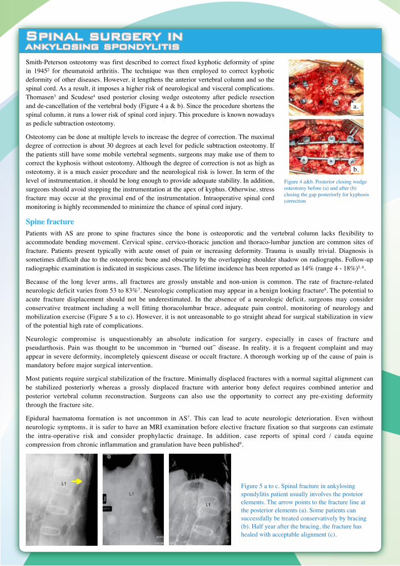
Spine fracturePatients with AS are prone to spine fractures since the bone is osteoporotic and the vertebral column lacks flexibility to accommodate bending movement. Cervical spine, cervico-thoracic junction and thoraco-lumbar junction are common sites of fracture. Patients present typically with acute onset of pain or increasing deformity. Trauma is usually trivial. Diagnosis is sometimes difficult due to the osteoporotic bone and obscurity by the overlapping shoulder shadow on radiographs. Follow-up radiographic examination is indicated in suspicious cases. The lifetime incidence has been reported as 14% (range 4 - 18%)5, 6.
Because of the long lever arms, all fractures are grossly unstable and non-union is common. The rate of fracture-related neurologic deficit varies from 53 to 83%7. Neurologic complication may appear in a benign looking fracture8. The potential to acute fracture displacement should not be underestimated. In the absence of a neurologic deficit, surgeons may consider conservative treatment including a well fitting thoracolumbar brace, adequate pain control, monitoring of neurology and mobilization exercise (Figure 5 a to c). However, it is not unreasonable to go straight ahead for surgical stabilization in view of the potential high rate of complications.
Neurologic compromise is unquestionably an absolute indication for surgery, especially in cases of fracture and pseudarthosis. Pain was thought to be uncommon in “burned out” disease. In reality, it is a frequent complaint and may appear in severe deformity, incompletely quiescent disease or occult fracture. A thorough working up of the cause of pain is mandatory before major surgical intervention.
Most patients require surgical stabilization of the fracture. Minimally displaced fractures with a normal sagittal alignment can be stabilized posteriorly whereas a grossly displaced fracture with anterior bony defect requires combined anterior and posterior vertebral column reconstruction. Surgeons can also use the opportunity to correct any pre-existing deformity through the fracture site.
Epidural haematoma formation is not uncommon in AS7. This can lead to acute neurologic deterioration. Even without neurologic symptoms, it is safer to have an MRI examination before elective fracture fixation so that surgeons can estimate the intra-operative risk and consider prophylactic drainage. In addition, case reports of spinal cord / cauda equine compression from chronic inflammation and granulation have been published9.
Dr. Dexter Yu-lung LEUNGClinical Assistant Professor (Honorary), Department of Ophthalmology & Visual Sciences, The Chinese University of Hong Kong Associate Consultant, Hong Kong Eye Hospital
Ophthalmological manifestations are common in patients with ankylosing spondylitis (AS). Uveitis is one of the commonest complications with 33.2% of AS patients developing uveitis during their lifetime.1 For those who have recurrent disease, the mean duration of cycles for recurrences and remissions can be as long as 17.0 years.1,2 The prevalence of uveitis was higher in patients with HLA-B27 positivity (39.8%) than in those who are HLA-B27 negative (13.6%)1,2 and in women than in men (33.3% vs. 28.5%).2 Uveitis refers to inflammation of iris and ciliary body (anterior uveitis), and choroid (posterior uveitis). Most uveitis in AS are anterior uveitis. Symptoms of anterior uveitis include eye pain, which is usually dull, caused by spasm of ciliary muscles secondary to anterior chamber inflammation and can radiate around the distribution of first branch of the trigeminal nerve. There will be an acute to subacute eye redness, or commonly, a “ciliary injection”, with the redness forming a circumferential red ring around the limbus (corneal-conjunctival junction). Ciliary injection is a result of inflammation of ciliary bodies located underneath the limbus. Vision is usually blurred but not excessively, due to anterior chamber inflammatory cells and flare from protein leakage. Photophobia is common. Complications from anterior uveitis include increase in intraocular pressure (IOP) and if persistent, may develop glaucoma with optic nerve damage, visual field defects and cataract. Cystoid macular edema is a complication presenting with decreased visual acuity, metamorphopsia (sensation of straight lines perceived as wavy), and micropsia/ macropsia (sensation of part of an object being miniaturized or magnified).
Ophthalmological manifestations and management in Ankylosing SpondylitisOphthalmological manifestations and management in Ankylosing Spondylitis
Inflammatory lesionsAndersson lesions refer to a spondylodiscitis that destroys the central portion of the intervertebral disc and adjacent vertebral body on radiographs10 (Figure 6). They occur at around 4% of AS patients and the radiological appearance may be confused with pseudarthrosis or infection. The main distinguishing feature is the absence of involvement of the posterior elements of the spine. Romanus lesions are much more common and appear in 53% of patients11. They are erosive changes at the anterior and posterior vertebral endplates that appear as radio-opaque lines on X-ray films. They also do not involve the posterior spinal elements. Differentiation between Andersson / Romanus lesions and pseudarthrosis / infection is important since the treatment regime is totally different.
ConclusionMedical treatment and conservative therapies are the first-line treatment for AS. The aims are to control inflammation, delay spinal deformity and maintain normal spinal posture. Surgery is indicated for fractures and other conditions causing intractable pain, disabling deformities and neurologic compromise.
Figure 6. The big solid arrow shows the Andersson lesions that the posterior spine elements are intact. The small broken arrow shows the Romanus lesion.
Figure 4 a&b. Posterior closing wedge osteotomy before (a) and after (b) closing the gap posteriorly for kyphosis correction
a.
b.
Figure 5 a to c. Spinal fracture in ankylosing spondylitis patient usually involves the posteior elements. The arrow points to the fracture line at the posterior elements (a). Some patients can successfully be treated conservatively by bracing (b). Half year after the bracing, the fracture has healed with acceptable alignment (c).
Smith-Peterson osteotomy was first described to correct fixed kyphotic deformity of spine in 19452 for rheumatoid arthritis. The technique was then employed to correct kyphotic deformity of other diseases. However, it lengthens the anterior vertebral column and so the spinal cord. As a result, it imposes a higher risk of neurological and visceral complications. Thomasen3 and Scudese4 used posterior closing wedge osteotomy after pedicle resection and de-cancellation of the vertebral body (Figure 4 a & b). Since the procedure shortens the spinal column, it runs a lower risk of spinal cord injury. This procedure is known nowadays as pedicle subtraction osteotomy.
Osteotomy can be done at multiple levels to increase the degree of correction. The maximal degree of correction is about 30 degrees at each level for pedicle subtraction osteotomy. If the patients still have some mobile vertebral segments, surgeons may make use of them to correct the kyphosis without osteotomy. Although the degree of correction is not as high as osteotomy, it is a much easier procedure and the neurological risk is lower. In term of the level of instrumentation, it should be long enough to provide adequate stability. In addition, surgeons should avoid stopping the instrumentation at the apex of kyphus. Otherwise, stress fracture may occur at the proximal end of the instrumentation. Intraoperative spinal cord monitoring is highly recommended to minimize the chance of spinal cord injury.

Spinal surgery in ankylosing spondylitisSpinal surgery in ankylosing spondylitis
Spinal surgery in ankylosing spondylitisSpinal surgery in ankylosing spondylitis
Reference1. Liao ZT, Pan YF, Huang JL, et al Scand J Rheumatol, 2009 Nov-Dec;38(6):455-92. Smith-Peterson MN, Larson CB, Aufranc OE. Osteotomy of the spine for correction of flexion deformity in rheumatoid arthritis. J Bone Joint Surg 1945;27:13. Thomasen E. Vertebral osteotomy for correction of kyphosis in ankylosing spondylitis. Clin Orthop 1985;194:1424. Scudese V, Calabro JJ. Vertebral wedge osteotomy. JAMA 1963;186:6275. Hitchon PW, From AM, Brenton MD, et al: Fractures of the thoracolumbar spine complicating ankylosing spondylitis. J Neurosurg 97 (2 Suppl):218-222,20026. Mitra D, Elvins DM, Speden DJ, et al: The prevalence of vertebral fractures in mild ankylosing spondylitis and their relationship to bone mineral density. Rheumatology 39:85-89, 20007. Weinstein PR, Karpmann RR, Gall EP, et al: Spinal cord injury, spinal fracture, and spinal stenosis in ankylosing spondylitis. J Neurosurg 57:609-616,19828. Wade W, Saltzstein R, Maiman D. Spinal fractures complicating ankylosing spondylitis. Arch Phys Med Rehabil 1989;70:3989. Good AE, Keller TS, Weatherbee L, et al: Spinal cord block with a destructive lesion of the dorsal spine in ankylosing spondylitis. Arthritis Rheum 25:218-222, 198210. Andersson O: Rontegenbilden vid spondylarthritis ankyylopoetica. Nord Med 30:2000-2002,193711. Hermann KA, Althoff CE, Schneider U, et al: Spinal changes in patients with spondyloarthritis: comparison of MR imaging and radiographic appearances. Radiographics 25:559-570,2005
Spine fracturePatients with AS are prone to spine fractures since the bone is osteoporotic and the vertebral column lacks flexibility to accommodate bending movement. Cervical spine, cervico-thoracic junction and thoraco-lumbar junction are common sites of fracture. Patients present typically with acute onset of pain or increasing deformity. Trauma is usually trivial. Diagnosis is sometimes difficult due to the osteoporotic bone and obscurity by the overlapping shoulder shadow on radiographs. Follow-up radiographic examination is indicated in suspicious cases. The lifetime incidence has been reported as 14% (range 4 - 18%)5, 6.
Because of the long lever arms, all fractures are grossly unstable and non-union is common. The rate of fracture-related neurologic deficit varies from 53 to 83%7. Neurologic complication may appear in a benign looking fracture8. The potential to acute fracture displacement should not be underestimated. In the absence of a neurologic deficit, surgeons may consider conservative treatment including a well fitting thoracolumbar brace, adequate pain control, monitoring of neurology and mobilization exercise (Figure 5 a to c). However, it is not unreasonable to go straight ahead for surgical stabilization in view of the potential high rate of complications.
Neurologic compromise is unquestionably an absolute indication for surgery, especially in cases of fracture and pseudarthosis. Pain was thought to be uncommon in “burned out” disease. In reality, it is a frequent complaint and may appear in severe deformity, incompletely quiescent disease or occult fracture. A thorough working up of the cause of pain is mandatory before major surgical intervention.
Most patients require surgical stabilization of the fracture. Minimally displaced fractures with a normal sagittal alignment can be stabilized posteriorly whereas a grossly displaced fracture with anterior bony defect requires combined anterior and posterior vertebral column reconstruction. Surgeons can also use the opportunity to correct any pre-existing deformity through the fracture site.
Epidural haematoma formation is not uncommon in AS7. This can lead to acute neurologic deterioration. Even without neurologic symptoms, it is safer to have an MRI examination before elective fracture fixation so that surgeons can estimate the intra-operative risk and consider prophylactic drainage. In addition, case reports of spinal cord / cauda equine compression from chronic inflammation and granulation have been published9.
Dr. Dexter Yu-lung LEUNGClinical Assistant Professor (Honorary), Department of Ophthalmology & Visual Sciences, The Chinese University of Hong Kong Associate Consultant, Hong Kong Eye Hospital
Ophthalmological manifestations are common in patients with ankylosing spondylitis (AS). Uveitis is one of the commonest complications with 33.2% of AS patients developing uveitis during their lifetime.1 For those who have recurrent disease, the mean duration of cycles for recurrences and remissions can be as long as 17.0 years.1,2 The prevalence of uveitis was higher in patients with HLA-B27 positivity (39.8%) than in those who are HLA-B27 negative (13.6%)1,2 and in women than in men (33.3% vs. 28.5%).2 Uveitis refers to inflammation of iris and ciliary body (anterior uveitis), and choroid (posterior uveitis). Most uveitis in AS are anterior uveitis. Symptoms of anterior uveitis include eye pain, which is usually dull, caused by spasm of ciliary muscles secondary to anterior chamber inflammation and can radiate around the distribution of first branch of the trigeminal nerve. There will be an acute to subacute eye redness, or commonly, a “ciliary injection”, with the redness forming a circumferential red ring around the limbus (corneal-conjunctival junction). Ciliary injection is a result of inflammation of ciliary bodies located underneath the limbus. Vision is usually blurred but not excessively, due to anterior chamber inflammatory cells and flare from protein leakage. Photophobia is common. Complications from anterior uveitis include increase in intraocular pressure (IOP) and if persistent, may develop glaucoma with optic nerve damage, visual field defects and cataract. Cystoid macular edema is a complication presenting with decreased visual acuity, metamorphopsia (sensation of straight lines perceived as wavy), and micropsia/ macropsia (sensation of part of an object being miniaturized or magnified).
Ophthalmological manifestations and management in Ankylosing SpondylitisOphthalmological manifestations and management in Ankylosing Spondylitis
Inflammatory lesionsAndersson lesions refer to a spondylodiscitis that destroys the central portion of the intervertebral disc and adjacent vertebral body on radiographs10 (Figure 6). They occur at around 4% of AS patients and the radiological appearance may be confused with pseudarthrosis or infection. The main distinguishing feature is the absence of involvement of the posterior elements of the spine. Romanus lesions are much more common and appear in 53% of patients11. They are erosive changes at the anterior and posterior vertebral endplates that appear as radio-opaque lines on X-ray films. They also do not involve the posterior spinal elements. Differentiation between Andersson / Romanus lesions and pseudarthrosis / infection is important since the treatment regime is totally different.
ConclusionMedical treatment and conservative therapies are the first-line treatment for AS. The aims are to control inflammation, delay spinal deformity and maintain normal spinal posture. Surgery is indicated for fractures and other conditions causing intractable pain, disabling deformities and neurologic compromise.
Figure 6. The big solid arrow shows the Andersson lesions that the posterior spine elements are intact. The small broken arrow shows the Romanus lesion.
Figure 4 a&b. Posterior closing wedge osteotomy before (a) and after (b) closing the gap posteriorly for kyphosis correction
a.
b.
Figure 5 a to c. Spinal fracture in ankylosing spondylitis patient usually involves the posteior elements. The arrow points to the fracture line at the posterior elements (a). Some patients can successfully be treated conservatively by bracing (b). Half year after the bracing, the fracture has healed with acceptable alignment (c).
Smith-Peterson osteotomy was first described to correct fixed kyphotic deformity of spine in 19452 for rheumatoid arthritis. The technique was then employed to correct kyphotic deformity of other diseases. However, it lengthens the anterior vertebral column and so the spinal cord. As a result, it imposes a higher risk of neurological and visceral complications. Thomasen3 and Scudese4 used posterior closing wedge osteotomy after pedicle resection and de-cancellation of the vertebral body (Figure 4 a & b). Since the procedure shortens the spinal column, it runs a lower risk of spinal cord injury. This procedure is known nowadays as pedicle subtraction osteotomy.
Osteotomy can be done at multiple levels to increase the degree of correction. The maximal degree of correction is about 30 degrees at each level for pedicle subtraction osteotomy. If the patients still have some mobile vertebral segments, surgeons may make use of them to correct the kyphosis without osteotomy. Although the degree of correction is not as high as osteotomy, it is a much easier procedure and the neurological risk is lower. In term of the level of instrumentation, it should be long enough to provide adequate stability. In addition, surgeons should avoid stopping the instrumentation at the apex of kyphus. Otherwise, stress fracture may occur at the proximal end of the instrumentation. Intraoperative spinal cord monitoring is highly recommended to minimize the chance of spinal cord injury.

The goals of treatment of uveitis in AS include prompt control of inflammation and minimization of risk of complications. Topical steroid such as prednisolone forte every 3 to 4 hours is the mainstay of treatment in a typical AS patient with acute anterior uveitis. The medication is used for a few weeks with a gradual tapering dose according to clinical response. Unsupervised use of topical steroid may cause side-effects such as glaucoma and cataract (usually after weeks to months of use). Under the supervision of ophthalmologist, the benefit of topical steroid outweights the risks of inflammatory ocular damages and any side-effects can be monitored. A mild mydriatic eyedrop may be indicated to keep the pupil mobile to prevent formation of posterior synechiae, which is a form of adhesion between the posterior iris and anterior lens capsule. This adhesion forms easily in anterior uveitis and will cause the pupil to be small and unable to dilate when needed. Vision may be dimmed if the pupil is bounded down to the anterior capsule in a meiotic phase. Such a small pupil will also render future cataract operation more difficult. The cycloplegic function of mydriatic drop will also relieve pain from ciliary spasms. Treatment of any accompanying rise in IOP depends on the actual level of rise and the baseline optic nerve head vulnerability. For acute symptomatic rise of IOP to >40-50 mmHg, an intravenous acetazolamide injection will be helpful in bringing down the IOP promptly, in order to prevent irreversible optic nerve damages. Topical glaucoma medication(s), such as beta-blockers (e.g. Timolol), with or without topical carbonic anhydrase inhibitors (e.g. Trusopt) and topical alpha-adrenergic agonist (e.g. Alphagan) may be used. Pilocarpine in general is contraindicated in acute glaucoma arising from uveitis, since pilocarpine worsens inflammation by destabilizing the intraocular blood-aqueous barriers, and it constricts the pupil, too. Prostaglandin analogue, (e.g. Xalatan, Travatan or Lumigan) may worsen inflammation and hence not recommended as first line treatment but in cases with resistant IOP rise to other glaucoma drops. Surgery will be required for those patients who had refractory IOP rise despite maximally tolerated topical glaucoma medications and include procedures such as trabeculectomy with mitomycin-C as chemoadjuvant; or glaucoma drainage implants such as Ahmed valve device implantation or Baerveldt tube implantation. The common aim of all these surgeries is to create a fistula, so that the aqueous humor can be diverted from the anterior chamber to a subconjunctival space (“bleb”), reducing IOP. Preliminary report suggested that the frequency of uveitis recurrences may be reduced by draining out the inflammatory mediators together with aqueous in the anterior chamber.
Cataracts are usually managed with phacoemulsification, using ultrasound energy to disrupt the cataract. An implantation of artificial intraocular lens will be needed for vision. The surgery may potentially flare up an uveitis and therefore, perioperative prophylaxis may be warranted.3 Cystoid macular edema can be managed by non-steroidal anti-inflammatory drugs (NSAIDs) such as voltaren. Recent advances include intravitreal injection of triamcinolone or anti-VEGF (vascular endothelial growth factor).4 Other minor ophthalmic manifestations in AS include immune episcleritis and conjunctivitis. Very mild and asymptomatic cases can be managed conservatively. For symptomatic patients, oral NSAIDs such as Indomethacin and Ibuprofen can be used. A mild topical steroid can sometimes be used in more patients with more severe diseases. Episcleritis in AS seldom transform into more complications such as nodular formation or necrotizing manifestations, hence systemic steroid is very rarely needed.
Recent studies have also suggested the efficacy of anti-tumor necrosis factor alpha in the treatment of uveitis in AS. Specifically, infliximab and adalimumab may decrease the rate of recurrences of uveitis. There was preliminary evidence that treatment with etanercept may be associated with significantly increased risk of uveitis in comparison with infliximab and adalimumab.5-6 If a patient develops uveitis during etanercept therapy, then a change to infliximab may be warranted. Lastly, more severe inflammation such as posterior uveitis and pan-uveitis (inflammation extending from anterior, intermediate to posterior segments of eyeball) may occur rarely. Systemic steroid treatment or other systemic immunosuppressants may be needed. Newer evidence on the horizon includes the Multicenter Uveitis Steroid Treatment Trial (MUST).7 This study examines the efficacy of steroid implant compared to systemic steroid in the management of difficult uveitis and may offer more effective and safe options for these patients.
Ophthalmological manifestations and management in Ankylosing SpondylitisOphthalmological manifestations and management in Ankylosing Spondylitis
Figure 5 Socking aid
Figure 4 Long handle reacherReference1. Linssen A, Rothova A, Valkenburg HA, et al. The lifetime cumulative incidence of acute anterior uveitis in a normal population and its relation to ankylosing spondylitis and histocompatibility antigen
HLA-B27. Invest Ophthalmol Vis Sci 1991;32:2568–78.2. Zeboulon N, Dougados M, Gossec L. Prevalence and characteristics of uveitis in the spondyloarthropathies: a systemic literature review. Ann Rheum Dis 2008;67: 955-959.3. Van Gelder RN, Leveque TK. Cataract surgery in the setting of uveitis. Curr Opin Ophthalmol 2009;20:42-5.4. Rothova A. Inflammatory cystoids macular edema. Curr Opin Ophthalmol 2007;18:487-92.5. Braun J, Baraliakos X, Listing J, et al. Decreased incidence of anterior uveitis in patients with ankylosing spondylitis treated with the anti-tumor necrosis factor agents infliximab and etanercept. Arthritis
Rheum 2005;52:2447–51.6. Lim LL, Graunfelder FW, Rosenbaum JT. Do tumor necrosis factor inhibitors cause uveitis? A registry-based study. Arthritis Rheum 2007;56:3248-52.7. The Multicenter Uveitis Steroid Treatment Trial Research Group. The Multicenter Uveitis Steroid Treatment Trial: Rationale, Design, and Baseline Characteristics. Am J Ophthalmol 2010 Jan 23. [Epub ahead
of print]
Enhancement of ADL (Activities of Daily Living ) for patients with Ankylosing Spondylitis (AS): Roles of occupational therapists Enhancement of ADL (Activities of Daily Living ) for patients with Ankylosing Spondylitis (AS): Roles of occupational therapists
Drinking is the most common basic task we do every day. When drinking from a cup or glass, a mild degree of neck extension is required to fill the mouth with fluids, followed by restoring the neck to neutral or a bit chin down position to enhance swallowing. Generally, we do this action smoothly without risk of choking. Due to decreased flexibility of the cervical spine, AS patients may have limited neck extension to assist this swallowing mechanism. As a result, choking is common and some may even experience pain and discomfort on forced neck extension. A flexi cup (Figure 1) with modified edge that fits the contour of the nose reduces the need of neck extension during drinking. Assistance with a drinking straw is recommended for patients who have very limited neck flexion and extension. However, only warm or cold fluid is recommended to avoid burning of tongue or lips if drinking hot soup or water from a straw (Figure 2).
Dressing is another essential daily task. Most people cannot imagine the spinal movements involved in dressing tasks. Without a stable spinal support, the shoulders and hips will not move smoothly to allow the garment to be fitted onto the body. AS patients have difficulty bending down to reach the feet to put on trousers, socks and shoes. Kyphosis in some patients may also hinder truncal movement. Wearing a jacket will be much easier than putting on a tight pullover shirt or T-shirt. Application of a dressing stick may help to pull the jacket to the other side of the body and ease wearing of the jacket. (Figure 3)
For pants or trousers, long handle reacher (Figure 4) can help position the pants to the legs and insert the legs into the pants. The sock aid is another good device to assist in wearing socks. The sock aid can widen the tight elastic opening of the sock such that it is easier to insert the foot into the sock (Figure 5). The long handle shoe horns can also assist in putting on and off shoes without much bending action. In terms of toileting and bathing, commodes and shower chairs may enable the user to sit stably to reduce risk of fall and to support the body during cleaning. Transfer in and out of bath tub can be difficult. A shower cabinet may be more suitable than a standard bath tub. Non-slip mat is essential to prevent slip and fall inside the bathroom. Hand rails at the side of toilet or bathing facilities can allow patients to hold onto the support during transfer.
In addition to the toilet facilities, firm bed mattress and sitting mattress may be considered to relieve long term pressure generated to the sacral region of the buttock and to prevent pressure sores. Wheelchair bound AS patients may have hip stiffness and knee flexion contracture. A high chair with firm back and neck support is necessary to support alignment of the body. In terms of work ergonomics, it is worthwhile to consult occupational therapist opinion for a visit to the work site and their prescription of furnitures with appropriate height and size. In this way, AS patients can be protected from chronic stress on the neck and back.
In conclusion, AS patients are faced with a lot of challenges in ADL during the course of their illness. Prescription of appropriate aids and gadgets will improve their functions and enhance their quality of life. The role of occupational therapist is important to evaluate individual needs for these aids and gadgets.
Reference: All pictures are adopted from homecraft Rolyan Company Ltd. www.homecraft-rolyan.com
Prof. Cecilia Wai-ping LI-TSANG, Professor and Associate Head, Department of Rehabilitation Sciences, The Hong Kong Polytechnic University
Mr. Calvin Chi-kong YIP, Cinial Associate, Department of Rehabilitation Sciences, The Hong Kong Polytechnic University
Ankylosing Spondylitis (AS) is a systemic disease which affects mainly the spine. In severe cases, it will result in fusion of the spinal joints causing limitation of spinal range of motion, thus affecting mobility and functions. The roles of an occupational therapist (OT) are to help clients suffering from AS to actively participate in activities and to engage into their living community. OT will first conduct a detail assessment to identify the performance that a client with AS concerns most and subsequently plan suitable intervention strategies and treatment to facilitate their independence and function.
To help AS patients to resolve the difficulties encountered in activities of daily living (ADL), prescription of suitable aids and gadgets for these patients would be needed. Some of the common ADL aids that help to resolve the ADL problems are listed below for references:
Figure 1 Flexi cup
Figure 2 one way straw
Figure 3 Dressing stick
Lastly, most AS patients can have accessed to medical care, but care in regard to the psychosocial aspects of living is limited. Encouraging patients to join patient self help group such as B27 Association and Hong Kong Ankylosing Spondylitis Association is a good way to enrich their social life. The Community Rehabilitation Network (CRN) also provides educational and therapeutic programs to help people with arthritis.
Ms Patsy Kai-ying CHAN,Registered Social Worker, Community Rehabilitation Network, The Hong Kong Society for Rehabilitation
‘You just don’t understand me…’ Even for family members and friends, many people cannot understand the sour and bitterness experienced by patients with Ankylosing spondylitis (AS). AS attacks mostly young male adults when they are at the prime of life for learning and development. The burden of AS results from pain, reduced function, and impaired well-being (Bostan, et al, 2003). Spinal deformity, morning stiffness, chronic pain, sleep problems and fatigue directly affect the psychological well-being, social and family role of AS patients. The quality of life of AS patients is poor especially in physical health related aspects (Rugienė, et al, 2008).
The level of pain and physical limitations on daily activities such as work and social activities is uncertain and fluctuates everyday. It is not too difficult to imagine AS patients are likely to have more depression, self blaming, anger, desperation, helplessness and low self-esteem. According to Lorig (2000), disease pain, depression, psychological stress and fatigue interacts in a cycle. The cycle is often vicious, the more depressed you are, the more pain you feel; the more pain you feel, the more stressed you become; the more stressed you become, the more depressed you are. Depression and other psychological distress makes AS patients tired which aggravates depression and pain. Therefore, the psychosocial needs of AS patients need to be addressed.
Psychosocial Needs of people with ASIn accordance with Lorig (2000), Backman (2002) suggested an interactive multidimensional relationship between psychosocial factors and physical limitations and pain. Psychosocial factors including the dimensions of psychological (cognitive, affective) and social (interacting with others, engaging in life activities) (Backman, 2002), influence pain perception and pain influences psychological well-being and social participation. Fear of change and of the unknown is often found at the early stage of diagnosis of AS. The uncertainty of health conditions and pain affect participation in all kinds of activities, and results in frustration, depression and sadness. Anger is also common in AS patients and is often expressed towards family and friends.
Interacting with others and engaging in social activities are essential to balance the psychological well-being and physical limitations in AS. Simple activities such as chatting and strolling with friends, joining a support group, meeting people with AS who have managed to live full and successful lives are the best ways to overcome distress and fear.
Getting Our Own Control with Cognitive Behavioural TherapyOne of the approaches in managing psychological distress is Cognitive Behavioral Therapy (CBT). Beck (1995) proposed that psychological distress occurs as a result of people’s dysfunctional thoughts which come from people’s negative automatic thoughts, dysfunctional rules, assumptions and core beliefs. These thought processes influence our feelings and behaviors. Under stressful life events, dysfunctional thoughts will be elicited automatically. Automatic thoughts are accumulative and built up from our life experience. These accumulative experiences will generate different types of automatic thoughts, like thinking pattern which drive one’s behaviour towards different life events. According to Beck (1995), these automatic thoughts commonly include ‘Absolutist thought’ - “If I ask for help, I am a failure”; ‘Catastrophizing thoughts’ – “Because of my back pain, I cannot work long, I will lose my job, my wife will leave me and I will end up sitting in a wheel chair”; ‘Personalization’ – “The dinner gathering is called off because of me, I am so ashamed to increase other people’s burden”. Automatic thoughts are targeted to be recognized and restructured into positive ones.
How to make one think more positive? First, try to stop the thoughts and relax: when negative automatic thoughts are followed by negative emotions, try to stop thinking negative and into too much details. The longer the negative thoughts stay in your mind, the longer you will be distressed. Prepare some cheer on phrases that calm you. Distraction behaviors such as taking a deep breath and listening to some relaxing music may also work. If the negative thoughts keep coming back, try to debate with ourselves if the thoughts are true, For example, “Has the dinner gathering been called off solely because of you, or may there be other factors?” Also, what’s the point of drilling into it further? If the dinner gathering is really called off because of you, do you need to be ashamed and there are no advantages to keep feeling ashamed forever. Instead of drilling into the negative thought of losing a job because of back pain, does it mean you will really lose your job? Does it mean you cannot find another job? There may be other jobs that suit you better.
Psychosocial Aspects in Ankylosing SpondylitisPsychosocial Aspects in Ankylosing Spondylitis
Psychosocial Aspects in Ankylosing SpondylitisPsychosocial Aspects in Ankylosing Spondylitis
ReferenceBackman CL. Psychosocial aspects in the management of arthritis pain, Arthritis Research & Therapy, 2002; 8(6): 221-228Beck JS. 1995. Cognitive therapy: basics and beyond. New York: Guilford Press. Bostan EE, Borman P, Bodur H, et al. Functional disability and quality of life in patients with ankylosing spondylitis. Rheumatol Int. 2003;23(3):121–6Lorig K and Fries JF. 2000. The arthritis helpbook: a tested self-management program for coping with arthritis and fibromyalgia. Cambridge, Mass: Perseus.Rugienė R, Kirdaitė G, Gražuleviciūtė E, et al. The quality of life and functional ability in patients with ankylosing spondylitis, Acta medica lituanica, 2008; 15(2): 99-103
The role of acupuncture in the management of rheumatic diseasesThe role of acupuncture in the management of rheumatic diseases
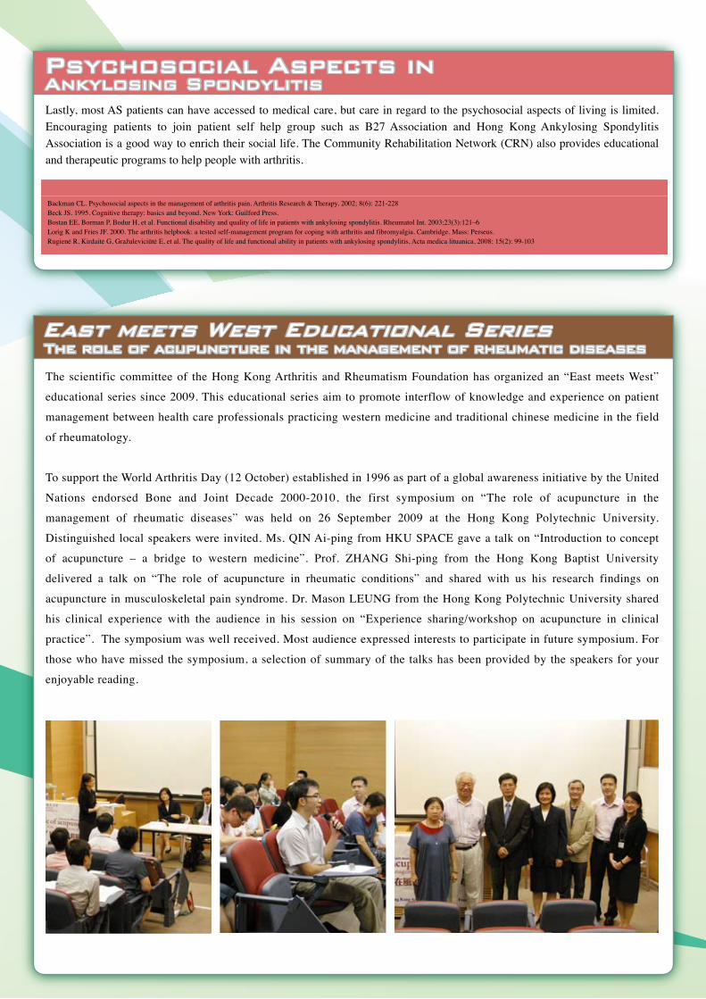
The scientific committee of the Hong Kong Arthritis and Rheumatism Foundation has organized an “East meets West”
educational series since 2009. This educational series aim to promote interflow of knowledge and experience on patient
management between health care professionals practicing western medicine and traditional chinese medicine in the field
of rheumatology.
To support the World Arthritis Day (12 October) established in 1996 as part of a global awareness initiative by the United
Nations endorsed Bone and Joint Decade 2000-2010, the first symposium on “The role of acupuncture in the
management of rheumatic diseases” was held on 26 September 2009 at the Hong Kong Polytechnic University.
Distinguished local speakers were invited. Ms. QIN Ai-ping from HKU SPACE gave a talk on “Introduction to concept
of acupuncture – a bridge to western medicine”. Prof. ZHANG Shi-ping from the Hong Kong Baptist University
delivered a talk on “The role of acupuncture in rheumatic conditions” and shared with us his research findings on
acupuncture in musculoskeletal pain syndrome. Dr. Mason LEUNG from the Hong Kong Polytechnic University shared
his clinical experience with the audience in his session on “Experience sharing/workshop on acupuncture in clinical
practice”. The symposium was well received. Most audience expressed interests to participate in future symposium. For
those who have missed the symposium, a selection of summary of the talks has been provided by the speakers for your
enjoyable reading.
East meets West Educational SeriesEast meets West Educational Series
Lastly, most AS patients can have accessed to medical care, but care in regard to the psychosocial aspects of living is limited. Encouraging patients to join patient self help group such as B27 Association and Hong Kong Ankylosing Spondylitis Association is a good way to enrich their social life. The Community Rehabilitation Network (CRN) also provides educational and therapeutic programs to help people with arthritis.
Ms Patsy Kai-ying CHAN,Registered Social Worker, Community Rehabilitation Network, The Hong Kong Society for Rehabilitation
‘You just don’t understand me…’ Even for family members and friends, many people cannot understand the sour and bitterness experienced by patients with Ankylosing spondylitis (AS). AS attacks mostly young male adults when they are at the prime of life for learning and development. The burden of AS results from pain, reduced function, and impaired well-being (Bostan, et al, 2003). Spinal deformity, morning stiffness, chronic pain, sleep problems and fatigue directly affect the psychological well-being, social and family role of AS patients. The quality of life of AS patients is poor especially in physical health related aspects (Rugienė, et al, 2008).
The level of pain and physical limitations on daily activities such as work and social activities is uncertain and fluctuates everyday. It is not too difficult to imagine AS patients are likely to have more depression, self blaming, anger, desperation, helplessness and low self-esteem. According to Lorig (2000), disease pain, depression, psychological stress and fatigue interacts in a cycle. The cycle is often vicious, the more depressed you are, the more pain you feel; the more pain you feel, the more stressed you become; the more stressed you become, the more depressed you are. Depression and other psychological distress makes AS patients tired which aggravates depression and pain. Therefore, the psychosocial needs of AS patients need to be addressed.
Psychosocial Needs of people with ASIn accordance with Lorig (2000), Backman (2002) suggested an interactive multidimensional relationship between psychosocial factors and physical limitations and pain. Psychosocial factors including the dimensions of psychological (cognitive, affective) and social (interacting with others, engaging in life activities) (Backman, 2002), influence pain perception and pain influences psychological well-being and social participation. Fear of change and of the unknown is often found at the early stage of diagnosis of AS. The uncertainty of health conditions and pain affect participation in all kinds of activities, and results in frustration, depression and sadness. Anger is also common in AS patients and is often expressed towards family and friends.
Interacting with others and engaging in social activities are essential to balance the psychological well-being and physical limitations in AS. Simple activities such as chatting and strolling with friends, joining a support group, meeting people with AS who have managed to live full and successful lives are the best ways to overcome distress and fear.
Getting Our Own Control with Cognitive Behavioural TherapyOne of the approaches in managing psychological distress is Cognitive Behavioral Therapy (CBT). Beck (1995) proposed that psychological distress occurs as a result of people’s dysfunctional thoughts which come from people’s negative automatic thoughts, dysfunctional rules, assumptions and core beliefs. These thought processes influence our feelings and behaviors. Under stressful life events, dysfunctional thoughts will be elicited automatically. Automatic thoughts are accumulative and built up from our life experience. These accumulative experiences will generate different types of automatic thoughts, like thinking pattern which drive one’s behaviour towards different life events. According to Beck (1995), these automatic thoughts commonly include ‘Absolutist thought’ - “If I ask for help, I am a failure”; ‘Catastrophizing thoughts’ – “Because of my back pain, I cannot work long, I will lose my job, my wife will leave me and I will end up sitting in a wheel chair”; ‘Personalization’ – “The dinner gathering is called off because of me, I am so ashamed to increase other people’s burden”. Automatic thoughts are targeted to be recognized and restructured into positive ones.
How to make one think more positive? First, try to stop the thoughts and relax: when negative automatic thoughts are followed by negative emotions, try to stop thinking negative and into too much details. The longer the negative thoughts stay in your mind, the longer you will be distressed. Prepare some cheer on phrases that calm you. Distraction behaviors such as taking a deep breath and listening to some relaxing music may also work. If the negative thoughts keep coming back, try to debate with ourselves if the thoughts are true, For example, “Has the dinner gathering been called off solely because of you, or may there be other factors?” Also, what’s the point of drilling into it further? If the dinner gathering is really called off because of you, do you need to be ashamed and there are no advantages to keep feeling ashamed forever. Instead of drilling into the negative thought of losing a job because of back pain, does it mean you will really lose your job? Does it mean you cannot find another job? There may be other jobs that suit you better.
Psychosocial Aspects in Ankylosing SpondylitisPsychosocial Aspects in Ankylosing Spondylitis
Psychosocial Aspects in Ankylosing SpondylitisPsychosocial Aspects in Ankylosing Spondylitis
ReferenceBackman CL. Psychosocial aspects in the management of arthritis pain, Arthritis Research & Therapy, 2002; 8(6): 221-228Beck JS. 1995. Cognitive therapy: basics and beyond. New York: Guilford Press. Bostan EE, Borman P, Bodur H, et al. Functional disability and quality of life in patients with ankylosing spondylitis. Rheumatol Int. 2003;23(3):121–6Lorig K and Fries JF. 2000. The arthritis helpbook: a tested self-management program for coping with arthritis and fibromyalgia. Cambridge, Mass: Perseus.Rugienė R, Kirdaitė G, Gražuleviciūtė E, et al. The quality of life and functional ability in patients with ankylosing spondylitis, Acta medica lituanica, 2008; 15(2): 99-103
The role of acupuncture in the management of rheumatic diseasesThe role of acupuncture in the management of rheumatic diseases
The scientific committee of the Hong Kong Arthritis and Rheumatism Foundation has organized an “East meets West”
educational series since 2009. This educational series aim to promote interflow of knowledge and experience on patient
management between health care professionals practicing western medicine and traditional chinese medicine in the field
of rheumatology.
To support the World Arthritis Day (12 October) established in 1996 as part of a global awareness initiative by the United
Nations endorsed Bone and Joint Decade 2000-2010, the first symposium on “The role of acupuncture in the
management of rheumatic diseases” was held on 26 September 2009 at the Hong Kong Polytechnic University.
Distinguished local speakers were invited. Ms. QIN Ai-ping from HKU SPACE gave a talk on “Introduction to concept
of acupuncture – a bridge to western medicine”. Prof. ZHANG Shi-ping from the Hong Kong Baptist University
delivered a talk on “The role of acupuncture in rheumatic conditions” and shared with us his research findings on
acupuncture in musculoskeletal pain syndrome. Dr. Mason LEUNG from the Hong Kong Polytechnic University shared
his clinical experience with the audience in his session on “Experience sharing/workshop on acupuncture in clinical
practice”. The symposium was well received. Most audience expressed interests to participate in future symposium. For
those who have missed the symposium, a selection of summary of the talks has been provided by the speakers for your
enjoyable reading.
East meets West Educational SeriesEast meets West Educational Series

Dr. Mason LEUNG,Associate Professor, Department of Rehabilitation Sciences, The Hong Kong Polytechnic University
A high percentage (up to 30%) of patients suffering from various rheumatic conditions have consulted complementary therapy especially acupuncture. The World Health Organisation (WHO), the National Center for Complementary and Alternative Medicine (NCCAM) of the National Institutes of Health (NIH), the American Medical Association (AMA) and various governments reported that acupuncture is safe when administered by well-trained practitioners using sterile needles, and that further research is warranted. A number of rheumatic conditions including low back pain, neck pain, osteoarthritis, fibromyalgia, or rheumatoid arthritis can benefit from acupuncture in terms of pain relief, improve function and/or decrease the rate of progression. However, there are common adverse effects from acupuncture. These include:
• Minor bleeding after removal of the needles, seen in roughly 3% of patients. Holding a cotton ball for about one minute over the site of puncture is usually sufficient to stop the bleeding.
• Hematoma, seen in about 2% of patients, which manifests as bruises. These usually resolves after a few days.
• Dizziness, seen in about 1% of patients. Some patients have a conscious or unconscious fear of needles which can produce dizziness and other symptoms of anxiety. Patients are usually managed by lying down in order to reduce likelihood of fainting.
All these adverse effects can be minimized and can be well managed even if these happen. In fact, the incidence of these complications is low, acupuncture is still a worthwhile therapy for consideration.
Experience sharing/workshop on acupuncture in clinical practiceExperience sharing/workshop on acupuncture in clinical practice
Dr. Shi-ping ZHANG,Associate Professor, School of Chinese Medicine, Hong Kong Baptist University
Acupuncture has been used extensively in the treatment of rheumatic conditions, including jaw pain, chronic neck pain, frozen shoulder, tennis elbow, tendonitis of the wrist and finger, ankylosing spondylitis, chronic low back pain, chronic knee pain, musculoskeletal pain of the leg and ankle, gout, and rheumatoid arthritis. The method of acupuncture treatment for these conditions involves mainly local points, in conjunction with electroacupuncture. In some cases acupuncture is used in combination with herbal or drug medication. The benefits of acupuncture treatment include reduction in pain and improvement in function. There have been a substantial number of clinical studies regarding the efficacy of acupuncture treatment for rheumatic conditions, and systematic reviews have been made available for chronic neck pain (Fu et al. 2009), frozen shoulder (Buchbinder et al. 2005), tennis elbow (Trinh et al. 2004), chronic low back pain (Furlan et al. 2008), chronic knee pain (White et al. 2007), and rheumatoid arthritis (Wang et al. 2008). Thus, acupuncture has short-term efficacy in the treatment of neck pain and lateral epicondyle pain, improving shoulder function at four weeks, effective pain relief and functional improvement for immediate and short-term treatment of chronic low back pain and chronic knee pain. However, in the treatment of rheumatoid arthritis, conflicting evidence exists in placebo-controlled trials concerning the efficacy of acupuncture. Two randomized controlled trials were conducted in the Hong Kong Baptist University. In chronic heel pain (plantar fasciitis), acupuncture using acupoint Daling (PC7) was found to be superior to acupoint Hegu (LI4) (Zhang et al. 2009). In chronic neck pain, there was no significant difference between electro-acupuncture and sham laser one month after treatment. The inconsistent findings of ours and others raise the need for careful design and cautious interpretation of acupuncture clinical trials. Large pragmatic trials are required to establish the long-term efficacy of acupuncture compared to conventional treatments in rheumatic conditions.
The role of acupuncture in rheumatic conditionsThe role of acupuncture in rheumatic conditions
Ms Ai-ping QINAssociate Professor, Division of Chinese Medicine, School of Professional and Continue Education The University of Hong Kong
Acupuncture in Traditional Chinese Medicine is a vibrant component of health management universally, which possesses a coherent and independent system. Its clinical indications constitute 43 kinds of diseases declared by WHO in 1979, gradually rising to 64 types in 1997. In a meeting on acupuncture held by National Institute of Health in USA in1996, the efficacy of acupuncture was proven by evidence based medicine on post-operative nausea and vomiting, chemotherapy nausea and vomiting and postoperative dental pain. Acupuncture is also regarded as an alternative for treating addiction, stroke rehabilitation, headache, menstrual cramps, tennis elbow, fibromyalgia, myofascial pain, osteoarthritis, low back pain and asthma.
Yet the character of acupuncture may not be demonstrable by a Randomized Controlled Trial. Traditional Chinese medicine and Western medicine present two different approaches of perspectives and thought. Acupuncture focuses on a holistic approach rather than a disease-oriented diagnostic and treatment model. Western Medicine diagnosed conditions such as gout, rheumatoid arthritis, systemic lupus erythematosus and osteoarthritis as one disease whereas in Chinese Medicine, as "Bi"-Painful Syndrome. It is further sub-classified into different syndromes, such as heat "Bi", damp "Bi", wondering "Bi", etc. Despite the different methods of treatment, the principle behind remains the same. Take the treatment of gout as an example as shown in the following table:
In summary, some Chinese medical concepts are elusive. It is difficult to reconcile with contemporary biomedical information. Yet it plays an important role in the diagnosis of diseases and formulation of acupuncture treatment.
Introduction to Concept of Acupuncture A bridge to Western MedicineIntroduction to Concept of Acupuncture A bridge to Western Medicine
Objective of the treatment Western Medicine Chinese Medicine
To manage symptoms of acute attack
Non-steroidal anti-inflammatory drugs
Local & Acupuncture points with Clearing heat function- to activate the Meridian Qi flow- to relieve pain
To prevent acute attack To inhibit conversion of purine into uric acid
- to activate general Meridian Qi flow - to dissolve dampness
To reduce serum uric acid To increase excretion of uric acid from the body
- to strengthen the spleen - to eliminate dampness
Hong Kong Arthritis & Rheumatism Foundation Ltd.Tel:(852) 2346 6336Fax:(852) 2346 6136
Email:[email protected]:www.hkarf.org
Rm 14, G/F, HKSR Lam Tin Complex,7 Rehab Path, Kwun Tong, Kowloon
Sponsor:
All materials published in CHARM represent the opinions of the authors responsible for the articles and do not reflect the official views or policy of the Hong Kong Arthritis & Rheumatism Foundation.
Publication of an advertisement in CHARM does not constitute endorsement or approval of the product or service promoted or of any claims made by the advertisers with respect to such products or services.
The Hong Kong Arthritis & Rheumatism Foundation and CHARM assume no responsibility for any injury and/or damage to persons or property arising from any use of execution of any methods, treatments, therapy, operations, instructions, ideas contained in the printed articles. Because of rapid advances in medicine, independent verification of diagnoses, treatment method and drug dosage should be made.
DisclaimerDisclaimer
CHARM is published by the Hong Kong Arthritis and Rheumatism Foundation, which is a charity entirely supported by voluntary contributions and public donations. If you wish to have further information about our work, or to make a donation or a monthly contribution, please contact us:
DonationDonation

3issueMARCH • 2010Care, Health, Arthritic Management
Editorial BoardEditorial Board
This is the first issue of CHARM in 2010. Members of the editorial board and I would like to wish you all Happy New Year of the Tiger!The theme of this issue is ankylosing spondylitis, an inflammatory deforming spinal disease. This disease has long been considered to be incurable with frustrating patients leaving clinics with loads of non-steroidal anti-inflammatory drugs, risking their lives from gastrointestinal bleeding. In this issue, we will read about update on management of ankylosing spondylitis involving the use of imaging techniques and serological tests to assist in early diagnosis as recent availability of biologic therapy offers new hope in the treatment of this disease. Another article covers the surgical aspect for the treatment of bony and neurological complications from advanced disease. As opthalmological complications such as uveitis is rather common in these patients, an ophthalmologist has been invited to write on this extra-articular manifestation of ankylosing spondylitis and the available treatment modalities. Lastly, since multi-disciplinary approach is required in the management of these patients, the important role of occupational therapist in the prescription of assistance of activities of daily living for these patients is not to be discounted.To support the World Arthritis Day established in 1996 as part of a global awareness initiative by the United Nations endorsed Bone and Joint Decade 2000-2010, the HKARF scientific committee has organized the first symposium of “East meets West” educational series on “The role of acupuncture in the management of rheumatic diseases” last September. This educational series aim to promote interflow of knowledge and experience on patient management between health care professionals practicing western medicine and traditional Chinese medicine. The symposium was well received. For those who have missed the meeting, a summary of their talks from the distinguished speakers can be found in this issue of CHARM.I hope you enjoy reading these articles.
Chief Editor:Dr. Temy Mo-yin Mok
Co-editor:Ms. Jane Lai-hung ChanDr. Hang-cheong ChengDr. Reann Wai-po ChuDr. Wai-keung LeungProf. Cecilia Wai-ping Li-TsangDr. Daniel Kam-hung NgMr. Peter King-kong PoonMr. David Po-wing Yau
Secretary:Ms Den Law
Invited guests:Ms Patsy Kai-ying ChanDr. Dexter Yu-lung LeungDr. Woon-leung NgDr. Yat-wa WongDr. Cheuk-wan YimMr. Calvin Chi-kong Yip
Dr. Cheuk-wan YIM, Specialist in Rheumatology, United Christian HospitalDr. Woon-leung NG, Consultant in Rheumatology, United Christian Hospital
IntroductionIn classic textbooks, the image of a male patient adopting a stooped, question mark posture with X ray appearance of bamboo-spine was deeply etched in our memories as the pathognomonic features of Ankylosing Spondylitis (AS). Today, such manifestations signify failures in public and patient awareness as well as gaps in our diagnostic-therapeutic cascade in much the same way as the encounter of metastatic disease in cancer presentation. While AS patients with mild disease and limited involvement are able to maintain almost full functional and employment capacity; a significant proportion may develop severe debilitation and skeletal restrictions. Early diagnosis and assessment is the critical step to enable effective therapeutic intervention before irreversible damages occur.
Update in Diagnosis and PrognosisThere is no single test to diagnose AS. According to the modified New York criteria in 1984, definite AS is defined as X-ray evidence of sacroiliitis of grade 2 or above plus any one of the followings: 1) inflammatory low back pain for more than three months; 2) limited lumbar motion or 3) restricted chest expansion. The drawback is that signs and symptoms of AS have to be present for as long as 8 to 10 years before changes are evident on X-ray. Computed Tomographic scan (CT-scan) can detect early bony erosions but there is significant radiation exposure. The advent of magnetic resonance imaging (MRI) has proven to be a milestone in the early diagnosis of AS through its ability to detect inflammatory signals in the bone marrow and soft tissues, bony erosions, arthritis and other chronic changes using different MRI sequences.
An Update on Ankylosing SpondylitisAn Update on Ankylosing Spondylitis
Editor's MessageEditor's Message
ENB-
AD-0
94-N
OV-
09