ec cpr, llc © 2010-20110 fluids, electrolytes, and intravenous therapy brian mitu, fnp, pa-c, faapa...
TRANSCRIPT

EC CPR, LLC © 2010-2011 1
Fluids, Electrolytes, and Intravenous Therapy
Fluids, Electrolytes, and Intravenous Therapy
Brian Mitu, FNP, PA-C, FAAPA
Internal Medicine, Cardiology
Instructor Basic/Advance EKG
National/Community Speaker Chronic Hepatitis B

EC CPR, LLC © 2010-2011 2
Learning Objectives
Theory
1. Recall the various functions fluid performs in the body.
2. Identify the body’s mechanisms for fluid regulation.
3. Review three ways in which body fluids are continually being distributed among the fluid compartments.
4. Distinguish the signs and symptoms of various electrolyte imbalances.
5. Discuss why the elderly have more problems with fluid and electrolyte imbalances.
6. Recognize the disorders that cause specific fluid and electrolyte imbalances.
7. Compare the major causes of acid-base imbalances.
8. State correct interventions to correct an acid-base imbalance.
9. Discuss the steps in managing an intravenous infusion.
10. Describe the measures used to prevent the complications of intravenous therapy.
11. Identify intravenous fluids that are isotonic.12. Discuss the principles of intravenous
therapy.
Clinical Practice
1. Assess patients for signs of dehydration.
2. Correctly assess for and identify edema and signs of overhydration.
3. Apply knowledge of normal laboratory values in order to recognize electrolyte imbalances.
4. Carry out interventions to correct an electrolyte imbalance.
5. Determine if a patient has an acid-base imbalance.
6. Carry out measures to prevent the complications of Intravenous therapy.
7. Compare interventions for the care of a patient receiving total parenteral nutrition with one undergoing intravenous therapy.
EC CPR, LLC © 2010-2011 3
Basic Concepts of Fluid and Electrolytes
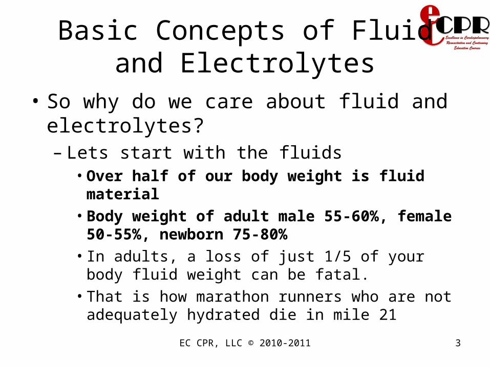
• So why do we care about fluid and electrolytes?– Lets start with the fluids
• Over half of our body weight is fluid material• Body weight of adult male 55-60%, female 50-
55%, newborn 75-80%• In adults, a loss of just 1/5 of your body fluid weight
can be fatal. • That is how marathon runners who are not
adequately hydrated die in mile 21

EC CPR, LLC © 2010-2011 4
Basic Concepts of Fluid and Electrolytes
• So why do we care about fluid and electrolytes?– Elderly patients are even more at risk. Why ?
• They have less muscle mass. This also means that a smaller amount of fluid loss can and will be detrimental
– The same can be said to infants. Why?• Because 80% of their body weight consist of fluids
EC CPR, LLC © 2010-2011 5
Basic Concepts of Fluid and Electrolytes
• So why do we care about fluid and electrolytes?– Electrolytes
• Are substances that become ions in solution and acquire the capacity to conduct electricity.
• Essential for normal function of our cells and our organs

EC CPR, LLC © 2010-2011 6
Basic Concepts of Fluid and Electrolytes
• So where are these fluids kept? – Body fluid compartments
• Each body fluid compartment has a particular composition of electrolytes/fluid, which differ from that of the other compartments
• Extracellular compartment ( 20% BW)– Tissue fluid (interstitial) ( 15% BW)– Intravascular compartment ( 4.5% BW)– Transcellular (1.5% BW)
• Intracellular compartment ( 40% of BW)– Refers to all the fluid inside the cell (most bodily fluid are
inside the cell)

EC CPR, LLC © 2010-2011 7
Basic Concepts of Fluid and Electrolytes
• Extracellular (ECF) compartment– 1/3 ( 20%) of body fluid– Comprised of 3 major components
• Intravascular– Fluid within the blood vessels. Plasma accounts for
about half of the total blood volume of the body
• Interstitial– Fluid that surrounds the cells – an example of
interstitial fluid is lymph
• Transcellular– Fluid found in the cerebrospinal column, pericardial
envelope, synovial joints, or intraocular space

EC CPR, LLC © 2010-2011 8
Basic Concepts of Fluid and Electrolytes
• Extracellular fluids provide– Nutrients for cell functioning
• Na• Ca• Cl• Glucose• Fatty acids• Amino Acids

EC CPR, LLC © 2010-2011 9
Basic Concepts of Fluid and Electrolytes
• Intravascular Component– Plasma
• Fluid portion of blood
– Made of:• Water• Plasma proteins
– Albumin– Clotting protein– Immunoglobulins (Antibodies)
• Small amount of other substances

EC CPR, LLC © 2010-2011 10
Basic Concepts of Fluid and Electrolytes
• Interstitial component– Made up of fluid between cells
• Surrounds cells • Transport medium for nutrients, gases, waste
products and other substances between blood and body cells
• Back-up fluid reservoir

EC CPR, LLC © 2010-2011 11
Basic Concepts of Fluid and Electrolytes
• Transcellular component– 1% of ECF– Located in joints, connective tissue, bones,
body cavities, CSF, and other tissues– Potential to increase significantly in abnormal
conditions • 3rd space

EC CPR, LLC © 2010-2011 12
Basic Concepts of Fluid and Electrolytes
• Intracellular (ICF)– Fluid within the cells themselves – 2/3 of body fluid (40%)– Located primarily in skeletal muscle mass– Provide nutrients for metabolism:
• High in K, Po4, protein• Moderate levels of Mg, So4
– Assists in cellular metabolism

EC CPR, LLC © 2010-2011 13
Basic Concepts of Fluid and Electrolytes
• Who are the major players of fluid and electrolyte balance?– Hypothalamus
• Thirst center – Stimulated by changes in water loss or increase
extracellular osmolality• Baroreceptors (carotid/ aortic) and stretch
receptors (atrial) as detectors – Impulses sent to the thirst control centers in the
hypothalamus

Basic Concepts of Fluid and Electrolytes
• Who are the major players of fluid and electrolyte balance?– Posterior Pituitary
• ADH ( Antidiuretic Hormone or Vasopressin)• Stimulated by changes in water loss or increase
extracellular osmolality– Heart
• ANP (Atrial Natriuretic Peptide)• In response to an increase in blood volume• Increase sodium excretion by increasing GFR and
inhibiting sodium reabsorption
EC CPR, LLC © 2010-2011 14

EC CPR, LLC © 2010-2011 15
Basic Concepts of Fluid and Electrolytes
• Who are the major players of fluid and electrolyte balance?– Kidneys/Adrenal glands (RAAS)
• Renin– Is released in response to decreased blood flow or
decreased renal pressure (sensed by receptors in the nephrons)
• Aldosterone – Is produced by the adrenal cortex (in response to
stimulation by angiotensin II) causing the tubules to excrete K+ while retaining Na 2+, adding to the reabsorption of water back into the vascular system.

EC CPR, LLC © 2010-2011 16

EC CPR, LLC © 2010-2011 17

EC CPR, LLC © 2010-2011 18

EC CPR, LLC © 2010-2011 19

EC CPR, LLC © 2010-2011 20
Movement of Fluid and Electrolytes
• Water Steady State• Amount Ingested =
Amount Eliminated

EC CPR, LLC © 2010-2011 21
Movement of Fluid and Electrolytes
• Electrolyte (Na+, K+, Ca++) Steady State
• Amount Ingested = Amount Excreted.
• Normal entry: Mainly ingestion in food.
• Clinical entry: Can include parenteral administration
• Just a remember that each fluid compartment has constant amount of fluid, and constant amount of electrolytes
EC CPR, LLC © 2010-2011 22
Movement of Fluid and Electrolytes
• What is the exact mechanism of fluid and electrolyte shift?– Passive transport
• Diffusion• Osmosis• Filtration• Hydrostatic pressure• Oncotic pressure
– Active transport• Sodium pump and ATP

EC CPR, LLC © 2010-2011 23
Movement of Fluid and Electrolytes
• Passive transport– Diffusion
• Movement of solutes from an area of higher concentration to an area of lower concentration in a solution and/or across a permeable membrane (permeable for that solute)
• Movement occurs until near equal state
– Osmosis• Movement of water through a selectively permeable
membrane from an area of low solute concentration to a higher concentration until equilibrium occurs
• Movement occurs until near equal concentration found

EC CPR, LLC © 2010-2011 24
Movement of Fluid and Electrolytes
• Osmolality– Concentration of body fluids – affects
movement of fluid by osmosis– Reflects hydration status– Measured by serum and urine – Solutes measured - mainly urea, glucose,
and sodium– Measured as solute concentration/Kg

EC CPR, LLC © 2010-2011 25
Movement of Fluid and Electrolytes
• Osmolality– Serum Osm/L = (serum Na x 2) + BUN/3 +
Glucose/18– Normal serum value - 280-300 mOsm/Kg– Serum <240 or >320 is critically abnormal – Normal urine Osm – 250 – 900 mOsm / kg
• Increase means dehydrated• Decrease means excess fluid
EC CPR, LLC © 2010-2011 26
Osmolality of a Solution
• Isotonic solutions– Means the solutions on both sides of a selectively
permeable membrane have established equilibrium or are equal in concentration Examples
• 0.9% NaCl, 5% dextrose water, 5% dextrose in 0.225% saline, LR
• Hypotonic solutions– The solution contains lower concentration of salt or
solute than another solution or lower osmolality than body fluid
– Less salt or > water than isotonic– 0.45% NaCl, 0.226%, 0.33% Nacl

EC CPR, LLC © 2010-2011 27
Osmolality of a Solution
• Hypertonic solution– A solution has higher concentration of
solutes than another solution or higher osmolality than body fluids
– 3%, 5% NaCl, D10%w, D5% with 0.9% NaCl, D5% with 0.45% NaCl, D5% LR

EC CPR, LLC © 2010-2011 28
Movement of Fluid and Electrolytes
• Passive transport– Filtration
• Movement of solutes and solvents through semipermeable membrane
• Influenced by hydrostatic pressure– Hydrostatic pressure is created by the pumping action of the
heart– Think of arterial end of the capillary– Supplying fluid to tissue cells
– Oncotic /osmotic pressure • Think of the venous end • Getting back the excess fluid and solutes that are in the
tissue and returned to the vascular compartment via lymph

EC CPR, LLC © 2010-2011 29
Movement of Fluid and Electrolytes
• Active transport:– Ion moves through a membrane from an area
of lower to higher concentration– Sodium pump and ATP– Substances that are transported actively
through the cell membrane• Sodium• Potassium, calcium, iron• Hydrogen, sugars, amino acids

EC CPR, LLC © 2010-2011 30
Fluid Volume deficit
• Description– Dehydration
• Occurs when the fluid intake of the body is not sufficient to meet the fluid needs of the body
– Goal of treatment• Is to restore fluid volume, replace electrolytes PRN, find
the cause
• Types– Isotonic fluid deficit/dehydration
• Both water and electrolytes are lost equally• Known as hypovolemia → ↓circulating blood volume,
tissue perfusion• Stimulates ADH
EC CPR, LLC © 2010-2011 31
Fluid Volume deficit
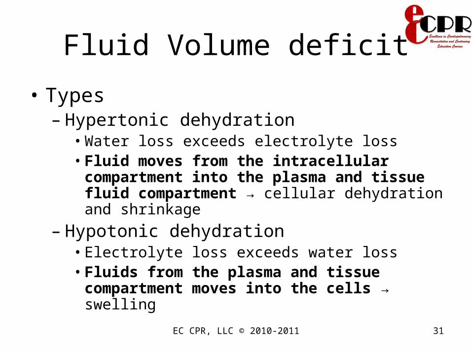
• Types– Hypertonic dehydration
• Water loss exceeds electrolyte loss• Fluid moves from the intracellular
compartment into the plasma and tissue fluid compartment → cellular dehydration and shrinkage
– Hypotonic dehydration• Electrolyte loss exceeds water loss• Fluids from the plasma and tissue
compartment moves into the cells → swelling

EC CPR, LLC © 2010-2011 32
Fluid Volume deficit
• Causes of fluid volume deficits– Isotonic dehydration
• Third spacing – peritonitis, intestinal obstruction, ascites, burns
• Hemorrhage• Altered intake, such as nothing by mouth (NPO)
– Hypertonic dehydration• Excessive perspiration, Ketoacidosis• Prolonged fevers, Diarrhea• ESRD, Diabetes Insipidus• Diuretic therapy

EC CPR, LLC © 2010-2011 33
Fluid Volume deficit
• Causes of fluid volume deficits– Hypotonic dehydration
• Chronic illness• Excessive fluid replacement (hypotonic)• RF, Chronic malnutrition• Gastrointestinal (GI) losses – vomiting,
nasogastric suctioning

EC CPR, LLC © 2010-2011 34
Fluid Volume Deficit: AssessmentFluid Volume Deficit: Assessment
• Cardiovascular– Thready, increase pulse rate– Decrease BP, Orthostatic hypotension– Flat neck and hand veins in dependent position– Diminished peripheral pulses
• Respiratory– Increase rate and depth of respiration
• Neuromuscular – Lethargy and coma– Fever

EC CPR, LLC © 2010-2011 35
Fluid Volume Deficit: AssessmentFluid Volume Deficit: Assessment
• Renal– Decrease urine output, increase specific
gravity• Integumentary
– Dry skin, poor turgor, tenting– Dry mouth
• GI– Wt loss, thirst
• Hypotonic dehydration– Muscle weakness
• Hypertonic dehydration– Pitting edema, hyperactive DTR

EC CPR, LLC © 2010-2011 36
Fluid Volume Deficit: AssessmentFluid Volume Deficit: Assessment
Mosby items and derived items © 2007 by Mosby, Inc., an affiliate of Elsevier, Inc.

EC CPR, LLC © 2010-2011 37
Fluid Volume Deficit: Nsg InterventionsFluid Volume Deficit: Nsg Interventions• Place client in shock position (on back with legs
elevated).• Fluid replacement
– Mild cases can be corrected via PO route– Significant losses (hemorrhage, anemia, 3rd space)
• Blood transfusion• Colloid solution ( albumin, dextran)• 0.9% NaCl or 154mEq/L Na+
– Solution of choice for normonatremic and mildly hyponatremic, hypotensive or shock pts
• Severe hyponatremia– Hypertonic saline (3.0% NaCl or 513 mEq/L)
Mosby items and derived items © 2007 by Mosby, Inc., an affiliate of Elsevier, Inc.

Fluid Volume Deficit: Nsg InterventionsFluid Volume Deficit: Nsg Interventions
• Monitor I&O. – Alert the primary care provider to urine output
less than 30 mL/hr for 2 consecutive hr.• Monitor vital signs and heart rhythm.• Monitor level of consciousness and maintain
client safety.• Treat underlying cause of fluid volume deficit.• Encourage the client to change positions
slowly.
EC CPR, LLC © 2010-2011 38

EC CPR, LLC © 2010-2011 39
Fluid Volume Deficit: ComplicationsFluid Volume Deficit: Complications
• Hypovolemic Shock– This can lead to vital organ hypoxia/anoxia
decreased hemoglobin, oxygen saturation, and pulse pressure (systolic-diastolic blood pressure).
– Administer oxygen.– Provide fluid replacement.– Perform hemodynamic monitoring.
Mosby items and derived items © 2007 by Mosby, Inc., an affiliate of Elsevier, Inc.

Fluid Volume Deficit: ComplicationsFluid Volume Deficit: Complications
• Hypovolemic Shock– Administer vasoconstrictors, coronary
vasodilators, and/or positive inotropes• dopamine (Inotropin)• Epinephrine• dobutamine (Dobutrex)• norepinephrine (Levophed)• phenylephrine (Neo-Synephrine)
EC CPR, LLC © 2010-2011 40

EC CPR, LLC © 2010-2011 41
Nausea and Vomiting• “Sick to my stomach"
• Pallor, mild diaphoresis, cold clammy skin, excessive salivation, and attempts to remain quiet and motionless
• Vomitus odor, color, contents (e.g., undigested food), and amount

EC CPR, LLC © 2010-2011 42
Medical Treatment• Antihistamines, sedative-hypnotics,
anticholinergics, and phenothiazines
• Complementary and alternative therapy
• NPO, then progressing slowly to a regular diet
• Carbonated drinks

EC CPR, LLC © 2010-2011 43
Diarrhea• Frequent watery bowel movements, abdominal
cramping, and general weakness • Watery stools often contain mucus and are
blood‑streaked; frequency could be as high as 15 to 20 liquid stools.
• Acute diarrhea and local irritation• Chronic and prolonged diarrhea in ulcerative
colitis, irritable bowel syndrome, allergies, lactose intolerance, and nontropical sprue
EC CPR, LLC © 2010-2011 44
Diarrhea• Dehydration, malnutrition, and anemia
• Bowel sounds likely to be loud gurgling and tinkling sounds that come in waves and are hyperactive
• Note and record the number of stools during the shift and the characteristics of each stool and associated pain

EC CPR, LLC © 2010-2011 45
Acute Diarrhea• Limit the intake of food• Progress to clear, bland liquids and to solids with
increased calories and high‑protein, high‑carbohydrate content
• Give rehydrating solutions containing glucose and electrolytes
• Avoid iced fluids, carbonated drinks, whole milk, roughage, raw fruits, and highly seasoned foods
EC CPR, LLC © 2010-2011 46
Medications for Diarrhea• Cause of the disorder and the length of time the
conditionMild cases:– Kaolin and bismuth preparations, (e.g., Kaopectate)
• Antispasmodic drugs such as belladonna or paregoric • Bismuth and "traveler's diarrhea" • Codeine, diphenoxylate (Lomotil), or loperamide
(Imodium)• Drugs and causative organisms• Metabolic acidosis and buffer solutions
EC CPR, LLC © 2010-2011 47
Nursing Management• Provide physical and mental rest, prevent
unnecessary loss of water and nutrients, protect the rectal mucosa, and replace fluids
• Maintain a calm and dignified manner, accept and understand the patient's behavior, and provide privacy and a restful environment

EC CPR, LLC © 2010-2011 48
Home Care: Fluid Volume Deficit
• Log of intake and output
• Small amounts of liquid every hour while awake
• Emergency department and intravenous fluids

EC CPR, LLC © 2010-2011 49
Fluid Volume Excess Fluid Volume Excess
• Description– Fluid intake or fluid retention exceeds the
fluid needs of the body or extracellular compartment
– Aka: fluid overload or overhydration
• Types– Isotonic overhydration
• Hypervolemia. Only the EC compartment expands

EC CPR, LLC © 2010-2011 50
Fluid Volume ExcessFluid Volume Excess
• Types– Isotonic overhydration
• Results in circulatory overload and tissue edema
• When severe it can worsen CHF and lead to pulmonary edema
– Hypertonic overhydration• Rare. Caused by excessive Na intake• Fluid is drawn from the ICF compartment and the
EC volume expands
EC CPR, LLC © 2010-2011 51
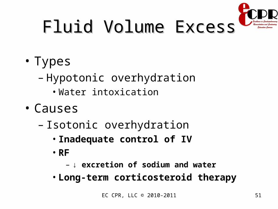
Fluid Volume ExcessFluid Volume Excess
• Types– Hypotonic overhydration
• Water intoxication
• Causes– Isotonic overhydration
• Inadequate control of IV• RF
– ↓ excretion of sodium and water
• Long-term corticosteroid therapy
EC CPR, LLC © 2010-2011 52
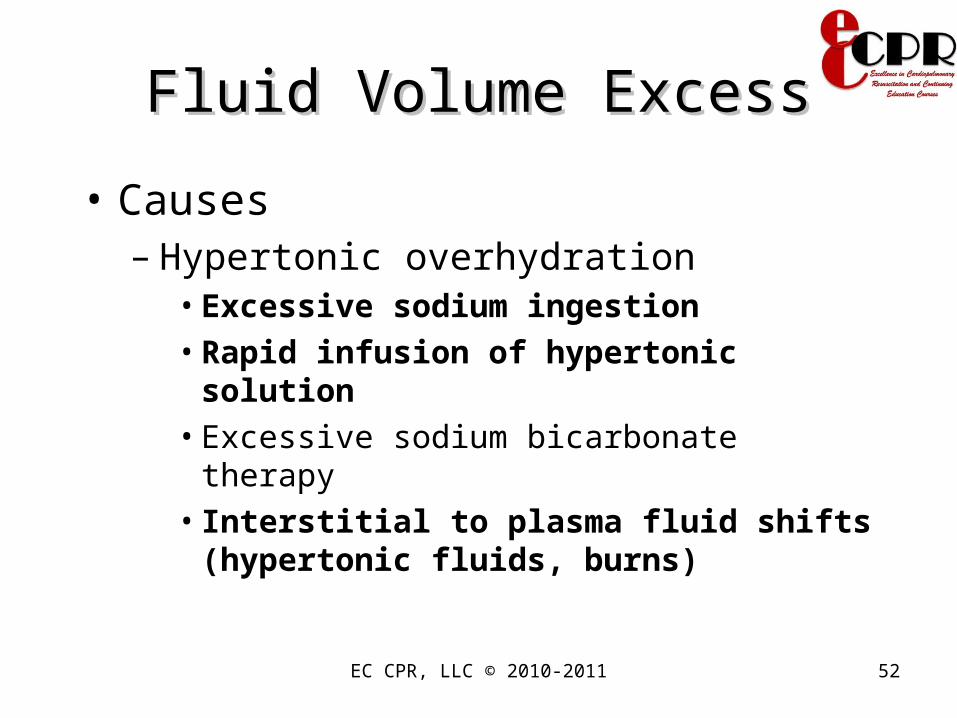
Fluid Volume ExcessFluid Volume Excess
• Causes– Hypertonic overhydration
• Excessive sodium ingestion• Rapid infusion of hypertonic solution• Excessive sodium bicarbonate therapy• Interstitial to plasma fluid shifts (hypertonic
fluids, burns)

Fluid Volume ExcessFluid Volume Excess
• Causes– Hypotonic overhydration
• Early RF• CHF, SIADH
– Chronic stimulus to the kidney to conserve sodium and water (heart failure, cirrhosis, glucocorticosteroids)
• Irrigation of wounds and body cavities with hypotonic fluids
EC CPR, LLC © 2010-2011 53

EC CPR, LLC © 2010-2011 54
Fluid Volume Excess: Risk Fluid Volume Excess: Risk FactorsFactors
• Overhydration– Water replacement without electrolyte
replacement such as strenuous exercise with profuse diaphoresis
Mosby items and derived items © 2007 by Mosby, Inc., an affiliate of Elsevier, Inc.
EC CPR, LLC © 2010-2011 55
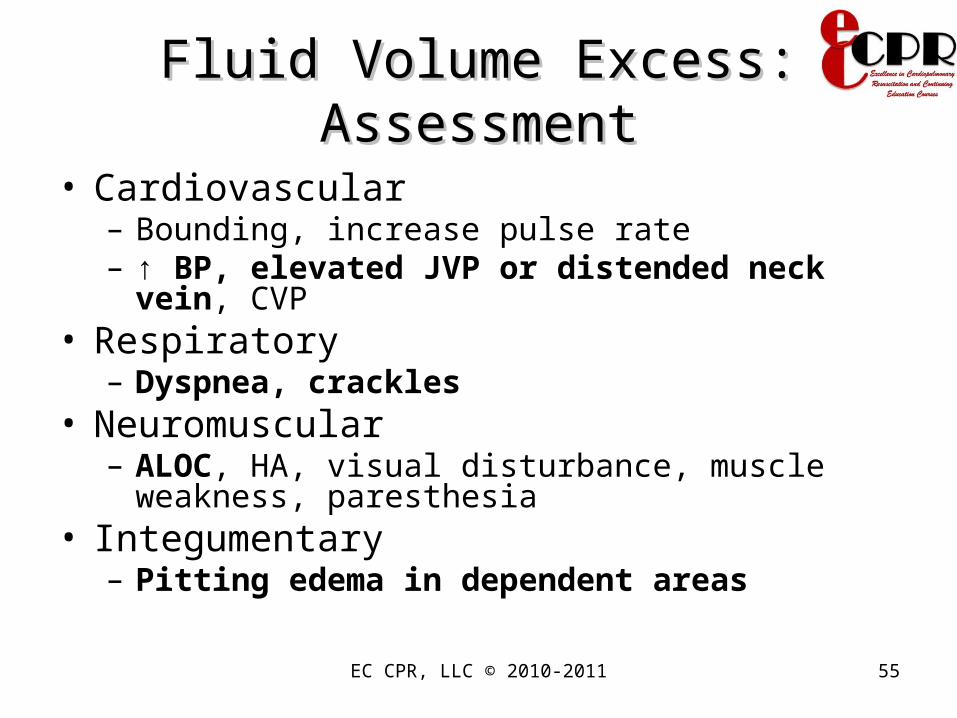
Fluid Volume Excess: AssessmentFluid Volume Excess: Assessment
• Cardiovascular– Bounding, increase pulse rate– ↑ BP, elevated JVP or distended neck vein, CVP
• Respiratory– Dyspnea, crackles
• Neuromuscular – ALOC, HA, visual disturbance, muscle weakness,
paresthesia• Integumentary
– Pitting edema in dependent areas

EC CPR, LLC © 2010-2011 56
Fluid Volume Excess: AssessmentFluid Volume Excess: Assessment
• GI– Hepatojugular reflux, ascites, liver
enlargement
• Labs– ↓ serum osmolality, hct, BUN, sodium,
urine specific gravity

EC CPR, LLC © 2010-2011 57
Fluid Volume Excess: Fluid Volume Excess: AssessmentAssessment
Mosby items and derived items © 2007 by Mosby, Inc., an affiliate of Elsevier, Inc.

EC CPR, LLC © 2010-2011 58
Excess Fluid Volume• Hematocrit:
– 35% to 54%, depending on age and sex
• Urine concentration: – Specific gravity: 1.003 to 1.030 (average range
is 1.010 to 1.025)

EC CPR, LLC © 2010-2011 59Mosby items and derived items © 2007 by Mosby, Inc., an affiliate of Elsevier, Inc.

EC CPR, LLC © 2010-2011 60
Fluid Volume Excess: Nsg Fluid Volume Excess: Nsg InterventionsInterventions
• Assess/monitor for signs of respiratory distress– including breath sounds and arterial blood gases
(ABGs).
• Position the client in semi-Fowler’s position.• Administer oxygen as needed.• Reduce IV flow rates.• Administer diuretics (osmotic, loop) as ordered.• Monitor daily I&O and weight.
Mosby items and derived items © 2007 by Mosby, Inc., an affiliate of Elsevier, Inc.

EC CPR, LLC © 2010-2011 61
Fluid Volume Excess: Nsg Fluid Volume Excess: Nsg InterventionsInterventions
• Limit fluid and sodium intake as ordered.• Assess/monitor and document peripheral vascular
system including– skin color, presence of edema (pretibial, sacral,
periorbital), and circulation to extremities.
• Turn and position the client at least every 2 hr.• Support arms and legs to decrease dependent
edema and promote venous return as appropriate.• Monitor for/treat skin breakdown.

EC CPR, LLC © 2010-2011 62
Fluid Volume Excess: Fluid Volume Excess: ComplicationsComplications
• Pulmonary Edema– Signs and symptoms include ascending
crackles, dyspnea at rest, and confusion.– Position the client in Fowler’s position.– Administer IV morphine. Why?
• ↑ pulmonary venous capacitance, ↓ left atrial pressure and anxiety → better ventilation
– Administer IV diuretic.– Prepare for possible intubation and
mechanical ventilation.Mosby items and derived items © 2007 by Mosby, Inc., an affiliate of Elsevier, Inc.

EC CPR, LLC © 2010-2011 63
Edema• Localized edema and generalized edema
• General causes of edema:– An increase in capillary hydrostatic pressure– A loss of plasma proteins– An obstruction of lymphatic circulation– An increase in capillary permeability
• Dependent edema

EC CPR, LLC © 2010-2011 64Mosby items and derived items © 2007 by Mosby, Inc., an affiliate of Elsevier, Inc.
Figure 17-3 Mechanisms of edema formation. Na+, Sodium; H2O, water. From Huether SE, McCance KL: Understanding pathophysiology, ed 3, St Louis, 2004, Mosby.
EC CPR, LLC © 2010-2011 65
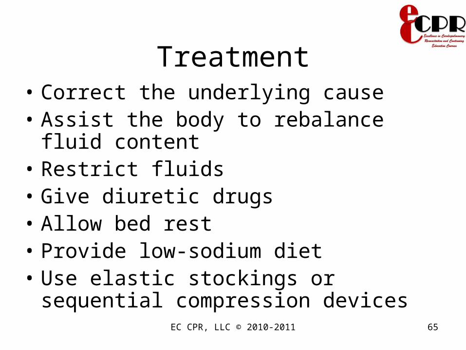
Treatment• Correct the underlying cause • Assist the body to rebalance fluid content• Restrict fluids• Give diuretic drugs• Allow bed rest• Provide low-sodium diet• Use elastic stockings or sequential
compression devices

EC CPR, LLC © 2010-2011 66
Home Care: Fluid Excess• Weigh daily
• Assess edema
• Know when to notify physician
EC CPR, LLC © 2010-2011 67
ElectrolytesElectrolytes
• Electrolytes are minerals (sometimes called salts) that are present in all body fluids.
• They regulate fluid balance, hormone production, and strengthen skeletal structures, as well as act as catalysts in nerve response, muscle contraction, and the metabolism of nutrients.
Mosby items and derived items © 2007 by Mosby, Inc., an affiliate of Elsevier, Inc.

EC CPR, LLC © 2010-2011 68Mosby items and derived items © 2007 by Mosby, Inc., an affiliate of Elsevier, Inc.
ElectrolytesElectrolytes• Electrolytes are distributed between
intracellular (ICF) and extracellular (ECF) fluid compartments.
• When dissolved in fluids, separate in to ions and conduct either – Cations: Positively charged ions
• magnesium, potassium, sodium, calcium – Anions: Negatively charged ions
• phosphate, sulfate, chloride, bicarbonate
• Measured in milliequivalents (mEq)• Serum levels indicate extracellular
concentration not inside the cells

EC CPR, LLC © 2010-2011 69
Electrolytes• Creation of an electrical impulse:
– Transmission of nerve impulses– Contraction of muscles– Excretion of hormones and other substances
from glandular cells
EC CPR, LLC © 2010-2011 70Mosby items and derived items © 2007 by Mosby, Inc., an affiliate of Elsevier, Inc.
EC CPR, LLC © 2010-2011 71
Osmolality • Concentration of the solution determined by
the number of solutes • Osmolality controls water movement and
the body fluid distribution in the intracellular and extracellular compartments
• Intracellular fluid and potassium• Extracellular fluid and sodium• Normal osmolality: 280 to 294 milliosmoles
per kilogram

EC CPR, LLC © 2010-2011 72Mosby items and derived items © 2007 by Mosby, Inc., an affiliate of Elsevier, Inc.
Sodium is the predominant Sodium is the predominant electrolyte in ECF. electrolyte in ECF.
• Sodium imbalances are usually associated with parallel changes in osmolality.
• Sodium is essential for the maintenance of acid-base balance, active and passive transport mechanisms, and maintaining irritability and conduction of nerve and muscle tissue.
• Normal serum sodium levels are 135 to 145 mEq/L.
EC CPR, LLC © 2010-2011 73
Hyponatremia
• Plasma Na+ of < 135 mEq/L
• Causes– Increased sodium excretion
• Excessive diaphoresis• Diuretics • Vomiting• Diarrhea• Wound drainage ,especially GI; Renal disease• Burns , nasogastric suctioning

EC CPR, LLC © 2010-2011 74
Hyponatremia
• Causes– Inadequate sodium intake
• NPO• Low-salt diet
– Dilution of serum sodium• Excessive irrigation/ ingestion of hypotonic
fluids, tap water enemas • Renal failure• Freshwater drowning• SIADH

EC CPR, LLC © 2010-2011 75
Hyponatremia
• Causes– Dilution of serum sodium
• CHF– Due to ↓ forward flow by the failing heart, the kidney
senses hypoperfusion and tries to ↑ percieved diminished intravascular volume via RAAS
– Furthermore, ADH gets activated also
• Cirrhosis– Pooling of blood in the mesenteric veins which
leads to decrease amount of blood seen by the kidneys
EC CPR, LLC © 2010-2011 76
Hyponatremia
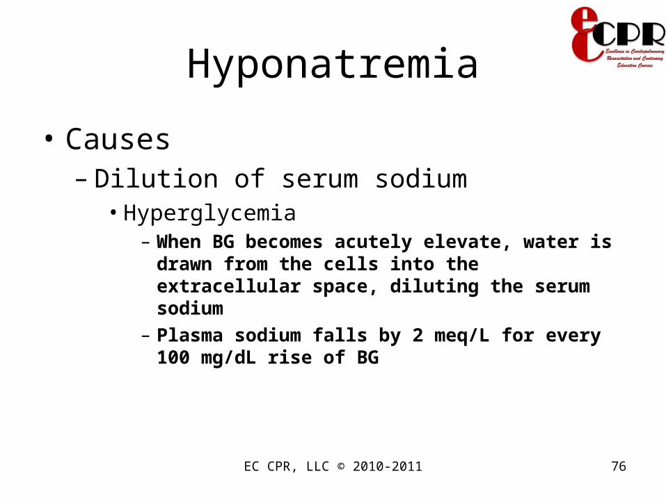
• Causes– Dilution of serum sodium
• Hyperglycemia– When BG becomes acutely elevate, water is drawn
from the cells into the extracellular space, diluting the serum sodium
– Plasma sodium falls by 2 meq/L for every 100 mg/dL rise of BG

EC CPR, LLC © 2010-2011 77

EC CPR, LLC © 2010-2011 78Mosby items and derived items © 2007 by Mosby, Inc., an affiliate of Elsevier, Inc.
Hyponatremia Hyponatremia
• Classification– Hypotonic hyponatremia < 280 mOsm/kg
• Fluid volume deficit (Hypovolemic) : Sodium loss greater than water loss
• Fluid volume excess (Hypervolemic): Water gain greater than sodium gain
• Normal fluid volume ( Euvolemic): Pure water gain
– Isotonic hyponatremia 280-290 mOsm/kg– Hyper hyponatremia > 290 mOsm/kg
EC CPR, LLC © 2010-2011 79
Hyponatremia: Assessment
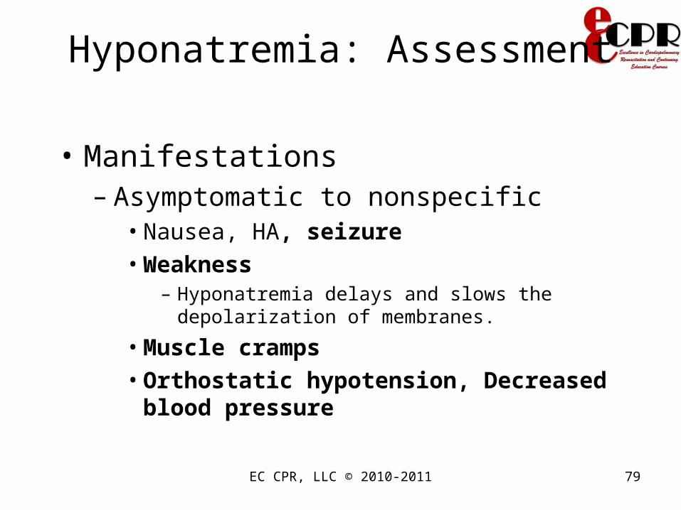
• Manifestations– Asymptomatic to nonspecific
• Nausea, HA, seizure• Weakness
– Hyponatremia delays and slows the depolarization of membranes.
• Muscle cramps• Orthostatic hypotension, Decreased blood
pressure
EC CPR, LLC © 2010-2011 80
Hyponatremia: Assessment
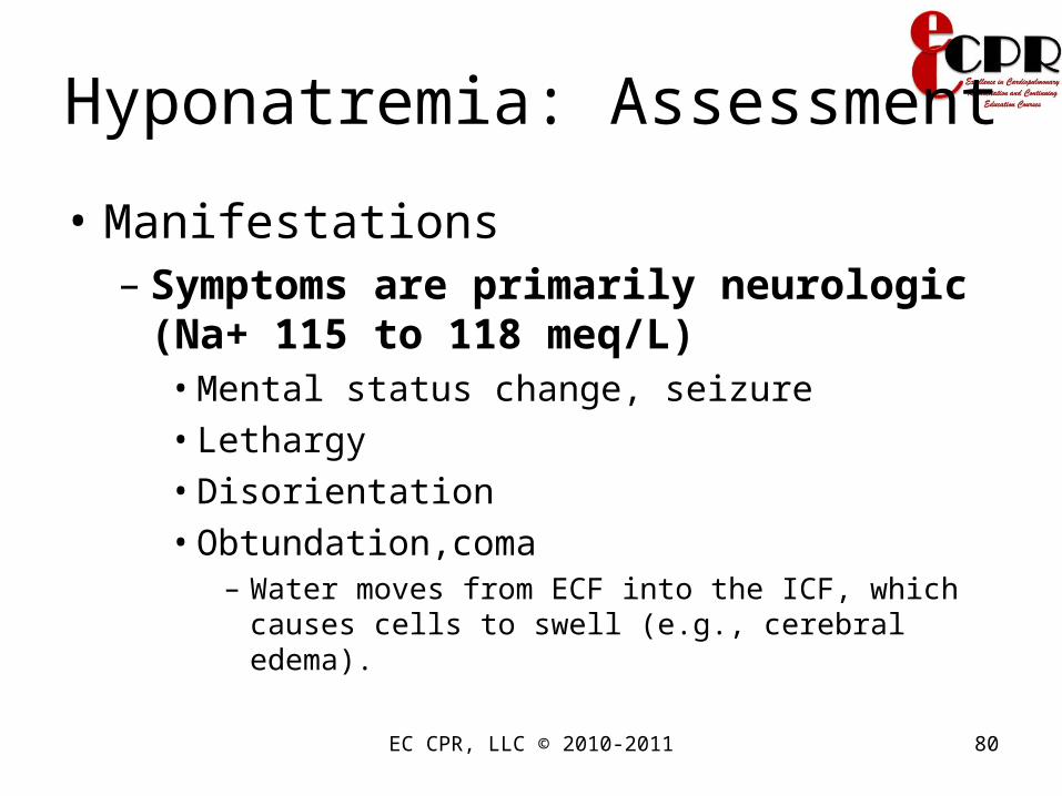
• Manifestations– Symptoms are primarily neurologic (Na+
115 to 118 meq/L)• Mental status change, seizure• Lethargy• Disorientation• Obtundation,coma
– Water moves from ECF into the ICF, which causes cells to swell (e.g., cerebral edema).

EC CPR, LLC © 2010-2011 81
Hyponatremia
• Diagnosis – H&P– Plasma osmolality (decrease)– Urine osmolality and volume
• Specific gravity
– Urine Na+ concentration ( increase or decrease depending on the cause)
– CT scan of the brain is done if SIADH is suspected
– C-XRAY to r/o lung pathology

EC CPR, LLC © 2010-2011 82
Hyponatremia: AssessmentHyponatremia: Assessment
Mosby items and derived items © 2007 by Mosby, Inc., an affiliate of Elsevier, Inc.

EC CPR, LLC © 2010-2011 83Mosby items and derived items © 2007 by Mosby, Inc., an affiliate of Elsevier, Inc.

EC CPR, LLC © 2010-2011 84
Hyponatremia: Nsg Hyponatremia: Nsg InterventionsInterventions
• Fluid overload: Restrict water intake as ordered (Hypervolemic)
• Acute Hyponatremia– Administer hypertonic oral and IV fluids as ordered.– Encourage foods and fluids high in sodium (cheese,
milk)• If pt is taking lithium, check lithium level
because hyponatremia causes lithium toxicity
• Give demeclocycline (Declomycin) if the cause is d/t excess ADH
Mosby items and derived items © 2007 by Mosby, Inc., an affiliate of Elsevier, Inc.

EC CPR, LLC © 2010-2011 85
Hyponatremia: Nsg InterventionsHyponatremia: Nsg Interventions
• Foods high in sodium– Bacon, butter, canned food, american/cottage
cheese– Frankfurters, ketchup, milk, soy sauce,
processed foods– White and whole-wheat bread
EC CPR, LLC © 2010-2011 86
Hyponatremia: Nsg Hyponatremia: Nsg InterventionsInterventions
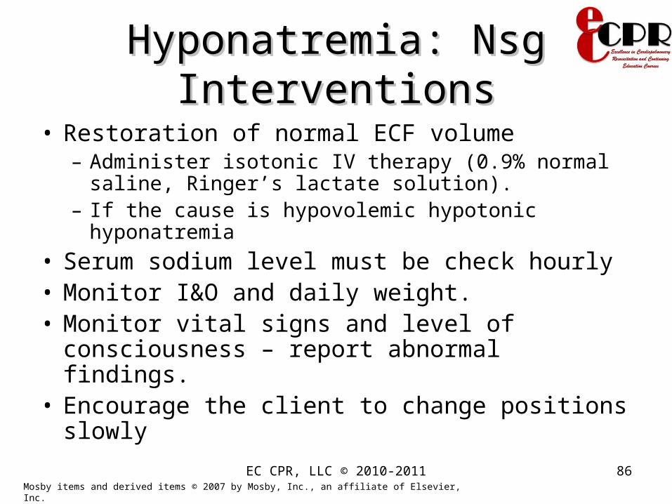
• Restoration of normal ECF volume– Administer isotonic IV therapy (0.9% normal saline,
Ringer’s lactate solution). – If the cause is hypovolemic hypotonic hyponatremia
• Serum sodium level must be check hourly• Monitor I&O and daily weight.• Monitor vital signs and level of consciousness –
report abnormal findings.• Encourage the client to change positions slowly
Mosby items and derived items © 2007 by Mosby, Inc., an affiliate of Elsevier, Inc.

EC CPR, LLC © 2010-2011 87
Hyponatremia: ComplicationsHyponatremia: Complications
• Seizures, Coma, and Respiratory Arrest– Seizure precautions and management– Life support interventions
• Note:– Avoid too rapid correction, because osmotic
imbalance may cause water to enter the brain cells → cerebral edema and potentially severe neurologic impairment (central pontine myelinolysis)
Mosby items and derived items © 2007 by Mosby, Inc., an affiliate of Elsevier, Inc.

EC CPR, LLC © 2010-2011 88
Hypernatremia
• Serum sodium level of > 145 meq/L• Water content of body fluid is deficient in
relation to sodium content• Hypernatremia causes significant
neurological, endocrine, and cardiac disturbances.
• Increased sodium causes hypertonicity of the serum. This causes a shift of water out of the cells, making the cells dehydrated.

EC CPR, LLC © 2010-2011 89
Hypernatremia
• Causes– Decrease sodium excretion (hypervolemic
Hypenatremia)• Corticosteroid• Cushing’s syndrome• RF• Hyperaldosteronism
– Increase sodium intake (PO/IV), sodium bicarbonate intake
– Decrease water intake (NPO)

EC CPR, LLC © 2010-2011 90
Hypernatremia
• Causes– Increase water loss ( hypovolemic
Hypernatremia)• Insensible losses
– Fever– Exercise– Heat exposure– Severe burns– Mechanically ventilated pts
• Diarrhea

EC CPR, LLC © 2010-2011 91
Hypernatremia
• Causes ( Euvolemic Hypernatremia)– Central Diabetes Insipidus
• Impaired ADH– Causes: destruction of posterior pituitary from trauma,
neurosurgery, granulomatous dz, neoplasm, vascular accident, infxn, idiopathic, hereditary
– Nephrogenic Diabetes Insipidus• Resistance to the action of ADH
– Causes: acquired or inherited» Lithium toxicity, hypercalcemia, hypokalemia,
conditions that impair medullary hypertonicity

EC CPR, LLC © 2010-2011 92
Hypernatremia
• How to diagnose if DI is central or nephrogenic?– Administer IV DDAVP– If the urine becomes concentrated, then the
problem is central because the kidney responded to the vasopressin
– If the urine is still diluted, then the problem is the kidneys because it did not respond to vasopressin

EC CPR, LLC © 2010-2011 93
Hypernatremia
• Manifestations– Thirst, polyuria, polydipsia (likes ice-cold water for
CDI)– Dry mouth, mucous membranes, decrease tears,
salivation, oliguria– Orthostatic hypotension– Major sxs are neurologic
• AMS (altered mental status)• Weakness• Neuromuscular irritability• Focal neurologic deficits, coma, seizures

EC CPR, LLC © 2010-2011 94
Hypernatremia• Signs and symptoms:
– Decreased urine output if compensatory ADH is being secreted
– Increased thirst with dry mucous membranes– Weakness and agitation– Good tissue turgor and firm subcutaneous
tissues

EC CPR, LLC © 2010-2011 95
Hypernatremia: AssessmentHypernatremia: Assessment
Mosby items and derived items © 2007 by Mosby, Inc., an affiliate of Elsevier, Inc.

EC CPR, LLC © 2010-2011 96Mosby items and derived items © 2007 by Mosby, Inc., an affiliate of Elsevier, Inc.

EC CPR, LLC © 2010-2011 97
Hypernatremia
• Diagnostic studies– Check serum sodium level– Check serum osmolality– Check urine volume and osmolality
EC CPR, LLC © 2010-2011 98
Hypernatremia: Nsg Hypernatremia: Nsg InterventionsInterventions
• Based on serum osmolarity
– Fluid Loss (Hypernatremia with hypovolemia)• Isotonic (0.9%) saline, followed by 0.45% saline
– Decrease excretion of sodium (Hypernatremia with euvolemia)
• Water drinking or 5% dextrose in water → excretion of sodium in the urine
Mosby items and derived items © 2007 by Mosby, Inc., an affiliate of Elsevier, Inc.

EC CPR, LLC © 2010-2011 99
Hypernatremia: Nsg Hypernatremia: Nsg InterventionsInterventions
Excess Sodium (Hypernatremia with hypervolemia)
• Encourage water intake and discourage sodium intake.
• Administer diuretics, such as loop diuretics.• Monitor level of consciousness and maintain
client safety.• Provide oral hygiene and other comfort
measures to decrease thirst.• Monitor I&O, and alert the primary care
provider of inadequate renal output.Mosby items and derived items © 2007 by Mosby, Inc., an affiliate of Elsevier, Inc.

EC CPR, LLC © 2010-2011 100
Hypernatremia: ComplicationsHypernatremia: Complications
• Cellular Dehydration, Convulsions, and Death– Seizure precautions and management– Life support interventions
• Note– Avoid rapid correction → pulmonary or
cerebral edema
Mosby items and derived items © 2007 by Mosby, Inc., an affiliate of Elsevier, Inc.

EC CPR, LLC © 2010-2011 101
PotassiumPotassium• Major intracellular cation (150mEq/L)• Normal plasma 3.5 -5.3mEq/L
– Note: K+ concentration is higher inside the cells than the serum
• Na-K pump actively transports Na+ out of the cell and K+ into the cell
• The passive outward diffusion of K+ generates the resting of membrane potential
• Renal excretion is the major route of elimination of excess K+
• Vital role in cell metabolism, transmission of nerve impulses, functioning of cardiac, lung, and muscle tissues, and acid base balance.
Mosby items and derived items © 2007 by Mosby, Inc., an affiliate of Elsevier, Inc.

EC CPR, LLC © 2010-2011 102
HypokalemiaHypokalemia
• Hypokalemia is a serum potassium less than 3.5 mEq/L.
• Causes– Inadequate potassium intake
• NPO• Rare cause because body has many mechanisms
of K+ regulation (aldosterone, insulin, catecholamine)
Mosby items and derived items © 2007 by Mosby, Inc., an affiliate of Elsevier, Inc.

EC CPR, LLC © 2010-2011 103
HypokalemiaHypokalemia
• Causes– Increased loss
• Renal loss d/t ↑ flow rate (washing away K+ → ↑ secretion of the tubules)
– Diuretic therapy– Hyperaldosteronism (adrenal adenoma
or carcinomas)

EC CPR, LLC © 2010-2011 104
HypokalemiaHypokalemia
• Causes– Increased loss
• GI losses– Diarrhea– Vomiting/NGT suctioning
» Direct or indirect K+ loss» Indirect loss via kidneys. Why?» Vomiting →loss of acid from the stomach» Loss of acid will →alkalosis (excess HCO3-)» Excess HCO3- makes into the kidneys (more
negative)» To maintain normal balance, the kidneys will
secrete positively charge k+ which → excretion of K+

EC CPR, LLC © 2010-2011 105
Hypokalemia
• Etiology– Movement from the blood into the cells
• Insulin excess• Beta agonist treatment
– Albuterol
• Alkalosis– Not enough H+ in the blood, so cells release H+ into
the blood in exchange for K+

EC CPR, LLC © 2010-2011 106Mosby items and derived items © 2007 by Mosby, Inc., an affiliate of Elsevier, Inc.
Causes and Effects of Hypokalemia

EC CPR, LLC © 2010-2011 107
Hypokalemia
• Manifestation– Cardiovascular
• Ventricular arrhythmias• Hypotension• Cardiac arrest• EKG
– Flattened T or inverted T wave– ST depression

EC CPR, LLC © 2010-2011 108
Hypokalemia
• Manifestation– Neuromuscular
• Malaise• Weakness, Flacid paralysis• Cramps• Smooth muscle involvement
– Ileus and constipation, abdominal pain, distention

EC CPR, LLC © 2010-2011 109
Hypokalemia: Nsg InterventionsHypokalemia: Nsg Interventions• Treat underlying cause. Usually not an
emergency unless cardiac manifestations are present
• Replacement of Potassium:– Encourage foods high in potassium (e.g.,
avocados, broccoli, dairy products, dried fruit, cantaloupe, bananas).
– Provide oral potassium supplementation• Liquid potassium chloride has an unpleasant taste• Should be taken with juice
Mosby items and derived items © 2007 by Mosby, Inc., an affiliate of Elsevier, Inc.

EC CPR, LLC © 2010-2011 110
Common food source for potassium
• Avocado• Bananas• Cantaloupe• Carrots• Fish• Mushrooms• Oranges
• Potatoes• Pork• Beef, veal• Raisins• Spinach• Strawberries• tomatoes

EC CPR, LLC © 2010-2011 111
Hypokalemia: Nsg InterventionsHypokalemia: Nsg Interventions
• Precaution with IV potassium– Never give IV push, IM, SC– Ensure that IV bag containing potassium is
properly labeled– Can cause phlebitis

EC CPR, LLC © 2010-2011 112
Hypokalemia: Nsg InterventionsHypokalemia: Nsg Interventions
– IV potassium supplementation• Never IV push (high risk cardiac arrest).• Monitor for phlebitis (tissue irritant).• Monitor for and maintain adequate urine output.
• Monitor for shallow ineffective respirations and diminished breath sounds.
• Monitor the client’s cardiac rhythm, level of consciousness, and bowel function.
Mosby items and derived items © 2007 by Mosby, Inc., an affiliate of Elsevier, Inc.

EC CPR, LLC © 2010-2011 113
Hypokalemia: ComplicationsHypokalemia: Complications
• Respiratory Failure– Monitor for hypoxemia and hypercapnia.– Intubation and mechanical ventilation may be
required.
• Cardiac Arrest– Perform continuous cardiac monitoring.– Treat life-threatening dysrhythmias.
• Hypokalemia potentiates digitalis intoxication
Mosby items and derived items © 2007 by Mosby, Inc., an affiliate of Elsevier, Inc.

EC CPR, LLC © 2010-2011 114
Hyperkalemia
• Plasma K+ >5.3mEq/L
• 3 basic mechanism/Etiology– ↑ intake
• Iatrogenic infusion of K+• Ingestion via eating would be quite difficult
unless there is a problem with the kidneys

EC CPR, LLC © 2010-2011 115
Hyperkalemia
• 3 basic mechanism/Etiology– ↓ urinary excretion
• Renal failure• ↓ inflow rate in the distal nephron → a
perceived high concentration of K+ in the nephron, thus inhibiting further secretion of K+ in the nephron (CHF, cirrhosis)
• Hypoaldosteronism/Addison’s disease

EC CPR, LLC © 2010-2011 116
Hyperkalemia
• 3 basic mechanism/Etiology– ↑ movement of K+ from the cells into the
blood stream• Processes that → breakdown of cells will
cause the release of k+– CRUSHING INJURIES– Rhabdomyolysis– Tumor lysis after chemotherapy– Pseudohyperkalemia
» Due to improper blood drawing technique– Acidosis
» To maintain homeostasis the body will trade K+ for H+

EC CPR, LLC © 2010-2011 117
Hyperkalemia
• 3 basic mechanism/Etiology– ↑ movement of K+ from the cells into the
blood stream• Insulin deficiency or resistance
– Insulin causes k+ to move into cells
• Beta-blockers– Like insulin, catecholamines causes K+ entry into cells
EC CPR, LLC © 2010-2011 118Mosby items and derived items © 2007 by Mosby, Inc., an affiliate of Elsevier, Inc.
Causes and Effects of Hyperkalemia
EC CPR, LLC © 2010-2011 119
Hyperkalemia
• Manifestation– Muscle weakness– Hypotension– Paresthesia– Paralysis– Cardiac dysrhythmia
• EKG > 6mEq/L– Peak T wave– Flattening of P wave– Prolongation of PR interval– Widening of QRS complex → V.tach, fib

EC CPR, LLC © 2010-2011 120
Hyperkalemia: Nsg Hyperkalemia: Nsg InterventionsInterventions
• Decrease potassium intake.– Stop infusion of IV potassium.– Withhold oral potassium.– Provide potassium-restricted diet. Avoid foods
high in potassium (e.g., avocados, broccoli, dairy products, dried fruit, cantaloupe, bananas).
Mosby items and derived items © 2007 by Mosby, Inc., an affiliate of Elsevier, Inc.
EC CPR, LLC © 2010-2011 121
Hyperkalemia: Nsg Hyperkalemia: Nsg InterventionsInterventions
• Increase potassium excretion.– Administer potassium-losing diuretics, such
as furosemide (Lasix), if renal function is adequate.
– Administer cation exchange resins such as sodium polystyrene sulfonate (Kayexalate).
– Perform dialysis.
Mosby items and derived items © 2007 by Mosby, Inc., an affiliate of Elsevier, Inc.

EC CPR, LLC © 2010-2011 122
Hyperkalemia: Nsg Hyperkalemia: Nsg InterventionsInterventions
• Promote movement of potassium from ECF to ICF.– Administer IV fluids with dextrose (glucose) and
regular insulin.– Administer sodium bicarbonate (reverse
acidosis).
• Monitor the client’s cardiac rhythm, and notify primary care provider of abnormal findings– Calcium gluconate to antagonize the effects of
hyperkalemia on the heart
Mosby items and derived items © 2007 by Mosby, Inc., an affiliate of Elsevier, Inc.

EC CPR, LLC © 2010-2011 123
Hyperkalemia: ComplicationHyperkalemia: Complication
• Cardiac Arrest– Perform continuous cardiac monitoring.– Treat life-threatening dysrhythmias.
Mosby items and derived items © 2007 by Mosby, Inc., an affiliate of Elsevier, Inc.

EC CPR, LLC © 2010-2011 124
Calcium
• Normal serum total Ca++ 9-10mg/dL• Essential for bone and neuromuscular
function• 99% of body ca++ is bone, 1% is in the ECF• 50% of serum Ca++ is ionized (free), remainder
is bound to albumin• Ca ++ metabolism is regulated by
parathyroid hormone (PTH), metabolites of vit.D, calcitonin– Parathyroid hormone serves to increase blood
concentrations of calcium. HOW?

Calcium
• Parathyroid hormone– Stimulates production of the biologically-active
form of vitamin D within the kidney.– Facilitates mobilization of calcium and phosphate
from bone.• Prevent detrimental increases in phosphate,
parathyroid hormone also has a potent effect on the kidney to eliminate phosphate (phosphaturic effect).
– Maximizes tubular reabsorption of calcium within the kidney, resulting in minimal losses of calcium in urine
EC CPR, LLC © 2010-2011 125

EC CPR, LLC © 2010-2011 126
Calcium
• Ca ++ metabolism is regulated by parathyroid hormone (PTH) and metabolites of vit.D– Vit D is absorbed from food and synthesized
in skin after exposure to sunlight• Liver converts Vit. D to 25-hydroxyvitamin D3,
which in turn converted by the kidney to 1,25D3• 1,25D3 promotes intestinal Ca++ absorption,
phosphate absorption

EC CPR, LLC © 2010-2011 127
Calcium
• PTH– ↑s serum Ca++ but ↓s serum phosphate
• Vitamin D– ↑s both serum Ca++ and phosphate levels
• Calcitonin– Antagonistic hormone to PTH and Vit.D– Little/ minor effect
• Note:– Diseases that affect the bones or the kidneys
can cause hyper- or hypocalcemia
EC CPR, LLC © 2010-2011 128
Figure 37-9 Regulation and function of parathyroid hormone.
• Parathyroid gland: Increases blood calcium concentration.Calcium and Magnesium

EC CPR, LLC © 2010-2011 129
Calcium
Important in:
• building bones and teeth
• muscle contraction
• blood clotting
• maintenance of cell membranes
• nerve transmission

EC CPR, LLC © 2010-2011 130
Hypocalcemia • serum calcium level less than 9.0 mg/dL.• Etiology
– Decrease intake– Decrease PTH
• Hypoparathyroidism– DiGeorge syndrome (congenital absence of parathyroid)– Autoimmune destruction
» HAM (hypoparathyroid, adrenal insufficiency, mucocutaneous candidiasis)
» APECED (autoimmune polyendocrinopathy candidiasis,ectodermal dystrophy)
– Familial– Surgical damage/removal– Magnesium (hypo- or hypermagnesemia can→hypocalcemia– Metastases
• Pseudoparathyroidism (PTH resistance)

EC CPR, LLC © 2010-2011 131
Hypocalcemia• Etiology
– Vit D deficiency• Decrease intake, malabsorption, renal dz, liver dz)• Rickets (Children with Vit D deficiency)• Osteomalacia (adults with Vit D deficiency)
– Increase excretion• Fanconi syndrome
– Shift to bone (hungry bone syndrome)• Happens after surgical correction of the abn PTH
– Binding of ca++• Hyperphosphatemia (rhabdomyolosis)• Pancreatitis (fats releases from pancreas bind ca++)
EC CPR, LLC © 2010-2011 132
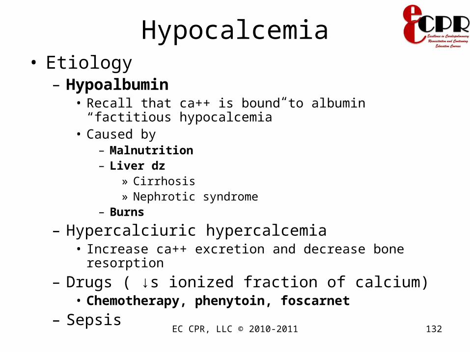
Hypocalcemia• Etiology
– Hypoalbumin• Recall that ca++ is bound to albumin “factitious
hypocalcemia”• Caused by
– Malnutrition– Liver dz
» Cirrhosis» Nephrotic syndrome
– Burns
– Hypercalciuric hypercalcemia• Increase ca++ excretion and decrease bone resorption
– Drugs ( ↓s ionized fraction of calcium)• Chemotherapy, phenytoin, foscarnet
– Sepsis

EC CPR, LLC © 2010-2011 133Mosby items and derived items © 2007 by Mosby, Inc., an affiliate of Elsevier, Inc.
Causes and Effects of Hypocalcemia

EC CPR, LLC © 2010-2011 134
Hypocalcemia: AssessmentHypocalcemia: Assessment• Manifestation
– Neuromuscular• Muscle twitches/tetany, seizure• Frequent painful muscle spasms at rest• Hyperactive deep tendon reflexes• Positive Chvostek’s sign (tap on facial nerve
triggers facial twitching)• Positive Trousseau’s sign (hand/finger spasms
with sustained blood pressure cuff inflation)

EC CPR, LLC © 2010-2011 135

EC CPR, LLC © 2010-2011 136

EC CPR, LLC © 2010-2011 137
Hypocalcemia: AssessmentHypocalcemia: Assessment
• Cardiovascular– Decreased myocardial contractility –
decreased heart rate and hypotension– Dysrhythmias, prolonged QT interval
• GI – hyperactive bowel sounds, diarrhea, abdominal cramping
• Respiratory– Laryngospasm– Respiratory failure
Mosby items and derived items © 2007 by Mosby, Inc., an affiliate of Elsevier, Inc.

EC CPR, LLC © 2010-2011 138
Hypocalcemia: Nsg InterventionsHypocalcemia: Nsg Interventions• Administer oral or IV calcium
supplements.– Aluminum hydroxide ↓s phosphorus levels →
↑calcium levels
– Vit. D to ↑ absorption calcium from the GI tract
• Encourage foods high in calcium including dairy products and dark green vegetables.
• Be aware of seizure precautions.Keep emergency equipment on standby.
Mosby items and derived items © 2007 by Mosby, Inc., an affiliate of Elsevier, Inc.

EC CPR, LLC © 2010-2011 139
Hypocalcemia: Nsg InterventionsHypocalcemia: Nsg Interventions
• Calcium gluconate IV administration– Warm solution to body temperature prior to
administration– Give slowly– Monitor EKG– Watch for infiltration

EC CPR, LLC © 2010-2011 140
Food source for calcium
• Cheese• Collard greens• Milk and soy milk• Rhubarb• Sardines• Spinach• Tofu• yogurt
EC CPR, LLC © 2010-2011 141
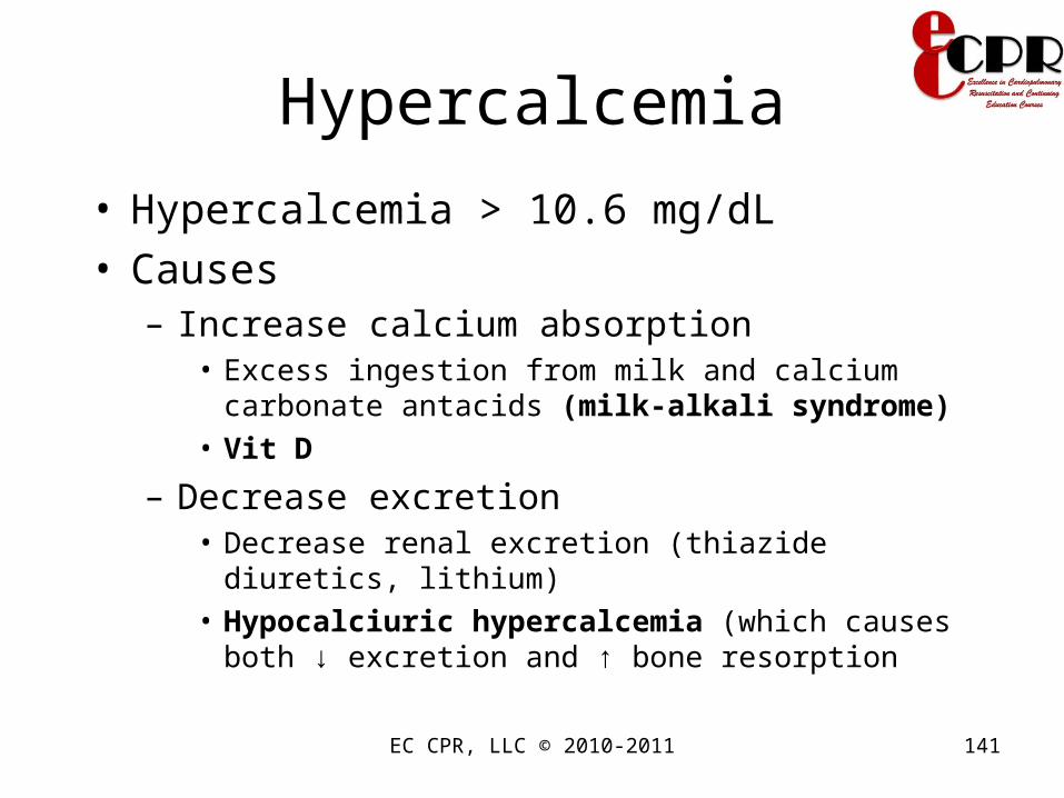
Hypercalcemia
• Hypercalcemia > 10.6 mg/dL• Causes
– Increase calcium absorption• Excess ingestion from milk and calcium carbonate
antacids (milk-alkali syndrome)• Vit D
– Decrease excretion• Decrease renal excretion (thiazide diuretics, lithium)• Hypocalciuric hypercalcemia (which causes both ↓
excretion and ↑ bone resorption

EC CPR, LLC © 2010-2011 142
Hypercalcemia
• Causes– Increase bone resorption
• Elevated parathyroid (hyperparathyroidism)• Elevated vit D (lymphoma, granulomatous
disease, milk-alkali syndrome)• Bone breakdown (secondary to mets to bone,
primary bone tumor, multiple myeloma, immobilization, hyperthyroidism)
– Hemoconcentration• Dehydration• Adrenal insufficiency

EC CPR, LLC © 2010-2011 143Mosby items and derived items © 2007 by Mosby, Inc., an affiliate of Elsevier, Inc.
Causes and Effects of Hypercalcemia
EC CPR, LLC © 2010-2011 144
Hypercalcemia
• Manifestation– “stones, bones, abdominal groans, moans,
and psychiatric overtones” means• Renal stones• Bone breakdown• Constipation/abdominal pain/ pancreatitis• Fatigue, weakness, arthralgia• Psychiatric disturbance
– Ekg: shorten QT interval • Mnemonic: Lots of Californians are short QTs
EC CPR, LLC © 2010-2011 145
Hypercalcemia: Nsg Interventions
• Calculate corrected calcium level if albumin is low– Serum calcium+0.8 (4-albumin)
• Monitor cardiac, respiratory status, neuromuscular, renal, GI status
• Place pt on cardiac monitor
• D/C IV or PO’s containing calcium, vit D, thiazide diuretic
• Prepare for dialysis if severe levels
• Position pt carefully due to risk of fracture
• Monitor for flank pain, strain urine

EC CPR, LLC © 2010-2011 146
Hypercalcemia: Nsg Interventions
• Treatment– Dilute blood Ca++
• 0.9% NaCl. Infuse 5-10 l over the 1st 24 hrs until dehydration is corrected
– Give furosemide IV (40-80mg q 2-4 hrs) to enhance excretion and prevent volume overload
EC CPR, LLC © 2010-2011 147
Hypercalcemia: Nsg Interventions
• Treatment– Metastatic bone dz
• Calcitonin (rapid, mild inhibition of bone resorption)• Pamidronate (delayed onset, but most potent than
calcitonin)
– Sarcoidosis, vit A or D intoxication, multiple myeloma, leukemia, breast ca
• Glucocorticoid (↑ ca++urinary excretion,↓ GI absorption of ca++
– Neoplasm• Resection, irridation
EC CPR, LLC © 2010-2011 148
Magnesium • Plays an important role in neuromuscular
function ( same as the effect of calcium )• 60% is in bone, and most of the remainder
is within cells• 1% is in ECF, serum levels do not reflect
total body Mg++ content; thus, levels have limited diagnostic value
• Normal 1.3-2.2 mEq/L• Excreted by the kidneys

Magnesium
• Imbalance of magnesium usually provokes alteration of calcium
• Hypermagnesemia can decrease PTH action ( hypocalcemia )– This is seen in pts being treated due to
preeclampsia
• Hypomagnesemia– Causes PTH resistance in end-organs and
eventually decreases PTH secretion EC CPR, LLC © 2010-2011 149

EC CPR, LLC © 2010-2011 150
HypomagnesemiaHypomagnesemia
• Serum magnesium level less than 1.3 mg/dL.
• Hypomagnesemia and hypokalemia shares the same etiologies
• Causes– ↓ intestinal absorption/insufficient intake
• Malnutrition, alcoholism• Prolonged diarrhea, laxative abuse• NGT aspiration, small bowel bypass• Malabsorption syndrome, Celiac, Crohn’s
disease

EC CPR, LLC © 2010-2011 151
HypomagnesemiaHypomagnesemia
• Causes– ↑ renal excretion
• Hypercalcemia• Osmotic diuresis• Loop diuretics• Aminoglycosides• Amphothericin B• Cisplatin• Cyclosporine• Hyperparathryroidism

EC CPR, LLC © 2010-2011 152
HypomagnesemiaHypomagnesemia
• Causes– Intracellular movement
• Hyperglycemia• Insulin administration• Sepsis
– Other• Hypoparathyroidism
Hypomagnesemia
• Diagnoses– Check urinary excretion, if > 10-30 mg/day for
renal wasting
EC CPR, LLC © 2010-2011 153

EC CPR, LLC © 2010-2011 154Mosby items and derived items © 2007 by Mosby, Inc., an affiliate of Elsevier, Inc.
Causes and Effects of Hypomagnesemia

EC CPR, LLC © 2010-2011 155
HypomagnesemiaHypomagnesemia
• Manifestation– Hypokalemia and hypocalcemia– Neurologic abnormalities
• Lethargy, Confusion• Tremor, Fasciculations• Ataxia, Nystagmus• Tetany, Seizure
– EKG • Prolonged PR and QT interval →
atrial and ventricular arrhythmia (esp if on digoxin)
EC CPR, LLC © 2010-2011 156
Hypomagnesemia: AssessmentHypomagnesemia: Assessment
• Neuromuscular – increased nerve impulse transmission (hyperactive deep tendon reflexes, paresthesias, muscle tetany), positive Chvostek’s and Trousseau’s signs
• GI – hypoactive bowel sounds, constipation, abdominal distention, paralytic ileus
Mosby items and derived items © 2007 by Mosby, Inc., an affiliate of Elsevier, Inc.

EC CPR, LLC © 2010-2011 157
Hypomagnesemia: Nsg Hypomagnesemia: Nsg InteventionsInteventions
• Discontinue magnesium-losing medications, such as loop diuretics.
• Administer oral or IV magnesium sulfate following safety protocols (check DTR for hyporeflexia)– Magoxide 250-500mg qd or bid– Mag sulfate 1-2 g IV, followed by 6g in 1liter/24hrs– Mag sulfate 200-800 mg/d IM in 4 divided doses
• Encourage foods high in magnesium, including dairy products and dark green vegetables
Mosby items and derived items © 2007 by Mosby, Inc., an affiliate of Elsevier, Inc.
EC CPR, LLC © 2010-2011 158
Hypomagnesemia: Nsg Hypomagnesemia: Nsg InteventionsInteventions
• Monitor cardiac, respiratory, neuromuscular, GI status
• Place pt on cardiac monitor
• Implement seizure precaution

Hypermagnesemia
• Serum magnesium level of >2.1mEq/dL
• Etiology– Decrease renal excretion
• Renal failure
– Increase intake• After therapy with Antacid or laxatives
– Maalox, Mylanta, Camalox, and Riopan
• During preeclampsia treatment with magnesium• Aspiration of sea water
EC CPR, LLC © 2010-2011 159

EC CPR, LLC © 2010-2011 160Mosby items and derived items © 2007 by Mosby, Inc., an affiliate of Elsevier, Inc.
Causes and Effects of Hypermagnesemia

EC CPR, LLC © 2010-2011 161
Hypermagnesemia
• Manifestation– s/sx on seen if Mg++ level is > 4 mEq/L– Impaired neuromuscular transmission
• Areflexia
– Lethargy, weakness, paralysis, respiratory failure
– Hypotension, bradycardia– EKG
• Prolonged PR, QRS widen, peaked T wave (hyperkalemia) , shorten QT → complete heart block, asystole

EC CPR, LLC © 2010-2011 162
Hypermagnesemia: Nsg InteventionsHypermagnesemia: Nsg Inteventions
• Teach pt to avoid abuse of laxatives and antacids
• Instruct pts with renal problems to avoid OTC meds that contain Mg++
• Judicious fluid intake• Monitor I/Os• Administer diuretics as ordered, calcium
gluconate 10-20ml IV over 10 minutes (antagonizes effects of magnesium)
• Dialysis

EC CPR, LLC © 2010-2011 163
Food sources for Mg++
• Avocado• Canned white tuna• Cauliflower• Green leafy
vegetables (spinach/broccoli)
• Milk• Oatmeal
• Peanut butter• Peas• Pork, beef, chicken• Potatoes• Raisins• Yogurt

EC CPR, LLC © 2010-2011 164
Phosphorus
• Critical for bone formation and cellular energy metabolism
• 85% of body phosphorus is in bone, remainder is within cells
• Only 1% is in ECF (do not reflect total body phosphorus stores)
• Phosphorus exists in the body as phosphate• Normal range 3.0-4.5 mg/dL• Must be drawn in fasting state (diurnal variation;
nadir in a.m.)
EC CPR, LLC © 2010-2011 165
Phosphorus
• Carbohydrate ingestion and glucose infusion lower serum phosphorus (do fasting), whereas high-phosphate meal raises it
• Major regulatory factors– PTH (lowers PO4 - via renal excretion)– Vit D ( increase PO4- by enhancing intestinal
absorption)– Insulin (lowers by shifting PO4- into cells– Dietary PO4- intake– Renal function (most important regulator)

EC CPR, LLC © 2010-2011 166
Hypophosphatemia
• Serum PO4- level of < 3mg/dL• Normal 2.7-4.5• Etiology
– Impaired intestinal absorption• Alcohol abuse and withdrawal ( beta
adrenergic effect )• Malabsorption• Oral phosphate binders• Refeeding after malnutrition,Hyperalimentation• Vit D deficiency
EC CPR, LLC © 2010-2011 167
Hypophosphatemia
• Etiology– Increased renal excretion
• Respiratory alkalosis• hyperPTH ( hypercalcemia; hypomagnesemia)
– Redistribution into cells• Severe burns• DKA therapy

EC CPR, LLC © 2010-2011 168
Hypophosphatemia
• Manifestation– Muscular abnormalities
• Weakness• Rhabdomyolysis• Impaired diaphragmatic function• Respiratory failure ( strong affinity of hgb to
oxygen d/t decrease of erythrocyte 2,3 diphosphoglycerate)
• Heart failure• Fractures, decrease reflex

EC CPR, LLC © 2010-2011 169
Hypophosphatemia
• Manifestation– Neurologic
• Paresthesia• Dysarthria• Confusion• Stupor• Sz• Coma
– Hemolysis, plt dysfunction, metabolic acidosis (rare)
– Rickets and osteomalacia in chronic hypophosphatemia

EC CPR, LLC © 2010-2011 170
Hypophosphatemia: Nsg InteventionsHypophosphatemia: Nsg Inteventions
• Assess for vit D deficiency, hyperPTH
• Assess for overuse of aluminum-containing antacids
• Elemental phosphorus 0.5-1.0 g bid or tid– Neutra-phos or neutra-phos K– Fleet phospho-Soda
• Check crt, phosphorus, ca++• SE: diarrhea, nausea
– Potasium phosphate or sodium phosphate IV TRO 6 hrs

EC CPR, LLC © 2010-2011 171
Hyperphosphatemia
• Serum PO4- > 4.5 mg/dL
• Etiology– Renal failure– hypoPTH– pseudohypoPTH– Rhabdomyolysis– Tumor lysis syndrome– Metabolic and respiratory acidosis– Excess phosphate administration

EC CPR, LLC © 2010-2011 172
Hyperphosphatemia• Manifestation
– s/sx are attributable to hypocalcemia and metastatic calcification of soft tissues
• BVs, cornea, skin, kidney, periarticular tissue
– Please refer to hypocalcemia for s/sx
• InterventionAdminister phosphate-binders that increase
fecal excretion of phosphorus• Calcium carbonate tid with meals• Sevelamer (avoids hypermag, calcemia, aluminum
toxicity)– Also lowers total chol– SE: GI complaints and metabolic acidosis

EC CPR, LLC © 2010-2011 173
Hyperphosphatemia
• Intervention– Aluminum hydroxide and aluminum carbonate
• No longer used in dialysis pts because of toxicity
– Calcium citrate should not be use with aluminum gels because it increases aluminum toxicity
– Instruct pt to avoid phosphate containing meds-laxatives and enemas
– Restrict dietary phosphate to 600-900mg/day
EC CPR, LLC © 2010-2011 174
Foods rich in phosphorus
• Fish
• Organ meats
• Nuts
• Pork, beef, chicken
• Whole-grain breads and cereals

EC CPR, LLC © 2010-2011 175
Anion ImbalancesImbalances of chloride, phosphate, and
bicarbonate accompany cation imbalances:
• Hypochloremia (below 95 mEq/L) and hyponatremia
• Hyperchloremia (above 103 mEq/L) with hypernatremia and metabolic acidosis

EC CPR, LLC © 2010-2011 176
Anion ImbalancesImbalances of chloride, phosphate, and
bicarbonate accompany cation imbalances
• Hypophosphatemia (below 3.0 mg/dL): – Aluminum-containing antacids that bind
phosphate– Vitamin D deficiency– Hyperparathyroidism
• Hyperphosphatemia (above 4.5 mg/dL) and renal failure

EC CPR, LLC © 2010-2011 177
Of all of the following patients, the nurse Of all of the following patients, the nurse recognizes that the individual who is most at recognizes that the individual who is most at
risk for a fluid volume deficit is a:risk for a fluid volume deficit is a:
• 6-month-old infant learning to drink from a cup
• 12-year-old child who is moderately active in 80° F weather
• 42-year-old patient with severe diarrhea
• 90-year-old patient with frequent headaches
Mosby items and derived items © 2007 by Mosby, Inc., an affiliate of Elsevier, Inc.

EC CPR, LLC © 2010-2011 178
The nurse anticipates that the The nurse anticipates that the patient with a fluid volume patient with a fluid volume excess will manifest a(n):excess will manifest a(n):
• Increased urine specific gravityIncreased urine specific gravity
• Decreased body weightDecreased body weight
• Increased blood pressureIncreased blood pressure
• Decreased pulse strengthDecreased pulse strength
Mosby items and derived items © 2007 by Mosby, Inc., an affiliate of Elsevier, Inc.