ebus-fnab: how to optimize your cytology samples, lhsc ... · ebus-fnab: how to optimize your...
TRANSCRIPT

EBUS-FNAB: HOW TO
OPTIMIZE YOUR CYTOLOGY
SAMPLES, LHSC
EXPERIENCE
Dr. Mariamma Joseph
Division Head of Cytopathology
LHSC and Western University

Objectives
• Brief overview of EBUS-FNA
• Strategies to optimize cytology samples
• Role of adequacy assessment (ROSE)
• Terminology, diagnostic challenges
• Lessons learned from LHSC • Where we were and where we are now…

Lung Cancer: Facts
• Lung cancer- leading cause of cancer death (male and female)
• In Canada 21,700 new cases and18,900 deaths/yr
• Mediastinal nodal staging is a key modality in management

Mediastinal Nodal Staging
• Imaging modalities – without tissue sampling • CT
• PET
• Integrated PET-CT
• Modern methods - with tissue sampling to improve staging accuracy
ENM – electromagnetic navigational biopsy (latest, very expensive)
Method Sensitivity Specificity
Mediastinoscopy 81 100
EBUS-FNA 89 100
EUS- FNA 89 100
EBUS+EUS FNA 91 100

Nodal Stations/Procedures

EBUS – FNA Procedure

EBUS – FNA Procedure

EBUS FNA: Advantages
• Minimally invasive procedure, cost effective
• Safe in experienced hands (major complication 0.15%)
• Excellent sensitivity, specificity, PPV, NPV
• Can sample lung and mediastinal nodes
• Lung ca including post chemo staging
• Metastatic cancer, melanoma, lymphoma (76% sensitivity)
• Granulomatous disease
• Sarcoid (86% sensitivity)
• Infection

EBUS Procedure at LHSC
• Started by Dr. Dave McCormack, respirologist (2010)
• Now 4 thoracic surgeons, 2 respirologists
• EBUS (6) EUS (2)
• 250 cases (2014)
• Standardized cytology practice

ROSE AND EBUS -FNA
The value of ROSE for EBUS-FNA is well recognized. A
recent metanalysis showed that with ROSE, sensitivity
is 88% (80% without)

DEBATE IS AROUND WHETHER ROSE SHOULD BE
PERFORMED FOR EVERY PROCEDURE OR ONLY
SELECTIVELY?
LHSC – ROSE DONE ON ALL CASES (8 CTS)
Mean # of passes = 4 (2-9)
Usually on 1- 2 samples
ROSE time: 25-55 minutes

Dr. McCormack, LHSC (300 cases)
For the diagnosis of malignancy our results show:
• Sensitivity: 95%
• Specificity: 100%
• Positive predictive value: 100%
• Negative predictive value: 93%
We are very happy with these results. Thanks to you and
your staff for being so supportive and helpful.
Dr. D. McCormack

It is well recognized that ……
• Success of EBUS-FNA depends on the combined skill
and competency of the pulmonologists, cytotechnologists
and cytopathologists – Total health care team
Clinicians
Pathologists Cytotechs
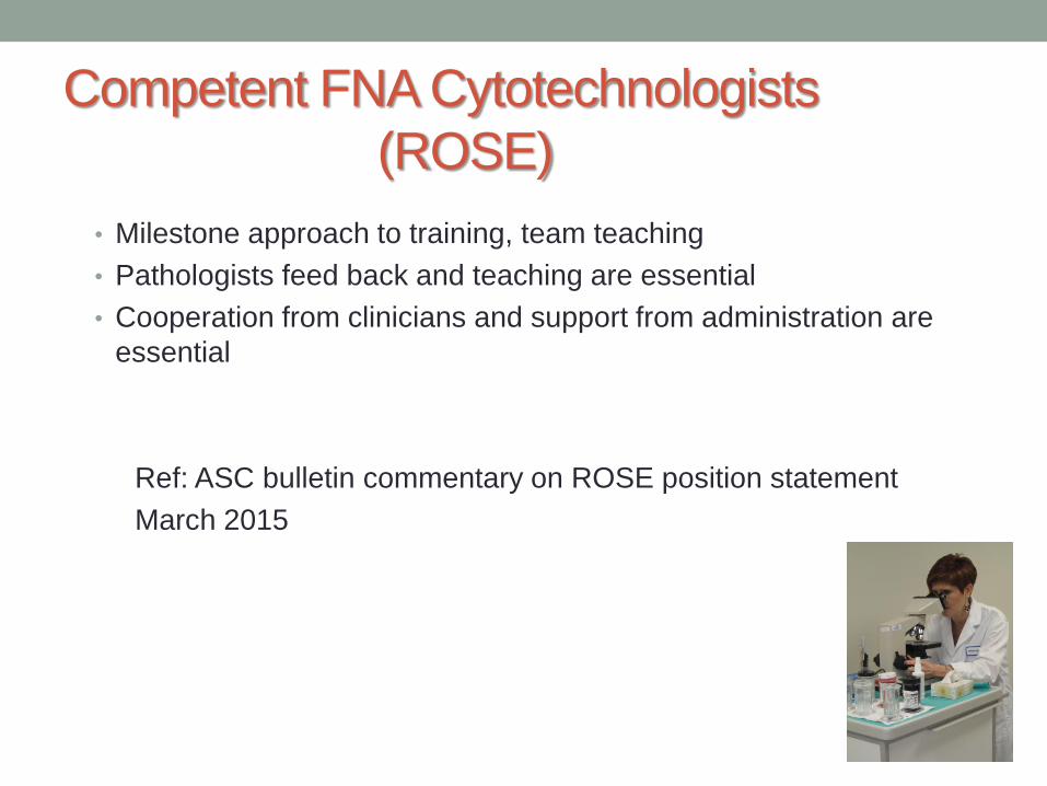
Competent FNA Cytotechnologists
(ROSE)
• Milestone approach to training, team teaching
• Pathologists feed back and teaching are essential
• Cooperation from clinicians and support from administration are
essential
Ref: ASC bulletin commentary on ROSE position statement
March 2015

Communication During ROSE
• ADEQUATE
• Specimen sample is adequate
• Specimen sample is adequate but please obtain more material for • Cell block
• FCM
• Microbiology
• NOT ADEQUATE
• Specimen sample is NOT adequate
• There is nothing here
• I need more, low cellularity
• We need to go back
Cytotechs cannot provide a diagnosis

EBUS FNA: OPTIMIZING
SAMPLES AND REPORTING
Sample processing
Adequacy
Terminology
Ancillary tests

FNAB + Needle
Rinses
Place all samples
in CytoLyt solution
Place some material
on a slide. Make 2
smears per pass. Air
dry one slide for Diff-
Quik stain and Rapid
On-site Evaluation.
Spray fix second slide
for Pap stain and final
evaluation
Thin Prep Slide
Cell Block
Pass 1 &
Pass 3 (as
needed)
Pass 2, 4 and Beyond
Pass 1&3 Needle
Rinses
Pap s
tain
D
iff Quik
Cytology Lab
For suspected
lymphoma, place
a sample in
Flow medium
Olymbus Needle

A: Formalin CB B: Formalin CB C: CytoLyt CCB vs HCB
Assess cellularity and select the appropriate block for immuno and molecular tests
ProCore Needle

Adequacy for Nodal Sampling
Criteria not well established
• 40 lymphocytes per HPF in cellular areas of slide
or pigmented macrophage clusters - Alsharif et al
Our criteria
Abundant lymphocytes >100/LPF in 5 fields or abundant
dust laden macrophage clusters and scant respiratory cells


Adenocarcinoma Small Cell Carcinoma

Cell Block, Granuloma, likely Sarcoid

LUNG CANCER REPORTING
TERMINOLOGY

Diagnostic Terminology in Cytology
• NE carcinomas
• Small cell carcinoma
• Possible LCNEC (immuno +)
• NSCLC with NE morphology
• Adenocarcinoma
• Squamous cell carcinoma
• NSCLC favour squamous cell ca (immuno +)
• NSCLC favour adenocarcinoma (immuno+)
• NSCLC NOS (use only rarely, immuno inconclusive)
Ref: Travis: Arch Pathol Lab Med. 2013;137:668–684
IASLC/ATS/ERS Classification –for details

Non small cell carcinoma
Marker Adenocarcinoma Squamous cell ca
TTF1 * + _
CK5/6, p63* or p40 - +
Napsin + -
+ -
Molecular EGFR or ALK + -
Arch Pathol Lab Med. 2013;137:668–684
Mucin*
- *Used at LHSC
- * mucin may or may not be used, PASD or mucicarmine
- If mixed immuno pattern call NSCLC NOS, may represent adenosquamous ca

Adenocarcinoma
TTF-1
TTF-1 Negative
P63 Positive
Squamous Cell Ca

Malignant Cells in Cell Block
Semi-quantitation by CTs
• Scant <10 cells
• Low 10-50 cells
• Medium 50-100 cells
• High >100 cells
• This would allow oncologists and pathologists to order
additional molecular tests (e.g. EGFR) on appropriate
samples, i.e. cell blocks with medium and high cellularity
are preferred.

EBUS-EUS Interpretation Challenges
• Cellular contaminants (en route)
• Tracheobronchial columnar cells
• Seromucinous glands, cartilage
• Esophageal squamous cells - EUS
• Atypical/dysplastic epithelium
• Reserve cell hyperplasia
• Mucinous metaplasia
• Epithelioid histiocytes
• Crushed lymphocytes
Can be challenging during ROSE
Tracheo bronchial
Esophagus

MOLECULAR TESTING:
EGFR, ALK Priority for NSCLC stage 4 cancers
Optimize sample size

Cell block: low cellularity
10-50 cells
Adenocarcinoma – LHSC 2010…still learning

Cell block cellularity:
>100 cells (high)
TTF-1 +, p63 –ve
Adequate sample for
EGFR and ALK testing
Adenocarcinoma, LHSC - 2014
DO NOT TRIM SIGN
HISTOGEL CB

Metastatic Melanoma 73 F, EBUS - FNA Subcarinal (#7) Node
HMB 45

EBUS FNA Practice - Summary
• Multidisciplinary team work, emphasize team teaching
• Use standardized procedures & diagnostic terminology
• Multidisciplinary Tissue Management Strategy
• Perform multiple aspirations (3-6)
• ROSE only on 2 samples, rest in needle rinse
• Optimize cell block (Histogel), avoid excessive trimming
• Minimal immuno for cell typing: p63/p40, TTF-1
• Preserve as much tissue for NSCLC molecular testing

Thank You