ebola virus uses clathrin-mediated endocytosis as … virus uses clathrin-mediated endocytosis as an...
TRANSCRIPT

Ebola virus uses clathrin-mediated endocytosis as an entry pathway
Suchita Bhattacharyya a,b,1, Kelly L. Warfield c,2, Gordon Ruthel c, Sina Bavari c,M. Javad Aman c,2, Thomas J. Hope a,⁎a Department of Cell and Molecular Biology, Feinberg School of Medicine, Northwestern University, 303 East Chicago Avenue, Chicago, IL 60611, USAb Department of Microbiology and Immunology, University of Illinois at Chicago, 835 South Wolcott Avenue, Chicago, IL 60612, USAc US Army Medical Research Institute of Infectious Diseases (USAMRIID), 1425 Porter Street, Frederick, MA 21702, USA
a b s t r a c ta r t i c l e i n f o
Article history:Received 21 December 2009Returned to author for revision15 January 2010Accepted 9 February 2010Available online 3 March 2010
Keywords:Ebola virusEntryClathrin endocytic pathwayChlorpromazineSucroseEps15
Ebola virus (EBOV) infects several cell types and while viral entry is known to be pH-dependent, the exactentry pathway(s) remains unknown. To gain insights into EBOV entry, the role of several inhibitors ofclathrin-mediated endocytosis in blocking infection mediated by HIV pseudotyped with the EBOV envelopeglycoprotein (EbGP) was examined. Wild type HIV and envelope-minus HIV pseudotyped with VesicularStomatitis Virus glycoprotein (VSVg) were used as controls to assess cell viability after inhibiting clathrinpathway. Inhibition of clathrin pathway using dominant-negative Eps15, siRNA-mediated knockdown ofclathrin heavy chain, chlorpromazine and sucrose blocked EbGP pseudotyped HIV infection. Also, bothchlorpromazine and Bafilomycin A1 inhibited entry of infectious EBOV. Sensitivity of EbGP pseudotyped HIVas well as infectious EBOV to inhibitors of clathrin suggests that EBOV uses clathrin-mediated endocytosis asan entry pathway. Furthermore, since chlorpromazine inhibits EBOV infection, novel therapeutic modalitiescould be designed based on this lead compound.
© 2010 Elsevier Inc. All rights reserved.
Introduction
Ebola virus (EBOV) causes a severe, fatal hemorrhagic fever withmortality rates as high as 90% (Feldmann et al., 2003). There iscurrently no effective anti-viral therapy available for EBOV infection.EBOV is an enveloped, negative-sense, single stranded RNA virus(Regnery et al., 1980) with a broad species and cell-type tropism(Wool-Lewis and Bates, 1998). The envelope glycoprotein (GP) isresponsible for entry (Elliott et al., 1985). Very little is known aboutthe pathway used by EBOV to enter target cells and its receptor(s)remains unidentified. It is known that EBOV entry in the context ofpseudotypes is pH-dependent (Chazal et al., 2001). Depletion ofmembrane cholesterol by β-methyl cyclodextrin inhibits EBOVinfection in a dose dependent manner (Yonezawa et al., 2005; Bavariet al., 2002). There have been conflicting reports on the endocyticpathway(s) used by EBOV to enter target cells. One group suggestedthat Ebola enters cells via caveolar uptake (Empig and Goldsmith,2002) and uses the folate receptor alpha as a cofactor for EbGP
mediated fusion (Chan et al., 2001). Another study has suggested thatEBOV uses clathrin as well as caveolae-mediated endocytosis for entryusing various chemical inhibitors of these pathways (Sanchez, 2007).However, most of the inhibitors caused considerable cell detachmentin this study and it was conducted only in Vero cells so cell-typespecific effects could not be ruled out. In contrast, two reports hadpreviously shown that neither caveolae nor folate receptor alpha arerequired for EbGP fusion function (Simmons et al., 2003; Sinn et al.,2003). In light of these controversies more analysis is required.
The major endocytic pathways include clathrin-mediated endo-cytosis, uptake by caveolae, macropinocytosis and phagocytosis. Somenon-clathrin (Nemerow and Cooper, 1984), non-caveolae-mediatedviral endocytic pathways have also been reported (Sanchez-SanMartin et al., 2004) but they are not well described.
Among all the known pathways of viral endocytosis, clathrin-mediated endocytosis is predominant. Key proteins involved inclathrin-mediated endocytosis include clathrin, Adaptor ProteinComplex-2 (AP-2) and epidermal growth factor receptor (EGFR)pathway substrate clone 15 (Eps15). Clathrin assembles into apolyhedral lattice on the inner side of the plasma membrane to formthe coated pit. Eps15 acts as an adaptor between the clathrin coatsand AP-2 (Keen, 1987). An AP-2 independent pathway for clathrin-mediated endocytosis has also been reported (Lakadamyali et al.,2006). Eps15 comprises of three structural domains: an N-terminalregion (DI) composed of three Eps15 homology regions, a centralcoiled-coil domain (DII) involved in oligomerization and a C-terminalregion (DIII) which contains the AP-2 binding sites. DI is required for
Virology 401 (2010) 18–28
⁎ Corresponding author. Department of Cell and Molecular Biology, Feinberg Schoolof Medicine, Northwestern University, Ward 8-140, 303 East Chicago Avenue, Chicago,IL 60611, USA. Fax: +1 312 503 2696.
E-mail address: [email protected] (T.J. Hope).1 Present address: Nomis Center for Immunobiology andMicrobial Pathogenesis, The
Salk Institute for Biological Studies, 10010 North Torrey Pines Road, La Jolla, CA 92037,USA.
2 Present address: Integrated BioTherapeutics, Inc., 20358 Seneca Meadows Parkway,Germantown, MA 20876, USA.
0042-6822/$ – see front matter © 2010 Elsevier Inc. All rights reserved.doi:10.1016/j.virol.2010.02.015
Contents lists available at ScienceDirect
Virology
j ourna l homepage: www.e lsev ie r.com/ locate /yv i ro
Report Documentation Page Form ApprovedOMB No. 0704-0188
Public reporting burden for the collection of information is estimated to average 1 hour per response, including the time for reviewing instructions, searching existing data sources, gathering andmaintaining the data needed, and completing and reviewing the collection of information. Send comments regarding this burden estimate or any other aspect of this collection of information,including suggestions for reducing this burden, to Washington Headquarters Services, Directorate for Information Operations and Reports, 1215 Jefferson Davis Highway, Suite 1204, ArlingtonVA 22202-4302. Respondents should be aware that notwithstanding any other provision of law, no person shall be subject to a penalty for failing to comply with a collection of information if itdoes not display a currently valid OMB control number.
1. REPORT DATE 9 FEB 2010
2. REPORT TYPE N/A
3. DATES COVERED -
4. TITLE AND SUBTITLE Ebola virus uses clathrin-mediated endocytosis as an entry pathway.Virology 401:18-29
5a. CONTRACT NUMBER
5b. GRANT NUMBER
5c. PROGRAM ELEMENT NUMBER
6. AUTHOR(S) Bhattacharyya, S Warfield, KL Ruthel, G Bavari, S Aman, MJ Hope, TJ
5d. PROJECT NUMBER
5e. TASK NUMBER
5f. WORK UNIT NUMBER
7. PERFORMING ORGANIZATION NAME(S) AND ADDRESS(ES) United States Army Medical Research Institute of Infectious Diseases,Fort Detrick, MD
8. PERFORMING ORGANIZATIONREPORT NUMBER TR-06-096
9. SPONSORING/MONITORING AGENCY NAME(S) AND ADDRESS(ES) 10. SPONSOR/MONITOR’S ACRONYM(S)
11. SPONSOR/MONITOR’S REPORT NUMBER(S)
12. DISTRIBUTION/AVAILABILITY STATEMENT Approved for public release, distribution unlimited
13. SUPPLEMENTARY NOTES The original document contains color images.
14. ABSTRACT Ebola virus (EBOV) infects several cell types and while viral entry is known to be pH-dependent, the exactentry pathway(s) remains unknown. To gain insights into EBOV entry, the role of several inhibitors ofclathrin-mediated endocytosis in blocking infection mediated by HIV pseudotyped with the EBOVenvelope glycoprotein (EbGP) was examined. Wild type HIV and envelope-minus HIV pseudotyped withvesicular stomatitis virus glycoprotein (VSVg) were used as controls to assess cell viability after inhibitingclathrin pathway. Inhibition of clathrin pathway using dominant-negative Eps15, siRNA-mediatedknockdown of clathrin heavy chain, chlorpromazine and sucrose blocked EbGP pseudotyped HIVinfection. Also, both chlorpromazine and Bafilomycin A1 inhibited entry of infectious EBOV. Sensitivity ofEbGP pseudotyped HIV as well as infectious EBOV to inhibitors of clathrin suggests that EBOV usesclathrin-mediated endocytosis as an entry pathway. Furthermore, since chlorpromazine inhibits EBOVinfection, novel therapeutic modalities could be designed based on this lead compound.
15. SUBJECT TERMS filovirus, Ebola, entry pathway, clathrin endocytic pathway, chlorpromazine, sucrose, Epst5
16. SECURITY CLASSIFICATION OF: 17. LIMITATION OF ABSTRACT
SAR
18. NUMBEROF PAGES
11
19a. NAME OFRESPONSIBLE PERSON
a. REPORT unclassified
b. ABSTRACT unclassified
c. THIS PAGE unclassified
Standard Form 298 (Rev. 8-98) Prescribed by ANSI Std Z39-18

correct coated pit targeting of Eps15. It was shown that in cellsexpressing mutants lacking Eps15 homology domains, punctatedistribution of AP-2 and clathrin is lost and endocytosis of transferrin,which is a specific marker of clathrin-mediated endocytosis (Hanoveret al., 1984) is inhibited (Benmerah et al., 1999). Certain chemicalagents such as chlorpromazine (Wang et al., 1993) and sucrose(Heuser and Anderson, 1989) are also known to prevent recycling ofclathrin to the plasma membrane thereby inhibiting clathrin-mediat-ed endocytosis.
In this study we examined whether EBOV requires the clathrin-mediated endocytic pathway for entry. Our results demonstratethat EbGP pseudotyped HIV as well as replication-competent EBOVuse clathrin-mediated endocytosis as an entry pathway. We havealso shown that chlorpromazine significantly inhibits replication-competent EBOV infection and hence is a potential lead candidate fordevelopment of anti-viral therapy against EBOV infection.
Results
EbGP mediated entry of the HIV pseudotype is pH-dependent
To analyze the pathway of Ebola virus entry, we used an envelope-minus HIV pseudotyped with the Ebola virus envelope glycoprotein(EbGP). For this analysis we utilized two control viruses, HIV using itsnative envelope and envelope-minus HIV pseudotyped with VSVg.HIV entry is pH-independent (Stein et al., 1987), while entrymediated by VSVg pseudotyped HIV is pH-dependent (Matlin et al.,1982). The control viruses were used to demonstrate target cellviability and specificity of the inhibitory disruption of the clathrinpathway. In all cases, the viral genomes contained the GFP genelocated in the Nef position within the HIV genome. Therefore, infectedcells could be readily identified for GFP expression using flowcytometry or microscopy. The initial analysis was designed to confirmthe previously reported role of acidification in the function of EbGP,VSVg and HIV envelopes. Vacuolar ATPases hydrolyze ATP, creating aproton gradient that causes acidification of endosomes. BafilomycinA1 (Baf A1) is a potent inhibitor of vacuolar ATPases and therebyprevents acidification of endosomes (Bowman et al., 1988). For thisanalysis, the titer of the different viral stocks was determined usinginfectivity studies. In this study, the different stocks were added atamounts that gave comparable levels of infectivity. When normalizedby p24 content, the VSVg pseudotyped virus was typically 25 timesmore infectious than replication-competent HIV and 79 times moreinfectious than the EbGP pseudotyped HIV after 4 h of exposure totarget cells.
Baf A1 treatment reduced both EbGP and VSVg mediated infec-tivity to near background levels, while HIV infectivity showed a minorincrease compared to the untreated control (Fig. 1). These resultsvalidated the functionality of the pseudotyping system and infectivityassays since a previous study has also reported that treatmentwith Baf A1 significantly inhibited EbGP and VSVg pseudotyped viralinfection, while enhancing HIV infection (Chazal et al., 2001).
Inhibition of EbGP mediated entry by chlorpromazine
To investigate the potential role of clathrin-mediated endocytosis,we first determined the ability of the drug chlorpromazine to inhibitEbGP mediated viral entry. Chlorpromazine is known to sequesterclathrin, causing it to assemble aroundendosomalmembranes therebypreventing recycling of clathrin to the plasmamembrane (Wang et al.,1993). A previous study used 10 μg/ml of chlorpromazine todetermine the involvement of clathrin pathway in influenza virusentry (Sieczkarski and Whittaker, 2002).
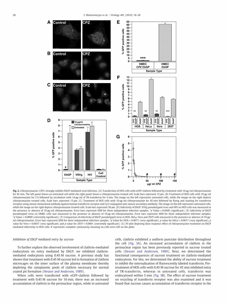
To determine the effect of chlorpromazine treatment on clathrinlocalization, cells were transiently transfected with eGFP-clathrin.After 24 h, the cells were treated with 10 μg/ml of chlorpromazine
for 45 min, fixed and analyzed by fluorescent deconvolution mi-croscopy. In the treated cells, clathrin was seen in random clustersin the cytoplasm, while in the untreated cells, a distinct punctatedistribution of clathrin was seen throughout the cell (Fig. 2A). Todetermine the functional consequences of chlorpromazine treat-ment, we examined if the endocytosis of transferrin, a knownmarker for clathrin-mediated endocytosis (Hanover et al., 1984),was altered. Fluorescently labeled transferrin was added to HOScells either pre-treated with 10 μg/ml chlorpromazine for 2 h oruntreated. After 5 min of incubation, the cells were rinsed to removeany unbound transferrin and fixed for fluorescent deconvolutionmicroscopy analysis. As shown in Fig. 2B, fluorescent transferrinwas localized in a perinuclear compartment, most likely recyclingendosomes, in untreated cells. In contrast, no internalized trans-ferrin was detected in the chlorpromazine treated cells. Thesestudies demonstrate that 10 µg/ml chlorpromazine potently inhibitsclathrin-mediated endocytosis. We also examined the effect ofchlorpromazine on the recycling of transferrin receptor and foundthat transferrin receptor is predominantly localized in the peri-nuclear region following treatment with chlorpromazine (Fig. 2C),while in the untreated cells, transferrin receptor is uniformly dis-tributed all over the cell. This result explained the absence of anytransferrin on the cell surface after chlorpromazine treatment inFig. 2B.
Next, we determined the effect of chlorpromazine treatment oninfection mediated by the EbGP, VSVg and HIV envelopes in HOS cells.As seen in Fig. 2D, treatment with 10 µg/ml chlorpromazine potentlyinhibited EbGP pseudotyped HIV infection without having any effecton infection with either wt HIV or VSVg pseudotyped HIV. The normalinfectivity by wt HIV and VSVg pseudotyped HIV demonstrated thatthe chlorpromazine treatment was not toxic to the target cells. Theeffect of chlorpromazine on EbGPmediated infectivity was also testedin HeLa, Vero, 293T and HMEC cell lines to determine whether theeffect was cell-type specific for HOS cells or could be seen in other celltypes including endothelial cells, which are one of the target cells forfiloviral infection. Chlorpromazine severely inhibited EbGP mediatedinfectivity in HMEC (Fig. 2E) and also HeLa, Vero and 293T cells(Fig. 2F), which suggests that inhibition of EbGP mediated infectivityby chlorpromazine is not restricted to a certain cell type but isprevalent among different cell types. The inhibitory effect ofchlorpromazine on EbGP mediated infectivity was found to be dosedependent for doses of 10 μg/ml or lower with maximum inhibitoryeffect seen at 10 μg/ml (Fig. 2G).
Fig. 1. Importance of acidification on EbGP mediated entry. Infectivity of EbGP, VSVgpseudotyped virus and HIV on HOS cells in the presence or absence of 50 nMBafilomycin A1 (Baf A1). Error bars represent SEM for three independent infectionsamples. *p Value for EbGP=0.0003 (extremely significant) and p value forVSVg=0.0003 (extremely significant).
19S. Bhattacharyya et al. / Virology 401 (2010) 18–28
Inhibition of EbGP mediated entry by sucrose
To further explore the observed involvement of clathrin-mediatedendocytosis on entry mediated by EbGP, we inhibited clathrin-mediated endocytosis using 0.45 M sucrose. A previous study hasshown that treatment with 0.45 M sucrose led to formation of clathrinmicrocages on the inner surface of the plasma membrane therebydepleting the cytoplasmic pools of clathrin necessary for normalcoated pit formation (Heuser and Anderson, 1989).
When cells were transfected with eGFP-clathrin followed bytreatment with 0.45 M sucrose for 10 min, there was an increasedaccumulation of clathrin in the perinuclear region, while in untreated
cells, clathrin exhibited a uniform punctate distribution throughoutthe cell (Fig. 3A). An increased accumulation of clathrin in theperinuclear region has been previously reported in sucrose treatedcells (Heuser and Anderson, 1989). Next, we determined thefunctional consequences of sucrose treatment on clathrin-mediatedendocytosis. For this, we determined the ability of sucrose treatmentto inhibit the internalization of fluorescently labeled transferrin. Pre-treatment of HOS cells with 0.45 M sucrose for 45 min inhibited entryof TR-transferrin, whereas in untreated cells, transferrin wasendocytosed within 5 min (Fig. 3B). The effect of sucrose treatmenton recycling of transferrin receptor was also examined and it wasfound that sucrose causes accumulation of transferrin receptor in the
Fig. 2. Chlorpromazine (CPZ) strongly inhibits EbGP mediated viral infection. (A) Transfection of HOS cells with eGFP-clathrin followed by treatment with 10 μg/ml chlorpromazinefor 45 min. The left panel shows an untreated cell while the right panel shows a chlorpromazine treated cell. Scale bars represent 15 μm. (B) Treatment of HOS cells with 10 μg/mlchlorpromazine for 2 h followed by incubation with 14 μg/ml of TR-transferrin for 5 min. The image on the left represents untreated cells, while the image on the right depictschlorpromazine treated cells. Scale bars represent 15 μm. (C) Treatment of HOS cells with 10 μg/ml chlorpromazine for 45 min followed by fixing and staining for transferrinreceptor usingmousemonoclonal antibody against human transferrin receptor and Cy3-conjugated anti-mouse secondary antibody. The image on the left represents untreated cells,while the image on the right depicts chlorpromazine treated cells. Scale bars represent 30 μm. (D) Infectivity of EbGP, VSVg pseudotyped virus and HIV on HOS cells wasmeasured inthe presence or absence of 10 μg/ml chlorpromazine. Error bars represent SEM for three independent infection samples. *p Value=0.0449 (significant). (E) Infectivity of EbGPpseudotyped virus on HMEC cells was measured in the presence or absence of 10 μg/ml chlorpromazine. Error bars represent SEM for three independent infection samples.*p Value=0.0009 (extremely significant). (F) Comparison of infectivity of EbGP pseudotyped virus in HOS, HeLa, Vero and 293T cells measured in the presence or absence of 10 μg/ml chlorpromazine. Error bars represent SEM for three independent infection samples. *p Value for HOS=0.0071 (very significant), p value for HeLa=0.0017 (very significant), pvalue for Vero=0.0037 (very significant) and p value for 293T=0.0001 (extremely significant). (G) XY plot depicting dose response effect of chlorpromazine treatment on EbGPmediated infectivity in HOS cells. # represents complete cytotoxicity meaning no cells were left on the plate.
20 S. Bhattacharyya et al. / Virology 401 (2010) 18–28
perinuclear region (Fig. 3C) and this explains why transferrin is notpresent on the cell surface upon sucrose treatment in Fig. 3B.
Next, we determined the effect of sucrose treatment on infectionmediated by the EbGP, VSVg, and HIV envelopes. A potent decrease inEbGPmediated infectivity was observed in sucrose treated cells, whileVSVg and HIV infectivity remained unaffected when compared tountreated controls (Fig. 3D). Since VSVg and HIV infectivity wereunaltered, it suggests that the sucrose treatment was not toxic tothe cells. Furthermore, we investigated the effect of sucrose treatmenton EbGP mediated infectivity in HMEC cells and as seen in Fig. 3E,sucrose severely inhibited EbGP mediated infectivity in endothelial
cells, which are one of the target cells of filoviral infection. We alsocompared the effect of sucrose treatment on EbGP mediated infec-tivity in HOS, HeLa, Vero and 293T cells and as seen in Fig. 3F, sucrosesignificantly inhibited EbGP mediated infectivity in all these cell linesindicating that the inhibitory effect of sucrose was not restricted toHOS cells.
One surprising observation was that VSVg pseudotyped viralinfection was not inhibited by either chlorpromazine or sucrosetreatment since it has been previously reported that VSV entryis mediated by clathrin-mediated endocytosis (Sun et al., 2005).To further investigate this process, we evaluated a combination
Fig. 3. EbGPmediated viral infection is inhibited by sucrose. (A) Transient transfection of HOS cells with eGFP-clathrin followed by treatment with 0.45 M sucrose for 10 min. The leftpanel shows an untreated cell, while the right panel shows sucrose treated cells. Scale bars represent 15 μm. (B) Treatment of HOS cells with 0.45 M sucrose for 45 min followed byincubation with 14 μg/ml of TR-transferrin for 5 min. The image on the left represents untreated cells, while the image on the right shows sucrose treated cells. Scale bars represent15 μm. (C) Treatment of HOS cells with 0.45 M sucrose for 10 min followed by fixing and staining for transferrin receptor using mouse monoclonal antibody against humantransferrin receptor and Cy3-conjugated anti-mouse secondary antibody. The image on the left represents untreated cells, while the image on the right depicts sucrose treated cells.Scale bars represent 30 μm. (D) Infectivity of EbGP, VSVg pseudotyped virus and HIV on HOS cells was measured in the presence or absence of 0.45 M sucrose. Error bars representSEM for three independent infection samples. *p Value=0.0047 (very significant). (E) Infectivity of EbGP pseudotyped virus on HMEC cells measured in the presence or absence of0.45 M sucrose. Error bars represent SEM for three independent infection samples. *p Value=0.0001 (extremely significant). (F) Comparison of infectivity of EbGP pseudotypedvirus on HOS, HeLa, Vero and 293T cells measured in the presence or absence of 0.45 M sucrose. Error bars represent SEM for three independent infection samples. *p Value forHOS=0.0091 (very significant), p value for HeLa=0.0018 (very significant), p value for Vero=0.0014 (very significant) and p value for 293T=0.0012 (very significant). (G) Effectof combination of chlorpromazine and sucrose on viral entry. Viral infectivity was measured following pre-treatment with a combination of 10 μg/ml chlorpromazine and 0.45 Msucrose for 45 min. Error bars represent SEM for three independent infection samples. *p Value for EbGP=0.0125 (significant) and p value for HIV=0.0309 (significant).
21S. Bhattacharyya et al. / Virology 401 (2010) 18–28

treatment with chlorpromazine and sucrose. Treatment with a com-bination of 10 μg/ml chlorpromazine and 0.45 M sucrose caused arelocalization of eGFP-clathrin to a perinuclear region and inhibitedthe endocytosis of fluorescently labeled transferrin (data not shown).In cells treated with a combination of 10 μg/ml chlorpromazine and0.45 M sucrose, EbGPmediated infectivity was reduced to backgroundlevels; VSVg infectivity remained unchanged, while HIV infectivitywas increased in treated cells when compared to untreated controls(Fig. 3G). We also examined the effect of increasing concentrationsof chlorpromazine on VSVg mediated infectivity in HOS cells andfound that concentrations between 50 μg/ml and 150 μg/ml werecompletely cytotoxic, while 10 μg/ml or lower concentrations did nothave any effect on infectivity (data not shown).
EbGP mediated entry is inhibited by expression of dominant-negativeEps15
Having established the effect of two chemical inhibitors ofclathrin-mediated endocytosis in inhibiting EbGP mediated entry,we next examined the effect of a molecular inhibitor of the clathrinpathway to see if it could also block EbGP mediated entry. For this, wetransfected HOS cells with a dominant-negative Eps15, DIII, which hasdeletion of all three Eps15 homology domains (Benmerah et al., 2000)or a control Eps15 plasmid D3Δ2, which is a 760 nucleotide insertanalogous to DIII but lacking the AP-2 binding sites (Benmerah et al.,2000). First, we examined the functional effect of these constructson transferrin endocytosis and found that in cells transfected withmRFP-DIII, transferrin entry was completely inhibited, while in D3Δ2
transfected cells, transferrin was endocytosed at similar levelscompared to adjoining untransfected cells (Fig. 4A).
Next, we examined the effect of dominant-negative and controlEps15 on viral entry mediated by EbGP, VSVg and wt HIV. There was agreater than 5-fold decrease in EbGP mediated infectivity in cellstransfected with dominant-negative DIII plasmid when compared tocells transfected with the D3Δ2 control plasmid over three indepen-dent experiments, while VSVg and HIV infectivity levels were com-parable in cells transfected with DIII or D3Δ2 plasmids (Fig. 4B). Thisresult confirmed that EbGP facilitates infection via clathrin-mediatedendocytosis. Further, we compared the role of Eps15 in EbGP medi-ated infectivity in HeLa, 293T and Vero cells and as seen in Fig. 4C,there was a considerable inhibition of EbGP mediated infectivity in allthree cell types transfected with dominant-negative Eps15 whencompared to cells transfected with control Eps15.
siRNA-mediated knockdown of clathrin inhibits EbGP mediated viralinfectivity
In order to further confirm the involvement of clathrin in EbGPmediated viral infectivity, we compared the effect of knockdown ofclathrin heavy chain on EbGP and VSVg pseudotyped virus andwt HIV entry in HOS cells. We used a previously well characterizedsiRNA duplex (Huang et al., 2004), which was shown to specificallyknockdown clathrin by 90–95% in HeLa cells without causing any off-target effects.
First, we performed a functional study to examine transferrinendocytosis upon clathrin knockdown. For this, HOS cells were
Fig. 4. Dominant-negative Eps15 inhibits EbGP mediated viral entry. (A) Transfection of HOS cells with mRFP-D3Δ2 control Eps15 plasmid (left panel) or mRFP-DIII dominant-negative (DN) Eps15 plasmid (right panel) followed by incubation with Fluorescein–transferrin for 30 min. The nuclei were stained with Hoechst. Red represents Eps15 transfectedcells, green represents transferrin and blue represents nuclei. Arrows indicate transferrin endocytosis in the D3Δ2 control Eps15 cells, which is comparable to neighboringuntransfected cells. Scale bar represents 30 μm. (B) Fold decrease in viral infectivity in HOS cells transfected with DIII DN Eps15when compared to D3Δ2 control Eps15 plasmid. Errorbars represent SEM for three independent experiments. *p Value for fold change in EbGP mediated infectivity compared to VSVg and HIV infectivity=0.0021 (very significant).(C) Comparison of fold decrease in EbGP mediated infectivity in HeLa, 293T and Vero cells transfected with DIII DN Eps15 when compared to D3Δ2 control Eps15 plasmid. Error barsrepresent SEM for three independent experiments. Typically, there were 20–30% transfected and infected cells (double positive cells) in cells transfected with the D3Δ2 controlplasmid and infected with EbGP virus in all four cell lines.
22 S. Bhattacharyya et al. / Virology 401 (2010) 18–28

transiently co-transfected with siRNA against clathrin heavy chainand eGFP plasmid (as a transfection marker) followed by incubationwith TR-transferrin for 30 min. In the siRNA-transfected cells, trans-ferrin entry was severely inhibited in cells expressing eGFP and thesecells also showed very low levels of clathrin thereby confirmingefficient knockdown of clathrin (Fig. 5A). In contrast, transferrinwas endocytosed in cells where clathrin expression was not down-regulated (cells not expressing eGFP). The efficiency of clathrinknockdown was further confirmed by western blotting, whichshowed a protein band of approximately 180 kD representing the
clathrin heavy chain in the control sample, while therewas no clathrinprotein detected in the siRNA sample (Fig. 5B). The blots were alsoprobed for GAPDH as a loading control and similar amounts of GAPDHwere detected in the siRNA-treated and control samples.
Next, we determined viral infectivity following siRNA-mediatedknockdown of clathrin. For this, we co-transfected HOS cells withthe siRNA against clathrin heavy chain and mCherry plasmid (as atransfection marker) followed by infection with EbGP, VSVg andreplication-competent HIV GFP reporter viruses. Control cells weretransfected with mCherry plasmid alone. The total number of trans-fected alone and transfected and infected cells was counted in eachpopulation after imaging several panels of cells from each coverslip.As seen in Fig. 5C, there was a 4.8 fold decrease in EbGP mediatedinfectivity in cells transfected with siRNA against clathrin whencompared to control cells. VSVg and wt HIV infectivity remainedunchanged in siRNA-transfected versus control cells. This result fur-ther confirmed that entry mediated by EbGP is clathrin dependent.
Bafilomycin A1 and chlorpromazine inhibit infection byreplication-competent Ebola virus
Given the significant difference in the morphology of the EbGPpseudotyped HIV and the authentic Ebola virus, it was importantto verify the results of the experiments with EbGP pseudotypedHIV in the context of real EBOV infection. For this purpose, we used arecombinant virus ZEBOV-GFP in which the GFP gene has beeninserted downstream of the gene for Ebola nucleoprotein (NP)(Towner et al., 2005). Infection of cells with this virus can bemonitored by visualization of GFP expression using microscopy orflow cytometry.
First, we examined if infectious EBOV entry requires acidificationof endosomes. For this, Vero E6 cells plated in triplicate in a 96 wellplate were treated with or without Baf A1 for 2 h followed by additionof ZEBOV-GFP at an MOI of 1. Cells were incubated with the virus for1 h then washed to remove the excess virus and the medium withoutdrug was replenished. After 48 h, the cells were fixed on the plate,nuclei were stained with Hoechst and the percentage of infected cellswas determined with a Discovery 1 screening device. As shownin Fig. 6A, 10 nM Baf A1 effectively blocked EBOV entry. This datasupports the findings with the pseudotyped virus system that acid-ification of endosomes is required for the entry of infectious EBOV.
Next, we examined the effect of chlorpromazine on ZEBOV-GFPinfection of Vero-E6 cells in a similar fashion as the above experiment,with the exception that the drug was kept in the culture for the entireincubation period. To ensure lack of cytotoxicity, we first treated cellswith increasing concentrations of chlorpromazine for 48 h and mea-sured cell viability by Sytox green exclusion assay using the Discovery1 microscope. No significant cytotoxicity was observed at concentra-tions up to 10 μg/ml whereas 30–40% decline in viability wasobserved at 20 μg/ml (Fig. 6B). We then examined the effect ofchlorpromazine on infection of ZEBOV-GFP. As shown in Fig. 6C,chlorpromazine caused a dose dependent inhibition of EBOV infectionwith almost complete inhibition at 10 μg/ml. Representative imagesfrom untreated and drug treated cells are shown in Fig. 6D. Next, weexamined the effect of various concentrations of chlorpromazine onZEBOV-GFP infectivity in HeLa cells and similar to Vero-E6 cells, therewas a dose dependent decline and viral infectivity dropped tobackground level at 10 μg/ml concentration (Fig. 6E). This dataindicated that the inhibitory effect of chlorpromazine on EBOVinfectivity was not restricted to Vero-E6 cells. Since chlorpromazinewas shown to block entry of transferrin, which is a known marker forclathrin-mediated endocytosis, inhibition of ZEBOV-GFP infectionfollowing treatment with chlorpromazine in Vero-E6 and HeLa cellssupports the results seen with EbGP pseudotyped virus to demon-strate that Ebola virus uses clathrin-mediated endocytosis as an entrypathway.
Fig. 5. EbGP mediated viral infection is inhibited by siRNA-mediated knockdown of clathrinheavy chain. (A) Co-transfection of HOS cells with eGFP and siRNA against clathrin heavychain followedby incubationwith14 μg/ml of TR-transferrin for 30 min. Cellswerefixed andstained for clathrin (shown in blue) usingmousemonoclonal antibody against clathrin heavychain and Cy5-conjugated anti-mouse secondary antibody. Arrows indicate transferrinendocytosis in the neighboring untransfected cells. Scale bar represents 15 μm. The sidepanels show individual channels in a single siRNA-transfected cell (co-transfectedwith eGFP)and demonstrate that there is very little clathrin and no internalized transferrin in that eGFP-expressing cell. (B) HOS cells in 12 well plates were transfected twice with siRNA againstclathrin followed by western blot detection of clathrin using mouse monoclonal antibodyagainst clathrin heavy chain. GAPDH was measured as a loading control. (C) HOS cells oncoverslips were transfected twice with siRNA against clathrin heavy chain and mCherryplasmid as a transfectionmarker. Control cellswere transfectedwithmCherry plasmid alone.48 h following the second transfection, the cells were incubatedwith virus for 4 h. 48 h post-infection, the cellswerefixedand theDNAwas stainedwithHoechst. Severalpanels of imageswere collected from each sample and the number of transfected cells and transfected andinfected cells were counted in each panel. Graph represents the fold decrease in viralinfectivity in siRNA-transfected cells when compared to control cells. Error bars representSEMfor three independentexperiments. *pValue for fold change inEbGPmediated infectivitycompared to VSVg andHIV infectivity=0.0006 (extremely significant). Typically, therewere20% transfected and infected cells (double positive cells) in control cells infected with EbGPvirus and 25% transfected and infected cells in siRNA-treated as well as control cells for VSVgand HIV infection.
23S. Bhattacharyya et al. / Virology 401 (2010) 18–28

Discussion
In this study, we present two lines of investigation that reveal thatEBOV utilizes the clathrin-mediated endocytic pathway to infect cells.First, using envelope-minus HIV pseudotyped with EbGP, we find thatviral infection is blocked by chemical and dominant-negative inhib-itors of clathrin endocytic pathway. Likewise, siRNA-mediated knock-down of clathrin heavy chain also inhibited infection with EbGPpseudotyped HIV. Importantly, the inhibition of clathrin-mediatedendocytosis was specific because chlorpromazine, sucrose, dominant-negative Eps15 and siRNA-mediated knockdown of clathrin hadno inhibitory effect on infection mediated by either VSVg or HIVenvelope. This specificity demonstrated that the inhibition of clathrin-
mediated endocytosis was not cytotoxic. Secondly, chlorpromazinecould potently inhibit infection with replication-competent EBOV.These studies validate the results obtained with the HIV pseudotypesystem and demonstrate that clathrin-mediated endocytosis isnecessary for infection with the filamentous particles characteristicof the filovirus Ebola.
The potential role of clathrin-mediated endocytosis may seemsurprising when considering the known small and spherical nature ofclathrin coated pits. Typically, they are on the order of 120 nm(Conner and Schmid, 2003). Ebola virus on the other hand has adiameter of 80 nm, but is filamentous in nature. It has previously beenreported that maximal infectivity of EBOV is associated with fila-mentous particles 970 nm long and EBOV particles can reach up to
Fig. 6. Baf A1 and chlorpromazine inhibit replication-competent EBOV infection. (A) Vero E6 cells were seeded in 96 well plates and pre-treated with 10 nM Baf A1 before infectionwith ZEBOV-GFP. After 1 h attachment of virus, cells were washed and incubated with medium alone for 48 h, and were fixed and the infected cells visualized and quantified byDiscovery 1 microscope for 9 regions per well. Error bars represent SEM for three independent infection samples. *p Value=0.0028 (very significant). (B) Effect of chlorpromazineon viability of Vero E6 cells. Cells were treated with increasing concentrations of chlorpromazine for 48 h and cell viability was measured by Sytox green staining and quantificationof cells using Discovery 1 microscope. (C) Vero cells were plated in 96 well plates and treated with different concentrations of chlorpromazine and infected with ZEBOV-GFP at anMOI of 1. After virus attachment and washing the excess virus, media containing the same concentrations of drug were replenished. After 48 h, the cells were fixed and percentinfection determined using Discovery 1 microscope for 9 regions per well. Error bars represent SEM for three independent infection samples. (D) Images captured with Discovery 1microscope from infected cells treated with medium alone (upper panel) or 10 μg/ml chlorpromazine (lower panel). Green represents infected cells and blue represents nuclearstaining with Hoechst. Scale bar represents 50 μm. (E) HeLa cells were treated with different concentrations of chlorpromazine and infected with ZEBOV-GFP as described above in(C). 48 h later, the cells were fixed and percent infection was determined using Discovery 1 microscope for 9 regions per well. Error bars represent SEM for three independentinfection samples.
24 S. Bhattacharyya et al. / Virology 401 (2010) 18–28

14 µm in length (Kiley et al., 1982). However, a previous reportreveals that clathrin-mediated endocytosis is not restricted to smallspherical coated pits. Listeria has been shown to utilize clathrin-mediated endocytosis to enter cells (Veiga and Cossart, 2005). Thelarge size of the Listeria, which extends to 2 μm (Giardini and Theriot,2001), suggests that clathrin coated pits should be able to readilyaccommodate the filamentous particles of EBOV.
Inhibition of clathrin-mediated endocytosis was previously shownto block wild type VSV infection (Sun et al., 2005). So a surprisingfinding from our study is that infection mediated by VSVg pseudo-typed HIV remained unaffected after disrupting the clathrin endocyticpathway using four different approaches. However, VSVg infectionwas inhibited by Baf A1, which agreed with the pH-dependence pre-viously reported for VSVg pseudotyped viruses and wild type VSV(Matlin et al., 1982). There could be several reasons why VSVgmediated entry was unaffected by inhibitors of the clathrin pathwayin our study. Perhaps, there are differences in the context of wild typevirus compared to the function of VSVg in the context of pseudotypedHIV. Alternatively, the use of certain entry pathways may be cell-typedependent. For example, in the case of influenza virus it has beenshown that the virus can enter via several pathways including clathrin(Rust et al., 2004), caveolae (Nunes-Correia et al., 2004) and a clathrinand caveolae independent pathway (Sieczkarski andWhittaker, 2002;Rust et al., 2004). Likewise, similar results have been reported foradenovirus (Varga et al., 1991; Svensson, 1985; Yoshimura, 1985;Meier et al., 2002). Therefore, past data and findings from this studyput together suggest that like influenza virus, VSV could perhaps usemultiple endocytic pathways for entry.
Like influenza virus and adenovirus, it is also possible that EBOVcould utilize other entry pathways in addition to clathrin-mediatedendocytosis under different circumstances. However, there appearsto be an important role for clathrin-mediated endocytosis in theinfection of HOS, HeLa, Vero, 293T and primary human endothelialcells with EbGP pseudotypes and Vero and HeLa cells with replication-competent EBOV as presented in this study.
Recent studies indicate that the viral endocytic pathways are notas well defined as was previously believed. Several studies haveshown that although some viruses such as HIV (Popik et al., 2002),Vaccinia virus (Chung et al., 2005), Coxsackievirus (Triantafilouand Triantafilou, 2003) and filoviruses (Bavari et al., 2002) coulduse lipid rafts as sites of entry and cholesterol depletion inhibits viralentry, these viruses may not necessarily enter via caveolae-mediatedendocytosis. Moreover, some studies have shown that clathrin-mediated endocytosis may occur in the absence of AP-2 (Lakadamyaliet al., 2006; Motley et al., 2003), which was initially considered to be acritical adaptor for this pathway. These data indicate that although avirus may use a certain pathway for entry, it could use proteins oradaptors that are different from what have been previously describedas essential and hence the use of certain inhibitors of a pathway maynot necessarily block viral entry.
There are currently no therapeutic modalities available for EBOVinfection. It is intriguing that chlorpromazine was highly effectivein blocking EBOV infection. Chlorpromazine is a well-known psycho-tropic drug approved by the Food and Drug Administration (Adamset al., 2005), which has also been shown to possess antimicrobialactivities (Kristiansen and Mortensen, 1987). Our findings suggestthat this drug could be further investigated as a lead for chemoinfor-matics-based design and development of a series of potentialinhibitors of EBOV replication. In this regard, it would be advanta-geous to examine other phenothiazine derivatives with lower psy-chotropic effects. Although chlorpromazine is shown to disrupt theassembly of clathrin (Wang et al., 1993), the detailed mechanism ofaction of chlorpromazine is not entirely understood. Chlorpromazineis also known to inhibit calmodulin (Marshak et al., 1985; Wrennet al., 1981) and can bind to phospholipid components of the plasmamembrane of endothelial cells (Hueck et al., 2000). Therefore, it
is possible that modulation of other cellular pathways may alsocontribute to the inhibitory effect of chlorpromazine on EBOVreplication.
Using EbGP pseudotyped HIV as well as replication-competentEBOV, the results of this study have provided extensive evidencethat EBOV uses clathrin-mediated endocytosis as an entry pathway.Another novel observation is the abrogation of EBOV infection bychlorpromazine, which could have important therapeutic implica-tions. The role of chlorpromazine in inhibiting EBOV infection inanimal models is under investigation.
Materials and methods
Cell lines, plasmids and reagents
HEK293T, HOS-CD4, HeLa and Vero cells were maintained inDulbecco's modified Eagle medium (DMEM) supplemented with 10%fetal bovine serum, penicillin–streptomycin–L-glutamine solution and10 μg/ml ciprofloxacin. Human microvascular endothelial cells(HMEC) were obtained from the American Type Culture Collection(ATCC) and grown in MCDB131 medium (Gibco) supplemented with0.7% FBS, 10 ng/ml EGF, 10 mM L-glutamine, 1 μg/ml hydrocortisoneand 50 U/ml penicillin–streptomycin.
The plasmid encoding Ebola Zaire GP (pCB6-EbGP) was obtainedfrom Dr. Paul Bates (Wool-Lewis and Bates, 1998). The VSVg and theHIV provirus (R7ΔEnvGFP) vectors were obtained from the NIH AIDSResearch and Reference Reagent Program. The R73X4EnvGFP plasmidwas provided by Dr. Mark Muesing (Fisher et al., 1986). The eGFP-clathrin plasmid was received from Dr. Lois Greene (Wu et al., 2001).The GFP dominant-negative Eps15 (DIII) and GFP control Eps15(D3Δ2) constructs were made by Alexandre Benmerah (Benmerahet al., 1999) and received from Dr. John Young.
The GFP-Eps15 plasmids were digested with AgeI and BsrGIrestriction enzymes to remove the GFP fragment and mRFP wasinserted using the rapid ligation kit (Roche).
Polyethylenimine (PEI), Linear, MW 25,000 (Polysciences, Inc.)was dissolved in distilled water to make 1 mg/ml stock solution.Chlorpromazine hydrochloride (Sigma) and sucrose (Fisher) weredissolved in distilled water to make 1 mg/ml and 5 M stock solutionsrespectively. Bafilomycin A1 (Baf A1) (Sigma) was dissolved in DMSOto make 20 μM stock solution. Texas Red (TR) and Fluorescein con-jugated transferrin (Molecular Probes) were reconstituted to 5 mg/mlsolutions in phosphate-buffered saline (PBS). Mousemonoclonal anti-human transferrin receptor antibody (Cat # 13-6800) was purchasedfrom Zymed Laboratories and Cy3 and Cy5 conjugated anti-mousesecondary antibodies were purchased from Jackson ImmunoResearchLaboratories, Inc.
Pseudotyped virus production
For preparing EbGP pseudotyped virus, HEK293T cells in a 15 cmplate were transiently co-transfected with 20 μg of pCB6-EbGP and30 μg of R7ΔEnvGFP HIV plasmids using 112 μl of PEI. 48 h post-transfection, the supernatant was collected, centrifuged at 1500 rpmfor 5 min and clarified by filtration through a 0.45-μm pore-size filter.VSVg pseudotyped GFP reporter virus was prepared similarly. Formaking replication-competent HIV, the cells were transfected with45 μg of R73X4EnvGFP plasmid. Viral content was measured using thep24 ELISA kit (Perkin Elmer Lifesciences) according to the manufac-turer's guidelines.
Infection with Ebola Zaire GFP virus and quantification of infectivity
GFP-expressing replication-competent EBOV Zaire (ZEBOV-GFP)was received from Dr. Jason Paragas (USAMRIID) (Towner et al.,2005). Vero E6 and HeLa cells were plated on black-wall 96 well plates
25S. Bhattacharyya et al. / Virology 401 (2010) 18–28

at a density of 50,000 cells/well. Inhibitory compounds were added tothe wells 2 h prior to infection. Virus at indicated MOI was diluted inEMEM medium and added to the wells. After 1 h attachment, theexcess virus was removed by washing twice with PBS and mediumcontaining the experimental compounds was added for the durationof the culture. Cells were then fixed by submerging the whole platein 10% neutral-buffered formalin for 3 days. The percentage of cellsexpressing GFP and the average fluorescence intensity of those cellswere measured with a Discovery-1 high content screening device(Molecular Devices Corp., Downingtown, PA) for 9 regions per well.These experiments were performed inside a Biosafety Level 4 (BSL-4)laboratory.
Flow cytometric analysis to measure viral transduction following variousinhibitor treatments
HOS cells in 12 well plates were pre-treated with 50 nM Baf A1 for30 min followed by overnight incubation with virus in the presence ofthe drug. To block clathrin-mediated endocytosis, HOS, HeLa, Vero,293T and HMEC cells in 12 well plates were pre-treated with either0.45 M sucrose for 10 min or 10 μg/ml chlorpromazine for 45 min or acombination of 10 μg/ml chlorpromazine and 0.45 M sucrose for45 min. The cells were incubated with virus for 4 h in the presenceof the drug. After 4 h, the cells were washed extensively with PBSfollowed by overnight incubation with the drug. Three independentinfection samples were analyzed for each virus. GFP fluorescence (as amarker of infectivity) was measured 48 h post-infection by flowcytometry using FACS Calibur (Becton Dickinson). Cells were gated onforward and side scatter and the same gates were used for treated andcontrol samples. 10,000 gated events were accrued and analyzed foreach sample. Each experiment was repeated three times and similarresults were obtained each time. The data from one representativeexperiment is shown in the figures.
Transfection with eGFP-clathrin followed by chlorpromazine or sucrosetreatment
HOS cells were plated on coverslips and transiently transfectedwith eGFP-clathrin using Effectene transfection reagent (Qiagen) asper the manufacturer's guidelines. 24 h post-transfection, cells weretreated with 0.45 M sucrose for 10 min or 10 μg/ml chlorpromazinefor 45 min or a combination of 10 μg/ml chlorpromazine and 0.45 Msucrose for 45 min. Cells were fixed with 3.7% formaldehyde(Polysciences Inc.) in PIPES buffer (0.1 M PIPES (pH 6.8), 2 mMMgCl2, and 1 mM EGTA). Cells were then rinsed with PBS and the DNAwas stained using Hoechst. Finally, the coverslips were mounted onglass slides with Gel Mount (Biomeda) containing antifade agents.Dried slides were imaged with an Olympus IX70 epifluorescentmicroscope fitted with an automated stage (Applied Precision Inc.)and images were captured in z-series on a CCD digital camera. Out offocus light was digitally removed using the Softworks deconvolutionsoftware (Applied Precision Inc.).
Immunofluorescence analysis of viral transduction after transfectionwith mRFP-Eps15 plasmids
HOS, HeLa, Vero and 293T cells were plated on coverslips followedby transient transfection with dominant-negative (DN) or controlmRFP-Eps15 plasmids using Effectene transfection reagent (Qiagen).24 h post-transfection, the cells were incubated with virus for 4 h.48 h later, the cells were fixed and the DNAwas stained with Hoechst.The coverslips were imaged as described in the Transfection witheGFP-clathrin followed by chlorpromazine or sucrose treatmentsection. Several panels of images were collected from differentsections of the coverslips and the number of transfected cells andtransfected and infected cells in each panel was counted. A minimum
of 100 transfected cells was counted in each case. The experiment wasrepeated three times and the percentage of transfected and infectedcells was determined in every population for each individualexperiment.
Immunofluorescence studies with TR or Fluorescein–transferrin
HOS cells were pre-incubated with 10 μg/ml of chlorpromazine for2 h or 0.45 M sucrose for 45 min or a combination of 10 μg/ml ofchlorpromazine and 0.45 M sucrose for 2 h. This was followed byincubation with 14 μg/ml of TR-transferrin for various time points. Inanother experiment, HOS cells were transiently transfected withdominant-negative or control mRFP-Eps15 plasmids. 24 h post-transfection, the cells were incubated with 14 μg/ml of Fluorescein–transferrin for various time points. In all the experiments, the cellswere rinsed extensively to remove any unbound transferrin, and werefixed, and the DNA was stained with Hoechst. The coverslips wereimaged as described in the Transfection with eGFP-clathrin followedby chlorpromazine or sucrose treatment section. The fluorescenceintensity settings for transferrin were equalized in the treated andcontrol images.
Intracellular detection of transferrin receptor by immunofluorescence
HOS cells were plated on coverslips and incubated with 10 μg/ml ofchlorpromazine for 45 min or 0.45 M sucrose for 10 min. The cells werefixed and stained for transferrin receptor using mouse monoclonalantibody against human transferrin receptor and Cy3-conjugatedsecondary antibody. The DNAwas stained with Hoechst. The coverslipswere imaged as described in the Transfection with eGFP-clathrinfollowed by chlorpromazine or sucrose treatment section. The fluores-cence intensity settings for transferrin receptor were equalized in thetreated and control images.
Immunofluorescence study to examine transferrin endocytosis followingsiRNA-mediated knockdown of clathrin
The siRNA duplex was synthesized as a 21-mer with UU overhangs(Dharmacon). The clathrin heavy chain target sequence was GCAAU-GAGCUGUUUGAAGA. The siRNA duplex was designed and synthe-sized exactly as described previously (Huang et al., 2004). The siRNAduplexwas resuspended in 1× siRNA universal buffer (Dharmacon) to20 μM before transfection.
HOS cells were seeded on coverslips and incubated in DMEMcontaining 10% FBS without antibiotics for 24 h. The cells were tran-siently transfected twice at 24 h intervals with 4 μl of siRNA duplex,1 μg/ml of eGFP (as a transfection marker) and 3 μl of Lipofectamine2000 reagent (Invitrogen) in 100 μl of Opti-MEM I medium. 4 h later,the transfection medium was replaced with regular DMEM supple-mented with antibiotics. 48 h following the second transfection, thecells were incubated with 14 μg/ml of TR-transferrin for 30 min. Cellswere rinsed extensively to remove any unbound transferrin, fixed andstained for clathrin usingmousemonoclonal antibody against clathrinheavy chain (Affinity Bioreagents) and Cy5-conjugated anti-mousesecondary antibody. The coverslips were mounted onto glass slidesand imaged as described in the Transfection with eGFP-clathrinfollowed by chlorpromazine or sucrose treatment section.
Western blotting to detect siRNA-mediated knockdown of clathrin
HOS cells were seeded in 12-well plates and incubated in 1 mlmedia containing 10% FBS without antibiotics for 24 h. Cells weretransfected twice at 24 h intervals with 4 μl of siRNA duplex and 3 μl ofLipofectamine 2000 reagent (Invitrogen) in 100 μl of Opti-MEM Imedium. 4 h later, the transfection medium was removed and regularmediumwas added to the cells. 48 h after the second transfection, the
26 S. Bhattacharyya et al. / Virology 401 (2010) 18–28

cells were lysed by adding IVKA lysis buffer (50 mM HEPES, 150 mMNaCl, 10% glycerol, 1% Triton X-100, 1 mMEGTA, 1.5 mMMgCl2, 1 mMNa-orthovanadate, 1 mM phenylmethylsulfonyl fluoride, and 1×protease inhibitor cocktail (Roche)) and then scraped off from theplate with a polyethylene cell lifter. The protein concentration in eachlysate was measured using the DC protein assay kit (Bio-Rad). Equalamounts of lysates were loaded onto 8% Tris–Glycine Sodium dodecylsulphate, polyacrylamide gel electrophoresis (SDS PAGE) gel; theproteins were separated by SDS PAGE and then transferred to a PVDFmembrane. The blot was probed for clathrin using mouse monoclonalantibody against clathrin heavy chain followed by detection usingenhanced chemiluminescence system (Pierce). The blots were alsoprobed for GAPDH as a loading control.
Immunofluorescence analysis of viral transduction followingsiRNA-mediated knockdown of clathrin
HOS cells were seeded on coverslips and incubated in 1 ml DMEMcontaining 10% FBS without antibiotics for 24 h. The cells werethen transfected twice at 24 h intervals with 4 μl of siRNA duplex,0.25 μg/ml mCherry plasmid (as a transfection marker) and 3 μl ofLipofectamine 2000 reagent (Invitrogen) in 100 μl of Opti-MEM Imedium. 4 h later, the transfectionmediumwas replaced with regularDMEM supplemented with antibiotics. 48 h following the secondtransfection, the cells were incubated with virus for 4 h. Control cellswere transfected with 0.25 μg/ml mCherry plasmid and 3 μl ofLipofectamine 2000 reagent (Invitrogen) in 100 μl of Opti-MEM Imedium. 48 h post-infection, the cells were fixed and the DNA wasstained with Hoechst. The coverslips were imaged and analyzed asdescribed in the Immunofluorescence analysis of viral transductionafter transfection with mRFP-Eps15 plasmids section. The experimentwas repeated three times and fold change in viral infectivity in siRNA-transfected cells compared to the control cells was calculated.
Statistical analysis of experimental data
p Values were determined by comparing treated versus controlsamples using a paired student t test with GraphPad InStat3 software.For the Eps15 and clathrin siRNA experiments, p values were cal-culated by comparing the fold decrease in EbGP mediated infectivitywith VSVg and HIV infectivity using the one way analysis of variance(ANOVA) test.
Acknowledgments
We thank Lois Greene (National Institutes of Health), John Young(The Salk Institute for Biological Studies), Paul Bates (University ofPennsylvania), Mark Muesing (Aaron Diamond AIDS ResearchCenter), David Rekosh and Marie-Louise Hammarskjold (Universityof Virginia), and the NIH AIDS reagent repository for providingplasmids and Jason Paragas (USAMRIID) for providing ZEBOV-GFP.This work was supported by the National Institutes of Health grantR21 AI054495 to T.J.H. T.J.H is an Elizabeth Glaser Scientist. Workperformed at USAMRIID was supported by a grant from DefenseThreat Reduction Agency (F_X012_04_RD_B).
Opinions, interpretations, conclusions, and recommendations arethose of the authors and are not necessarily endorsed by the U.S.Army.
References
Adams, C.E., Rathbone, J., Thornley, B., Clarke, M., Borrill, J., Wahlbeck, K., Awad, A.G.,2005. Chlorpromazine for schizophrenia: a Cochrane systematic review of 50 yearsof randomised controlled trials. BMC Med. 3, 15.
Bavari, S., Bosio, C.M., Wiegand, E., Ruthel, G., Will, A.B., Geisbert, T.W., Hevey, M.,Schmaljohn, C., Schmaljohn, A., Aman, M.J., 2002. Lipid raft microdomains: a
gateway for compartmentalized trafficking of Ebola and Marburg viruses. J. Exp.Med. 195 (5), 593–602.
Benmerah, A., Bayrou, M., Cerf-Bensussan, N., Dautry-Varsat, A., 1999. Inhibition ofclathrin-coated pit assembly by an Eps15 mutant. J. Cell Sci. 112 (Pt 9), 1303–1311.
Benmerah, A., Poupon, V., Cerf-Bensussan, N., Dautry-Varsat, A., 2000. Mapping ofEps15 domains involved in its targeting to clathrin-coated pits. J. Biol. Chem. 275(5), 3288–3295.
Bowman, E.J., Siebers, A., Altendorf, K., 1988. Bafilomycins: a class of inhibitors ofmembrane ATPases from microorganisms, animal cells, and plant cells. Proc. Natl.Acad. Sci. U. S. A. 85 (21), 7972–7976.
Chan, S.Y., Empig, C.J., Welte, F.J., Speck, R.F., Schmaljohn, A., Kreisberg, J.F., Goldsmith,M.A., 2001. Folate receptor-alpha is a cofactor for cellular entry by Marburg andEbola viruses. Cell 106 (1), 117–126.
Chazal, N., Singer, G., Aiken, C., Hammarskjold, M.L., Rekosh, D., 2001. Humanimmunodeficiency virus type 1 particles pseudotyped with envelope proteinsthat fuse at low pH no longer require Nef for optimal infectivity. J. Virol. 75 (8),4014–4018.
Chung, C.S., Huang, C.Y., Chang, W., 2005. Vaccinia virus penetration requirescholesterol and results in specific viral envelope proteins associated with lipidrafts. J. Virol. 79 (3), 1623–1634.
Conner, S.D., Schmid, S.L., 2003. Regulated portals of entry into the cell. Nature 422(6927), 37–44.
Elliott, L.H., Kiley, M.P., McCormick, J.B., 1985. Descriptive analysis of Ebola virusproteins. Virology 147 (1), 169–176.
Empig, C.J., Goldsmith, M.A., 2002. Association of the caveola vesicular system withcellular entry by filoviruses. J. Virol. 76 (10), 5266–5270.
Feldmann, H., Jones, S., Klenk, H.D., Schnittler, H.J., 2003. Ebola virus: from discovery tovaccine. Nat. Rev. Immunol. 3 (8), 677–685.
Fisher, A.G., Feinberg, M.B., Josephs, S.F., Harper, M.E., Marselle, L.M., Reyes, G., Gonda,M.A., Aldovini, A., Debouk, C., Gallo, R.C., et al., 1986. The trans-activator gene ofHTLV-III is essential for virus replication. Nature 320 (6060), 367–371.
Giardini, P.A., Theriot, J.A., 2001. Effects of intermediate filaments on actin-basedmotility of Listeria monocytogenes. Biophys. J. 81 (6), 3193–3203.
Hanover, J.A., Willingham, M.C., Pastan, I., 1984. Kinetics of transit of transferrin andepidermal growth factor through clathrin-coated membranes. Cell 39 (2 Pt 1),283–293.
Heuser, J.E., Anderson, R.G., 1989. Hypertonic media inhibit receptor-mediated endo-cytosis by blocking clathrin-coated pit formation. J. Cell Biol. 108 (2), 389–400.
Huang, F., Khvorova, A., Marshall, W., Sorkin, A., 2004. Analysis of clathrin-mediatedendocytosis of epidermal growth factor receptor by RNA interference. J. Biol. Chem.279 (16), 16657–16661.
Hueck, I.S., Hollweg, H.G., Schmid-Schonbein, G.W., Artmann, G.M., 2000. Chlorprom-azine modulates the morphological macro- and microstructure of endothelial cells.Am. J. Physiol. Cell Physiol. 278 (5), C873–C878.
Keen, J.H., 1987. Clathrin assembly proteins: affinity purification and a model for coatassembly. J. Cell. Biol. 105 (5), 1989–1998.
Kiley, M.P., Bowen, E.T., Eddy, G.A., Isaacson, M., Johnson, K.M., McCormick, J.B., Murphy,F.A., Pattyn, S.R., Peters, D., Prozesky, O.W., Regnery, R.L., Simpson, D.I., Slenczka,W.,Sureau, P., van der Groen, G., Webb, P.A., Wulff, H., 1982. Filoviridae: a taxonomichome for Marburg and Ebola viruses? Intervirology 18 (1–2), 24–32.
Kristiansen, J.E., Mortensen, I., 1987. Antibacterial effect of four phenothiazines.Pharmacol. Toxicol. 60 (2), 100–103.
Lakadamyali, M., Rust, M.J., Zhuang, X., 2006. Ligands for clathrin-mediated endocytosisare differentially sorted into distinct populations of early endosomes. Cell 124 (5),997–1009.
Marshak, D.R., Lukas, T.J., Watterson, D.M., 1985. Drug–protein interactions: binding ofchlorpromazine to calmodulin, calmodulin fragments, and related calcium bindingproteins. Biochemistry 24 (1), 144–150.
Matlin, K.S., Reggio, H., Helenius, A., Simons, K., 1982. Pathway of vesicular stomatitisvirus entry leading to infection. J. Mol. Biol. 156 (3), 609–631.
Meier, O., Boucke, K., Hammer, S.V., Keller, S., Stidwill, R.P., Hemmi, S., Greber, U.F.,2002. Adenovirus triggers macropinocytosis and endosomal leakage together withits clathrin-mediated uptake. J. Cell Biol. 158 (6), 1119–1131.
Motley, A., Bright, N.A., Seaman, M.N., Robinson, M.S., 2003. Clathrin-mediatedendocytosis in AP-2-depleted cells. J. Cell Biol. 162 (5), 909–918.
Nemerow, G.R., Cooper, N.R., 1984. Early events in the infection of human Blymphocytes by Epstein–Barr virus: the internalization process. Virology 132 (1),186–198.
Nunes-Correia, I., Eulalio, A., Nir, S., Pedroso de Lima, M.C., 2004. Caveolae as anadditional route for influenza virus endocytosis inMDCK cells. Cell. Mol. Biol. Lett. 9(1), 47–60.
Popik, W., Alce, T.M., Au, W.C., 2002. Human immunodeficiency virus type 1 uses lipidraft-colocalized CD4 and chemokine receptors for productive entry into CD4(+)T cells. J. Virol. 76 (10), 4709–4722.
Regnery, R.L., Johnson, K.M., Kiley, M.P., 1980. Virion nucleic acid of Ebola virus. J. Virol.36 (2), 465–469.
Rust, M.J., Lakadamyali, M., Zhang, F., Zhuang, X., 2004. Assembly of endocyticmachinery around individual influenza viruses during viral entry. Nat. Struct. Mol.Biol. 11 (6), 567–573.
Sanchez, A., 2007. Analysis of filovirus entry into Vero E6 cells, using inhibitors ofendocytosis, endosomal acidification, structural integrity, and cathepsin (B and L)activity. J. Infect. Dis. 196 (Suppl 2), S251–S258.
Sanchez-San Martin, C., Lopez, T., Arias, C.F., Lopez, S., 2004. Characterization ofrotavirus cell entry. J. Virol. 78 (5), 2310–2318.
Sieczkarski, S.B., Whittaker, G.R., 2002. Influenza virus can enter and infect cells in theabsence of clathrin-mediated endocytosis. J. Virol. 76 (20), 10455–10464.
27S. Bhattacharyya et al. / Virology 401 (2010) 18–28

Simmons, G., Rennekamp, A.J., Chai, N., Vandenberghe, L.H., Riley, J.L., Bates, P., 2003.Folate receptor alpha and caveolae are not required for Ebola virus glycoprotein-mediated viral infection. J. Virol. 77 (24), 13433–13438.
Sinn, P.L., Hickey, M.A., Staber, P.D., Dylla, D.E., Jeffers, S.A., Davidson, B.L., Sanders, D.A.,McCray Jr., P.B., 2003. Lentivirus vectors pseudotyped with filoviral envelopeglycoproteins transduce airway epithelia from the apical surface independently offolate receptor alpha. J. Virol. 77 (10), 5902–5910.
Stein, B.S., Gowda, S.D., Lifson, J.D., Penhallow, R.C., Bensch, K.G., Engleman, E.G., 1987.pH-independent HIV entry into CD4-positive T cells via virus envelope fusion to theplasma membrane. Cell 49 (5), 659–668.
Sun, X., Yau, V.K., Briggs, B.J., Whittaker, G.R., 2005. Role of clathrin-mediated endo-cytosis during vesicular stomatitis virus entry into host cells. Virology 338 (1),53–60.
Svensson, U., 1985. Role of vesicles during adenovirus 2 internalization into HeLa cells.J. Virol. 55 (2), 442–449.
Towner, J.S., Paragas, J., Dover, J.E., Gupta, M., Goldsmith, C.S., Huggins, J.W., Nichol, S.T.,2005. Generation of eGFP expressing recombinant Zaire ebolavirus for analysis ofearly pathogenesis events and high-throughput antiviral drug screening. Virology332 (1), 20–27.
Triantafilou, K., Triantafilou, M., 2003. Lipid raft microdomains: key sites forCoxsackievirus A9 infectious cycle. Virology 317 (1), 128–135.
Varga, M.J., Weibull, C., Everitt, E., 1991. Infectious entry pathway of adenovirus type 2.J. Virol. 65 (11), 6061–6070.
Veiga, E., Cossart, P., 2005. Listeria hijacks the clathrin-dependent endocytic machineryto invade mammalian cells. Nat. Cell Biol. 7 (9), 894–900.
Wang, L.H., Rothberg, K.G., Anderson, R.G., 1993. Mis-assembly of clathrin lattices onendosomes reveals a regulatory switch for coated pit formation. J. Cell Biol. 123 (5),1107–1117.
Wool-Lewis, R.J., Bates, P., 1998. Characterization of Ebola virus entry by usingpseudotyped viruses: identification of receptor-deficient cell lines. J. Virol. 72 (4),3155–3160.
Wrenn, R.W., Katoh, N., Schatzman, R.C., Kuo, J.F., 1981. Inhibition by phenothiazineantipsychotic drugs of calcium-dependent phosphorylation of cerebral cortexproteins regulated by phospholipid or calmodulin. Life Sci. 29 (7), 725–733.
Wu, X., Zhao, X., Baylor, L., Kaushal, S., Eisenberg, E., Greene, L.E., 2001. Clathrinexchange during clathrin-mediated endocytosis. J. Cell Biol. 155 (2), 291–300.
Yonezawa, A., Cavrois, M., Greene, W.C., 2005. Studies of Ebola virus glycoprotein-mediated entry and fusion by using pseudotyped human immunodeficiency virustype 1 virions: involvement of cytoskeletal proteins and enhancement by tumornecrosis factor alpha. J. Virol. 79 (2), 918–926.
Yoshimura, A., 1985. Adenovirus-induced leakage of co-endocytosed macromoleculesinto the cytosol. Cell Struct. Funct. 10 (4), 391–404.
28 S. Bhattacharyya et al. / Virology 401 (2010) 18–28