early ontogeny of the human femoral bicondylar angle
TRANSCRIPT
AMERICAN JOURNAL OF PHYSICAL ANTHROPOLOGY 95:183-195 (1994)
Early Ontogeny of the Human Femoral Bicondylar Angle CHRISTINE TARDIEU AND ERIK TRINKAUS UA 1137 du C.N.R.S., Laboratoire d'Anatomie Comparee, Museum National d'Histoire Naturelle, 75005 Paris, France (C.T.); Department of Anthropology, University of New Mexico, Albuquerque, New Mexico 87131, and Laboratoire d'Anthropologie, UA 376 du C.N.R.S., Uniuersite ak Bordeaux I, 33405 Talence, France (E.T.)
KEY WORDS Postcrania, Femur, Development, Human pale- onto 1 o gy
ABSTRACT The presence of a femoral bicondylar angle consistently and significantly greater than 0" has been a hallmark of hominid bipedality, but its pattern of development has not been documented. We have therefore compiled cross-sectional data on the development of the articular bicondylar angle for a clinical sample of modern humans and of the metaphyseal bicondy- lar angle for two Recent human skeletal samples, one predominantly Euro- pean in origin and the other Amerindian. All three samples exhibit a pattern of a bicondylar angle of 0" at birth and then a steady average increase in the angle from late in the first year postnatal, through infancy, and into the juvenile years. The two skeletal samples reach low adult values by approxi- mately 4 years postnatal, whereas the clinical sample with a lowered activity level appears to attain consistent adult values slightly later (approximately 6 years postnatal). In addition, two modern human individuals, one nonambu- latory and the other minimally ambulatory, show no and little development, respectively, of a bicondylar angle. These data, in conjunction with clinical and experimental observations on the potential and form of angular changes during epiphyseal growth, establish a high degree of potential for plasticity in the development of the human bicondylar angle and the direct association of a bipedal locomotion and (especially) posture with the developmental emer- gence of a human femoral bicondylar angle. o 1994 Wiley-Liss, Inc.
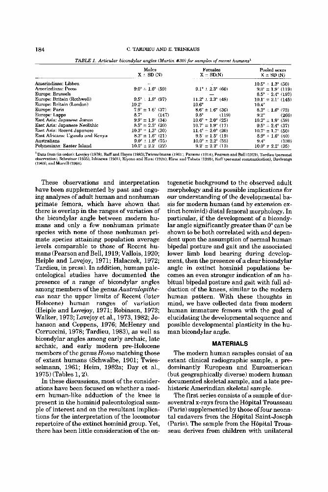
It has long been recognized that one of the distinctive features of the hominid lower limb, normally associated with the adoption of bipedal locomotion, is the presence of ad- duction of the knee (genu valgus) and its associated skeletal reflection, a femoral bi- condylar (divergence, obliquity, inclination, condyle-shaft, condylo-diaphyseal) angle significantly greater than 0") with sample means in the vicinity of 8-11" (Parsons, 1914; Pearson and Bell, 1919; Walmsley, 1933; LeGros Clark, 1947; Kern and Straus, 1949; Heiple and Lovejoy, 1971; McHenry and Corruccini, 1978; Tardieu, 1981, 1983, in press, Stern and Susman, 1983) (see Ta- ble 1). This elevated bicondylar angle is nor- mally assumed to represent a consequence of the need to maintain essentially horizon-
tal mediolateral metaphyseal and infra- condylar planes of the knee joint, to mini- mize the transverse shear component of joint reaction force at the tibiofemoral syn- ovial joint and (during development) across the epiphyseal cartilages of the knee (Smith, 1962; Amtmann, 1979), and to facilitate flexion-extension of the knee in a parasagit- tal plane, while positioning the knee close to the sagittal trajectory of the body's center of gravity in a bipedal striding gait, in the con- text of a large interacetabular distance.
Received November 5, 1993; accepted April 20, 1994. Address reprint requests to Christine Tardieu, Laboratoire
#Anatomic Comparee, Museum National #Histoire Naturelle, 55 rue Buffon, 75005 Paris, France.
0 1994 WILEY-LISS, INC
184 C. TARDIEU AND E. TRINKAUS
TABLE 1 . Articular bicondvlar aneles (Martin #30) for samdes of recent humans1
Males X ? SD (N)
Females X f SD(N)
Pooled sexes X ? SD (N)
10.5" ? 1.3" (50) Amerindians: Libben - - Amerindians: Pecos 9.0" ? 1.6" (59) 9.1" k 2.3" (60) 9.0" ? 1.9" (119) Europe: Brussels - - 8.5" ? 2.4" (197) Europe: Britain (Rothwell) 9.5" t 1.8" (97) 11.2" ? 2.3" (48) 10.1" t 2.1" (145) Europe: Britain (London) 10.2" 10.6" 10.4" Europe: Paris 7.9" t 1.6" (37) 8.6" ? 1.6" (36) 8.3" 2 1.6" (73) Europe: Lapps 8.7" (147) 9.8" (119) 9.2" (266) East Asia: Japanese Jomon 10.2" t 1.9" (59) East Asia: Japanese Neolithic 9.5" k 2.4" (37) East Asia: Recent Japanese 10.7" t 1.7" (50) East Africans: Uganda and Kenya 8.9" ? 1.6" (40)
9.9" t 1.9" (34) 8.5" t 2.3" (20)
10.3" t 1.3" (30) 8.3" -t 1.6" (21)
10.6" t 2.0" (25) 10.7" ? 1.9" (17) 11.4" t 2.0" (20) 9.5" f 1.5" (19)
Australians 9.0" 2 1.8" (75) 10.0" ? 2.2" (55) 9.4" (130) Polynesians: Easter Island 10.5" ? 2.2 (22) 9.2" 2 2.2" (13) 10.0" 2 2.2" (35)
*Data fmm (in order): Lovejoy (19781, Ruff and Hayes (1983),Twiesselmann (19611, Parsons (1914), Pearson and Bell (19191, Tardieu (personal observation), Sehreiner (19351, Ishisawa (1931), Kiyono and Hirai (1928), Hirai and Tabata (19281, Ruff (personal communication), Davivongs (1963), and Murrill(1968).
These observations and interpretation have been supplemented by past and ongo- ing analyses of adult human and nonhuman primate femora, which have shown that there is overlap in the ranges of variation of the bicondylar angle between modern hu- mans and only a few nonhuman primate species with none of those nonhuman pri- mate species attaining population average levels comparable to those of Recent hu- mans (Pearson and Bell, 1919; Vallois, 1920; Heiple and Lovejoy, 1971; Halaczek, 1972; Tardieu, in press). In addition, human pale- ontological studies have documented the presence of a range of bicondylar angles among members of the genus Australopithe- cus near the upper limits of Recent (later Holocene) human ranges of variation (Heiple and Lovejoy, 1971; Robinson, 1972; Walker, 1973; Lovejoy et al., 1973,1982; Jo- hanson and Coppens, 1976; McHenry and Corruccini, 1978; Tardieu, 19831, as well as bicondylar angles among early archaic, late archaic, and early modern pre-Holocene members of the genus Homo matching those of extant humans (Schwalbe, 1901; Twies- selmann, 1961; Heim, 1982a; Day et al., 1975) (Tables 1,2).
In these discussions, most of the consider- ations have been focused on whether a mod- ern human-like adduction of the knee is present in the hominid paleontological sam- ple of interest and on the resultant implica- tions for the interpretation of the locomotor repertoire of the extinct hominid group. Yet, there has been little consideration of the on-
togenetic background to the observed adult morphology and its possible implications for our understanding of the developmental ba- sis for modern human (and by extension ex- tinct hominid) distal femoral morphology. In particular, if the development of a bicondy- lar angle significantly greater than 0" can be shown to be both correlated with and depen- dent upon the assumption of normal human bipedal posture and gait and the associated lower limb load bearing during develop- ment, then the presence of a clear bicondylar angle in extinct hominid populations be- comes an even stronger indication of an ha- bitual bipedal posture and gait with full ad- duction of the knees, similar to the modern human pattern. With these thoughts in mind, we have collected data from modern human immature femora with the goal of elucidating the developmental sequence and possible developmental plasticity in the hu- man bicondylar angle.
MATERIALS The modern human samples consist of an
extant clinical radiographic sample, a pre- dominantly European and Euroamerican (but geographically diverse) modern human documented skeletal sample, and a late pre- historic Amerindian skeletal sample.
The first series consists of a sample of dor- soventral x-rays from the Hopital Trousseau (Paris) supplemented by those of four neona- tal cadavers from the Hopital Saint-Joseph (Paris). The sample from the H8pital Trous- seau derives from children with unilateral

HUMAN BICONDYLAR ANGLE ONTOGENY 185
TABLE 2. Articular bicondvlar aneles of AustraloDithecus and rtre-Hofocene Homo fossil femora'
Australopithecus A.L. 129-la A.L. 333-4 A.L. 333w-56 Sts 34 TM 1513 KNM-ER 993
Homo cf. habilis KNM-ER 1472 KNM-ER 1481A
Late archaic Homo La Ferrassie 2 Fond-de-Foret 1 Neandertal 1
Tabun 1 Early modern Homo
Cro-Magnon 4328 Cro-Magnon 4329 Minatogawa 1 Minatogawa 3 Minatogawa 4 Paviland 1 Pfedmosti 3 Pfedmosti 4
SPY 2
Articular bicondylar angle
8.5" 9"
(12.5")
7" 9"
12" 9", 10" 9" 9" go, 9" 7", 7"
Sex Source
F M? M M? F
-
F? M F F M M F
Lovejoy, 1978 Lovejoy, 1978 Walker, 1973
Day et al., 1975 Day et al., 1975
Heim, 1982a Twiesselmann, 1961 Schwalbe, 1901 Twiesselmann, 1961
Baba and Endo, 1982 Baba and Endo, 1982 Baba and Endo, 1982
Matiegka, 1938 Matiegka, 1938
Piedmosti 9 lo", 10" M Matiegka, 1938 Piedmosti 10 13", 14" F Matiegka, 1938 Pfedmosti 14 12", 14" M Matiegka, 1938 La Rochette 1 9" M Klaatsch and Lustig, 1914 Skhul4 9", 10" M McCown and Keith, 1938
'Values in parentheses are approximate, either due to damage to the femoral condyles (e.g., KNM-ER 993 and Tabun 1) or the preservation of relatively little of the distal femoral diaphysis ( e g , A.L. specimens, Sts 34, and TM 1513). Unless noted otherwise, measurements are from the authors' measurements on the original specimens. Right and left values are provided, as available.
defects of the locomotor anatomy including varying degrees of congenital dislocation of one hip and unequal longitudinal growth of the limbs. All of them were following a nor- mal pattern of learning to walk, although some were delayed in achieving full bipedal- ity. Nevertheless, all of them were capable of normal weight-bearing on the unaffected lower limb, even though their levels of activ- ity were undoubtedly lower than those of clinically normal children. In each case the measurements were taken on the normal side, so as to avoid direct effects of the devel- opmental abnormality. The total sample in- cludes 70 observations taken from 19 indi- viduals (7 males, 12 females) distributed between birth and 184 months (15.3 years). Of the 70 observations, 27 derive from the males and 43 from the females. Sixty-five of the observations derive from longitudinal growth series of variable lengths for 11 chil- dren; hence, there are variable numbers of multiple observations from these 11 individ-
uals between the ages of 3 and 11 years. Given the pooled semi-longitudinal nature of this sample, it is treated as a cross-sec- tional sample in the analysis.
The second sample includes femora of 25 documented skeletons from the collections of the Musee de l'Homme (Paris), which are primarily European in origin. They range in age from 8 months gestation to 18 years postnatal. Combined with these are 23 docu- mented Euroamerican individuals from 6 months gestation to about 16 years postna- tal from the collections of the Maxwell Mu- seum of Anthropology (Albuquerque), with 14 of the 32 femora for which age-at-death is documented provided by individuals less than 1 year postnatal and about half of those being from fetal (premature birth) speci- mens. Given the predominantly European origin of both of these documented samples and their common origins from industrial- ized society contexts, they have been pooled together in the analysis as a "documented

186 C. TARDIEU AND E. TRINKAUS
skeletal sample. For this sample with a total of 48 individuals, documented age is avail- able for 34 of them.
The third sample includes 31 femora from immature individuals of unknown sex from the Amerindian late prehistoric (Puebloan) Rio Grande pueblos of Pottery Mound and Kuaua, New Mexico, in the collections of the Maxwell Museum of Anthropology; they span the period from birth to early adoles- cence, based on femoral length, epiphyseal formation and fusion, and (for some) associ- ated dentitions. The majority represent in- fants and juveniles, with adolescents (given normal paleodemographic patterns [e.g., Lovejoy et al., 1977; Mobley, 1980; Palkov- ich, 1981; Storey, 19921) being rare.
The modern human developmental series therefore consist of one predominantly low activity level but otherwise fully ambulatory living human radiographic sample, one (pooled) modern industrialized-society skel- etal sample, and one prehistoric horticul- tural skeletal sample. In addition, we have included observations on two individuals with congenital abnormalities which pre- vented them from achieving normal bipedal posture or locomotion. One had congenital muscular hypotonia of the trunk and never walked. The other had congenital cerebral palsy with only minimal motor control of the lower limb; he did not walk before the age of 6 years, at which time he was able to walk minimally with an orthopedic walker. Bi- condylar angle was measured on radio- graphs of their femora taken at 12 and 7 years of age, respectively.
In addition, data are summarized for adult articular bicondylar angles in Recent human samples (Table 11, so as to provide a mature reference for evaluation of the im- mature bicondylar angles.
Ideally, similar developmental series would be available for paleontological sam- ples of hominid species. However, only four immature fossil hominids preserve suffi- cient amounts of the distal femoral epiphy- sis, or at least of the metaphyseal surface, to provide bicondylar angles (A.L. 333-110, A.L. 333-111 (both A. ufurensis), KNM-WT 15000 (early H. erectus), and La Ferrassie 6 (late archaic Homo) [Lovejoy et al., 1982; Heim, 1982b; Walker and Leakey, 199311,
TABLE 3. Femoral metaphyseal bicondylar angles for immature fossil hominid femora'
Metaphyseal bicondylar
anele
Australopithecus afarensis A.L. 333-110 A.L. 333-111
Homo erectus KNM-WT 15000
Late archaic Homo La Ferrassie 6
'The La Ferrassie 6 values are from Heim (1982b); the others were measured on casts, by C.T. for the Hadar femora and by C.B. Ruff for KNM-WT 15000.
even though there is some fossilization dam- age to the distal femoral metaphyses of all four of these individuals. The other pre- served premodern immature femora from the hominid fossil record (Sinel-nikov and Gremyatski, 1949; White, 1980; Heim, 198213; Cotrozzi et al., 1985; Madre-Dupouy, 1992) do not have the distal metaphyseal surface sufficiently intact for measurement of the bicondylar angle. Moreover, most of these specimens appear to be at or above the developmental age during which the adult human bicondylar angle is normally at- tained.
We have therefore largely limited our analysis to Recent human remains which provide adequate samples and developmen- tal age ranges. However, in the discussion reference will be made to bicondylar angles from the immature fossil femora (Table 3), even though an accurate (e.g., dentally de- termined) age-at-death is available only for KNM-WT 15000 (Smith, 1993).
METHODS In those cases (radiographic and skeletal)
for which a documented age is known, the development of the bicondylar angle is com- pared to chronological age, in months with birth represented by zero and fetal (or pre- mature birth) ages indicated by negative values. In addition, the bicondylar angles of the two skeletal samples are compared to diaphyseal femoral length, used primarily as an indicator of developmental age, given the close association of femoral length with age (Johnston, 1962; Anderson et al., 1964).

HUMAN BICONDYLAR ANGLE ONTOGENY 187
Bicondylar angle in adult femora is de- fined as the angle between the sagittal plane perpendicular to the infracondylar plane and the longitudinal axis of the femo- ral diaphysis, measured in the coronal plane of the dorsal femoral condyles (e.g., Martin, 1928 (measurement #30); see also Heiple and Lovejoy, 1971). In the radiographic sample, it was possible to employ the same measurement definition, since the in vivo anatomical relationship between the infra- condylar plane and the diaphyseal axis is observable.
Since the observed bicondylar angle po- tentially can be affected by internal or exter- nal rotation of the femur relative to the ra- diographic plane, a femur was x-rayed in 15" of internal and external rotation, as well as in the neutral position. The rotation pro- duced * 1" of change in the bicondylar angle. Consequently, on those x-rays for which in- ternal or external rotation was observable (by the position of the patella), the measured angle was corrected as appropriate. All of the x-rays were corrected for parallax en- largement assuming a linear enlargement proportional to the distances between the source, the subject, and the film.
For the skeletal remains, given the pro- cess of ossification of the femoral epiphyses and their eventual fusion to the diaphysis, it is not possible to apply directly to immature femora the criteria of measurement em- ployed on adult material and the radio- graphic sample. We have therefore rede- fined the skeletal measurements with respect to immature remains. The bicondy- lar angle was taken as the angle between the diaphyseal axis and the sagittal plane perpendicular to the distal metaphyseal plane; the metaphyseal plane was defined by the two most distally projecting points on the medial and lateral portions of the meta- physeal surface (Fig. l). Diaphyseal length was then taken as the direct distance paral- lel to the diaphyseal axis between the inter- section of the diaphyseal axis and the meta- physeal surface (almost always in the middle of the notch between the medial and lateral portions of the metaphyseal surface) and the most proximal point on the diaphy- seal axis, adjacent to the metaphyseal sur- face for the epiphysis of the greater tro-
Fig. 1. Anterior view of a Recent human immature femur illustrating the diaphyseal axis (a-a), the diaphy- seal length measurement (b-b), and the metaphyseal bicondylar angle (go"-@) as defined here (see text for further discussion).
chanter but not including any portion of the neck (Fig. 1).
To distinguish these two bicondylar angle measurement techniques, the former will be referred to as the articular bicondylar angle and the latter as the metaphyseal bicondylar angle. Measurement of both bicondylar an- gles on a small sample (N = 9) of femora permitting the measurement of both angles provided a mean difference of 1.9" and a range of differences of 0-3"; in every case the articular bicondylar angle was greater than or equal to the metaphyseal one. It is there- fore inappropriate to directly compare data based on the two measurements, even though they closely approximate each other.

188 C. TARDIEU AND E. TRINKAUS
In any case, the articular bicondylar angle more closely reflects the orientation of the distal femur as it relates to the mechanics of the synovial joint, and the metaphyseal one relates more closely to the pattern of growth, including angular changes, at the distal fe- mur during development, especially since approximately 70% of the longitudinal dia- physeal growth occurs a t the distal meta- physis (Bisgard and Bisgard, 1935; Taussig et al., 1976).
The growth patterns for bicondylar angles were assessed primarily through bivariate plots of the angle vs. documented age or fem- oral length, as available. To illustrate the trends through development in these pri- marily cross-sectional samples, and given the nonlinear nature of developmental se- quences, we have included Lowess smoothed lines through the bivariate data plots, a technique of nonparametric robust locally weighted regression (Cleveland, 1979; Efron and Tibshirani, 1991; see also Leigh, 1992; Ruff et al., 1994). In this technique, a speci- fied window of points (the smooth interval) is negatively weighted by the distance from the target point on the 3c axis, which is then used to derive a local least squares regres- sion which defines the target point (y) esti- mate; this procedure is repeated for every point along the curve to produce the series of connected points that result in the smoothed line. In other words, Lowess "produces a smooth [line] by running along the X values and finding the predicted values from a weighted average of nearby Y values" (Wilkinson, 1990). The lines provided here were calculated using NCSS (Hintze, 19911, and the dimensions of the smooth intervals are specified in the figure captions. Those smooth interval dimensions (15 and 20 in the cases here) represent the number of data points included within the interval (Hintze, 1991); those intervals were visually deter- mined to provide a curve which, as accu- rately as possible, represents the nonlinear distribution of bicondylar angle values rela- tive to developmental age or femoral length.
RESULTS Modern human adult femora exhibit artic-
ular bicondylar angles which normally range from approximately 5" to approxi-
mately 14", but sample averages remain be- tween approximately 8" and approximately 11" (Table 1). Differences between males and females can be modest and not statisti- cally significant (P > 0.05; the Australian Pecos, Parisian, Easter Island, recent Japa- nese and Jomon samples [t-test assuming heteroscedasticityl) or they can be more pro- nounced and statistically significant (P < 0.01; East African sample; P < 0.001: Medieval British, and Japanese Neolithic samples). However, in the 11 samples for which sex-specific data are available, the fe- male mean is higher than the male one in 10 of them (all except the small Easter Island sample), presumably as a result of the gen- erally larger interacetabular distance rela- tive to femoral length in females compared to males.
In the rather small samples of adult Aus- tralopithecus and pre-Holocene Homo fem- ora which provide reasonably secure esti- mates of articular bicondylar angles, the specimens attributed to Homo (archaic Homo: 10.1" 2 2.3", N = 7; early modern Homo: 9.7" * 1.9", N = 13) fall well within the normal ranges of variation of Recent hu- mans, whereas the six Australopithecus femora for which bicondylar angle can be estimated (12.7" * 2.4", N = 6) have values which fall at the top of the Recent human ranges of variation (Table 2). In the late ar- chaic and early modern Homo samples for which sex is known or reasonably approxi- mated for the majority of the specimens, the females have a clearly higher mean than the males in the late archaic group (12.0" vs. 7.8"), whereas the early modern males have a slightly higher mean than the early mod- ern females (10.2' vs. 9.6").
The plots of bicondylar angle vs. docu- mented age (in months) for the immature radiographic and documented skeletal sam- ples exhibit similar overall patterns (Fig. 2). In both of them, bicondylar angle starts at 0" at birth and then increases during infancy and the juvenile years to reach adult values of at least 6-8" between 4 and 8 years post- natal. The main differences between the two samples involve the timing of the final in- crease in bicondylar angle from the neonatal value of 0"; in the smaller documented Sam- ple the average values rise steadily and con-

HUMAN BICONDYLAR ANGLE ONTOGENY 189
L r - 50 100 150 200
- 1 I I
M A A
J * . 0 50 100 150 200 250
Age fin months)
Fig. 2. Bivariate plots of bicondylar angle (in de- grees) vs. chronological age (in months, with birth = 0) for samples of Recent Europeans (or predominantly Eu- ropean-derived populations). Lowess smoothed lines are provided to indicate the general pattern of increase in bicondylar angle with age; the smooth interval for each = 20. Top: Plot of articular bicondylar angle vs. age for a radiographic sample of modern European children with minimal congental deficiencies of the contralateral limb. Bottom: Metaphyseal bicondylar angle vs. age for an age-documented skeletal sample of European and predominantly European-derived children.
tinuously to reach values between 6" and 8" between 4 and 5 years postnatal, whereas the radiographic sample has a slower in- crease beyond approximately 5" to reach similar adult levels closer to 6 years postna- tal. This slight difference would be accentu- ated by the higher values produced by the articular bicondylar angle measurement employed for the radiographic sample. How- ever, the amount of variation within each
-00 50 100 150
Age (in months)
Fig. 3. Bivariate plot of articular bicondylar angle (in degrees) vs. chronological age (in months) for males (squares) and females (triangles) in the radiographic sample of modern European children with minimal con- gental deficiencies of the contralateral limb. Lowess lines (smooth interval = 20) are provided for each sex. The resultant Lowess lines are essentially indistin- guishable, despite a slight separation in the late juve- nile age range.
sample makes it unlikely that there is sig- nificant difference between the two sam- ples. Indeed, if the bicondylar angles of the two samples from age 36-72 months (or 3 and 6 years) are compared, the samples are statistically indistinguishable (t-test P = 0.705); the degree of difference is in- creased if 1.9" (the average difference be- tween articular and metaphyseal angles in the small sample with both measurements) is added to the values for the documented sample, but the difference remains statisti- cally insignificant (t-test P = 0.137). There is therefore a suggestion of a slower and pro- longed attainment of adult bicondylar an- gles in the lower activity level radiographic sample but with only a slight difference in the overall pattern.
In the radiographic sample, there is little difference between the sexes in the develop- ment of the bicondylar angle (Fig. 3). The female Lowess curve rises a t a trivially faster rate during the first four years post- natal, but then both sex-specific curves fluc- tuate (in part due to sampling) through the late juvenile and early adolescent years.

190 C. TARDIEU AN D E. TRINKAUS
mented sample vs. 409.1 2 22.2 mm (N = 84) for an Amerindian sample from the same sites as the immature femora) and the slightly greater femoral lengths among Eu- roamericans for a given developmental age provided by Anderson et al. (1964) in con- trast to those provided by Johnston (1962) for a North American (but nonsouthwestern US) prehistoric Amerindian sample. Adjust- ing for different measurement techniques, a femoral diaphyseal length of approximately 110 mm should represent about 1 year post- natal for the Amerindian sample, and a dia- physeal length of approximately 140 mm should represent about 2 years postnatal (Johnston, 1962). In the documented sam- ple, following Anderson et al. (1964), femo- ral diaphyseal lengths closer to 130 mm and 160 mm should represent about 1 and 2 years postnatal, respectively. In addition, the predominantly European-derived docu- mented sample should attain femoral dia- physeal lengths of approximately 200 mm by the end of the third year postnatal, whereas similar lengths were probably not reached by the Amerindian sample, follow- ing Johnston (19621, until the fifth year.
Using these femoral length growth pa- rameters as a guide, the documented skele- tal sample exhibits metaphyseal bicondylar angles of 0" through fetal life and into in- fancy, followed by a steady increase in the angle beginning late in the first year postna- tal and continuing through the remainder of the first approximately 3 years of postnatal life, reaching adult values by the end of this period. The Amerindian sample exhibits considerably more variation in the immedi- ately postnatal period, with most bicondylar angle values between birth and approxi- mately 2 years postnatal being between 2" and 5", despite a persistence of 0" measure- ments through the first year. There is then a steady increase up to adult values by what is probably approximately 3 4 years postna- tal, even though this trend is made tenuous by the dearth of specimens longer than ap- proximately 150 mm.
These data, despite sample size limita- tions and difficulties in assigning develop- mental ages based on long bone lengths, provide a relatively consistent pattern. Bi- condylar angles, whether articular or meta-
l I 2 120) P
A I
100 200 300 400
100 200 300
Femoral Diaphyseal Length imm) 400
Fig. 4. Bivariate plots of metaphyseal bicondylar an- gle (in degrees) vs. diaphyseal femoral length (in milli- meters) for samples of European and predominately Eu- ropean-derived children (top) and of prehistoric Puebloan Amerindian children (bottom). Lowess smoothed lines (smooth interval = 15) are provided for each.
Generally similar patterns are evident in the comparisons of metaphyseal bicondylar angle to femoral diaphyseal length for the documented and Amerindian skeletal sam- ples (Fig. 4). In this, it is necessary to bear in mind that the independent variable (femo- ral intermetaphyseal diaphyseal length) was most likely larger for a given develop- mental age in the predominantly European- derived documented sample than in the pre- historic Amerindian sample. This is based on the contrasts in adult femoral bicondylar length between the two samples (453.5 2 32.7 mm (N = 55) for a Euroamerican docu-

HUMAN BICONDYLAR ANGLE ONTOGENY 191
physeal, are approximately 0" prenatally and at birth, largely remain near 0" through most of the first year postnatal, and then rise relatively steadily through the next 2-3 years of postnatal life, reaching low adult values usually around 4 years of age. There is some variation in the rate of attainment of these adult values, with the clinical radio- graphic sample (with probably reduced ac- tivity levels) achieving them slightly later than the other samples.
These observations are supplemented by those on the two individuals with congenital defects preventing normal bipedal posture and locomotion. The individual who had not walked for the first 12 years of life had a bicondylar angle of 0", whereas the one who began to walk with a walker at age 6 years exhibited a bicondylar angle of 1.5" one year later, or 2.9 standard deviations below the mean (6.8" * 1.8", N = 12) of the individuals in the clinical radiograph sample between the ages of 6 and 8 years.
The immature fossil human femora for which reasonable bicondylar angle esti- mates can be determined (Table 3) have val- ues which fall well within the ranges ex- pected for them. The probably late juvenile to adolescent A. afarensis femora have metaphyseal bicondylar angles well within the ranges of older immature Recent hu- mans documented here, even though the A.L. 333-111 value of approximately 11" is at the upper limit of those ranges of variation (Fig. 4). The value of approximately 8" for KNM-WT 15000 is in the middle of the ranges of variation for early adolescent Re- cent humans (Figs. 2, 41, and those of La Ferrassie 6, a 3-5-year-old (Heim, 1982b; Tompkins and Trinkaus, 19871, are close to a mean (5.0" rt 1.7", N = 10) of individuals between 3 and 5 years old in the radio- graphic sample, much as the mean (9.5" & 2.3", N = 5) of adult late archaic hu- mans (Table 2) is similar to those of several Recent human samples (Table 1).
DISCUSSION These data therefore indicate that the hu-
man bicondylar angle, whether measured across the distal femoral metaphyseal or condylar plane, is approximately 0" prena- tally and immediately postnatally. Without
normal postural and locomotor loading, as indicated by the two clinical cases pre- sented, it remains at or close to 0" through development. With normal or near-normal bipedal posture and locomotion, it goes through an angular development, in which a clear angle usually appears during the sec- ond year of life postnatal and continues to develop, reaching low adult values generally by the fourth or fifth year postnatal. In cases of reduced loading from congenital abnor- malities restricting locomotor levels, as in our radiographic sample, the developmental increase in the angle may be delayed but nonetheless follows a pattern similar to that observed in the other samples.
This chronology of bicondylar angle devel- opment closely parallels the developmental chronology of the acquisition of walking in young children. Most children begin to walk toward the end of the first year of postnatal life, perfecting the technique and increas- ingly loading the legs during the subsequent couple of years (Scoles, 1988; Le Metayer, 1992). More importantly, a t birth the legs habitually assume a marked genu varus po- sition, with average tibiofemoral angles (be- tween the tibia1 and femoral diaphyseal axes in the coronal plane of the leg) of ap- proximately 15" (Salenius and Vankka, 1975). As they begin to stand and walk, the tibiofemoral angle decreases, passing 0" be- tween 1.5 and 2 years on average and reach- ing a peak valgus position of about 10" around 3 years, only to decrease to a rela- tively constant approximately 6" by 6-7 years (Salenius and Vankka, 1975).
Consequently, there is little loading of the leg in bipedal posture and locomotion prior to late in the first year postnatal and little loading with the knee in a valgus position until about 2 years postnatal, at which time the child usually is both actively bipedal and is maintaining the leg in a full, or even exag- gerated, valgus position. It is during this time period that there is most of the change in the bicondylar angle, although it contin- ues to increase for several additional years.
This general chronological correlation of locomotor development and the emergence of a bicondylar angle, combined with the ab- sence of such an angle in the two non- or minimally locomotor clinical cases, strongly
192 C. TARDIEU AND E. TRINKAUS
suggests that the development of a bicondy- lar angle is dependent upon the levels and especially patterns of biomechanical loading at the knee commensurate with a normal human bipedal posture and gait. Further- more, given the presence of only a sugges- tion of a difference between the lower loco- motor activity level radiographic sample and the other samples, it appears that pres- ence of normal weight-bearing by the lower limb in a bipedal posture is as important as, if not more important than, locomotor activ- ity levels in determining the development of a bicondylar angle.
Such a posturaflocomotor connection to bicondylar angle development would have to be through differential mediolateral meta- physeal apposition during longitudinal fem- oral growth. Not only is the majority of the longitudinal growth of the femur the result of distal metaphyseal apposition (Bisgard and Bisgard, 1935; Taussig et al., 19761, but theoretical, clinical, and experimental work indicates that such a mechanism is likely. Pauwels (1965) suggested that increased compression on the medial portion of the dis- tal femoral epiphyseal cartilage (as a result of the vector of the center of gravity being medial of the knee) and the increasingly Val- gus position of the knee as a child acquires an upright, bipedal posture, would lead to additional medial metaphyseal apposition and the formation of a bicondylar angle. Even though high levels of compressive force (probably above normal physiological loads) will retard metaphyseal apposition (Arkin and Katz, 19561, experimental (e.g., Karaharju et al., 1976) and clinical (e.g., Frost, 1979) observations support the con- tention that moderate increases in compres- sion on the cartilage (within normal physio- logical levels) will stimulate metaphyseal apposition. Furthermore, Wallace and Hoff- man (1992) have shown that subsequent to diaphyseal angular deformities from frac- tures in children, an average of 85% of the initial deformity was corrected and, more- over, that 74% of the correction occurred through differential angular growth at the epiphysis/metaphysis. These results have been experimentally duplicated (e.g., Ry- oppy and Karaharju, 1974; Karaharju et al., 1976; Abraham, 19891, with surgically
induced angular osteotomies in animals resulting in differential epiphyseaY metaphyseal growth that corrected, at least in part, the artificially induced angular de- formities.
It therefore appears that normal apposi- tional responses of the epiphyseal cartilage to changed distributions of levels of com- pressive force across the cartilage are ade- quate to account for the general correlation between normal bicondylar angle and pos- tural development and for the failure of it to develop in non- or minimally bipedal indi- viduals. However, regardless of the mecha- nism involved, it is clear that there is consid- erable potential for plasticity in the angular orientation of the epiphyseal plate relative to the diaphysis and that normal develop- mental mechanisms serve to maintain the epiphyseal plate, to the extent possible, in a biomechanically appropriate orientation, whether those angular changes are required by normal locomotor development or abnor- mal posttraumatic deformities.
Additionally, even though fossil hominid femora which are adequately complete and sufficiently immature to document such early ontogenetic changes are absent from the hominid fossil record, the data available for immature archaic hominid bicondylar angles are commensurate with a pattern of bicondylar angle development similar to that of Recent humans.
These data and developmental consider- ations also suggest that, on average, a greater degree of genu valgus will tend to accentuate the observed bicondylar angle. Assuming that the g e m valgus characteris- tic of hominids is the result of a need to position the knee below the center of grav- ity, shorter femora relative to interacetabu- lar distance should accentuate thisgenu Val- gus. In Recent humans, the usual, although not always significant, higher mean bi- condylar angles among females vs. males may indeed reflect this. For example, in one sample (the Parisian one), females have both higher bicondylar angles (Table 1) and higher indices of interacetabular distance to femoral articular length (43.6 2 3.2, N = 36 vs. 40.1 ? 2.6, N = 37 for males [Tardieu, unpub. data]), even though there is no significant correlation between this in-
HUMAN BICONDYLAR ANGLE ONTOGENY 193
dex and bicondylar angle across the pooled- sex sample (r = 0.256). In addition, the ap- parently relatively large interacetabular distances of members of Australopithecus (as documented by A.L. 288-1 and Sts 14 [Berge et al., 19841) may be a contributory factor to their tendency to have high bi- condylar angles, whatever factors were de- termining those large interacetabular dis- tances (Berge et al., 1984; T a p e and Lovejoy, 1986).
CONCLUSIONS These considerations therefore indicate
that the emergence of a bicondylar angle in early hominids, and its persistence through the Hominidae, is likely to have been the result of developmental plasticity, respond- ing to differential levels of compressive force acting upon the epiphyseal cartilage to pro- duce differential mediolateral metaphyseal apposition. Furthermore, these data on the development of the bicondylar angle in nor- mal and clinically abnormal modern human children indicate that a pattern of habitual bipedal posture and locomotion, with the center of gravity displaced medial of the knee and the subsequent development of a valgus position of the knee, is required to promote this lateral deviation of the femoral diaphysis relative to the articular and meta- physeal planes of the distal femur. What- ever the full postural and locomotor reper- toires of early, or later, archaic hominids might have been (Senut, 1981; Susman et al., 1984; Berge, 1993; Trinkaus, 1986; La- timer et al., 1987; Lovejoy, 1988; Latimer and Lovejoy, 1990; McHenry, 1991; Tardieu, 1991), the universal presence of distinct, non-African-ape-like, bicondylar angles among them supports the contention that habitual bipedal posture was an important component of their postural and locomotor repertoires and that it emerged early in de- velopment.
ACKNOWLEDGMENTS We thank J.P. Damsin at the Hopital
Trousseau and N. Khouri a t the Hopital Saint-Joseph for access to the radiographic series and clinical information on individu- als used in our modern human radiographic samples and A. Langaney and S. Rhine for
access to the collections of the Musee de l'Homme and Maxwell Museum of Anthro- pology, respectively. Y. Coppens, D. Johan- son, A. Langaney, W. J . Kennedy, and C.B. Stringer provided access to original fossil human remains in their care, and C.B. Ruff kindly provided data summaries for the Pe- cos and East African samples plus the meta- physeal bicondylar angle estimate for KNM- WT15000. This work has been funded in part by the Centre National de la Recherche Scientifique (UA 1137 and UA 376), the Col- lege de France, NSF BNS76-14344 A01, and the L.S.B. Leakey Foundation. C.B. Ruff provided helpful comments on an earlier version of this paper. To all of them we are grateful.
LITERATURE CITED Abraham E (1989) Remodeling potential of long bones
following angular osteotomies. J. Pediatr. Orthop. 9:37-43.
Amtmann E (1979) Biomechanical interpretation of form and structure of bones: Role of genetics and func- tion in growth and remodeling. In ME Morbeck, H Preuschoft, and N Gomberg (eds.): Environment, Be- havior, and Morphology: Dynamic Interactions in Pri- mates. Stuttgart: Fischer Verlag, pp. 347-366.
Anderson M, Messner MB, and Green WT (1964) Distri- bution of lengths of the normal femur and tibia in children from one to eighteen years of age. J . Bone Joint Surg. [Am] 46:1197-1202.
Arkin AM, and Katz JF (1956) The effects of pressure on epiphyseal growth. J . Bone Joint Surg. [Am] 38:105& 1076.
Baba H, and Endo B (1982) Postcranial skeleton of the Minatogawa man. In H Suzuki and K Hanihara (eds.): The Minatogawa Man. The Upper Pleistocene Man from the Island of Okinawa. Bull. Univ. Mus., Univ. Tokyo 19:61-195.
Berge C (1993) L'Evolution de la Hanche et du Pelvis des Hominides. Paris: C.N.R.S.
Berge C, Orban-Segebarth R, and Schmid P (1984) Ob- stetrical interpretation of the Australopithecine pel- vic cavity. J. Hum. Evol. 13:573-587.
Bisgard JD, and Bisgard ME (1935) Longitudinal growth of long bones. Arch. Surg. 31.568.
Cleveland WS (1979) Robust locally weighted regression and smoothing scatterplots. J. Am. Stat. Assoc. 74: 829-836.
Cotrozzi S, Mallegni F, and Radmilli AM (1985) Femur d'un enfant Neandertalien, dans la Buca del Tasso a Metata, Alpi Apuane (Italie). L'Anthropol. 89:111- 116.
Davivongs V (1963) The femur of the Australian Aborig- ine. Am. J. Phys. Anthropol. 21:457-467.
Day MH, Leakey REF, Walker AC, and Wood BA (1975) New hominids from East Rudolf, Kenya. Am. J. Phys. Anthropol. 42t461-470.

C. TARDIEU AND E. TRINKAUS 194
Efron B, and Tibshirani R (1991) Statistical data analy- sis in the computer age. Science 253:390-395.
Frost HM (1979) A chondral modeling theory. Calcif. Tissue Int. 28:181-200.
Halaczek B (1972) Die Langknocken der Hinter Extrem- itat bei Simischen Primaten. Zurich: Juris Druck und Verlag.
Heim J L (1982a) Les hommes fossiles de La Ferrassie 11. Arch. Inst. Paleont. Hum. 38:l-272.
Heim J L (198213) Les Enfants Neandertaliens de La Fer- rassie. Paris: Masson.
Heiple KG, and Lovejoy CO (1971) The distal femoral anatomy ofdustralopithecus. Am. J . Phys. Anthropol. 35:75-84.
Hintze J L (1991) Number Cruncher Statistical System Version 5.03 9/91. Kaysville, U T NCSS.
Hirai T, and Tabata T (1928) Anthropologische Unter- suchungen iiber das Skelett der rezenten Japaner. Die unteren Extremitat. J . Anthropol. SOC. Nippon 43(Suppl):1-176.
Ishisawa M (1931) Anthropologische Untersuchungen uber das Skelett der Yoshiko-Steinzeitmenschen. 111. Teil. Die unteren Extremitaten. J . Anthropol. SOC. Nippon 45:l-192.
Johanson D, and Coppens Y (1976) A preliminary ana- tomical diagnosis of the first PlioPleistocene hominid discoveries in the CentralAfar, Ethiopia. Am. J . Phys. Anthropol. 45:217-222.
Johnston FE (1962) Growth of long bones of infants and young children at Indian Knoll. Am. J. Phys. Anthro- pol. 20:249-254.
Karaharju EO, Ryoppy SA, and Makinen RJ (1976) Re- modelling by asymmetrical epiphyseal growth. J. Bone Joint Surg. IBrl58r122-126.
Kern HM, and Straus WL Jr (1949) The femur of Ple- sianthropus transvaalensis. Am. J . Phys. Anthropol. 753-78.
Kiyono K, and Hirai T (1928) Anthropologische Unter- suchungen iiber das Skelett der Steinzeit Japaner. IV. Due untere Extremitat. J . Anthropol. SOC. Nippon.
Klaatsch H, and Lustig W (1914) Morphologie der pala- olithischen Skelettreste des mittleren Aurignacien der Grotte von La Rochette. Arch. Anthropol. 41:81- 126.
Latimer B, and Lovejoy CO (1990) Hallucial tarsometa- tarsal joint in Australopithecus afarensis. Am. J . Phys. Anthropol. 82:12&133.
Latimer B, Ohman JC, and Lovejoy CO (1987) Talocru- ral joint of African Hominoids: Implications for Aw- tralopithecus afarensis. Am. J. Phys. Anthropol. 74: 155-175.
Le Metayer M (1992) Evolution de la locomotion au cours des trois premieres annees de la vie. Motricite Cerebrale, madaptation, Neurologie du Developpe- ment 13:81-103.
LeGros Clark WE (1947) Observations on the an- atomy of the fossil Australopithecinae. J . Anat. 81: 300-333.
Leigh SR (1992) Patterns of variation in the ontogeny of primate body size dimorphism. J . Hum. Evol. 23: 27-50.
Lovejoy CO (1978) A biomechanical review of the loco- motor diversity of early hominids. In CJ Jolly (ed.):
43(Supp1.):303494.
Early Hominids of Afi-ica. New York St. Martin's Press, pp. 403439.
Lovejoy CO (1988) Evolution of human walking. Sci. Am. 259(5):11%125.
Lovejoy CO, Heiple KG, and Burstein AH (1973) The gait of Australopithecus. Am. J. Phys. Anthopol. 38:
Lovejoy CO, Meindl RS, Pryzbeck TR, Barton TS, Heiple KG, and Kotting D (1977) Paleodemography of the Libben Site, Ottawa County, Ohio. Science 198:291- 293.
Lovejoy CO, Johanson DC, and Coppens Y (1982) Homi- nid lower limb bones recovered from the Hadar For- mation: 1974-1977 collections. Am. J . Phys. Anthro- pol. 57:679-700.
Madre-Dupouy M (1992) L'Enfant du Roc-de-Marsal. Paris: C.N.R.S.
Martin R (1928) Lehrbuch der Anthropologie, 2nd ed. Jena: Fischer Verlag.
Matiegka J (1938) Homo Pkedmostensis. Fosilni Clovek z Piedmosti na Morave 11. Prague: Nakladem Ceske Akademie Ved a Umeni.
McCown TD, and Keith A (1939) The Stone Age of Mount Carmel 11. Oxford: Clarendon.
McHenry HM (1991) First steps? Analyses of the post- cranium of early hominids. In Y Coppens and B Senut (eds.): Origine(s) de la Bipedie chez les Hominides. Paris: C.N.R.S., pp. 133-141.
McHenry HM, and Corruccini RS (1978) The femur in early human evolution. Am. J. Phys. Anthropol. 49: 47-88,
Mobley CM (1980) Demographic structure of Pecos Indi- ans: A model based on life tables. Am. Antiq. 45:51% 530.
Murrill RI (1968) Cranial and Postcranial Skeletal Re- mains from Easter Island. Minneapolis: University of Minnesota Press.
Palkovich AM (1981) Demography and disease patterns in a protohistoric plains group: A study of the Mo- bridge Site (39WW1). In RL Jantz and DH Ubelaker (eds.): Progress in Skeletal Biology of Plains Popula- tions. Plains Anthropol. Mem. 17:71-84.
Parsons FG (1914) The characters of the english thigh bone. J . Anat. Physiol. 48:23%267.
Pauwels F (1965) Gesammelte Abhandlungen zur funk- tionellen Anatomie des Bewegungsapparates. Berlin: Springer Verlag.
Pearson K, and Bell J (1919) A study of the long bones of the English skeleton. Drapers' Company Memoirs Bi- ometric Series XI. Cambridge: Cambridge University Press.
Robinson J T (1972) Early Hominid Posture and Locomo- tion. Chicago: University of Chicago Press.
Ruff CB, and Hayes WC (1983) Cross-sectional geome- try of Pecos Pueblo femora and tibiae-a biomechani- cal investigation: I. Method and general patterns of variation. Am. J . Phys. Anthropol. 60:359381.
Ruff CB, Trinkaus E, and Walker A (1994) Postcranial robusticity in Homo, 111: Ontogeny. Am. J. Phys. An- thropol. 93:35-54.
Ryoppy SA, and Karaharju EO (1974) Alteration of epiphyseal growth by an experimentally pro- duced angular deformity. Acta Orthop. Scand. 45: 49M98.
757-780.

HUMAN BICONDYLAR ANGLE ONTOGENY 195
Salenius P, and Vankka E (1975) The development of the tibiofemoral angle in children. J . Bone Joint Surg. 57A:25%261.
Schreiner KE (1935) Zur Osteologie der Lappen. Inst. Sammenlignende Kulturforskning Serie B 18:l-294.
Schwalbe G (1901) Der Neanderthalschadel. Bonner Jahrbucher 106:l-72.
Scoles PV (1988) Pediatric Orthopedics in Clinical Prac- tice, 2nd ed. Chicago: Year Book Med. Pub.
Senut B (1981) L'Humerus et ses Articultions chez les Hominides Plio-Pleistocenes. Paris: CNRS.
Sinel-nikov NA, and Gremyatskij MA (1949) Bones of the skeleton of the Neandertal child from the cave of Teshik-Tash, southern Uzbekistan. (In Russian.) In MA Gremyatskij and MF Nesturkh (eds.): Teshik- Tash. Moscow: Moscow State University, pp. 123-135.
Smith BH (1993) The physiological age of KNM-WT 15000. In A Walker and R Leakey (eds.): The Narioko- tome Homo erectus Skeleton. Cambridge: Harvard University Press, pp. 195-220.
Smith JW (1962) The structure and stress relations of fibrous epiphyseal plates. J. Anat. 96.209-225.
Stern JT, and Susman RL (1983) The locomotor anat- omy of Australopithecus afarensis. Am. J . Phys. An- thropol. 60:27%317.
Storey R (1992) Life and Death in the Ancient City of Teotihuacan. Tuscaloosa: University of Alabama Press.
Susman RL, Stern JT, and Jungers WL (1984) Arboreal- ity and bipedality in the Hadar hominids. Folia Pri- matol. 43:113-156.
Tague RG, and Lovejoy CO (1986) The obstetric pelvis of A.L. 288-1 (Lucy). J . Hum. Evol. 15:237-255.
Tardieu C (1981) Morphofunctional analysis of the artic- ular surfaces of the knee joint in primates. In AB Chiarelli and RS Corruccini (eds.): Primate Evolution- ary Biology. Berlin: Springer Verlag, pp. 6&%80.
Tardieu C (1983) L'Articulation du Genou. Analyse Mor- pho-fonctionelle chez les Primates; Application aux Hominides Fossiles. Paris: C.N.R.S.
Tardieu C (1991) Etude comparative des deplacements du centre de gravite pendant la marche. Mise a l'epreuve dune hypothese evolutive. In Y Coppens and B Senut (eds.): Origineb) de la Bipedie chez les Hominides. Paris: C.N.R.S., pp. 49-58.
Tardieu C (in press) Morphogenese de la diaphyse femo- rale chez l'homme. Signification fonctionelle et evolu- tive. Folia Primatol.
Taussig G, Delor MH, and Masse P (1976) Les alt6r- ations de croissance de I'extremite superieure du fe- mur. Rev. Chir. Orthop. 62:191-210.
Tompkins RL, and Trinkaus E (1987) La Ferrassie 6 and the development of Neandertal pubic morphology. Am. J . Phys. Anthropol. 73:233-239.
Trinkaus E (1986) The Neandertals and modem human origins. Annu. Rev. Anthropol. 15t193-218.
Twiesselmann F (1961) Le femur Neanderthalien de Fond-de-Foret (Province de Liege). Mem. Inst. Roy. Sci. Nat. Belgique 148:l-164.
Vallois H V (1920) L'epiphyse inferieure du femur chez les Primates. L'angle de divergence et ses varia- tions. Bull. Mem. SOC. Anthropol. Paris Serie 6,10230- 107.
Walker A (1973) New Australopithecus femora from East Rudolf, Kenya. J . Hum. Evol. 2:545-556.
Walker A, and Leakey R (1993) The postcranial bones. In A Walker and R Leakey (eds.): The Nariokotome Homo erectus Skeleton. Cambridge: Harvard Univer- sity Press, pp. 95-160.
Wallace ME, and Hoffman EB (1992) Remodelling of angular deformity after femoral shaft fractures in children. J . Bone Joint Surg. [Brl74:765-769.
Walmsley T (1933) The vertical axes of the femur and their relations. A contribution to the study of the erect position. J . Anat. 67:284-300.
White TD (1980) Additional fossil hominids from La- etoli, Tanzania: 1976-1979 specimens. Am. J . Phys. Anthropol. 53:487504.
Wilkinson L (1990) SYGRAPH: The System for Graph- ics. Evanston: SYSTAT Inc.