ear molding in newborn infants with auricular deformities
TRANSCRIPT

RECONSTRUCTIVE
Ear Molding in Newborn Infants withAuricular Deformities
H. Steve Byrd, M.D.Claude-Jean Langevin,
M.D., D.M.D.Lorraine A. Ghidoni, M.D.
Dallas, Texas
Background: A review of a single physician’s experience in managing over 831infant ear deformities (488 patients) is presented.Methods: The authors’ methods of molding have advanced from the use ofvarious tapes, glues, and stents, to a comprehensive yet simple system that shapesthe antihelix, the triangular fossa, the helical rim, and the overly prominentconchal-mastoid angle (EarWell Infant Ear Correction System).Results: The types of deformities managed, and their relative occurrence, areas follows: (1) prominent/cup ear, 373 ears (45 percent); (2) lidding/lop ear,224 ears (27 percent); (3) mixed ear deformities, 83 ears (10 percent) (all hadassociated conchal crus); (4) Stahl’s ear, 66 ears (8 percent); (5) helical rimabnormalities, 58 ears (7 percent); (6) conchal crus, 25 ears (3 percent); and(7) cryptotia, two ears (0.2 percent). Bilateral deformities were present in 340patients (70 percent), with unilateral deformities in 148 patients (30 percent).Fifty-eight infant ears (34 patients) were treated using the final version of theEarWell Infant Ear Correction System with a success rate exceeding 90 percent(good to excellent results). The system was found to be most successful whenbegun in the first week of the infant’s life. When molding was initiated after 3weeks from birth, only approximately half of the infants had a good response.Conclusions: Congenital ear deformities are common and only approximately30 percent self-correct. These deformities can be corrected by initiating appro-priate molding in the first week of life. Neonatal molding reduces the need forsurgical correction with results that often exceed what can be achieved with thesurgical alternative. (Plast. Reconstr. Surg. 126: 1191, 2010.)
Infant auricular deformities are classified eitheras malformation or deformation. Malforma-tions are characterized by a partial absence of
the skin or cartilage resulting in a constricted orunderdeveloped pinna, whereas deformations arecharacterized by a misshaped but fully developedpinna.1 Molding is best suited for deformations,but is useful in cases of less severe malformation.
Our first experience with ear molding was in1989 with a unilateral Stahl’s ear deformity. Evi-dence in the Japanese literature2–4 showed prom-ising results with ear molding at that time. Ourtechnique then involved rather crude fabricationof helical rim stents from thermoplastic dentalcompound supported with Steri-Strips (3M, St.
Paul, Minn.) to hold the ear in place. This wasmaintained for approximately 4 weeks and pro-duced an outcome with improved shape but
From the Department of Plastic Surgery, University of TexasSouthwestern Medical Center at Dallas; Children’s MedicalCenter Dallas; and the Department of Pediatrics, BaylorUniversity Medical Center.Received for publication January 18, 2010; accepted March22, 2010.Copyright ©2010 by the American Society of Plastic SurgeonsDOI: 10.1097/PRS.0b013e3181e617bb
Disclosures: Dr. Byrd has a royalty agreement withBecon Medical for his work on the EarWell device;neither of the other authors has any commercialassociations or financial disclosures that might poseor create a conflict of interest with information pre-sented in this article. No funding was received forthe work presented in this article.
Supplemental digital content is available forthis article. A direct URL citation appears inthe printed text; simply type the URL addressinto any Web browser to access this content. Aclickable link to the material is provided in theHTML text of this article on the Journal’s Website (www.PRSJournal.com).
www.PRSJournal.com 1191

lacked the full aesthetic definition of a normal ear.We also quickly realized that when infants withdeformity were seen after 3 weeks of age, moldinghad to be maintained for a very prolonged inter-val, often exceeding 3 months and requiring aninordinate time commitment from physician andparents. Sadly, only approximately half of the in-fants treated with late-onset molding, after 3 weeksof age, had good outcomes. As a result, we adoptedrather restrictive policies for the use of ear mold-ing. Infants seen at birth with auricular deformitywere reevaluated at 5 to 7 days. If the deformityhad not improved, ear molding was initiated. If theears were assuming a normal shape, no treatmentwas offered. In a subgroup of infants who hadsome improvement, observation was continuedinto the second week, and ear molding was initi-ated if spontaneous correction was not observedby the end of the week. Over time, we realized thatpostponing molding to await spontaneous recov-ery only increased the likelihood of failure orpoor outcome and increased the interval requiredfor molding.
Parallel to these observations, virtually 100percent of children and adults seen for otoplastygave a history of the deformity being present sincebirth. This coupled with the seemingly universalteaching in pediatrics that these deformitieswould self-correct led to a study in a local nurseryof 100 prospective births. Observations and pho-tographs of all infant ears suspected of being ab-normal were made by the attending pediatricianand nursing staff. Of the 100 infants, 39 were
noted to have misshapen ears. Of the 200 infantears seen, 58 were deemed misshapen (29 per-cent). We estimate that approximately one-thirdof the misshapen ears present at birth self-correctin the first week, leaving 15 to 20 percent of new-borns as candidates for molding.
PATIENTS AND METHODS
Seven Patterns of DeformationProminent/cup ear may be secondary to a wid-
ened, conchal-mastoid angle, an absent antiheli-cal fold, or a combination of both (Fig. 1, left). Theprominent/cup ear deformity was the most com-mon type and present in 373 ears (45 percent).The antihelical fold requires posterior stentingwith anterior pressure to deepen the scapha,whereas the conchal-mastoid angle is only ame-nable to anteriorly directed forces.
Lidding/lop ear constitutes a folding over ofthe upper third of the ear and was present in 224ears (27 percent) (Fig. 2, left). This may be lim-ited to the helical rim (lidding) or completefolding over of the rim and scapha (lop ear). Inthis group, it is not only critical to fold the rim andear back in position, but also to reconstruct thesuperior limb of the triangular fossa that repre-sents a continuance of the antihelical fold out tothe helical rim.
Mixed deformities comprise various combina-tions of ear deformities and often involve the pres-ence of a conchal crus with prominent/cup ear
Fig. 1. Prominent/cup ear before (left) and after therapy (right).
Plastic and Reconstructive Surgery • October 2010
1192

(Fig. 3, left). Mixed deformities were seen in 83ears (10 percent).
The Stahl’s (Spock) ear is also a deformityinvolving the upper third of the pinna and waspresent in 66 ears (8 percent) (Fig. 4, left). It ischaracterized by a transverse cartilaginous crusextending from the normal Y of the antihelicalfold out to the helical rim. The normal superiorlimb of the triangular fossa is absent or deformed
by the presence of the transverse crus. The up-per third of the helical rim may be flattened,failing to maintain the aesthetic curvature thatis seen in the lower and mid thirds of the ear.The key to the correction of this deformity in-volves the creation of the normal, superior limbof the triangular fossa; obliteration of the ab-normal, transverse crus; and reshaping of thehelical rim and scapha.
Fig. 2. Lidding/lop ear before (left) and after therapy (right).
Fig. 3. Mixed ear deformity before (left) and after therapy (right).
Volume 126, Number 4 • Ear Molding in Infants
1193

Helical rim deformities may present as an ab-sent rim failing to have any curvature and justextending as a flattened appendage off the scapha(Fig. 5, left). The rim may be compressed so thatit folds over, touching the lateral aspect of theantihelix and obliterating the scapha. When com-pressed, it must be distinguished from a Tanzer IIconstricted deformity where an actual skin andcartilage shortage is present. In addition, the rim
may be irregular or misshapen. Helical rim defor-mities were found in 58 ears (7 percent).
The conchal crus is a convex crus crossing themidportion (cymba conchae) and extending ontothe vertical wall of the concha (Fig. 6, left). It waspresent in 25 ears (3 percent) as an isolated de-formity but was present in all of the 83 ears withmixed deformities. The conchal crus frequentlyappears as a continuation of the helical rim across
Fig. 4. Stahl’s ear before (left) and after therapy (right).
Fig. 5. Helical rim deformity before (left) and after therapy (right).
Plastic and Reconstructive Surgery • October 2010
1194
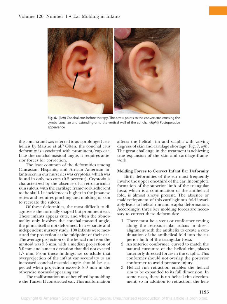
the concha and was referred to as a prolonged crushelicis by Matsuo et al.3 Often, the conchal crusdeformity is associated with prominent/cup ear.Like the conchal-mastoid angle, it requires ante-rior forces for correction.
The least common of the deformities amongCaucasian, Hispanic, and African American in-fants seen in our nurseries was cryptotia, which wasfound in only two ears (0.2 percent). Cryptotia ischaracterized by the absence of a retroauricularskin sulcus, with the cartilage framework adherentto the skull. Its incidence is higher in the Japaneseseries and requires pinching and molding of skinto recreate the sulcus.
Of these deformities, the most difficult to di-agnose is the normally shaped but prominent ear.These infants appear cute, and when the abnor-mality only involves the conchal-mastoid angle,the pinna itself is not deformed. In a separate andindependent nursery study, 100 infants were mea-sured for projection at the midpoint of their ear.The average projection of the helical rim from themastoid was 5.3 mm, with a median projection of5.0 mm and a mean deviation that did not exceed1.7 mm. From these findings, we conclude thatoverprojection of the infant ear secondary to anincreased conchal-mastoid angle should be sus-pected when projection exceeds 8.0 mm in theotherwise normal-appearing ear.
The malformation most benefited by moldingis the Tanzer II constricted ear. This malformation
affects the helical rim and scapha with varyingdegrees of skin and cartilage shortage (Fig. 7, left).The great challenge in the treatment is achievingtrue expansion of the skin and cartilage frame-work.
Molding Forces to Correct Infant Ear DeformityBirth deformities of the ear most frequently
involve the upper one-third of the ear. Incompleteformation of the superior limb of the triangularfossa, which is a continuation of the antihelicalfold, is almost always present. The absence ormaldevelopment of this cartilaginous fold invari-ably leads to helical rim and scapha deformation.Accordingly, three key molding forces are neces-sary to correct these deformities:
1. There must be a stent or conformer restingalong the retroauricular sulcus in directalignment with the antihelix to create a con-tinuation of the antihelical fold into the su-perior limb of the triangular fossa.
2. An anterior conformer, curved to match thenatural curvature of the helical rim, placesanteriorly directed forces in the scapha. Thisconformer should not overlap the posteriorconformer to avoid pressure injury.
3. Helical rim retraction enables the helicalrim to be expanded to its full dimension. Insome cases, there is no helical rim develop-ment, so in addition to retraction, the heli-
Fig. 6. (Left) Conchal crus before therapy. The arrow points to the convex crus crossing thecymba conchae and extending onto the vertical wall of the concha. (Right) Postoperativeappearance.
Volume 126, Number 4 • Ear Molding in Infants
1195

cal rim itself must be created to provide thedelicate cartilaginous arch that characterizesthe rim.
Midconchal DeformitiesIn reviewing infants who had poor outcome or
failures in molding, almost all were limited to arecurrence of overprojection. Many of these earswere noted to have a conchal crus bisecting themidsection of the cymba conchae and extendingonto the vertical wall of the concha. Sometimes,this crus was only seen when the pinna was pushedback to its normal position. The antihelical foldwas well shaped; however, the conchal-mastoid an-gle was widened. Generally, this produced over-projection across the midportion of the ear butsometimes affected the upper third as well.
A subset of children in this group had well-shaped and normal-projecting ears when moldingwas terminated at 6 weeks. However, over thecourse of 6 months, recurrent overprojection wasseen. This overprojection was again most notablein the conchal-mastoid angle. These children werealso unique in that they had a family history ofprominent ear deformity. It may be that the ge-netic influence in these children extends well be-yond the period of molding therapy and destinesthem to late recurrence. Attempts at taping moldsin the conchal hollow to correct the conchal crusand conchal-mastoid angle were not successful.The inability to apply enough anterior force withtaping and the consequent obliteration of the ex-
ternal auditory canal severely compromised anyefforts at molding this area.
With evidence that the conchal crus was a fac-tor in increasing the conchal-mastoid angle andproducing a prominent ear deformity, we createda molding device that would allow correction ofthe upper third as previously described and, at thesame time, allow anterior forces to be applied tothe concha that would flatten the conchal crus andcorrect the conchal-mastoid angle. A nonocclusivehypoallergenic adhesive was required to hold fora prolonged interval the device to an infant’s earwithout damaging the skin.
The final result incorporating all of these re-quirements was a two-piece cradle system thatslipped over the pinna and attached to the skinsurrounding the ear. The posterior cradle incor-porated a posterior conformer that guided thefold of the antihelix into the superior limb of thetriangular fossa while providing an adhesive sur-face to hold the device against the scalp. A retrac-tor system was subsequently developed to shapeand hold the helical rim in position. The retractorwas designed so that the helical rim snapped intoits preshaped arched cavity and the adjustable rimof the retractor served as an anterior stent to thescapha. The retractor then attached to the inneradhesive surface of the posterior cradle, allowingretraction and expansion of the rim and scaphawhen indicated. A soft compressible conchalformer was designed with an opening toward theexternal auditory canal and was made to fit in the
Fig. 7. A Tanzer II constricted ear before (left) and after therapy (right).
Plastic and Reconstructive Surgery • October 2010
1196

conchal cavity. The wall of the conchal former wascreated to rest against the vertical wall of the con-cha so that it would exert a downward force at thetakeoff of the concha from the skull (conchal-mastoid angle). The height of the conchal former
can be varied by the addition of compressiblefoam to its surface.
The anterior shell was attached to the posteriorcradle, allowing direct anterior forces to be appliedto the conchal former and retractor system. By mak-
Fig. 8. The EarWell Infant Ear Correction System. (Above, left) Infant with Stahl’s ear deformity. Thescalp hair is shaved approximately 2 cm around the ear and prepared with alcohol to create a hairlessfootprint to facilitate adherence of the posterior cradle against the scalp. (Above, right) The posteriorcradle is placed, making sure to orient the posterior conformer into the antihelix and the futureproposed superior limb of the triangular fossa. The arrow points to the posterior conformer. Note theimprovement in the helical rim deformity by the placement of the posterior guide. (Below, left) Place-ment of the retractor system to shape and hold the helical rim in position (black arrows). Theseretractors are held in place by the inner adhesive surface of the posterior cradle. A soft compressibleconchal former is placed within the conchal cavity (blue arrow). The height of this device can beaugmented by the addition of compressible foam to its surface. (Below, right) The clear and perfo-rated anterior shell attaches to the posterior cradle, allowing direct anterior forces to be applied tothe conchal former and retractor system.
Volume 126, Number 4 • Ear Molding in Infants
1197

ing the construct of the cradle clear and with per-forations, moisture collection was minimized anddirect visualization of the skin was permitted. Theadhesive of the device lasts approximately 2 weeks,requiring two additional applications for a total treat-ment period of 6 weeks. A universal size EarWell device(EarWell InfantEarCorrectionSystem;BeconMedicalLtd., Tucson, Ariz.) was created for babies whoseweight ranged from 4.5 to 8 pounds, and a second,larger sized version was fashioned for large-ear infantsweighing more than 8 pounds. Fifty-eight infants ears(34 patients) were treated with the final version of theEarWell System (Fig. 8). (See Video, SupplementalDigital Content 1, which demonstrates the use of theEarWell System, http://links.lww.com/PRS/A202).
RESULTSFinal auricular morphologic results were classi-
fied as excellent (normal shape), good (near normalshape with some degree of abnormality), and poor(slight or no improvement). From the final con-struct of the EarWell Infant Ear Correction System,58 infant ears (34 patients) have been managed witha success rate exceeding 90 percent (good to excel-lent results) (Figs. 1 through 7, right). The aestheticdetail among these ears far exceeds the outcome inour patients treated with various stents, tapes, andglues and also surpasses the surgical results in olderchildren. We are reluctant to say that prominent eardeformity has been eliminated with this device, be-cause we believe there is a subset of children thatgrow into this deformity, possibly manifesting be-yond our period of observation. Nevertheless, withthis new construct, we have been able to consistently
correct the conchal crus, which we believe is one ofthe underlying factors in prominent ear deformity.No premature infants or infants less than 4 poundswere treated. One 12-pound infant with very largeears had treatment initiated with the large EarWelldevice but had to be converted to retention tapesafter 2 weeks because of his overly large ears. He hada good but not excellent outcome after 6 weeks. Ofthe various deformities seen, this system corrects all,with the exception of cryptotia, which is still man-aged with various molding materials, splints, andstretching devices.5–9
Complications have been minor and few. Threepatients (5 percent) had localized skin excoriationsor breakdown: one in the cymba conchae from theconchal former, one along the helical rim/scaphajunction from the retractor system, and one poste-riorly in the retroauricular sulcus from the posteriorconformer (Fig. 9). There was no cartilage erosion.Each instance of skin injury was caused by the pos-terior cradle becoming loose and allowing move-ment and malposition of the internal parts. Tippingof the conchal former onto the fixed “nonfloating”part of the cymba conchae led to the breakdownshown. Parents are now instructed to return to theclinic when the adhesive of the EarWell device be-gins to loosen, which occurs on average 2 weeks afterapplication. One infant developed an erythematousrash around the ear that required disruption of ther-apy for 2 days. This rash was diagnosed as moniliaskin infection and was treated with topical antifungalcream before reapplication of the EarWell System.In all of these cases, the final outcomes were deemedexcellent.
DISCUSSIONIt is well established and apparent that the teach-
ing and belief that all infants born with misshapenears will self-correct is not only wrong but also leadsto an indifference to early diagnosis, which may pre-clude successful nonsurgical treatment with mold-ing. Several ways to splint the deformed ears havebeen reported, with satisfactory results.2–4,10–15 Ourexperience agrees with that of Merlob et al.16 and aprospective Canadian study17 where in the former,none of the control infants showed self-correction,and in the latter, 33 percent showed self-correction,most of which occurred early in life. Our recom-mendation to withhold the molding therapy untilthe end of the first week is aimed at identifying thoseinfants who may demonstrate self-correction. In ourexperience, also approximately one-third showedthis tendency toward self-correction, whereas the re-mainder warranted immediate treatment.
Video. Supplemental Digital Content 1 demonstrates use of theEarWell, http://links.lww.com/PRS/A202.
Plastic and Reconstructive Surgery • October 2010
1198

Several studies minimize the importance ofthe timing of molding therapy. Yotsuyanagi et al.9reported an excellent outcome in over 50 percentof children treated with ear molding with an av-erage age of 3.6 years. It is important to note thatover half of the deformities were cryptotias, a de-formity in which the cartilaginous framework isnormal and only requires expansion of the retro-auricular skin. Their average time of molding ther-apy was 2.1 months, which was longer than in ourpatients. Muraoka et al.12 also reported good re-sults in treated patients between 5 months and 5years old. In contrast, Matsuo et al.3,18 have shownthat deformities of the upper third (lop ear andStahl’s ear) respond to therapy only during theneonatal period, even though protruding ears andcryptotia may respond later. Tan et al.19 are addi-tional advocates for early diagnosis and initiationof molding therapy. Schonauer et al.13 recom-mend waiting 48 to 72 hours after birth beforeapplying their splint. In our study, the outcomesare clearly better when molding is instituted in thefirst 5 to 7 days of life and less favorable whenbegun after 3 weeks.
The responsiveness of the ear cartilage is great-est in the newborn because of maternal estrogen.20
Hyaluronic acid, an important constituent of earcartilage, is increased by estrogen, and is respon-sible for the malleable nature of the neonatalear.21–25 The circulating estrogen levels decreaserapidly to levels similar to those in older children
by 6 weeks of age.26 Breast-feeding was felt to ex-tend these levels and require a longer interval formolding, probably secondary to the maternal es-trogen in breast milk as previously speculated.19
Infants with failed or poor outcomes havebeen largely limited to the prominent/cup earcategory. Some had an associated conchal crusand were treated before the introduction of theconchal former. Others had a family history ofprominent ear and, despite a good response tomolding, experienced a gradual relapse aftertherapy was stopped. We now use retentiontapes in these infants to extend therapy out to3 months.
CONCLUSIONSCongenital ear deformities are common, and
only approximately 30 percent self-correct. Thesedeformities can be corrected by initiating appro-priate molding in the first week of life. Neonatalmolding reduces the need for surgical correctionwith results that often exceed what can beachieved with the surgical alternative.
H. Steve Byrd, M.D.Department of Plastic Surgery
University of Texas Southwestern Medical Center atDallas
1801 Inwood RoadDallas, Texas 75390
Fig. 9. Minor complications related to the EarWell device. (Left) Skin excoriation in the cymbaconchae from the conchal former. (Right) Skin excoriation along the helical rim/scapha junc-tion from the retractor system being placed too close to the posterior conformer.
Volume 126, Number 4 • Ear Molding in Infants
1199

REFERENCES1. Tan ST, Abramson DL, MacDonald DM, Mulliken JB. Mold-
ing therapy for infants with deformational auricular anom-alies. Ann Plast Surg. 1997;38:263-268.
2. Kurozumi N, Ono S, Ishida H. Non-surgical correction of acongenital lop ear deformity by splinting with Reston foam.Br J Plast Surg. 1982;35:181-182.
3. Matsuo K, Hirose T, Tomono T, et al. Nonsurgical correctionof congenital auricular deformities in the early neonate: Apreliminary report. Plast Reconstr Surg. 1984;73:38-51.
4. Nakajima T, Yoshimura Y, Kami T. Surgical and conservativerepair of Stahl’s ear. Aesthetic Plast Surg. 1984;8:101-107.
5. Dancey A, Jeynes P, Nishikawa H. Acrylic ear splints for treat-ment of cryptotia. Plast Reconstr Surg. 2005;115:2150-2152.
6. Hirose T, Tomono T, Matsuo K, et al. Cryptotia: Our clas-sification and treatment. Br J Plast Surg. 1985;38:352-360.
7. Hirose T, Tomono T, Yamamoto K. Non-surgical correctionfor cryptotia using simple apparatus. In: Foneseca J, ed.Transactions of the 7th International Congress of Plastic and Re-constructive Surgery, Rio de Janeiro, May 1979. Sao Paulo, Brazil:Catgraf; 1980.
8. Park C. Correction of cryptotia using an external stretchingdevice. Ann Plast Surg. 2002;48:534-538.
9. Yotsuyanagi T, Yokoi K, Urushidate S, Sawada Y. Nonsurgicalcorrection of congenital auricular deformities in children olderthan early neonates. Plast Reconstr Surg. 1998;101:907-914.
10. Bernal-Sprekelsen M, Krummel FJ. Conservative correctionof congenital deformities of the auricle (in German). Laryn-gorhinootologie 1990;69:581-585.
11. Brown FE, Colen LB, Addante RR, Graham JM Jr. Correctionof congenital auricular deformities by splinting in the neo-natal period. Pediatrics 1986;78:406-411.
12. Muraoka M, Nakai Y, Ohashi Y, Sasaki T, Maruoka K, Fu-rukawa M. Tape attachment therapy for correction of con-genital malformations of the auricle: Clinical and experi-mental studies. Laryngoscope 1985;95:167-176.
13. Schonauer F, La Rusca I, Molea G. Non-surgical correctionof deformational auricular anomalies. J Plast Reconstr AesthetSurg. 2009;62:876-883.
14. Tan ST, Shibu M, Gault DT. A splint for correction of con-genital ear deformities. Br J Plast Surg. 1994;47:575-578.
15. Ullmann Y, Blazer S, Ramon Y, Blumenfeld I, Peled IJ. Earlynonsurgical correction of congenital auricular deformities.Plast Reconstr Surg. 2002;109:907-913.
16. Merlob P, Eshel Y, Mor N. Splinting therapy for congenitalauricular deformities with the use of soft material. J Perinatol.1995;15:293-296.
17. Smith W, Toye J, Reid A, Smith R. Nonsurgical correction ofcongenital ear abnormalities in the newborn: Case series.Paediatr Child Health 2005;10:327-331.
18. Matsuo K, La Rusca I, Molea G. Non-surgical correction ofcongenital auricular deformities. Clin Plast Surg. 1990;17:383-395.
19. Tan S, Wright A, Hemphill A, Ashton K, Evans J. Correctionof deformational auricular anomalies by moulding: Resultsof a fast-track service. N Z Med J. 2003;116:U584.
20. Kenny FM, Angsusingha K, Stinson D, Hotchkiss J. Uncon-jugated estrogens in the perinatal period. Pediatr Res. 1973;7:826-831.
21. Hardingham TE, Muir H. The specific interaction of hyal-uronic acid with cartillage proteoglycans. Biochim Biophys Acta1972;279:401-405.
22. Hascall VC, Heinegard D. Aggregation of cartilage pro-teoglycans: II. Oligosaccharide competitors of the proteo-glycan-hyaluronic acid interaction. J Biol Chem. 1974;249:4242-4249.
23. Schiff M, Burn HF. The effect of intravenous estrogens onground substance. Arch Otolaryngol. 1961;73:43-51.
24. Uzuka M, Nakajima K, Ohta S, Mori Y. The mechanism ofestrogen-induced increase in hyaluronic acid biosynthesis,with special reference to estrogen receptor in the mouseskin. Biochim Biophys Acta 1980;627:199-206.
25. Uzuka M, Nakajima K, Ohta S, Mori Y. Induction of hyal-uronic acid synthetase by estrogen in the mouse skin. BiochimBiophys Acta 1981;673:387-393.
26. Hung W, August G, Glasgow AM. The ovary. In: Hung W,August G, Glasgow AM, eds. Pediatric Endocrinology. New York:Medical Examination Publishing; 1978.
Instructions for Authors: UpdateEthical Approval of Studies/Informed Consent
Authors of manuscripts that describe experimental studies on either humans or animals must supply to theEditor a statement that the study was approved by an institutional review committee or ethics committee andthat all human subjects gave informed consent. Such approval should be described in the Methods sectionof the manuscript. For studies conducted with human subjects, the method by which informed consent wasobtained from the participants (i.e., verbal or written) also needs to be stated in the Methods section.
In those situations where a formal institutional review board process is not available, the authors must indicatethat the principles outlined in the Declaration of Helsinki have been followed. More information regardingthe Declaration of Helsinki can be found at http://www.wma.net/e/policy/b3.htm.
Plastic and Reconstructive Surgery • October 2010
1200