eae teaching course - clinical applications of...
TRANSCRIPT

European Society of Cardiology copyright -All right reserved
Ventricular-arterial coupling
Prof. Dr. Alan FraserCardiff University

European Society of Cardiology copyright -All right reserved
Ventricular–arterial coupling
• Increased loading triggers LV hypertrophy
• Ventricular response is asymmetrical
• Central arterial pressure
• Conduit arterial stiffness
• Ventricular and vascular elastance
• Arterial waves and energy

European Society of Cardiology copyright -All right reserved
Central arterial pressure changes with age
Peripheral pressure
amplification
Central pressure
augmentation
McDonald’s Blood Flow in Arteries

European Society of Cardiology copyright -All right reserved
AUGMENTATION
Applanation tonometry
Stiff conduit arteries cause increased central aortic pressure, isolated systolic hypertension,
and increased arterial pulse pressure
Wave speed is a powerful prognostic indicator

European Society of Cardiology copyright -All right reserved
• BETA INDEX
- Pressure-independent Young’s modulus of stiffness
- Adjusted for BP
- No units
• EPSILON
- Pressure-strain elastic modulus
- Measured in kPa
Imaging arterial stiffness
logn (Ps / Pd)
(Ds - Dd / Dd)b =
(Ps - Pd) Dd
(Ds - Dd)Ep =
Anterior wall
Posterior wall
Diameter

European Society of Cardiology copyright -All right reserved
Pooled observations in 144 subjects – Age
J Swampillai
Conduit arterial stiffness
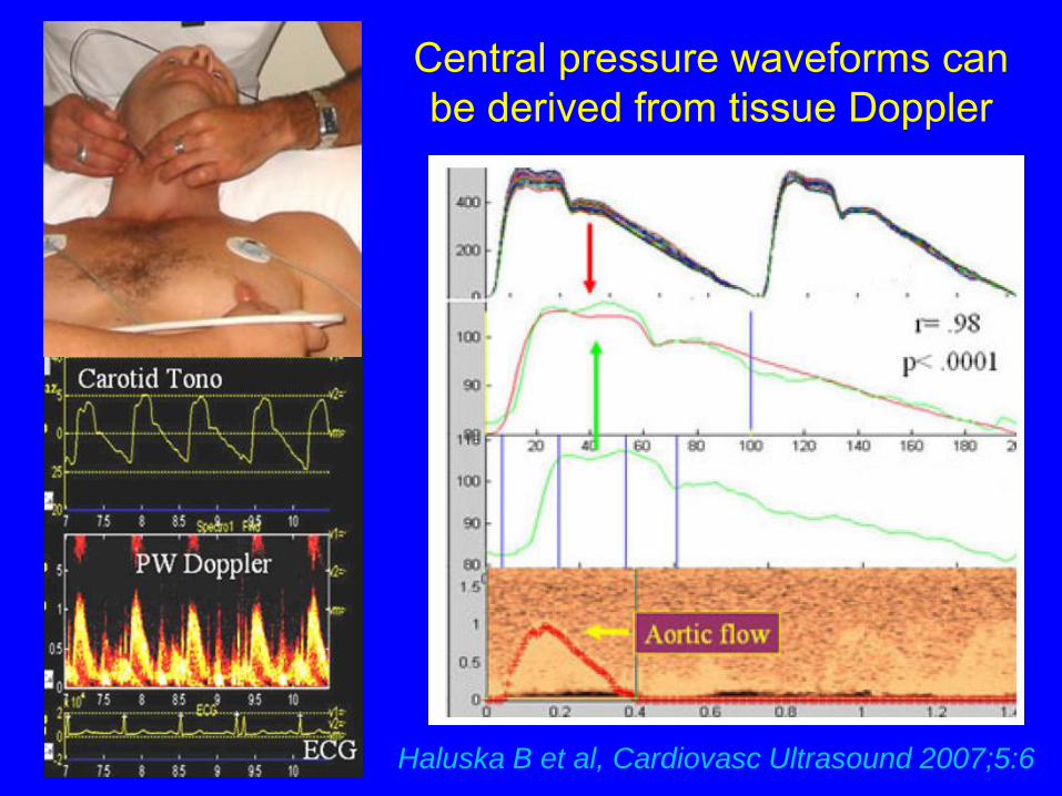
European Society of Cardiology copyright -All right reserved Haluska B et al, Cardiovasc Ultrasound 2007;5:6
Central pressure waveforms can be derived from tissue Doppler

European Society of Cardiology copyright -All right reserved
Tissue velocity of aortic wall motion

European Society of Cardiology copyright -All right reserved Williams A et al
Aortic wall motion in Marfan syndromeProspective, double-blind, randomised cross-over
Time to peak systolic velocity of anterior aortic wall motion
-10
-5
0
5
10
15
20
25
30
Perindopril Verapamil Atenolol
D ms
4 mg 240 mg SR 75 mg
ArchAbdo
p = 0.003

European Society of Cardiology copyright -All right reserved
Arterial stiffness is a determinant of VO2 max
Hundley, JACC 2001; 38: 796
Distensibility
VO2 max (ml/min)
10-3 mmHg-1

European Society of Cardiology copyright -All right reserved
VO
2m
ax
(m
l/k
g/m
in)
Arterial compliance & peak exercise capacity are
related
& both decline with risk factors
& severity of the metabolic
syndrome
Wong C et al,
AJC 2005;96:1686

European Society of Cardiology copyright -All right reserved
Myocardial dysfunction in metabolic syndromecorrelates with number of diagnostic features
Wong C et al, Am J Cardiol 2005; 96: 1686-91
393 subjects with negative stress echocardiography

European Society of Cardiology copyright -All right reserved
Pressure
Volume
ESP
DP
ESV EDV
Left ventricular pressure-volume loop
·
·Isovolumic
contraction
Ejection
Filling
Isovolumic
relaxation
End-diastolic pressure volume relationship
End-systolic pressure volume relationship contractility

European Society of Cardiology copyright -All right reserved
Pressure
Volume
ESP
DP
ESV EDV
·
· ·
The traditional conceptof ventricular-arterial coupling
Elastance =change in pressure for a given change in volume

European Society of Cardiology copyright -All right reserved
Elastance
Change in pressure for a unit change in volume(mmHg/ml)
• Arterial elastance
EA = ESP / SV (SV stroke volume)
Higher elastance = greater sensitivity to volume change, more variable pressure
• Ventricular elastanceELV = ESP / ESV (ESV LV end-systolic volume)
Net arterial load exerted on the ventricle
European Society of Cardiology copyright -All right reserved
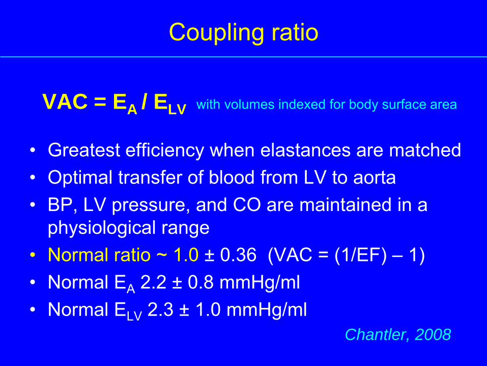
VAC = EA / ELV with volumes indexed for body surface area
• Greatest efficiency when elastances are matched• Optimal transfer of blood from LV to aorta • BP, LV pressure, and CO are maintained in a
physiological range• Normal ratio ~ 1.0 ± 0.36 (VAC = (1/EF) – 1) • Normal EA 2.2 ± 0.8 mmHg/ml• Normal ELV 2.3 ± 1.0 mmHg/ml
Chantler, 2008
Coupling ratio

European Society of Cardiology copyright -All right reserved
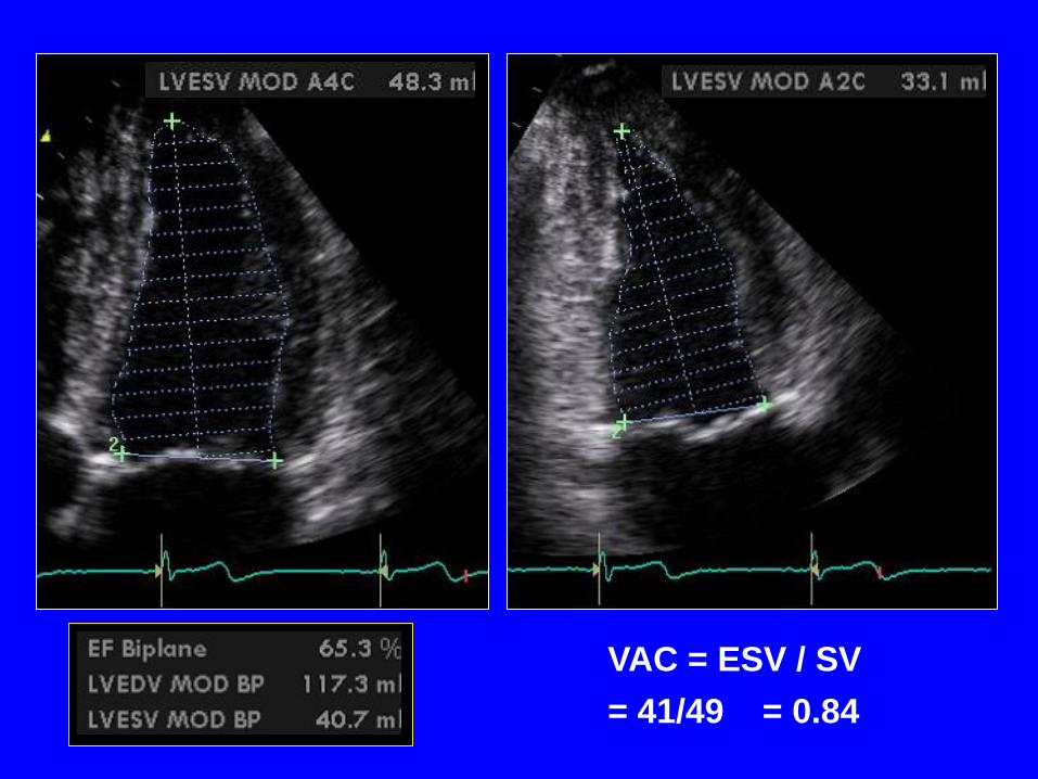
M aged 30, triathlete
SV = 49ml
European Society of Cardiology copyright -All right reserved
VAC = ESV / SV
= 41/49 = 0.84

European Society of Cardiology copyright -All right reserved Circulation 2003; 107: 714-20

European Society of Cardiology copyright -All right reserved
Myocardial contractility in the left ventricle is
inversely related to arterial compliance
Kawaguchi M et al, Circulation 2003; 107: 714-20

European Society of Cardiology copyright -All right reserved
Journal of Biomechanical
Engineering 1990; 112: 323
Wave intensity = dP/dt . dU/dt

European Society of Cardiology copyright -All right reserved
Velocity Pressure
Forward compression wave
Forward expansion wave
Backward compression wave
Backward expansion wave
There are 4 types of arterial waves
The integral of wave intensity is energy

European Society of Cardiology copyright -All right reserved
Wave intensity and reflection
ControlAbdominal
Aorta DiaphragmDescending
Aorta
Ramsey, Jones & Sugawara

European Society of Cardiology copyright -All right reserved
Wall tracking of common carotid artery
Diameter / distension waveform, calibrated as
estimate of pressure
Simultaneous measurement of blood velocity at same site
Non-invasive
wave intensity

European Society of Cardiology copyright -All right reserved
Anterior wall
Posterior wall
Aloka SSD 5500 7.5 MHz linear array transducer
Pressure
Velocity
Compression
wave
Expansion
wave
Reflections
dP/dt.dU/dt

European Society of Cardiology copyright -All right reserved
Beta
index
Pressure-
strain
elastic
modulus
Integral of mid-
systolic wave
reflections
Longitudinal S -0.45** -0.37** -0.37**Longitudinal E -0.53** -0.47** -0.50**Radial S -0.07 -0.02 -0.06 Radial E -0.28* -0.25* -0.13Ejection fraction -0.21 -0.16 -0.15E/Ea 0.48** 0.50** 0.34**
Ventricular-arterial couplingConduit arterial stiffness impairs long-axis function
Vinereanu D et al, Heart 2003; 89: 449

European Society of Cardiology copyright -All right reserved
1. Peak velocity of radial
shortening of the left ventricle
coincides with arrival of reflected
waves
2. Generation of longitudinal shortening
persists against reflections
Page C et al,
Int J Cardiol 2009 e pub
0 1 2 3 4
-5
0
5
10
15
20
FCW
BCW
FEW
50 70 90 110 130 150 170 190 310
-0.015
-0.01
-0.005
0
0.005
0.01
0.015
Radial
Long-
axis
Wave
intensity
in the
ascending
aorta
W/m2
Velocity of
shortening
m/s
Time ms

European Society of Cardiology copyright -All right reserved Rakebrandt F et al, Ultrasound Med Biol 2009; 35: 266-77
The major “determinant” of reflected wave energy
is left ventricular systolic function
3
3,5
4
4,5
5
5,5
6
6,5
4,5 5 5,5 6 6,5 7 7,5
Logn FCW integral
r = 0.81, p<0.0001
MALE
FEMALE
Logn
BCW
integral
from
wave
intensity
studies
n 106, 57 M, aged 46 ± 12 (22-71) years

European Society of Cardiology copyright -All right reserved
The heart does not operate independently ..
.. it’s part of an integrated
cardiovascular symptom
Why do we have to understand V-A coupling ?
- to understand mechanisms of LV dysfunction
- because conduit arterial stiffness, central arterial pressure, and wave reflections can be important new therapeutic targets