e-sayfa Özgün görüntüler e-page original images e-15
TRANSCRIPT
E-sayfa Özgün Görüntüler E-page Original Images E-15
Aortic valve aneurysm: a result or reason?
Aort kapak anevrizması: Neden mi, sonuç mu?
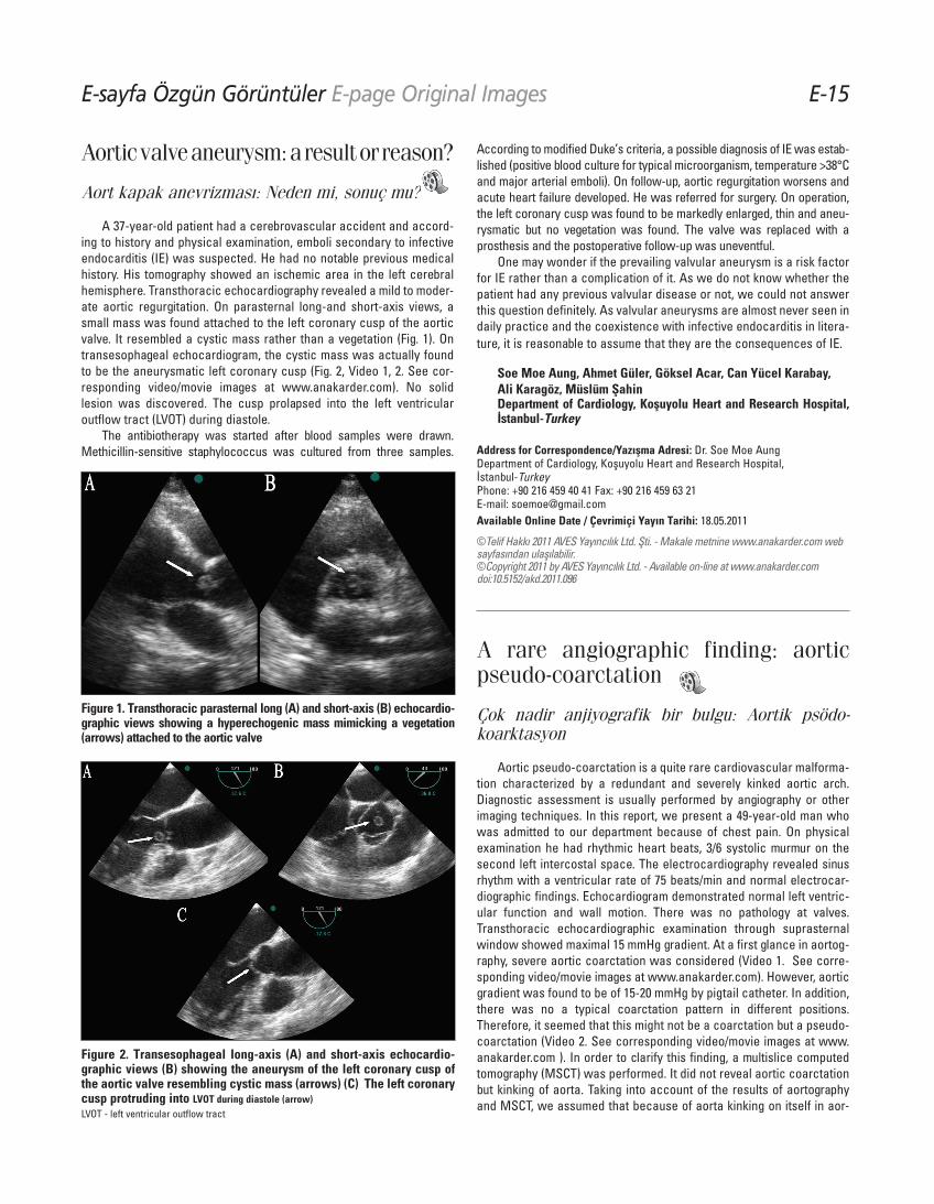
A 37-year-old patient had a cerebrovascular accident and accord-ing to history and physical examination, emboli secondary to infective endocarditis (IE) was suspected. He had no notable previous medical history. His tomography showed an ischemic area in the left cerebral hemisphere. Transthoracic echocardiography revealed a mild to moder-ate aortic regurgitation. On parasternal long-and short-axis views, a small mass was found attached to the left coronary cusp of the aortic valve. It resembled a cystic mass rather than a vegetation (Fig. 1). On transesophageal echocardiogram, the cystic mass was actually found to be the aneurysmatic left coronary cusp (Fig. 2, Video 1, 2. See cor-responding video/movie images at www.anakarder.com). No solid lesion was discovered. The cusp prolapsed into the left ventricular outflow tract (LVOT) during diastole.
The antibiotherapy was started after blood samples were drawn. Methicillin-sensitive staphylococcus was cultured from three samples.
According to modified Duke’s criteria, a possible diagnosis of IE was estab-lished (positive blood culture for typical microorganism, temperature >38°C and major arterial emboli). On follow-up, aortic regurgitation worsens and acute heart failure developed. He was referred for surgery. On operation, the left coronary cusp was found to be markedly enlarged, thin and aneu-rysmatic but no vegetation was found. The valve was replaced with a prosthesis and the postoperative follow-up was uneventful.
One may wonder if the prevailing valvular aneurysm is a risk factor for IE rather than a complication of it. As we do not know whether the patient had any previous valvular disease or not, we could not answer this question definitely. As valvular aneurysms are almost never seen in daily practice and the coexistence with infective endocarditis in litera-ture, it is reasonable to assume that they are the consequences of IE.
Soe Moe Aung, Ahmet Güler, Göksel Acar, Can Yücel Karabay, Ali Karagöz, Müslüm ŞahinDepartment of Cardiology, Koşuyolu Heart and Research Hospital, İstanbul-Turkey
Address for Correspondence/Yaz›şma Adresi: Dr. Soe Moe AungDepartment of Cardiology, Koşuyolu Heart and Research Hospital, İstanbul-TurkeyPhone: +90 216 459 40 41 Fax: +90 216 459 63 21 E-mail: [email protected] Online Date / Çevrimiçi Yayın Tarihi: 18.05.2011
©Telif Hakk› 2011 AVES Yay›nc›l›k Ltd. Şti. - Makale metnine www.anakarder.com web sayfas›ndan ulaş›labilir.©Copyright 2011 by AVES Yay›nc›l›k Ltd. - Available on-line at www.anakarder.comdoi:10.5152/akd.2011.096
A rare angiographic finding: aortic pseudo-coarctation
Çok nadir anjiyografik bir bulgu: Aortik psödo-koarktasyon
Aortic pseudo-coarctation is a quite rare cardiovascular malforma-tion characterized by a redundant and severely kinked aortic arch. Diagnostic assessment is usually performed by angiography or other imaging techniques. In this report, we present a 49-year-old man who was admitted to our department because of chest pain. On physical examination he had rhythmic heart beats, 3/6 systolic murmur on the second left intercostal space. The electrocardiography revealed sinus rhythm with a ventricular rate of 75 beats/min and normal electrocar-diographic findings. Echocardiogram demonstrated normal left ventric-ular function and wall motion. There was no pathology at valves. Transthoracic echocardiographic examination through suprasternal window showed maximal 15 mmHg gradient. At a first glance in aortog-raphy, severe aortic coarctation was considered (Video 1. See corre-sponding video/movie images at www.anakarder.com). However, aortic gradient was found to be of 15-20 mmHg by pigtail catheter. In addition, there was no a typical coarctation pattern in different positions. Therefore, it seemed that this might not be a coarctation but a pseudo-coarctation (Video 2. See corresponding video/movie images at www.anakarder.com ). In order to clarify this finding, a multislice computed tomography (MSCT) was performed. It did not reveal aortic coarctation but kinking of aorta. Taking into account of the results of aortography and MSCT, we assumed that because of aorta kinking on itself in aor-
Figure 1. Transthoracic parasternal long (A) and short-axis (B) echocardio-graphic views showing a hyperechogenic mass mimicking a vegetation (arrows) attached to the aortic valve
Figure 2. Transesophageal long-axis (A) and short-axis echocardio-graphic views (B) showing the aneurysm of the left coronary cusp of the aortic valve resembling cystic mass (arrows) (C) The left coronary cusp protruding into LVOT during diastole (arrow)LVOT - left ventricular outflow tract
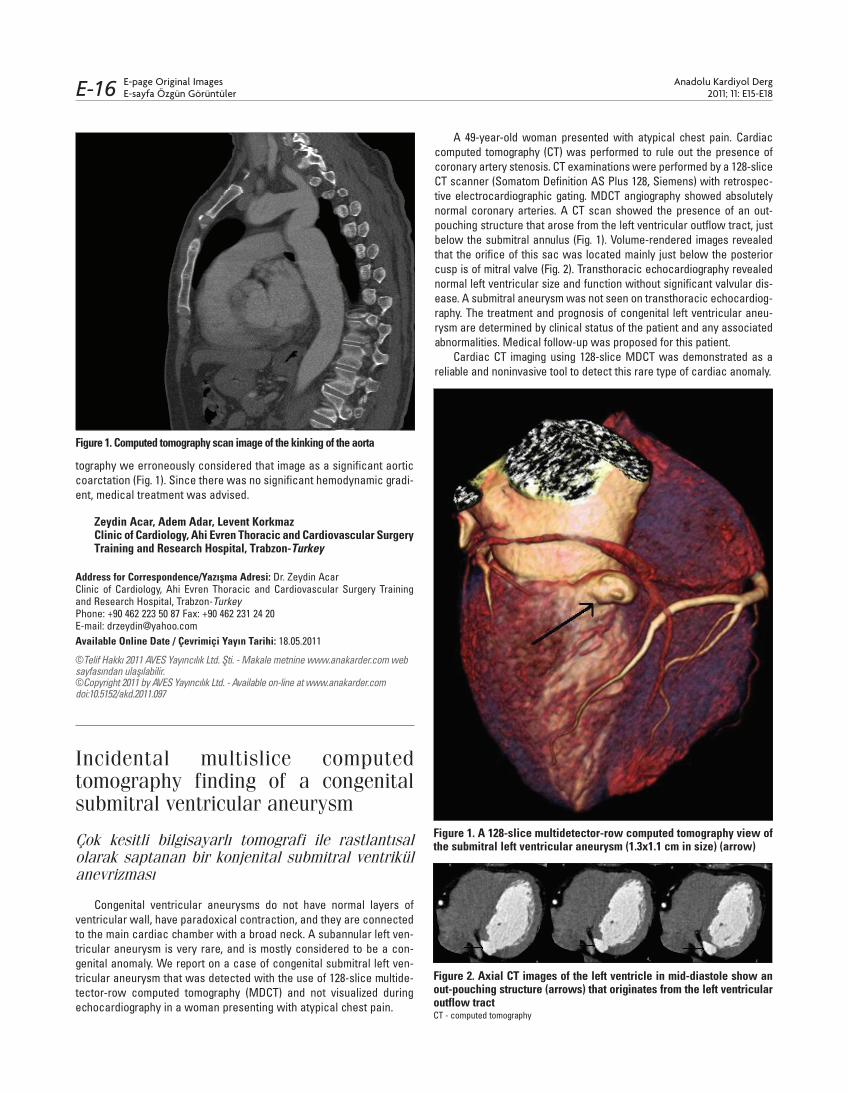
tography we erroneously considered that image as a significant aortic coarctation (Fig. 1). Since there was no significant hemodynamic gradi-ent, medical treatment was advised.
Zeydin Acar, Adem Adar, Levent KorkmazClinic of Cardiology, Ahi Evren Thoracic and Cardiovascular Surgery Training and Research Hospital, Trabzon-Turkey
Address for Correspondence/Yaz›şma Adresi: Dr. Zeydin AcarClinic of Cardiology, Ahi Evren Thoracic and Cardiovascular Surgery Training and Research Hospital, Trabzon-TurkeyPhone: +90 462 223 50 87 Fax: +90 462 231 24 20 E-mail: [email protected] Online Date / Çevrimiçi Yayın Tarihi: 18.05.2011
©Telif Hakk› 2011 AVES Yay›nc›l›k Ltd. Şti. - Makale metnine www.anakarder.com web sayfas›ndan ulaş›labilir.©Copyright 2011 by AVES Yay›nc›l›k Ltd. - Available on-line at www.anakarder.comdoi:10.5152/akd.2011.097
Incidental multislice computed tomography finding of a congenital submitral ventricular aneurysm
Çok kesitli bilgisayarlı tomografi ile rastlantısal olarak saptanan bir konjenital submitral ventrikül anevrizması
Congenital ventricular aneurysms do not have normal layers of ventricular wall, have paradoxical contraction, and they are connected to the main cardiac chamber with a broad neck. A subannular left ven-tricular aneurysm is very rare, and is mostly considered to be a con-genital anomaly. We report on a case of congenital submitral left ven-tricular aneurysm that was detected with the use of 128-slice multide-tector-row computed tomography (MDCT) and not visualized during echocardiography in a woman presenting with atypical chest pain.
A 49-year-old woman presented with atypical chest pain. Cardiac computed tomography (CT) was performed to rule out the presence of coronary artery stenosis. CT examinations were performed by a 128-slice CT scanner (Somatom Definition AS Plus 128, Siemens) with retrospec-tive electrocardiographic gating. MDCT angiography showed absolutely normal coronary arteries. A CT scan showed the presence of an out-pouching structure that arose from the left ventricular outflow tract, just below the submitral annulus (Fig. 1). Volume-rendered images revealed that the orifice of this sac was located mainly just below the posterior cusp is of mitral valve (Fig. 2). Transthoracic echocardiography revealed normal left ventricular size and function without significant valvular dis-ease. A submitral aneurysm was not seen on transthoracic echocardiog-raphy. The treatment and prognosis of congenital left ventricular aneu-rysm are determined by clinical status of the patient and any associated abnormalities. Medical follow-up was proposed for this patient.
Cardiac CT imaging using 128-slice MDCT was demonstrated as a reliable and noninvasive tool to detect this rare type of cardiac anomaly.
Figure 1. Computed tomography scan image of the kinking of the aorta
Figure 1. A 128-slice multidetector-row computed tomography view of the submitral left ventricular aneurysm (1.3x1.1 cm in size) (arrow)
Figure 2. Axial CT images of the left ventricle in mid-diastole show an out-pouching structure (arrows) that originates from the left ventricular outflow tractCT - computed tomography
E-page Original ImagesE-sayfa Özgün Görüntüler
Anadolu Kardiyol Derg 2011; 11: E15-E18E-16