dysmenorrhea: a pilot study connectivity in the pain
TRANSCRIPT
Page 1/22
Acupuncture Treatment Compensated the Altered FunctionalConnectivity in the Pain Modulation System on PrimaryDysmenorrhea: A Pilot StudyCheng-Hao Tu ( [email protected] )
China Medical University https://orcid.org/0000-0002-7695-5570Yu-Chen Lee
China Medical University Hospital Department of AcupunctureYing-Yu Chen
China Medical University Hospital Department of Chinese Medicine GynecologyChun-Ming Chen
China Medical University Hospital Department of Medical ImagingWen-Chi Lu
China Medical University Graduate Institute of Acupuncture ScienceYi-Hung Chen
China Medical University Graduate Institute of Acupuncture ScienceSu-Tso Yang
China Medical University Hospital Department of Medical Imaging
Research
Keywords: primary dysmenorrhea, acupuncture, Sanyinjiao, descending pain modulation system, periaqueductal gray matter,functional connectivity, resting-state functional magnetic resonance imaging
Posted Date: November 12th, 2020
DOI: https://doi.org/10.21203/rs.3.rs-104133/v1
License: This work is licensed under a Creative Commons Attribution 4.0 International License. Read Full License

Page 2/22
AbstractBackground
Primary dysmenorrhea (PDM) is the most commonly encountered gynecological problem in reproductive-age women. Wepreviously reported altered functional connectivity between periaqueductal gray matter and other pain-related brain regions inwomen with PDM, indicating a maladaptation in the descending pain modulation system. Clinically, acupuncture has beensuggested as an effective treatment of PDM. Previous animal studies revealed that acupuncture on speci�c acupoint canproduced analgesic effect via descending pain modulation system. In the present study, we used resting-state functional magneticresonance imaging to investigate possible changes in descending pain modulation after acupuncture treatment in women withPDM.
Methods
Thirty-four right-handed adult women with PDM participated in this randomized, single-blinded, sham-controlled study. Eachpatient was randomly allocated to an 8-week verum or sham acupuncture intervention on the bilateral acupoint Sanyinjiao (SP6).Resting-state functional magnetic resonance imaging was conducted before, during, and after the intervention session to measurethe spontaneous activity in brain. The functional connectivity maps between periaqueductal gray matter and other pain-relatedbrain regions were generated to reveal the status of descending pain modulation systems.
Results
After 8 weeks, both groups reported decreased menstrual pain. However, the patterns of functional connectivity changes indescending pain modulation system were different after the verum or sham intervention. The effect of the verum acupunctureintervention may be underpinned by compensatory and/or resilience changes in descending pain modulation systems, whereasthat of the sham acupuncture intervention may be underpinned by cognitive reappraisal of pain
Conclusions
The dissimilar pattern of the FC alterations after the verum and sham acupuncture indicating that different brain mechanismsmay be responsible for the menstrual pain relief in verum or sham acupuncture intervention. These results may contribute to thediscussion of whether acupuncture treatment induces therapeutic physiological alterations or its bene�t is attributable to theplacebo effect.
BackgroundPrimary dysmenorrhea (PDM) is the most common gynecological problem among reproductive-age women. Around 10–20% ofwomen experience PDM, causing them to be absent from work or school; thus, PDM affects their daily life (1). Women with PDMexperience cramping pain during menses, which may be due to an abnormal concentration of prostanoids, uterinehypercontractility, or reduced uterine blood �ow (2). Furthermore, they have a lower pain threshold in not only the referred pain sitebut also the non-referred pain site, indicating altered pain perception in the central nervous system (3, 4). We previously reportedseveral abnormal functional and structural changes in brain regions associated with visceral sensation and pain processing (5, 6),as well as how altered functional connectivity (FC) between periaqueductal gray matter (PAG) and other pain-related brain regionsin women with PDM indicated the maladaptation of the descending pain modulation system (7).
Acupuncture has been suggested to be an effective treatment for PDM (8). A recent meta-analysis including 60 randomizedcontrol trials reported that manual acupuncture is more effective for menstrual pain relief than no treatment and nonsteroidal anti-in�ammatory drugs (9). Among these 60 trials, the acupoint Sanyinjiao (SP6) was the most frequently used for treating PDM (9).Acupuncture might alleviate PDM through peripheral or central mechanisms. Peripherally, manual acupuncture on SP6signi�cantly improves uterine artery blood �ow (10). However, blood prostanoid concentrations were not signi�cantly different inresponse to electroacupuncture treatment on SP6 and that on other acupoints (11). Thus, the treatment effect of acupuncture onPDM may be mediated by vascular response but not changes in blood prostanoid levels.

Page 3/22
How acupuncture exerts the central effect on PDM has not been fully explored. Acupuncture on speci�c acupoints can produce ananalgesic effect through descending pain modulation systems, especially by modulating PAG activity (12). Furthermore, thetermination of chronic pain or of repetitive painful stimuli may be associated with functional and structural resilience changes inthe brain (13–15). Therefore, we explored the central mechanisms possibly underlying the effect of acupuncture treatment onPDM. We hypothesized that acupuncture resilience the altered FC in the descending pain modulation systems between PAG andother pain-related brain areas on PDM.
Materials And Methods
SubjectsThis study was conducted in accordance with the Declaration of Helsinki. The protocol was approved by the Institutional ReviewBoard of China Medical University Hospital, Taiwan (CMUH105-REC1-027). All patients were recruited from the internet and thenreferred to and diagnosed by the Department of Chinese Medicine Gynecology, China Medical University Hospital, Taiwan. Allpatients received a full explanation of the study and signed written informed consent forms.
The inclusion criteria for subject were as follows: a) aged 20–35 years, b) right-handed, c) regular menstrual cycle of 27–32 days,and d) average menstrual pain level in the past 6 months > 4 points on a 10-point numerical rating scale. The exclusion criteriawere as follows: a) history of pituitary gland disorder; b) history of organic pelvic or reproductive system disease(s); c) history ofmental disorder(s), such as major depressive disorder, bipolar disorder, general anxiety disorder, claustrophobia, or schizophrenia;d) history of brain trauma or neurological disease(s), such as epilepsy, stroke, Parkinson’s disease, or dementia; e) history of orcurrent pregnancy or plans to get pregnant; f) presence of metal implants or a pacemaker; g) use of oral contraceptives,psychiatric medicines, Chinese herbal medicine, or acupuncture treatment in the past 6 months or taking analgesic drugs 24 hbefore the magnetic resonance imaging (MRI) scan; and h) unsuited for acupuncture treatment, as determined by the physiciansof the Department of Chinese Medicine Acupuncture, China Medical University Hospital, Taiwan.
Study designThis study was a randomized, single-blind, sham-controlled study. All subjects were randomly allocated to the verum or shamacupuncture group at a 1:1 ratio by using a computer-generated list of random numbers. All subjects were not been inform theallocated group until the end of the experiment. During the inception interview, subject’s general PDM experience in the past wasassessed, and handedness were assessed using the Chinese version of the Edinburgh Handedness Inventory (16).Transabdominal ultrasound was performed to exclude possible organic diseases in the reproduction system that may lead tosecondary dysmenorrhea. The resting-state functional MRI (rfMRI) scans were conducted on weeks 0, 4, and 8 of verum or shamacupuncture intervention sessions, respectively. To reduce the possible interference of menstrual phase, the MRI scans wereconducted during the follicular phase (days 5–12 of the menstrual cycle). The latest menstrual pain experience and psychologicalstate were also assessed after each MRI scan. Venous blood samples were taken to evaluate the concentrations of estrogen,progesterone, and testosterone within 2 days before or after MRI scans. If the subject had pregnancy concerns, a urine test strippregnancy test was performed.
Acupuncture interventionAll acupuncture interventions were performed by attending physicians with more than 10-year experience in the outpatient clinicsof the Department of Chinese Medicine Acupuncture, China Medical University Hospital, Taiwan. Subjects received a 20-minmanual acupuncture intervention on bilateral SP6 twice per week for 8 consecutive weeks (total 16 interventions). The acupointselection was based on a recent meta-analysis study which reported that manual acupuncture is more effective for menstrualpain relief and SP6 was the most frequently used for treating PDM (9). The acupoint was cleaned with an alcohol swab, and an O-ring was placed on the acupoint and covered by white surgical tape to conceal the penetration depth of the needle. In the verumacupuncture group, the standard disposable sterile acupuncture needle (diameter: 0.22 mm, length: 30 mm) was used to penetratevertically into SP6 to a depth of approximately 25 mm. In the sham acupuncture group, the Streitberger’s placebo needle (Asia-med GmbH. Pullach, Germany), which can effectively serve as the placebo needle in acupuncture (17, 18), was used to avoid skinpenetration. In both verum and sham group, the retention time of needles on SP6 was 20 minutes and no manipulation on the
Page 4/22
needles during the retention time. To verify the effectiveness of single blinding procedure, the subjects were asked whether theyreceived verum or sham acupuncture after they completed whole 8-week intervention.
Menstrual pain experience and psychological assessmentsThe Chinese version of the McGill Pain Questionnaire (MPQ) was used to assess the multidimensional menstrual pain experience(19). The total score of the pain rating index represents the menstrual pain experience, whereas that of the present pain intensityrepresents menstrual pain intensity. In addition, the Chinese version of Spielberger’s State-Trait Anxiety Inventory (STAI) and theChinese version of Beck’s Depression Inventory II (BDI II) were used to assess the level of anxiety and depression, respectively (20,21).
Measurement of blood gonadal hormone levelBlood concentrations of estrogen, progesterone, and testosterone were assessed using the chemiluminescent immunoassaysandwich method in the Department of Laboratory Medicine, China Medical University Hospital, Taiwan.
Image acquisitionAll brain images were acquired with an 8-channel head coil in a 3.0 T MRI scanner (Signa HDxt, GE Healthcare, Chicago, IL, USA).For rfMRI, four blank scans and 200 resting scans were continuously acquired with an ascending interleaved echo-planar imagingsequence (repetition time = 2500 ms; echo time = 30 ms; �ip angle = 90°; matrix = 64 × 64; �eld of view = 224 × 224 mm2; slicenumber = 40; and slice thickness = 3.5 mm). High-resolution, three-dimensional, T1-weighted anatomical images were alsoacquired with a spoiled gradient echo sequence (repetition time: 7.356 ms; echo time: 2.736 ms; �ip angle = 12°; matrix = 224 × 224 × 170; �eld of view = 224 × 224 × 170 mm3). All scans were acquired in a dimly lit shielding room. Before scanning, subjectswere instructed to relax, not to move their head, and keep their eyes opened during the scan. The whole time for scanningprocedure (including preparation time) was about 20 minutes.
Image preprocessingAfter discarding blank scans, the rfMRI scans were preprocessed using Statistical Parametric Mapping 12 (Wellcome Centre forHuman Neuroimaging, University College London, London, UK) with MATLAB 2018a (MathWorks, Sherborn, MA, USA). The rfMRIimages were �rst corrected for slice acquisition times and then realigned to correct the head motions during image scan. Subjectwith head motion above 1.75 mm maximum displacement in x-, y-, or z-direction or 1.5 degrees of angular motion were excludedfor further analysis. The T1-weighted anatomical image was coregistered to the mean image generated from the realignment stepand then normalized into the Montreal Neurological Institute reference space with default template using SPM12. Thenormalization parameters were then applied to all rfMRI images for spatial normalization with the resampled voxel size of 2 × 2 × 2 mm3. Finally, the normalized rfMRI images were smoothed with a three-dimensional Gaussian kernel (8-mm full width at halfmaximum).
The FC maps of PAG and other brain regions were generated by Data Processing and Analysis for Brain Imaging 4.3 with MATLAB2018a (22). In preprocessed images, the brain activities in each voxel were linearly detrended and bandpass �ltered (0.01–0.08 Hz), and the confounding variables, namely six head movement parameters, the global mean signal, the mean signal ofwhite matter, and the mean signal of the cerebrospinal �uid, were regressed out. The mean time-series activity in the seed regionof PAG (a spherical region centered at the coordinates [− 4, − 26, − 14] with a 3-mm radius (23)) was extracted, and voxel-wisecorrelation analysis was conducted to generate the correlation maps of PAG. Finally, z-standardization was performed to generatestandardized FC maps for statistical analysis.
Statistical analysisThe statistical analyses regarding demographic data, menstrual pain experience, psychological assessment, and blood gonadalhormone levels were conducted using SPSS v21 (IBM, Armonk, NY, USA). To compare intergroup differences in demographic dataand general PDM experience, a two-sample t test was conducted. The two-way mixed-model analysis of variance was used to testthe possible interaction effects between groups (verum and sham) and times (before, during, and after) on menstrual painexperience, psychological status, and blood gonadal hormone levels. The signi�cance was set at p < 0.05.
Page 5/22
The statistical analysis for standardized FC maps of PAG was conducted using Statistical Parametric Mapping 12. The two-waymixed-model analysis of variance was constructed as the statistical model to estimate overall variance. The intragroupcomparisons between different times in each group and interaction effect between groups and times were tested using t-contrastsin the model. A correlation analysis was also conducted between the menstrual pain experience and FC maps of PAG across 8-weeks intervention in each group using two-way mixed-model analysis of covariance. Considering that the fMRI study often havevery low statistical power (24), a less stringent signi�cance threshold (uncorrected p < 0.005 at the voxel level with a cluster size > 40, corresponding to an uncorrected cluster-level p < 0.35) was applied to balance the chance of Type I and Type II error assuggested by a previous study (24).
Results
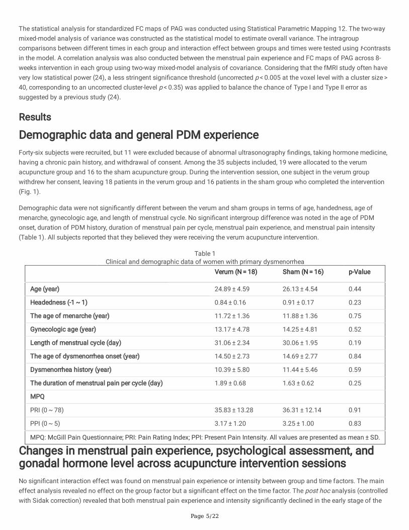
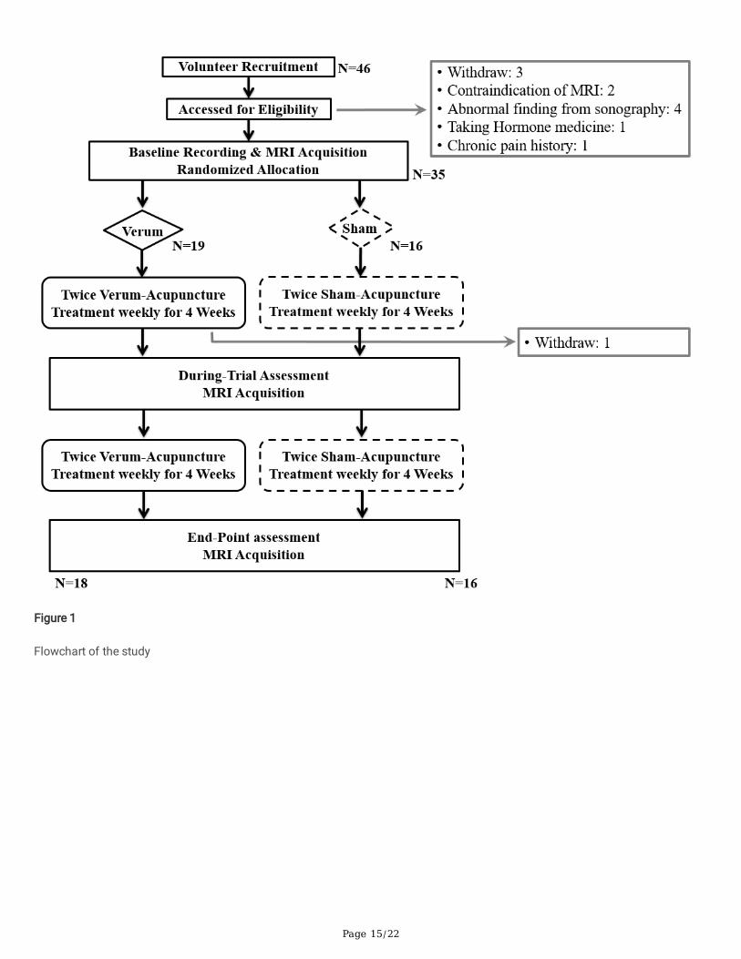
Demographic data and general PDM experienceForty-six subjects were recruited, but 11 were excluded because of abnormal ultrasonography �ndings, taking hormone medicine,having a chronic pain history, and withdrawal of consent. Among the 35 subjects included, 19 were allocated to the verumacupuncture group and 16 to the sham acupuncture group. During the intervention session, one subject in the verum groupwithdrew her consent, leaving 18 patients in the verum group and 16 patients in the sham group who completed the intervention(Fig. 1).
Demographic data were not signi�cantly different between the verum and sham groups in terms of age, handedness, age ofmenarche, gynecologic age, and length of menstrual cycle. No signi�cant intergroup difference was noted in the age of PDMonset, duration of PDM history, duration of menstrual pain per cycle, menstrual pain experience, and menstrual pain intensity(Table 1). All subjects reported that they believed they were receiving the verum acupuncture intervention.
Table 1Clinical and demographic data of women with primary dysmenorrhea
Verum (N = 18) Sham (N = 16) p-Value
Age (year) 24.89 ± 4.59 26.13 ± 4.54 0.44
Headedness (-1 ~ 1) 0.84 ± 0.16 0.91 ± 0.17 0.23
The age of menarche (year) 11.72 ± 1.36 11.88 ± 1.36 0.75
Gynecologic age (year) 13.17 ± 4.78 14.25 ± 4.81 0.52
Length of menstrual cycle (day) 31.06 ± 2.34 30.06 ± 1.95 0.19
The age of dysmenorrhea onset (year) 14.50 ± 2.73 14.69 ± 2.77 0.84
Dysmenorrhea history (year) 10.39 ± 5.80 11.44 ± 5.46 0.59
The duration of menstrual pain per cycle (day) 1.89 ± 0.68 1.63 ± 0.62 0.25
MPQ
PRI (0 ~ 78) 35.83 ± 13.28 36.31 ± 12.14 0.91
PPI (0 ~ 5) 3.17 ± 1.20 3.25 ± 1.00 0.83
MPQ: McGill Pain Questionnaire; PRI: Pain Rating Index; PPI: Present Pain Intensity. All values are presented as mean ± SD.
Changes in menstrual pain experience, psychological assessment, andgonadal hormone level across acupuncture intervention sessionsNo signi�cant interaction effect was found on menstrual pain experience or intensity between group and time factors. The maineffect analysis revealed no effect on the group factor but a signi�cant effect on the time factor. The post hoc analysis (controlledwith Sidak correction) revealed that both menstrual pain experience and intensity signi�cantly declined in the early stage of the

Page 6/22
intervention (weeks 0 to 4) and during the entire intervention (weeks 0 to 8), but no signi�cant change was found in the late stage(weeks 4 to 8). Furthermore, no signi�cant difference was observed in psychological status and gonadal hormone level (Table 2).
Table 2Menstrual pain experience, psychological assessment, and gonadal hormone level across acupuncture intervention sessions
Verum (N = 18) Sham(N = 16) p-Value
Week 0 Week 4 Week 8 Week 0 Week 4 Week 8 Interaction Group Time
MPQ
PRI total (0 ~ 78)
33.61 ± 13.22
23.17 ± 12.20
26.11 ± 13.79
34.31 ± 14.78
26.69 ± 15.70
23.81 ± 19.46
0.52 0.88 0.001#&
PPI (0 ~ 5) 2.83 ± 1.38
2.00 ± 0.97
2.17 ± 1.20
2.75 ± 0.93
2.19 ± 0.98
2.00 ± 1.37
0.77 0.94 0.009#&
BDI 8.11 ± 6.33
7.89 ± 5.80
7.72 ± 9.41
10.69 ± 9.51
6.38 ± 3.65
7.63 ± 5.77
0.32 0.86 0.23
STAI
State (20 ~ 80)
43.17 ± 8.96
41.67 ± 7.30
40.78 ± 10.94
43.38 ± 9.98
43.13 ± 8.88
40.81 ± 9.30
0.89 0.83 0.29
Trait (20 ~ 80)
47.33 ± 7.22
45.61 ± 4.05
45.72 ± 10.01
46.94 ± 9.77
45.06 ± 8.95
43.56 ± 9.63
0.76 0.68 0.15
Hormones
Estradiol(pg/ml)
87.89 ± 73.90
63.44 ± 30.63
116.28 ± 108.75
77.69 ± 57.44
94.63 ± 63.10
117.44 ± 81.97
0.45 0.65 0.05
Progesterone(ng/ml)
2.77 ± 4.47
0.75 ± 1.29
2.67 ± 5.11
1.23 ± 2.61
2.26 ± 4.70
2.77 ± 4.12
0.24 0.98 0.40
Testosterone(ng/ml)
0.52 ± 0.28
0.58 ± 0.28
0.64 ± 0.27
0.51 ± 0.15
0.49 ± 0.15
0.51 ± 0.41
0.05 0.32 0.05
MPQ: McGill Pain Questionnaire; BDI: Beck’s Depression Inventory; STAI: State-Trait Anxiety Inventory; PRI: Pain Rating Index;PPI: Present Pain Intensity; #: Signi�cant difference between weeks 0 and 4; &: Signi�cant difference between weeks 0 and 8.All values are presented as mean ± SD.
Changes in FC maps across acupuncture sessionsDue to scanner scheduling issue, three and four subjects in the verum and sham groups, respectively, were unable to undergo therfMRI on week 4. Thus, only 15 and 12 subjects, respectively, were included in the imaging analysis.
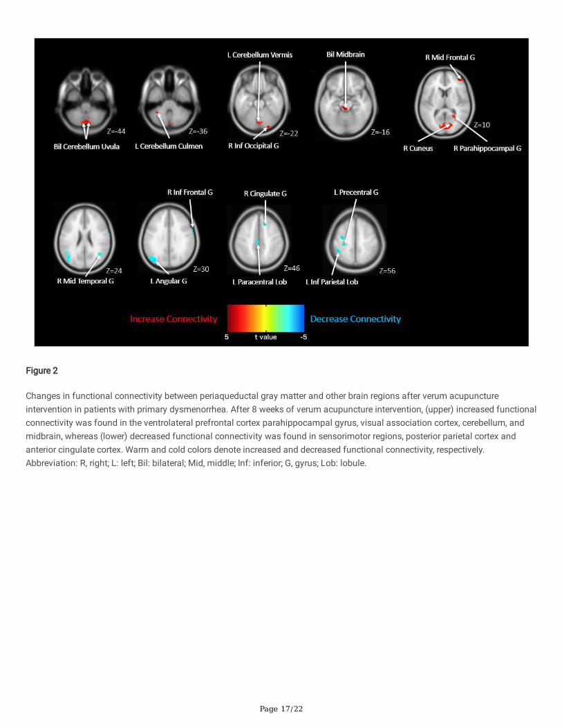
After the 8-week acupuncture intervention, in the verum group, increased FC was found in the right middle frontal gyrus, rightinferior occipital gyrus, right cuneus, right parahippocampal gyrus, midbrain, and cerebellum, and decreased FC was evident in theright inferior frontal gyrus, right middle temporal gyrus, right anterior cingulate gyrus, left precentral gyrus, left inferior parietallobule, and left angular gyrus (Fig. 2, Table 3). In the sham group, increased FC was found in the right superior parietal lobule, lefttemporal gyrus, midbrain, and cerebellum, whereas decreased FC was evident in the right rectal gyrus, right superior frontal gyrus,right inferior frontal gyrus, left inferior temporal gyrus, and bilateral inferior parietal lobule (Fig. 3, Table 3). After the 8-weekintervention, the interaction analysis revealed that compared with the sham group, the verum group had a higher increase in FC inthe right middle frontal gyrus, right caudate body, and cerebellum and a higher decrease in FC in the left precentral gyrus, bilateralprecuneus, and brainstem (Table 3).

Page 7/22
Table 3Changes in functional connectivity map after acupuncture intervention
Week 0 > Week 8 Week 0 < Week 8
Coordinate Coordinate
AnatomicalArea
BA Size Zmax x y z Anatomical Area BA Size Zmax x y z
Verum
R AntCingulateG
32 112 3.86 18 24 42 Bil Midbrain 285 4.20 -4 -28 -16
L InfParietalLob
40 124 3.68 -44 -44 56 3.21 6 -42 -6
LPrecentralG
4/6 347 3.56 -24 -30 70 R Cuneus 18 912 3.71 4 -64 2
L AngularG
39 419 3.44 -40 -78 30 R Mid Frontal G 10 182 3.59 40 50 14
LParacentralLob
31 47 3.37 -6 -24 46 R Inf Occipital G 18 45 3.57 26 -92 -22
R InfFrontal G
9 47 3.36 64 6 30 Bil CerebellumUvula
248 3.48 8 -76 -44
R MidTemporalG
39 87 3.24 42 -60 24 2.86 -2 -86 -34
L CerebellumCulmen
45 3.09 -34 -50 -36
L CerebellumVermis
79 3.08 0 -78 -20
RParahippocampalG
30 46 2.67 18 -44 8
Sham
R Rectal G 25 63 4.12 14 12 -22 L Midbrain 52 4.06 -8 -26 -16
11 68 3.35 14 30 -22 LParahippocampalG
53 3.50 -26 -22 -14
R SupFrontal G
8 70 3.45 10 26 54 R CerebellumVermis
142 3.34 0 -50 -36
Bil InfParietalLob
40 147 3.30 -52 -48 36 R Sup ParietalLob
7 60 3.29 34 -82 44
43 3.03 54 -42 28 L Mid Temporal G 21 60 3.09 -44 -6 -20
L InfTemporalG
21 148 3.26 -66 -28 -20 L CerebellarTonsil
47 3.06 -12 -46 -46
R InfFrontal G
47 121 3.19 50 38 0

Page 8/22
Week 0 > Week 8 Week 0 < Week 8
44 77 3.04 60 18 16
Verum > Sham
RBrainstem
43 3.67 20 -22 -50 Bil CerebellumCulmen
81 3.77 -4 -36 -6
BilPrecuneus
19 112 3.64 34 -82 44 3.65 4 -40 -6
19 102 3.21 -34 -76 34 R Mid Frontal G 46/10 322 3.52 44 48 10
LPrecentralG
4/6 168 3.33 -24 -30 68 R Caudate Body 60 3.50 12 -6 24
BA: Brodmann area; Size: number of voxels in the cluster; Zmax: peak Z value; L: left; R: right; Bil: bilateral; Sup: superior; Inf:inferior; Mid: middle; Med: medial; Ant: anterior; G: gyrus; Lob: lobule; N: nucleus.
After the early stage of intervention, in the verum group, increased FC was evident in the left medial frontal gyrus, left superiortemporal gyrus, left parahippocampal gyrus, and cerebellum, and decreased FC was evident in the right superior frontal gyrus, leftprecentral gyrus, left paracentral lobule, and bilateral precuneus. In the sham group, increased FC was identi�ed in the right middleoccipital gyrus, left precentral gyrus, and cerebellum, whereas decreased FC was apparent in the right medial frontal gyrus, rightsuperior temporal gyrus, right middle temporal gyrus, and cerebellum. The interaction analysis revealed that compared with thesham group, the verum group had a higher increase in FC in the right middle temporal gyrus, left superior temporal gyrus, andcerebellum and a higher decrease in FC in the right middle occipital gyrus and left superior frontal gyrus (Table S1).
After the late stage of intervention, in the verum group, increased FC was apparent in the left middle frontal gyrus, left middleoccipital gyrus, left precuneus, bilateral cuneus, and bilateral parahippocampal gyrus, whereas decreased FC was evident in theright inferior parietal lobule, left medial frontal gyrus, left supramarginal gyrus, and bilateral middle temporal gyrus. In the shamgroup, increased FC was identi�ed in the right inferior parietal lobule, left precentral gyrus, left superior temporal gyrus, bilateralposterior cingulate gyrus, and cerebellum, and decreased FC was apparent in the right middle frontal gyrus, right inferior frontalgyrus, left inferior parietal lobule, left fusiform gyrus, bilateral superior frontal gyrus, and bilateral anterior cingulate gyrus. Theinteraction analysis revealed that compared with the sham group, the verum group had a higher increase in FC in the leftparacentral lobule, left fusiform gyrus, left caudate body, and bilateral superior frontal gyrus and a higher decrease in FC in theright interior parietal lobule, left precentral gyrus, left middle temporal gyrus, and cerebellum (Table S2).
Correlation between menstrual pain experience and the FC of PAGIn the verum group, menstrual pain experience was positively correlated with the FC of PAG in the right middle frontal gyrus, rightsubcallosal gyrus, left precentral gyrus, and bilateral caudate head and negatively correlated with the right inferior temporal gyrus,right lingual gyrus, left postcentral gyrus, and left superior parietal lobule (Fig. 4, Table 4). In the sham group, menstrual painexperience was positively correlated with the FC of PAG in the right inferior frontal gyrus, left middle temporal gyrus, left fusiformgyrus, left cingulate gyrus, bilateral cuneus, and cerebellum and negatively correlated with the right inferior temporal gyrus, leftcaudate body, bilateral thalamus, and cerebellum (Fig. 4, Table 4). The interaction analysis revealed that compared with the shamgroup, the menstrual pain experience in the verum group was more positively correlated with the FC of PAG in the right middlefrontal gyrus, right postcentral gyrus, right inferior temporal gyrus, right caudate head, left precentral gyrus, and left middletemporal gyrus and more negatively correlated in the left cuneus, left anterior cingulate gyrus, and cerebellum (Table 4).

Page 9/22
Table 4Correlation between menstrual pain experience and functional connectivity of PAG
Positive Negative
Coordinate Coordinate
AnatomicalArea
BA Size Zmax x y z Anatomical Area BA Size Zmax x y z
Verum
Bil CaudateHead
463 3.92 12 18 6 L Postcentral G 2 59 4.01 -30 -42 72
44 2.83 -14 18 4 R Inf Temporal G 20 90 3.84 58 -28 -28
R SubcallosalG
13 463 3.32 18 16 -14 L Sup ParietalLob
7 79 3.51 -40 -60 60
R Mid Frontal G 6 137 3.28 34 6 54 R Lingual G 18 87 3.28 12 -78 -14
L Precentral G 6 88 3.18 -26 -20 54
Sham
Bil Cuneus 19 927 4.65 32 -86 20 R Inf Temporal G 20 89 4.47 36 -8 -44
19 635 3.68 -16 -98 20 R CerebellumCulmen
75 3.37 8 -60 -26
L MidTemporal G
21 62 3.26 -54 8 -16 L Caudate Body 69 3.21 -20 -6 24
L Ant CingulateG
32 61 3.26 -16 14 32 Bil Thalamus 73 2.99 -4 -2 4
L Fusiform G 37 74 3.20 -38 -62 -16 2.82 2 -8 4
L CerebellumLob
48 3.08 -4 -68 -52
R Inf Frontal G 47 43 3.06 28 12 -22
Verum > Sham
R Inf TemporalG
20 64 3.85 36 -6 -40 Bil Cuneus 19 432 3.85 -26 -88 34
R Mid Frontal G 6 212 3.51 34 6 56 19 356 3.72 34 -90 18
R CaudateHead
187 3.48 12 18 6 R CerebellumTuber
76 3.68 30 -90 -38
L Precentral G 6 87 3.37 -26 -20 56 L Cerebellum Lob 91 3.21 -26 -74 -58
L MidTemporal G
21 62 3.21 -36 6 -38 L Cingulate G 24 52 3.13 -14 14 34
R Postcentral G 3 46 3.00 22 -30 62
BA: Brodmann area; Size: number of voxels in the cluster; Zmax: peak Z value; L: left; R: right; Bil: bilateral; Sup: superior; Mid:middle; Inf: inferior; Ant: anterior; G: gyrus; Lob: lobule.
DiscussionWe conducted this rfMRI study to investigate the possible central mechanisms of acupuncture treatment on PDM in terms of FCchanges within the descending pain modulation systems. Both verum and sham acupuncture signi�cantly reduced menstrual
Page 10/22
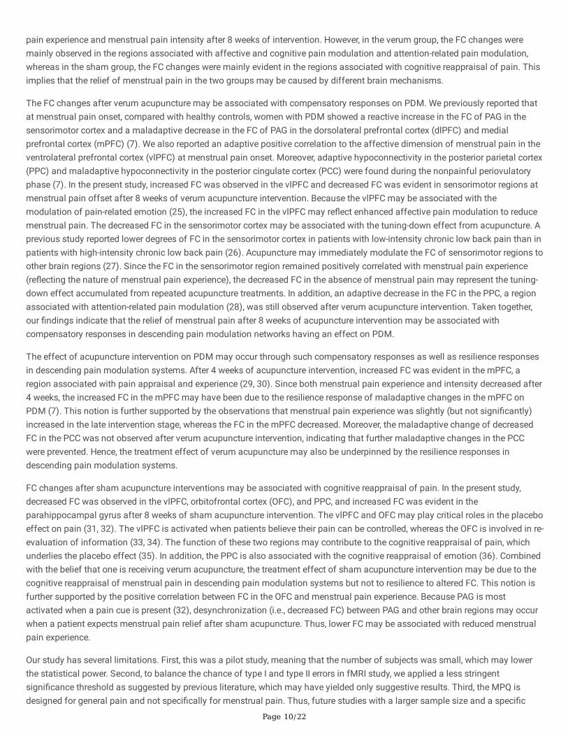
pain experience and menstrual pain intensity after 8 weeks of intervention. However, in the verum group, the FC changes weremainly observed in the regions associated with affective and cognitive pain modulation and attention-related pain modulation,whereas in the sham group, the FC changes were mainly evident in the regions associated with cognitive reappraisal of pain. Thisimplies that the relief of menstrual pain in the two groups may be caused by different brain mechanisms.
The FC changes after verum acupuncture may be associated with compensatory responses on PDM. We previously reported thatat menstrual pain onset, compared with healthy controls, women with PDM showed a reactive increase in the FC of PAG in thesensorimotor cortex and a maladaptive decrease in the FC of PAG in the dorsolateral prefrontal cortex (dlPFC) and medialprefrontal cortex (mPFC) (7). We also reported an adaptive positive correlation to the affective dimension of menstrual pain in theventrolateral prefrontal cortex (vlPFC) at menstrual pain onset. Moreover, adaptive hypoconnectivity in the posterior parietal cortex(PPC) and maladaptive hypoconnectivity in the posterior cingulate cortex (PCC) were found during the nonpainful periovulatoryphase (7). In the present study, increased FC was observed in the vlPFC and decreased FC was evident in sensorimotor regions atmenstrual pain offset after 8 weeks of verum acupuncture intervention. Because the vlPFC may be associated with themodulation of pain-related emotion (25), the increased FC in the vlPFC may re�ect enhanced affective pain modulation to reducemenstrual pain. The decreased FC in the sensorimotor cortex may be associated with the tuning-down effect from acupuncture. Aprevious study reported lower degrees of FC in the sensorimotor cortex in patients with low-intensity chronic low back pain than inpatients with high-intensity chronic low back pain (26). Acupuncture may immediately modulate the FC of sensorimotor regions toother brain regions (27). Since the FC in the sensorimotor region remained positively correlated with menstrual pain experience(re�ecting the nature of menstrual pain experience), the decreased FC in the absence of menstrual pain may represent the tuning-down effect accumulated from repeated acupuncture treatments. In addition, an adaptive decrease in the FC in the PPC, a regionassociated with attention-related pain modulation (28), was still observed after verum acupuncture intervention. Taken together,our �ndings indicate that the relief of menstrual pain after 8 weeks of acupuncture intervention may be associated withcompensatory responses in descending pain modulation networks having an effect on PDM.
The effect of acupuncture intervention on PDM may occur through such compensatory responses as well as resilience responsesin descending pain modulation systems. After 4 weeks of acupuncture intervention, increased FC was evident in the mPFC, aregion associated with pain appraisal and experience (29, 30). Since both menstrual pain experience and intensity decreased after4 weeks, the increased FC in the mPFC may have been due to the resilience response of maladaptive changes in the mPFC onPDM (7). This notion is further supported by the observations that menstrual pain experience was slightly (but not signi�cantly)increased in the late intervention stage, whereas the FC in the mPFC decreased. Moreover, the maladaptive change of decreasedFC in the PCC was not observed after verum acupuncture intervention, indicating that further maladaptive changes in the PCCwere prevented. Hence, the treatment effect of verum acupuncture may also be underpinned by the resilience responses indescending pain modulation systems.
FC changes after sham acupuncture interventions may be associated with cognitive reappraisal of pain. In the present study,decreased FC was observed in the vlPFC, orbitofrontal cortex (OFC), and PPC, and increased FC was evident in theparahippocampal gyrus after 8 weeks of sham acupuncture intervention. The vlPFC and OFC may play critical roles in the placeboeffect on pain (31, 32). The vlPFC is activated when patients believe their pain can be controlled, whereas the OFC is involved in re-evaluation of information (33, 34). The function of these two regions may contribute to the cognitive reappraisal of pain, whichunderlies the placebo effect (35). In addition, the PPC is also associated with the cognitive reappraisal of emotion (36). Combinedwith the belief that one is receiving verum acupuncture, the treatment effect of sham acupuncture intervention may be due to thecognitive reappraisal of menstrual pain in descending pain modulation systems but not to resilience to altered FC. This notion isfurther supported by the positive correlation between FC in the OFC and menstrual pain experience. Because PAG is mostactivated when a pain cue is present (32), desynchronization (i.e., decreased FC) between PAG and other brain regions may occurwhen a patient expects menstrual pain relief after sham acupuncture. Thus, lower FC may be associated with reduced menstrualpain experience.
Our study has several limitations. First, this was a pilot study, meaning that the number of subjects was small, which may lowerthe statistical power. Second, to balance the chance of type I and type II errors in fMRI study, we applied a less stringentsigni�cance threshold as suggested by previous literature, which may have yielded only suggestive results. Third, the MPQ isdesigned for general pain and not speci�cally for menstrual pain. Thus, future studies with a larger sample size and a speci�c
Page 11/22
menstrual pain questionnaire may be needed to elucidate the brain mechanisms underlying the effect of acupuncture treatmenton PDM.
ConclusionsIn conclusion, our results demonstrated that both verum and sham acupuncture intervention on bilateral SP6 can signi�cantlyrelieve menstrual pain. However, the two interventions exhibited different patterns of FC changes in altered descending painmodulation systems. The effect of the verum acupuncture intervention may be underpinned by compensatory and/or resiliencechanges, whereas that of the sham acupuncture intervention may be underpinned by cognitive reappraisal of pain. The results ofthis pilot study may be a basis for further discussion of whether acupuncture treatment induces physiological therapeuticalterations or simply induces the placebo effect.
AbbreviationsBDI IIBeck’s Depression Inventory IIdlPFCdorsolateral prefrontal cortexFCfunctional connectivitymPFCmedial prefrontal cortexMPQMcGill Pain QuestionnaireMRImagnetic resonance imagingOFCorbitofrontal cortexPAGperiaqueductal gray matterPCCposterior cingulate cortexPDMprimary dysmenorrhea;PPCposterior parietal cortexrMRIresting-state magnetic resonance imagingSTAIState-Trait Anxiety InventoryvlPFCventrolateral prefrontal cortex
DeclarationsEthics approval and consent to participate
This study was conducted in accordance with the Declaration of Helsinki. The protocol was approved by the Institutional ReviewBoard of China Medical University Hospital, Taiwan (CMUH105-REC1-027). All patients received a full explanation of the studyand signed written informed consent forms.
Page 12/22
Consent for publication
Not applicable.
Availability of data and materials
All data analyzed during this study are available from the corresponding author on reasonable request.
Competing interest
The authors declare that they have no competing interests.
Funding
This work was supported by the Ministry of Science and Technology, Taiwan (MOST 106-2314-B-039-012, 107-2314-B-039-058-MY2, and 108-2813-C-039-141-B) and the China Medical University, Taiwan (CMU 102-ASIA-19, 107-N-24, and 108-MF-03).
Author’s contributions
Conceptualization, C.H.T. and S.T.Y.; Patient diagnosis, Y.Y.C.; Patient intervention, Y.C.L.; Data acquisition, C.M.C. and W.C.L;Formal analysis, C.H.T and W.C.L.; Writing—original draft preparation, C.H.T.; Writing—review and editing, C.H.T, S.T.Y, and Y.H.C. Allauthors have read and agree with the �nal manuscript.
Acknowledgements
The authors thank the English-editing service by Wallace Academic Editing, Taipei, Taiwan for this manuscript.
References1. French L. Dysmenorrhea. Am Fam Physician. 2005;71(2):285-91.
2. Dawood MY. Primary dysmenorrhea: advances in pathogenesis and management. Obstet Gynecol. 2006;108(2):428-41.
3. Bajaj P, Madsen H, Arendt-Nielsen L. A comparison of modality-speci�c somatosensory changes during menstruation indysmenorrheic and nondysmenorrheic women. Clin J Pain. 2002;18(3):180-90.
4. Granot M, Yarnitsky D, Itskovitz-Eldor J, Granovsky Y, Peer E, Zimmer EZ. Pain perception in women with dysmenorrhea.Obstet Gynecol. 2001;98(3):407-11.
5. Tu CH, Niddam DM, Chao HT, Chen LF, Chen YS, Wu YT, et al. Brain morphological changes associated with cyclic menstrualpain. Pain. 2010;150(3):462-8.
�. Tu CH, Niddam DM, Chao HT, Liu RS, Hwang RJ, Yeh TC, et al. Abnormal cerebral metabolism during menstrual pain inprimary dysmenorrhea. Neuroimage. 2009;47(1):28-35.
7. Wei SY, Chao HT, Tu CH, Li WC, Low I, Chuang CY, et al. Changes in functional connectivity of pain modulatory systems inwomen with primary dysmenorrhea. Pain. 2016;157(1):92-102.
�. Organization WH. Acupuncture: Review and Analysis of Reports on Controlled Clinical Trials. Geneva: World HealthOrganization; 2003.
9. Woo HL, Ji HR, Pak YK, Lee H, Heo SJ, Lee JM, et al. The e�cacy and safety of acupuncture in women with primarydysmenorrhea: A systematic review and meta-analysis. Medicine (Baltimore). 2018;97(23):e11007.
10. Yu YP, Ma LX, Ma YX, Ma YX, Liu YQ, Liu CZ, et al. Immediate effect of acupuncture at Sanyinjiao (SP6) and Xuanzhong(GB39) on uterine arterial blood �ow in primary dysmenorrhea. J Altern Complement Med. 2010;16(10):1073-8.
11. Shi GX, Liu CZ, Zhu J, Guan LP, Wang DJ, Wu MM. Effects of acupuncture at Sanyinjiao (SP6) on prostaglandin levels inprimary dysmenorrhea patients. Clin J Pain. 2011;27(3):258-61.
12. Lin JG, Chen WL. Acupuncture analgesia: a review of its mechanisms of actions. Am J Chin Med. 2008;36(4):635-45.

Page 13/22
13. Rodriguez-Raecke R, Niemeier A, Ihle K, Ruether W, May A. Brain gray matter decrease in chronic pain is the consequence andnot the cause of pain. J Neurosci. 2009;29(44):13746-50.
14. Teutsch S, Herken W, Bingel U, Schoell E, May A. Changes in brain gray matter due to repetitive painful stimulation.Neuroimage. 2008;42(2):845-9.
15. Bingel U, Herken W, Teutsch S, May A. Habituation to painful stimulation involves the antinociceptive system--a 1-year follow-up of 10 participants. Pain. 2008;140(2):393-4.
1�. Yang N, Waddington G, Adams R, Han J. Translation, cultural adaption, and test-retest reliability of Chinese versions of theEdinburgh Handedness Inventory and Waterloo Footedness Questionnaire. Laterality. 2018;23(3):255-73.
17. Xie CC, Wen XY, Jiang L, Xie MJ, Fu WB. Validity of the "streitberger" needle in a chinese population with acupuncture: arandomized, single-blinded, and crossover pilot study. Evid Based Complement Alternat Med. 2013;2013:251603.
1�. Streitberger K, Kleinhenz J. Introducing a placebo needle into acupuncture research. Lancet. 1998;352(9125):364-5.
19. Hui YL, Chen AC. Analysis of headache in a Chinese patient population. Ma Zui Xue Za Zhi. 1989;27(1):13-8.
20. Wu PC, Huang TW. Gender-related invariance of the Beck Depression Inventory II for Taiwanese adolescent samples.Assessment. 2014;21(2):218-26.
21. Ma WF, Liu YC, Chen YF, Lane HY, Lai TJ, Huang LC. Evaluation of psychometric properties of the Chinese Mandarin versionState-Trait Anxiety Inventory Y form in Taiwanese outpatients with anxiety disorders. Journal of psychiatric and mental healthnursing. 2013;20(6):499-507.
22. Yan CG, Wang XD, Zuo XN, Zang YF. DPABI: Data Processing & Analysis for (Resting-State) Brain Imaging. Neuroinformatics.2016;14(3):339-51.
23. Kong J, Tu PC, Zyloney C, Su TP. Intrinsic functional connectivity of the periaqueductal gray, a resting fMRI study. Behav BrainRes. 2010;211(2):215-9.
24. Cremers HR, Wager TD, Yarkoni T. The relation between statistical power and inference in fMRI. PLoS One.2017;12(11):e0184923.
25. Garcia-Larrea L, Peyron R. Pain matrices and neuropathic pain matrices: a review. Pain. 2013;154 Suppl 1:S29-43.
2�. Kong J, Spaeth RB, Wey HY, Cheetham A, Cook AH, Jensen K, et al. S1 is associated with chronic low back pain: a functionaland structural MRI study. Mol Pain. 2013;9:43.
27. Bian Y, He X, Hu S, Li C, Xu C, Kan H, et al. Functional Connectivity Modulation by Acupuncture in Patients with Bell's Palsy.Evid Based Complement Alternat Med. 2016;2016:5928758.
2�. Villemure C, Bushnell MC. Mood in�uences supraspinal pain processing separately from attention. J Neurosci.2009;29(3):705-15.
29. Apkarian AV, Baliki MN, Farmer MA. Predicting transition to chronic pain. Curr Opin Neurol. 2013;26(4):360-7.
30. Baliki MN, Chialvo DR, Geha PY, Levy RM, Harden RN, Parrish TB, et al. Chronic pain and the emotional brain: speci�c brainactivity associated with spontaneous �uctuations of intensity of chronic back pain. J Neurosci. 2006;26(47):12165-73.
31. Benedetti F, Mayberg HS, Wager TD, Stohler CS, Zubieta JK. Neurobiological mechanisms of the placebo effect. J Neurosci.2005;25(45):10390-402.
32. Wager TD, Rilling JK, Smith EE, Sokolik A, Casey KL, Davidson RJ, et al. Placebo-induced changes in FMRI in the anticipationand experience of pain. Science. 2004;303(5661):1162-7.
33. Wiech K, Kalisch R, Weiskopf N, Pleger B, Stephan KE, Dolan RJ. Anterolateral prefrontal cortex mediates the analgesic effectof expected and perceived control over pain. J Neurosci. 2006;26(44):11501-9.
34. Kringelbach ML, Rolls ET. The functional neuroanatomy of the human orbitofrontal cortex: evidence from neuroimaging andneuropsychology. Prog Neurobiol. 2004;72(5):341-72.
35. Tracey I. Getting the pain you expect: mechanisms of placebo, nocebo and reappraisal effects in humans. Nat Med.2010;16(11):1277-83.
3�. Buhle JT, Silvers JA, Wager TD, Lopez R, Onyemekwu C, Kober H, et al. Cognitive reappraisal of emotion: a meta-analysis ofhuman neuroimaging studies. Cereb Cortex. 2014;24(11):2981-90.

Page 14/22
Figures
Figure 1
Flowchart of the study
Page 15/22
Figure 1
Flowchart of the study

Page 16/22
Figure 2
Changes in functional connectivity between periaqueductal gray matter and other brain regions after verum acupunctureintervention in patients with primary dysmenorrhea. After 8 weeks of verum acupuncture intervention, (upper) increased functionalconnectivity was found in the ventrolateral prefrontal cortex parahippocampal gyrus, visual association cortex, cerebellum, andmidbrain, whereas (lower) decreased functional connectivity was found in sensorimotor regions, posterior parietal cortex andanterior cingulate cortex. Warm and cold colors denote increased and decreased functional connectivity, respectively.Abbreviation: R, right; L: left; Bil: bilateral; Mid, middle; Inf: inferior; G, gyrus; Lob: lobule.
Page 17/22
Figure 2
Changes in functional connectivity between periaqueductal gray matter and other brain regions after verum acupunctureintervention in patients with primary dysmenorrhea. After 8 weeks of verum acupuncture intervention, (upper) increased functionalconnectivity was found in the ventrolateral prefrontal cortex parahippocampal gyrus, visual association cortex, cerebellum, andmidbrain, whereas (lower) decreased functional connectivity was found in sensorimotor regions, posterior parietal cortex andanterior cingulate cortex. Warm and cold colors denote increased and decreased functional connectivity, respectively.Abbreviation: R, right; L: left; Bil: bilateral; Mid, middle; Inf: inferior; G, gyrus; Lob: lobule.

Page 18/22
Figure 3
Changes in functional connectivity between periaqueductal gray matter and other brain regions after sham acupunctureintervention in patients with primary dysmenorrhea. After 8 weeks of sham acupuncture intervention, (upper) increased functionalconnectivity was found in the superior parietal lobule, middle temporal gyrus, parahippocampal gyrus, cerebellum, and midbrain,whereas (lower) decreased functional connectivity was found in the medial prefrontal cortex, ventrolateral prefrontal cortex,orbitofrontal cortex, posterior parietal cortex, and inferior temporal gyrus. Warm and cold colors denote increased and decreasedfunctional connectivity, respectively. Abbreviation: R, right; L: left; Bil: bilateral; Sup: superior; Mid, middle; Inf: inferior; G, gyrus; Lob:lobule.

Page 19/22
Figure 3
Changes in functional connectivity between periaqueductal gray matter and other brain regions after sham acupunctureintervention in patients with primary dysmenorrhea. After 8 weeks of sham acupuncture intervention, (upper) increased functionalconnectivity was found in the superior parietal lobule, middle temporal gyrus, parahippocampal gyrus, cerebellum, and midbrain,whereas (lower) decreased functional connectivity was found in the medial prefrontal cortex, ventrolateral prefrontal cortex,orbitofrontal cortex, posterior parietal cortex, and inferior temporal gyrus. Warm and cold colors denote increased and decreasedfunctional connectivity, respectively. Abbreviation: R, right; L: left; Bil: bilateral; Sup: superior; Mid, middle; Inf: inferior; G, gyrus; Lob:lobule.

Page 20/22
Figure 4
Correlation between the changes in functional connectivity and menstrual pain experience after acupuncture intervention inpatients with primary dysmenorrhea. (Left) After 8 weeks of verum acupuncture intervention, a positive correlation betweenfunctional connectivity and menstrual pain experience was found in the motor region, orbitofrontal cortex, and caudate nucleus,whereas a negative correlation was found in the somatosensory association cortex, visual association cortex, and inferiortemporal gyrus. (Right) After 8 weeks of sham acupuncture intervention, a positive correlation between functional connectivityand menstrual pain experience was found in the orbitofrontal cortex, anterior cingulate cortex, visual association cortex, basaltemporal cortex, and cerebellum, whereas a negative correlation was found in the thalamus, caudate nucleus, and cerebellum.Warm and cold colors denote positive and negative correlations, respectively. Abbreviation: R, right; L: left; Bil: bilateral; Mid,middle; Inf: inferior; G, gyrus; Lob: lobule.

Page 21/22
Figure 4
Correlation between the changes in functional connectivity and menstrual pain experience after acupuncture intervention inpatients with primary dysmenorrhea. (Left) After 8 weeks of verum acupuncture intervention, a positive correlation betweenfunctional connectivity and menstrual pain experience was found in the motor region, orbitofrontal cortex, and caudate nucleus,whereas a negative correlation was found in the somatosensory association cortex, visual association cortex, and inferiortemporal gyrus. (Right) After 8 weeks of sham acupuncture intervention, a positive correlation between functional connectivityand menstrual pain experience was found in the orbitofrontal cortex, anterior cingulate cortex, visual association cortex, basaltemporal cortex, and cerebellum, whereas a negative correlation was found in the thalamus, caudate nucleus, and cerebellum.Warm and cold colors denote positive and negative correlations, respectively. Abbreviation: R, right; L: left; Bil: bilateral; Mid,middle; Inf: inferior; G, gyrus; Lob: lobule.
Supplementary Files
This is a list of supplementary �les associated with this preprint. Click to download.
Supplement.docx

Page 22/22
Supplement.docx