durham e-theses protein binding contrast agents for ...etheses.dur.ac.uk/2886/1/2886_972.pdf ·...
TRANSCRIPT

Durham E-Theses
Protein binding contrast agents for potential use in
MRI
Thompson, Nicola C.
How to cite:
Thompson, Nicola C. (2005) Protein binding contrast agents for potential use in MRI, Durham theses,Durham University. Available at Durham E-Theses Online: http://etheses.dur.ac.uk/2886/
Use policy
The full-text may be used and/or reproduced, and given to third parties in any format or medium, without prior permission orcharge, for personal research or study, educational, or not-for-pro�t purposes provided that:
• a full bibliographic reference is made to the original source
• a link is made to the metadata record in Durham E-Theses
• the full-text is not changed in any way
The full-text must not be sold in any format or medium without the formal permission of the copyright holders.
Please consult the full Durham E-Theses policy for further details.
Academic Support O�ce, Durham University, University O�ce, Old Elvet, Durham DH1 3HPe-mail: [email protected] Tel: +44 0191 334 6107
http://etheses.dur.ac.uk

Protein Binding Contrast Agents For Potential Use In
MRI
Nicola C. Thompson
Department of Chemistry
University of Durham
A thesis submitted in part-fulfilment for the degree of Doctor of Philosophy
January 2005 A copyright of this thesis rests with the author. No quotation from it should be published without his prior written consent and information derived from it should be acknowledged.
- 1 S E P 2005
Abstract
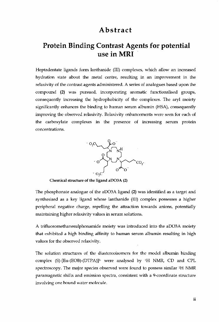
Protein Binding Contrast Agents for potential use in MRI
Heptadentate ligands fo rm lanthanide (III) complexes, which allow an increased
hydration state about the metal centre, resulting in an improvement i n the
relaxivity of the contrast agents administered. A series of analogues based upon the
compound (2) was pursued, incorporating aromatic functionalised groups,
consequently increasing the hydrophobicity of the complexes. The aryl moiety
significantly enhances the binding to human serum albumin (HSA), consequently
improving the observed relaxivity. Relaxivity enhancements were seen for each of
the carboxylate complexes i n the presence of increasing serum protein
concentrations.
O [ J
- o > ^ V ^ ^ ' c O j -
o o
Chemical structure of the ligand aD03A (2)
The phosphonate analogue of the aD03A ligand (2) was identified as a target and
synthesised as a key ligand whose lanthanide (111) complex possesses a higher
peripheral negative charge, repelling the attraction towards anions, potentially
maintaining higher relaxivity values i n serum solutions.
A trifluoromethanesulphonamide moiety was introduced into the aD03A moiety
that exhibited a high binding affini ty to human serum albumin resulting i n high
values for the observed relaxivity.
The solution structures of the diastereoisomers for the model albumin binding
complex (S)-[Eu-(EOB)-(DTPA)]2- were analysed by i H NMR, CD and CPL
spectioscopy. The major species observed were found to possess similar N M R
paramagnetic shifts and emission spectra, consistent w i th a 9-coordinate stiucture
involving one bound water molecule.
u

Declaration
The work described herein was carried out i n the Department of Chemistry,
University of Durham between October 2001 and December 2004. A l l the work is
my own unless otherwise stated and no part of i t has been submitted for a degree at
this or any other university.
Statement of Copyright
The copyright of this thesis rests w i t h the author. No quotation f r o m i t should be
published without prior consent and information derived f r o m i t should be
acknowledged.
l U

Acknowledgments
I wou ld like to thank my supervisor Professor David Parker, firstly, for his
guidance and advice, and secondly for his insight and helpful suggestions,
especially when everything seemed to go wrong.
To all my friends in the analytical department; Dr. Alan Kenwright, Ian McKeag
and Catherine Heffeman, for their advice, cups of tea, tissues and making sense of
my swiggly spectra; Dr. Mike Jones and Lara Turner for their assistance w i t h mass
spectrometry, source of gossip and their contribution towards my employment;
Lenny Lauchlan for help w i t h HPLC analysis; Jarka Dostal for C H N analysis and
Dr. Tony Royston for N M R D work.
Special thanks to all the past and present PhD students and Post Docs of CG27;
esepcaiUy those present i n earUer times whose advice w i t h synthetic chemistiy,
especially column chromatography was indispensable, to the girls for their laughs,
tears and friendship; and everyone else for the f u n , gossip and cakes we have
shared; Paul Atkinson, Alex Badari, Gabriella Bobba, Yann Bretonniere, AUeen
Congreve, Rachel Dickins, Elisa Elemento, Juan Carlos Frias, David Fulton, Filip
Kielar, Mark O'Halloran, Robek Pal, Robert Poole, Horst Puschmann, Kanthi
Senanayke and Simon Welsh.
To Aileen Congreve for the headaches she gave i n developing something that
boimd so well to protein! and for all her help and assistance w i t h submission of this
thesis
Thank you to all the other people i n the department who have made me smile over
the last three years and making chemistry enjoyable.
The submission of this thesis was not only a personal academic mission but also a
family affair, special thanks to my parents for their never fai l ing support, love and
encouragement throughout and for insisting the greatest gi f t I could give myself
was an education.
Finally, a special thank you for Neal, for his unconditional love throughout the last
four years; I thank you for keeping me sane and for never losing fai th i n me.
I V

Dedicated in Loving Memory of my Brother Steven,
For never really leaving my side, you have given me the strength, courage and
opportunity to persevere whilst watching down on me f r o m Heaven.
Theres a lot of comfort in the thought
That sorrow, grief and woe
Are sent into our lives sometimes
To help our soles to groiv.
Helen Steiner Rice

Abbreviations
aD03A
BOC
B O M
CD
CPL
CT
COSY
cyclen
D C M
D 0 3 A
DOTA
DOTP
DTPA
D T P A - B M A
EDC
EOB-DTPA
EPR
Et
EtOAc
EtOH
Gd-BOPTA
HSA
Hz
I.R.
ISC
Ln
m.p.
MeCN
MeOH
MP-2269
l,4,7,10-tetraazacyclododecane-l,4,7-tri(adipic acid)
fer?-butoxycarbonyl
benzyloxymethyl
circular dichroism
circular polarised luminescence
computed tomography
correlation spectroscopy
1,4,7,10-tetraazacyclododecane
dichloromethane
1,4,7,10-tetraazacyclododecane-1,4,7-triacetate
1,4,7,10-tetraazacyclododecane-1,4,7,10-tetraacetate
1,4,7,10-tetraazacyclododecane-1,4,7,10-tetrakis(methylene
phosphonate)
diethylenetriaminepentaacetate
diethylenetriaminepentaacetate-bis(methylamide)
l-(3-dimethylaminopropyl)-3-ethylcarbodiimide
ethoxybenzyl diethylenetriaminepentaacetate
electron paramagnetic resonance
ethyl
ethyl acetate
ethanol
gadobenate dimeglumine
human serum albumin
hertz
infra red
intersystem crossing
lanthanide
melting point
acetonitrile
methanol
4-pentyl-bicyclo[2.2.2]octan-l-carboxyl-di-L-
V I
M R I
MS
MS-325
N M R
N M R D
PCTA-[12]
PEG
q
Rip
rip
Rf
RIME
Ti
t
Bu
TETA
TFA
THF
TLC
tosyl
Tren Me 3,2 HOPO
U V T
aspartyllysine-DTPA
Magnetic Resonance Imaging
mass spectrometry
diphenylcyclohexyl phosphodiester-Gd-DTPA
Nuclear Magnetic Resonance
Nuclear Magnetic Relaxation Dispersion
3,6,9,15-tetraazabicyclo[9.3.1 Jpentadeca-1 (15), 11,13-triene-
3,6,9-triacetic acid
poly(ethylene glycol)
number o f primary coordination sphere water molecules
relaxation rate caused by paramagnetic species
relaxivity caused by paramagnetic species
retention factor
Receptor Induced Magnetisation Enhancement
longitudinal relaxation time
tert-huty\
1,4,8,11 -tetraazacyclotetradecane-1,4,8,11 -tetraacetate
trifluoroacetic acid
tetrahydrofiiran
Thin Layer Chromatography
/?-toluenesulfonyl
A'^,A^',A'^"-tris[(3-hydroxy-l-methyl-2-oxo-l,2-didehydro
pyrid-4-yl)-carboxamidoethyl] amine
ultra violet
rotational correlation time/ reorientational correlation time
V l l
Table of Contents
INTRODUCTION 2
1.1 CONTRAST AGENTS 2
1.2 RELAXATION THEORY 3
1.3 INNER SPHERE RELAXATION 5
1.4 HEPTADENTATE LIGANDS 6
1.4.1 [Gd{D03A)J 6
1.4.2 Derivatives of [Gd(D03A)J 7
1.4.3 Pyridine - containing triaza macrocyclic ligands 9
1.4.4 Heptadentate tripodal ligands 12
1.5 OUTER SPHERE RELAXATION 16
1.5.1 Second Sphere coordination 17
1.5.2 Nuclear Magnetic Relaxation Dispersion (NMRD) profiles 18
1.5.3 [Gd(DOTP)t 18
1.5.4 Derivatives offGd(DOTP)f' 20
1.6 ROTATIONAL M O T I O N 23
1.6.1 Internal Flexibility of Macromolecular Gd (HI) Complexes 25
1.7 W A T E R EXCHANGE 26
1.8 SECOND GENERATION CONTRAST AGENTS 29
1.8.1 Hepatotropic Agents 29
1.9 PROTEIN BINDING 30
1.9.1 Human Serum Albumin 31
1.9.2 Binding requirements 34
1.9.3 Receptor Induced Magnetisation Enhancement (RIME) 35
1.9.4 MS-325 36
1.9.5 MP-2269 38
1.9.6 [Gd(BOTPA)f- and [Gd(EOB-DTPA)f-with HSA 40
2 DI-AQUA GADOLINIUM C O M P L E X E S AND PROTEIN I N T E R A C T I O N 43
2.1 INTRODUCTION 43
2.2 aD03A 43
v i i i
2.3 PROTEIN BINDING 45
2.4 EMISSIVE LANTHANIDE COMPLEXES 46
2.4.1 Lanthanide Luminescence 46
2.4.2 Excitation Methods •••..46
2.5 SYNTHESIS 48
2.6 LUMINESCENCE EMISSION SPECTRA 51
2.7 LANTHANIDE HYDRATION STATES 54
2.8 RELAXOMETRIC STUDIES 55
2.8.1 Relaxivity .....55
2.8.2 Relaxivity Measurements 55
2.8.3 Relaxivity measurements offGdL'f. [GdL^f- and [GdL^f' 56
2.8.4 Nuclear Magnetic Resonance Dispersion (NMRD) Profiles 55
2.9 COMPARATIVE STUDY OF STRUCTURALLY RELATED G A D O L I N I U M COMPLEXES 63
2.9.1 Complexes with increased rigidity 66
2.9.2 A Gadolinium Acridone complex 67
2 . 1 0 EFFECT OF P H UPON RELAXIVITY 71
2 . 1 1 A D 0 2 A BASED SYSTEMS 72
2 . 1 2 SUGGESTIONS FOR FURTHER WORK 74
2 . 1 3 SUMMARY 74
PHOSPHONATE BASED M A C R O C Y C L I C COMPLEXES 76
3.1 INTRODUCTION A N D SYNTHESIS 76
3.2 RELAXIVITY MEASUREMENTS 82
3.3 SEMI QUANTITATIVE ASSESSMENT OF ANION EFFECT ON RELAXIVITY 85
3.4 PROTEIN B I N D I N G STUDIES 86
3.5 CONCLUSIONS 88
SULPHONAMIDE BASED SYSTEMS 90
4.1 INTRODUCTION 9 0
4.2 Z I N C 9 0
4.3 SULPHONAMIDES 9 0
4.4 PROTEIN BINDING STUDIES 92
ix
4.5 INTERNAL FLEXIBILITY 96
4.6 EFFECTS OF ANIONS ON RELAXIVITY 101
4.7 EFFECT OF P H ON R E L A X I V I T Y 102
4.8 DERIVATIVES OF COMPLEX [3]. . 103
4.9 SYNTHESIS OF DERIVATIVES 103
4 .10 RELAXOMETRIC STUDIES OF [GdL'°f , [ G d L " f A N D [GdL'^]' ' 107
4 .11 SYNTHESIS OF L " 107
4 .12 SUMMARY 110
5 NMR AND C H I R O P T I C A L EXAMINATION OF T H E DIASTEREOISOMERS
O F (S)- [EU-(EOBDTPA)]^ 112
5.1 INTRODUCTION 112
5.2 PROJECT A I M S 116
5.3 SOLUTION N M R ANALYSIS 117
5.4 CHIROPTICAL SPECTROSCOPY 124
5.5 CIRCULAR DICHROISM 124
5.6 C D MEASUREMENTS OF (S ) - [EU - (E0B -DTPA ) (H20 ) f 125
5.7 C I R C U L A R L Y P O L A R I S E D L U M I N E S C E N C E 126
5.8 C P L MEASUREMENTS OF ( S ) - [ E U - ( E O B - D T P A ) ( H 2 0 ] f 128
5.9 CONCLUSIONS 130
5.10 PROTEIN B I N D I N G STUDIES 133
6 E X P E R I M E N T A L 139
6.1 S Y N T H E T I C P R O C E D U R E S A N D C H A R A C T E R I S A T I O N 139
6.2 RELAXIVITY MEASUREMENTS 140
6.3 P H O T O P H Y S I C A L M E A S U R E M E N T S 140
6.4 C H A P T E R 2 E X P E R I M E N T A L 141
6.4.1 Ligand Synthesis 141
6.5 COMPLEX SYNTHESIS 158
6.6 CHAPTER 3 EXPERIMENTAL 162
6.6.1 Ligand Synthesis 162
6.7 CHAPTER 4 EXPERIMENTAL 169
X

6.7. / Ligand Synthesis 169
6.8 COMPLEX SYNTHESIS 180
6.9 CHAPTER 5 EXPERIMENTAL 182
LECTURE COURSES ATTENDED 2001/2002 193
CONFERENCES ATTENDED 193
SEMINARS ATTENDED 193
2 0 0 1 193
2 0 0 2 194
2003 195
PUBLICATIONS 197
X I

Chapter 1 Introduction
Chapter 1
Introduction

Chapter 1 Introduction
Introduction In radiology. Magnetic Resonance Imagingi^-^ MRI , has revolutionised the
diagnosis of many diseases. As a consequence of its rapid development i t has
become a routine medical procedure. The technique wou ld have been of lesser
importance had i t not been for the significant advances i n contrast agent media,
which have considerably improved the quaUty and uti l i ty of the images obtained.
In the early days of contrast enhancement i t was estimated less than 10% of all MRI
procedures wou ld require contrast agent enhancement. This figure was greatly
underestimated, as reaUsticaUy, i t is now estimated that over 35% of examinations
employ contrasting media.^
Gadolinium (III) is a paramagnetic metal ion commonly used in MRI contiast
agents due to its unique magnetic properties. It possesses seven unpaired /
elections, and has a relatively slow election spin relaxation rate, resulting in almost
idyllic magnetic properties for use as a contiast agent.s However, the paramagnetic
gadolinium (III) ion is extiemely toxic at concentiations required for M R I and
therefore cannot be used directly as the free aqua ion. For this reason it must be
administered in the f o r m of a stable complex, unable to release metal ions before
excretion f r o m the body.^
1.1 Contrast Agents
Polyaminocarboxylic ligands have proved successful i n forming Gd (III) complexes
w i t h efficient relaxivity properties, high thermodynamic stability and acceptable
toxicity.'''7 There are four that are commonly used in the clinic see Figure 1.1 M*.
The ligands of these chelates belong to two different types of stiucture: acyclic
(open chain) compounds and macrocycUc compounds. [Gd(DTPA)]2-
(Gadopentetate, Magnevist), is an acycUc compound and was the first contiast
agent permitted for clinical use. The first macrocyclic agent [Gd(DOTA)]-
(DOTAREM), followed, which offered increased stabiUty due to more effective
capsulation of the metal ion by the ligand, resulting in a decrease in entiopy upon
metal complexation.i The ionic characteristics of both compounds caused
undesirable side effects in some patients, including painful intiavenous
2
Chapter 1 Introduction
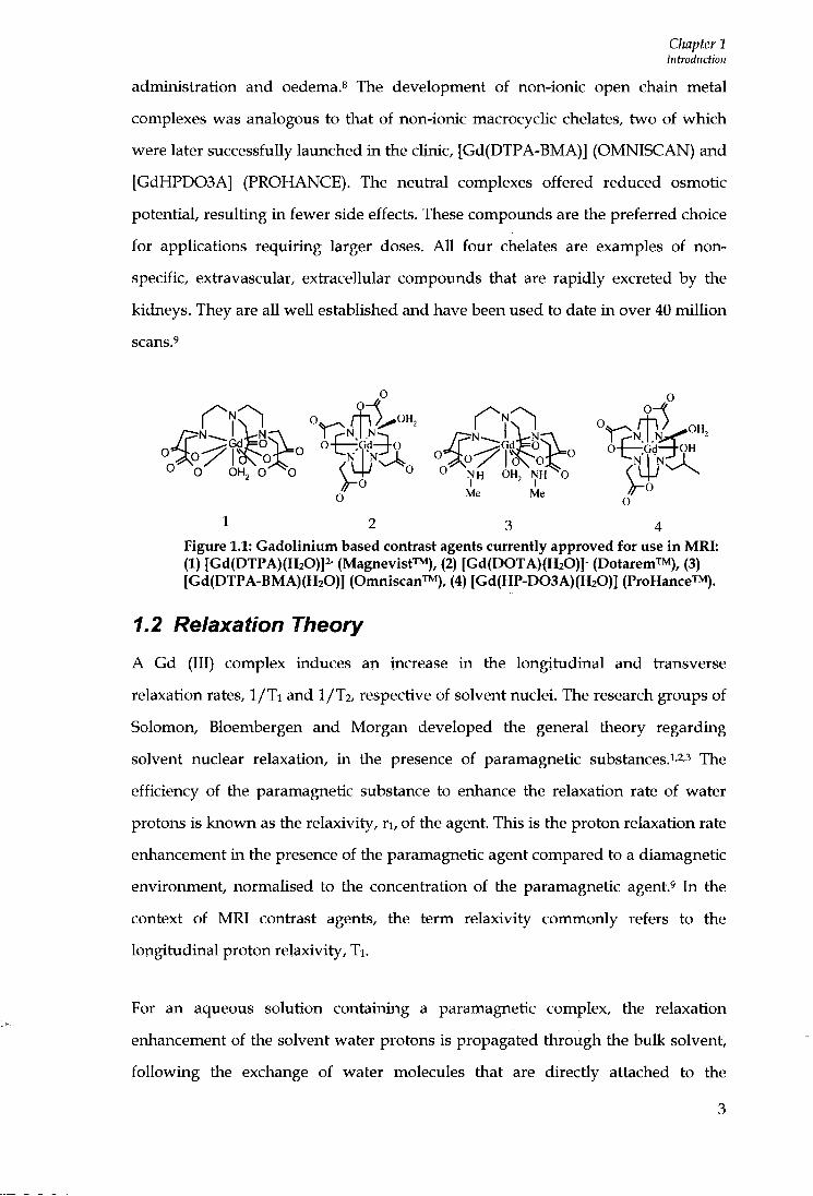
administration and oedema.^ The development of non-ionic open chain metal complexes was analogous to that of non-ionic macrocyclic chelates, two of which were later successfully laimched in the clinic, [Gd(DTPA-BMA)] (OMNISCAN) and [GdHPD03A] (PROHANCE). The neufa-al complexes offered reduced osmotic potential, resulting in fewer side effects. These compoimds are the preferred choice for applications requiring larger doses. AU four chelates are examples of nonspecific, extiavascular, extiacellular compounds that are rapidly excreted by the kidneys. They are all well established and have been used to date i n over 40 mi l l ion scans.'
0 0
O O OHj 0 - ^ 0 O-l-' 0 0 NH OH, NH^O O-V o'^" Me Me
1 2 3 4 Figure 1.1: Gadolinium based contrast agents currently approved for use in MRI: (1) [Gd(DTPA)(H20)]2- (Magnevist™), (2) [Gd(D0TA){H20)]- (DotaremTM), (3) [Gd(DTPA-BMA)(H20)] (Omniscan™), (4) [Gd(HP-D03A)(H20)] (ProHance™).
1.2 Relaxation Theory
A Gd (III) complex induces an increase in the longitudinal and tiansverse
relaxation rates, 1/Ti and 1/12, respective of solvent nuclei. The research groups of
Solomon, Bloembergen and Morgan developed the general theory regarding
solvent nuclear relaxation, i n the presence of paramagnetic substances.i'2'3 The
efficiency of the paramagnetic substance to enhance the relaxation rate of water
protons is known as the relaxivity, r i , of the agent. This is the proton relaxation rate
enhancement in the presence of the paramagnetic agent compared to a diamagnetic
environment, normalised to the concentiation of the paramagnetic agent.' In the
context of M R I contiast agents, the term relaxivity commonly refers to the
longitudinal proton relaxivity, Ti .
For an aqueous solution containing a paramagnetic complex, the relaxation
enhancement of the solvent water protons is propagated through the bulk solvent,
fo l lowing the exchange of water molecules that are directly attached to the
3

Chapter 1 Introduction
gadolinium centre.i" This mechanism is depicted as the inner sphere contribution to the overall relaxivity, see Figure 1.2.
Inner sphere relaxivity
Gd -H
Figure 1.2: Schematic representation of a Gd (III) chelate wi th one inner sphere water molecule, surrounded by bulk water; XR refers to the rotational correlation time of the molecule, Xm the residence time of the inner sphere water molecule and Gd-H the distance between the coordinated water proton and the Gd electron spin.
It wUl be seen from equations 1.2 and 1.3 below, that the inner sphere contribution
is also influenced by correlation times involving rotation (TR) , proton exchange (xm),
and the electionic relaxation (TS).1'2'1O Solvent molecules of the bulk also experience
the paramagnetic effect as they diffuse in the surroundings of the paramagnetic
centie. The effect of this random tianslational diffusion is defined as outer sphere
relaxation.'' The total paramagnetic relaxation rate enhancement due to the
paramagnetic agent is therefore given in equation 1.1, where the subscripts TS',
'OS' and 'SS' refer to the inner, outer and second sphere respectively.
1 1 ( 1 ^ OS
+ (1.1)
There are three different types of water (inner, second and outer sphere), these are
pictorially represented in the molecular dynamic simulation (Figure 1.3), obtained
for [Gd(DOTA)]- in aqueous solution.io

Chapter 1 Introduction
Figure 1.3: Three different types of water molecules around a Gd (III) complex as obtained by molecular dynamics simulation of [Gd(D0TA)(H20)]' in aqueous solution. The inner sphere water is directly coordinated to the metal (the oxygen is red). Second sphere water molecules are on the hydrophilic side of the complex, close to carboxylate groups (their oxygens represented as green balls). Outer sphere or bulk ivater molecules have no preferential orientation (shown in white).!"
1.3 Inner Sphere Relaxation
The inner sphere term represents the major contribution to the overall proton
relaxation rate according to the interpretations of the theory of Solomon,
Bloembergen and Morgan (SBM theory). A more detailed explanation of this theory
is well documented in the literature.iA^s
For currently used monomeric gadolinium based contrast agents, the outer and
inner sphere relaxation mechanisms contiibute roughly the same amotmt to the
observed proton relaxivity at the imaging fields used in MRI of 20-60 MHz.^ The
inner sphere relaxation mechanism is described in basic terms in Equations 1.2-1.3,
where c is the molal concentiation, q is the number of bound water nuclei per Gd
ion (hydration number), 1/Tim is the longitudinal proton relaxation rate, Xm is the
lifetime of a water molecule in the inner sphere of the complex, T R is the
reorientational correlation time of the metal proton vector, and Xs the longitudinal
spin relaxation time of the metal ion.
eg 55.5
Chapter 1 Introduction
(1.2)
1 1 1 1 — = + + —
(1.3)
The equations show that through appropriate modification of the inner sphere
term, the relaxivity achievable can be greatly increased, compared to the outer
sphere, which is more dif f icul t to optimise. However, increases i n hydration states
are often accompanied by a decrease in the thermodynamic and kinetic stability of
the complex w i t h respect to dissociation, l imi t ing the choice of ligand to either
heptadentate (q=2) or i n some cases hexadentate systems (q=3). The equations
show that by increasing the hydration number f r o m one to two, i t is possible to
double the observed relaxivity. The Solomon-Bloembergen-Morgan theory predicts
when all three influencing factors such as rotation, electron paramagnetic relaxation
and water exchange are simultaneously optimised, maximum proton relaxivities
for Gd (III) complexes i n the order of 100 mM-is-i can be achieved.^'!" These are very
high compared to the measured values of relaxivity obtained for currently available
agents of 4-5 mM-^s-i.
1.4 Heptadentate Ligands
1.4.1 [Gd(D03A)]
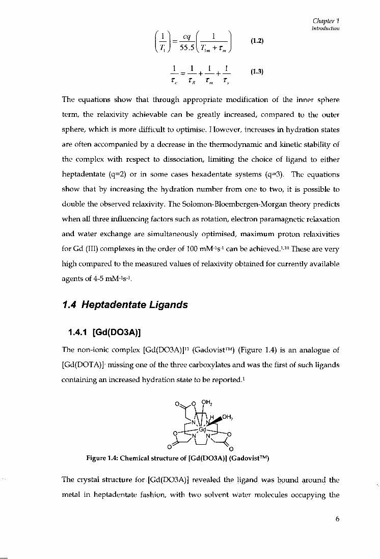
The non-ionic complex [Gd(D03A)]" (GadovisfTM) (Figure 1.4) is an analogue of
[Gd(DOTA)]- missing one of the three carboxylates and was the first of such ligands
containing an increased hydration state to be reported.^
Figure 1.4: Chemical structure of [Gd(D03A)] (Gadovist™)
The crystal structure for [Gd(D03A)] revealed the ligand was bound around the
metal i n heptadentate fashion, w i t h two solvent water molecules occupying the

Chapter 1 Introduction
remaining coordination sites.12 Similarly to the parent complex, [Gd(DOTA)]-, [Gd(D03A)] was found to distribute rapidly into extracellular space and was then eliminated by glomerular fi l tration; no particular protein binding was displayed.^3 A n increased hydration state led to the predicted gain in relaxivity of 4.8 mM-is-i (20 MHz , 313 K, p H 7.3), compared to the monomeric parent w i t h a value of 3.5 mM-is-i (20 MHz , 313 K, p H 7.?>)}» This value was much lower than expected for such a heptadentate system. This is explained by the strong tendency of the [Gd(D03A)] ligand to readily bind the endogenous anion, hydrogencarbonate, which in vivo is present i n large concentrations. The carbonate anion binds in a bidentate fashion displacing two water molecules forming a ternary complex, the formation of which is clearly indicated by a reduction in the measured relaxivity. Despite the disappointing results [Gd(D03A)] , has been frequently functionalised.
1.4.2 Derivatives of [Gd(D03A)]
The derivative [Gd(D03MA)] (Figure 1.5)" incorporates three methyl acetic
pendant arms, encompassing the metal ion in similar heptadentate fashion.
Stability studies confirm the complex has added thermodynamic and kinetic
inertness compared to the parent, resulting f r o m the increased rigidity obtained
f r o m addition of the a-methyl groups. The water exchange time, X m of 68 ns is
almost an opt imum value for [Gd(D03MA)] . This is much quicker than the parent
analogue [Gd(D03A)] , for which X m is 180 ns. This should yield a greater relaxivity
value, yet the measured value reported of 4.4 mM-is-i for [Gd(D03MA)] was
actually sUghtly less than for the parent complex.
Me—< r~\ . H ^ O H , -N. ,5K
O Gd h OH, N-
Me Me
Figure 1.5: Chemical structure of [Gd(D03MA)]
Parker et fl/'^ reported the triamide derivative, [Gd(D03A)-Ala] and its unusual
interaction wi th hydrogen carbonate. Under ambient conditions water was
prevented f rom directly binding to the paramagnetic centie, whereas i n acidic
Chapter 1 Introduction
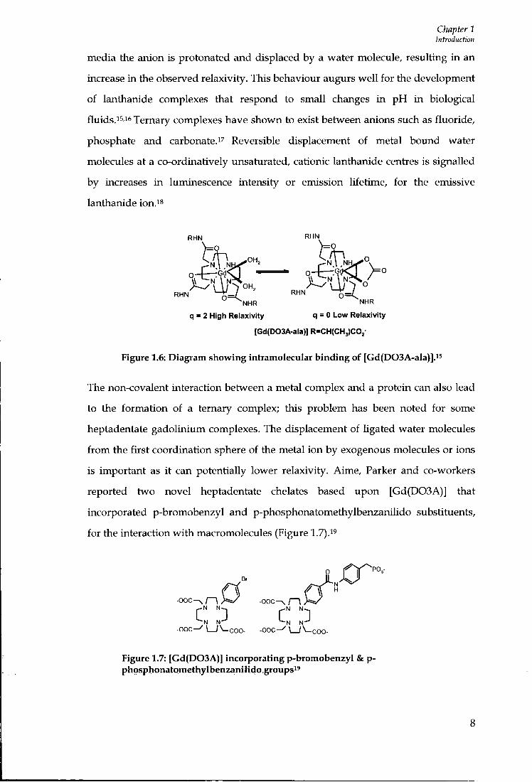
media the anion is protonated and displaced by a water molecule, resulting in an increase in the observed relaxivity. This behaviour augurs wel l for the development of lanthanide complexes that respond to small changes in p H in biological fluids.15,16 Ternary complexes have shown to exist between anions such as fluoride, phosphate and carbonate.^7 Reversible displacement of metal bound water molecules at a co-ordinatively unsaturated, cationic lanthanide centres is signalled by increases in luminescence intensity or emission lifetime, for the emissive lanthanide ion.i^
RHN RHN
R H N ^ ^ J RHN " ^ N H R NHR
q = 2 High Relaxivity q = 0 Low Relaxivity
[Gd(D03A-ala)] R=CH(CH3)C0j-
Figure 1.6: Diagram showing intramolecular binding of [Gd(D03A-ala)]."
The non-covalent interaction between a metal complex and a protein can also lead
to the formation of a ternary complex; this problem has been noted for some
heptadentate gadoliniimi complexes. The displacement of ligated water molecules
f r o m the first coordination sphere of the metal ion by exogenous molecules or ions
is important as i t can potentially lower relaxivity. Aime, Parker and co-workers
reported two novel heptadentate chelates based upon [Gd(D03A)] that
incorporated p-bromobenzyl and p-phosphonatomethylbenzanilido substituents,
for the interaction w i t h macromolecules (Figure 1.7).^^
Br
f 1 «
- O O C ^ [ - \ > = a / - O O C ^ r i
- O O C ^ \ _ / \ - C 0 0 - - O O C - ^ \ _ / \ - C O 0 -Figure 1.7: [Gd(D03A)] incorporating p-bromobenzyl & p-phosphonatomethylbenzanilido groups^'
Chapter 1 Introduction
Upon binding to human serum albumin the expected relaxation was not observed owing to the displacement of two water molecules f r o m the inner sphere, by a donor atom (likely to be a carboxylate group) on the protein and possibly the phosphate anions present f r o m the buffer solution. Moreover, i t should be noted that no ternary complex formation was observed in the presence of the macromolecular substrate, poly-p-cyclodextrin.i^
1.4.3 Pyridine - containing triaza macrocyclic ligands
In earlier work, Aime and co-workers reported the synthesis of 3,6,10-16-
tetraazabicyclo[10.3.1]hexadeca-l(16),12,14-triene (PCTA), a novel heptadentate
ligand involving the insertion of a pyridine moiety i n the macrocyclic ring, w i th
three carboxymethyl arms (Figure 1.8) .20 Competition experiments w i t h the
europium (III) complex confirmed the complex had good thermodynamic stability
even in acidic media. The acid promoted dissociation pathway below p H 3, was
found to be less rapid than for [Gd(DTPA)]2-, suggesting the complex was suitable
for use in vivo. This prompted the synthesis of the gadolinium (III) analogue in
order to assess its use as a MRI contrast agent. The relaxivity was reported as 6.3
mM-is-i (20 MHz , 298 K), approximately 35% higher than [Gd(DOTA)]- and similar
to the value reported for [Gd(D03A)], resulting f r o m increased hydration and
optimised water exchange lifetimes. The stiuctural, dynamic, thermodynamic and
relaxometric properties of this novel complex provided the backbone for the
research of new neutial chelates.
The pyridine-containing triaza macrocycle tiiacetate ligand (PCTA), and its
complexation properties to alkaline earth metals and transition metal cations was
first reported by Stetter et al.^ The synthesis and characterisation of the PCTA
ligand w i t h various lanthanides were later investigated by Aime and co-workers22
(Figure 1.8), involving a series of macrocycUc ligands, including 13 and 14
membered ring compoxmds.22

Chapter 1 Introduction
HO^O
o f J o
Figure 1.8: The ligand PCTA containing a pyridine moiety in the macrocyclic ring and three acetic acid arms.
The PCTA Ugand acts as a heptadentate chelator towards the metal ion allowing
two water molecules to complete coordination. The di-aqua hydration state was
confirmed by examination of the luminescence Ufetimes of the Tb (III) and Eu (III)
complexes.23 The longitudinal proton relaxivity exhibited for [Gd(PCTA)] is
reported as being 5.1 mM-^s-i (20 MHz , 313 K, p H 7), significantly higher tiian that
of [Gd(DOTA)]-. The pyridine based system is reported to have a shorter electionic
relaxation time, tso w i t h respect to [Gd(DOTA)]-, resulting in a shorter water
exchange time, T M 70 ns (298 K), which consequently does not l imi t the relaxivity at
low fields ( T M < T I M ) , imlike Gd (III) chelates currently i n clinical use. StabiUty
studies showed the complex had similar thermodynamic and kinetic properties to
[Gd(D03A)], therefore the release of highly toxic gadolinium ions in vivo is
reduced. It is expected that the pyridine moiety increases the stereochemical
rigidity of the complex and also allows fimctionaUsation towards specific targets.
Hovland et aP-'^ reported the introduction of a lipophiUc moiety (OC12H25) at the 4
position of the pyridine uni t of [GdPCTA]-[12], resulting in micelle aggregation,
due to the amphiphilic structure of the complex i n aqueous solution. Micellar
formation increases the rotational correlation time, T r due to an increased molecular
volume that should result i n an increased relaxivity. As miceUar formation is
concentration dependent, greater relaxivity values were reported when the critical
micelle concenh-ation, (CMC) was 0.15 m M (298 K). A t higher CMC values, the Ti
relaxivity reaches a plateau of 29.2 mM-is-i, whereas at lower CMC concentiations
of 0.05 m M the lowest relaxivity value measured was 11.9 mM-^s-' (20 M H z , 298 K,
p H 7). This is somewhat higher than the value reported for monomeric
[Gd(PCTA)]-[12] system of 6.9 mM-is-i (20 MHz , 298 K, p H 7), which has a similar
molecular volume.
10

Chapter 1 Introduction
The related complex [Gd(PCTP)]-[12] bears three bulky methylenephosphonic arms (Figure 1.9), w i t h respect to the carboxylate compound [Gd(PCTA)]-[12]; this results i n increased steric hindrance causing a reduction in the hydration state.25
OHr N N O, '—N N- O
OH OH
Figure 1.9: Chemical struchire of ligand PCTP-[12]
However, this is not accompanied by a reduction i n the observed relaxivity. The
reported relaxivity value is actually slightly higher at 5.3 mM-is-i for [Gd(PCTP)]-
[12], compared to 5.1 mM-is-^ (20 M H z , 298 K, p H 7.5) for the carboxylate analogue.
This is explained by the formation of a more wel l defined second coordination
sphere for the phosphonate species.
The complex [Gd(PCP2A)] consists of two acetic arms and one
methylenephosphonic arm, as reported by Aime et al?^ Again, the Ugand is
arranged in heptadentate fashion allowing coordination of two inner sphere water
molecules. The steric hindrance of only one phosphonate group is not so great to
prevent the water molecules f r o m coordinating to the metal centre; i t also has the
advantage of creating second sphere effects. These arise f r o m the formation of
hydrogen bonds between solvent molecules and the oxygens of the
methylenephosphonic groups, which may contiibute to an increased relaxivity. The
relaxivity of [Gd(PCP2A)] was reported as 8.3 mM-is-i (20 MHz , 298 K, p H 7),
which was independent of p H across the range 3 - 9.5. Below p H 3.0 an increase i n
relaxivity was observed, which was explained by the possible increase i n hydration
associated w i t h protonation of the phosphonate group. Sinularly to some other d i -
aqua complexes, ternary formation w i t h dissolved carbonate ions was observed at
basic p H , accompanied by a reduction in relaxivity. [Gd(PCP2A)] was suggested to
be sufficiently stable for use in vivo. ^^O N M R determinations of water exchange
revealed a fast exchange mechanism for the complex, an important pre-requisite for
11

Chapter 1 Introduction
coupling to macromolecular substrates.
1.4.4 Heptadentate tripodal ligands
The heptadentate tripodal ligand Hstpaa containing three pyridinecarboxylate
binding units was reported by Bretonniere et flZ^/ (Figure 1.10), w i t h a relaxivity
value of 13.3 mM-is-i (60 MHz , 298 K).
Figure 1.10: Chemical structure of the tripodal ligand Hstpaa ^
This was significantly higher than the value obtained for [Gd(D03A)] or
[Gd(PCTA)]-[12] of 6.1 mM-is-i and 6.9 mM-is-i respectively. Unfortunately, the
complex is only partially soluble in water; the introduction of hydrophilic
substituents on the pyridine rings may increase the solubility of the complex. The
incorporation of hydrophobic groups on the pyridine rings may also increase the
aff ini ty of the complex towards macromolecules.
In earlier stages of contrast development, the tripodal gadolinium complex
[Gd(TREN-Me-3,2-HOPO)] was reported by Xu et al,^ which promised a new class
of lanthanide complexes for use as contrast agents (Figure 1.11).
Figure 1.11: Chemical structure of IGd(TREN-Me-3,2-HOPO)]2»
12

Chapter 1 Introduction
The hydroxyl-pyridinonate (HOPO) monoanions are bidentate Ugands that f o r m effective multidentate sequestering agents. The geometry imposed promotes stiong chelation of metal ions i n general, and of Gd (111) i n particular.^** Crystal structure analyses show the ligand binds i n hexadentate fashion to the metal ion, through the hydroxypyridinone oxygens. Two inner sphere water molecules complete the octadentate coordination state of gadolinium. This is a similar state to the gadolinium aqua ion [Gd(H20)8]'* and differs f r o m that of the nine coordinate polyaminocarboxylate complexes.29 Micskei et aP° noted there is a relatively low energy difference between the eight and nine coordinated gadoUnium energy states. As a result of octadentate binding, water exchange occurs via a rapid associative mechanism, contributing to the higher relaxivity value of the complex of 10.5 mM-is-i (20 MHz, 300 K). This value is approximately 2.5 times greater than [Gd(DTPA)]2- and can be explained due to the increased hydration state and increased molecular size, which increases the reorientational correlation time, T R . Ideally, the complex was reported as highly thermodynamically stable and unaffected by the presence of physiological bidentate chelators such as acetate, lactate and malonate. However, solubility problems were apparent preventing detailed magnetic characterisation and practical appUcability. Consequently, there have been several reports of [Gd(TREN-Me-3,2-HOPO)] derivatives containing substituents i n the TREN moiety, as wel l as the attachment of various HOPO chelators. Cohen and co-workers^i synthesised and characterised three new complexes [Gd(TREN-Me-3,2-HOPOSAM)], [Gd(TREN-Me-3,2-HOPOTAM)]- and [Gd(TREN-Me-3,2-HOP01AM)], by replacing one of the hydroxypyridinone groups wi th groups designed to regulate properties such as charge, solubiUty and stability of the resulting complex. The rapid water exchange values for complexes of [Gd(TREN-Me-3,2-HOPO)] are near optimum, ideal for obtaining maximum relaxivities f r o m the formation of molecules w i t h greater T R . Raymond and co-workers3233,34 reported the derivatives of [Gd(TREN-Me-3,2-HOPO)] w i t h poly (ethyleneglycol) (PEG) groups incorporated, increasing molecular masses of the complexes to the range of 2,000 to 5,000. The polyethylene groups also had a dual purpose; they were highly water-soluble and were suggested to bind weakly to HSA across a wide p H range. However, there were concerns associated w i t h the
13

Chapter 1 Introduction
rapid internal flexibility of the PEG groups.34 The derivative [Gd(TREN-HOPO-TAM)] (Figure 1.12) has also been investigated in related work.^i
N
NH
OH O
Figure 1.12: Structure of [Gd(TREN-HOPO-TAM)]3i
The complex contains two inner sphere water molecules and displays fast water
exchange unt i l PEG groups are incorporated. A reduction in the hydration number
of two to one was observed for [Gd(TREN-HOPO-TAM-PEG-5000)], which is
explained by partial displacement of the water molecule by the PEG oxygen
donors. The longitudinal relaxivity value for [Gd(TREN-HOPO-TAM-PEG-5000)] is
9.1 mM-is-i (20 MHz , 298 K, p H 7.5) compared to that of the parent [Gd(TREN-
HOPO-TAM)] of 8.8 mM-is-', which is poor considering such large increase in
molecular weight and hence greater T R value. This is a consequence of the
reduction in hydration number and the rapid internal motion of the PEG groups. In
addition, i t should be noted that a significant increase i n water exchange rate, T M
f rom 15 to 31 + 2 ns was reported, upon addition of PEG groups.'*^
Modification of the inner sphere effect seems an obvious manner of synthesising
contrast agents w i t h increased relaxivity, yet attempts to synthesise such systems
that have progressed into the clinic have failed for several reasons such as those
already discussed.
In conclusion, dramatic decreases in the thermodynamic and kinetic stabilities w i t h
respect to acid or cation mediated dissociation have prevented some complexes
being safe for use in vivo. The significant binding of the complex to endogenous
anions in serum, such as phosphate or carbonate, displacing one or more of the
inner sphere water molecules in ternary complexes, thus lowering relaxivity, has
14

Chapter 1 Introduction
been reported for many heptadentate species.35
According to Merbach and co-workers^, the long water exchange lifetimes of
monoaqua complexes results f r o m the dissociative mechanism operating between
the coordinated molecules and the bulk solvent. On going f r o m octadentate ligands
to heptadentate species, a different mechanism of water exchange may be expected
which may result i n an increased exchange rate.^* However, for some heptadentate
ligands the water exchange rates measured have been similar i n magnitude to those
of octadentate complexes, resulting i n a quenching effect of the observed relaxivity.
For some complexes designed to bind to serum albumin in a non-covalent fashion,
problems associated wi th the removal of inner sphere waters molecules, as a result
of the interaction w i t h side chain carboxylates i n Glu or Asp residues were also
observed.
1.4. 5 [Gd(aD03A)]^-
- t : ;—.Gd-j—OH, r i ^ G d ^ ^ O H ,
-OjC
Figure 1.13: Chemical structures of [Gd(aD03A)]»- and IGd(gD03A)]3-respectively
The complex [Gd(D03MA)] was a starting point for the synthesis and
characterisation of the derivatives [Gd(aD03A)]3- and [Gd(gD03A)]3- (Figure 1.13),
reported by Parker et a/. ^ It was hypothesised that the anionic pendant arms would
inhibit the binding of bi-dentate anions such as carbonate and phosphate, retaining
the two water molecules involved in the coordination of the metal ion.
Luminescence studies of the europium analogues i n aqueous solution at p H 7,
revealed [Gd(gD03A)]3- experiences intiamolecular (seven ring) carboxylate
ligation, displacing one of the bound waters and suppressing intramolecular
binding in the p H range 4 to 7.5 (Figure 1.14).
15

Chapter 1 Introduction
O . O
H0,C " V o OH2 " V o OHj,
J H-^ ^ J N -^ I
^ i ^ ^ - C O . H ^
COjH CO^H
Figure 1.14: Intramolecular (seven ring) carboxylate ligation of [Gd(gD03A)]3-
The homologous complex [Gd(aD03A)]3- exhibited very different behaviour,
remaining a di-aqua species at ambient p H . Emission spectra examined i n a
simulated extiacellular backgroxmd containing various competing anions over the
p H range 3.5-10.5, revealed that whilst carbonate was preferentially bound, i t only
formed a ternary complex above p H 7.5. Relaxometric studies revealed a l imi t ing
maximal relaxivity was reached at p H 7.3 of 10.1 mM-is-i (65.6 MHz , 293 K). Beyond
p H 7.3 the formation of a ternary complex was observed. The complex was
examined in the presence of serum, and a high relaxivity of 12.3 mM-is-i (65.6 MHz ,
293 K) was maintained, suggesting no interaction w i t h side chain residues of the
protein molecules. A n empirical screening method was used to assess kinetic
stability, which involved taking several relaxivity measurements over a specific
time period (pH 7, phosphate buffer). Endogenous cations such as Zn^*, which may
promote dissociation of the free Gd (III) ion resulting i n a reduction in the
relaxivity, were added. The study found that [Gd(aD03A)]3- was ten times more
chemicaUy inert than clinically used [Gd(DTPA)]2-. Therefore, [Gd(aD03A)]3- offers
ideal prerequisites for the introduction of hydrophobic aromatic groups that can
bind strongly to proteins, increasing the T R effect and the overall relaxivity of the
complex.3738
1.5 Outer Sphere Relaxation
As previously mentioned the relaxivity due to outer sphere relaxation mechanisms
contributes to about half of the overall observed relaxation for clinically used,
monomeric gadolinium based contrast agents. The outer sphere relaxivity stems
f rom modulation of dipolar proton interactions brought about by loosely diffusing
water molecules f r o m the bulk solvent and the paramagnetic centie.^' The outer
16
Chapter 1 Introduction
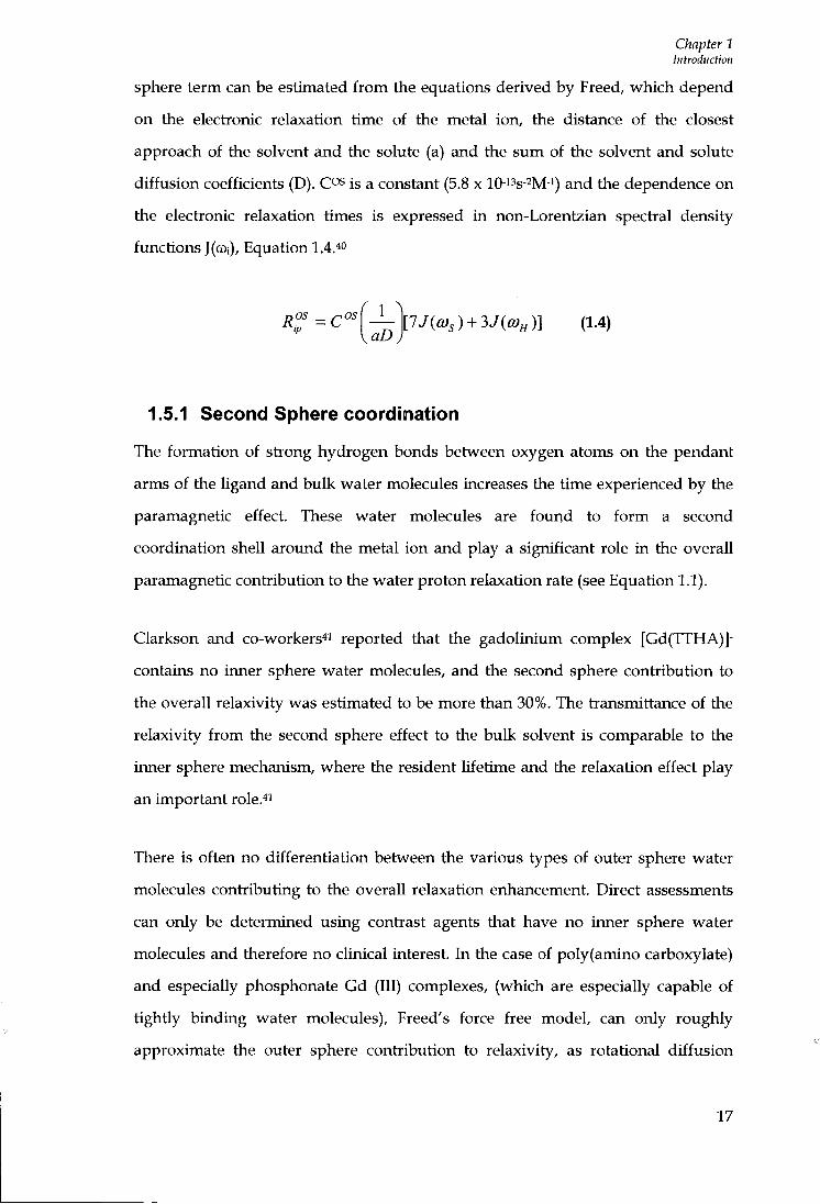
sphere term can be estimated f r o m the equations derived by Freed, which depend on the electronic relaxation time of the metal ion, the distance of the closest approach of the solvent and the solute (a) and the sum of the solvent and solute dif fusion coefficients (D). C^s is a constant (5.8 x lO-i's-^M-i) and the dependence on the electronic relaxation times is expressed i n non-Lorentzian spectral density functions J(a)i), Equation 1.4.4"
nOS ^OSf 1 \lJia),) + 3JiQ)„)] (1.4)
1.5.1 Second Spliere coordination
The formation of stiong hydrogen bonds between oxygen atoms on the pendant
arms of the ligand and bulk water molecules increases the time experienced by the
paramagnetic effect. These water molecules are fo imd to fo rm a second
coordination shell around the metal ion and play a significant role i n the overall
paramagnetic contiibution to the water proton relaxation rate (see Equation 1.1).
Clarkson and co-workers^i reported that the gadolinium complex [Gd(TTHA)]-
contains no inner sphere water molecules, and the second sphere contribution to
the overall relaxivity was estimated to be more than 30%. The tiansmittance of the
relaxivity f r o m the second sphere effect to the bulk solvent is comparable to the
inner sphere mechanism, where the resident lifetime and the relaxation effect play
an important role.4'
There is often no differentiation between the various types of outer sphere water
molecules contiibuting to the overall relaxation enhancement. Direct assessments
can only be determined using contrast agents that have no inner sphere water
molecules and therefore no clinical interest. In the case of poly(amino carboxylate)
and especially phosphonate Gd (111) complexes, (which are especially capable of
tightly binding water molecules), Freed's force free model, can only roughly
approximate the outer sphere contiibution to relaxivity, as rotational diffusion
17

Chapter I Introduction
effects must be taken into account.
1.5.2 Nuclear Magnetic Relaxation Dispersion (NMRD) profiles
Gadolinium complexes of the ligands DOTP and TTHA contain no inner sphere
water molecules and therefore their N M R D profiles directly reflect the second and
outer sphere relaxivity (Figure 1.15). Measuring the relaxation rates of an abundant
nuclear species as a fvmction of the magnetic f ield over a wide range, typically 0.01
to 100 M H z is referred to as relaxometry. The profile is a plot of nuclear magnetic
relaxation rate usually 1/Ti , as a frmction of the magnetic f ield on a logarithmic
scale, which is also referred to as a Nuclear Magnetic Relaxation Dispersion
(NMRD)prof i l e . 3 '«
i6a(DOTP)f»-
I • • • • B n ,
|Ori(rTHA,I*
Figure 1.15: NMRD curves for two (q=0) complexes, [Gd(DOTP)]5- and [Gd(TTHA)]3- (298 K), showing that not all 'outer sphere' complexes of similar molecular volume are equivalent.^
Investigating a sample where certain thermodynamic properties are modified such
as pressure and temperature, can influence the chemical or physical state of the
species. However, variation of the magnetic f ield has no influence on the chemistry
of the sample and is a valuable means of determining the different interaction
mechanisms and dynamic processes influencing relaxation behaviour.^
1.5.3 [Gd(DOTP)]'-
Attractive alternatives to the carboxylic acid donor are the phosphonic acid (PO3H2)
and the related phosphinic acids (PRO2H). The phosphonic acid is a stionger acid
than RCO2H, so that protonation not only of the free Ugand but also of the
18
Chapter 1 Introduction
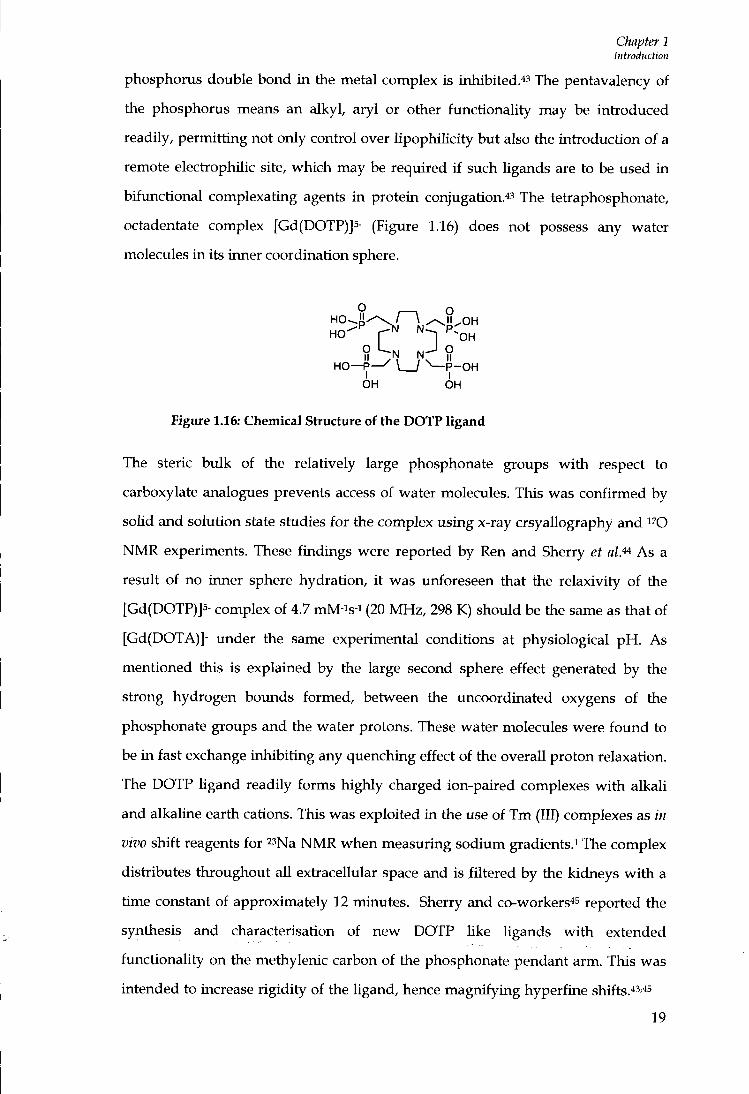
phosphorus double bond i n the metal complex is inhibited.''^ The pentavalency of the phosphorus means an alkyl, aryl or other functionality may be introduced readily, permitting not only control over lipophiKcity but also the introduction of a remote electiophilic site, which may be required i f such ligands are to be used in bifunctional complexating agents i n protein conjugation.43 The tetraphosphonate, octadentate complex [Gd(DOTP)]5- (Figure 1.16) does not possess any water molecules i n its inner coordination sphere.
Ho ' ^ f -OH
HO—P- \ I ^P-OH OH OH
Figure 1.16: Chemical Structure of the DOTP ligand
The steric bulk of the relatively large phosphonate groups wi th respect to
carboxylate analogues prevents access of water molecules. This was confirmed by
solid and solution state studies for the complex using x-ray crsyaUography and ^''O
NMR experiments. These findings were reported by Ren and Sherry et al.'^ As a
result of no inner sphere hydration, i t was imforeseen that the relaxivity of the
[Gd(DOTP)]5- complex of 4.7 mM-is-i (20 M H z , 298 K) should be the same as that of
[Gd(DOTA)]- under the same experimental conditions at physiological p H . As
mentioned this is explained by the large second sphere effect generated by the
stiong hydrogen bounds formed, between the vmcoordinated oxygens of the
phosphonate groups and the water protons. These water molecules were f o i m d to
be in fast exchange inhibit ing any quenching effect of the overall proton relaxation.
The DOTP ligand readily forms highly charged ion-paired complexes wi th alkali
and alkaline earth cations. This was exploited in the use of T m (III) complexes as in
vivo shift reagents for '^Na N M R when measuring sodium gradients.^ The complex
distributes throughout al l extracellular space and is filtered by the kidneys w i t h a
time constant of approximately 12 minutes. Sherry and co-workers^s reported the
synthesis and characterisation of new DOTP Uke Ugands w i t h extended
functionality on the methylenic carbon of the phosphonate pendant arm. This was
intended to increase rigidity of the ligand, hence magnifying hyperfine shifts.'''''''^
19

Chapter 1 Introduction
The use of [Gd(DOTP)]5- as a contiast agent for M R l has been unsuccessful as the complex is highly charged and despite its stiong association w i t h sodium ions is not very convenient due to osmolality problems.
1.5.4 Derivatives of [Gd(DOTP)]^-
As more conclusive evidence proved phosphonate and phosphinate moieties could
result i n an efficient second coordination sphere, increasing the observed relaxivity,
many research groups have investigated anionic species for the basis of improving
the efficacy of contiast agents. This includes attachment of hydrophobic groups to
the phosphinate backbone in order to increase the rigidity, or introducing specific
moieties capable of increasing the binding interaction to serum albumin.47
Parker and co-workers reported the synthesis and characterisation of the highly
rigid, kinetically stable, eight coordinate benzylphosphinate complex,
[Gd(BzDOTP)]- (Figure 1.17).4M8,49
o=p'Ar7\)
Figure 1.17: Chemical structure of [Gd(BzDOTP)]-.3
The N M R D profile of [Gd(BzDOTP)]- was simplified due to lack of inner sphere
contributions and of characteristic shape and amplitude, for a complex only
affected by the tianslational diffusion model of pure outer sphere relaxivity. Stiong
binding interactions were reported for [Gd(BzDOTP)]- w i th bovine serum albumin
( K A = 9.1 X 10³ M-i 298 K ) , leading to a marked relaxivity enhancement for a (q = 0)
gadolinium complex. A remarkably high efficacy of the complex in liver and bile
was observed i n M R l examination tiials.3 The high relaxivity enhancement upon
binding is likely to result f rom the mobile protons of the protein that are dipolarly
relaxed, by their proximity to the paramagnetic centie. In addition, the highly
20
CMpter 2 Introduction
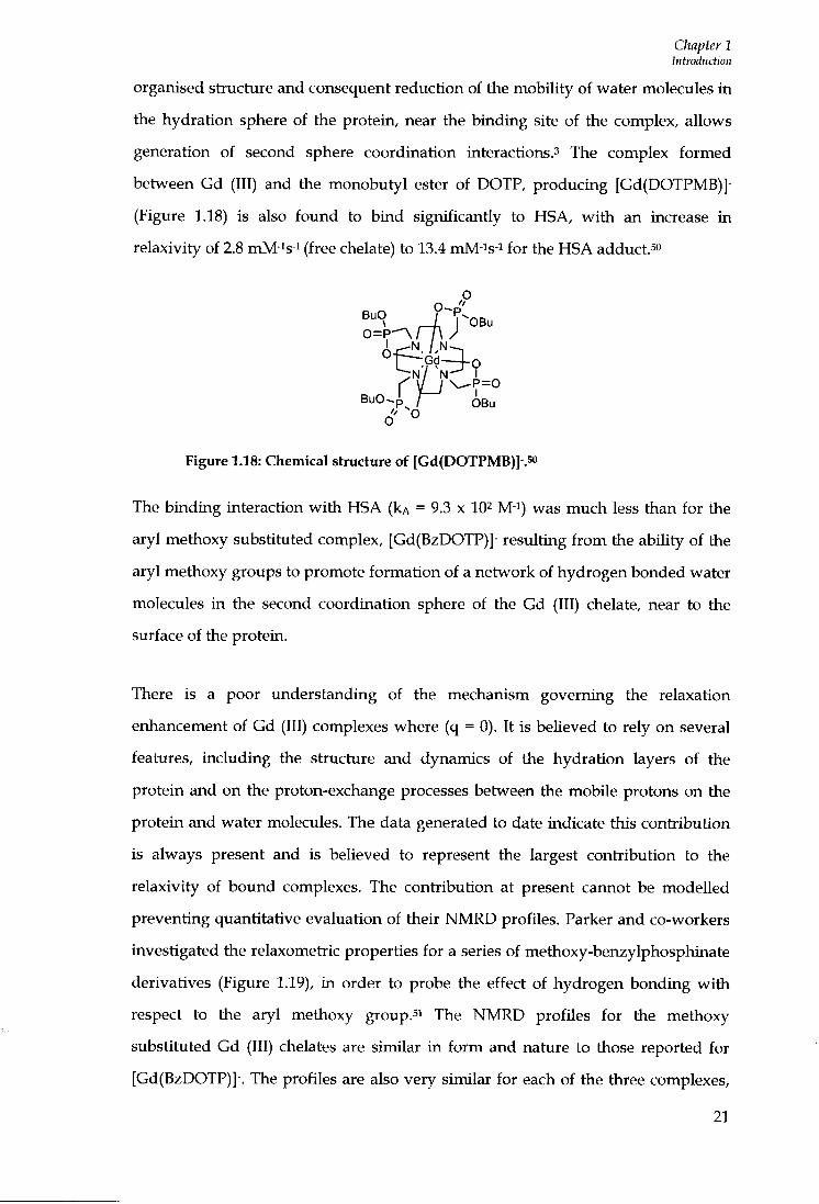
organised structure and consequent reduction of the mobility of water molecules i n the hydration sphere of the protein, near the binding site of the complex, allows generation of second sphere coordination interactions.^ The complex formed between Gd (III) and the monobutyl ester of DOTP, producing [Gd(DOTPMB)]-(Figure 1.18) is also found to b ind significantly to HSA, w i t h an increase in relaxivity of 2.8 mM-is-i (free chelate) to 13.4 mM-is-i for the HSA adduct.so
O ' °
BuO / f -nD
o=p^rA /
o" °
Figure 1.18: Chemical structure of [Gd(DOTPMB)]-.5o
The binding interaction w i t h HSA ( k A = 9.3 x 10^ M-i) was much less than for the
aryl methoxy substituted complex, [Gd(BzDOTP)]- resulting f r o m the ability of the
aryl methoxy groups to promote formation of a network of hydrogen bonded water
molecules in the second coordination sphere of the Gd (III) chelate, near to the
surface of the protein.
There is a poor understanding of the mechanism governing the relaxation
enhancement of Gd (III) complexes where (q = 0). It is believed to rely on several
features, including the structure and dynamics of the hydration layers of the
protein and on the proton-exchange processes between the mobile protons on the
protein and water molecules. The data generated to date indicate this contribution
is always present and is believed to represent the largest contribution to the
relaxivity of bound complexes. The contribution at present cannot be modelled
preventing quantitative evaluation of their N M R D profiles. Parker and co-workers
investigated the relaxometiic properties for a series of methoxy-benzylphosphinate
derivatives (Figure 1.19), in order to probe the effect of hydrogen bonding wi th
respect to the aryl methoxy group.si The N M R D profiles for the methoxy
substituted Gd (III) chelates are similar in f o r m and nature to those reported for
[Gd(BzDOTP)]-. The profiles are also very similar for each of the three complexes,
21

Chapter 1 bitrodudion
consistent with Gd (III) chelates of identical molecular size, coordination polyhedron and hydration sphere. Slight differences were observed in the Xs values; this resulted from the different substitution sites of the methoxy groups on the aromatic ring. The binding interaction of GdL^ with bovine serum albumin of (Kd = 1.1 X 10⁻⁴ (jjn3 mol-i) was significantly higher than for the parent complex, [Gd(BzDOTP)] of (Kd = 2.8 x 10⁻⁴ dm3 mol-i), leading to an approximate enhancement in the relaxivity of more than 20%, as a result of more efficient hydrogen bonding in the second coordination sphere of the gadolinium ion, near to the protein surface. The derivatives showed selective biUary clearance at low concentrations of complex, a slower rate of clearance from tumour tissue and impressive proton relaxation enhancement in the presence of protein, rendering them suitable as potential MRI contiast agents.
o" R - P = 0 R
I IN IN-—,11 -
- O ' ? ^ \ _ J >
Figure 1.19: Chemical structure of methoxybenzyl ligands^i
Macrocycles bearing pendant phosphonic or phosphinic groups exhibit excellent
coordination selectivity and high thermodynamic stability. Protonation of the
phosphrnate or phosphonate oxygen atom is difficult to accomplish in both the free
ligand and in the metal complex, resulting in metal complexes that are stable to
proton catalysed dissociation pathways, required for in vivo imaging. Structural
variations of the phosphorus substituents may effectively contiol the hydro-
lipophilic properties of both ligand and metal complexes, which may ultimately
provide a fine-timing of the conjugation with proteins and other biologically active
molecules.52.53
A new hexadentate macrocyclic ligand, Me2D02PMe, containing two monoethyl
ester phosphonate arms has been synthesised by Bianchini et al^^ (Figure 1.20).
22

Chapter 1 Introduction
O
E t O - P ^ CH3
H3C \ I ^ P — O E t
OH
Figure 1.20: Chemical structure of the ligand MezDOZPMeSz
The hexadentate ligand involves coordination of the four nitrogen atoms of the
macrocycle and two oxygen atoms from the phosphonate groups, which allows
addition of two or three water molecules to complete coordination. The data
showed the stability of [Gd(Me2D02PMe) was very low with respect to the
complexes of copper and zinc. This is a likely consequence of the strong effect of the
nature and number of pendant monoethyl ester phosphonate groups. The stability
results also show [Gd(Me2D02PMe)], is far less stable than the Gd (III) complex of
the carboxylate macrocycle D02A, consistent with the weakly basic P(0)(OEt)0-
groups. However, complexation of the ligand to copper, [Cu(Me2D02PMe)] or zinc
[Zn(Me2D02PMe)] shows remarkable thermodynamic stability in the pH range 2.5
to 7, since neither protonated nor hydroxylated species were apparently formed.52
1.6 Rotational Motion
At the magnetic field stiengths used in MRI (20 - 60 MHz) the longitudinal time of
bound water protons is most critically contioUed by the rotational correlation time,
T R , according to Equation 1.3. For current clinically used contiast agents, fast
rotation is the Umiting factor for proton relaxivity. This is demonstiated by the
NMRD profile for a low molecular weight complex, in which the effect on relaxivity
is considered, based upon changes in the TR value (see Figure 1.21).
23
Chapter 1 Introduction
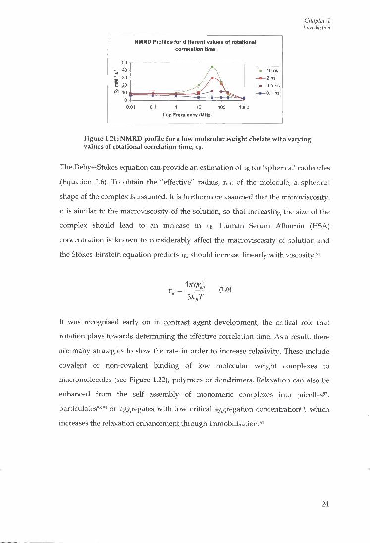
NMRD Profiles for different values of rotational correlation time
50 - 40
E 20 -I ^ 10
10 ns -2 ns -0.5 ns -0.1 ns
0.01 0.1 1 10 100
Log Frequency (MHz)
1000
Figure 1.21: NMRD profile for a low molecular weight chelate with varying values of rotational correlation time, T R .
The Debye-Stokes equation can provide an estimation of T R for 'spherical' molecules
(Equation 1.6). To obtain the "effective" radius, reff, of the molecule, a spherical
shape of the complex is assumed. It is furthermore assumed that the microviscosity,
q is similar to the macroviscosity of the solution, so that increasing the size of the
complex should lead to an increase in T R . Human Serum Albumin (HSA)
concentration is known to considerably affect the macroviscosity of solution and
the Stokes-Einstein equation predicts T R , should increase linearly with viscosity.54
3kJ (1.6)
It was recognised early on in contrast agent development, the critical role that
rotation plays towards determining the effective correlation time. As a result, there
are many stiategies to slow the rate in order to increase relaxivity. These include
covalent or non-covalent binding of low molecular weight complexes to
macromolecules (see Figure 1.22), polymers or dendrimers. Relaxation can also be
enhanced from the self assembly of monomeric complexes into micelless^,
particulates58'59 or aggregates with low critical aggregation concentration*'", which
increases the relaxation enhancement through immobilisation.^^
24

Chapter 1 Introduction
Slowly Tumbling Protein
Human \ Hydrophobic] i Serum r~ Albumin
Rigid
Gd Chelate Spacer
Figure 1
Figure 1.22: Diagram representing the non-covalent interaction of a protein and a Gd (III) clielate.
Besides increasing the reorientational time, T R , hence the relaxivity of such an
albumin based agent the reversible interaction of the protein presents two
additional advantages: a safe clearance of the agent and a longer residence time in
the blood pool compared to the unbound agent. This behaviour is favourable in
angiographic applications.*'^
1.6.1 Internal Flexibility of Macromolecular Gd (III) Complexes
As is expected, T R , values for some Gd (111) chelates are seen to increase with
increasing molecular weight, however, the relationship is far from linearly
predicted from the Debye-Stokes equation, resulting from the internal flexibility of
some systems.3 As some of the chelates investigated are not particularly rigid
themselves, internal motion which determines relaxivity may be considerably faster
than the overall tumbling motion of the whole molecule. Therefore, the Gd (111)
chelate should be fixed covalently or by non-covalent binding to a macromolecule
or dendrimer via a short rigid linking unit to reduce local flexibility. Linear
polymers are especially susceptible and in these complexes the rotational
correlation time, T R , is dominated by segmental motions which are independent of
molecular weight. Internal flexibility effects have also been reported for non-
covalently bound Gd (111) chelate- protein adducts. The complex MP-2269 has a
reported T R value of 0.1 ns when boimd to serum albumin. This value is one order
of magnitude lower than the rotational correlation time of the protein molecule,
consistent with inefficient rotational coupling.^-«
25
Chapter 1 Introduction
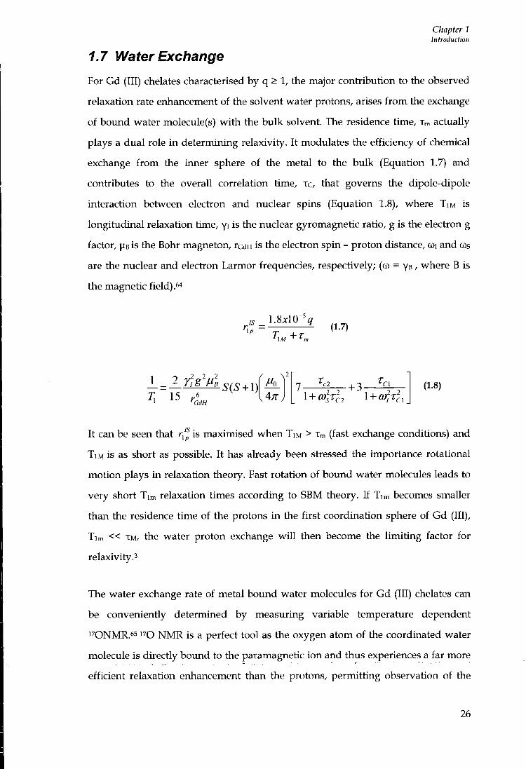
1.7 Water Exchange
For Gd (III) chelates characterised by q > 1, the major contribution to the observed
relaxation rate enhancement of the solvent water protons, arises from the exchange
of bound water molecule (s) with the bulk solvent. The residence time, Xm actually
plays a dual role in determining relaxivity. It modulates the efficiency of chemical
exchange from the inner sphere of the metal to the bulk (Equation 1.7) and
contiibutes to the overall correlation time, xc, that governs the dipole-dipole
interaction between election and nuclear spins (Equation 1.8), where TIM is
longitudinal relaxation time, yi is the nuclear gyromagnetic ratio, g is the electron g
factor, PB is the Bohr magneton, rcdH is the election spin - proton distance, coi and oos
are the nuclear and election Larmor frequencies, respectively; (co = ys / where B is
the magnetic field) . 4
1.8^10-^^ (1.7)
1 _ 2 j j g ' M l
15 SiS + \)
'GdH
- + 3- "Cl
l + 6?/Tc, (1.8)
It can be seen that r,p is maximised when TIM > Xm (fast exchange conditions) and
T I M is as short as possible. It has already been stiessed the importance rotational
motion plays in relaxation theory. Fast rotation of bound water molecules leads to
very short Tim relaxation times according to SBM theory. If Tim becomes smaller
than the residence time of the protons in tlie first coordination sphere of Gd (III),
Tim « XM, the water proton exchange wiU then become the limiting factor for
relaxivity.3
The water exchange rate of metal bound water molecules for Gd (III) chelates can
be convenientiy determined by measuring variable temperature dependent
1 7 0 N M R . 6 5 N M R is a perfect tool as the oxygen atom of the coordinated water
molecule is directly bound to the paramagnetic ion and thus experiences a far more
efficient relaxation enhancement than the protons, permitting observation of the
26
Chapter 1 Introduction
exchange rate directly.^^
The residence lifetime is very important in relation to the relaxation of high
molecular weight MRI contrast agents. Theory predicts the optimal values for water
exchange, Xm are in the range of 20-50 ns at 298 K.^ Initially, the water exchange
rates of polyaminocarboxylate complexes were believed to be faster than the Gd
aqua ion (Kex ~ 10 s-i), too fast to influence proton relaxivity. However, extensive
studies by Merbach and co-workers reported water exchange rates determined by
1 7 0 NMR for [Gd(DTPA)(H20)]2- and [Gd (D0TA)(H20) ] - as 303 and 244 ns (298 K ) ,
which are far from optimum.3'58 The nine coordinate Gd (III) polyaminocarboxylate
complexes all have positive activation volumes, indicative of a dissociatively
activated water exchange mechanism. This is expected, as there is no longer space
for a second water to enter before the departure of the bound water molecule. The
eight coordinate tiansition state is energetically unstable, requiring high activation
energy, resulting in a decreased rate constant. Hence, the lower exchange rates on
the lanthanide poly(aminocarboxylate) complexes differ from the faster dissociative
interchange, water exchange mechanism of [Gd(H20)8]^*.
Efforts have been devoted to optimising residence lifetimes, alleviating limitation of
a complex's relaxivity. In complex design, amide linkages have tended to be
avoided since it has been suggested that complexes with this structural motif may
prolong residence times. It has also been noted that water exchange rates at
positively charged centres are much slower than those for neutral complexes which,
in turn, are slower than for negatively charged species. ' Therefore, intioduction of
negative charge into the neighbourhood of the Gd (III) bound water molecule may
effectively increase the observed relaxivity.58 These empirical observations are more
appropriately assessed in terms of the perturbation of the local water structure by
the complex counterions or integral substituents, and the variation of the
strength/length of the Gd-OH2 bond. o The C 4 substitution of the ethylenic bridge
of DTPA by a larger substituent such as an ethoxybenzyl group, benzyl, or a (4,4-
diphenylcyGlohexyl)phosphanooxymethyl group has been shown to decrease the
water exchange by 25-42% as a result of increased steric crowding around the
27
Chapter 1 Introduction
bound water siieJ^
Merbach et aP"^ reported the behaviour of the complex [Gd(TRITA)]-, which is a
derivative of the macrocyclic chelate [Gd(DOTA)]-, in which one ethylene bridge is
replaced by a propylene bridge. Similarly, to [Gd(DOTA)]-, [Gd(TRrTA)] - contains
one water molecule in its inner sphere. However, the water exchange rate was
found to be faster as a consequence of the increased steric compression around the
bound water molecule, induced by a larger macrocycle. A slight reduction in
thermodynamic stability resulted for [Gd(TRITA)(H20)]- but may still be stable
enough for use in vivo. Therefore, the chelate is a potential synthon for the
development of high relaxivity, macromolecular agents. ^ addition, the distance
between the plane of the carboxylate oxygens and the metal was found to be longer
in the TRITA complex. This resulted in the boimd water molecule being in closer
proximity to the negatively charged carboxylates, facilitating the dissociative
mechanism of water exchange. s
The nature of the second coordination sphere also has an effect on water exchange
rates. The complex [Gd-D03A-propionate(H20)]- has a much shorter Ufetime of 8
ns, compared to that of [Gd-D03A-alanine(H20)] which is significantly longer, (180
ns, 298 K).73'74 This is explained by the hydrogen bonding interaction between the
metal bound water molecule and the positively charged a-amino group of the
amino acid.
Upon formation of an adduct with a macromolecule effective complex rotation is
obviously slowed. However, the effect upon water exchange is often harder to
quantify. Aime and co-workers reported a significant decrease in water exchange
rates for two Gd (III) complexes upon albumin binding, via electiostatic or
hydrophobic interactions.^s They deduced these conclusions from the data
produced by the NMRD profiles of the individual complexes. This was explained
by the obstiuction of the water coordination site when the complex was boimd to
protein. In conteast, Merbach and co-workers, used independent techniques such as
i 'O NMR and EPR including NMRD assessments and concluded the decrease in
28
Chapter 1 Introduction
exchange was not as large as reported by Aime et al?-"^^ It can be concluded that incorporation of Gd (III) complexes into macromolecular systems may affect water exchange kinetics, therefore it is important to design a complex which displays optimal residence lifetimes prior to macromolecular formation.
1.8 Second Generation Contrast Agents
The first generation of contrast agents previously discussed are low molecular
weight, highly hydrophilic gadolinium based chelates, which consequently are
distiibuted rather unselectively throughout extracellular fluids. The inability to
localize selectivity in a desired area lessens the diagnostic potential of these agents.
The development of second generation Gd (III) based systems stems from the MRI
complexes endowed with higher relaxivities, or systems that are responsive or
specifically targeted to certain sites in vivo.
Responsive or smart agents are able to report about the physiochemical
environment in which they are distributed. There are a variety of complexes whose
relaxivity can be modulated to changes in physiochemical environments, such as
plj76,77,7s,79 metal ion concentration, partial pressure of oxygen,7'8o degree of
glycation of proteins, concentration of Ca2+ si, concentration of zinc^^ and the
presence of enzymes. - 84 A diverse range of applications has been investigated and
are reported in the literature.
1.8.1 Hepatotropic Agents
A targeted or 'organ specific' contrast agent is defined as an agent that is selectively
taken up by a particular type of cell (e.g. Kupffer cells of hepatocytes) and thereby
only enhances organs where these cells are present either in the liver, spleen or
lymph nodes.3 The infusion of the chelate [Gd(DTPA)]2- in rats results in marginal
hepatic uptake and negligible biliary recovery, the contrast agent is non-specific
and the excretion pathway for this complex occurs by rapid glomerular filtiation.ss
Markedly different from the [Gd(DTPA)]2- are the pharmacodynamics of
gadobenate dimegluminese, [Gd(BOPTA)]2- and gadoxetic acid, [Gd(EOB-DTPA)]2-.
29
Chapter 1 Introduction
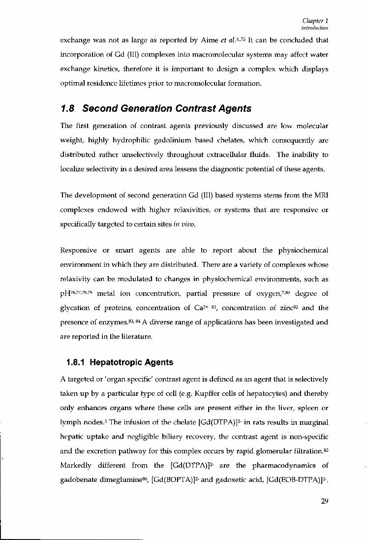
These are analogues of the parent compotmd [Gd(DTPA)]2-, with added hydrophobic substituents incorporated onto the DTPA backbone (see Figure 1.23) 7 These compounds are believed to be taken up by the hepatocytes and once inside or surface immobilised, the relaxivity increases because of the interactions with macromolecular structures. They therefore offer the advantages of improving detection and characterisation of liver lesions in the early extracellular distribution by fast dynamic imaging, followed by delayed imaging where the contiast is dependent upon the specificity of the agent. The complexes are excreted via the bile duct, gall bladder and intestines.ss-ss Although the compounds target the liver for [Gd(BOPTA)]2-, only 2-4% of the injected compound is actually absorbed by the liver, whereas for [Gd(EOB-DTPA)]2-, approximately 50% of the compound is absorbed by the organ.**
- O O C ^ N '
-ooc^
r coo-
N C O O -
' coo-N C O O
B O P T A
- O O C C O O -
E O B - D T P A
Figure 1.23: Chemical structures of the ligands BOFTA and EOB-DTPA
[Gd(BOPTA)]2- (MultiHance^'^) has been approved for use in patients in Europe but
is still pending approval by the FDA. The complex has also been investigated for
MR imaging in brain tumours and as a possible agent for neutron capture therapy
(NCT), a growing area of research.'o [Gd(EOB-DTPA)]2- (Eovist '*') is presently
undergoing cUnical phase III studies. In addition to the promising results for MRl,
[Gd(EOB-DTPA)]2- has also been tested successfully in patients as a potential
contiast agent for computed tomography (CT). ^
1.9 Protein Binding
The formation of adducts with HSA represents one of the major methods of
increasing relaxivity of Gd (III) complexes, associated with lengthening of the
molecular reorientational time, T R upon formation of a macromolecular adduct. This
can be pursued through the formation of both covalent and non-covalent
30

Chapter 1 Introduction
interactions with macromolecules. Each approach has been studied in depth and several interesting systems have been developed.^ Covalentiy bound complexes have proven to be more suitable for delivering the particular derivative to the site of interest, however, there are major drawbacks for in vivo applications such as their metabolic fate.'i The toxicological problems associated with the use of covalently bound conjugates have prompted the search for paramagnetic chelates able to form non-covalent bonds with endogenous proteins."* Within this field, most of the work published focuses on the non-covalent interactions with macromolecules. This requires small molecules with suitable moieties that are organised to recognise the target protein.^i As a result of binding the new functionalised compound may possess similar properties to covalently bound conjugates, such as providing excellent images of the blood pool, yet the excretory pathway remains that of the metal complex having a higher elimination in plasma.'^
1.9.1 Human Serum Albumin
Human serum albumin (HSA) is present in the blood with a concentration of
approximately 40 mgml-i (0.6 mM).92.93 it is synthesised in the liver and exported as
a non-glycosylated protein, and is the major transport protein for unesterified fatty
acids. The serum protein is also capable of binding a diverse range of metabolites,
drugs and organic compoimds. Those compoimds that bind most strongly are
hydrophobic organic anions of medium size and long chain fatty acids. The
remarkable binding properties of albumin account for its role in both the efficacy
and delivery rate of many drugs. Anti-coagulants and general anaesthetics, are
transported in the blood whilst bound to albumin (often more than 90% of drug is
bound), which has stimulated a great deal of research on the nature of drug binding
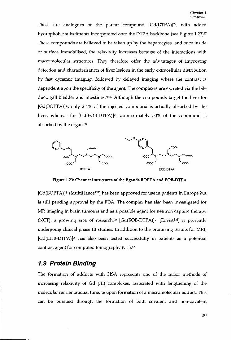
sites.i2 The protein is monomeric and heart shaped, consisting of 585 amino acids
with a total molecular weight of 66,400 Da.'-* It is organised into three homologous
a helical domains labelled (l-HI), and each domain is comprised of two sub-
domains, A and B, which contain six and four a helices respectively.'^
31
Chapter 1 introduction
Figure 1.24: Crystal structiu-e of un-liganded HSA. The sub-domains are colour coded as follows: lA, red: IB, light-red, IIA, green, IIB, light-green, IIIA blue, IIIB, light-blue's
Several studies have attempted to map the locations of fatty acid binding sites and
primary drug binding sites on the protein. The fatty acid sites are distiibuted
throughout the protein and involve all six sub-domains. By contiast many drugs
bind to one of the two primary binding sites located on the protein, these are
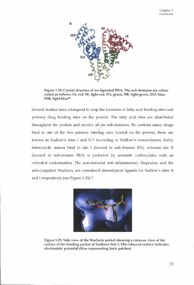
known as Sudlow's sites I and 11.''' According to Sudlow's nomenclature, bulky
heterocyclic anions bind to site I (located in sub-domain IIA), whereas site I I
(located in sub-domain IIIA) is preferred by aromatic carboxylates with an
extended conformation. The non-steroidal anti inflammatory Ibuprofen, and the
anti-coagulant Warfarin, are considered stereotypical Ugands for Sudlow's sites I I
and I respectively (see Figure 1.25).'*7
Figure 1.25: Side view of the Warfarin pocket showing a cutaway view of the surface of the binding pocket of Sudlows Site I. The coloured surface indicates electrostatic potential (blue representing basic patches)
32

Chapter 1 Introduction
Drug binding sites are now known to be located in sub-domains IIA and IIIA. A particularly large hydrophobic cavity is present in the IIA subdomain's and the geometry of the pocket of IIA is found to be quite different to that of IIIA. Bombieri et fl/'i reported that human serum albumin consists of 14 cavities. Cavity 14 was found to be a large, hydrophobic pocket situated between the lA and IIA subdomains. Four highly polar amino acid residues surround the cavity and may allow the entrance of a ligand into the region.^i The binding interaction between the Gd (III) chelate BOPTA and HSA was investigated. [Gd(BOPTA)]2- was found to dock into cavity 14 (Figure 1.26). The results indicate that the approach of the metal complex is dictated more by the electronic properties of the complex than its geometry.'!
»
Figiu-e 1.26: MEP representation of the cavity 14 docked with [Gd(BOPTA)]2
Data generated from the binding interactions of a variety of substrates with serum
proteins provide useful information relating to contrast agent design. The non-
covalent interaction of Gd (III) complexes with HSA can be conveniently studied by
relaxometiic methods using a low-resolution spectiometer operating at a fixed
frequency. Those typical for MRI applications operate in the range of 20 - 60 MHz
nowadays. Titration of a dilute solution containing the Gd (III) chelate and the
protein causes an increase in the relaxation rate when a binding interaction between
the complex and protein occurs, as a result of the increased reorientational
correlation time of the macromolecular adduct.'^
33

Chapter 1 Introduction
1.9.2 Binding requirements
The effect of hydrophobicity of the Gd (III) chelates and the affinity for serum
albumin was investigated by Aime and co-workers for a series of macrocyclic Gd
(III) complexes based upon [Gd(D0TA)(H20)]-, bearing an increased number of
lipophilic residues (BOM = benzyloxymethyl) (Figure 1.27) .3-4
- o o c ^ / — \ ^ c o o - - o o c - x / — \ ^ c o o - - o o c ^ n ^ c o o - - o o c ^ n ^ c c x i -
-ooc -^ \ I ^ c o o - -OOC—( \ I ^ c o o - - o o c ^ \ ; \ - c o o - •" • • - ^ L J ^ c o o - - o o c ^ L J •v-coo- - o o c - ^ L J V -
DOTA(BOM) trans-DOTA(BOM)j cis-D0TA(B0M)2 DOTA(BOM)3
Figure 1.27: Chemical structures of a series of ligands bearing increasing lipophilic residues
Upon interaction with serum albumin an increase in relaxivity was observed for
each of the complexes resulting in a lengthening of the rotational correlation time,
TR . ' The data obtained showed the binding affinity of KA = 1.7 x 10³ m-I was greatest
for the tii-substituted product [Gd(DOTA)-(BOM)3]-, this was of the same order of
magnitude as that reported for endogenous substiates like testosterone and
aldosterone.ioo Competition studies of [Gd(DOTA)-(BOM)3]-, showed that tiie
complex was displaced from the protein when equimolar amounts of Warfarin and
Ibuprofen were added, suggesting [Gd(DOTA)-(BOM)3]' interacts with both
subdomains IIA and IIIA.^o"
The binding affinity for the two isomers (cis and b-ans) of [Gd(DOTA)-(BOM)2]-
were of similar order KA =3.2 x 10^ M-i and KA = 3.6 x 10^ M-i respectively. For the
chelate containing only one (BOM) group [Gd(DOTA)-(BOM)]- only a weak
interaction was formed with serum albumin KA = < 1 x 10^ M-i.^ Comparison of the
relaxivity values of the Gd (III) derivatives in water is shown in the associated
NMRD profiles (Figure 1.28).
34
Chapter 1 Introduction
15
T 12 1
9
f 6 OC
3
25 *C • DOTA
••• DOTA(BOM) —o D0TA(B0M)3
Q _
outer ^liene
0.1 1 10 100 Proton LaiiTfor Fr©querK:y/MHz
1000
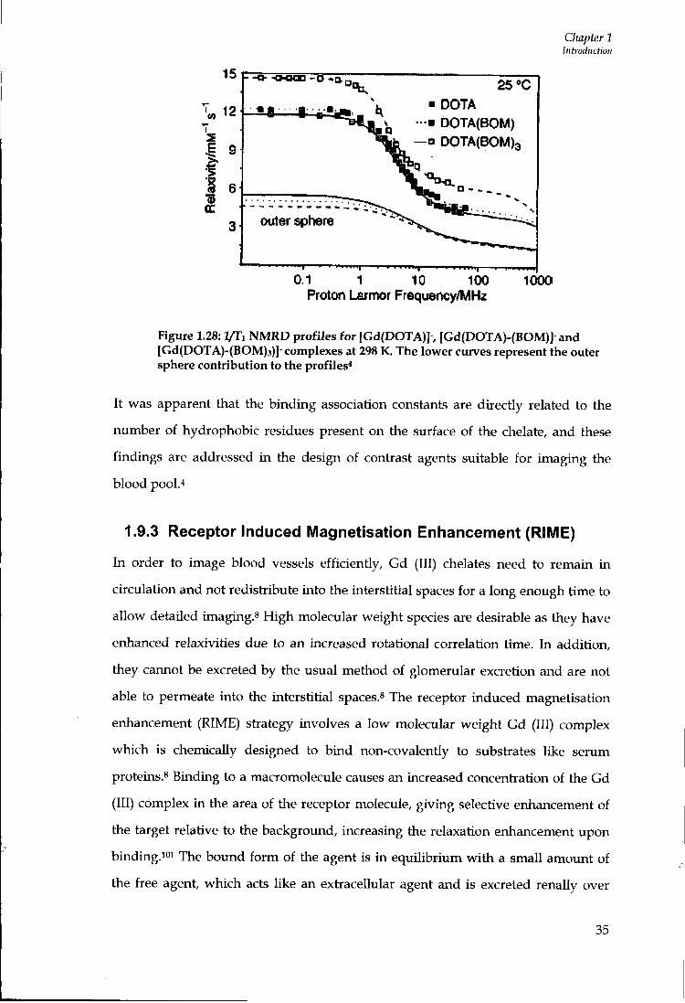
Figure 1.28: l/Ti NMRD profiles for [Gd(DOTA)]-, [Gd(DOTA)-(BOM)]-and [Gd(DOTA)-(BOM)3)]-complexes at 298 K. The lower curves represent the outer sphere contribution to the profiles*
It was apparent that the binding association constants are directly related to the
number of hydrophobic residues present on the surface of the chelate, and these
findings are addressed in the design of contrast agents suitable for imaging the
blood pool.''
1.9.3 Receptor Induced Magnetisation Enhancement (RIME)
In order to image blood vessels efficiently, Gd (III) chelates need to remain in
circulation and not redistribute into the interstitial spaces for a long enough time to
allow detailed imaging.^ High molecular weight species are desirable as they have
enhanced relaxivities due to an increased rotational correlation time. In addition,
they cannot be excreted by the usual method of glomerular excretion and are not
able to permeate into the interstitial spaces. The receptor induced magnetisation
enhancement (RIME) strategy involves a low molecular weight Gd (III) complex
which is chemically designed to bind non-covalentiy to substrates like serum
proteins.8 Binding to a macromolecule causes an increased concentration of the Gd
(III) complex in the area of the receptor molecule, giving selective enhancement of
the target relative to the background, increasing the relaxation enhancement upon
binding.ioi The bound form of the agent is in equilibrium with a small amount of
the free agent, which acts like an extracellular agent and is excreted renally over
35

Chapter 1 Introduction
time, avoiding the problems associated with slow excretion sometimes exhibited by covalently bound macromolecular systems. Binding allows selective enhancement of arteries and veins, relaxation enhancement due to increased molecular tumbling time and increased half-Ufe of the drug in vivo, allowing the radiologist time to image multiple body regions and to employ pulse sequences which give high resolution images. 02 Relatively rigid paramagnetic macromolecular complexes have been generated in vivo, in the blood pool, through reversible non-covalent hydrophobic interactions of small lipophilic Gd (III) chelates with serum. These include MS-325 (AngioMARK™, EPIX), MP-2269 (Mallinckrodt), [Gd(BOPTA)(H20)]2- (Multihance^M, Bracco) and [Gd(EOB-DTPA)(H20)]2-(Eovisf'"'^, Schering).
1.9.4 IVIS-325
MS-325 was the first representative of an RIME contiast agent and is currently in
phase III clinical trials. (Figure 1.29).ioi The complex is monomeric and is based
upon the parent compound [Gd(DTPA)]2-.
OH, 0
Figure 1.29: Chemical structure of MS-325
A pendant biphenylcyclohexyl moiety attached via a phosphodiester link is
inserted into the DTPA backbone, which increases the binding affinity towards
HSA, at the same time representing a steric impediment to prevent dissociation of
the complex.io^ MS-325 is reported to be more thermodynamically and kineticaUy
inert to metal ion substitution that the parent compound, [Gd(DTPA)]2-. Studies in
rabbits (12-16) revealed an average blood half-life for MS-325 of ~ 2.5 hours,
compared with ~ 10 minutes for non-bound agents. 03 Yhe blood persistence of the
36
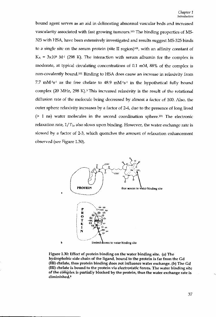
Chapter 1 Introduction
bound agent serves as an aid in delineating abnormal vascular beds and increased vascularity associated with fast growing tumours.io' The binding properties of MS-325 with HSA, have been extensively investigated and results suggest MS-325 binds to a single site on the serum protein (site I I region)ioi, with an affinity constant of KA = 3x10"* M-i (298 K ) . The interaction with serum albumin for the complex is moderate, at typical circulating concentiations of 0.1 mM, 88% of the complex is non-covalently bound. f Binding to HSA does cause an increase in relaxivity from 7.7 mM-^s-i as the free chelate to 48.9 mM-is-i in the hypothetical fully bound complex (20 MHz, 298 K).^ This increased relaxivity is the result of the rotational diffusion rate of the molecule being decreased by almost a factor of 100. Also, the outer sphere relaxivity increases by a factor of 2-4, due to the presence of long lived (> 1 ns) water molecules in the second coordination sphere.ioi The electionic relaxation rate, 1/Tie also slows upon binding. However, the water exchange rate is slowed by a factor of 2-3, which quenches the amount of relaxation enhancement observed (see Figure 1.30).
I f H
P R O T E I N free access to water binding site
limited Access to water binding site
Figure 1.30: Effect of protein binding on the water binding site, (a) The hydrophobic side chain of the ligand, bound to the protein is far from the Gd (III) chelate, thus protein binding does not influence water exchange, (b) The Gd (III) chelate is bound to the protein via electrostatic forces. The water binding site of the complex is partially blocked by the protein, thus the water exchange rate is diminished.*
37

Chapter 1 Introduction
1.9.5 MP-2269
MP-2269 (Figure 1.31) is a monomeric non aromatic Gd (III) complex. Similarly to
MS-325, i t is based upon [Gd(DTPA)]2-. The hydrophobic perityIbicyclo-[2.2.2]
octane side chains interact w i t h serum albumin allowing the compound to remain
in the intravascular space sufficiently long for imaging to be carried out fol lowing
intravenous injection.io^
The negative charges f r o m both the aspartyl linkers and the terminal [Gd(DTPA)]2-
group impart water solubility.i**
HOOC, HOOC^ / v ^ / - . . . A , ^NH, A „ A r A / ^ - ^ C H 3
Nn 1 ^ I — « L i HOOC COOH MP.2269
Figure 1.31: Chemical Structure of MP-2269
If the distance between the metal and proteia binding site of the Ugand is relatively
large then water exchange is not affected by the protein interaction, as was seen for
the Gd (III) chelate MP-2269 (Figure 1.30).8 However, the TR value of MP-2269
bound to bovine serum albumin was determined independently using deuterium
NMR. Adzamli et fl/ios reported that the TR value of the bovmd contrast agent is 8 ns,
sigruficantly shorter than the TR value expected for a fuUy immobilised ligand of 20-
22 ns. This indicates there is some segmental mobility of the contrast agent w i t h
respect to the protein one. The remarkable internal flexibility has been attributed to
the relatively long distance between the Gd-binding and the protein binding sites of
the molecule.8 Upon interaction w i t h BSA the relaxivity was found to increase f r o m
5.64 mM-is-i to 24.5 mM-is-i (20 MHz , 310 K).8
The problems associated w i t h increased residence times, Xm, upon formation of a
HSA adduct, as discussed earlier, l imi t the efficacy of some newer, second
generation contrast agents. The complex [Gd(DTPA)(BOM)3] (Figure 1.32) was
studied in parallel to MS-325, where i t was reported that the water exchange rates
38
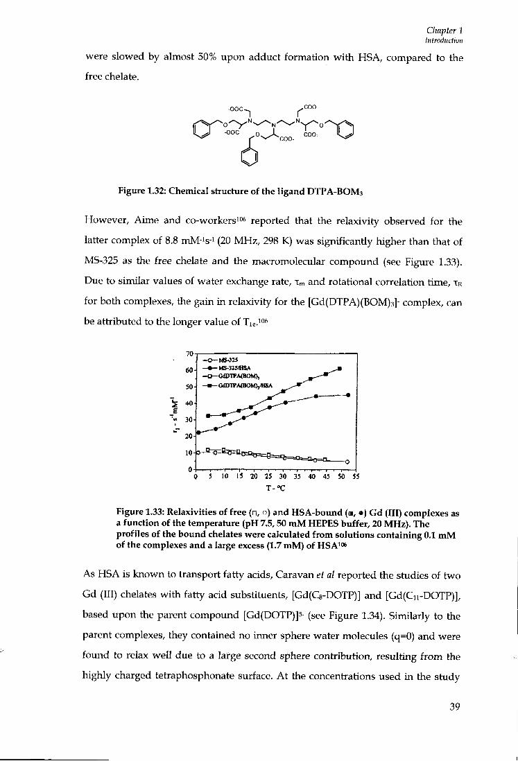
Chapter 1 Introduction
were slowed by almost 50% upon adduct formation wi th HSA, compared to the free chelate.
-ooc-
-ooc o ^ ^ ^ ^ ioo-
0 Figure 1.32: Chemical structure of the ligand DTPA-BOM3
However, Aime and co-workers^o^ reported that the relaxivity observed for the
latter complex of 8.8 mM-is-i (20 M H z , 298 K) was significantly higher than that of
MS-325 as the free chelate and the macromolecular compound (see Figure 1.33).
Due to similar values of water exchange rate, Xm and rotational correlation time, TR
for both complexes, the gain i n relaxivity for the [Gd(DTPA)(BOM)3]- complex, can
be attributed to the longer value of Tie. "^
70
60
50
30
-•-NS3-323VHSA -0-G<lDTI>A<HOM), - • — aiDlTAtBOMjyHSA
0 1 10 15 20 25 30 35 40 45 50 55
T - o C
Figure 1.33: Relaxivities of free (•, o) and HSA-bound («, • ) Gd (III) complexes as a function of the temperature (pH 7.5,50 mM HEPES buffer, 20 MHz). The profiles of the bound chelates were calculated from solutions containing 0.1 mM of the complexes and a large excess (1.7 mM) of HSAit"*
As HSA is known to transport fatty acids. Caravan et al reported the studies of two
Gd (111) chelates wi th fatty acid substituents, [Gd(C8-DOTP)] and [Gd(Cii-DOTP)],
based upon the parent compound [Gd(DOTP)]5- (see Figure 1.34). Similarly to the
parent complexes, they contained no inner sphere water molecules (q=0) and were
found to relax wel l due to a large second sphere contribution, resulting f r o m the
highly charged tetraphosphonate surface. A t the concentrations used in the study
39
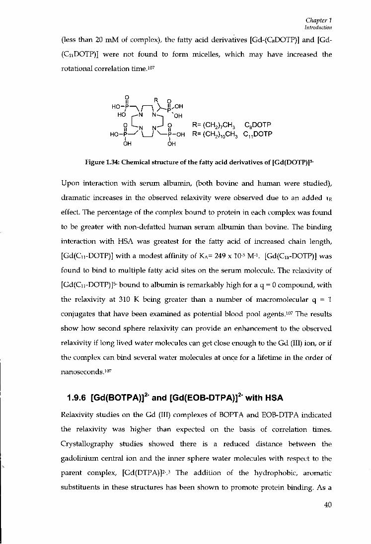
Chapter 1 Introduction
(less than 2 0 m M of complex), the fatty acid derivatives [Gd-(C8DOTP)] and [Gd-(CiiDOTP)] were not found to f o r m micelles, which may have increased the rotational correlation time.io^
R Q
HO ^OH
O L.M J O I I
^ O R = (CH2)7CH3 C g D O T P
H O - P ^ \ _ / \ - P - O H R=(CH2) ioCH3 C ^ p O T P
OH OH
Figure 1.34: Chemical structure of the fatty acid derivatives of [Gd(DOTP)]5-
Upon interaction w i t h serum albumin, (both bovine and human were studied),
dramatic increases in the observed relaxivity were observed due to an added TR
effect. The percentage of the complex bound to protein i n each complex was found
to be greater w i t h non-defatted human serum albumin than bovine. The binding
interaction w i t h HSA was greatest for the fatty acid of increased chain length,
[Gd(Cn-DOTP)] w i t h a modest aff ini ty of K A = 2 4 9 x 10⁻³ m-\ [Gd(Cii-DOTP)] was
found to bind to multiple fatty acid sites on the serum molecule. The relaxivity of
[Gd(Cii-DOTP)]s- bovmd to albumin is remarkably high for a q = 0 compound, w i t h
the relaxivity at 3 1 0 K beiag greater than a number of macromolecular q = 1
conjugates that have been examined as potential blood pool agents.^o^ xhe results
show how second sphere relaxivity can provide an enhancement to the observed
relaxivity if long lived water molecules can get close enough to the Gd (III) ion, or if
the complex can bind several water molecules at once for a lifetime in the order of
nanoseconds.^''^
1.9.6 [Gd(B0TPA)]2- and [Gd(EOB-DTPA)]^- with HSA
Relaxivity studies on the Gd (111) complexes of BOPTA and EOB-DTPA indicated
the relaxivity was higher than expected on the basis of correlation times.
Crystallography studies showed there is a reduced distance between the
gadolinium central ion and the inner sphere water molecules w i t h respect to the
parent complex, [Gd(DTPA)]2-.3 The addition of the hydrophobic, aromatic
substituents i n these structures has been shown to promote protein binding. As a
4 0

Chapter 1 Introduction
result of the modest non-covalent interactions wi th serum albumin, both complexes act as blood pool contrast agents and can provide useful contrasts to the compound MS-325. Relatively modest changes in the structure of a Gd (III) chelate can have major effects upon protein binding; the results of this allow fine tuning in the design of complexes to produce the required response. For example, MS-325 is reported as being 94.3% bound i n a 4.5% solution of HSA. h i contrast, [Gd(EOB-DTPA)]2- is reported as only 10% bound in a 5% solution of HSA w i t h a concentiation of 1.5 mmol L-i. MS-325 and EOB-DTPA have been found to interact w i t h several sites on the protein molecule. A t increased concentiations of complex as many as 20 to 25 molecules can bind to a single protein molecule.« Upon binding to serum albumin, there is an increased TR effect for [Gd(EOB-DTPA)]2- that Chen et aP reported as 9.8 ns, which is much less than the increased effect seen for MS-325 of 20-30 ns, measured under the same experimental conditions. A n increase i n tumbling can be observed for [Gd(EOB-DTPA)]2- i n the frequency range of 40-60 MHz, the fields used i n medical MRI (see Figure 1.35).
I Bound lynniMlcn
0"— 0LO1
Proton Frequency (MIfe)
Figure 1.35: NMRD profile of bound and unbound populations of [Gd(EOB-DTPA)J2- in 4% HSA solution
41

Chapter 2 Di-aqim Gadolinium Complexes and Protein Interaction
Chapter 2
Di-aqua Gadolinium Complexes and Protein
Interaction
42
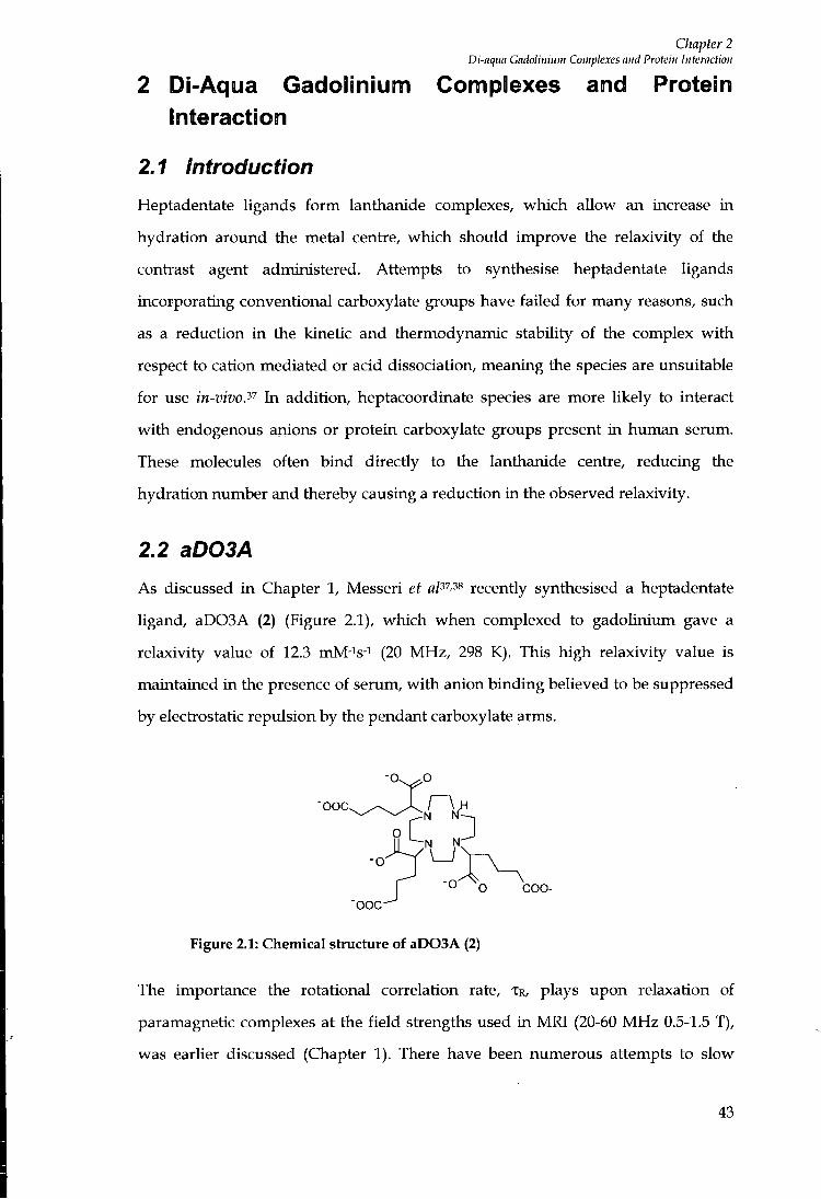
Chapter 2 Di-aqtm Gadolinium Complexes and Protein Interaction
2 Di-Aqua Gadolinium Complexes and Protein Interaction
2.1 Introduction
Heptadentate ligands f o r m lanthanide complexes, which allow an increase in
hydration around the metal centre, which should improve the relaxivity of the
contiast agent administered. Attempts to synthesise heptadentate ligands
incorporating conventional carboxylate groups have failed for many reasons, such
as a reduction in the kinetic and thermodynamic stability of the complex wi th
respect to cation mediated or acid dissociation, meaning the species are unsuitable
for use in-vivo.'^'^ In addition, heptacoordinate species are more likely to interact
w i t h endogenous anions or protein carboxylate groups present in human serum.
These molecules often bind directly to the lanthanide centie, reducing the
hydration number and thereby causing a reduction in the observed relaxivity.
2.2 aD03A
As discussed i n Chapter 1, Messeri et fl/37,38 recently synthesised a heptadentate
ligand, aD03A (2) (Figure 2.1), which when complexed to gadolinium gave a
relaxivity value of 12.3 mM-is-i (20 MHz , 298 K). This high relaxivity value is
maintained i n the presence of serum, w i t h anion binding beUeved to be suppressed
by electrostatic repulsion by the pendant carboxylate arms.
"DOC
N N
COO-"OOC
Figure 2.1: Chemical structure of aD03A (2)
The importance the rotational correlation rate, TR, plays upon relaxation of
paramagnetic complexes at the f ie ld strengths used in M R l (20-60 M H z 0.5-1.5 T),
was earlier discussed (Chapter 1). There have been numerous attempts to slow
43
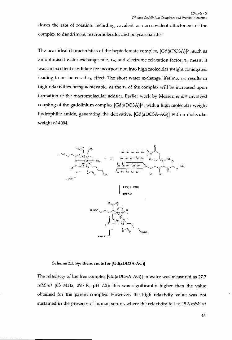
Chapter 2 Di-nqiin CadoUnium Complexes and Protein Interaction
down the rate of rotation, including covalent or non-covalent attachment of the
complex to dendrimers, macromolecules and polysaccharides.
The near ideal characteristics of the heptadentate complex, [Gd(aD03A)]3-, such as
an optimised water exchange rate, Xm, and electionic relaxation factor, Xs, meant i t
was an excellent candidate for incorporation into high molecular weight conjugates,
leading to an increased TR effect. The short water exchange lifetime, Xm, results i n
high relaxivities being achievable, as the XR of the complex w i l l be increased upon
formation of the macromolecular adduct. Earlier work by Messeri et aP^ involved
coupling of the gadolinium complex [Gd(aD03A)]3-, w i t h a high molecular weight
hydrophilic amide, generating the derivative, [Gd(aD03A-AG)] w i t h a molecular
weight of 4094.
+ 3
I 1 1 1 1 \ OH OH OH OH OH ^
OH OH OH OH OH
I I I I i \ OH OH OH OH OH N
I I I i OH OH OH OH OH
EDO / HOBt
pH 6.0
r3"'~] O Gd
Scheme 2.1: Synthetic route for [Gd(aD03A-AG)]
The relaxivity of the free complex [Gd(aD03A-AG)] i n water was measured as 27.7
mM-is-i (65 MHz, 293 K, p H 7.2); this was significantly higher than the value
obtained for the parent complex. However, the high relaxivity value was not
sustained i n the presence of human serum, where the relaxivity fel l to 15.5 mM-is-i
44

Chapter 2 Di-aqua Gadolinium Complexes and Protein Interaction
(65 MHz, 293 K, p H 7.2). This is explained by the neutraUsation of the overall
charge of the high molecular weight conjugate. Upon formation of three amide
bonds, the peripheral negative charge is neutralised, hence the complex becomes
more susceptible to binding endogenous anions such as carbonate, lactate or
phosphate.
2.3 Protein Binding
The most significant results to date to slow down the rate of rotation, TR, for a Gd
(III) complex involve the non-covalent binding of a Gd (III) complex to the slowly
tumbling macromolecule human serum albumin, through incorporation of
hydrophobic substituents. It is wel l established that the presence of hydrophobic
moieties are the basic requirements for binding a substrate to serum as wel l as the
effect of negative charges on the complex.^
[Gd(aD03A)]3- has a peripheral negative charge, hence the introduction of a
hydrophobic moiety should enhance the binding affinity for the serum protein,
resulting in an increase in the rotational correlation time, which should represent
an ideal heptadentate contrast agent. Inserting an aromatic functionalised group
w o u l d increase the hydrophobicity of the complex as wel l as facilitating studies
into the luminescence behaviour of the related Eu (III) or Tb (III) complexes by
using the aryl moiety as a sensitiser for lanthanide emission.
As a result of extensive research involving the study of human serum albumin,
including competitive assays and detailed investigations regarding the binding
interaction of several drugs and metabolites, detailed information regarding the
binding domains of the protein has been obtained.^ This provides definition of the
structural properties that may enhance binding.
45
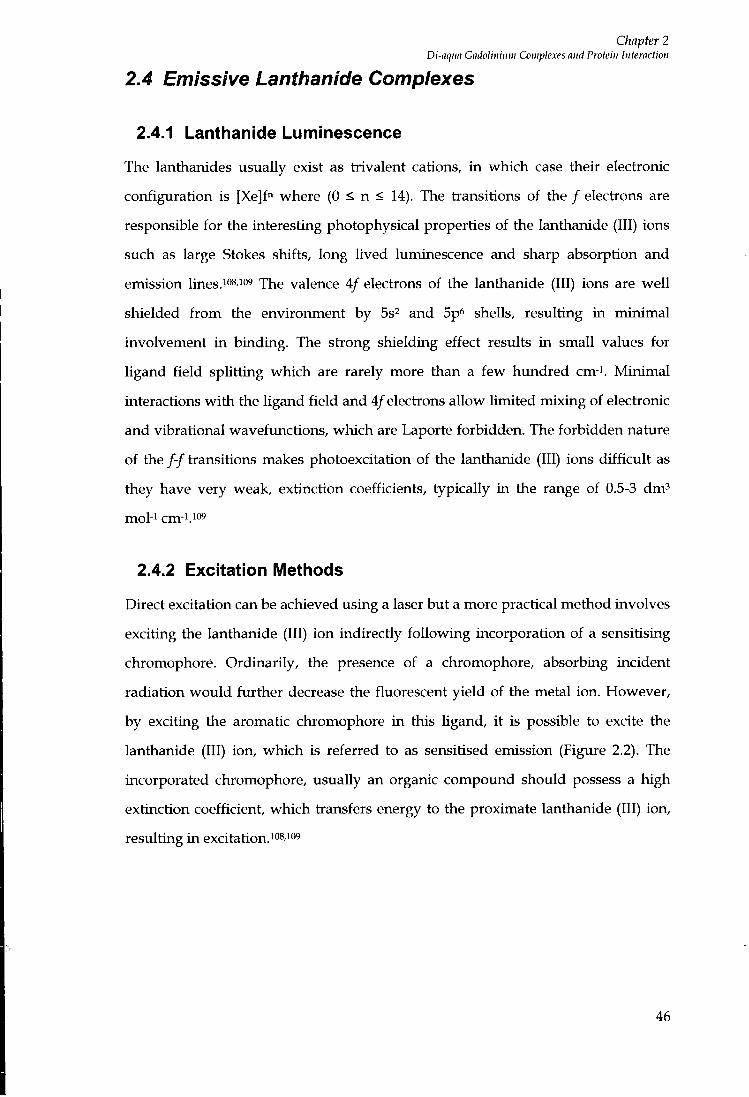
Chapter 2 Di-aqua Gadolinium Complexes and Protein Interaction
2.4 Emissive Lanthanide Complexes
2.4.1 Lanthanide Luminescence
The lanthanides usually exist as trivalent cations, in which case their electronic
configuration is [Xe]f" where (0 < n < 14). The transitions of the / electrons are
responsible for the interesting photophysical properties of the lanthanide (111) ions
such as large Stokes shifts, long Uved luminescence and sharp absorption and
emission lines.io^.iog jhe valence 4/ electrons of the lanthanide (111) ions are wel l
shielded f r o m the environment by Ss and Sp^ shells, resulting in minimal
involvement in binding. The strong shielding effect results in small values for
ligand f ield splitting which are rarely more than a few hundred cm-i. Min imal
interactions w i t h the ligand f ie ld and 4/electrons allow limited mixing of electronic
and vibrational wavefimctions, which are Laporte forbidden. The forbidden nature
of the / - / transitions makes photoexcitation of the lanthanide (111) ions dif f icul t as
they have very weak, extinction coefficients, typically i n the range of 0.5-3 d m '
mol-i cm-1.109
2.4.2 Excitation Metliods
Direct excitation can be achieved using a laser but a more practical method involves
exciting the lanthanide (111) ion indirectly fo l lowing incorporation of a sensitising
chromophore. Ordinarily, the presence of a chromophore, absorbing incident
radiation wou ld further decrease the fluorescent yield of the metal ion. However,
by exciting the aromatic chromophore i n this ligand, i t is possible to excite the
lanthanide (111) ion, which is referred to as sensitised emission (Figure 2.2). The
incorporated chromophore, usually an organic compound should possess a high
extinction coefficient, which tiansfers energy to the proximate lanthanide (111) ion,
resulting in excitation.^os io^
46
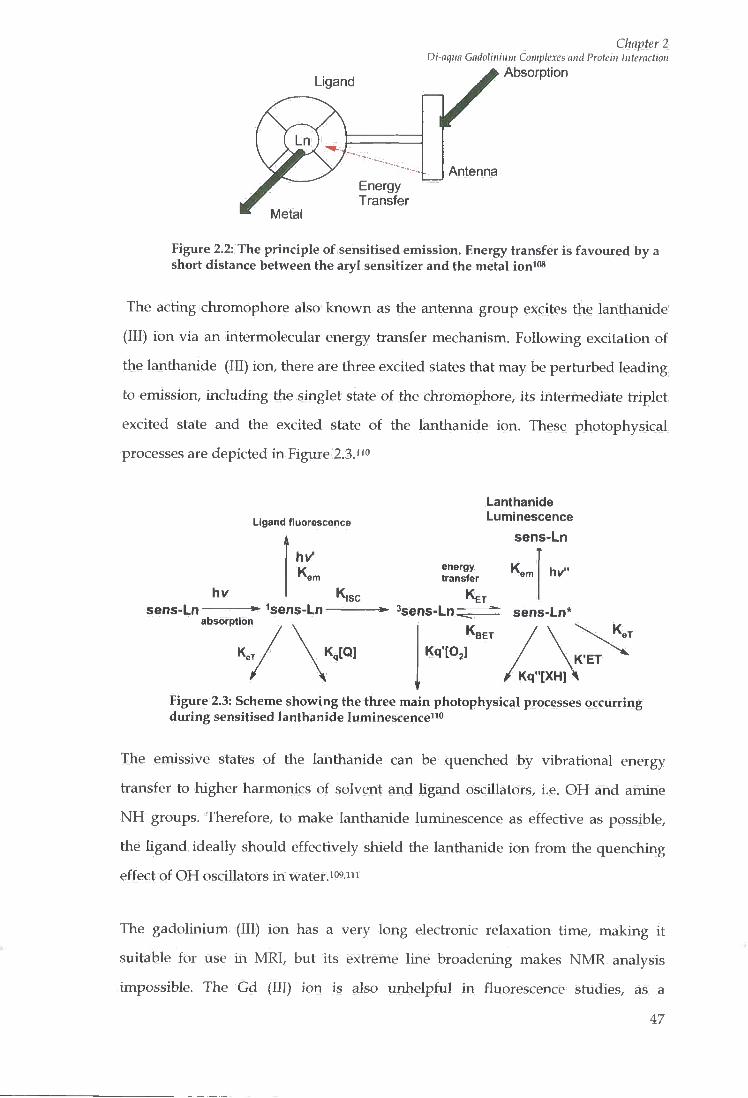
Ligand
Chapter 2 Di-aqua Gndolinium Complexes and Protein Interaction
Absorption
Antenna Energy Transfer
Metal
Figure 2.2: The principle of sensitised emission. Energy transfer is favoured by a short distance between the aryl sensitizer and the metal ion "*
The acting chromophore also known as the antenna group excites the lanthanide
(111) ion via an intermolecular energy transfer mechanism. Following excitation of
the lanthanide (III) ion, there are three excited states that may be perturbed leading
to emission, including the singlet state of the chromophore, its intermediate triplet
excited state and the excited state of the lanthanide ion. These photophysical
processes are depicted in Figure 2.3.1'*'
Ligand fluorescence
sens-Ln sens-Ln absorption
Lanthanide Luminescence
sens-Ln
energy transfer
K,
Ken
^sens-Ln; ET
hv"
sens-Ln* K BET
Kq"[XH] Figure 2.3: Scheme showing the three main photophysical processes occurring during sensitised lanthanide luminescence""
The emissive states of the lanthanide can be quenched by vibrational energy
transfer to higher harmonics of solvent and ligand oscillators, i.e. O H and amine
N H groups. Therefore, to make lanthanide luminescence as effective as possible,
the ligand ideally should effectively shield the lanthanide ion f r o m the quenching
effect of O H oscillators i n water.i"?"'
The gadolinium (111) ion has a very long electronic relaxation time, making it
suitable for use m MRl , but its extreme line broadening makes N M R analysis
impossible. The Gd (III) ion is also unhelpful i n fluorescence studies, as a
4 7

Chapter 2 Di-aqua Gadolinium Complexes and Protein Interaction
consequence of the large difference between the ground state ( S 7/2) and its first
excited state {^Fy/i). The neighbouring ion Eu (III) ion emits fairly strongly in the
visible region of the spectrum when directly excited at 397 n m and does not possess
the limitations observed for Gd (III). Therefore, as a result of similarities i n their
ionic radii, Eu (III) analogues can be used as val id models to provide information
regarding the properties of gadoUnium (III) complexes in solution.
2.5 Synthesis
Initial work was directed towards the synthesis of the macrocyclic Ugand 1,4,7,-tris-
[4'-methoxycarbonyl-l'-methoxycarbonylbutyl]-l,4,7,10-tetraazacyclododecane also
known as aD03A (2), using the synthetic route established by Messeri et a/ -ss. This
is shown in Scheme 2.2.
1 .SOCI2 2. Br, .COOMe
3. MeOH Br (1)
MeCN
(2)
I O COOMe MoOOC^
Scheme 2.2: Synthetic route for the preparation of aD03A (2)
1,4,7,10-Tetraazacyclododecane was alkylated using 2.9 equivalents of dimethyl 2-
bromo-adipate (1) in the presence of potassium carbonate as the selected base and
hot acetonitrile. Attempts had been made to synthesise the product using the
weaker base, sodium hydrogen carbonate, which was beUeved to increase the
selectivity of the reaction, producing an improved yield of the required t r i -
substituted product, rather than the over alkylated tetra substituted specie.
However, the weaker base resulted in an increased amount of the undesired
carbamate by-product being produced. This results f r o m hydrogen carbonate attack
on one or more of the cyclen r ing N - H groups. Although, carbamate by products
were still present f r o m alkylation methods using potassium carbonate, the yield
48
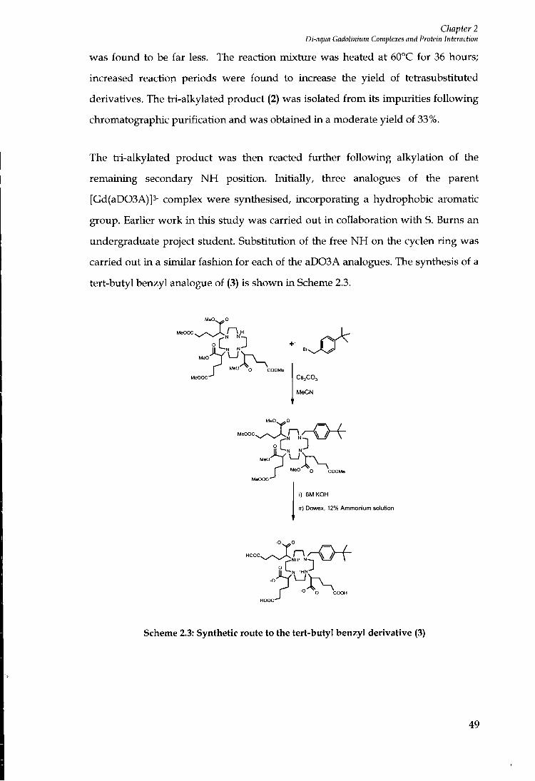
Chapter 2 Di-aqiia Gadolinium Complexes and Protein Interaction
was fo imd to be far less. The reaction mixture was heated at 60°C for 36 hours;
increased reaction periods were fo imd to increase the yield of tetrasubstituted
derivatives. The tri-alkylated product (2) was isolated f r o m its impurities fo l lowing
chromatographic purification and was obtained in a moderate yield of 33%.
The tri-alkylated product was then reacted further fo l lowing alkylation of the
remaining secondary N H position. Initially, three analogues of the parent
[Gd(aD03A)]3- complex were synthesised, incorporating a hydrophobic aromatic
group. Earlier work i n this study was carried out i n collaboration w i t h S. Burns an
undergraduate project student. Substitution of the free N H on the cyclen r ing was
carried out i n a similar fashion for each of the aD03A analogues. The synthesis of a
tert-butyl benzyl analogue of (3) is shown in Scheme 2.3.
M e O ^ O
M e O O C , ^ , „ . v ^ r \ ^
MeO'
MeOOC-
II N ' ' B , ^ J .
MeO
CS2CO3
MeCN
I I N K
MeO
i) 6M KOH
ii) DowBx, 12% Ammonium solution
O COOH HOOC"^
Scheme 2.3: Synthetic route to the tert-butyl benzyl derivative (3)
49
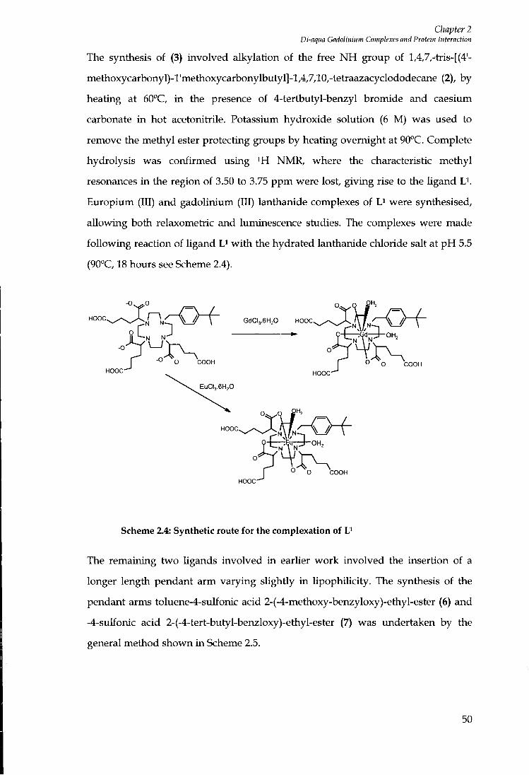
Chapter 2 Di-aqua Gadolinium Complexes and Protein Interaction
The synthesis of (3) involved alkylation of the free N H group of l,4,7,-tris-[(4'-
methoxycarbonyl)-l'methoxycarbonylbutyl]-l,4,7,10,-tetraazacyclododecane (2), by
heating at 60°C, in the presence of 4-tertbutyl-benzyl bromide and caesium
carbonate in hot acetonitrile. Potassium hydroxide solution (6 M) was used to
remove the methyl ester protecting groups by heating overnight at 90°C. Complete
hydrolysis was confirmed using i H NMR, where the characteristic methyl
resonances in the region of 3.50 to 3.75 ppm were lost, giving rise to the ligand L^.
Europium (III) and gadoUnium (III) lanthanide complexes of were synthesised,
allowing both relaxometric and luminescence studies. The complexes were made
fol lowing reaction of ligand w i t h the hydra ted lanthanide chloride salt at p H 5.5
(90°C, 18 hours see Scheme 2.4).
HOOC GdCI, .6H,0
HOOC COOH
HOOC
COOH
HOOC
EuCU.6H,0
HOOC
COOH HOOC
Scheme 2.4: Synthetic route for the complexation of U
The remaining two ligands involved in earlier work involved the insertion of a
longer length pendant arm varying slightly i n lipophilicity. The synthesis of the
pendant arms toluene-4-sulfonic acid 2-(-4-methoxy-benzyloxy)-ethyl-ester (6) and
-4-sulfonic acid 2-(-4-tert-butyl-benzloxy)-ethyl-ester (7) was undertaken by the
general method shown in Scheme 2.5.
50
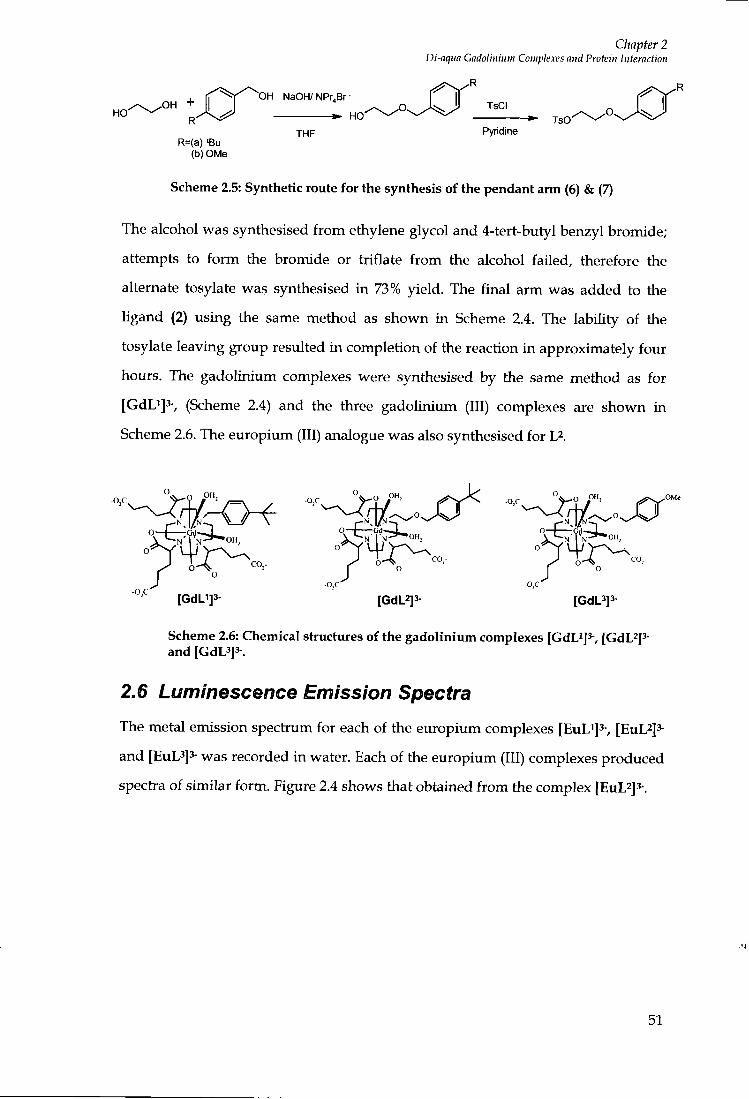
Chapter 2 Di-aqua Gadolinium Complexes and Protein Interaction
, tf'^'^Sv'^^OH NaOH/NPr.Br- f^'^^' HO ^ f ^ ^ X ^ ^ H O ^ / ^
R=(a) 'Bu (b) OMe
THF Pyridine
Scheme 2.5: Synthetic route for the synthesis of the pendant arm (6) & (7)
The alcohol was synthesised f r o m ethylene glycol and 4-tert-butyl benzyl bromide;
attempts to f o r m the bromide or triflate f r o m the alcohol failed, therefore the
alternate tosylate was synthesised in 73% yield. The final arm was added to the
ligand (2) using the same method as shown in Scheme 2.4. The lability of the
tosylate leaving group resulted in completion of the reaction in approximately four
hours. The gadolinium complexes were synthesised by the same method as for
[GdLi]3-, (Scheme 2.4) and the three gadoUnium (III) complexes are shown in
Scheme 2.6. The europium (III) analogue was also synthesised for L^.
^"^^^^ ''-i^l-'Xr [GdV]3- [GdL2]3- [GdL3]3-
Scheme 2.6: Chemical structures of the gadolinium complexes [CdL^]^, [GdL^p and [GdL3]3-.
2.6 Luminescence Emission Spectra
The metal emission spectium for each of the europium complexes [EuLi]^-, [EuL^]^-
and [EuL3]3- was recorded in water. Each of the europium (III) complexes produced
spectra of similar form. Figure 2.4 shows that obtained f r o m the complex [EuL^js-.
5 1

Chapter 2 Di-aqua Gadolinium Complexes and Protein Interaction
18000
16000
14000
12000
s 10000
S 8000
4000 H
Wavelength (nm)
Figure 2.4: Europium emission spectrum obtained for [EuL^]^ in water, \exc = 309 nm, 420 nm filter.
The spectrum was found to consist of five distinct bands. From left to right these
bands represented the AJ = 0 , 1 , 2, 3, 4 transitions, originating f r o m the emissive ^Do
level of Eu (111) and terminating in the ground state ^Fo, ^Fi, ^Fs, ^F, , levels
respectively. Figure 2.5 shows the energy level diagram of the 4/levels involved in
Eu (111) luminescence.109'110'111
23.000
21.000
17,000
5,000 4,000 3.000 2,000 1,0(K)
0
2
1
Figure 2.5: Partial energy level diagram representing the 4/energy levels involved in Eu (III) luminescence
52
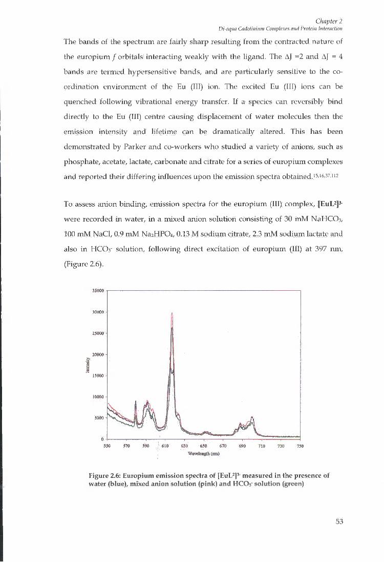
Chapter 2 Di-aqua Gadolinium Complexes and Protein Interaction
The bands of the spectrum are fairly sharp resulting f r o m the contracted nature of
the europium / orbitals interacting weakly w i t h the ligand. The AJ =2 and AJ = 4
bands are termed hypersensitive bands, and are particularly sensitive to the co
ordination environment of the Eu (111) ion. The excited Eu (111) ions can be
quenched fol lowing vibrational energy transfer. If a species can reversibly bind
directly to the Eu (111) centre causing displacement of water molecules then the
emission intensity and lifetime can be dramatically altered. This has been
demonstrated by Parker and co-workers who studied a variety of anions, such as
phosphate, acetate, lactate, carbonate and citiate for a series of europium complexes
and reported their differing influences upon the emission spectra obtained.i5,i6,37,u2
To assess anion binding, emission spectra for the europium (III) complex, [EuL^js-
were recorded in water, i n a mixed anion solution consisting of 30 m M NaHCOs,
100 m M NaCI, 0.9 m M Na2HP04, 0.13 M sodium citiate, 2.3 m M sodium lactate and
also in HCO3- solution, fo l lowing direct excitation of europium (III) at 397 nm,
(Figure 2.6).
.15000
25000
20000 i
IftUfill
m 6M 670 WiT<fco«Ih(iim)
" 0
Figure 2.6: Europium emission spectra of [EuL^]^ measured in the presence of water (blue), mixed anion solution (pink) and HCOa' solution (green)
53
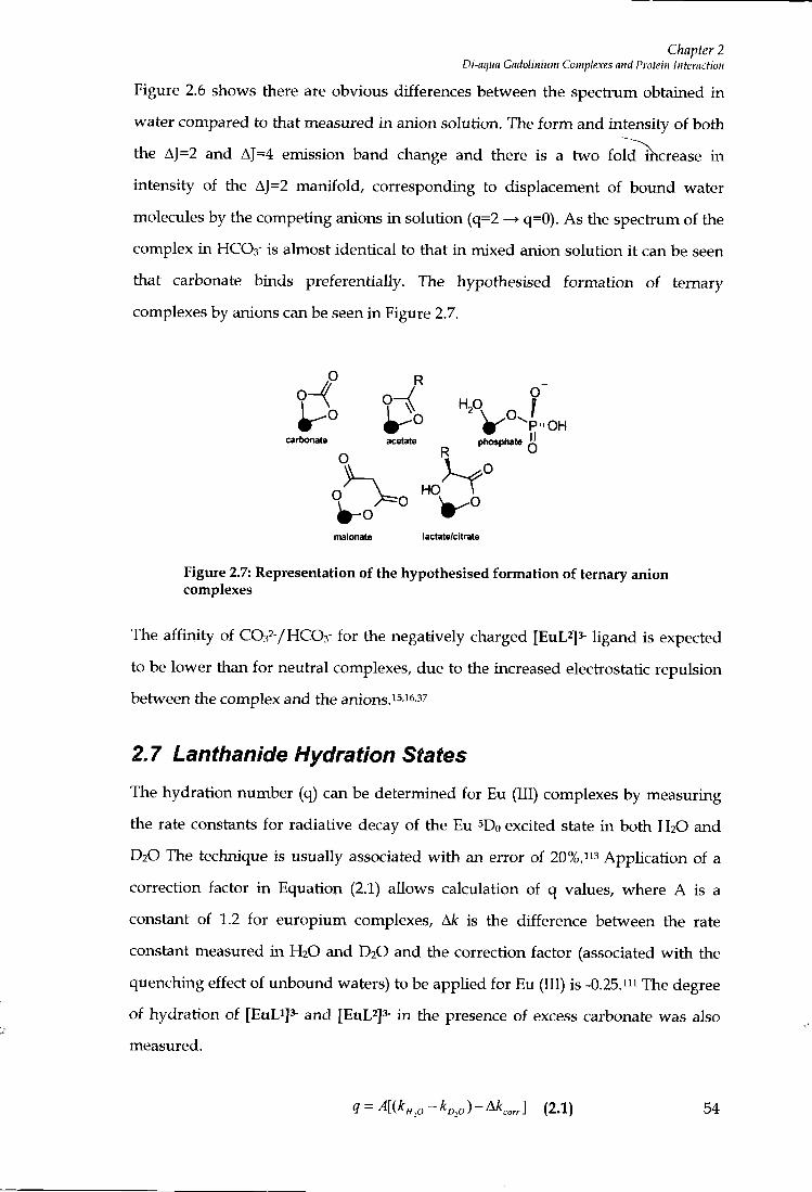
Chapter 2 Di-aqua Gadolinium Complexes and Protein Interaction
Figure 2.6 shows there are obvious differences between the spectrum obtained i n
water compared to that measured in anion solution. The f o r m and intensity of both
the AJ=2 and AJ=4 emission band change and there is a two fold^hcrease i n
intensity of the AJ=2 manifold, corresponding to displacement of boxmd water
molecules by the competing anions i n solution (q=2 q=0). As the spectrum of the
complex in HCO3- is almost identical to that i n mixed anion solution i t can be seen
that carbonate binds preferentially. The hypothesised formation of ternary
complexes by anions can be seen in Figure 2.7.
.0 R
H ri H P ?
cartjonate acetate phosphate il
o °
malonate lactate/citrate
Figure 2.7: Representation of the h)^othesised formation of ternary anion complexes
The affinity of CO32-/HCO3- for the negatively charged [EuL^Js- ligand is expected
to be lower than for neutral complexes, due to the increased electiostatic repulsion
between the complex and the anions.i^-i^-^^
2.7 Lanthanide Hydration States
The hydration number (q) can be determined for Eu (111) complexes by measuring
the rate constants for radiative decay of the Eu ^Do excited state in both H2O and
D2O The technique is usually associated wi th an error of 20%.i'^ Application of a
correction factor i n Equation (2.1) allows calculation of q values, where A is a
constant of 1.2 for europium complexes, A/c is the difference between the rate
constant measured in H2O and D2O and the correction factor (associated w i t h the
quenching effect of unbound waters) to be applied for Eu (111) is -0.25."i The degree
of hydration of [EuLi ]3- and [EuL^]^ i n the presence of excess carbonate was also
measured.
<} = A[{kH,O-I^D,0)-^Korr] (2.1) 54
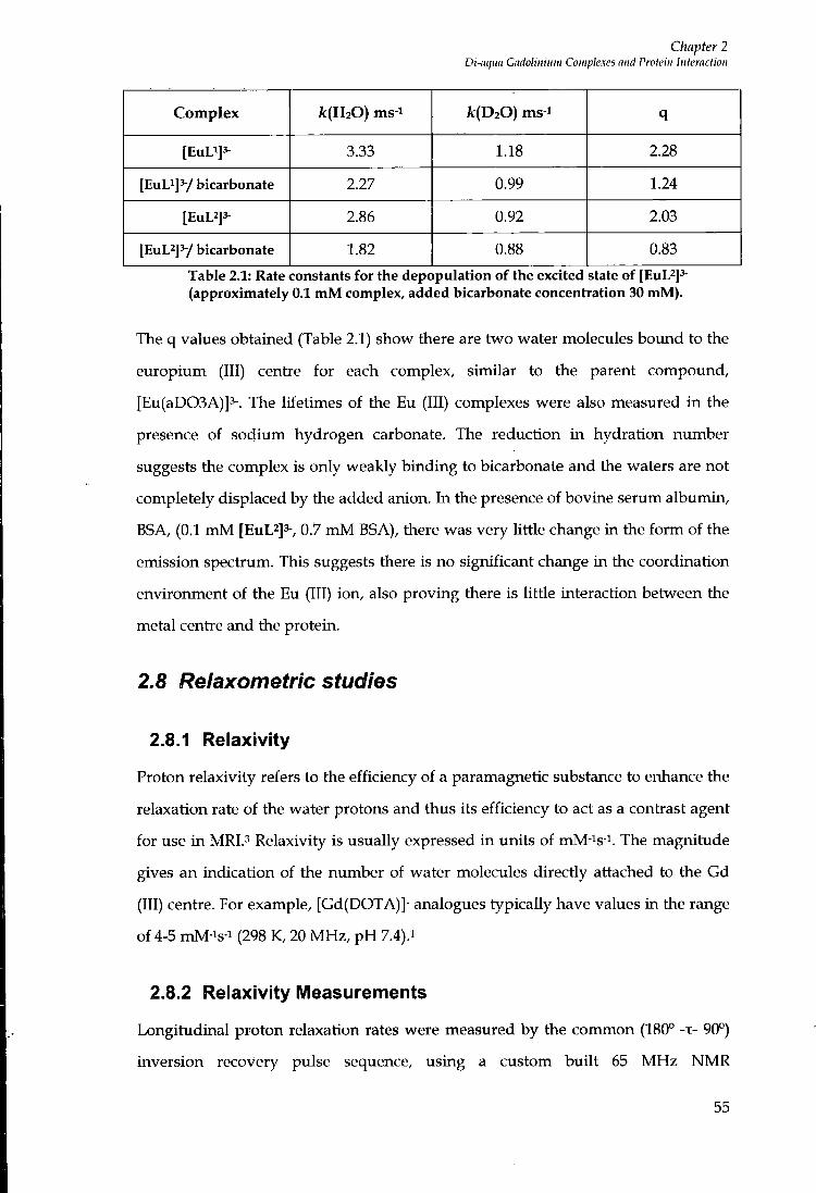
Chapter 2 Di-nqua Gadolimum Complexes mid Protein Interaction
Complex A:(H20) ms-i /c(D20) ms 1 q
[EuLi]3- 3.33 1.18 2.28
[EuLi]^/ bicarbonate 2.27 0.99 1.24
[EuL2]3- 2.86 0.92 2.03
[EuL^] V bicarbonate 1.82 0.88 0.83
Table 2.1: Rate constants for the depopulation of the excited state of [EuL^]^ (approximately 0.1 mM complex, added bicarbonate concentration 30 mM).
The q values obtained (Table 2.1) show there are two water molecules bound to the
europium (111) centre for each complex, similar to the parent compound,
[Eu(aD03A)]3-. The lifetimes of the Eu (111) complexes were also measured i n the
presence of sodium hydrogen carbonate. The reduction in hydration number
suggests the complex is only weakly binding to bicarbonate and the waters are not
completely displaced by the added anion. In the presence of bovine serum albumin,
BSA, (0.1 m M [EuL2]3-, 0.7 m M BSA), there was very little change in the f o r m of the
emission spectrum. This suggests there is no significant change in the coordination
environment of the Eu (111) ion, also proving there is little interaction between the
metal centre and the protein.
2.8 Relaxometric studies
2.8.1 Relaxivity
Proton relaxivity refers to the efficiency of a paramagnetic substance to enhance the
relaxation rate of the water protons and thus its efficiency to act as a contrast agent
for use i n M R l . ' Relaxivity is usually expressed in units of mM-is-i. The magnitude
gives an indication of the number of water molecules directly attached to the Gd
(111) centre. For example, [Gd(DOTA)]- analogues typically have values in the range
of 4-5 mM-is-i (298 K, 20 M H z , p H 7.4).i
2.8.2 Relaxivity IVIeasurements
Longitudinal proton relaxation rates were measm-ed by the common (180° -T- 90°)
inversion recovery pulse sequence, using a custom buil t 65 M H z NMR
55

Chapter 2 Di-aqua Gadolinium Complexes and Protein Interaction
Spectrometer, operating at ambient temperature 295 K or a Bruker Minispec Mq60
instrument operating at 20 M H z and 310 K. Relaxivity values can be calculated
according to the Equations 2.2 and 2.3, where (d) is the diamagnetic contribution
and (p) is the paramagnetic contiibution to the observed relaxation rate.i-^
i = (2-2) ^1
^ i „ = — 1 ^ V (2-3)
The diamagnetic contiibutions of water, ni t i ic acid and aqueous solutions of human
serum albumin were determined f r o m the relaxivity measurements for a series of
gadolinium standards in water, i n collaboration w i t h A. Congreve. The
diamagnetic contiibutions determined for the Bruker Minispec instrument (310 K)
were 0.25 s-i (water), 0.26 s-i (2.5 M nit i ic acid solution) and 0.38 s-i (0.7 m M human
serum albumin solution). The diamagnetic correction values for water and nitric
acid determined for the 65 M H z spectiometer by M . Lowe were 0.38 s-i and 0.26 s-i
respectively.
The concentiation of gadolinium in the aqueous relaxivity samples was determined
fol lowing digestion using an equal volume of 5 M niti ic acid. The relaxivity of free
gadolinium (III) i n 2.5 M nit i ic acid at 20 M H z , 310 K was measured as 8.74 mmol-
is-i, compared to the value of 11.66 rrunol-'s-i at 65 M H z , 295 K. The relaxivity of the
samples was determined using Equation 2.4.
, (2.4)
2.8.3 Relaxivity measurements of [GdL^]^-, [GdL^]^- and [GdL^]^-
The relaxivities of the three gadolinium complexes were also measured i n the
presence of bovine serum albumin to assess the enhancement upon binding Table
56
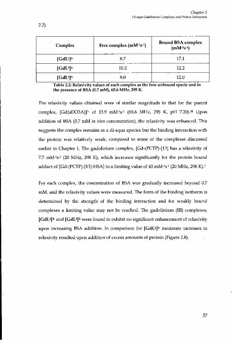
Chapter 2 Di-aqua Gadotiitium Complexes and Protein Interaction
2.2).
Complex Free complex (mM-^s-i) Bound BSA complex
(mM-is-i)
[GdLi]3- 8.7 17.1
[GdL2]3- 10.2 12.2
[GdL3]3^ 9.0 12.0
Table 2.2: Relaxivity values of each complex as the free unbound specie and in the presence of BSA (0.7 mM), 65.6 MHz, 295 K.
The relaxivity values obtained were of similar magnitude to that for the parent
complex, [Gd(aD03A)]3- of 13.9 mM-is-i (65.6 MHz , 295 K, p H 7.20).38 Upon
addition of BSA (0.7 m M in vivo concentration), the relaxivity w as enhanced. This
suggests the complex remains as a di-aqua species but the binding interaction wi th
the protein was relatively weak, compared to some of the complexes discussed
earlier i n Chapter 1. The gadolinium complex, [Gd-(PCTP)-[13] has a relaxivity of
7.7 mM-is-^ (20 M H z , 298 K), which increases significantly for the protein bound
adduct of [Gd-(PCTP)-[13]-HSA] to a l imi t ing value of 45 mM-is-i (20 MHz, 298 K).3
For each complex, the concentiation of BSA was gradually increased beyond 0.7
m M , and the relaxivity values were measured. The f o r m of the binding isotherm is
determined by the strength of the binding interaction and for weakly bovmd
complexes a l imit ing value may not be reached. The gadolinium (111) complexes,
[GdL2]3- and [GdL^Js-were found to exhibit no significant enhancement of relaxivity
upon increasing BSA addition. In comparison for [GdD]3- moderate increases in
relaxivity resulted upon addition of excess amounts of protein (Figure 2.8).
57
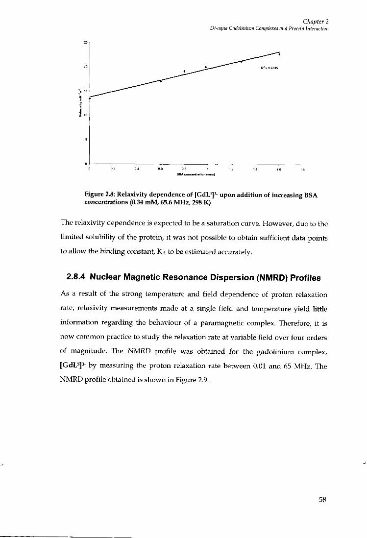
Chapter 2 Di-aqm Gadolinium Complexes and Protein Interaction
1 I J £ 10
BSA concentration mmol
Figure 2.8: Relaxivity dependence of [GdU]^ upon addition of increasing BSA concentrations (0.34 mM, 65.6 MHz, 298 K)
The relaxivity dependence is expected to be a saturation curve. However, due to the
l imited solubiUty of the protein, i t was not possible to obtain sufficient data points
to allow the binding constant, K A to be estimated accurately.
2.8.4 Nuclear Magnetic Resonance Dispersion (NIVIRD) Profiles
As a result of the strong temperature and f ie ld dependence of proton relaxation
rate, relaxivity measurements made at a single f ield and temperature yield little
information regarding the behaviour of a paramagnetic complex. Therefore, i t is
now common practice to study the relaxation rate at variable f ie ld over four orders
of magnitude. The N M R D profile was obtained for the gadolinium complex,
[GdL3]3- by measuring the proton relaxation rate between 0.01 and 65 MHz. The
N M R D profile obtained is shown in Figure 2.9.
58
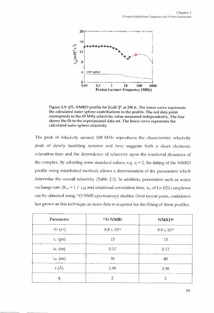
Chapter 2 Di-aqua Gadolitiinm Complexes and Protein Interaction
4 J outer jp/iere
0,01 0,1 1 10 100 1000 Proton Larmor Frequency (MHz)
Figure 2.9: l / T i N M R D profile for [GdLi]^- at 298 K. The lower curve represents the calculated outer sphere contributions to the profile. The red data point corresponds to the 65 MHz relaxivity value measured independently. The line shows the fit to the experimental data set. The lower curve represents the calculated outer sphere relaxivity.
The peak of relaxivity around 100 M H z reproduces the characteristic relaxivity
peak of slowly tumbling systems and here suggests both a short electionic
relaxation time and the dependence of relaxivity upon the rotational dynanucs of
the complex. By adopting some standard values, e.g. q = 2, the f i t t ing of the N M R D
profile using established methods allows a determination of the parameters which
determine the overall relaxivity (Table 2.3). In addition, parameters such as water
exchange rate (Kev = 1 / Xm) and rotational correlation time, TR, of Ln (111) complexes
can be obtained using i^O NMR spectioscopy studies. Over recent years, confidence
has grown in this technique as more data is acquired for the f i t t ing of these profiles.
Parameter N M R " NMRDb
' A2 (s-2) 8.8 X 1 0 " 8.8 X 10¹⁹
Tv (ps) 15 15
TR (ns) 0.12 0.12
Tm (ns) 38 40
r ( A ) 2.98 2.98
q 2 2
59
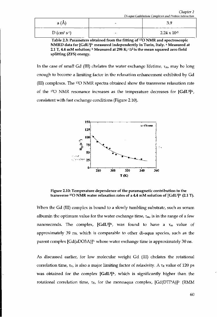
Chapter 2
a (A) - 3.9
D (cm2 s-i) - 2.24 X 10⁻⁵
Table 2.3: Parameters obtained from the fitting of ^^O N M R and spectroscopic N M R D data for [GdL^]^ measured independently in Turin, Italy. ' Measured at 2.1 T, 4.4 m M solution; ^ Measured at 298 K; "A is the mean squared zero field splitting (ZFS) energy.
In the case of small Gd (III) chelates the water exchange lifetime, Xm, may be long
enough to become a l imit ing factor i n the relaxation enhancement exhibited by Gd
(III) complexes. The ^^O N M R spectra obtained show the transverse relaxation rate
of the i^o N M R resonance increases as the temperature decreases for [GdL^]^-,
consistent w i th fast exchange conditions (Figure 2.10).
: . l 1.0 KMR
280 300 320 340 360 T(K)
Figure 2.10: Temperature dependence of the paramagnetic contribution to the transverse ^^O NMR water relaxation rates of a 4.4 mM solution of [GdL'J^- (2.1 T) .
When the Gd (III) complex is boimd to a slowly tumbling substrate, such as serum
albumin the opt imum value for the water exchange time, Xm, is i n the range of a few
nanoseconds. The complex, [GdL^]^-, was found to have a Xm value of
approximately 39 ns, which is comparable to other di-aqua species, such as the
parent complex [Gd(aD03A)]3- whose water exchange time is approximately 30 ns.
As discussed earlier, for low molecular weight Gd (III) chelates the rotational
correlation time, XR, is also a major l imi t ing factor of relaxivity. A XR value of 120 ps
was obtained for the complex [GdL^]^-, which is significantly higher than the
rotational correlation time, XR, for the monoaqua complex, [Gd(DTPA)]2- (RMM
60
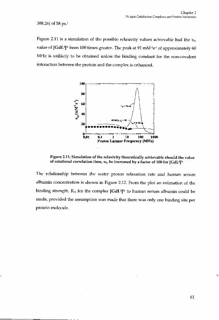
Chapter 2 Di-aqua Gadolinium Complexes and Protein Interaction
388.26) of 58 ps.
Figure 2.11 is a simulation of the possible relaxivity values achievable had the TR,
value of [GdL3]3- been 100 times greater. The peak at 91 mM-'s ' of approximately 60
M H z is unlikely to be obtained unless the binding constant for the non-covalent
interaction between the protein and the complex is enhanced.
100
80
60
I 3 40
20
0 0.01 0,1 1 10 100 1000
Proton Larmor Frequency (MHz)
Figure 2.11: Simulation of the relaxivity theoretically achievable should the value of rotational correlation time, t r , be increased by a factor of 100 for [GdL^]^
The relationship between the water proton relaxation rate and human serum
albumin concentiation is shown in Figure 2.12. From the plot an estimation of the
binding strength, K A for the complex [GdL3]3- ^ human serum albumin could be
made, provided the assumption was made that there was only one binding site per
protein molecule.
61
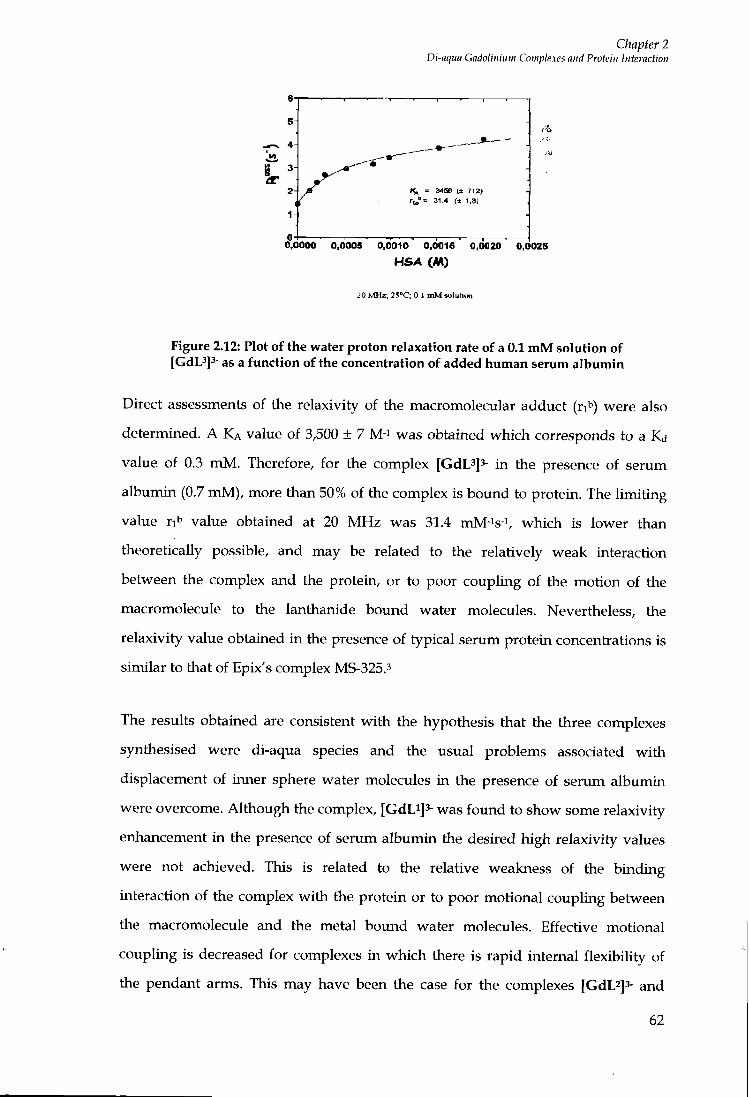
Chapter 2 Di-aqua Gadolinium Complexes and Protein Interaction
31.4 (± 1.3)
0,0000 0.0005 0,0010 0,0015 0,0020 0,0025 HSA (M)
20 MHz; 25°C; 0,J aiMwlulioii
Figure 2.12: Plot of the water proton relaxation rate of a 0.1 m M solution of [GdL^]^ as a function of the concentration of added human serum albumin
Direct assessments of the relaxivity of the macromolecular adduct (ri'') were also
determined. A K A value of 3,500 ± 7 M-i was obtained which corresponds to a Kd
value of 0.3 m M . Therefore, for the complex [CdL^J^- in the presence of serum
albumin (0.7 m M ) , more than 50% of the complex is bound to protein. The l imit ing
value r i ' ' value obtained at 20 M H z was 31.4 mM-^s-^, which is lower than
theoretically possible, and may be related to the relatively weak interaction
between the complex and the protein, or to poor coupling of the motion of the
macromolecule to the lanthanide bound water molecules. Nevertheless, the
relaxivity value obtained in the presence of typical serum protein concentrations is
similar to that of Epix's complex MS-325.3
The results obtained are consistent w i t h the hypothesis that the three complexes
synthesised were di-aqua species and the usual problems associated wi th
displacement of inner sphere water molecules in the presence of serum albumin
were overcome. Although the complex, [GdL^J^- was found to show some relaxivity
enhancement i n the presence of serum albumin the desired high relaxivity values
were not achieved. This is related to the relative weakness of the binding
interaction of the complex w i t h the protein or to poor motional coupling between
the macromolecule and the metal bound water molecules. Effective motional
coupling is decreased for complexes in which there is rapid internal flexibility of
the pendant arms. This may have been the case for the complexes [GdL^J^- and
62
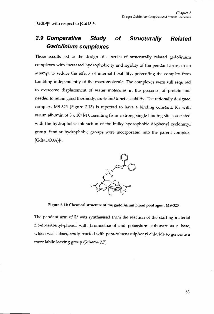
Chapter 2 Di-aqua Gadolinium Complexes and Protein Interaction
[GdL3]3- w i t h respect to [GdLi]3-.
2.9 Comparative Study of Structurally Related Gadolinium complexes
These results led to the design of a series of stiucturally related gadolinium
complexes w i t h increased hydrophobicity and rigidi ty of the pendant arms, i n an
attempt to reduce the effects of internal flexibility, preventing the complex f rom
tumbling independently of the macromolecule. The complexes were still required
to overcome displacement of water molecules i n the presence of protein and
needed to retain good thermodynamic and kinetic stability. The rationally designed
complex, MS-325 (Figure 2.13) is reported to have a binding constant, K A w i t h
serum albumin of 3 x 10^ M-i , resulting f r o m a strong single binding site associated
w i t h the hydrophobic interaction of the bulky hydrophobic di-phenyl cyclohexyl
group. Similar hydrophobic groups were incorporated into the parent complex,
[Gd(aD03A)]3-.
Figure 2.13: Chemical structure of the gadolinium blood pool agent MS-325
The pendant arm of was synthesised f r o m the reaction of the starting material
3,5-di-tertbutyl-phenol w i t h bromoethanol and potassium carbonate as a base,
which was subsequently reacted wi th para-toluenesulphonyl chloride to generate a
more labile leaving group (Scheme 2.7).
63
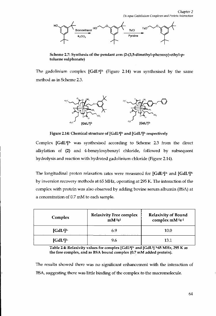
Chapter 2 Di-aqim Cadoliiiiuw Complexes and Protein Interaction
HO
Bromoethanol
K2CO3
HO XsCI
TsO
Pyridine
Scheme 2.7: Synthesis of the pendant arm (2-(3,5-dimethyl-phenoxy)-ethyl-p-toluene sulphonate)
The gadolinium complex [GdL*]^ (Figure 2.14) was synthesised by the same
method as in Scheme 2.3.
N I N
Figure 2.14: Chemical structure of [GdL*]^ and [GdL^]^ respectively
Complex [GdL5]3- was synthesised according to Scheme 2.3 f r o m the direct
alkylation of (2) and 4-benzyloxybenzyI chloride, fol lowed by subsequent
hydrolysis and reaction w i t h hydra ted gadolinium chloride (Figure 2.14).
The longitudinal proton relaxation rates were measured for [GdL*]3- and [GdL5]3-
by inversion recovery methods at 65 MHz, operating at 295 K. The interaction of the
complex w i t h protein was also observed by adding bovine serum albumin (BSA) at
a concentration of 0.7 m M to each sample.
Complex Relaxivity Free complex Relaxivity of Bound complex mM-^s-i
6.9 10.0
[GdL5]3^ 9.6 13.1
Table 2.4: Relaxivity values for complex [GdL«]3- and [GdL^] 3-65 MHz, 295 K as the free complex, and as BSA bound complex (0.7 m M added protein).
The results showed there was no significant enhancement w i t h the interaction of
BSA, suggesting there was Uttle binding of the complex to tiie macromolecule.
64
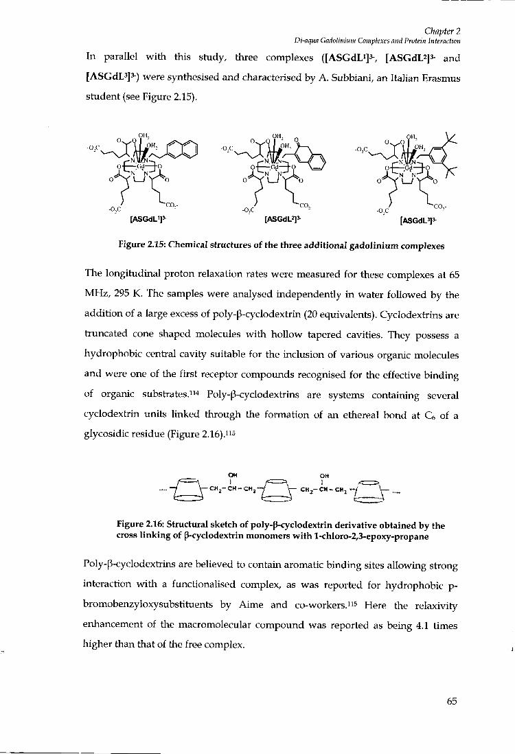
Chapter 2 Di-nqua Gadolinium Complexes and Protein Interaction
In parallel w i th this study, three complexes ([ASGdLijs-, [ASGdL2]3- and
[ASGdL3]3-) were synthesised and characterised by A. Subbiani, an Italian Erasmus
student (see Figure 2.15).
OH,
OH,
EN. MAIN ~ , — G d — ^ O
-0,C
N N"
-o,c ~C0,-
N N N N 0- X \__y r o
-o,c [ASGdL']'^ [ASGdL^]^ [ASGdL5]3 -
Figure 2.15: Chemical structures of the three additional gadolinium complexes
The longitudinal proton relaxation rates were measured for these complexes at 65
M H z , 295 K. The samples were analysed independently i n water fol lowed by the
addition of a large excess of poly-p-cyclodextiin (20 equivalents). Cyclodextrins are
truncated cone shaped molecules w i t h hoUow tapered cavities. They possess a
hydrophobic central cavity suitable for the inclusion of various organic molecules
and were one of the first receptor compoxmds recognised for the effective binding
of organic substrates."'' Poly-p-cyclodextrins are systems containing several
cyclodextiin units linked through the formation of an ethereal bond at Ce of a
glycosidic residue (Figure 2.16)."s
OH C « , - C H - C H ,
OH
CHj-CH-CHj -y
Figure 2.16: Structural sketch of poly-p-cyclodextrin derivative obtained by the cross linking of p-cyclodextrin monomers with l-chloro-2,3-epoxy-propane
Poly-p-cyclodextiins are believed to contain aromatic binding sites allowing stiong
interaction w i t h a functionaUsed complex, as was reported for hydrophobic p-
bromobenzyloxysubstituents by Aime and co-workers."^ Here the relaxivity
enhancement of the macromolecular compound was reported as being 4.1 times
higher than that of the free complex.
65
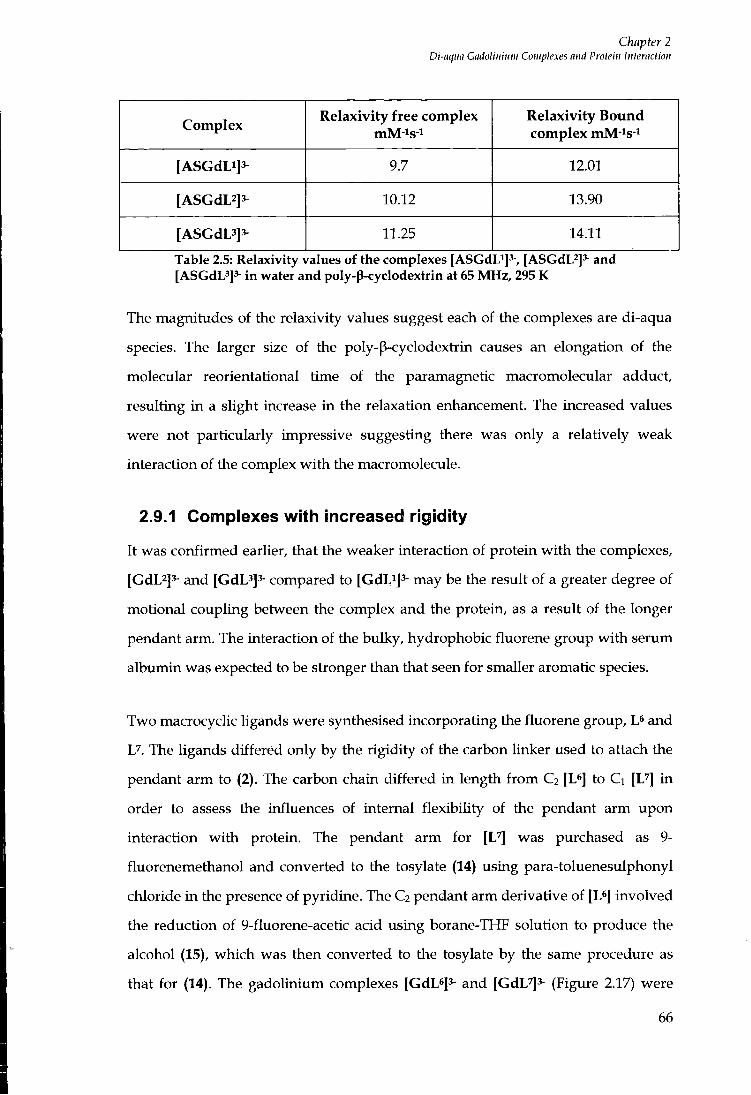
Chapter 2 Di-aqua Gadolinium Complexes and Protein Interaction
Complex Relaxivity free complex Relaxivity Bound
complex mM-^s-i
[ASGdLi]3^ 9.7 12.01
[ASGdL2]3- 10.12 13.90
[ASGdL3]3- 11.25 14.11
Table 2.5: Relaxivity values of the complexes [ASGdLi]^-, [ASGdL^Js- and [ASGdL3]3- in water and poly-p-cyclodextrin at 65 MHz, 295 K
The magnitudes of the relaxivity values suggest each of the complexes are di-aqua
species. The larger size of the poly-p-cyclodextiin causes an elongation of the
molecular reorientational time of the paramagnetic macromolecular adduct,
resulting i n a slight increase in the relaxation enhancement. The increased values
were not particularly impressive suggesting there was only a relatively weak
interaction of the complex w i t h the macromolecule.
2.9.1 Complexes with increased rigidity
It was confirmed earlier, that the weaker interaction of protein w i t h the complexes,
[GdL2]3- and [GdL3]3- compared to [GdLi]^- may be the result of a greater degree of
motional coupling between the complex and the protein, as a result of the longer
pendant arm. The interaction of the bulky, hydrophobic fluorene group w i t h serum
albumin was expected to be stionger than that seen for smaller aromatic species.
Two macrocyclic ligands were synthesised incorporating the fluorene group, L* and
U. The ligands differed only by the rigidity of the carbon linker used to attach the
pendant arm to (2). The carbon chain differed in length f r o m C2 [L*] to Ci [L''] i n
order to assess the influences of internal flexibili ty of the pendant arm upon
interaction wi th protein. The pendant arm for [ L ^ was purchased as 9-
fluorenemethanol and converted to the tosylate (14) using para-toluenesulphonyl
chloride i n the presence of pyridine. The C2 pendant arm derivative of [L*] involved
the reduction of 9-fluorene-acetic acid using borane-THF solution to produce the
alcohol (15), which was then converted to the tosylate by the same procedure as
that for (14). The gadolinium complexes [GdL6]3- and [GdL7]3- (Figure 2.17) were
66
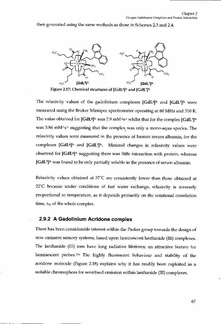
Chapter 2 Di-aqiia Gadolinium Complexes and Protein Interaction
then generated using the same methods as those in Schemes 2.3 and 2.4.
-o,c -o,c O OH,
-o,c^ [GdL6]3- o,c' [GdL^3-
Figure 2.17: Chemical structures of (GdL*]3- and [CdL^]'-
The relaxivity values of the gadolinium complexes [GdL*]^ and [GdL^^- vvere
measured using the Bruker Minispec spectrometer operating at 60 M H z and 310 K.
The value obtained for [GdL*]3- was 7.9 mM-is-i whilst that for the complex [GdL^^-
was 3.86 mM-^s-i suggesting that the complex was only a mono-aqua species. The
relaxivity values were measured in the presence of human serum albumin, for the
complexes [GdL^J^- and [GdL^]^-. Minimal changes in relaxivity values were
observed for [GdL*]^- suggesting there was little interaction w i t h protein, whereas
[GdL^]^ was fo imd to be only partially soluble in the presence of serum albumin.
Relaxivity values obtained at 37°C are consistently lower than those obtained at
22°C because under conditions of fast water exchange, relaxivity is inversely
proportional to temperature, as i t depends primarily on the rotational correlation
time, XR, of the whole complex.
2.9.2 A Gadolinium Acridone complex
There has been considerable interest w i th in the Parker group towards the design of
new emissive sensory systems, based upon luminescent lanthanide (111) complexes.
The lanthanide (111) ions have long radiative lifetimes; an attractive feature for
luminescent probes."* The highly fluorescent behaviour and stability of the
acridone molecule (Figure 2.18) explains why i t has readily been exploited as a
suitable chromophore for sensitised emission wi th in lanthanide (III) complexes.
67

Chapter 2 Di-aqua Gadolinium Complexes and Protein Interaction
O
Figure 2.18: Chemical structure of acridone
Monosubstituted N-acridoneethyl 1,4,7,10-tetiaazacyclododecane (Figure 2.19) was
donated by Y. Bretonneire, a post doctoral researcher wi th in the Parker group.
N N
Figure 2.19: Chemical structure of monosubstituted N-acridoneethyl 1,4,7,10-tetraazacyclododecane
The remaining free N H groups of monosubstituted N-acridoneethyl 1,4,7,10-
tetiaazacyclododecane were alkylated using excess dimethyl 2-bromo-adipate (1) in
the presence of hot acetonitiile and caesium carbonate as base, to produce (18)
(Figure 2.20), which was purif ied by colxmm chromatography. Following,
hydrolysis of (18) in the presence of l i th ium hydroxide (1 M) the ligand [L*] (Figure
2.20) was complexed using the hydrated lanthanide chloride salts of both europium
(111) and gadoliruum (III), to give [GdLs]^- and [EuL8]3-.
68
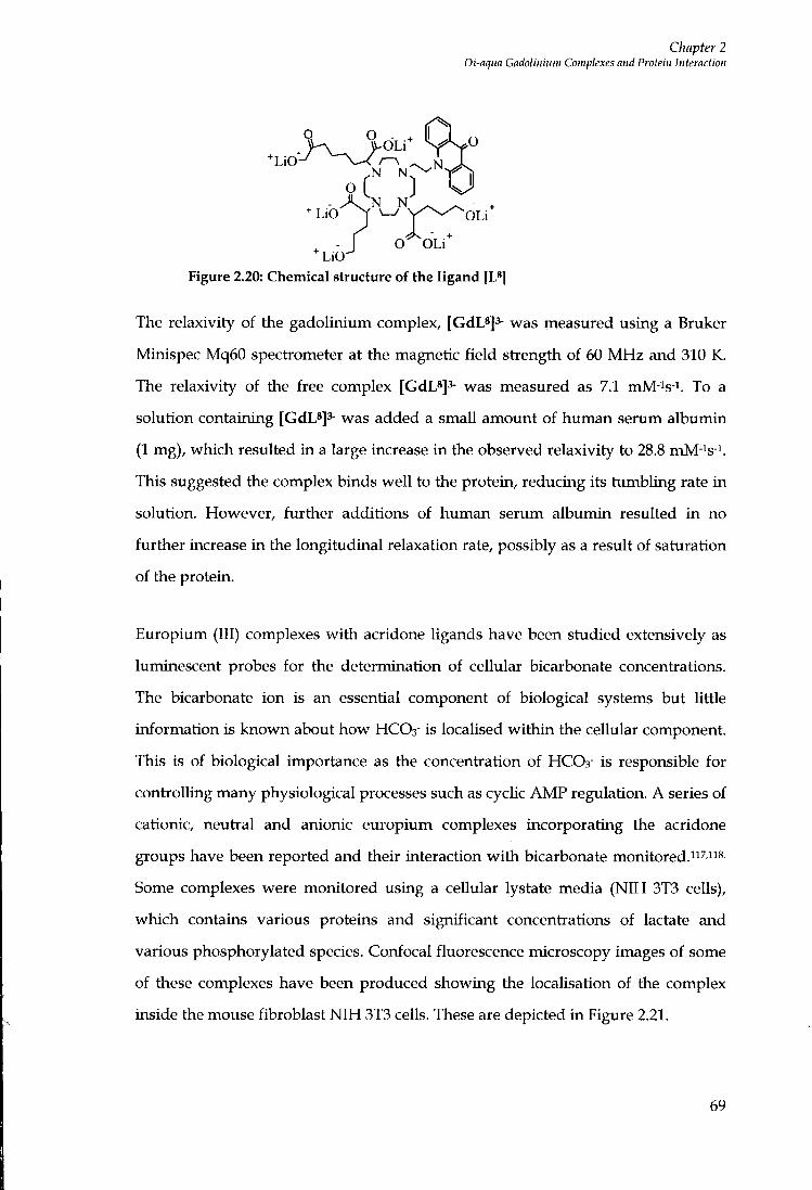
Chapter 2 Di-aqua Gadolinium Complexes and Protein Interaction
N N
- L i O > ^ Y ^ ^ ^ O L i
Figure 2.20: Chemical structure of the ligand [L*]
O O L i L i O -
The relaxivity of the gadolinium complex, [GdL*]3- was measured using a Bruker
Minispec Mq60 spectrometer at the magnetic f ie ld strength of 60 M H z and 310 K.
The relaxivity of the free complex [GdL^J^- was measured as 7.1 mM-is-i. To a
solution containing [GdLs]^- was added a small amotmt of human serum albumin
(1 mg), which resulted i n a large increase i n the observed relaxivity to 28.8 mM-is-i.
This suggested the complex binds wel l to the protein, reducing its tumbling rate i n
solution. However, further additions of human serum albumin resulted i n no
further increase in the longitudinal relaxation rate, possibly as a result of saturation
of the protein.
Europium (III) complexes wi th acridone ligands have been studied extensively as
luminescent probes for the determination of cellular bicarbonate concentrations.
The bicarbonate ion is an essential component of biological systems but httle
information is known about how HCOs" is locaUsed wi th in the cellular component.
This is of biological importance as the concentiation of HCO3- is responsible for
contiolhng many physiological processes such as cycUc A M P regulation. A series of
cationic, neutial and anionic europium complexes incorporating the acridone
groups have been reported and their interaction w i t h bicarbonate monitored."^ "8.
Some complexes were monitored using a cellular lystate media ( N I H 3T3 cells),
which contains various proteins and significant concentiations of lactate and
various phosphorylated species. Confocal fluorescence microscopy images of some
of these complexes have been produced showing the locaUsation of the complex
inside the mouse fibroblast N I H 3T3 cells. These are depicted in Figure 2.21.
69
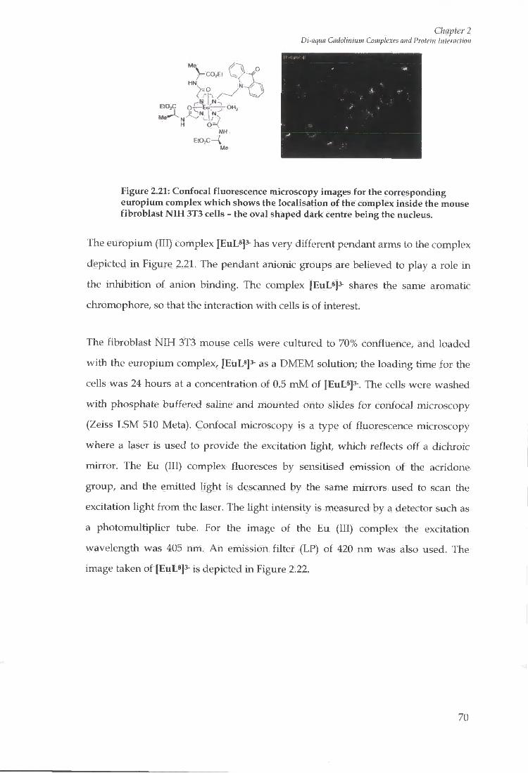
CO^t W
H 0"<
Chapter 2 Di-aqtia Gadolinium Complexes and Protein Interaction
. 1
*• r .
Figure 2.21: Confocal fluorescence microscopy images for the corresponding europium complex whicli shows the localisation of the complex inside the mouse fibroblast NIH 3T3 cells - the oval shaped dark centre being the nucleus.
The europium (III) complex [EuL*]3- has very different pendant arms to the complex
depicted in Figure 2.21. The pendant anionic groups are believed to play a role i n
the inhibit ion of anion binding. The complex [EuL*]^ shares the same aromatic
chromophore, so that the interaction wi th cells is of interest.
The fibroblast N I H 3T3 mouse cells were cultured to 70% confluence, and loaded
wi th the europium complex, [EuL^js- as a D M E M solution; the loading time for the
cells was 24 hours at a concentration of 0.5 m M of [EuL*]3-. The cells were washed
wi th phosphate buffered saline and mounted onto slides for confocal microscopy
(Zeiss LSM 510 Meta). Confocal microscopy is a type of fluorescence microscopy
where a laser is used to provide the excitation light, which reflects off a dichroic
mirror. The Eu (III) complex fluoresces by sensitised emission of the acridone
group, and the emitted light is descanned by the same mirrors used to scan the
excitation light f r o m the laser. The light intensity is measured by a detector such as
a photomultiplier tube. For the image of the Eu (III) complex the excitation
wavelength was 405 nm. A n emission filter (LP) of 420 nm was also used. The
image taken of [EuL*]'- is depicted in Figure 2.22.
71)
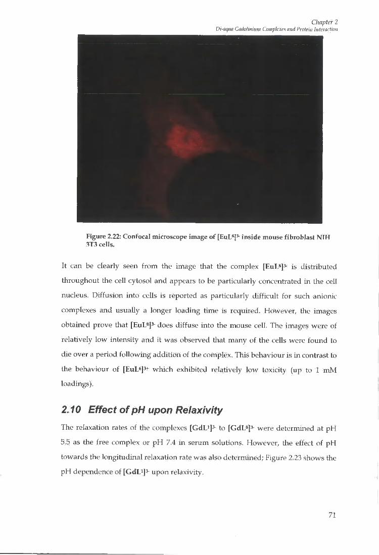
Chapter 2 Di-aqun Gadolinium Complexes and Protein Interaction
Figure 2.22: Confocal microscope image of [EuL*]^ inside mouse fibroblast NIH 3T3 cells.
It can be clearly seen f r o m the image that the complex [EuL8]3- is distributed
throughout the cell cytosol and appears to be particularly concentrated in the cell
nucleus. Diffusion into cells is reported as particularly di f f icul t for such anionic
complexes and usually a longer loading time is required. However, the images
obtained prove that [EuL^js- does diffuse into the mouse cell. The images were of
relatively low intensity and it was observed that many of the cells were found to
die over a period fol lowing addition of the complex. This behaviour is in contrast to
the behaviour of [EuL^]3+ which exhibited relatively low toxicity (up to 1 m M
loadings).
2.10 Effect of pH upon Relaxivity The relaxation rates of the complexes [GdLiJ^- to [GdL*]3- were determined at p H
5.5 as the free complex or p H 7.4 in serum solutions. However, the effect of p H
towards the longitudinal relaxation rate was also determined; Figure 2.23 shows the
p H dependence of [GdE^]^ upon relaxivity.
71

Chapter 2 Di-aqtia Gadolinium Complexes and Protein Interaction
12
10
8
Is E >. 6 >
2
5.5 6.5 7.5
PH
8.5 9.5 10
Figure 2.23: Effect of pH upon relaxivity for [GdLi]^ (310 K, 20 MHz)
The observed relaxivity typically does not change below p H 6.8, however,
increasing the p H above this value reveals a small reduction in the relaxivity value.
This suggests that competitive anion binding may be occurring to the metal ion
centre, in the f o r m of carbonate f r o m atmospheric carbon dioxide. The bidentate
carbonate anion binds to the gadolinium centre creating a ternary complex. The p H
dependence of the parent complex [Gd(aD03A)]3- was determined by Messeri.'^-*
Here a decrement was seen in the observed relaxivity beyond p H 8.5 due to
carbonate binding to the gadolinium (III) centre. This was confirmed f r o m the
luminescence of the europium (III) complex. A t physiological p H , the parent
complex, [Gd(aD03A)]3- was found to possess two inner sphere water molecules.
For complexes [GdLi]3- to [GdL*]^, where the free N H group on the cyclen r ing is
substituted, the binding of anions at physiological p H is significant. This may be
explained by a loss of the hydrogen bonding network allowing easier displacement
of bound water molecules by bidentate ligands.
2.11 aD02A based systems The network of hydrogen bonding promoted by the free N H group seen in
[Gd(aD03A)]3- may explain why the complex is less resistant to tlie binding of
bidentate anions at physiological p H , compared to the tetrasubstituted complexes
72
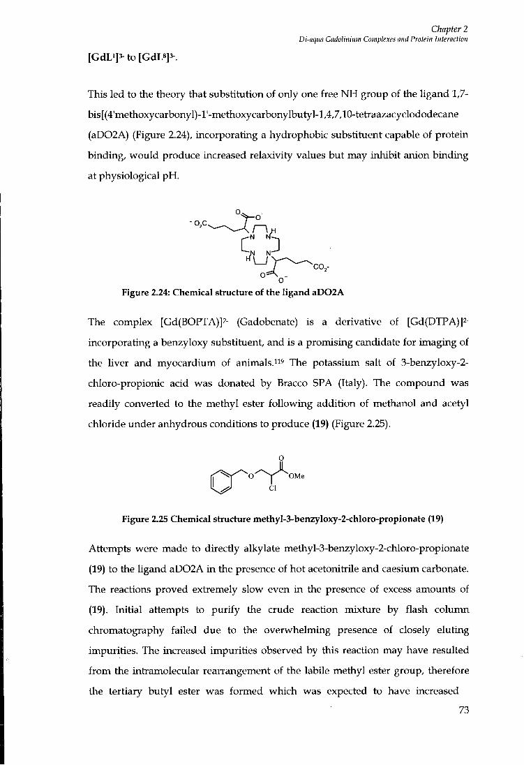
Chapter 2 Di-aqim CadoHiiium Complexes and Protein Interaction
[GdLi]3- to [GdL8]3^.
This led to the theory that substitution of only one free N H group of the ligand 1,7-
bis[(4'methoxycarbonyl)-l'-methoxycarbonylbutyl-l,4,7,10-tetraazacyclododecane
(aD02A) (Figure 2.24), incorporating a hydrophobic substituent capable of protein
binding, wou ld produce increased relaxivity values but may inhibit anion binding
at physiological p H .
•o,c
° o Figure 2.24: Chemical structure of the ligand aD02A
The complex [Gd(BOPTA)]2- (Gadobenate) is a derivative of [Gd(DTPA)]2-
rncorporating a benzyloxy substituent, and is a promising candidate for imaging of
the liver and myocardium of animals."'' The potassium salt of 3-benzyloxy-2-
chloro-propionic acid was donated by Bracco SPA (Italy). The compound was
readily converted to the methyl ester fo l lowing addition of methanol and acetyl
chloride under anhydrous conditions to produce (19) (Figure 2.25).
o
CI
Figure 2.25 Chemical structure methyl-3-benzyIoxy-2-chloro-propionate (19)
Attempts were made to directly alkylate methyl-3-benzyloxy-2-chloro-propionate
(19) to the Ugand aD02A in the presence of hot acetonitrile and caesium carbonate.
The reactions proved extremely slow even in the presence of excess amounts of
(19). Initial attempts to pur i fy the crude reaction mixture by flash column
chromatography failed due to the overwhelming presence of closely eluting
impurities. The increased impurities observed by this reaction may have resulted
f r o m the intramolecular rearrangement of the labile methyl ester group, therefore
the tertiary butyl ester was formed which was expected to have increased
73
Chapter 2 Di-aqua Gadolinium Complexes and Protein Interaction
stability. Similarly, the reaction of tert-butyl-3-benzyloxy-2-chloro-propionate w i th
aD02A proved unsuccessful. The yield of required product may be enhanced by
increasing the chemical reactivity of the leaving group of the tert-butyl ester. Tert-
butyl-3-benzyloxy-2-iodo-propionate was produced fo l lowing a Finkelsteini^o
reaction, carried out i n the presence of acetone and sodium iodide. Frustratingly,
still no reaction occurred w i t h the addition of aD02A. The initial reaction route was
repeated using an increased scale i n attempt to directly alkylate aD02A using
excess amounts of (19). Careful purification by flash column chromatography
resulted in very small yield of the required product (less than 5%). However,
subsequent hydrolysis and purification using strong cation exchange resin resulted
in the yield of product being negligible.
2.12 Suggestions for Further Work The aD02A based system still remains of interest as the binding of anions may be
significantly decreased due to the increased hydrogen bonding network. Attempts
to synthesise a similar based system fol lowing substitution of the benzyloxy
pendant arm for a more hydrophobic tri-phenyl substituent may prove more
successful.
2.13 Summary The relaxivity enhancement obtained for [GdD]3- and [GdL^]^- i n the presence of
serum protein concentrations was significant but much less than the theoretical
maximum for di-aqua gadolinium complexes. A comparative analysis of several
structurally related complexes gave no significant benefit, although an N-ethyl-
acridone complex shows some promise.
The protein binding interaction of commercially important contrast agents is
discussed later i n Chapter 6, where only modest enhancements i n relaxivity were
displayed. This stresses the significance of some of the complexes that have been
discussed here as potential contrast agents for MRI.
74

Chapter 3 Phosphonate based mncrocyclic compounds
Chapter 3
Phosphonate based macrocyclic compounds
75

Chapter 3 Pliosphonale based macroajclic compounds
3 Phosphonate based macrocyclic complexes
3.1 Introduction and Synthesis The phosphonate analogue (Figure 3.1) of the aD03A ligand (2) was identified in
Chapter 2 as a key ligand, which in its lantlianide complexes wou ld possess a
higher peripheral negative charge. In addition the dual basicity of the P(V) acid
system allows for introduction of 3 ester groups whilst retaining a (-3) peripheral
charge.
R O - P
° L J o
. y o —I II R O ' ^ R O - P
O
Figure 3.1: The Phosphonate target molecule based upon the ligand aD03A (2)
The synthetic pathway began by protection of 5-bromopentanoic acid, involving
conversion to the tert-butyl ester, fol lowing the method of Schmidt et fl/.^^i Yh^
alcohol was stirred i n oxalyl chloride to generate the acid chloride, which was, in
turn reacted w i t h tert butanol in the presence of pyridine. The pyridine acts as a
nucIeophiUc catalyst and a base preventing a high acid concentration f rom being
generated. The white pyr id in ium salts produced f r o m the reaction were filtered off
using a short column of silica gel.
'BuOH . Br " 'BuO
pyridine
Scheme 3.1: Synthesis of tert-butyl-5-(diethoxy-phosphonyl)-pentanoate (21)
The bromo functionality of the ester (21) was converted into a phosphonate ester by
heating under reflux in the presence of excess trimethylphosphite, via a Michaelis-
Arbusovi22 reaction, to yield t-butyl-5-(dimethylphosphonyl)-pentanoate as the
desired product, i n moderate yield (Scheme 3.2).
76

Chapter 3 Phosphonate based macrocyclic compounds
P(OMe) X ^ ^ t B u O ' ^ ^ - ' ' ' ^ ^ P - O M e
OMe
Scheme 3.2: 5-(diinethylphosphonate)-pentanoic acid-(tert-butyl) ester
Analysis of the reaction mixture by phosphorus NMR and electrospray mass
spectrometry revealed the presence of dimethylated products. Such behaviour is
consistent w i th reactive methyl groups i n the phosphite reagent being susceptible
to de-alkylation, fo l lowing attack of Br- by an Sn2 type mechanism. The MeBr
product then again reacts w i t h excess phosphite reagent, resulting i n the generation
of the two impurities shown i n Scheme 3.3. Formation of such impurities f r o m
Arbusov reactions has previously been reportedi23. However, these have been
found to be less prevalent w i t h triethylphosphite as a reagent.
Br- Br MeBr + P(0Me)3 Me—P-OMe Me—P-OMe
OMe OH
+ MeBr + MeBr Scheme 3.3: Generation of Arbusov reaction impurities
Accordingly, tert-butyl protected 5-bromopentanoic acid (21) was reacted wi th
excess triethylphosphite, no dealkylation products were produced and tert-butyl-5-
(diethoxy-phosphonyl)-pentanoate (22) was isolated w i t h a large increase in yield.
Deprotection of the tert-butyl ester (22) group using trifluoroacetic acid proceeded
quantitatively to yield (23). Bromination alpha to the car boxy lie acid group was
undertaken by using a Hell-VoLhard-Zelinskii24 reaction using thionyl chloride and
molecular bromine. The reaction was quenched by pouring onto cooled ethanol,
resulting in the formation of the ethyl ester, (24).
77
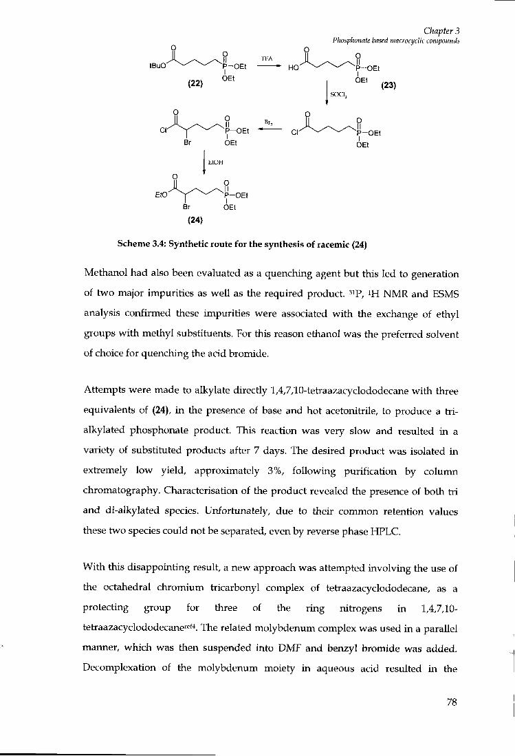
tBuG
Chapter 3 Phosphonate based niacrocijdic compounds
T F A
o
CI
Br
P - O E t
OEt
O II P - O E t I
OEt
Br,
P - O E t
P - O E t
OEt
O
EtOH
Etc Br
P-OEt I
OEt
(24)
Scheme 3.4: Synthetic route for the synthesis of racemic (24)
Methanol had also been evaluated as a quenching agent but this led to generation
of two major impurities as wel l as the required product, ^ip, i H N M R and ESMS
analysis confirmed these impurities were associated wi th the exchange of ethyl
groups w i t h methyl substituents. For this reason ethanol was the preferred solvent
of choice for quenching the acid bromide.
Attempts were made to alkylate directly 1,4,7,10-tetraazacyclododecane w i t h three
equivalents of (24), in the presence of base and hot acetonitrile, to produce a t r i -
alkylated phosphonate product. This reaction was very slow and resulted in a
variety of substituted products after 7 days. The desired product was isolated in
extremely low yield, approximately 3%, fo l lowing purification by column
chromatography. Characterisation of the product revealed the presence of both t r i
and di-alkylated species. Ur^fortunately, due to their common retention values
these two species could not be separated, even by reverse phase HPLC.
Wi th this disappointing result, a new approach was attempted involving the use of
the octahedral chromium tiicarbonyl complex of tetraazacyclododecane, as a
protecting group for three of the r ing nitrogens in 1,4,7,10-
tetraazacyclododecanef'^f'*. The related molybdenum complex was used in a parallel
manner, which was tlien suspended into DMF and benzyl bromide was added.
Decomplexation of the molybdenum moiety i n aqueous acid resulted i n the
78

Chapter 3 Phosphonate based macroajclic compounds
isolation of the monoalkylated amine. Alkylat ion of the remaining three nitrogens
of the cyclen r ing was attempted by adding excess (24) i n the presence of dry
acetonitrile and base, and heating under reflux. Again, attempts were made to
pur i fy the crude reaction mixture, to isolate the desired tetra-substituted product,
f r o m which the protecting group could be removed fo l lowing hydrogenation.
Unfortunately, the desired product was never isolated f r o m its major impurities.
The next plan of attack involved slightly modify ing the method of Yang et fl/i^s
which involved the reaction of three equivalents of ethyl trifluoroacetate in the
presence of methanol and tiiethylamine to produce a protected, tiisubstitued
cyclen. 1,4,7,10 tris-trifluoromethyl carbonyl-12-N-4 (25) was isolated in respectable
yield of 80%. The required product was isolated by passing through a short silica
gel column. The tri-substituted product has a much higher Rf value than any of the
other related species. Subsequent alkylation of the free N H group of the
macrocycle, using BOC anhydride was achieved in methanol (26). Again, a high
yield of material was obtained fo l lowing purification by column chromatography.
Stiaightforward deprotection of the ethyl trifluoroacetate arms was carried out in
the presence of potassium hydroxide, to yield the monocarbamate (27). Excess
amounts of (24) were added to (27) in the presence of caesium carbonate as base
and hot acetonitiile. The required product (28) was isolated fol lowing careful
purification by flash column chromatography w i t h a low reaction yield of 22%. The
BOC protecting group was removed using tiifluoroacetic acid i n the presence of
dichloromethane to yield (29) quantitatively (Scheme 3.5).
79
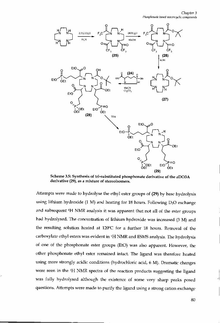
Chapter 3 Phosphonate based macroajclic compounds
O
_ - N N l - ^ (CF,co)p F3C PN ( B O C > P F3C J ^ N N--^ / C
N."'^ N"^ '^^^
" W L j y o C F , C F , C F , C F ,
(25) (26)
o (24) o EtO ^ " P - O E t
N N MeCN Cs^CO,
I OEt E t c I
E t O - P N N
N N
I OEt E t c I
Scheme 3.5: Synthesis of tri-substituted phosphonate derivative of the aD03A derivative (29), as a mixttu-e of stereoisomers.
Attempts were made to hydrolyse the ethyl ester groups of (29) by base hydrolysis
using l i th ium hydroxide (1 M) and heating for 18 hours. Following D2O exchange
and subsequent NMR analysis it was apparent that not all of the ester groups
had hydrolysed. The concentration of Uthium hydroxide was increased (3 M) and
the resulting solution heated at 120''C for a further 18 hours. Removal of the
carboxylate ethyl esters was evident i n NMR and ESMS analysis. The hydrolysis
of one of the phosphonate ester groups (EtO) was also apparent. However, the
other phosphonate ethyl ester remained intact. The ligand was therefore heated
using more strongly acidic conditions (hydrochloric acid, 6 M ) . Dramatic changes
were seen in the ^H N M R spectra of the reaction products suggesting the ligand
was fu l ly hydrolysed although the existence of some very sharp peaks posed
questions. Attempts were made to pur i fy the ligand using a strong cation exchange
80

Chapter 3 Phosphonate based macroq/clic compounds
resin (Dowex 50 W). The product was not retained on the column, eluting quickly
in water. This product was analysed by ^H and " C N M R where i t was obvious
f r o m a reduction in the number of carbon signals and no resonance to represent the
(NCH2) groups of the cyclen ring that the complex had degraded. A t this early
point i t was imclear whether the problems had occurred during acid hydrolysis or
base hydrolysis so the reactions were repeated. The reaction of (29) w i th l i th ium
hydroxide (0.5 M) fol lowed by gentle heating (60°C, 18 hours) confirmed
degradation of the product was occurring at this stage. In the presence of increased
concentration of base and increased temperatures the yield of this imdesirable
product wou ld have been more significant. Attempts were made to directly
hydrolyse all ethyl ester groups of (29) using hydrochloric acid (6 M) upon heating
under reflux for 18 hours. ^H N M R confirmed some ethyl signals remained of
which no change was observed fol lowing further heating for another 18 hours.
Earlier work wi th in the Parker groupies and the work of Sherry et al*^ for
phosphinate hydrolysis employed the stronger acid, hydrogen bromide in acetic
acid. The reaction conditions were tested for the hydrolysis of aU ethyl ester groups
of (24). The Ugand (24) was heated at 100°C for 18 hours i n the presence of excess
hydrogen bromide acetic acid solution. The mixture was allowed to cool to room
temperature and added dropwise into a cooled solution of diethyl ether; some
precipitate formed. The diethyl ether was removed vmder reduced pressure. The
acetic acid was removed f r o m the solution as its toluene azeotrope. i H N M R
analysis confirmed fuU hydrolysis of the Ugand had occurred. The cyclen derivative
wi th phosphonate peripheral arms (29) was fu l l y hydrolysed upon heating for 48
hours i n the presence of hydrogen bromide solution i n acetic acid. The p H of the
acidic solution was increased to p H 5.5 and complexation of the europium (III) and
gadolinium (III) complexes was carried out, as in Scheme 3.6
81

Chapter 3 Phosphonate based macrocyclic compounds
EtO—P
E t c — P
H B r / A c e t i c acid
OEt
H O - P
N +HN
NH+ N
H O - P HO—P
LnCla-GHjO
pH 6.0
H O — P
O
O,
N 0 4 - ;Ln ho
V - o
H O - P H O - P [LnL9]3
Scheme 3.6: Synthetic route for the complexation of [L']
3.2 Relaxivity Measurements The relaxivity of the gadolinium complex [GdL']^- was measured at 60 M H z and
310 K. The p H of the complex i n solution was 6.8 and the relaxivity value obtained
was 7.3 mM-is-i. This is similar in magnitude to previously studied lanthanide
complexes possessing two inner sphere water molecules at the lanthanide (111)
centre, suggesting that [ G d L ' ] ^ is a di-aqua species.
The N M R D profile for the complex [GdL']^- was measured i n water at p H 6.8,
between the magnetic fields 0.01 to 20 M H z in Durham at three different
temperatures 288 K, 298 K and 310 K (Figure 3.2). The profile was found to be
typical of a di-aqua gadoliniumi'^ complex and resembled that obtained for
[Gd(aD03A)(H20)2]3-.37.38
82

Chapter 3 Phosphonate based macrocyclic compounds
30
20
.> X
• 288 K • 298 K
310 K
100 10.00
Magnetic Field IMHz 100.00
Figure 3.2 NMRD profile for [GdL'p at 288 K, 298 K and 310 K operating at the magnetic fields 0.01 to 20 MHz. An additional point for the relaxivity of [GdL»]^ at 60 MHz, 310 K is added.
To assess the characteristics of complex behaviour i n solution, the relaxivity of
[GdL9] was measvu-ed as a f imct ion of p H at 60 M H z , 310 K (Figure 3.3.). The
relaxivity values were found to be constant across the p H range 3.5 to 6.
(A S
E
> '5 (0
1 ^
pH
Figure 3.3: Relaxivity as a function of pH for [GdL']*^
83
Chapter 3 Phosphonate based macroajclic compounds
As the p H was increased beyond p H 6 there was an obvious decrease in the
observed relaxivity. This may result f r o m the di-aqua complex being susceptible to
the binding of dissolved carbonate i n solution deriving f r o m atmospheric carbon
dioxide at increased p H . Alternatively, the reduction may be associated w i t h
deprotonation of the three phosphonate O H groups i n the side-arms, although the
apparent protonation constant observed (at 6.8) is slightly lower than expected for
such systems. The marked change in relaxivity may also be related to a large
variation in the second sphere contribution to the observed relaxivity and/or the
onset of 'intermolecular' phosphonate Ligation. This latter effect wou ld be expected
to be concentration, p H and time dependent.
The reversibiUty of this p H dependence was examined, but a time-dependent
phenomenon was observed. Using a sample that had aged in solution for 2 days,
the p H was increased; particulate matter was found to precipitate out of solution.
The amormt of precipitate formed was observed to be time-dependent, suggesting
some f o r m of oligomerisation reaction was occurring. Oligomer formation is
concentiation dependent so the relaxation rate of the complex was measured as a
function of complex concentiation (Figure 3.4) for a freshly prepared sample of the
complex. The linearity of the paramagnetic relaxation rate versus concentiation plot
(Figure 3.4) is not i n accord w i t h oligomerisation at p H 6.8, on the experimental
timescale used (< 2 hours).
84

Chapter 3 Phosphonate based macrocyclic compounds
r-
' M 1.
c
atii
0.
X « 0. a: 0.
0 0.05 0.1 0.15 0.2 0.25
Concentration of [GdL*]^" (mM)
Figure 3.4: Concentration dependency of relaxivity for (GdL']'- (pH 6.8, freshly prepared solution
3.3 Semi quantitative assessment of anion effect on relaxivity
As i t was beUeved that anions were binding to the gadolinium centre causing a
reduction in the observed relaxivity, the relaxivity of [GdL']^ was recorded as a
fimction of the concentration of biological anion background. The simulated anion
background solution contained carbonate (30 mM) , phosphate (0.9 mM), citiate
(0.13 m M ) and lactate (2.3 mM) , and the p H of the added anion solution was 7.3.
The complex [GdL']^- was taken as a solid and dissolved into the anion background
solution. The longitudinal relaxation time, Ti of the solution was measured but the
value was extremely long similar to that of pure water solutions containing no
paramagnetic material. Aliquots (20 pi) of the anion solution were also added to a
solution of [GdL9]3- and the relaxivity was measured at 60 MHz, 310 K. Instantly,
upon addition of anions the relaxivity was found to decrease significantly
suggesting complete displacement of bound water molecules by competing anions.
Previous research wi th in the Parker group has shown the most common anions that
are found to displace inner sphere water molecules f r o m lanthanide (111) complexes
are phosphate, lactate and carbonate.i5-i6-"2,i27 Xo assess which anion was most
85

Chapter 3 Pliosphonate based mncrocydic compounds
prominent i n the displacement of inner sphere water molecules, carbonate (sodium
hydrogen carbonate, to a l imi t ing p H of 8.7), lactate (D-Lactic acid sodium salt
solution, p H 7.7), and phosphate (di-sodium hydrogen phosphate to a l imi t ing p H
of 8.6), were added individually i n large excess, as solids to solutions containing the
same concentration of [GdL']^. The relaxivity was then measured at 60 M H z , 310 K
(Table 3.1).
Anion Relaxivity mM-is-i (60 MHz, 310 K)
no anions 7.9
hydrogencarbonate 4.9
lactate 8.2
hydrogenphosphate 3.0
Table 3.1: Relaxivity values obtained depending on anion used
The results show that carbonate and phosphate both lead to relaxivity reductions
consistent w i t h the displacement of water molecules f r o m the gadolnium (III)
centre. However, i t was noted that i n the presence of hydrogenphosphate the
relaxivity value reduces to a value consistent w i t h that of purely outer sphere
effects. Lactate is not found to compete w i t h inner sphere water molecules even
when the concentration of lactate present i n solution is significantly increased. The
relaxivity value measured for [GdL']3- is actually slightly higher i n the presence of
lactate than i t is as the free complex.
3.4 Protein Binding Studies
The relaxivity of the carboxylate analogue [Gd(aD03A)]3- of the complex [GdL']3-
was not found to be significantly enhanced i n the presence of serum albumin.
However, the phosphonate analogue may exhibit differ ing behaviour as the
solution speciation of the complex may not be the same. In the presence of human
serum albumin at a concentration of (0.6 mM) , similar to in-vivo concentration, the
relaxivity is dramatically enhanced f r o m 7.9 mM-is-i to 43.5 m M 's- . The protein
binding study was repeated for a solution containing a lower concentration of
complex [GdL9]3-. The results revealed a relaxivity enhancement f r o m 8.5 to 30.2
86
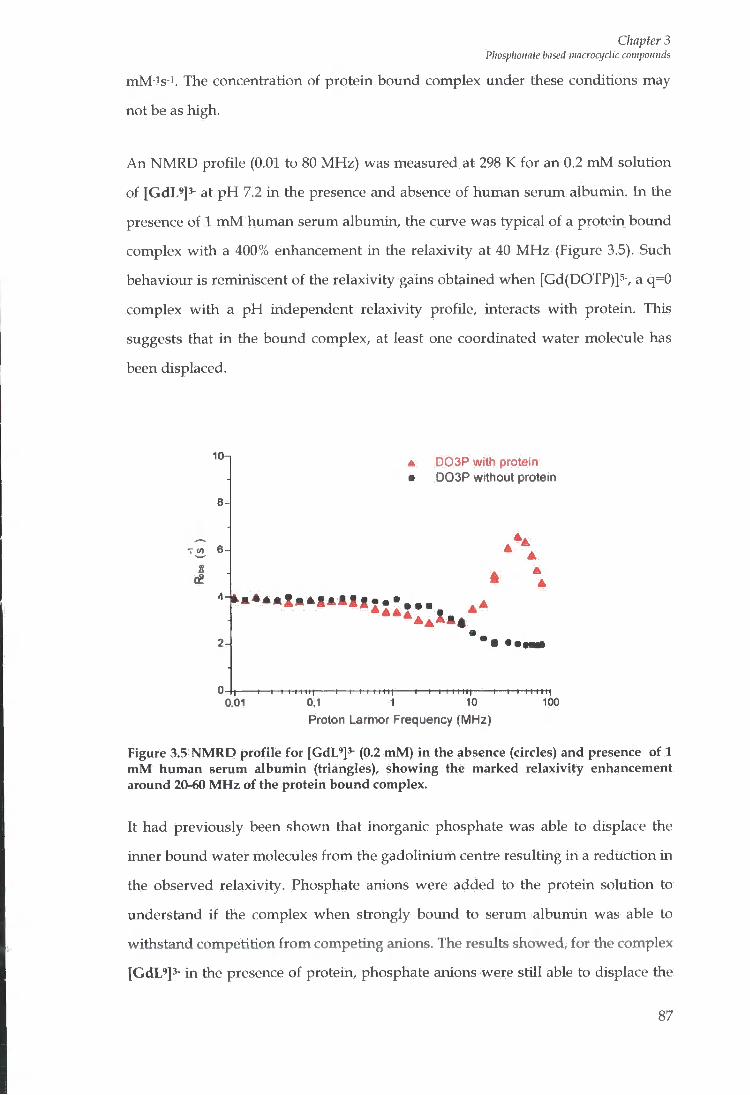
Chapter 3 Phosphoiiate based macroajdic compounds
mM-is-1. The concentration of protein bound complex under these conditions may
not be as high.
A n N M R D profile (0.01 to 80 MHz) was measured at 298 K for an 0.2 m M solution
of [GdL ' ]3- at p H 7.2 i n the presence and absence of human serum albumin. In the
presence of 1 m M human serum albumin, the curve was typical of a protein bound
complex w i t h a 400% enhancement in the relaxivity at 40 M H z (Figure 3.5). Such
behaviour is reminiscent of the relaxivity gains obtained when [Gd(DOTP)]5-, a q=0
complex wi th a p H independent relaxivity profile, interacts w i t h protein. This
suggests that i n the bound complex, at least one coordinated water molecule has
been displaced.
10n
iit 6
J
2A
A D03P with protein • D03P without protein
0,01 -1—I I 111 I I I -1 1—I I I I M | -1—I I I I I I I
0,1 1 10 Proton Larmor Frequency (MHz)
- I — I I 111111 1 0 0
Figure 3.5 NMRD profile for [GdL']^- (0.2 mM) in the absence (circles) and presence of 1 mM human serum albumin (triangles), showing the marked relaxivity enhancement around 20-60 MHz of the protein bound complex.
It had previously been shown that inorganic phosphate was able to displace the
inner bound water molecules f r o m the gadolinium centre resulting in a reduction in
the observed relaxivity. Phosphate anions were added to the protein solution to
understand if the complex when strongly bound to serum albumin was able to
withstand competition f r o m competing anions. The results showed, for the complex
[GdL'l^- in the presence of protein, phosphate anions were still able to displace the
87
Chapter 3 Pliosphoimte based mncroajdic compounds
inner sphere water molecules resulting in a large reduction in the observed
relaxivity.
3.5 Conclusions
[GdL']^ was synthesised and characterised successfully as a di-aqua complex.
Unlike [Gd(aD03A)]3- i t does show a marked PRE effect i n binding serum
albumin, similarly to [Gd(DOTP)]5- . However, i t is sensitive to substitution of the
coordinated waters by common added anions, hence reducing the observed
relaxivity.
88
Chapter 4 Siilphonamide Bused Systems
Chapter 4
Sulphonamide Based Systems
89
Chapter 4 Sulphoimmide Based Systems
4 Sulphonamide Based Systems
4.1 Introduction
Human serum albumin (HSA) is a 67 kDa protein and is the most abimdant protein
in blood plasma, typically present at a concentration of 0.7 m M . It consists of three
structurally homologous, largely helical (67%) domains I , I I , and I I I . Each domain is
further divided into two sub-domains denoted A and B.'s The main physiological
role of HSA deals w i t h the transport of a huge number of substrates. Most
information regarding the binding properties of drugs and substrates has been
identified on the basis of extensive competitive assays. - ^ i t was apparent f r o m
previous research that serum albumin contains many metal binding sites w^hich
have different binding specificities, the most common belonging to Ni^*, Cu^* and
Zn2+. I t was we l l established that HSA contained binding sites that have a high
affini ty for zinc yet unt i l recently very little was known about the whereabouts of
this binding pocket. Stewart et fl/i28 proposed, fo l lowing detailed X-ray
crystallography studies, that the zinc site lies at the interface of the domains I and
I I , and is essential i n the tiansport of the metal ion and its delivery to cells and
tissues.
4.2 Zinc
Zinc is essential for many physiological processes, such as the development and
function of all cells, maintaining the immune system and transmission of genetic
information. The concentiation of Zn2+ i n blood plasma is approximately 19 | i M ,
most of which is bound to serum albumin w i t h an affini ty constant of log Ka = -
7.53.129
4.3 Sulptionamides
Previously the simple pyr idy l based tiifluoromethanesulphonamides systems (1)
and (2) (Figure 4.1) were identified as suitable zinc (II) binding ligands, capable of
forming ML2 complexes at ambient p H when the free concentiation of zinc is i n the
micro to nanomolar range.i^o
90

Chapter 4 Sulphonamide Based Systems
H N . I S - C F
Figure 4.1: Chemical structure of simple pyridyl based trifluoromethanesulphonamides ligands o
The ligands were incorporated into various macrocyclic ligands wi th the intention
of increasing the aff ini ty of zinc for such a system, which may produce a suitable
derivative that can be integrated into practical luminescent or pZn sensitive M R I
probes.
The pyr idy l trifluoromethanesulphonamide moiety was incorporated into the
macrocyclic system using an ethyl or a propylamide group linkage. The pendant
arms of the macrocyclic system also differed, which affected the peripheral charge
(Figure 4.2).
R O — ^ R = H
N - S - C F , H I I '
0
n = 1 or 2
Figure 4.2: Pyridyl trifluoromethanesulphonamide lanthanide complex
The lanthanide (111) complexes [1] to [4] (Figure 4.3) were synthesised and
characterised by Aileen Congreve^^o w i t h proposed binding affinities for zinc. Due
to the high concentration of zinc found in human serum albumin it was anticipated
that these complexes may bind to the serum protein w i t h a relatively high affinity.
91

Chapter 4 Sulphoiiamide Based Systems
V o V° ° O
•O "O
(1) ^ (2)
, o 9, / . 0 9. 0 -
^ N ^ I O H N - S - C F 3 " - N I ^ N ^
^ o N -
C F , H
o (3) o W
Figure 4.3: Chemical structures of the lanthanide complexes [1] to [4]
4.4 Protein Binding Studies
Complexes [1] to [4] were first synthesised and characterised by A. Congreve in
Durham. The relaxivities of the free gadolinium complexes [1] to [4] were
determined at 60 MHz , 310 K (Table 4.1). The observed longitudinal relaxivity
values for complexes [1] and [2] were much lower than those for complexes [3] and
[4]. This was a consequence of the hydration state of the complex. The D 0 3 A based
systems [1] and [2] contain only one inner sphere water molecule, which is
consistent w i t h their lower relaxivity values and was confirmed by luminescence
studies of the europium analogue. The relaxivity values obtained for complexes [3]
and [4] based upon aD03A (2) were significantly higher than for [1] and [2],
indicating that they are di-aqua based systems, similar to the parent complex
[Gd(aD03A)]3-.
92
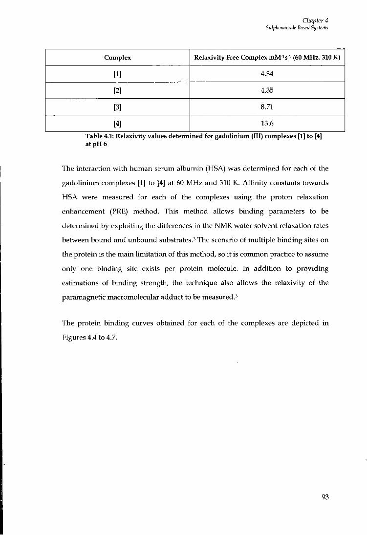
Chapter 4 Snlphoiiamide Based Systems
Complex Relaxivity Free Complex mM-is-i (60 MHz, 310 K)
[1] 4.34
[2] 4.35
[3] 8.71
[4] 13.6
Table 4.1: Relaxivity values determined for gadolinium (III) complexes [1] to [4] at pH 6
The interaction w i t h human serum albumin (HSA) was determined for each of the
gadolinium complexes [1] to [4] at 60 M H z and 310 K. Af f in i t y constants towards
HSA were measured for each of the complexes using the proton relaxation
enhancement (PRE) method. This method allows binding parameters to be
determined by exploiting the differences i n the N M R water solvent relaxation rates
between boimd and imbound substiates.^ The scenario of multiple binding sites on
the protein is the main limitation of this method, so i t is common practice to assume
only one binding site exists per protein molecule. In addition to providing
estimations of binding stiength, the technique also allows the relaxivity of the
paramagnetic macromolecular adduct to be measured.^
The protein binding curves obtained for each of the complexes are depicted in
Figures 4.4 to 4.7.
93
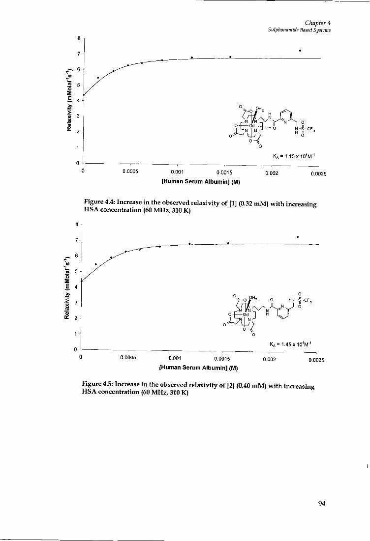
Chapter 4 Siilphoiwmide Based Systems
N - S - C F " O
K A = 1 . 1 5 X 1 0 ' M
0.0005 0.0025 0.001 0 . 0 0 1 5
[Human Serum Albumin] (M) 0.002
Figure 4.4: Increase in the observed relaxivity of [1] (0.32 mM) with increasing HSA concentration (60 MHz, 310 K)
o S E.
.5 > a
p H , o O H N - S - C F , I! N .
- G D 1 H
O
K A = 1 . 4 5 X 1 0 V
0.0005 0.001 0 . 0 0 1 5
[Human Serum Albumin] (IVI) 0.002 0.0025
Figure 4.5: Increase in the observed relaxivity of [2] (0.40 mM) with increasing HSA concentration (60 MHz, 310 K)
94

Chapter 4 Sulphonamide Based Systems
a 30
o 4 — G D - 4 . . , ^ N - S - C F , H ^
O ^ 0 0
0.0005 0.001 0 .0015
[Human Serum Albumin] (M) 0.002 0.0025
Figure 4.6: Increase in the observed relaxivity of [3] (0.25 mM) with increasing HSA concentration (60 MHz, 310 K)
E 1 5
> « 1 0 a>
0 HN-S -CF ,
KA = 8 .55X10 ' 'M" '
0 .0005 0.001 0 . 0 0 1 5
[Human Serum Albumin] (IM) 0.002 0.0025
Figure 4.7: Increase in the observed relaxivity of [4] (0.12 mM) with increasing HSA concentration (60 MHz, 310 K)
The binding studies show an increase in the observed relaxivity for each of the
complexes upon addition of HSA (Table 4.2). The results were significantly higher
for the negatively charged complexes based on the parent compoimd,
[Gd(aD03A)]^, compared to the neutrally charged D 0 3 A based systems. As i t is
wel l established that protein binding requires hydrophobic substituents and
negatively charged groupss this may explain the much larger enhancement towards
95
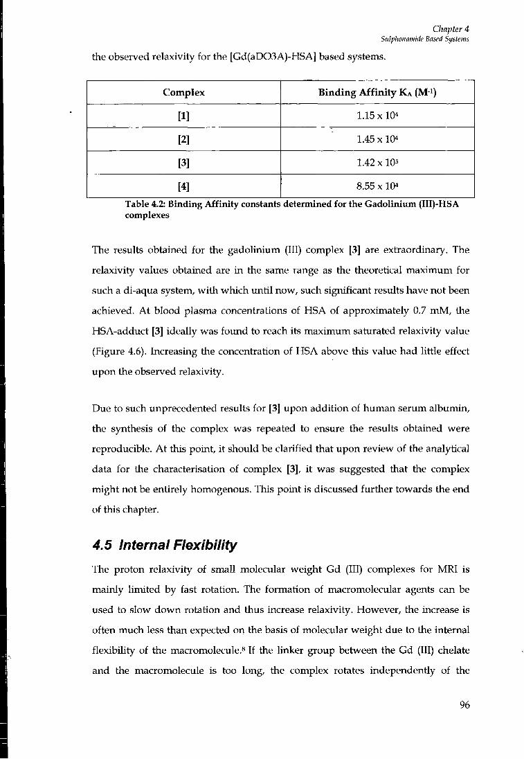
Chapter 4 Sulphonamide Based Systems
the observed relaxivity for the [Gd(aD03A)-HSA] based systems.
Complex Binding Affinity K A (M- i )
[1] 1.15 X 10⁴
[2] 1.45x10⁴
[3] 1.42x10⁵
[4] 8.55 x 10⁴
Table 4.2: Binding Affinity constants determined for the Gadolinium (III)-HSA complexes
The results obtained for the gadoUnium (III) complex [3] are extraordinary. The
relaxivity values obtained are i n the same range as the theoretical maximum for
such a di-aqua system, w i t h which unt i l now, such significant results have not been
achieved. A t blood plasma concentrations of HSA of approximately 0.7 m M , the
HSA-adduct [3] ideally was fo imd to reach its maximum saturated relaxivity value
(Figure 4.6). Increasing the concentration of HSA above this value had little effect
upon the observed relaxivity.
Due to such unprecedented results for [3] upon addition of human serum albumin,
the synthesis of the complex was repeated to ensure the results obtained were
reproducible. A t this point, i t should be clarified that upon review of the analytical
data for the characterisation of complex [3], i t was suggested that the complex
might not be entirely homogenous. This point is discussed further towards the end
of this chapter.
4.5 Internal Flexibility
The proton relaxivity of small molecular weight Gd (III) complexes for M R I is
mainly l imited by fast rotation. The formation of macromolecular agents can be
used to slow down rotation and thus increase relaxivity. However, the increase is
often much less than expected on the basis of molecular weight due to the internal
flexibihty of the macromolecule.^ If the linker group between the Gd (III) chelate
and the macromolecule is too long, the complex rotates independently of the
96
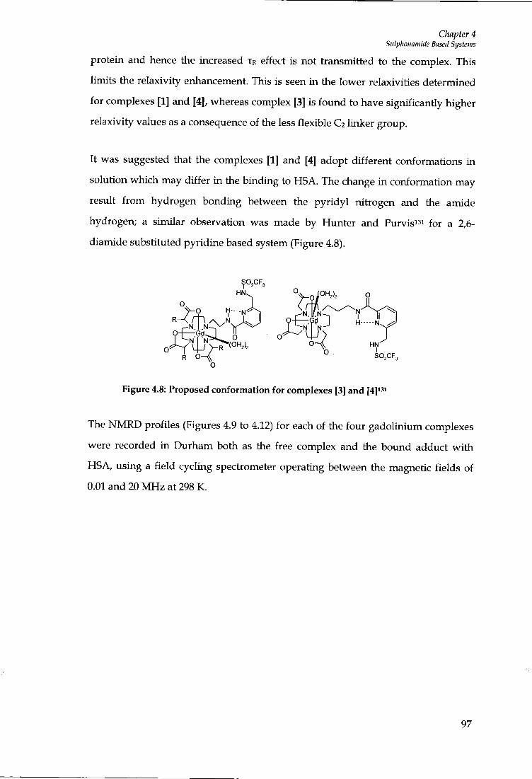
Chapter 4 Sulplioiiamide Based Systems
protein and hence the increased T R effect is not transmitted to the complex. This
limits the relaxivity enhancement. This is seen in the lower relaxivities determined
for complexes [1] and [4], whereas complex [3] is found to have significantly higher
relaxivity values as a consequence of the less flexible Ca linker group.
It was suggested that the complexes [1] and [4] adopt different conformations in
solution which may differ i n the binding to HSA. The change in conformation may
result f r o m hydrogen bonding between the pyr idy l nitrogen and the amide
hydrogen; a similar observation was made by Hunter and Purvis" ' for a 2,6-
diamide substituted pyridine based system (Figure 4.8).
° k o - i ° SO,CF3
o
Figure 4.8: Proposed conformation for complexes [3] and
The N M R D profiles (Figures 4.9 to 4.12) for each of the four gadolinium complexes
were recorded i n Durham both as the free complex and the bound adduct w i th
HSA, using a f ie ld cycling spectrometer operating between the magnetic fields of
0.01 and 20 M H z at 298 K.
97

Chapter 4 Sulphonamide Based Systems
E
5 15
Proton Latmor Frequency (MHz)
Figure 4.9: NMRD profile of [1] in the presence n and absence A of HSA at 298 K
™ 15.0
'0 10.0
Proton Lannor Frequency (MHz)
Figure 4.10: NMRD profile of [2] in the presence • and absence A of HSA at 298 K
The relaxivity of the D 0 3 A based systems [1] and [2] was doubled on formation of
an adduct w i t h HSA (Figures 4.9 and 4.10). As the N M R D profile was only
measured to 20 MHz , the additional point obtained at 60 M H z was added. This
gave rise to the typical high f ield relaxivity peak in the region of 20-60 MHz,
resulting f r o m an increased T R effect. -'*''* The N M R D profiles show the T R effect is
greater for the more r igid spacer (C2) for the Gd (III) complex [1] due to more
98

Chapter 4 Sidplwiiamide Based Systems
efficient coupling of the complex w i t h respect to the macromolecule. This
behaviour was also mirrored in the aD03A based systems, suggesting the C 3 linker
allows too much rotational freedom, l imit ing the observed relaxivity.
Proinn I armnr Frfiniifinr.u ^MH7^
Figure 4.11: NMRD profile of [3] in the presence • and absence A of HSA at 298 K
' E 25-00
i .> 20.00
o Ct 15.00
* * A A A
Prnton I armor Frfiniifinnv ^MH7^
Figure 4.12: NMRD profile of [4] in the presence • and absence A of HSA at 298 K
The Solomon-Bloembergen-Morgan theory predicts that gadolinium (III) complexes
possessing two inner sphere water molecules and optimised values of rotation,
election paramagnetic relaxation and water exchange times, can have maximum
99
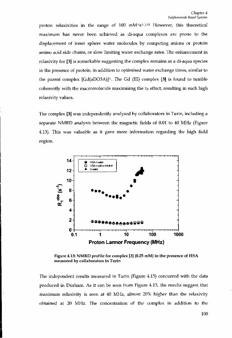
Chapter 4 Sulphonamide Based Systems
proton relaxivities i n the range of 100 mM-^s-^.^io However, this theoretical
maximum has never been achieved as di-aqua complexes are prone to the
displacement of inner sphere water molecules by competing anions or protein
amino acid side chains, or slow l imit ing water exchange rates. The enhancement i n
relaxivity for [3] is remarkable suggesting the complex remains as a di-aqua species
i n the presence of protein, in addition to optimised water exchange times, similar to
the parent complex [Gd(aD03A)]3-. The Gd (III) complex [3] is found to tumble
coherently w i t h the macromolecule maximising the T R effect, resulting in such high
relaxivity values.
The complex [3] was independently analysed by collaborators i n Turin, including a
separate N M R D analysis between the magnetic fields of 0.01 to 40 M H z (Figure
4.13). This was valuable as i t gave more information regarding the high field
region.
14
12H
10
i 8-|
6
4
2
T 0^
I I I 1 1 1 1
9 HSAinweter
O HSA in saline sdu6on •
••••••
10 100 1000 0.1 1 Proton Larmor Frequency (MHz)
Figure 4.13: NMRD profile for complex [3] (0.25 mM) in the presence of HSA measured by collaborators in Turin
The independent results measured in Tur in (Figure 4.13) concurred w i t h the data
produced in Durham. As i t can be seen f r o m Figure 4.13, the results suggest that
maximum relaxivity is seen at 40 MHz , almost 20% higher tiian the relaxivity
obtained at 20 MHz. The concentiation of the complex in addition to the
100
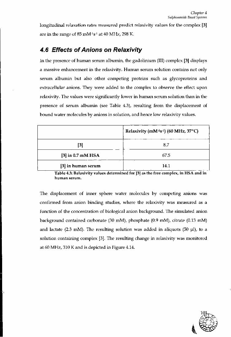
Chapter 4 Siilphonamide Based Systems
longitudinal relaxation rates measured predict relaxivity values for the complex [3]
are i n the range of 85 mM - i s - i at 40 M H z , 298 K.
4.6 Effects of Anions on Relaxivity
I n the presence of human serum albumin, the gadolinium (III) complex [3] displays
a massive enhancement i n the relaxivity. Human serum solution contains not only
serum albumin but also other competing proteins such as glycoproteins and
extracellular anions. They were added to the complex to observe the effect upon
relaxivity. The values were significantly lower i n human serum solution than in the
presence of serum albumin (see Table 4.3), resulting f r o m the displacement of
bound water molecules by anions i n solution, and hence low relaxivity values.
Relaxivity (mM-is-i) (60 MHz, 37" C)
[3] 8.7
[3] in 0.7 m M H S A 67.5
[3] in human serum 14.1
Table 4.3: Relaxivity values determined for [3] as the free complex, in HSA and in human serum.
The displacement of inner sphere water molecules by competing anions was
confirmed f r o m aruon binding studies, where the relaxivity was measured as a
function of the concentiation of biological anion background. The simulated anion
background contained carbonate (30 mM) , phosphate (0.9 mM) , citrate (0.13 mM)
and lactate (2.3 mM) . The resulting solution was added in aliquots (50 pi) , to a
solution containing complex [3]. The resulting change in relaxivity was monitored
at 60 MHz, 310 K and is depicted in Figure 4.14.
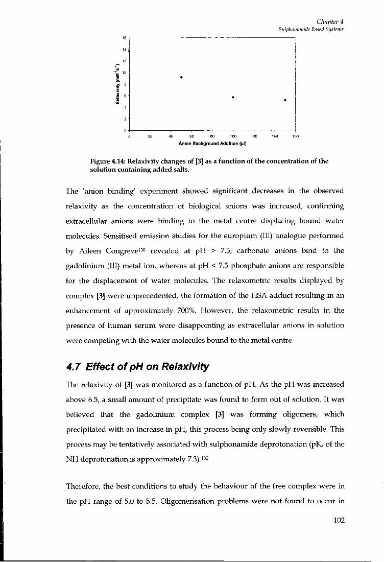
Chapter 4 Sulphoimwide Based Systems
40 60 80 100 120
Anion Background Addition ()J)
Figure 4.14: Relaxivity changes of [3] as a function of the concentration of the solution containing added salts.
The 'anion binding' experiment showed significant decreases in the observed
relaxivity as the concentration of biological anions was increased, confirming
extracellular anions were binding to the metal centre displacing bound water
molecules. Sensitised emission studies for the europium (III) analogue performed
by AUeen Congrevei-''o revealed at pH > 7.5, carbonate anions bind to the
gadolinium (III) metal ion, whereas at p H < 7.5 phosphate anions are responsible
for the displacement of water molecules. The relaxometric results displayed by
complex [3] were unprecedented, the formation of the HSA adduct resulting in an
enhancement of approximately 700%. However, the relaxometric results i n the
presence of human serum were disappointing as extracellular anions in solution
were competing wi th the water molecules bound to the metal centre.
4.7 Effect of pH on Relaxivity
The relaxivity of [3] was monitored as a function of p H . As the p H was increased
above 6.5, a small amoimt of precipitate was found to fo rm out of solution. I t was
believed that the gadolinium complex [3] was forming oligomers, which
precipitated w i t h an increase in p H , this process being only slowly reversible. This
process may be tentatively associated w i t h sulphonamide deprotonation (pKa of the
N H deprotonation is approximately 7.3)."2
Therefore, the best conditions to study the behaviour of the free complex were in
the p H range of 5.0 to 5.5. Oligomerisation problems were not found to occur i n
102
Chapter 4 Siilphoiinwide Based Systems
protein solutions as the complex is solubilised; no significant pH variation was seen
in the range 5.5 to 7.5.
The tendency to forrr\ oligomers is also inhibited in the presence of competing
anions but the observed relaxivity is low^ered as a consequence. No problems
associated with oUgomer formation was seen for systems with C 3 linker groups, [2]
and [4] and the C2 complex with acetate arms [1] rather than carboxylate arms.
4.8 Derivatives of Complex [3]
Three Ugands were synthesised with respect to the chemical structure of [3] with
the intention of assessing which part of the molecule was responsible for the
significant binding affinity for HSA.
The trifluorosulphonamide functional group was also substituted for a less
hydrophobic trimethanesulphonamide, which also has a much higher pKa value, so
that problems associated with oUgomerisation may be suppressed.
4.9 Synthesis of derivatives
The synthesis of L^" was carried out using the established synthetic methods
reported by Congrevei30432 (Scheme 4.1).
103

Chapter 4 Stilphoimmide Based Systems
HO OHE'OH.H- NaBH.
° (30, °
. 0 ^ ^ ^ ^OH CH,SO,CI N
(31) NaNj, nBUjNCI
Hj. Pd/C BoCjO EtOAc
(32)
B r ' N ^boc CBr,, PPh,
HO
(35) cyclen MeCN
H H ^N^
N ^boc
CH,CL
H H0(CHj)2NHj
^boc "boc
(34) (33)
MeO
(36)
N N
OMe
(38)
Scheme 4.1: Synthetic Route for the production of compound 38
Pyridine-2,6-dicarboxylic acid was converted into diethyl-pyridine-2,6-
dicarboxylate (30) fo l lowing esterification w i t h anhydrous ethanol. The product
was reduced to yield the alcohol (31) fo l lowing reaction w i t h sodium borohydride,
which was subsequently converted to the azide (32). The azide was hydrogenated
using palladium on carbon catalyst, using 40 psi hydrogen gas using a Parr
hydrogenator apparatus. The resulting amide was protected using BOC anhydride
i n the same reaction pot to produce (33) in moderate yield. EDC coupling of (33)
w i t h ethanolamine resulted in formation of the hydroxy amide (34). This was
converted into the corresponding bromide (35), fo l lowing reaction w i t h carbon
tetrabromide and triphenylphosphine in dichloromethane. MonoaLkylation of
cyclen was achieved by reaction of (35) w i t h cyclen i n large excess, i n chloroform.
The product was extracted f r o m dichloromethane to yield (36). Alkylat ion of tlie
three remaining r ing nitrogen atoms was achieved by stirring (36) i n the presence of
104
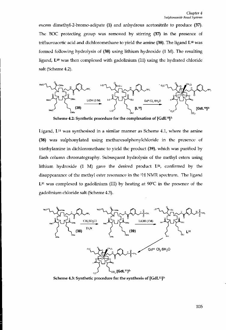
Chapter 4 Siilphonamide Based Systems
excess dimethyl-2-bromo-adipate (1) and anhydrous acetonitrile to produce (37).
The BOC protecting group was removed by stirring (37) in the presence of
trifluoroacetic acid and dichloromethane to yield the amine (38). The Ugand L^" was
formed fol lowing hydrolysis of (38) using l i th ium hydroxide (1 M) . The resulting
ligand, L^o was then complexed w i t h gadolinium (III) using the hydrated chloride
salt (Scheme 4.2).
N N N N
N N LiOH (3 M) Gd"i .C, .6H,0
OMb [GdU»]3-
OLi C O , -
Scheme 4.2: Synthetic procedure for the complexation of [GdU"]'-
Ligand, L " was synthesised in a similar manner as Scheme 4.1, where the amine
(38) was sulphonylated using methanesulphonylchloride i n the presence of
triethylamine in dichloromethane to yield the product (39), which was purif ied by
flash column chromatography. Subsequent hydrolysis of the methyl esters using
l i th ium hydroxide (1 M) gave the desired product L^i, confirmed by the
disappearance of the methyl ester resonance in the NMR spectrum. The ligand
L^i was complexed to gadolinium (III) by heating at 90°C in the presence of the
gadolinium chloride salt (Scheme 4.3).
(39)
C H , S 0 , C 1
Gd'" CI3.6H2O
-co,-[GdUi]3-
Scheme 4.3: Synthetic procedure for the synthesis of [GdU^]^
105

The synthesis of the ligand L12 is depicted in Scheme 4.4.
Chapter 4 Stilphonamide Based Systems
, 0 H EDC PhjP / CBr^
OH
o (40) Br
o (41)
CH,CI
MeO^C/^^^Y^
MeO' V L J
MeO,C (43)
(1) MeO'
OMe
o
" L J " (42)
CO,Me
LiOH
LiO. LIO,C
N N
-Ofi. OH,),
o
LIO,C CO±t [GdLi2]3- ^co,-
Scheme 4.4: Chemical synthesis of the gadolinium complex [GdL^2]3-
Picolinic acid was converted to the corresponding ester by stirring the acid i n the
presence of ethanol w i t h acetyl chloride used as a catalyst. EDC coupling of
picolinic ethyl ester w i th ethanolamine afforded the amide (40).
Triphenylphosphine and carbon tetrabromide were used to convert (40) into the
corresponding bromide (41). In the presence of a large excess of cyclen, which also
acted as a base, compound (41) was added to yield (42). Attempts to directly
alkylate the free N H position of l,4,7-tris-[(4'-methoxycarbonyl)-l'-methoxy-
carbonylbutyl]-l,4,7,10-tetraazacyclododecane (2) were unsuccessful as a number of
impurities were produced, and the desired product could only be isolated in very
low yield. The remaining three N H groups of the cyclen r ing were alkylated in the
presence of excess dimethyl-2-bromo-adipate (1), caesium carbonate and
acetonitiile to produce (43). The hydrolysis of (43) was achieved by stirring the
106
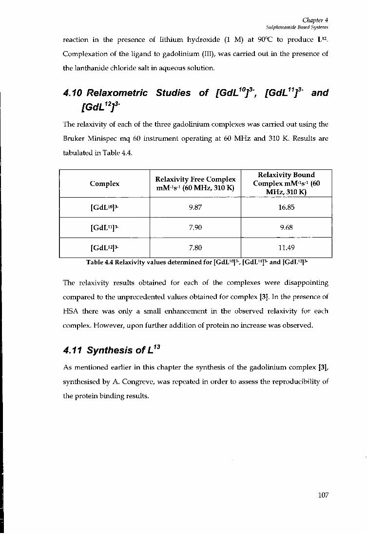
Chapter 4 Sulplwnamide Based Systems
reaction in the presence of l i th ium hydroxide (1 M) at 90°C to produce L^^.
Complexation of the Ugand to gadolinium (III), was carried out i n the presence of
the lanthanide chloride salt i n aqueous solution.
4.10 Relaxometric Studies of [GdL'Y> [GdU't and [GdU^f
The relaxivity of each of the three gadolinium complexes was carried out using the
Bruker Minispec mq 60 instrument operating at 60 M H z and 310 K. Results are
tabulated in Table 4.4.
Complex Relaxivity Free Complex mM-is-i (60 MHz, 310 K)
Relaxivity Bound Complex mM-^s-i (60
MHz, 310 K)
[GdLi»]3- 9.87 16.85
[GdL"]3- 7.90 9.68
[GdLi2]3- 7.80 11.49
Table 4.4 Relaxivity values determined for [GdLi»]3-, {GdU^f- and [GdU^p
The relaxivity results obtained for each of the complexes were disappointing
compared to the unprecedented values obtained for complex [3]. In the presence of
HSA there was only a smaU enhancement i n the observed relaxivity for each
complex. However, upon further addition of protein no increase was observed.
4.11 Synthesis of
As mentioned earUer i n this chapter the synthesis of the gadolinium complex [3],
synthesised by A. Congreve, was repeated in order to assess the reproducibility of
the protein binding results.
107
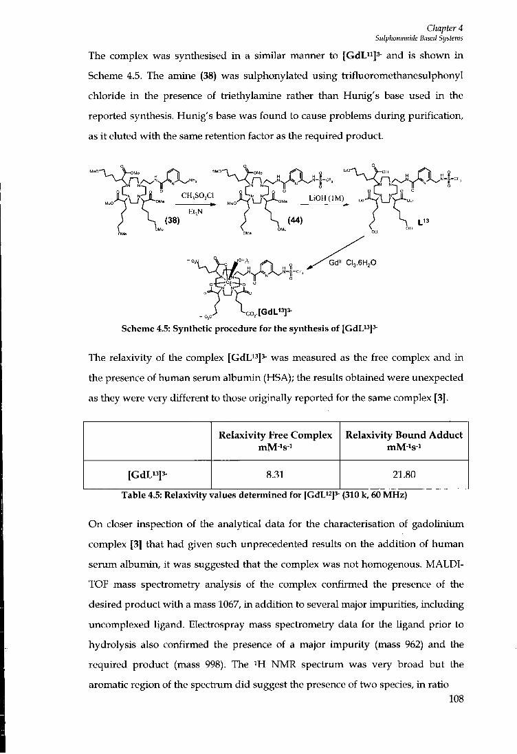
Chapter 4 Sulphoimnide Based Systetns
The complex was synthesised in a similar manner to [GdL"]^- and is shown i n
Scheme 4.5. The amine (38) was sulphonylated using trifluoromethanesulphonyl
chloride i n the presence of triethylamine rather than Hunig's base used i n the
reported synthesis. Hunig's base was found to cause problems during purification,
as i t eluted w i t h the same retention factor as the required product.
" ^ r i ^ / X . . " ^ ^ ^ U " ^ A ^ / L i H II
EtjN (38) ; S (44)
. „ / \ o , [ G d L " ] 3 -
Scheme 4.5: Synthetic procedure for the synthesis of [GdLi^JJ-
The relaxivity of the complex [GdLi^js- was measured as the free complex and i n
the presence of human serum albumin (HSA); the results obtained were unexpected
as they were very different to those originally reported for the same complex [3].
Relaxivity Free Complex Relaxivity Bound Adduct
[GdL"]3- 8.31 21.80
Table 4.5: Relaxivity values determined for [CdL^^Js- (310 k, 60 MHz)
O n closer inspection of the analytical data for the characterisation of gadoliniimi
complex [3] that had given such unprecedented results on the addition of human
serum albumin, i t was suggested that the complex was not homogenous. M A L D I -
TOF mass spectrometry analysis of the complex confirmed the presence of the
desired product w i th a mass 1067, i n addition to several major impurities, including
uncomplexed ligand. Electrospray mass spectrometry data for the Ugand prior to
hydrolysis also confirmed the presence of a major impuri ty (mass 962) and the
required product (mass 998). The N M R spectrum was very broad but the
aromatic region of the spectium d id suggest the presence of two species, i n ratio
108
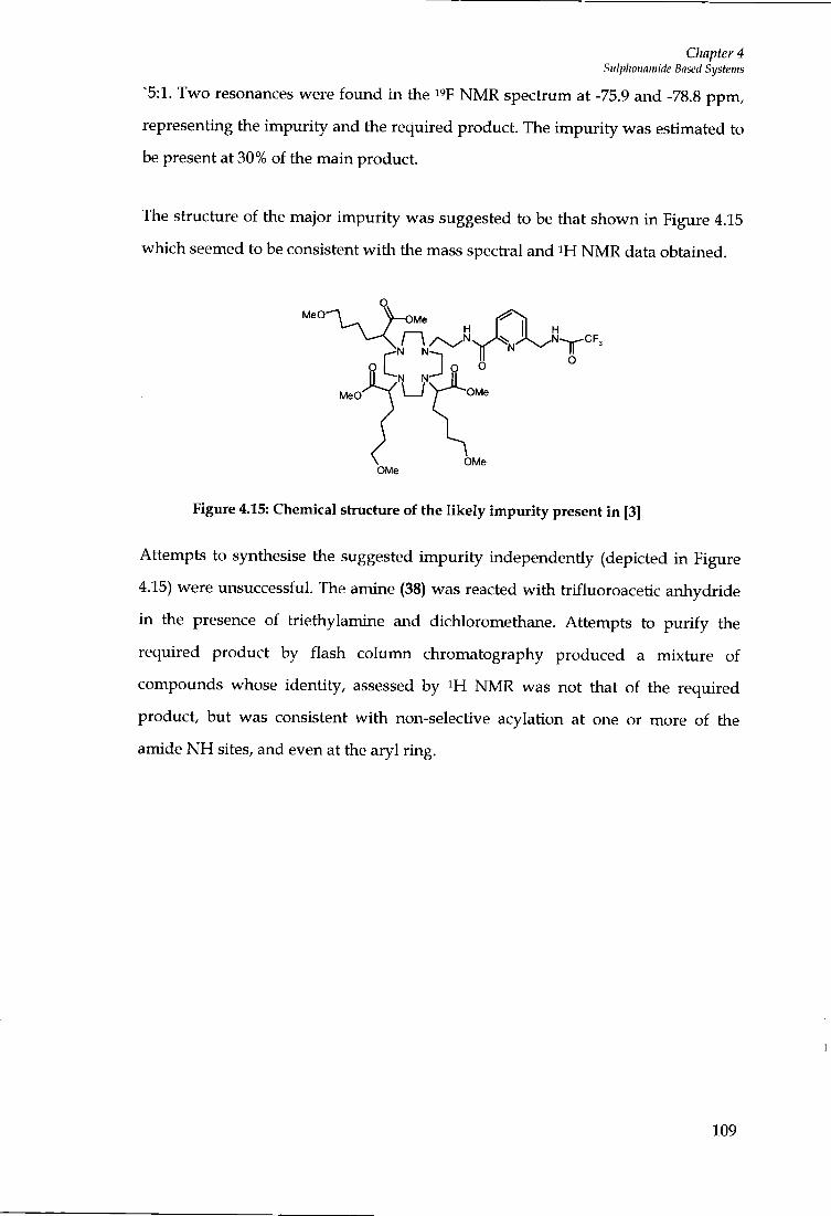
Chapter 4 Sulplionmiiide Based Systems
5:1. Two resonances were found in the I 'F N M R spectrum at -75.9 and -78.8 ppm,
representing the impuri ty and the required product. The impuri ty was estimated to
be present at 30% of the main product.
The structure of the major impuri ty was suggested to be that shown in Figure 4.15
which seemed to be consistent w i t h the mass spectral and N M R data obtained.
MeO
N N
N N
OMe OMe
Figure 4.15: Chemical structure of the likely impurity present in [3]
Attempts to synthesise the suggested impuri ty independently (depicted i n Figure
4.15) were unsuccessful. The amine (38) was reacted wi th trifluoroacetic anhydride
in the presence of triethylamine and dichloromethane. Attempts to pur i fy the
required product by flash column chromatography produced a mixture of
compounds whose identity, assessed by N M R was not that of the required
product, but was consistent w i t h non-selective acylation at one or more of the
amide N H sites, and even at the aryl ring.
109
Chapter 4 Sulphonamide Based Systems
4.12 Summary
The relaxivity results obtained for each of the derivative complexes were
disappointing compared to the xmprecedented values obtained for complex [3]. The
most significant enhancement reported i n the study was associated w i t h the protein
bound adduct for the free amide complex, [CdLi^J^-. However, the enhancement
observed in saturated protein solutions was minimal. None of the complexes
including that of [CdL^^Js-, which initially was beheved to be the same as complex
[3], gave the unprecedented relaxivity enhancements that were observed for [3].
The less hydrophobic methylsulphonamide complex, [CdL^^J^- was found to have
minimal interaction towards human serum alburtiin w i t h respect to the
trifluorosulphonamide complex [3]. This may suggest that as the sulphonamide pKa
of [GdL"] is higher than that of complex [3], (ca. 12 compared to 7.5), and
consequentiy protonated at ambient p H , i t may be that deprotonation of the
sulphonamide N H is required for a strong interaction wi th protein.
The insolubility problems associated w i t h complex [3] at neutral p H , a
phenomenon which was found to be time dependent, result i n reproducibility
issues, l imit ing the use of such system as a potential contrast agent. Further work
may include the attachment of more water-soluble fimctionaUsed groups to
complex [3], in an attempt to reduce any oligomerisation, and the associated p H
and time dependent effects.
110

Chapter 5 NMR and cliiroptical examination of tite diastereisomers of (S)-EOB-DTPA
Chapter 5
NMR and chiroptical examination of the
diastereoisomers of (S) -
[Eu-(EOBDTPA)]2-
111

Chapter 5 NMR and cliiroptical examination of tite diastereisomers of (S)-EOB-DTPA
5 NMR and chiroptical examination of the diastereoisomers of (S)- [Eu-(EOBDTPA)]^"
5.1 Introduction
[Gd(DTPA)]2- or Gadopentetate Dimeglumine, marketed as Magnevist ™ by
Schering AG, was the first Gd (111) containing compound ever to be approved by
the FDA in 1988; i t has since been used i n more than 25 mil l ion people.^
C O , -
N N N
Figure 5.1: [DTPA]s^
I t is an example of an extracellular, hydrophiUc, low molecular weight gadolinium
chelate. These types of contiast agent are non-specific compounds, that fo l lowing
intravenous injection are rapidly excreted f r o m the vascular compartments into
interstitial spaces and do not pass plasma membranes, excretion occurring via the
urinary system.i33 Current research in this area seeks to devise methods of targeting
the paramagnetic complex to selected tissues, and a first step i n this direction was
the discovery that gadolinium complexes of certain analogues of DTP A, such as
BOPTA and EOB-DTPA, clear the body via the biliary system rather than the renal
system.134
Eto
co,-2
N N 2
Figure 5.2: [EOB-DTPA]5-
[Gd-(EOB-DTPA)]2- (international non proprietary name: gadoxetic acid, disodium
salt), which is currently undergoing phase 111 clinical trials, exhibits very different
pharmacodynamic behaviour and hence a distinctive biodisti ibution profile in
human patients. It shows the specific uptake by hepatocytes not by cancerous
tissue, allowing the detection and differentiation of hepatic tumours. In addition to
112
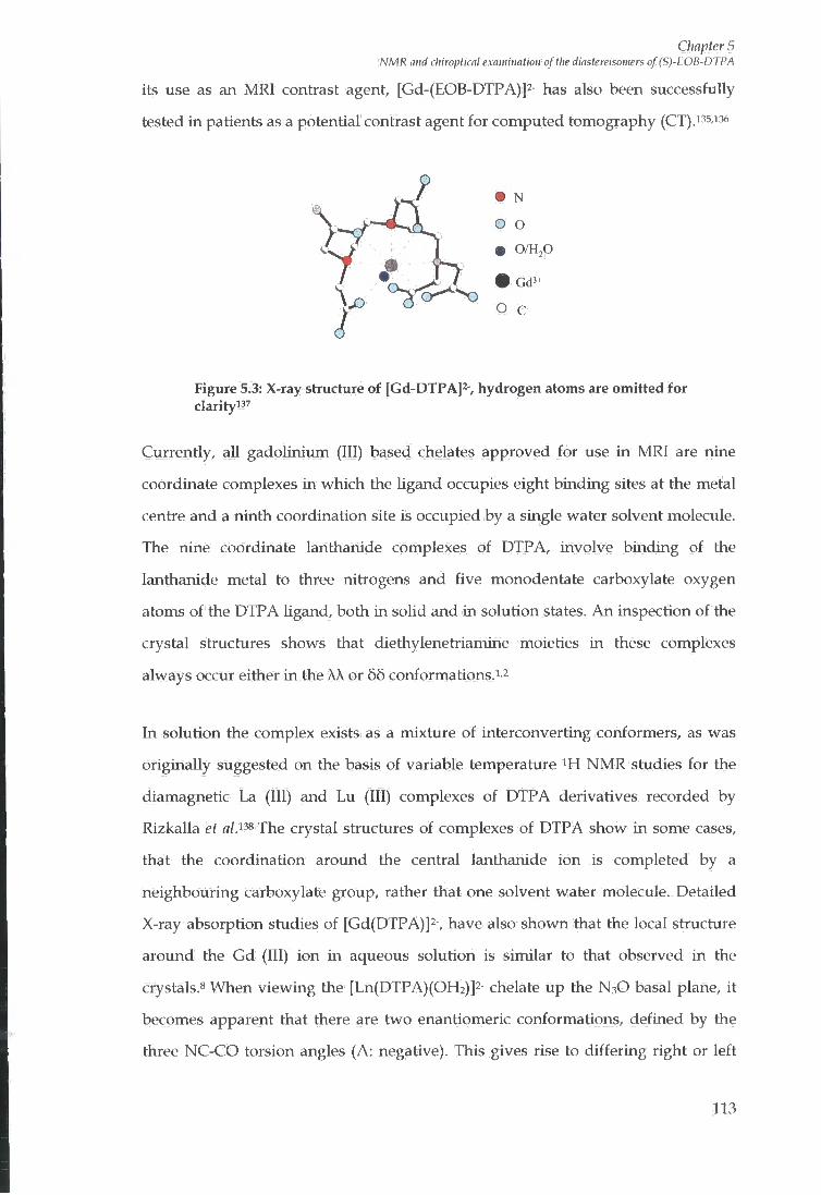
Chapter 5 NMR and chiroptical examination of tlie dmstereisomers of (S)-EOB-DTPA
its use as an M R l contrast agent, [Gd-(EOB-DTPA)]2- has also been successfully
tested in patients as a potential contrast agent for computed tomography (CT).i35,i36
• 0/H,0
Figure 5.3: X-ray structure of [Gd-DTPA]^-, hydrogen atoms are omitted for clarityi37
Currently, all gadolinium (III) based chelates approved for use i n M R l are nine
coordinate complexes in which the ligand occupies eight binding sites at the metal
centre and a ninth coordination site is occupied by a single water solvent molecule.
The nine coordinate lanthanide complexes of DTP A, involve binding of the
lanthanide metal to three nitrogens and five monodentate carboxylate oxygen
atoms of the DTPA Ugand, both i n solid and in solution states. A n inspection of the
crystal structures shows that diethylenetriamine moieties i n these complexes
always occur either i n the AA or 65 conformations.^ ^
In solution the complex exists as a mixture of interconverting conformers, as was
originally suggested on the basis of variable temperature i H N M R studies for the
diamagnetic La (III) and Lu (III) complexes of DTPA derivatives recorded by
Rizkalla et alA^ The crystal structures of complexes of DTPA show in some cases,
that the coordination around the central lanthanide ion is completed by a
neighbouring carboxylate group, rather that one solvent water molecule. Detailed
X-ray absorption studies of [Gd(DTPA)]2-, have also shown that the local structure
arotmd the Gd (111) ion in aqueous solution is similar to that observed in the
crystals.8 When viewing the [Ln(DTPA)(OFl2)]2- chelate up the N3O basal plane, i t
becomes apparent that there are two enantiomeric conformations, defined by the
three NC-CO torsion angles (A: negative). This gives rise to differ ing right or left
113

Chapter 5 NMR and chiroptical exawiiiatioii of the diastereisomers of (S)-EOB-DTPA
handed orientations of three of the acetate arms, associated w i t h different A or A
helicites about the metal centie (Figures 5.4 and 5.5).
N'
Figure 5.4: In the A complex, the NCCO torsion angle is negative
Figure 5.5: View up the Ln-water vector, or down the N3O basal plane, for the A complex
As a result, the two conformers XA and 66, distinguished by the helicity of the C-C
ethylene bond relative to the Ln (III) N - N plane, may interconvert through a
shuffl ing of the acetate groups accompanied by a f l ip in the diethylenetriamine
backbone. The nitrogen atom N-6, i n this case, is rendered chiral upon
complexation.127
The introduction of the lipophilic ethoxybenzyl moiety, creates a stereogenic centie
at C-4 (Figure 5.2), as i n the case of (S)-EOB-DTPA and renders each of the
conformations diastereoisomeric. Thus, there are 4 diastereoisomeric complexes for
(S)-[Ln-(EOB-DTPA)(H20)]2- (see Figure 5.6), distinguished by an S or R
configuration at N and by a A / A helicity about the metal centre. The analysis does
not include consideration of the preferred 6 or A confirmation of each of the NCCN
chelate rings.
114

Chapter 5 NMR and chiroptical examination oftlie diastereisoniers of (S)-EOB-DTPA
o o
o q : N
/ ,
o
Figure 5.6: Four possible diastereoisomeric complexes for (S)-[Eu-EOB-DTPA(H20)]2-
The Ugand EOB-DTPA used throughout the investigation was of (S) absolute
configuration; the compound was synthesised and donated by Schering Research
laboratories AG, Berlin Germany. None of the (R) enantiomer was provided for
study.
Protein binding shidies of [Gd-(S)-(EOB-DTPA)(H20)]2- versus [Gd-(R)-(EOB-
DTPA)(H20)]2-, i n the presence of human serum albumin, and the non-covalent
binding of the paramagnetic complexes to the macromolecule had earlier been
studied. Each isomer was found to bind i n a non-covalent manner to human serum
albumin. However, the affini ty of the (S) isomer for the protein was much greater
than that for the (R) isomer."? This may have been a reason for the preference of
the (S) enantiomer being studied rather than the (R) corifiguration.
Previous work on (S)-EOB-DTPA carried out by Schmitt-Willich et a/"" involved
complexation of the ligand w i t h the lanthanide metal gadolinium, generating two
chiral centies, hence two diastereoisomers of (S)-[Gd-(EOB-DTPA)(H20)]2-. It was
shown that the two diastereoisomers could be separated by reversed phase HPLC,
resulting i n a ratio of the two components of 65:35 (Figure 5.7).
115
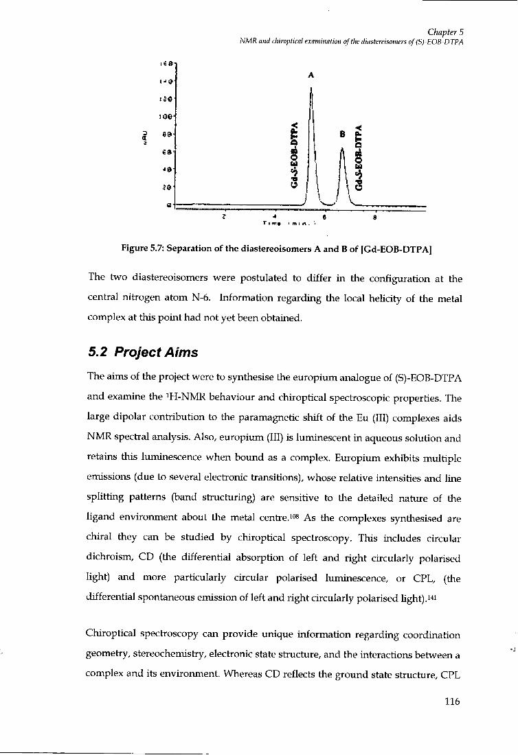
Chapter 5 NMR and chiroptical examination of the diastereisomers of (S)-EOB-DTPA
3 tt
| g 0
a
i
* e
Figure 5.7: Separation of the diastereoisomers A and B of [Gd-EOB-DTFA]
The two diastereoisomers were postulated to differ in the configuration at the
cential nitrogen atom N-6. Irrformation regarding the local helicity of the metal
complex at this point had not yet been obtained.
5.2 Project Aims
The aims of the project were to synthesise the europium analogue of (S)-EOB-DTPA
and examine the ^H-NMR behaviour and chiroptical spectroscopic properties. The
large dipolar contribution to the paramagnetic shift of the Eu (III) complexes aids
N M R spectial analysis. Also, europium (III) is luminescent i n aqueous solution and
retains this luminescence when bound as a complex. Europium exhibits multiple
emissions (due to several electionic transitions), whose relative intensities and line
splitting patterns (band structuring) are sensitive to the detailed nature of the
ligand environment about the metal centre. o^ As the complexes synthesised are
chiral they can be studied by chiroptical spectroscopy. This includes circular
dichroism, CD (the differential absorption of left and right circularly polarised
light) and more particularly circular polarised luminescence, or CPL, (the
differential spontaneous emission of left and right circularly polarised light) .^^i
Chiroptical spectroscopy can provide unique information regarding coordination
geometry, stereochemistry, electronic state stiucture, and the interactions between a
complex and its environment. Whereas CD reflects the ground state structure, CPL
116

Chapter 5 NMR and chiropticnl examination of the diastereisomers of (S)-EOB-DTPA
probes the structure of the excited state, which can provide us w i t h information
about the helicity of the metal complex arotmd the europium centie.127 From the
extensive knowledge previously obtained wi th in the Parker research group,
concerning structurally related chiral complexes based upon the cyclen skeleton,
and the effects of complex geometry and isomerism, comparative analysis w i t h this
DTPA based system could be made.127,142
5.3 Solution NMR analysis
The europium (III) complex [EuL^*] of (S)-EOB-DTPA was prepared by a slightly
modified method to that already established for the synthesis of the gadolinium
(III) analogue reported by Schmitt-WilUch et alA*o Following heating of the ligand in
the presence of equimolar quantities of europium (III) chloride, suspended in
equivalent volumes of water and methanol mixture, the solution p H was adjusted
to p H 5.5., using 0.1 M sodium hydroxide. The mixture was then concentrated by
reduced pressure and the resulting concentiate added dropwise to a warmed
solution of ethanol. Upon heating, a turbid solution resulted, which was
centiifuged, the white solid collected, washed wi th ethanol and dried. This
solidified product was dissolved into water and the p H adjusted to remove any free
lanthanide material present as the free lanthanide hydroxide, which if un-removed
may have affected the quality of the analytical spectia obtained.
Similarly to earlier work, the complex (S)-[Eu-(EOB-DTPA)(H20)]2-, [ E u L " ] was
analysed again by reversed phase HPLC, which revealed the presence of two
species (diastereoisomers), which were individually collected by preparative HPLC.
The isometric ratios of 57:43 were very similar to those found for the related Gd
(65:35) and La (60:40) analogues.i^o The separated complexes when stored i n
solution were held at 4°C, to minimise the rate of isomer interconversion. i H NMR
stability studies for the separated diastereoisomers of [EuL^*], were evaluated over
a three month time period at this temperature, and evidence for <5%
interconversion was foxmd. However, at ambient temperature, interconversion was
found to occur, reaching equilibrium wi th in approximately 72 hours. This value
was i n conflict w i t h the behaviour of the separated (S)-[Gd-(EOB-DTPA)]2-
117
Chapter 5 NMR and citiropticnl examination of the diastereisomers of (S)-EOB-DTPA
diastereoisomers earlier studied by Schmitt et al^*°'^ where interconversion was
found to be very slow, the half-life time of one of the Gd (III) isomers being 13100
hours at p H 9 and 25°C. A t thermodynamic equiUbrium, the ratio of the isomers
was found to be 65:35. The first order rate constant for the interconversion between
the isomers is p H dependent, suggesting that the isomerisation is an acid catalysed
process similar to the dissociation of the complex.8'i4o N M R studies of the two (S)-
[La-(EOB-DTPA)]2- complexes, had earlier given no evidence for the presence of
enantiomers that differ i n the orientation of the acetate groups. Although four are
theoretically possible for the system, two are mirror images of one another and
therefore discrimination by NMR is impossible. Proton N M R analysis of the
europium mixture of the complex (S)-[Eu-(EOB-DTPA)]2-, [EuL^^] revealed the
presence of two species (D2O, 200 MHz) , Figure 5.8. Although it was obvious that
there was one major isomer, the shifted resonances of the minor isomer were
significantly broader than those associated w i t h the major species.
118

15
Chapter 5 NMR and chiroptical examination of tlie diastereisomers of (S)-EOB-DTPA
18 17
ppm
Figure 5.8: NMR spectrum (200 MHz, D2O, 296 K) of the mixture of diastereoisomers of (S)-[Eu-(EOB-DTPA)]2- [EuL"].
A detailed variable temperature N M R study of the mixture was undertaken over
the range of 4°C to 92°C. The broadened, shifted resonances of the minor isomer
were found to be much sharper upon heating. Upon closer inspection of the
completed variable temperature study, the spectral data revealed there were only
two species i n solution, each w i t h an equal number of isochronous resonances."^
There was no f i r m evidence to suggest that an exchange process between the two
observed isomers was occurring, as line broadening variations involving the minor
isomer were not echoed by spectral changes in resonances of the major species
(Figures 5.9 and 5.10). The temperature dependence of the chemical shift variations
for every resonance in each isomer was the same and wi th in experimental error
fol lowed the expected T dependence of the paramagnetic shift.io^ Such behaviour
119

Chapter 5 NMR mid chiroptical examiimtion of the diastereisoiiiers of (S)-EOB-DTPA
precludes the presence of an intramolecular exchange process between the
observed species, on the N M R timescale.
4 ° C
22 °C
32 °C
72 °C
j I I I I I I I I I I I I I I I I I M j I I I I j I I I 1 I I I I I I ! I I I I I I I I I I I I I j I I I I I I I I I I
35 25 15 5 -5 ppm
Figure 5.9: Variable temperature proton NMR spectra (300 MHz, D2O) of the stereoisomeric mixture of (S)-[Eu-(EOB-DTPA)]2-, [EuL"] between 4°C and 72°C.
120

Chapter 5 NMR nnH rhirnntirn! PYnwiuntinn of fhp din'^tprpi'inmpr^ nf (fi]~FnH-nTPA
4 ° C
16 °C
32 °C
42 °C
62 °C
I I 1 I I I I I I I 1 I ) I I I I I I 1 I [ I 1 1 I I 3 5 2 5 1 5
I I I I I I I 1 I I I I 1 I I I I
- 5 P P ni
Figure 5.10: Variable temperature proton NMR spectra (300 MHz, D2O) of the stereoisomeric mixture (S)-[Eu-(EOB-DTPA)]2-, |EuL"] between 4°C to 62°C.
The proton spectrum of the major isomer was fu l l y assigned using COSY (proton -
proton correlation) experimental methods (Figure 5.11), the interpretation of
spectra being aided by comparison to earUer studies on structurally related
derivatives of cyclen fo l lowing the work of other members of the research
group. 143,144,145 j j ^ g resonances of each proton i n the complex were anisochronous,
except for the ortho/meta aryl protons at +28.9 and +10.9 ppm. The three protons at
C4 and C5 f o r m a mutually coupled sub-set, w i t h the axial proton, Hs'*'*, resonating
to highest frequency at 28.0 ppm. Similarly, the four protons at C? and Cg in the
urvsubstituted 5-ring chelate, formed a distinctive mutually coupled sub-set of
resonances, w i t h Hs"'* resonating at 29.0 ppm. Each proton in the five C H 2 C O
groups is chemical shift non-equivalent, and five mutually coupled sets were
121
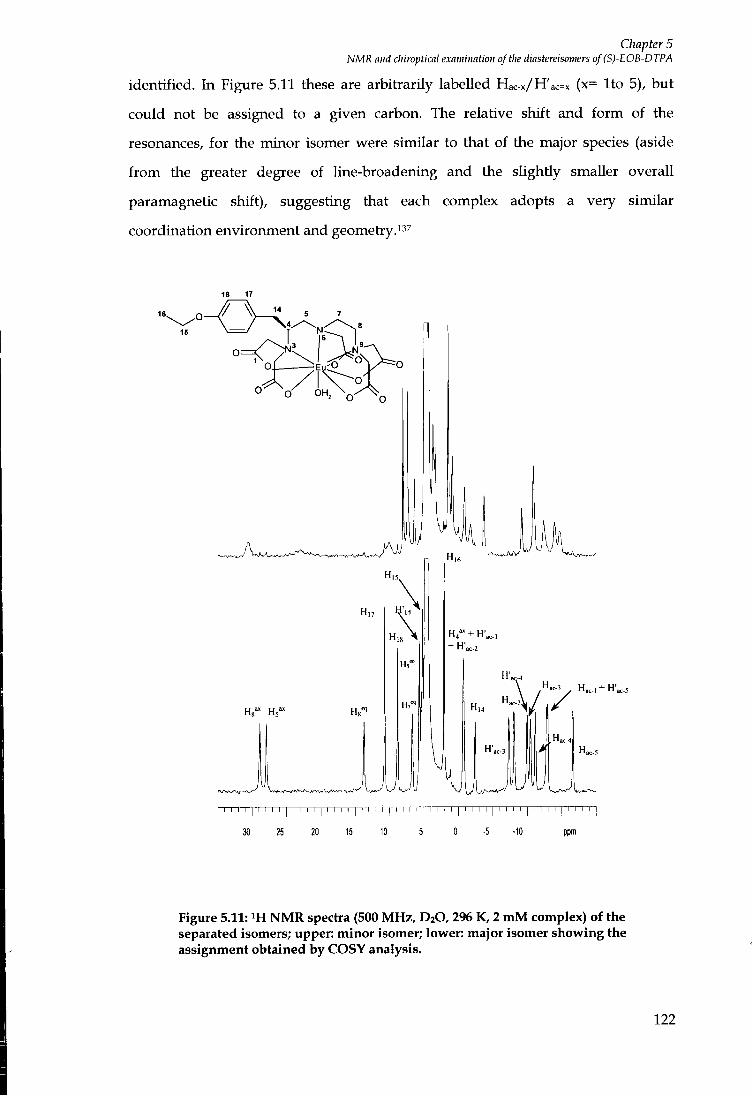
Chapter 5 NMR and chiroptkal examination of the diastereisomers of (S)-EOB-DTPA
identified. In Figure 5.11 these are arbitrarily labelled Hac-x/H'ac=x (x= I to 5), but
could not be assigned to a given carbon. The relative shift and form of the
resonances, for the minor isomer were similar to that of the major species (aside
f r o m the greater degree of line-broadening and the slightly smaller overall
paramagnetic shift), suggesting that each complex adopts a very similar
coordination environment and geometry."^
15
18 17
H,5.
H,8
Hi"
UUUli
H,6
H4" + H',c,]
+ H',c.2
4
I I M I I I M I I I I I I I I I I I I I I I I I I I I I I I I I I I I I I I I I I I I I M I I I I M [
30 25 20 15 10 5 0 -5 -10 ppm
Figure 5.11: iH NMR spectra (500 MHz, D20,296 K, 2 mM complex) of the separated isomers; upper: minor isomer; lower: major isomer showing the assignment obtained by COSY analysis.
122
Chapter 5 NMR and chiroptical examination of the diastereisoniers of(S)-EOB-DTPA
Inner sphere proton relaxivity is linearly proportional to the number of water
molecules directly coordinated to the central Ln (III) metal ion as shown by
Equation 5.1, where c is the molal concentiation, q is the number of bound water
molecules per Ln ion (hydration number), Pm is the mole fraction of the bound
water molecules, Xm is the lifetime of the water molecule residing on the Ln (III)
centre (equal to the reciprocal water exchange rate l/Zcex), and 1/Tim is the
longitudinal proton relaxation rate.3
]_ cq
55.5 = P (5.1)
T +T
The above equation shows clearly that proton relaxivities cannot be determined
without knowing the hydration number. Determination of hydration nimrber can
be calculated f r o m solid state stiuctures, however there are examples of a change in
hydration number observed f r o m the solid state to the solution state. Therefore, the
determination of hydration number i n aqueous solution is important and there are
several experimental techniques available, although none can be directly applied to
Gd (III) chelates."!
The hydration states for the Eu (III) complex [EuL^^] were determined by laser-
induced luminescence, where the difference in luminescence lifetimes measured in
H2O and D2O solutions, is related to the hydration number, q of the complex based
upon the fol lowing equation, where x is the luminescence lifetime and A L H is a
proportionality constant specific to a given lanthanide. For Eu (III) complexes, A =
1.05 ms-1.3
^ = A « « o - < o ) (5.2)
I major 1 major The values obtained for the major isomer were C^z/jO = T -65 ms-^; ^o^o =0.50
I minor » minor ms-i;q=l.l) and for the minor isomer ( '^HjO =1.63 ms-^; f^o^o = 0-51 rns-^; q = 1.0);
the values are consistent w i t h the presence of a single quenching water molecule.
123
Chapter 5 NMR and chtmptical examination of tlie diastereisomers of (S)-EOB-DTPA
The results are i n agreement w i t h the measured hydration value for the complex
[Dy-(EOB-DTPA)]2-, determined by the induced water ^ Q shifts for the Dy (111)
metal ion, where a hydration value of 1.2 was measured.^
5.4 Chiroptical Spectroscopy
The chirahty of the complex (S)-[Eu(EOB-DTPA)(H20)]2-, [EuL"] aUows chiroptical
techniques such as circular dichroism (CD) and circular polarised luminescence
(CPL) to be used. Chiroptical properties of lanthanides are particularly sensitive to
ligand coordination geometry and stereochemistry, electronic state structure, and
the interactions between a complex and its environment. In chiroptical
spectroscopy, different mechanisms are used to determine band intensities and
compared to conventional spectroscopy; further stiuctural information may be
available.^27
5.5 Circular Dichroism
If l ight is composed of two plane waves of equal ampUtude but differing i n phase
by 90°, then the Ught is said to be circularly polarised. Circular dichroism (CD) is
observed when optically active matter absorbs left and right handed circular
polarised light slightly differently.i^s When circular polarised light passes through
the optically active sample w i t h a different absorbance. A, for the two components,
the amplitude of the stionger absorbed component w i l l be smaller than that of the
less absorbed component. The consequence is that a projection of the resulting
amplitude wiU now yield an elUpse instead of the usual line. This occurrence of
ellipicity is called circular dichroism."6 The technique requires a source of
monochromatic left and right circularly polarised light produced by a photoelastic
modulator, (in older instiuments a Pockels cell), which produces alternatively right
and left circularly polarised light w i t h a switching frequency of about 50 kHz,
which is detected by a photomultipler tube. The current output of the tube is
converted to a voltage signal, the mean intensity of which is proportional to the
total CD. The other signal is fed into a lock-in amplifier, where the alternating
current (AC) ripple present i n the large direct current (DC) background is amplified
124
Chapter 5 NMR and chiropticiil examination oftlie diastereisomers of (S)-EOB-DTPA
by means of a phase sensitive detector, and is proportional to the CD intensity."7
The CD experiment thus produces two signals. One signal results f r o m the
differential absorption of one component over the other and is proportional to the
circular dichroism. The remaining signal is averaged and related to the mean Ught
absorption. The ratio of these signals is the absorption dissymmetry factor, gabs
defined by the fol lowing equation, where £L and ER correspond to extinction
coefficients for left and right circularly polarised components of the emitted
radiation.146
Sabs (^) ^ 2(g^ - £ r ) (5.3)
(£ • ,+£« )
A CD spectrum is a plot of Ae versus wavelength and since different electronic
transitions involve the redistribution of electroris of different handedness, any one
molecule may give rise to both positive and negative CD signals.
5.6 CD measurements of (S)'[Eu-(EOB-DTPA)(H20)t
The CD spectrum of (S)-EOB-DTPA ligand in water was recorded in the ultra-violet
region, (pH 7.5, 295K) and compared to the spectra obtained for the Eu (III)
complex [EuLi4]. Two bands were seen for the enantiomerically pure free ligand; a
positive band at 245 n m (g245 = +3 x lO-^) and a negative band at 276 n m (gabs = -6 x
10-4) were distinguished. The CD spectra obtained for each of the diastereoisomers
of [EuLi*] were found to be very similar to those of the free ligand, w i th a slight
reduction in the measured dichroism (approximately 40% lower i n terms of the
measured g values). Generally, circular dichroism measurements require a higher
concentration of the lanthanide complex as most 4 / - / transitions are parity
forbidden and have very low molar absorptivities, (values for e for Eu (III)
transitions are typically < 1 dm^ moH cm-i).i27 For this reason, i t was decided to
measure the high resolution CPL spectra instead, as this technique combines
instrument sensitivity w i t h structure selectivity and can be carried out on dilute
solutions of lanthanide complex, e.g. 0.5 to 10.0 m M solutions. CPL also has the
125
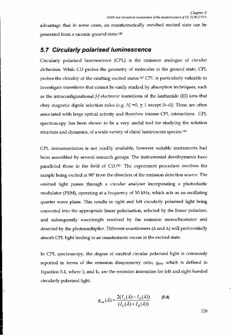
Chapter 5 NMR and chiroptical examination of the diastereisomers of (S)-EOB-DTPA
advantage that in some cases, an enantiomerically enriched excited state can be
generated f r o m a racemic ground state. ^s
5.7 Circularly polarised luminescence
Circularly polarised luminescence (CPL) is the emission analogue of circular
dichroism. While CD probes the geometry of molecules i n the ground state, CPL
probes the chirality of the emitting excited states.i47 CPL is particularly valuable to
investigate transitions that cannot be easily studied by absorption techniques, such
as the intraconfigurational / - / electronic transitions of the lanthanide (111) ions that
obey magnetic dipole selection rules (e.g. AJ =0, + 1 except 0<->0). These are often
associated w i t h large optical activity and therefore intense CPL interactions. CPL
spectroscopy has been shown to be a very useful tool for studying the solution
structure and dynamics, of a wide variety of chiral luminescent species. '*'
CPL instrumentation is not readily available, however suitable instruments had
been assembled by several research groups. The instrumental developments have
paralleled those in the field of CD.™ The experiment procedure involves the
sample being excited at 90° f r o m the direction of the emission detection source. The
emitted light passes through a circular analyser incorporating a photoelastic
modulator (PEM), operating at a frequency of 50 kHz, which acts as an oscillating
quarter wave plane. This results in right and left circularly polarised light being
converted into the appropriate Unear polarisation, selected by the linear polarizer,
and subsequently wavelength resolved by the emission monochromator and
detected by the photomultiplier. Different enantiomers (A and A) w i l l preferentially
absorb CPL light leading to an enantiomeric excess in the excited state.
In CPL spectroscopy, the degree of emitted circular polarised light is commonly
reported i n terms of the emission dissymmetry ratio, gem, which is defined in
Equation 5.4, where I I and 1R, are the emission intensities for left and right handed
circularly polarised light.
2(1,(A)-1M)) (5.4)
126
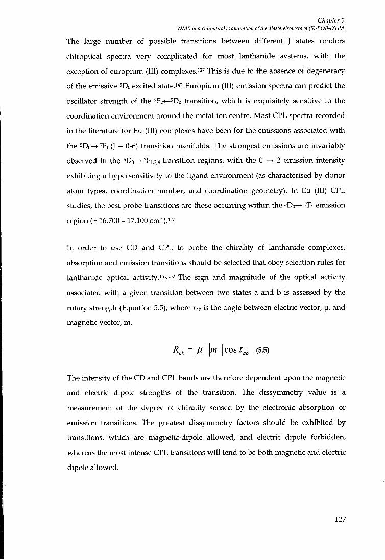
Chapter 5 NMR and chiroplical examination of tiie diastereisomers of (S)-EOB-DTPA
The large number of possible transitions between different J states renders
chiroptical spectia very complicated for most lanthanide systems, w i t h the
exception of europium (111) complexes.127 This is due to the absence of degeneracy
of the emissive ^Do excited state.i*^ Europium (111) emission spectra can predict the
oscillator strength of the 7F2<— Do tiansition, which is exquisitely sensitive to the
coordination environment around the metal ion centre. Most CPL spectra recorded
in the literature for Eu (111) complexes have been for the emissions associated w i t h
the 5Do^ ^F] (J = 0-6) tiansition manifolds. The stiongest emissions are invariably
observed i n the ^Do- ^ ^Fi,2,4 tiansition regions, w i t h the 0 ^ 2 emission intensity
exhibiting a hypersensitivity to the ligand environment (as characterised by donor
atom types, coordination number, and coordination geometiy). In Eu (111) CPL
studies, the best probe tiansitions are those occurring wi th in the ^Do^ ^Fi emission
region (~ 16,700 - 17,100 cm-i).i27
In order to use CD and CPL to probe the chirality of lanthanide complexes,
absorption and emission tiansitions should be selected that obey selection rules for
lanthanide optical activity.151/152 The sign and magnitude of the optical activity
associated w i t h a given tiansition between two states a and b is assessed by the
rotary stiength (Equation 5.5), where Tab is the angle between electiic vector, p, and
magnetic vector, m.
Kt,= m COST^ (5.5)
The intensity of the CD and CPL bands are therefore dependent upon the magnetic
and electiic dipole stiengths of the tiansition. The dissymmetiy value is a
measurement of the degree of chirality sensed by the electionic absorption or
emission tiansitions. The greatest dissymmetiy factors should be exhibited by
tiansitions, which are magnetic-dipole allowed, and electiic dipole forbidden,
whereas the most intense CPL tiansitions w i l l tend to be both magnetic and electiic
dipole allowed.
127
Chapter 5 NMR and chiroptical examination of tlie diastereisomers of (S)-EOB-DTPA
5.8 CPL measurements of (S)-[Eu-(E0B'DTPA)(H20]f-The metal based CPL emission spectrum in the tiansition To—^^Do was measured
for each of the separated isomers of the complex (S)-[Eu-(EOB-DTPA)(H20)]2-,
[EuL^*] complex i n an aqueous media, at p H 7.5, 282 K. The lower temperature was
chosen to minimise the risk of any isomer interconversion on the experimental time
scale, and revealed the presence of only one peak centied at 579.7 n m (Figure 5.12).
1.0
0.B E 3
0.6 A
0.2 H
(6ulE0B-DTPA)f' lasmer l (ftOI MInDjO. pH = 7.5,282K.
614.6 rni)
0.0 578 S79 680 581
Wavelength (nm)
Figure 5.12: The ^Fo^^Do CPL emission spectra for (S)-[Eu-(EOB-DTPA)(H20)]2-, [EuLi*] showing each isomer was the same.
CPL spectia were measured for the sDo^^Fi transition, fo l lowing excitation at the
maximum of the ^Do^^Fo excitation tiansition at 579.7 nm. The magnitudes of the
gem values were slightly different between the two isomers (see Figure 5.13). The
CPL spectia were also measured for the 5Do^''F2 tiansition fo l lowing excitation at
589.4 nm. The magnitudes of the gem values were only slightly different for the two
isomers w i t h the f o r m and sequence for the observed bands being the same (Figure
5.14).
128
Chapter 5 NMR mid chiroptical examination of the diastereisomers of (S)-EOB-D TPA
§ 0,8
AJ
S8S soo s»s m
WaveJftrtgth (nm)
S £ 0.8
0,8
| 0 , 4 i
I 0,2 I
V
0.0 SSS S90 595 m
Wavotfingth (nm)
0.04
0.02
0.00
•O.02
Figure 5.13: Emission (lower) and circularly polarised emission (upper) spectra showing the MD-allowed AJ = 1 transitions for (S)-[Eu-(EOB-DTPA)]2- for the major (left) and minor isomer (right) (10 mM complex, pH 7.5, 9°C, DjO, Kxc = 589.4 nm).
The magnitude of the gem values was only slightly different for the 2 isomers wi th
the f o r m and sign sequence for the observed bonds being the same. Thus,
gl'^^ = +0 .10(±0 .0 l ) in each case, whereas gl^ = -0.04 (major) and -0.02 (minor)
and gll^ = -0.05 (major) and -0.02 (minor).
For the electric dipole AJ =2 transition, the CPL spectra were similar, w i th
g l ' " = - 0 . 1 0 ( ± 0 . 0 l ) and -0.08 w i t h g,^,f =+0.06(+0.01) for each isomer (Figure
5.14).
129

1.0 I C 0 8
f e e
I 0.2
Q.O
r v
Chapter 5 NMR and cliiroptical examination of t)ie diastereisomers of (S)-EOB-DTPA
O.fti
-a.oi A/
•0.02
-0.03
-0.04
610 «20 630 Wavelength (ran)
0.6
0.4
0 .0
0.O0
\ 4.01 Ai i
•0.02
-0.0J
•0.04
610 620 630 Wavetongth (nm)
Figure 5.14: Emission (lower) and circularly polarised emission (upper) spectra showing the ED-allowed AJ=2 transitions for (S)-EOB-DTPA, for the major (left) and minor isomers (right) (10 mM complex, pH 7 ,5 ,9°C, D2O, \exc = 589.4 nm).
5.9 Conclusions I t can be concluded that the europium (III) complex of (S)-EOB-DTPA, [EuL"]
exists in solution as two isomers, which are i n slow exchange at ambient p H and
temperature on the N M R timescales. The similarity i n the paramagnetic shifted
N M R spectium implies an identical coordination environment for the two isomeric
species i.e. each has the same coordination number geometry, w i th the same donor
arm layout. However, i t was apparent f r o m variable temperature N M R
experiments that the minor isomer differed i n intermolecular exchange dynamics
f r o m the major isomer. The similar CPL spectia of each isomer provided
compelling evidence that each of the isomers possessed the same time-averaged
helicity about the principle axis around the metal centie. Otherwise, more
significant differences in their emission dissymmetry factors and in their sign and
sign sequence (A and A), for the observed bands in the magnetic-dipole allowed
AJ=1 manifold, i.e. a - / - to + sequence would have been observed.
It is concluded that each of the isomers for the (S) complex differs only in its
configuration at the cential nitiogen. This is consistent w i th the temperature and
p H dependence of their rate of interconversion. The overall helicity for each isomer
130
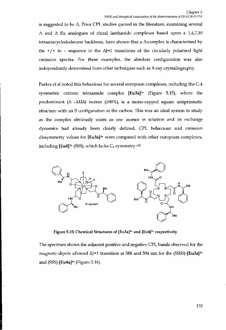
Chapter 5 NMR and chiroptical examination of the diastereisomers of (S)-EOB-DTPA
is suggested to be A. Prior CPL studies quoted i n the literature, examining several
A and A Eu analogues of chiral lanthanide complexes based upon a 1,4,7,10
tetiaazacyclododecane backbone, have shown that a A-complex is characterised by
the + / + to - sequence i n the AJ=1 tiansitions of the circularly polarised light
emission spectia. For these examples, the absolute configuration was also
independently determined f r o m other techniques such as X-ray crystallography.
Parker et al noted this behaviour for several europium complexes, including the C-4
symmetiic cationic tetiaamide complex [Eu3a]3+ (Figure 5.15), where the
predominant (A -AAAA) isomer (>90%), is a mono-capped square antiprismatic
stiucture w i t h an S configuration at the carbon. This was an ideal system to study
as the complex obviously exists as one isomer i n solution and its exchange
dynamics had already been clearly defined. CPL behaviour and emission
dissymmetiy values for [Eu3a]3+ were compared w i t h other europium complexes,
including [Eu4]3+ (SSS), which lacks Cn symmetry.i^^
Me.
HN^
Nr-* / Me y J N IN l ^ ~ - T ^ ^ f l ^
° L J o H N ^ ^ O " 1 ^ M e ^ N ^ ^ \ _ / ^
X=solvent O — \ Me NH
.Me
Me
Figure 5.15: Chemical Structures of [Eu3a]3+ and [Eu4]3+ respectively
The spectium shows the adjacent positive and negative CPL bands observed for the
magnetic-dipole allowed AJ=1 tiansition at 588 and 594 run for the (SSSS)-[Eu3a]3+
and (SSS)-[Eu4a]3^ (Figure 5.16).
131

Chapter 5 NMR and chiroptical examination of the diastereisomers of (S)-EOB-DTPA
(30
Figure 5.16 : Circualrly polarised emission spectra for (SSSS)-[Eu3a]3+ and (SSS)-[Eu4]3+ in water (1 mM complex, 295 K, Xexc = 255 nm.
The spectrum was compared to that obtained for the isomeric (RR) diamide
complex, where the complex shows mirror image behaviour, hence A heUcity
around the metal centre, corresponding to a reverse for the adjacent bands of the
AJ=1 transition at 588 and 594 n m (Figure 5.17).
M e
AJ =1 0.02
Q.Oi
a.oo« H
-n n?
HN OH,
-N \ / N H,0- -M
\ Kl -N \ N •OH,
NH
M e
Figure 5.17: Circularly polarised luminescence spectra for trans-(RR)-[Eu6]3+ in MeCN solution (1 mM complex, 295 K, Kc^ 255 nm).
Thus, i t can be concluded that the two isomers existing in solution were of A
helicity about the EU-OH2 vector.
132
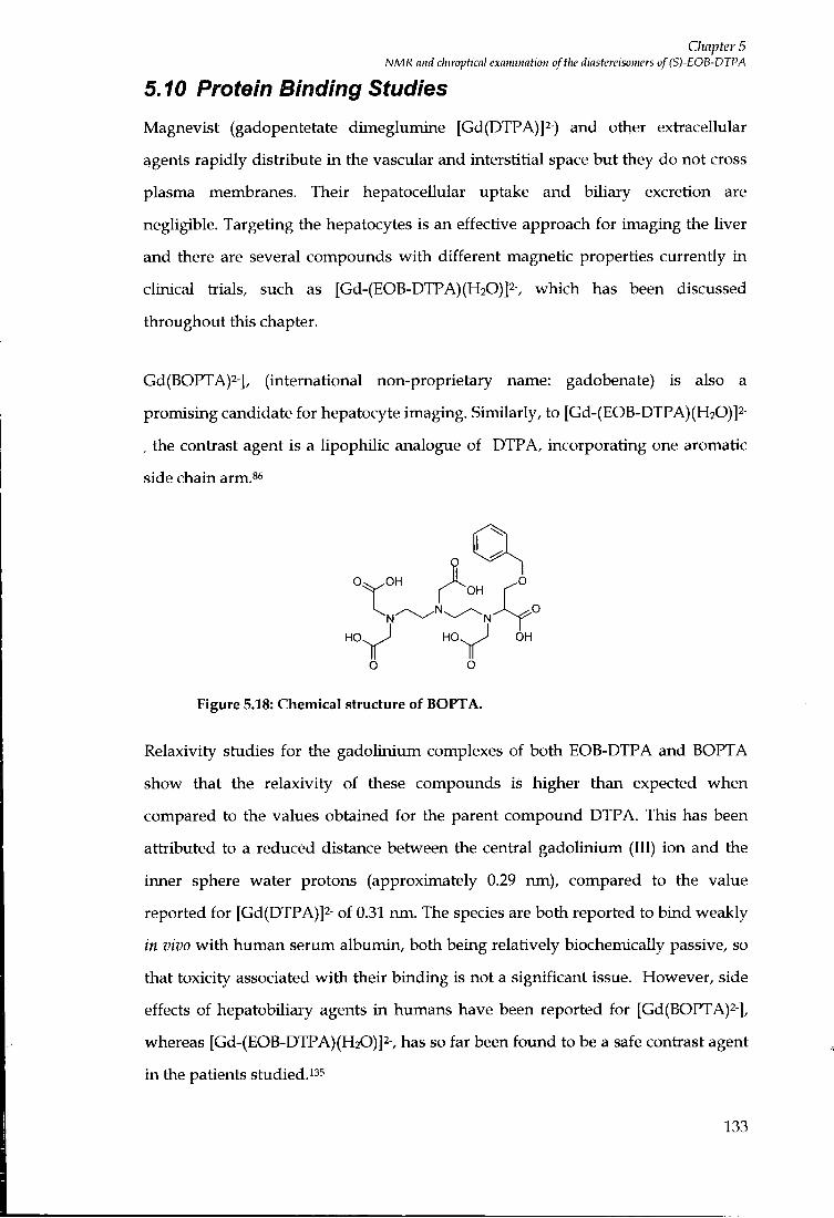
Chapter 5 NMR and chiroptical examination of tlie diastereisomers of (S)-EOB-DTPA
5.10 Protein Binding Studies
Magnevist (gadopentetate dimeglumine [Gd(DTPA)]2-) and other extracellular
agents rapidly distribute i n the vascular and interstitial space but they do not cross
plasma membranes. Their hepatocellular uptake and biliary excretion are
negligible. Targeting the hepatocytes is an effective approach for imaging the liver
and there are several compounds w i t h different magnetic properties currently in
clinical trials, such as [Gd-(EOB-DTPA)(H20)]2-, which has been discussed
throughout this chapter.
Gd(BOPTA)2-], (international non-proprietary name: gadobenate) is also a
promising candidate for hepatocyte imaging. Similarly, to [Gd-(EOB-DTPA)(H20)]2-
, the contrast agent is a lipophilic analogue of DTP A, incorporating one aromatic
side chain arm.^^
o
O O
Figure 5.18: Chemical structure of BOPTA.
Relaxivity studies for the gadolinium complexes of both EOB-DTPA and BOPTA
show that the relaxivity of these compounds is higher than expected when
compared to the values obtained for the parent compound DTP A. This has been
attributed to a reduced distance between the central gadoHnium (III) ion and the
inner sphere water protons (approximately 0.29 nm), compared to the value
reported for [Gd(DTPA)]2- of 0.31 nm. The species are both reported to bind weakly
in vivo w i t h human serum albumin, both being relatively biochemically passive, so
that toxicity associated w i t h their binding is not a significant issue. However, side
effects of hepatobiliary agents i n humans have been reported for [Gd(BOPTA)2-],
whereas [Gd-(EOB-DTPA)(H20)]2-, has so far been found to be a safe contrast agent
in the patients studied."^
133

Chapter 5 NMR and cliiropticnl examiiintioti of t)w diastereisomers of (S)-EOB-DTPA
[Gd-(EOB-DTPA)(H20)]2- exhibits a Ti relaxivity as measured in water at 0.47 Tesla
of 4.9 mM-is-i, compared to gadopentetate dimeglumine [Gd-(DTPA)]2- w i th a
relaxivity value of 3.7 mM-^s-^ However, a much higher Ti value of 8.2 mM-^s-i is
exhibited for [Gd-(EOB-DTPA)(H20)]2- i n the presence of human serum whereas
the relaxivity in human serum for [Gd(DTPA)]2- is 5.0 mM-^s-i 36 Hence, a greater
degree of protein binding, creating an increase i n molecular weight, hence an
increased t r effect, is exhibited by [Gd-(EOB-DTPA)(H20)]2- compared to [Gd-
(DTPA)]2-. [Gd(BOPTA)]2- is also found to bind moderately to human serum
albumin.^
The BOPTA hgand (4-carboxy-5,8,ll-tris(carboxymethyl)-l-phenyl-2-oxa-5,8,ll-
triazatridecan-13-oic acid) was kindly donated by Bracco SpA, Italy. The
homogeneity of the ligand sample was confirmed by i H and i^C NMR. Gadolinium
(111) complexes of BOPTA and EOB-DTPA were examined in protein binding
studies, al lowing a comparison w i t h earlier work (Chapter 2) and wi th the
literature. The protein binding interaction of each of the gadolinium complexes was
assessed by measuring the proton relaxation rates at 60 MHz , 37°C, in the presence
of water and then titrating the paramagnetic solution wi th increasing
concentrations of human serum albumin.
The protein binding curve for the gadolinium complex of [Gd-(EOB-DTPA)(H20)]2-
is shown in Figure 5.19.
134
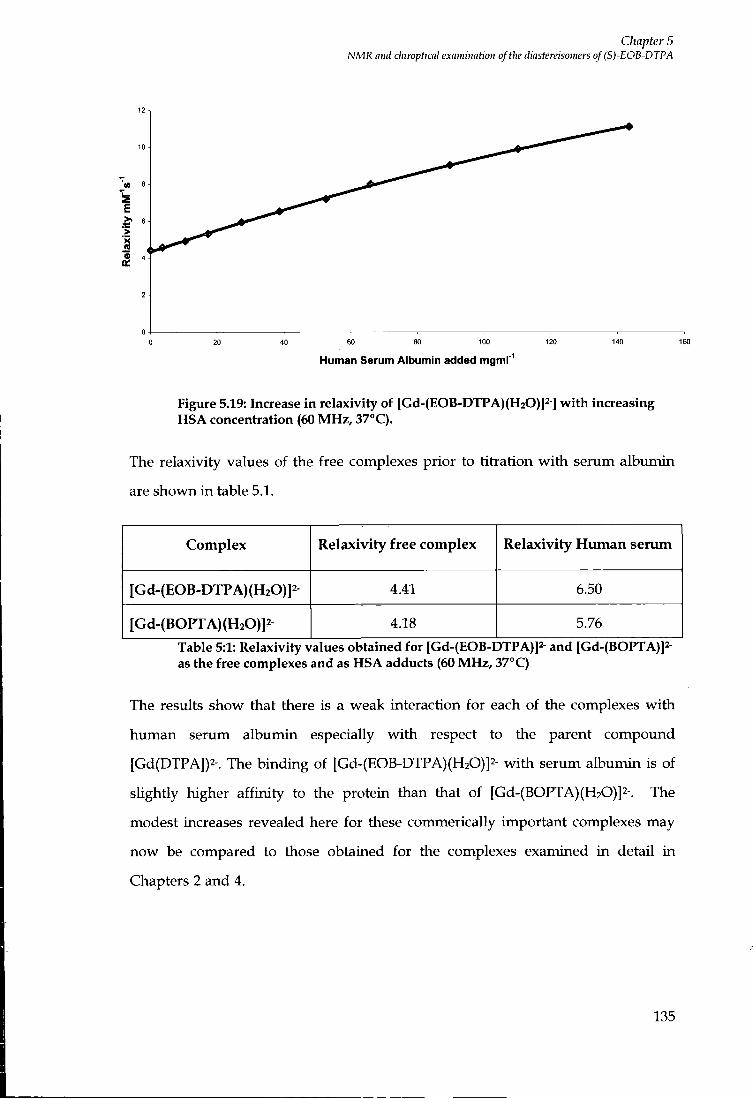
Chapter 5 NMR and chiroptknl exaimimtion oftlie diastereisomers of (S)-EOB-DTPA
Human Serum Albumin added mgml'
Figure 5.19: Increase in relaxivity of [Gd-(EOB-DTPA)(H20)]2-] with increasing HSA concentration (60 MHz, 37°C).
The relaxivity values of the free complexes prior to titration w i t h serum albumin
are shown in table 5.1.
Complex Relaxivity free complex Relaxivity Human serum
[G d-(EOB-DTPA) (H20)]2- 4.41 6.50
[Gd-(BOPTA)(H20)]2 4.18 5.76
Table 5:1: Relaxivity values obtained for [Gd-(EOB-DTPA)]2 and [Gd-(BOPTA)]2-as the free complexes and as HSA adducts (60 MHz, 37"C)
The results show that there is a weak interaction for each of the complexes wi th
human serum albumin especially w i t h respect to the parent compound
[Gd(DTPA])2-. The binding of [Gd-(EOB-DTPA)(H20)]2- w i t h serum albumin is of
sUghtly higher aff ini ty to the protein than that of [Gd-(BOPTA)(H20)]2-. The
modest increases revealed here for these commerically important complexes may
now be compared to those obtained for the complexes examined in detail i n
Chapters 2 and 4.
135

Conclusions and Suggestions for further work
Conclusions and Suggestions for further work
Although i t was hoped that heptadentate ligands based upon aD03A,
incorporating aromatic substituents, capable of binding to serum albumin may
result i n contrast agents w i t h very high relaxivities, these studies i n this thesis have
shown the relaxivity enhancements were not as large as hoped for. The complex,
[Gd(aD03A)]3- was not found to bind endogenous anions, such as carbonate,
phosphate and lactate at physiological p H . However, addition of hydrophobic
substituents on the ring nitrogen atom, producing a variety of tetrasubstituted
species, results i n complexes wi th increased affinities towards anions. It may be
possible to selectively increase the affinity towards serum albumin, yet prevent the
binding of anions fo l lowing an increased hydrogen bonding network based on an
aD02A based system (Figure 0.1).
o " o
o
N N
H
Figure 0.1: Schematic diagram of a putative aD02A based system
The phosphonate derivative of the aD03A ligand was recogrused as possessing a
higher negative peripheral charge in the lanthanide (III) complex, which was
anticipated to inhibit the binding of anions. The gadolinium (III) complex was
found to be a di-aqua species wi th similar relaxivities i n the p H range 3.5 to 6.0.
Above p H 6, carbonate and phosphate were fo imd to compete w i t h inner sphere
water molecules. The problems associated w i t h oUgomerisation and the time
dependence of p H reversibility, rendered this compound unsuitable for in vivo use.
The high relaxivities displayed in the presence of serum solutions d id suggest that
suitably derivatised compounds may yet be suitable for use as contrast agents.
Therefore, this idea may be pursued further by the synthesis and characterisation of
the compound depicted in Figure 0.2, fo l lowing the substitution of the ethyl groups
136
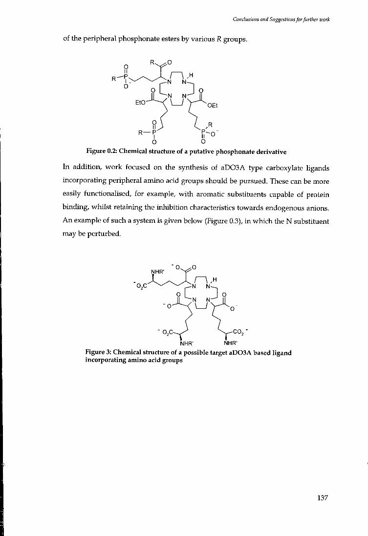
Conclusions and Suggestions for further work
of the peripheral phosphonate esters by various R groups.
R-
O ~ V
o I J o O
o Figure 0.2: Chemical structure of a putative phosphonate derivative
In addition, work focused on the synthesis of aD03A type carboxylate ligands
incorporating peripheral amino acid groups should be pursued. These can be more
easily functionalised, for example, w i th aromatic substituents capable of protein
binding, whilst retaining the inhibition characteristics towards endogenous anions.
A n example of such a system is given below (Figure 0.3), in which the N substituent
may be perturbed.
NHR'
N N
N N
NHR' NHR' Figure 3: Chemical structure of a possible target aD03A based ligand incorporating amino acid groups
137
Cluipter 6 Experimental
Chapter 6
Experimental
138
Clmpter 6 Experimental
6 Experimental
6.1 Synthetic Procedures and Characterisation
Reactions requiring anhydrous conditions were carried out using Schlenk-line
techniques under an atmosphere of dry argon. Solvents were dried f r o m an
appropriate drying agent when required and water was purif ied by the 'PuritesxiLL
plus' system w i t h a conductivity value of < 0.04 |iScm-i-. Reagents were used as
received f r o m suppUers unless otherwise stated.
Fluorescent silica plates (254 nm) were used for thin layer chromatography (Merck
A r t 5554) and visuaUsed using UV (254 nm) or iodine staining. Flash column
chromatography was carried out using silica purchased f r o m Merck (silica gel 60,
230-400 mesh). Ion exchange chromatography was performed using strong cation
exchange resin Dowex 50W x 4-200 or weakly acidic carboxylic acid cation
exchange resin Amberlite IRC-50.
N M R spectra were acquired using a Varian Mercury 200 spectrometer operating at
199.99 M H z (iH), 50.29 M H z ( " Q , 80.95 M H z (3iP), or a Varian Unity 300
spectrometer at 299.91 M H z (iH) and 75.41 M H z ("C) or a Varian Mercury 400
specb-ometer at 399.97 M H z (^H) and 50.58 M H z ("C) or a Varian kiova 500
spectiometer at 499.87 M H z (iH) and 125.71 (i^C). Specti-a were referenced
internally to the residual proton-solvent resonances and are reported relative to
TMS. Chemical shifts are given in values of p p m w i t h coupling constants i n Hz.
SpUtting patterns are described as singlet (s), doublet (d), triplet (t), quartet (q), or
multiplet (m). Variable temperature ^H N M R studies and two dimensional spectra
were recorded on a Varian Inova 500.
Electrospray mass spectra were acquired using a VG plat iorm (II) spectrometer
(Fisons instiuments), operating in positive or negative ion mode wi th methanol
used as a carrier solvent, typically using a cone voltage of 60 V and a source
temperature of 110°C. Accurate mass measurements were recorded at the
University of Durham using a Micromass LCT at 5000 resolution using sodium
139

Chapter 6 Experimental
iodide as a reference source.
l.R spectra were recorded on a Perkin-Ehner FT-IR 1720X spectrometer w i t h
GRAMS Analyst operating software; Melting points were determined on a
Reichert-Kofler block melting point apparatus and are uncorrected. Combustion
analysis was preformed using an Exeter Analytical Inc CE-440 elemental analyser.
6.2 Relaxivity Measurements
Longitudinal proton relaxation times were measured at 65 M H z using a custom
buil t 65.6 M H z Varian spectrometer operating at 295 K or at 60 M H z using a Bruker
Minispec m q 60 N M R analyser, operating at 310 K; both operated using an
inversion recovery technique. The concentration of gadoUnium in the sample was
typically in the range of 0.1-0.5 m M , measured by accurately digesting the complex
in nitric acid (5 M) for a period of time ranging f r o m 18 hours to approximately 48
hours. The relaxation rate of the digested sample was measured and the
concentration determined using equations 2.2-2.4 (Chapter 2).
Variable temperature proton-solvent longitudinal relaxation times were measured
in the range of 0.01 to 20 M H z in Durham on a Stelar Sputmaster-FFC 2000 Fast
Field Cycling NMR relaxometer (Stelar, Mede (PV), Italy) by means of the inversion
recovery technique (thirty two experiments, four scans). The reproducibility of Ti
measurements was w i th in 1%. The temperature was controlled by a Stelar VTC 90
airf low heater equipped w i t h copper constantan thermocouple: the actual
temperature i n the probe head was measured w i t h a Fluke 53 k / j digital
thermometer w i th an uncertainty of 0.5 K.
6.3 Photophysical Measurements
p H Measurements were made using a Jenway 3320 p H meter (fitted w i t h a BDH
glass and combination electrode microsampler) calibrated using p H 4, 7, and 10
buffer solutions.
Ultraviolet absorbance spectra were recorded using a Perkin Elmer Lambda 900
140

CImpter 6 Experimental
U V / V I S / N I R spectrometer operating UV WinLab L800/L900 software. Samples were contained in quartz cuvettes w i t h a path length of 1 cm. A l l spectra were acquired w i t h respect to a purite water reference measured i n a matched cell.
Luminescence spectra were recorded using a S.A. Fluorolog 3-11 system operating
using DataMax software. Quartz fluorescence cuvettes were employed w i t h a path
length of 1cm. Second order diffraction effects were obviated as a result of using a
cut-off filter to remove the scattered light before i t enters the emission
monochromator.
Excited state lifetimes were measured using a Perkin Elmer LS-55 spectrometer.
Measurements were made fo l lowing excitation of the sample by a short pulse of
light (348 nm) fol lowed by monitoring the integrated intensity of light (620 n m for
europium) emitted during a fixed gate time, tg, a delay time, f,;, later. A t least 20
delay times were used covering 3 or more lifetimes. A gate time of 0.1 ms was
used, and the excitation and emission slits were set to bandpass values of 2.5 and 15
nm respectively. The obtained decay curves were f i t ted using Microcal Origin.
6.4 Chapter 2 Experimental
6.4.1 Ligand Synthesis
Dimethyl 2-bromo-adipate (1)^^
O
O Br
A solution of monomethyladipic acid (21 g, 131 rrmiol) i n thionyl chloride (38 ml)
and carbon tetrachloride (60 ml) was heated to 65°C for 40 minutes. N -
Bromosuccinimide (27.8 g, 157 mmol), carbon tetrachloride (50 ml) and hydrogen
bromide (5 drops) were then added and the resulting orange solution was heated at
85°C for 2 hours. The resulting brown solution was cooled w i t h an ice bath and
quenched w i t h methanol (80 ml). The suspension which formed was stirred 141
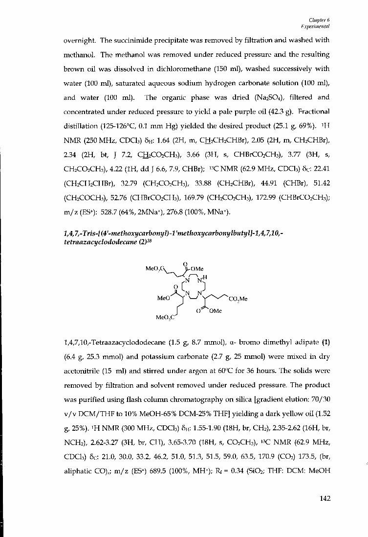
Chapter 6 Experimental
overnight. The succinimide precipitate was removed by fi l t rat ion and washed wi th methanol. The methanol was removed under reduced pressure and the resulting brown oi l was dissolved in dichloromethane (150 ml), washed successively wi th water (100 ml), saturated aqueous sodium hydrogen carbonate solution (100 ml), and water (100 ml). The organic phase was dried (Na2S04), filtered and concentiated tmder reduced pressure to yield a pale purple oi l (42.3 g). Fractional distillation (125-126°C, 0.1 m m Hg) yielded the desired product (25.1 g, 69%). NMR (250 MHz, CDCI3) 5H : 1.64 (2H, m, CHaCHaCHBr), 2.05 (2H, m, CHaCHBr), 2.34 (2H, bt, J 7.2, CH2CO2CH3), 3.66 (3H, s, CHBrCOzCHa), 3.77 (3H, s, CH2CO2CH3), 4.22 ( I H , dd J 6.6, 7.9, CHBr); N M R (62.9 MHz, CDCI3) 6c: 22.41 (CH2CH2CHBr), 32.79 (CH2CO2CH3), 33.88 (CH2CHBr), 44.91 (CHBr), 51.42 (CH2COCH3), 52.76 (CHBrCOzCHs), 169.79 (CH2CO2CH3), 172.99 (CHBrC02CH3); m / z (ES+): 528.7 (64%, 2MNa^), 276.8 (100%, MNa*).
l,4JrTris-[(4'-methoxycarbonyl)-l'methoxycarbonylbutyl]-l,4,7,10,-tetraazacyclododecane (2P^
MeO^G^__^^^^^ OMe
o L MeO J ^ Y ^ - ^ C O j M e
J O OMe MeO^C
1,4,7,10,-Tetraazacyclododecane (1.5 g, 8.7 mmol), a- bromo dimethyl adipate (1)
(6.4 g, 25.3 mmol) and potassium carbonate (2.7 g, 25 mmol) were mixed in dry
acetonitrile (15 ml) and stirred under argon at 60°C for 36 hours. The solids were
removed by fi l trat ion and solvent removed under reduced pressure. The product
was pur i f ied using flash column chromatography on silica [gradient elution: 70/30
v / v D C M / T H F to 10% MeOH-65% DCM-25% THF] yielding a dark yellow oi l (1.52
g, 25%). i H NMR (300 MHz, CDCI3) 6H : 1.55-1.90 (18H, br, CH2), 2.35-2.62 (16H, br,
NCH2), 2.62-3.27 (3H, br, CH), 3.65-3.70 (18H, s, CO2CH3), i- C N M R (62.9 MHz,
CDCI3) 6c: 21.0, 30.0, 33.2, 46.2, 51.0, 51.3, 51.5, 59.0, 63.5, 170.9 (CO2) 173.5, (br,
aliphatic CO),; m / z (ES^) 689.5 (100%, MH^); Rf = 0.34 (Si02; THF: DCM: MeOH
142

Ctmpter 6 Experimental
6:3:1); IR (thin fihn) Vmax (cm-i): 2919, 2850, 1729 (CO stietch) 1460, 1266, 1178, 918, 825.
1-2 '-(4-Tert-butyl-benzyl)-4,7,10-Ms-l(4'tnethoxycarbonyl)-l '-methoxycarbonylbutyl-l,4,7,10-tetraazacyclododecane (3)
MeO.'" 9\^0Me
MeO r ]
MeOX
l,4,7,-Tris-[(4'-methoxycarbonyl)-l'methoxycarbonylbutyl]-l,4,7,10,-
tetiaazacyclododecane (2) (360 mg, 0.52 mmol) was dissolved in dry acetonitiile
(5 ml). 4-(Tert-butyl) benzylbromide (118.7 mg, 0.52 mmol) and caesium carbonate
(170 mg, 0.52 mmol) were added. The resulting mixture was stirred overnight at
60°C under argon. The solids were removed by f i l t ia t ion and the solvent removed
under reduced pressure to yield a pale brown oil . The residue was purif ied by flash
column chromatography on silica [gradient elution-100% D C M to 10% MeOH-90%
DCM, Rf=0.42 (95% D C M - 5% MeOH)] producing a yellow oil (290 mg, 66%). i H
NMR (200 MHz, CDCI3) 6H : 1.29 (9H, s, (CH3)3C), 1.55-1.90 (18H, br, CH2), 2.42-2.35
(16H, br, NCH2) , 2.62-3.27 (3H, br, CH), 3.65-3.70 (18H, s, CO2CH3), 7.33 (2H, d,
A r H , J8.4 Hz), 7.45 (2H, d, A r H , J 8.4 Hz), m / z (ES^) 834 (100%, M+) , 835 (25%,
MH+); Accurate Mass Found: 835.5109 (MH+), C43H71N4O12 requires 835.5068.
2-(4-Tert-butyl-benzyloxy)-ethanol (4)
4-Tert-butyl benzyl bromide (11.0 g, 49.1 mmol), ethylene glycol (30.48 g, 0.49 mol),
sodium hydroxide (1.96 g, 49.1 mmol) and tetrapropylammonium bromide (55 mg,
0.21 mmol) were mixed in THF (150 ml), and the mixture was boiled under reflux
for 18 hours. The solvent was removed vmder reduced pressure and the residue
143

CImpter 6 Experimental
washed w i t h water (25 ml), ether (2 x 50 ml) and chloroform (25 ml) . The combined organic extracts were dried and the solvent removed under reduced pressure. Column chromatography on sUica [gradient elution; hexane to 20% methanol-hexane, Rf=0.16 (hexane)] gave a yellow oi l (7.68 g, 75%). i H NMR (200 MHz, CDCI3) 6H : 1.32 (9H, s, (CH3)3C), 2.28 ( I H , bs, OH), 3.58 (2H, t, CH2CH2OH, J 4.5Hz), 3.74 (2H, t, CH2OH, J 4.5 Hz), 4.53 (2H, s, 'Bu-CeHi-CHz), 7.26-7.30 (2H, d, A r H , ] 8.4 Hz), 7.36-7.41 (2H, d, A r H , J 8.4 Hz); " C N M R (69.2 MHz , CDCI3) 5c: 31.13, 34.52, 61.87, 71.30, 73.09,125.38,127.70,134.87,150.84; m / z (ES^) 231.0 (100%, MNa+), 439.3 (2MNa+); Accurate mass found 231.1357, Ci3H2o02Na requires 231.1361.
2-(4-Methoxy-benzyloxy)-ethanol (5)
2-(4-Methoxy-benzyloxy)-ethanol was synthesized by a similar method to that used
for (4), using 4-methoxybenzyl chloride (7.83 g, 50 mmol), ethylene glycol (31.04 g,
0.5 mol), sodium hydroxide (2.00 g, 50 mmol), tetrapropylammonium bromide (56
mg, 0.21 mmol) and THF (150 ml) to yield a yellow oi l (6.67 g, 73%).iH N M R (200
M H z , CDCI3) 5H: 2.06 ( I H , bs, OH), 3.58 (2H, t, CH2CH2OH , J 5.2Hz,), 3.71 (2H, t,
CH2OH, J 3.4Hz), 3.80 (3H, s, CH3O), 4.49 (2H, s, C6H4-C H2), 6.86-6.90 (2H, d, A r H ,
J 8.5Hz), 7.24-7.29 (2H, d, A r H , J 8.5 Hz), "C N M R (100.6 MHz , CDCI3) 5c: 55.50
(OCH3), 62.14, 71.29, 73.20, 114.09, 129.71, 130.25, 159.54; m / z (ES^) 204.9 (100%,
MNa+), 387.0 (60% 2MNa+); Accurate mass found 205.0859, CioHwOsNa requires
205.0865.
2-[4-(l,l-Dimethylethyl) phenyl) tnethoxy]-2ethyl p-toulene sulphonate (6)
A solution of 2-(4-tert-butyl-benzyloxy)-ethanol (4) (1.04 g, 5 mmol) in pyridine (20
ml) was cooled to <0°C imder argon. p-Toluene-sulfonyl chloride (0.935 g, 5 mmol)
144

CImpter 6 Experimental
was added in five equal parts and the solution maintained below 0 °C for 24 hours. The reaction mixture was poured onto crushed ice (150 g) and stirred unt i l fu l ly melted. The solid was removed by fi l trat ion and washed w i t h water to give a white solid (1.36 g, 75%). m N M R (250 MHz, CDCI3) 6H : 1.31 (9H, s, (CH3)3C), 2.43 (3H, s, CH3), 3.65 (2H, t, CH2CH2OTS, J 4.8 Hz), 4.18 (2H, t, CHj-tosyl, J 4.8 Hz), 4.45 (2H, s, »Bu-C6H4-CH2), 7.18-7.21 (2H, d, CH3C(CH)2, J 8.5 Hz), 7.33-7.36 (4H, dt, 'Bu-ArH) , 7.79 (2H, d, tosyl A r H , J 6.6 Hz); " C NMR (62.9 M H z , CDCI3) 5c: 21.61, 31.31, 34.51, 67.39, 69.27, 73.03,125.31,127.55,127.96,129.76,133.02,134.47,144.70,150.81; m / z (ES+) 385.1 (90%, MNa^), 747.3 (50%, 2MNa^); Elemental Analysis Found C% 65.94, H % 7.24, C20H26O4S requires C% 66.27, H % 7.23; Mp: 45-46 °C.
2-[(4-Methoxyphenyl)methoxy]-4-methyl benzenesulphonate (7)
TsO
Toulene-4-sulfonic acid 2-(4-methoxy-benyloxy)-ethyl-ester (7) was synthesised
using a similar method to that used for (6), using 2-(4-methoxy-benzyloxy)-ethanol
(5) (0.911 g, 5 mmol), p-toluene-sulfonyl chloride (1.9 g, 10 mmol) and pyridine (18
ml) to yield a white solid (1.20 g, 72%). i H NMR (200 M H z , CDCI3) 6H : 2.43 (3H, s,
CH3-C6H4), 3.62 (2H, t, CH2CH2OTS , J 4.8 Hz), 3.80 (3H, s, OCH3), 4.17 (2H, t,
CH2OTS, J 4.8 Hz), 4.41 (2H, s, CH3O-C6H4-CH2), 6.85 (2H, d, CHsOArH, J 8.6 Hz),
7.18 (2H, d, CH3CCH, J 8.4 Hz), 7.30 (2H, d, CHsO-ArH, J 8.4 Hz), 7.78 (2H, d,
tosyl-ArH, J 8.4 Hz), m / z (ES^) 358.9 (85%, MNa+), 695.1 (25%, 2MNa+); Elemental
Analysis: Found C% 60.78, H % 6.06; C17H20O5S requires C% 60.71, H % 5.99; Mp: 44-
45 °C
l-l2'A'l-Tert-hutyl-henzyloxy)-ethyl]-4J,10-tns4(4'-methoxycarhonyl)-V-methoxycarbonylbutyl-l,4:,7,10-tetraazacyclododecane (8)
MeQ ^3
MeOX r MeO o CO Me
145

Cliapter 6 Experimental
The amine (2) (160 mg, 0.233 mmol), the tosylate (6) (84 mg, 0.233 mmol) and caesium carbonate (75.6 mg, 0.233 mmol) were mixed in acetonitrile (4 ml) imder argon and the mixture was boiled under reflux for 4 hours. The solids were removed by f i l t ia t ion and the residue purif ied by flash column chromatography on silica [gradient elution; DCM to 10% MeOH-DCM, Rf =0.2 (DCM)] to yield a dark brown oi l (74 mg, 34 %) . i H N M R (200 M H z , CDCI3) 6H: 1.31 (9H, s, (CH3)3C), 1.55-1.85 (18H, br, CH2), 2.64-2.29 (16H, br, NCH2), 3.30-3.20 (3H, br, CH), 3.65 (18H, s, CO2CH3), 3.78 (2H, t, NCH2CH2O, J 4.8 Hz), 4.18 (2H, t, NCH2CH2O, J 4.8 Hz), 4.48 (2H, s, C6H4-CH2), 7.23 (2H, d, A r H , J 8.2 Hz), 7.32 (2H, d, A r H , J 8.2 Hz); m / z (ES+) 879.7 (100%, M+); Accurate mass found: 879.5329 (MH+), C45H74N4O13 requires 879.5330.
l-[2'-(4-Methoxy-benzyloxy)-ethyl]-4,7,10-tris-[(4'-methoxycarbonyl)-l'-methoxycarbonylbutyl-l,4,7,10-tetraazaq/clododecane (9)
MeO,Q O^OMe -sS N^OMe
P MeO o CO^Me
Compotmd (9) was synthesised by a similar method to that of (8), using (2) (258mg,
0.37mmol), (7) (120mg, 0.37mmol), caesium carbonate (122mg, 0.37mmol) and
acetonih-ile (5 ml) to yield a dark brown oi l (92.6 mg, 29%). ^H N M R (200 MHz,
CDCI3) 5H : 1.58-1.85 (br, 18H, CH2), 2.30-2.55 (br, 16H, NCH2), 3.20-3.40 (br, 3H,
CH), 3.40-3.65 (br, 18H, CO2CH3), 3.61 (t, J=6.2Hz, 2H, NCH2CH2O), 3.74 (s, 3H,
OCH3), 4.40 (t, J=6.2Hz, 2H, NCH2CH2O), 4.45 (s, 2H, C6H4-CH2, 6.84 (d, J=8.6Hz,
2H, meta A r H ) , 7.22 (d, J=8.6Hz, 2H, ortho A r H ) , m / z (ES^) 853.6 (100%, M*);
Accurate mass found: 853.4810, C42H68N4O14 requires 853.4808.
l-f2'-(4-Tert-bMty/-&en2y0-4,7,10-tns-f(4'carbo«xy)-l'-cflrboni//bMti/y-l,4,7,10-tetraazacyclododecane [ L^]
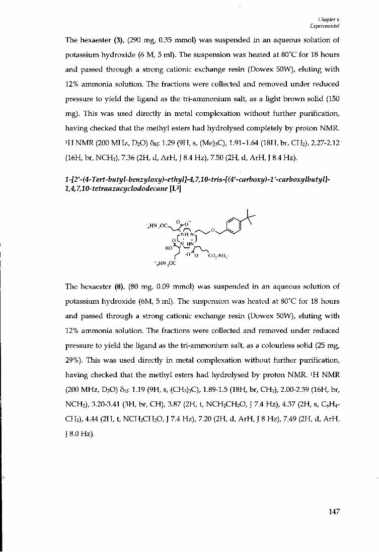
Chapter 6 Ex-perimental
The hexaester (3), (290 mg, 0.35 mmol) was suspended i n an aqueous solution of potassium hydroxide (6 M , 5 ml) . The suspension was heated at 80°C for 18 hours and passed through a stiong cationic exchange resin (Dowex 50W), eluting w i t h 12% ammonia solution. The fractions were collected and removed imder reduced pressure to yield the ligand as the tri-ammonium salt, as a Ught brown soUd (150 mg). This was used directly i n metal complexation without further purification, having checked that the methyl esters had hydrolysed completely by proton NMR. •H N M R (200 MHz, D2O) 5H : 1.29 (9H, s, (Me)3C), 1.91-1.64 (18H, br, CH2), 2.27-2.12 (16H, br, NCH2), 7.36 (2H, d, A r H , J 8.4 Hz), 7.50 (2H, d, A r H , J 8.4 Hz).
l-l2'-(4-Tert-butyl-benzyloxy)-ethyl]-4J,10-tris-[(4'-carboxy)-l'-car^^ 1,4,7,10-tetraazacyclododecane [L^]
. H N - P C ^ O ^ -
f-NH
%HN-pC
The hexaester (8), (80 mg, 0.09 rrunol) was suspended in an aqueous solution of
potassium hydroxide (6M, 5 ml) . The suspension was heated at 80°C for 18 hours
and passed through a strong cationic exchange resin (Dowex 50W), eluting wi th
12% ammonia solution. The fractions were collected and removed under reduced
pressure to yield the ligand as the tii-ammonium salt, as a colourless solid (25 mg,
29%). This was used directly i n metal complexation wi thout further purification,
having checked that the methyl esters had hydrolysed by proton NMR. i H NMR
(200 MHz, D2O) 6H: 1.19 (9H, s, (CH3)3C), 1.89-1.5 (18H, br, CH2), 2.00-2.39 (16H, br,
NCH2), 3.20-3.41 (3H, br, CH), 3.87 (2H, t, NCH2CH2O, J 7.4 Hz), 4.37 (2H, s, C6H4-
CH2), 4.44 (2H, t, NCH2CH2O, J 7.4 Hz), 7.20 (2H, d, A r H , J 8 Hz), 7.49 (2H, d, A r H ,
J 8.0 Hz).
147

Cluipter 6 Experimental
l-[2^-(4-Methoxy-benzyloxy)-ethyl]-4,7,10-tris-[(4^-carboxy)-V-carboxylbutyl]-1,4,7,10-tetraazacyclododecane [L^]
rOMe
r -0 o CO,-NHj+ +4HN-,0C
The hexaester (9), (92.6 mg, 0.11 mmol) was suspended in an aqueous solution of
potassium hydroxide (6M, 5 ml) . The suspension was heated at 80°C for 18 hours
and passed through a strong cationic exchange resin (Dowex 50 W), eluting w i t h
12% ammonia solution. The fractions were collected and removed xmder reduced
pressure to yield the ligand as the tri-ammonium salt, as a colourless solid (40.7 mg,
47%). This was used directly i n metal complexation without further purification,
having checked that the methyl esters had hydrolysed completely. ^H N M R (D2O,
200 MHz) 5H : 1.51-1.99 (18H, br, CH2), 2.00-2.50 (16H, br, NCH2), 3.20-3.44 (3H, br,
CH), 3.63 (2H, t, NCH2CH2, J 6.2 Hz), 3.74 (3H, s, OCH3), 4.39 (2H, t, NCH2CH2O, J
5.8 Hz), 4.43 (2H, s, CeHi-CHs), 6.99 (2H, d, A r H , J 8.1 Hz), 7.37 (2H, d, A r H , J 8.4
Hz).
2-(3,5-Dimethyl-phenoxy)-ethanol (10)
H O ^ ° ~ C ^
3,5 Di-(tertbutyl) phenol (11.1 g, 53.4 mmol), bromoethanol (3.8 ml , 53.4 mmol) and
potassium carbonate (7.4 g, 53.4 mmol) were dissolved i n acetonitrile (40 ml) and
the mixture boiled imder reflux for approximately 18 hours. The solid precipitate
that formed was filtered and washed using dichloromethane (10 ml), which was
removed under reduced pressure to yield a white, crystalUne solid. The solid
residue was dissolved into dichloromethane (3 ml) and puri f ied by colvunn
chromatography using silica, to yield a pale yellow, oily l iqu id (4.8 g, 36%);
148
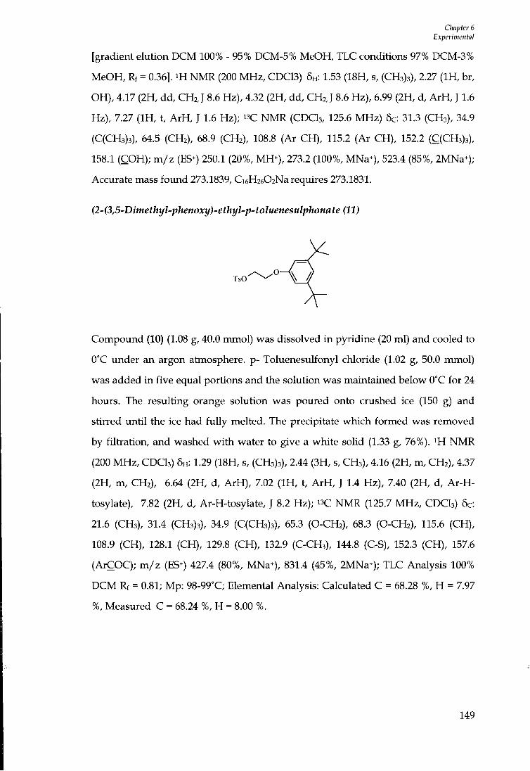
CImpter 6 Experimental
[gradient elution DCM 100% - 95% DCM-5% MeOH, TLC conditions 97% DCM-3% MeOH, Rf = 0.36]. iH NMR (200 MHz, CDC13) 5H: 1.53 (18H, s, (CH3)3), 2.27 ( I H , br, OH), 4.17 (2H, dd, CHzJ 8.6 Hz), 4.32 (2H, dd, CH2, J 8.6 Hz), 6.99 (2H, d, A r H , J 1.6 Hz), 7.27 ( I H , t, A r H , J 1.6 Hz); " C NMR (CDCI3, 125.6 MHz) 5c: 31.3 (CH3), 34.9 (C(CH3)3), 64.5 (CH2), 68.9 (CH2), 108.8 (Ar CH) , 115.2 (Ar CH), 152.2 (C(CH3)3), 158.1 (COH); m / z (ES^) 250.1 (20%, MH^), 273.2 (100%, MNa*), 523.4 (85%, 2MNa^); Accurate mass foxmd 273.1839, Ci6H2602Na requires 273.1831.
(2-(3,5-Dimethyl-phenoxy)-ethyl-p-toluenesulphonate (11)
TsO
Compound (10) (1.08 g, 40.0 mmol) was dissolved i n pyridine (20 ml) and cooled to
O'C under an argon atmosphere, p- Toluenesulfonyl chloride (1.02 g, 50.0 mmol)
was added in five equal portions and the solution was maintained below 0°C for 24
hours. The resulting orange solution was poured onto crushed ice (150 g) and
stirred unt i l the ice had fu l ly melted. The precipitate which formed was removed
by f i l t ia t ion, and washed w i t h water to give a white solid (1.33 g, 76%). ^H NMR
(200 M H z , CDCI3) 6H : 1.29 (18H, s, (CH3)3), 2.44 (3H, s, CH3), 4.16 (2H, m, CH2), 4.37
(2H, m, CH2), 6.64 (2H, d, A r H ) , 7.02 ( I H , t, A r H , J 1.4 Hz), 7.40 (2H, d, A r - H -
tosylate), 7.82 (2H, d, Ar-H-tosylate, J 8.2 Hz); isC NMR (125.7 MHz , CDCI3) 5c:
21.6 (CH3), 31.4 (CH3)3), 34.9 (C(CH3)3), 65.3 (O-CH2), 68.3 (O-CH2), 115.6 (CH),
108.9 (CH), 128.1 (CH), 129.8 (CH), 132.9 (C-CH3), 144.8 (C-S), 152.3 (CH), 157.6
(ArCOC); m / z (ES^) 427.4 (80%, MNa+), 831.4 (45%, 2MNa^); TLC Analysis 100%
D C M Rf = 0.81; Mp: 98-99°C; Elemental Analysis: Calculated C = 68.28 %, H = 7.97
%, Measured C = 68.24 %, H = 8.00 %.
149
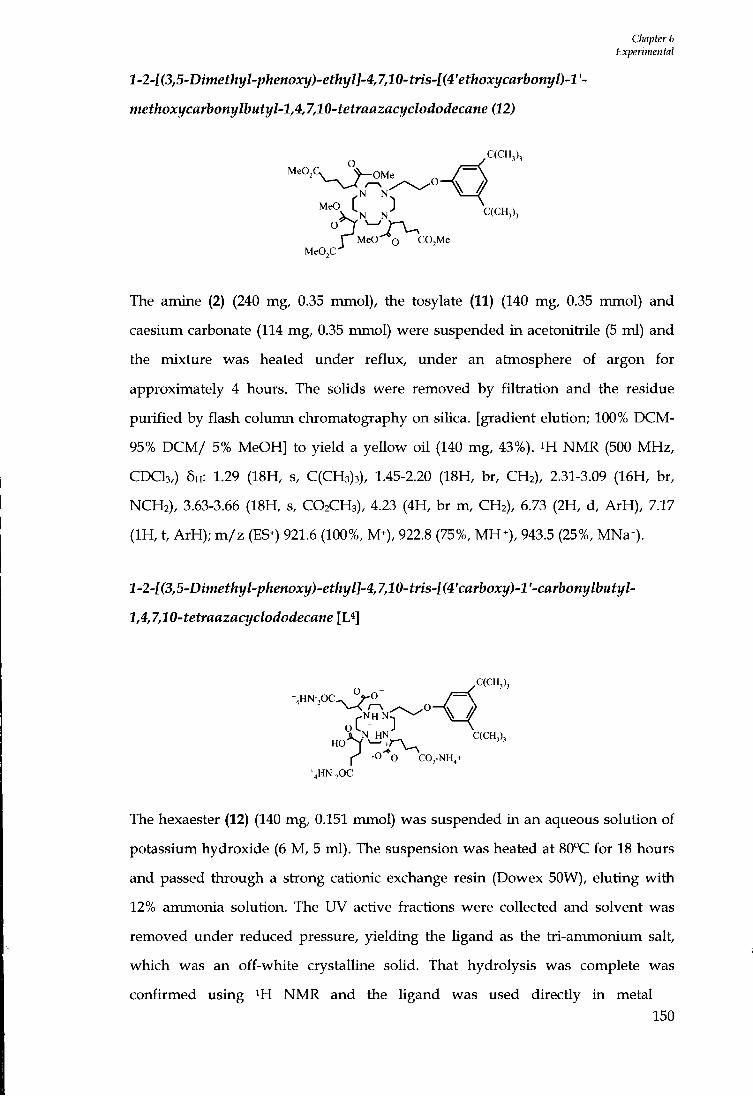
Chapter 6 Experimental
l-2-[(3,5-Dimethyl-phenoxy)-ethyl]-4J,10-tris-[(4'ethoxycarbonyl)-l'-
methoxycarbonylbutyl-1,4,7,10-tetraazacyclododecane (12)
, N N MeO C ]
" N N
p MeO o ' ^ C C M e
V C(CH,),
MeO,C'
The amine (2) (240 mg, 0.35 mmol), the tosylate (11) (140 mg, 0.35 mmol) and
caesium carbonate (114 mg, 0.35 mmol) were suspended i n acetonitrile (5 ml) and
the mixture was heated under reflux, under an atmosphere of argon for
approximately 4 hours. The solids were removed by fi l trat ion and the residue
purif ied by flash column chromatography on siUca. [gradient elution; 100% D C M -
95% D C M / 5% MeOH] to yield a yellow oi l (140 mg, 43%). i H N M R (500 MHz,
C D C I 3 , ) 5H: 1.29 (18H, s, C(CH3)3), 1.45-2.20 (18H, br, CH2), 2.31-3.09 (16H, br,
NCH2), 3.63-3.66 (18H, s, CO2CH3), 4.23 (4H, br m, CH2), 6.73 (2H, d, ArH) , 7.17
( I H , t, ArH) ; m / z (ES^) 921.6 (100%, M^), 922.8 (75%, M H ^ ) , 943.5 (25%, MNa+).
l-2-[(3,5-Dimethyl-phenoxy)-ethyl]-4,7,10-tris-[(4'carboxy)-l'-carbonylbutyl-
1,4,7,10-tetraazacyclododecane [L*]
%HN-,OC. O - /C(CH3),
HO 1^ -Co COj-NH,+
+4HN-2OC
The hexaester (12) (140 mg, 0.151 mmol) was suspended in an aqueous solution of
potassium hydroxide (6 M , 5 ml). The suspension was heated at 80°C for 18 hours
and passed through a strong cationic exchange resin (Dowex SOW), eluting wi th
12% ammonia solution. The UV active fractions were collected and solvent was
removed under reduced pressure, yielding the Ugand as the tri-ammonium salt,
which was an off-white crystalline solid. That hydrolysis was complete was
confirmed using ^H N M R and the Ugand was used directly i n metal
150
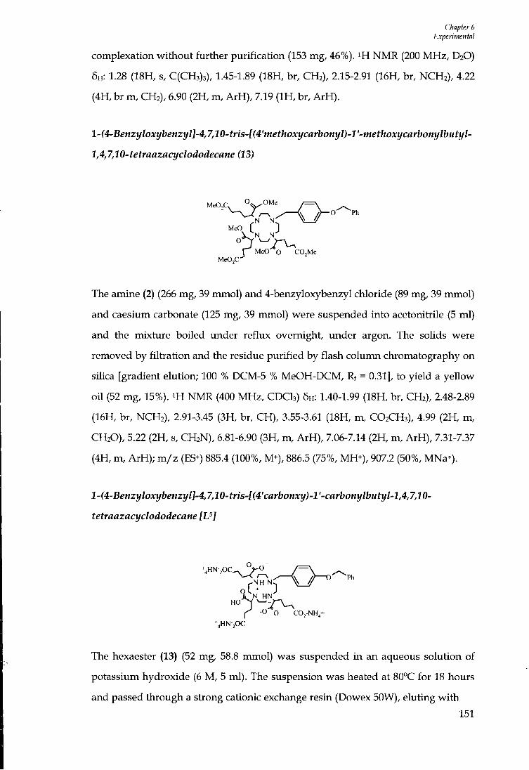
Chapter 6 Experimental
complexation without further purification (153 mg, 46%). i H NMR (200 M H z , D2O) 5H: 1.28 (18H, s, C(CH3)3), 1.45-1.89 (18H, br, CH2), 2.15-2.91 (16H, br, NCH2), 4.22 (4H, br m , CH2), 6.90 (2H, m, A r H ) , 7.19 ( I H , br, A r H ) .
l-(4-Benzyloxybenzyl]-4,7,10-tns-l(4'methoxycarbonyl)-l'-methoxycarbonylbutyl-
1,4,7,10-tetraazacyclododecane (13)
MeO,C, "O"^^
N N: ^ / Ph
MeO r 1 P MeO O COjMe
M e C C ^
The arrune (2) (266 mg, 39 mmol) and 4-benzyloxybenzyl chloride (89 mg, 39 rrm\ol)
and caesium carbonate (125 mg, 39 mmol) were suspended into acetonitrile (5 ml)
and the mixture boiled under reflux overnight, imder argon. The solids were
removed by f i l t rat ion and the residue purif ied by flash column chromatography on
silica [gradient elution; 100 % DCM-5 % MeOH-DCM, Rf = 0.31], to yield a yellow
oi l (52 mg, 15%). m NMR (400 MHz, CDCI3) 5H: 1.40-1.99 (18H, br, CH2), 2.48-2.89
(16H, br, NCH2), 2.91-3.45 (3H, br, CH), 3.55-3.61 (18H, m, CO2CH3), 4.99 (2H, m,
CH2O), 5.22 (2H, s, CH2N), 6.81-6.90 (3H, m, A r H ) , 7.06-7.14 (2H, m, A r H ) , 7.31-7.37
(4H, m, ArH) ; m / z (ES^) 885.4 (100%, M+), 886.5 (75%, MH+), 907.2 (50%, MNa*).
l-(4-Benzyloxybenzyl]-4,7,10-tris-[(4'carbonxy)-l'-carbonylbutyl-l,4,7,10-
tetraazacyclododecane [U]
% H N , 0 C ^ / - O
X N HN
f -O O COj-NH^+ %HN-pC
The hexaester (13) (52 mg, 58.8 mmol) was suspended in an aqueous solution of
potassium hydroxide (6 M , 5 ml). The suspension was heated at 80°C for 18 hours
and passed through a strong cationic exchange resin (Dowex 50W), eluting w i t h
151
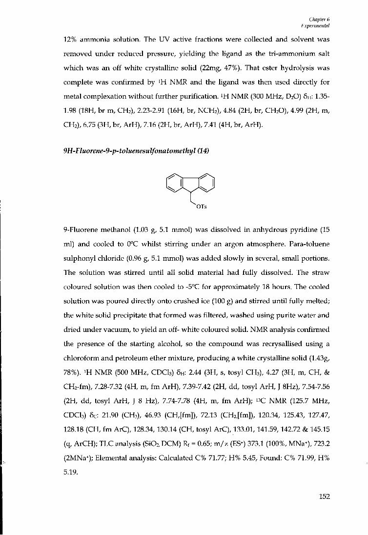
Chapter 6 Experimental
12% ammonia solution. The UV active fractions were collected and solvent was removed under reduced pressure, yielding the ligand as the tri-ammonium salt which was an off white crystaUine soUd (22mg, 47%). That ester hydrolysis was complete was confirmed by NMR and the Ugand was then used directly for metal complexation without further purification. N M R (300 MHz, D2O) 5H: 1.35-1.98 (18H, br m, CH2), 2.23-2.91 (16H, br, NCH2), 4.84 (2H, br, CH2O), 4.99 (2H, m, CH2), 6.75 (3H, br, A r H ) , 7.16 (2H, br, ArH) , 7.41 (4H, br, A r H ) .
9H-Fluorene-9-p'-toluenesulfonatomethyl (14)
OTs
9-Fluorene methanol (1.03 g, 5.1 mmol) was dissolved i n anhydrous pyridine (15
ml) and cooled to 0°C whilst stirring under an argon atmosphere. Para-toluene
sulphonyl chloride (0.96 g, 5.1 mmol) was added slowly i n several, small portions.
The solution was stirred unt i l all solid material had fu l l y dissolved. The stiaw
coloured solution was then cooled to -5°C for approximately 18 hours. The cooled
solution was poured directly onto crushed ice (100 g) and stirred unt i l f u l l y melted;
the white soUd precipitate that formed was filtered, washed using purite water and
dried imder vacuum, to yield an off- white coloured soUd. N M R analysis confirmed
the presence of the starting alcohol, so the compoimd was recrysallised using a
chloroform and petroleum ether mixture, producing a white crystalline solid (1.43g,
78%). N M R (500 MHz, CDCI3) 5H: 2.44 (3H, s, tosyl CH3), 4.27 (3H, m, CH, &
CH2-fm), 7.28-7.32 (4H, m, f m A r H ) , 7.39-7.42 (2H, dd, tosyl A r H , J 8Hz), 7.54-7.56
(2H, dd, tosyl A r H , J 8 Hz), 7.74-7.78 (4H, m, f m ArH) ; " C N M R (125.7 MHz,
CDCI3) 5c: 21.90 (CH3), 46.93 (CH,[fm]), 72.13 (CH2,[fm]), 120.34, 125.43, 127.47,
128.18 (CH, f m ArC), 128.34,130.14 (CH, tosyl ArC), 133.01,141.59,142.72 & 145.15
(q, ArCH); TLC analysis (Si02,DCM) Rf = 0.65; m / z (ES^) 373.1 (100%, MNa^), 723.2
(2MNa-); Elemental analysis: Calculated C% 71.77; H % 5.45, Found: C% 71.99, H %
5.19.
152

CImpter 6 Experimental
2-(9H-Fluoren-9-yl)-ethanol (15)
OH
9-Fluorene-acetic acid (200 mg, 0.90 mmol) was dissolved into dry tetrahydrofuran
(2 ml) and stirred under argon at -40°C. Borane-THF solution (490 \iL, 1.0 mmol)
was added dropwise and the resulting mixture was stirred at room temperature for
approximately 18 hours. The clear solution was cooled to 0°C and quenched using
methanol (5 ml). The solution was stirred for a further 2 hours under an argon
atmosphere. The reaction mixture was then tieated w i t h diethyl ether (20 ml) and
washed successively w i t h water, saturated aqueous sodium carbonate solution and
water. The organic layer was dried (MgS04), and the organic solvent removed
under reduced pressure, to yield a white solid (80 mg, 42%). i H N M R (400 MHz,
CDCI3) 6H: 1.25-1.30 ( I H , br, OH), 2.29-2.34 (2H, q, CH2, J 6.8 Hz), 3.59-3.62 (2H, t,
CH2, J 6.8 Hz), 4.13-4.16 ( I H , t, CH, J 5.6 Hz), 7.32-7.84 (4H, m, A r H ) , 7.54-7.56 (4H,
dd, A r H , J 7.6 Hz); "C N M R (125.7 MHz, CDCI3) 5c: 35.99, 46.86 (CH2), 60.52 (CH),
120.28, 124.72, 127.29, 127.44 (ArC), 141.23, 147.07 (q, C); m / z (ES^) 233.0 (70%,
MNa*); Elemental analysis found C% 80.20, H % 6.99 C15H14O. 4/5 H2O requires C%
80.18, H % 6.99.
9H-fluorene-9-p-toluenesulfonatoethyl (16)
OTs
2-(9H-Fluoren-9-yl)-ethanol (15) (35 mg, 0.167 mmol) was dissolved into anhydrous
pyridine (10 ml) and stirred under argon at 0 °C. p-Toluene sulphonyl chloride (32
mg, 0.167 mmol) was added slowly i n two equal parts. The solution was stirred
until solids were fu l ly dissolved maintaining a temperature of 0°C, then cooled to -
5''C for approximately 18 hours. The orange coloured solution was poured directly
153

Cliapter 6 Experimental
onto crushed ice (100 g) and stirred unt i l the ice had fuUy melted. The white solid
precipitate that formed was filtered, washed using distilled water and dried over
P2O5 under vacuum, to yield a white soUd. ^H NMR (CDCI3, 400 MHz) 6H: 2.21-2.26
(2H, q, CH2, J 6.4 Hz), 3.91-3.99 (3H, m, CH2 & CH), 7.19-7.28 (8H, m, ArH) , 7.62-
7.63 (2H, d, A r H tosyl, J 8 Hz), 7.65-7.66 (2H, d, A r H tosyl); " C N M R (CDCI3,100.6
MHz) 6c: 21.62 (CH3), 32.35 (CH2), 43.68 (CH2), 67.75 (CH), 120.03, 124.29, 127.09,
127.90 (CH-fm ArC), 128.90,129.80 (CH tosyl ArC), 145.69,140.89,134.70,133.05 (q,
ArC); m / z (ES^) 387.1 (50%, MNa^).
l-2'-(9-Propyl-9H-fluorene)-4,7,10-tris-[(4'methoxycarbonyl)-l'-
niethoxycarbonylbutyl-l,4,7,10-tetraazacyclododecane (17)
MeO
9H-fluorene-9-p-toluenesulfonatoethyl (16) (295 mg, 0.80 mmol) was dissolved in
dry acetonitrUe (5 ml). l,4,7,-Tris-[(4'-methoxycarbonyl)-l'methoxycarbonylbutyl]-
1,4,7,10,-tetraazacyclododecane (2) (280 mg, 0.41 mmol) and caesium carbonate (264
mg, 0.81 mmol) were then added. The resulting mixture was heated imder reflux
for approximately 36 hours under an argon atmosphere. The solid residues were
removed by fi l trat ion and washed wi th dichloromethane (20 ml) . The organic
solvent was removed by reduced pressure, to yield a crude, brown oily product
(295 mg). The required product was purif ied by flash chromatography on siKca
[100% DCM- 95% DCM- 5% MeOH]. The fractions were collected and solvent
removed by reduced pressure to yield a brown oily product (75 mg, 21%). H N M R
(300 M H z , CDCI3) 6H: 1.41-1.77 (16H, br m, CH2), 2.26-2.33 (6H, br m, CH2), 2.41-
3.10 (12H, br m, CH2), 3.42-3.48 (3H, br, CH) , 3.68 (21H, m, CO2CH3 & CH2 & CH),
7.34-7.67 (8H, br m, ArH) ; m / z (ES+) 881.3 (100%, M H ) , 463.5 (48%, V2MNa^); TLC
154

Cliapter 6 Experimental
[Si02 D C M - 95% D C M - 5% MeOH] Rf = 0.35.
1-2 '-(9-Propyl-9H-fluorene)-4,7,10-tris-[ (4'carboxy)-l '-carbonylbutyl-1,4,7,10-
tetraazacyclododecane
- N H N
f -O O C02-NH^+
%HN-jOC
The hexaester (17) (74 mg, 0.084 mmol) was dissolved into l i th ium hydroxide
solution (1 M , 10 ml), and the solution was heated under reflux for approximately
72 hours. The resulting mi lky white solution was neutialized to p H 6.5 prior to ion
exchange using hydrochloric acid (O.IM). The product was loaded on a stiong
cation exchange column (Dowex -50) and washed using Purite water p H 6. The
product was eluted f r o m the column using 12% ammonia solution, which was
removed f rom the compound by reduced pressure, yielding the ligand as the t r i -
ammonium salt, a white crystalline solid. That ester hydrolysis was complete was
confirmed using ^H NMR and the ligand was used directly for metal complexation
without further purification (36 mg, 49%). As the synthetic pathway is analogous
for the C2 and the C i appended aD03A Hgand system [L*] and [L^ respectively; the
synthetic details wiU only be described for the C2 variant.
l-2'-(N-Ethyl-acridone),4,7,10-trisl(4'methoxycarbonyl)-l'-methoxy carbonylbutyl
(18)
MeOX'
COjMe
N N
N N
MeOX
155

Chapter 6 Experimental
Mono-substituted N-acridoneethyl 1,4,7,10-tetraazacyclododecane (215 mg, 0.55 mmol) was donated by Y. Bretonneire, University of Durham. The compound was dissolved into dry acetonitrile (5 ml). Dimethyl 2-bromo adipate (1) (425 mg, 1.67 mmol) and caesium carbonate (547 mg, 1.67 mmol) were added. The resulting mixture was heated under reflux for approximately 96 hours. The solids were removed by fi l trat ion and washed w i t h acetonitrile, the solution being concentrated by reduced pressure to yield a brown crude mixture. The required product was purif ied by flash column chromatography on silica [Si02, D C M ^ 3 % M e O H in DCM] (67 mg, 13%). ^H N M R (CDCI3, 500 MHz) 5H: 1.57-1.82 (16H, br, CH2), 2.28-3.12 (19H, br, NCH2 x 16H & C H x 3H), 3.59-3.72 (20H, br, CO2CH3 x 18H & CH2), 4.51 (2H, br, CH2), 7.33 (2H, m, ArH) , 7.59 ( I H , br, A r H ) , 7.78 (3H, m, A r H ) , 8.59 (2H, dd, A r H , J 7 Hz); " C N M R partial assignment (CDCI3, 125.6 MHz) 5c: 19.79, 20.93, (CH), 25.63, 29.96, 31.28 (CH2), 33.54, 33.81 (NCH2), 51.81 (OCH3), 121.83, 128.48, 134.58 (ArC), 173.87 (C=0); m / z (ES^) 976.3 (100%, MCu^); A suitable accurate mass could not be obtained for this compound.
l-2'-(N-ethyl-acridone),4,7,10-trisl(4'carboxy)-r-carbonylbutyl [L*]
Q Q
,N N
LiO-
A suspension of (18) (60 mg, 0.065 mmol) and l i th ium hydroxide (54 mg) i n water
(1 ml) was left stirring at room temperature for 24 hours. The hydrolysis of the
methyl esters was monitored by ^H NMR. The ligand was complexed in sitii. ^H
N M R was used to confirm aU methyl esters were hydrolysed. ^H N M R (200 MHz,
D2O) 6H: 1.41-3.01 (37 H , br, CH2'sand NCH2's), 7.51-8.70 (8H, br, A r H ) .
Methyl-3-benzyloxy-2-chloro-propionate (19)
o
^ CI OMe
156

Chapter 6 Experimental
The potassium salt of 3-benzyloxy-2-chloro-propionic acid (0.53 g, 2.097 mol), was dried thoroughly and then dissolved in dry methanol (10 ml). Acetyl chloride (0.2 ml) was added and the resulting mixture was heated under reflux under an argon atmosphere for 18 hours. The solution was cooled to room temperature and dissolved into diethyl ether (30 ml). The organic phase was washed successively using water (20 ml) sodium bicarbonate solution (pH 8.5, 20 ml) and water (20 ml). The organic phase was dried (Mg2S04), and the organic solvent removed at reduced pressure to yield a ti-ansparent oily product (0.50 g, 87%). i H N M R (CDCI3, 500 MHz) 6H: 7.23-7.28 (5H, m, A r H ) , 4.52 (2H, s, CHzAr), 4.34-4.37 ( I H , t, CH, J 6 Hz), 3.71-3.74 (4H, m, OCH3 & CH); i3C N M R (CDCI3, 125.7 MHz) 6c: 53.39 (OCH3), 54.79 (C*), 71.16 (CH2C*), 73.82 (Ar-CH2), 128.03, 128.25, 128.77 (ArCH), 137.54 (q, C), 168.89 (C=0); m / z (ES^) 251.0 (100%, MNa^), 267.7 (48%, M K - ) ; Accurate mass found 251.0457, CnHisOjClNa requires 251.0451.
l-3'-(BenzyIoxy-propionic acid methyl ester)-4,7,-di-[4' (methoxycarbonyl)-l' -
methoxycarbonylbutyl]-l,4,7,10-tetraazacyclododecane (20)
o MeO
o OMe
OMe OMe
Methyl-3-benzyloxy-2-chloro-propionate (19) (118 mg, 0.474 mol) and 1,7-bis-
[(4 methoxycarbonyl)-l-methoxycarbonylbutyl-l,4,7,10-tetraazacyclododecane,
(245 mg, 0.474 mol) were suspended in dry acetonitrile (5 mL). Caesium carbonate
(154 mg, 0.474 mol) was added and the resulting mixture heated under reflux under
an argon atmosphere for approximately 48 hours. The solid residue was filtered
and washed using dichloromethane. The combined organic washings were
evaporated under reduced pressure yielding a crude brown oily product (270 mg).
The required product was purif ied by flash chromatography on silica [100% D C M -
95% D C M - 5% MeOH]. The fractions were collected and solvent removed at
reduced pressure to yield a b rown oily product (22 mg, 6%). i H N M R (CDCI3, 400
157
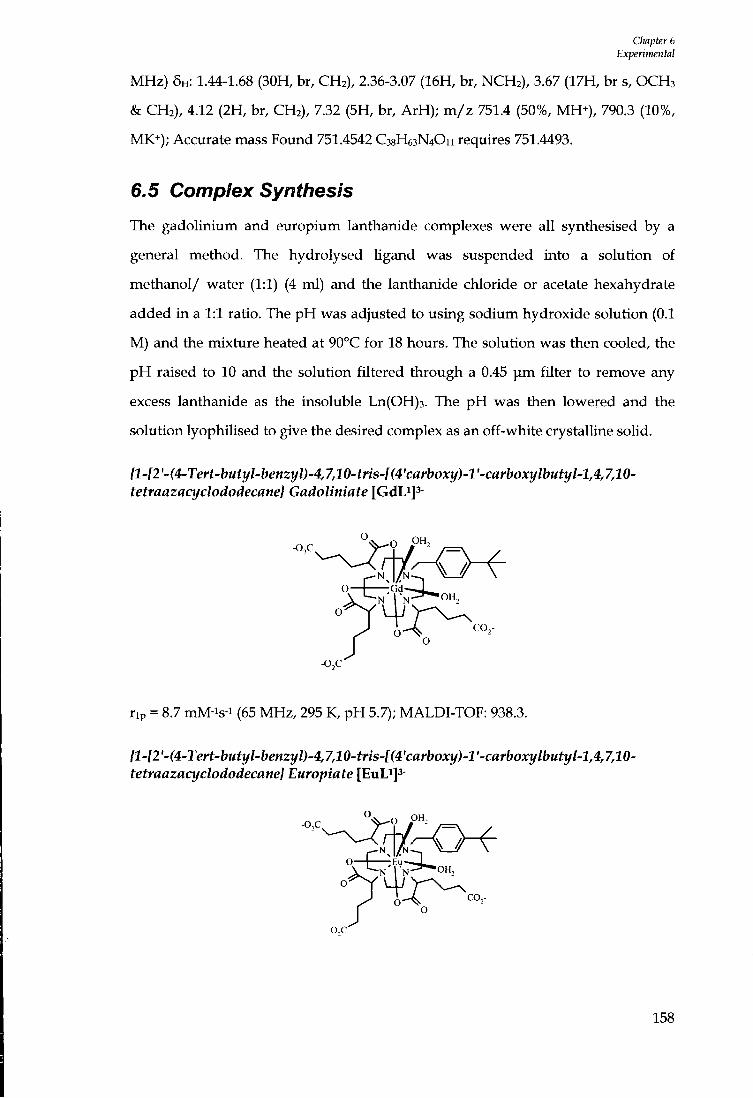
Chapter 6 Experimental
MHz) 5H: 1.44-1.68 (30H, br, CH2), 2.36-3.07 (16H, br, NCH2) , 3.67 (17H, br s, O C H 3 & CH2), 4.12 (2H, br, CH2), 7.32 (5H, br, ArH) ; m / z 751.4 (50%, MH^) , 790.3 (10%, MK*); Accurate mass Found 751.4542 C38H63N4O11 requires 751.4493.
6.5 Complex Synthesis
The gadolinium and europium lanthanide complexes were all synthesised by a
general method. The hydrolysed hgand was suspended into a solution of
methanol/ water (1:1) (4 ml) and the lanthanide chloride or acetate hexahydrate
added in a 1:1 ratio. The p H was adjusted to using sodium hydroxide solution (0.1
M) and the mixture heated at 90°C for 18 hours. The solution was then cooled, the
p H raised to 10 and the solution filtered through a 0.45 | i m filter to remove any
excess lanthanide as the insoluble Ln(OH)3. The p H was then lowered and the
solution lyoplulised to give the desired complex as an off-white crystalline solid.
{l-[2'-(4-Tert-butyl-benzyl)-4,7,10-tns-[(4'carboxy)-l'-carboxylbutyl-l,4,7,10-tetraazacyclododecane) Gadoliniate [GdLi]3-
-o,c O pH,
OH,
CO,
rip = 8.7 mM-is-i (65 MHz, 295 K, p H 5.7); MALDI-TOF: 938.3.
{l-l2'-(4-Tert-butyl-henzyl)-4,7,10-tns-l(4'carboxy)-l'-carboxylbutyl-l,4,7,10-tetraazacyclododecane} Europiate [EuLiJ^-
-o,c OH,
o— :Eu~-J^
J cc-O
158

Ouipter 6 Experiinentnl
^H N M R (200 MHz, D2O): The ^H spectrum was exchanged broadened.
{l-l2'-(4-Tert-hutyl-hmzyloxy)-ethyl]-4J,10-tris4(4'carhoxy)-l'-carhoxyhutyl-1,4,7,10-tetraazacyclododecane} Gadoliniate [GdL^js-
o. -o,c.
0—\ Gd L
CO,-
rip^ 10.2 mM-i s-i (65 MHz, 295 K, p H 5.3); m / z (ES-) 947 [M-].
{l-l2'A4-Tert-hutyl-henzyloxy)-4J,10-tns-[(4'carhoxy)-V-carhoxylhutyl-l,4J,10-tetraazacyclododecane] Europiate [EuL^]^-
o -o,c
0 P"2
° o
OH,
N CO,-
i H N M R (200 MHz, D2O): The spectium was exchanged broadened; m / z (ES ) 943
[ M ]
{l-[2'-(4-Methoxy-benzyloxy)-ethyl]-4,7,10-tris-[(4'carboxy)-l'-carboxybutyl-1,4,7,lO-tetraazacyclododecane] Gadoliniate [GdL^Js-
o H —
co,-
rip = 9.0 mM-is-i (65 MHz, 295 K, p H 5.7)
159
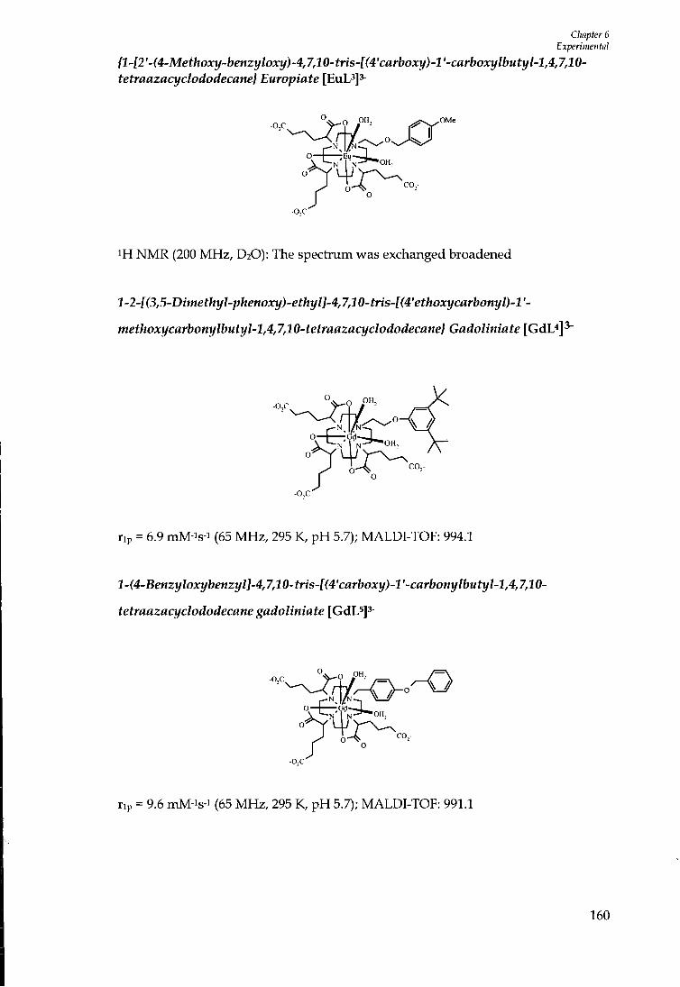
Chapter 6 Experimental
{l-l2'-(4-Methoxy-benzyloxy)-4,7,10-tris-[(4'carboxy)-l'-carboxylbutyl-l,4,7,10-tetraazacyclododecane} Europiate [ E u L 3 ] 3 -
-o,c
0—\ - E u - — i . OH,
co,-
^H N M R (200 MHz, D2O): The spectrum was exchanged broadened
l-2-[(3,5-Dimethyl-phenoxy)-ethyl]-4,7,10-tris-[(4'ethoxycarbonyl)-l'-
methoxycarbonylbutyl-l,4,7,10-tetraazacyclododecane} Gadoliniate [GdL«]^
-o,c
o o'
rip = 6.9 mM-is-i (65 MHz, 295 K, p H 5.7); MALDl-TOF: 994.1
l-(4-Benzyloxybenzyl]-4,7,10-tris-[(4'carboxy)-l'-carbonylbutyl-l,4,7,10-
tetraazacyclododecane gadoliniate [GdL^Js-
O — r.A^ I OH,
CO,-
rip = 9.6 mM-is-i (65 MHz, 295 K, p H 5.7); MALDl-TOF: 991.1
160
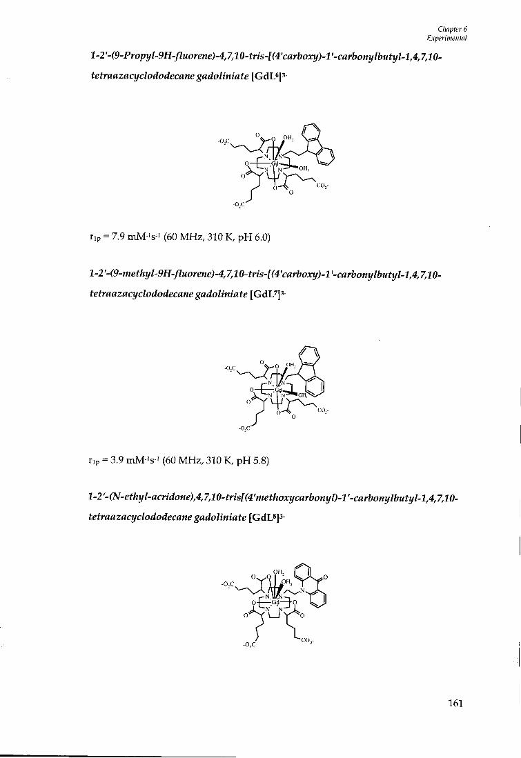
CImpter 6 Experimental
l-2^(9-Pro;?yWH-/7Morene^4,7,10-fns-K4'cflr{»oxi/)-l'-cflrbony/bMti//-l,4,740-
tetraazacyclododecane gadoliniate [GdLsjs-
rip = 7.9 mM-is-i (60 MHz, 310 K, p H 6.0)
l-2'-(9-methyl-9H-fluorme)-4,7,10-tris-[(4'carboxy)-l'-carbonylbu^
tetraazacyclododecane gadoliniate [GdL^3-
O OH,
rip = 3.9 mM-is-i (60 MHz, 310 K, p H 5.8)
2-2'-(N-e^/ly^flc^^^o«e),4,7,I0-^Wsf(4'me^/^oxyc«r&otty/>-^'-c«rbo«y/&M^y/-l,4,7,10-
tetraazacyclododecane gadoliniate [ G d L 8 ] 3 -
OH,
- 0 , C - C O , .
161
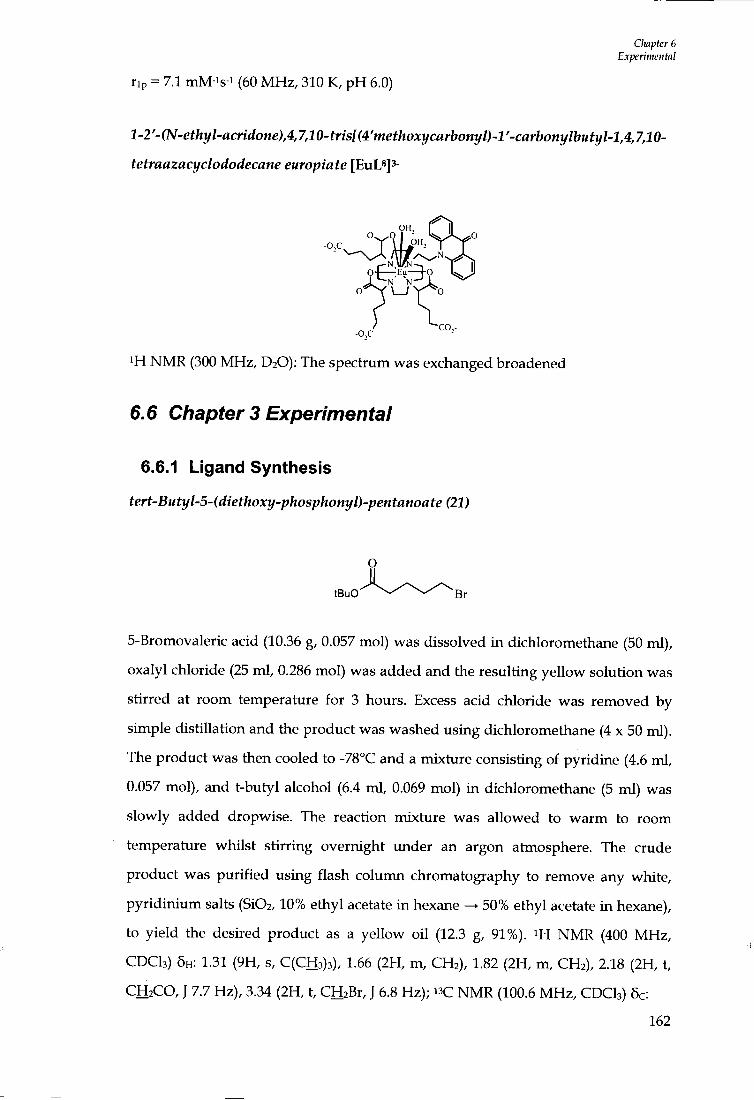
CImpter 6 Experimental
rip = 7.1 mM-is-i (60 M H z , 310 K, p H 6.0)
l-2'-(N-ethyl-acridone),4,7,10-tris[(4'methoxycarbonyl)-l'-carbonylbutyl-l,4,7,10-
tetraazacyclododecane europiate [EuL*]3-
EIM MAIN ~ , — E u — f O
-o,c
i H N M R (300 MHz, D2O): The spectrum was exchanged broadened
6.6 Chapter 3 Experimental
6.6.1 Ligand Synthesis
tert-Butyl-5-(diethoxy-phosphonyl)-pentanoate (21)
5-Bromovaleric acid (10.36 g, 0.057 mol) was dissolved in dichloromethane (50 ml),
oxalyl chloride (25 ml , 0.286 mol) was added and the resulting yellow solution was
stirred at room temperature for 3 hours. Excess acid chloride was removed by
simple distillation and the product was washed using dichloromethane (4 x 50 ml).
The product was then cooled to -78°C and a mixture consisting of pyridine (4.6 ml ,
0.057 mol), and t-butyl alcohol (6.4 ml , 0.069 mol) i n dichloromethane (5 ml) was
slowly added dropwise. The reaction mixture was allowed to warm to room
temperature whilst stirring overnight under an argon atmosphere. The crude
product was purif ied using flash column chromatography to remove any white,
pyr id in ium salts (Si02,10% ethyl acetate in hexane —> 50% ethyl acetate in hexane),
to yield the desired product as a yellow oil (12.3 g, 91%). i H N M R (400 MHz,
C D C I 3 ) 6H: 1.31 (9H, s, C (CH3)3) , 1.66 (2H, m, C H 2 ) , 1.82 (2H, m, C H 2 ) , 2.18 (2H, t,
C H 2 C O , J 7.7 Hz), 3.34 (2H, t, CHiBr, J 6.8 Hz); " C NMR (100.6 MHz, C D C I 3 ) 6c:
162
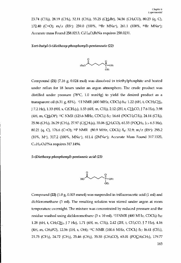
CImpter 6 Experimental
23.74 (CH2), 28.19 (CH3), 32.11 (CH2), 33.25 (CH2Br), 34.56 (CH2CO), 80.23 (q, C) , 172.40 (C=0); m / z (ES^): 259.0 (100%, 79Br MNa+), 261.1 (100%, siBr MNa*); Accurate mass Found 258.0213, C9Hi602BrNa requires 258.0231.
Tert-butyl-5-(diethoxy-phosphonyl)-pentanoate (22)
tBuO P - O E t
Compound (21) (7.16 g, 0.024 mol) was dissolved in triethylphosphite and heated
under reflux for 18 hours under an argon atmosphere. The crude product was
distilled under pressure (78°C, 1.0 mmHg) to yield the desired product as a
fa-ansparent oi l (6.31 g, 83%). i H N M R (400 MHz, CDCI3) 5H: 1.22 (6H, t, OCH2CH3,
J 7.2 Hz), 1.33 (9H, s, C(CH3)3), 1.53 (6H, m, CH2), 2.12 (2H, t, CH2CO, J 7.6 Hz), 3.98
(4H, m, CH2OP); " C NMR (125.6 MHz , CDCI3) 5c: 16.61 (POCH2CH3), 24.14 (CH2),
25.86 (CH2), 26.29 (CH2), 27.97 (C(CH3)3), 35.06 (CH2CO), 61.53 (POCH2, JCP 6.5 Hz),
80.21 (q, C), 176.6 (C=0); 3iP N M R (80.9 M H z , CDCI3) 5p: 32.9; m / z (ES+): 295.2
(10%, M+), 317.2 (100%, MNa^), 611.4 (2MNa^); Accurate Mass Found 317.1525,
Ci3H2705PNa requires 317.1494.
5-(Diethoxy-phosphonyl)-pentanoic acid (23)
P - O E t
Compound (22) (1.0 g, 0.003 mmol) was suspended in trifluoroacetic acid (1 ml) and
dichloromethane (3 ml). The resulting solution was stirred under argon at room
temperature overnight. The mixture was concentiated by reduced pressure and the
residue washed using dichloromethane (3 x 10 ml) . ^ H N M R (400 M H z , CDCI3) 5H:
1.28 (6H, t, CH2CH3, J 7 Hz), 1.71 (6H, m, CH2), 2.42 (2H, t, CH2CO, J 7 Hz), 4.16
(4H, m, CH2PO), 12.56 ( I H , s, OH); N M R (100.6 MHz , CDCI3) 5c: 16.41 (CH3),
21.75 (CH2), 24.72 (CH2), 25.46 (CH2), 35.55 (CH2CO), 63.01 (POCH2CH3), 179.77
163
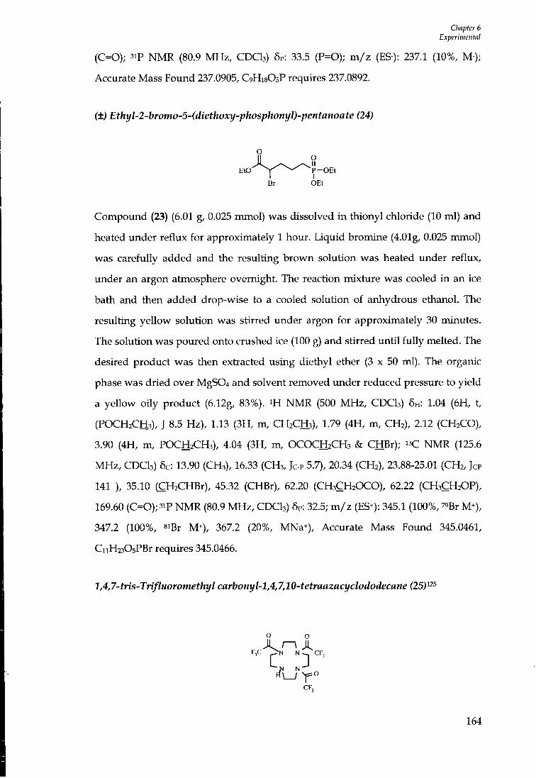
CImpter 6 Experimental
(C=0); 3iP NMR (80.9 MHz, CDCI3) 5P: 33.5 (P=0); m/z (ES-): 237.1 (10%, M-); Accurate Mass Found 237.0905, C9H18O5P requires 237.0892.
(±) Ethyl-2-bromo-5-(diethoxy-phosphonyl)-pentanoate (24)
o
Br OEt
Compound (23) (6.01 g, 0.025 mmol) was dissolved in thionyl chloride (10 ml) and
heated under reflux for approximately 1 hour. Liquid bromine (4.01g, 0.025 mmol)
was carefully added and the resulting brown solution was heated under reflux,
under an argon atmosphere overnight. The reaction mixture was cooled in an ice
bath and then added drop-wise to a cooled solution of anhydrous ethanol. The
resulting yellow solution was stirred under argon for approximately 30 minutes.
The solution was poured onto crushed ice (100 g) and stirred until fully melted. The
desired product was then extiacted using diethyl ether (3 x 50 ml). The organic
phase was dried over MgS04 and solvent removed under reduced pressure to yield
a yellow oily product (6.12g, 83%). ^H NMR (500 MHz, CDCI3) 5H: 1.04 (6H, t,
(POCH2CH3), J 8.5 Hz), 1.13 (3H, m, CH2CH3), 1.79 (4H, m, CH2), 2.12 (CH2CO),
3.90 (4H, m, POCH2CH3), 4.04 (3H, m, OCOCH2CH3 & CHBr); " C NMR (125.6
MHz, CDCI3) 5c: 13.90 (CH3), 16.33 (CH3, Jc-p 5.7), 20.34 (CH2), 23.88-25.01 (CH2, Jcp
141 ), 35.10 (CH2CHBr), 45.32 (CHBr), 62.20 (CH3CH2OCO), 62.22 (CH3CH2OP),
169.60 (C=0 ) ;3 ip NMR (80.9 MHz, CDCI3) 5p: 32.5; m/z (ES+): 345.1 (100%, 79Br M+),
347.2 (100%, 8iBr M+), 367.2 (20%, MNa^), Accurate Mass Found 345.0461,
CnH2305PBr requires 345.0466.
1,4,7-tris-Trifluoromethyl carbonyl-1,4,7,10-tetraazacyclododecane (25) 125
FX I>N N-^ CF,
CF,
164

Chapter 6 Experimental
1,4,7,10-Tetraazacyclododecane (1.01 g, 5.87 mmol) and triethylamine (0.8 ml , 6.98 mmol) were dissolved into anhydrous methanol and cooled to -0°C. Ethyl trifluoroacetate (3.30 g, 0.023 mmol) was added dropwise over a 30 minute period, the mixture was left stirring under an argon atmosphere for approximately 4 hours, and the temperature was allowed to increase to room temperature. The solvent was removed by reduced pressure to yield a crude yellow product, which was purif ied by flash column chromatography (Si02,1% MeOH in D C M 3% MeOH in DCM) (1.9 g, 70%). Rf (Si02,100 EtOAc = 0.295) ^H N M R (400 MHz, CDCI3) 5H: 1.49 ( I H , br s, N H ) , 2.87 (4H, m, NCH2), 3.52-3.65 (8H, m, NCH2), 3.85 (4H, m, NCH2); " C NMR (100.6 MHz, CDCI3) 5c: 20.93 (CH2), 46.91-52.81 (m, NCH2), 116.44 (q, CF3, JC-F ~ 278 Hz, further split due to conformers), 156.78-158.22 (m, C=0, existence of conformers and long range C-F coupling); m / z (ES+) 483.3 (100%, MNa+), 943.1 (75% 2MNa^).
l,t-Butoxycarbonyl-4,7,10-tris-trifluoromethylcarbonyl-l,4,7,10-
tetraazacyclododecane (26)
o o I
N \_J Y°
CF, CF,
1,4,7-tris-trifluoromethyl carbonyl-l,4,7,10-tetraazacyclododecane (25) (1.86 g, 4.0
mmol) was dissolved into dry methanol (20 ml), di-tert-butyl-dicarbonate (1.02 g,
4.7 mmol) was added and the resulting mixture was left stirring at room
temperature, under an argon atmosphere, for approximately 5 hours. The solvent
was removed under reduced pressure to yield an oily crude product. The desired
product was purif ied by flash colunm chromatography (Si02, D C M —> 2% EtOAc in
DCM), as a colourless oi l (1.35 g, 57%), Rf (10% EtOAc in D C M = 0.54). ^H NMR
(400 M H z , CDCI3) 5H: 1.33 (9H, s, C(CH3)3), 3.34-3.73 (16H, m, NCH2); i^C NMR
(100.6 M H z , CDCI3) 5c: 20.62 (CH2), 27.98 (C(CH3)3), 48.54-50.70 (m, NCH2), 81.04
(C(CH3)3), 116.03 (q, CF3, JC-F ~ 287 Hz, further split due to conformers), 156.67-
158.70 (m, C=0 , existence of conformers and long range C-F coupling); (m/z (ES*)
165
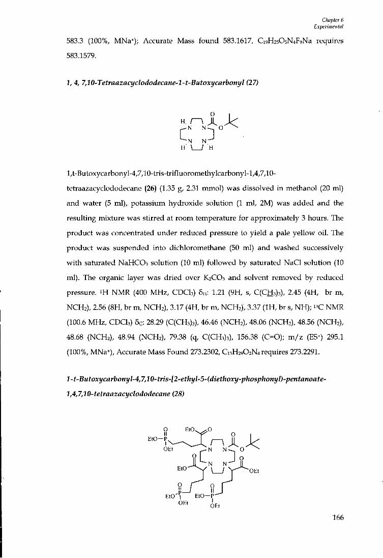
Cliapter 6 Experimental
583.3 (100%, MNa*); Accurate Mass fo imd 583.1617, Ci9H2505N4F9Na requires 583.1579.
1, 4, 7,10-Tetraazacyclododecane-l-t-Butoxycarbonyl (27)
H . A J < O ^
M M •N N H \ _ ^ H
l,t-Butoxycarbonyl-4,7,10-tris-trifluoromethylcarbonyl-l,4,7,10-
tetraazacyclododecane (26) (1.35 g, 2.31 mmol) was dissolved in methanol (20 ml)
and water (5 ml), potassium hydroxide solution (1 ml , 2M) was added and the
resulting mixture was stirred at room temperature for approximately 3 hours. The
product was concentrated under reduced pressure to yield a pale yellow oi l . The
product was suspended into dichloromethane (50 ml) and washed successively
wi th saturated NaHCOs solution (10 ml) followed by saturated NaCl solution (10
ml). The organic layer was dried over K2CO3 and solvent removed by reduced
pressure. N M R (400 MHz, CDCI3) 5H: 1.21 (9H, s, C(CH3)3), 2.45 (4H, br m,
NCH2), 2.56 (8H, br m, NCH2), 3.17 (4H, br m, NCH2), 3.37 ( I H , br s, NH) ; " C NMR
(100.6 MHz, CDCI3) 6c: 28.29 (C(CH3)3), 46.46 (NCH2), 48.06 (NCH2), 48.56 (NCH2),
48.68 (NCH2), 48.94 (NCH2), 79.38 (q, C(CH3)3), 156.38 (C=0); m / z (ES+) 295.1
(100%, MNa-), Accurate Mass Found 273.2302, C13H29O2N4 requires 273.2291.
l-t-Butoxycarbonyl-4,7,10-tris-[2-ethyl-5-(diethoxy-phosphonyl)-pentanoate-
1,4,7,10-tetraazacyclododecane (28)
E t O — P
N N
N N
E t O - P
OEt
166
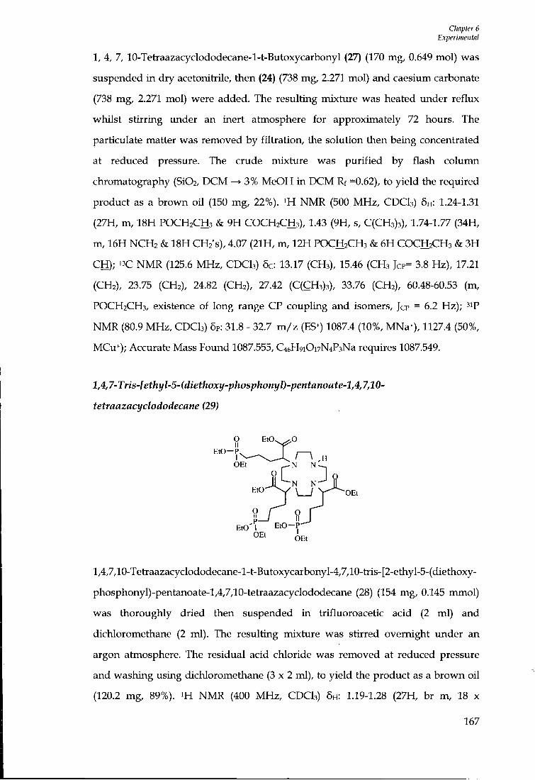
Chapter 6 Experimeiitnl
1, 4, 7, 10-Tetraazacyclododecane-l-t-Butoxycarbonyl (27) (170 mg, 0.649 mol) was suspended in dry acetonitrile, then (24) (738 mg, 2.271 mol) and caesium carbonate (738 mg, 2.271 mol) were added. The resulting mixture was heated under reflux whilst stirring under an inert atmosphere for approximately 72 hours. The particulate matter was removed by fil tration, the solution then being concentiated at reduced pressure. The crude mixture was purif ied by flash column chromatography (Si02, D C M 3% MeOH in D C M Rf =0.62), to yield the required product as a brown oi l (150 mg, 22%). NMR (500 MHz, CDCI3) 6H: 1.24-1.31 (27H, m, 18H POCH2CH3 & 9H COCH2CH3), 1.43 (9H, s, C(CH3)3), 1.74-1.77 (34H, m, 16H NCH2 & 18H CHz's), 4.07 (21H, m, 12H POCH2CH3 & 6H COCH2CH3 & 3H CH); " C N M R (125.6 MHz, CDCI3) 6c: 13.17 (CH3), 15.46 (CH3 JCP= 3.8 Hz), 17.21 (CH2), 23.75 (CH2), 24.82 (CH2), 27.42 (C(CH3)3), 33.76 (CH2), 60.48-60.53 (m, POCH2CH3, existence of long range CP coupling and isomers, Jcp = 6.2 Hz); ''ip NMR (80.9 MHz , CDCI3) 5P: 31.8 - 32.7 m / z (ES+) 1087.4 (10%, MNa^), 1127.4 (50%, MCu+); Accurate Mass Found 1087.555, C46H9iOi7N4P3Na requires 1087.549.
l,4,7-Tris-[ethyl-5-(diethoxy-phosphonyl)-pentanoate-l,4,7,10-
tetraazacyclododecane (29)
0 ^0 II
OEt •N N - ^
•N N " ^ • L J V
EtO p — '
' 1 EtO OEt OEt
' O E t
l,4,7,10-Tetiaazacyclododecane-l-t-Butoxycarbonyl-4,7,10-tiis-[2-ethyl-5-(diethoxy-
phosphonyl)-pentanoate-l,4,7,10-tetiaazacyclododecane (28) (154 mg, 0.145 mmol)
was thoroughly dried then suspended i n tiifluoroacetic acid (2 ml) and
dichloromethane (2 ml). The resulting mixture was stirred overnight under an
argon atmosphere. The residual acid chloride was removed at reduced pressure
and washing using dichloromethane ( 3 x 2 ml), to yield the product as a brown oil
(120.2 mg, 89%). i H NMR (400 M H z , CDCI3) 5H: 1.19-1.28 (27H, br m, 18 x
167

CImpter 6 Experimental
POCH2CH3 & 9 X COCH2CH3), 1.64-2.34 (38H, br m, 16 x NCH2 & 21 x CH2 & NCH) , 4.03-4.14 (21H, 12 x POCH2CH3 & 6 x COCH2CH3 & 3 x CH), 3ip N M R (80.9 M H z , CDCI3) 5p: 32.9 (s, POCH2CH3); m / z (ES+) 965.3 (10%, MH^), 1003.2 (100%, MK+), 1027.2 (10%, MCu+); Accurate mass Found 965.5210, C4iH840i5N4P3 requires 965.5146.
l,4,7-Tris-(di-phosphonyl)-pentanoate-l,4,7,10-Tetraazaq/clododecane[U]
- II
' \ - H • N N -O H I IN IN
?\L J o M ' II N N
0 - P 0 - P
l,4,7-Tris-[2-ethyl-5-(diethoxy-phosphonyl)-pentanoate-l,4,7,10-
tetraazacyclododecane (29) (90 mg, 0.093 mmol) was suspended into hydrogen
bromide solution i n acetic acid (5 ml). The resulting mixture was heated under
reflux at 90°C for eighteen hours. The solution was allowed to cool to room
temperature and was then added dropwise to a cooled solution of diethyl ether.
The product was insoluble i n ether and formed a yellow oily precipitate. The
product was concentrated at reduced pressure. Toluene was added (5 x 10 ml)
which was also removed at reduced pressure in an attempt to remove any
remaining acetic acid. NMR (200 MHz, D2O) 6H: 1.64-1.93 (18H, br m, CH2), 2.52-
3.58 (16H br m, NCH2), 3.92 4.32 (3H, m, NCH), 3ip N M R (D2O, 80.9 MHz) 5P: 31.7 -
33.0 (s, POCH2CH3).
168

Chapter 6 Experiwental
6.6 Complex Synthesis
1,4,7-Tris-5-(di-phosphonyl)-pentanoate-l,4,7,10-Tetraazacyclododecane [GdL']^-
O - j - \Gd O
- o - U n j
OH OH
l,4,7-Tris-5-(di-phosphonyl)-pentanoate-l,4/7,10-Tetraazacyclododecane (33 mg,
0.047 mmol) was dissolved into water (2 ml). The p H of the acidic solution was
adjusted to p H 5.5 using sodium hydroxide solution (0.1 M) . Gadolinium(IlI)
chloride hexahydrate (17 mg, 0.047 mmol) was added, the p H being re-adjusted to
p H 5.5. The resulting brown solution was heated under reflux for 18 hours. The
cloudy brown solution was allowed to cool to room temperature and was
centrifuged to remove any particulate matter. The filtrate was collected and the p H
of the solution increased to p H 10 using sodium hydroxide solution (0.1 M) for
approximately 20 minutes. The solution was filtered through a 0.45 p m syringe
filter to remove any free gadolinium hydroxide precipitate. The p H was then
lowered to p H 5.5 using hydrochloric acid (0.1 M) . The solution was lyophiUsed to
yield a white solid. (22 mg, 55%). Relaxivity value for [GdL9]3- 7.3 mM-is-i (pH 6.8,
310 K, 60 MHz) .
6.7 Chapter 4 Experimental
6.7.1 Ligand Synthesis
Diethyl-pyridine-2,6-dicarboxylate (30)
, 0 . J L ^ ^ . 0 .
o o
2,6 Pyridine carboxyUc acid (10.2 g, 0.062 mol) was dried under vacuum and then
169

CImpter 6 Experimental
suspended into dry ethanol (20 ml). Concentrated sulphuric acid was added (0.8 ml) and the resulting white mixture was heated under reflux for approximately 18 hours. The transparent reaction mixture was cooled and quenched using excess sodium hydrogen carbonate and stirred for approximately 30 minutes. The solvent was evaporated at reduced pressure producing a white soUd, which was then suspended into dichloromethane (40 ml) and washed successively w i t h water, saturated sodium hydrogen carbonate solution and water. The organic phase was dried using MgS04 and solvent removed at reduced pressure to yield a white, crystalline soUd (7.88 g, 58 %). ^H NMR (400 M H z , CDCI3) 5H: 1.39 (6H, t, CH3, J 7.2 Hz), 4.43 (4H, q, CH2CH3, J 7.2 Hz), 7.96 ( I H , t, H4, J 7.2 Hz), 8.23 (2H, d, H3 & H5, J 7.2 Hz); " C N M R (100.6 MHz , CDCI3) 6c: 14.16 (2 CH3), 62.23 (2 CH2), 127.76 (C4), 138.16 (C3 & C5), 148.63 (q, C2 & Ce), 164.57 (C=0); m / z (ES*) 246.1 (50%, MNa*), 469.0 (100%, 2MNa^).
6-Hydroxymethyl-pyridine-2-carboxylic acid ethyl ester^^HSl)
Sodium borohydride (0.73 g, 0.019 rmnol) was added to a solution of (30) (7.15 g,
0.032) i n dry ethanol (100 ml). The resulting white suspension was heated under
reflux for approximately 18 hours under an argon atmosphere. The reaction
mixture was allowed to cool to room temperature and quenched w i t h water (80
ml). The solvent was then evaporated at reduced pressure to approximately 30 ml ,
which was then extracted using chloroform (3 x 80 ml) . The organic fractions were
collected and dried using sodium sulphate, and organic solvent removed at
reduced pressure to yield a white solid (3.50 g, 60%). « N M R (400 MHz, CDCI3)
5H: 1.35 (3H, t, CH3, J 7.2 Hz), 3.76 ( I H , brs, OH), 4.38 (2H, q, CH2CH3, J 7.2 Hz), 4.79
(2H, s, CH2OH), 7.46 ( I H , d, H5, J 7.6 Hz), 7.76 ( I H , t, H4, J 7.6 Hz), 7.94 ( I H , d, H3, J
7.6 Hz); m / z (ES+) 204.1 (100%, MNa*).
170
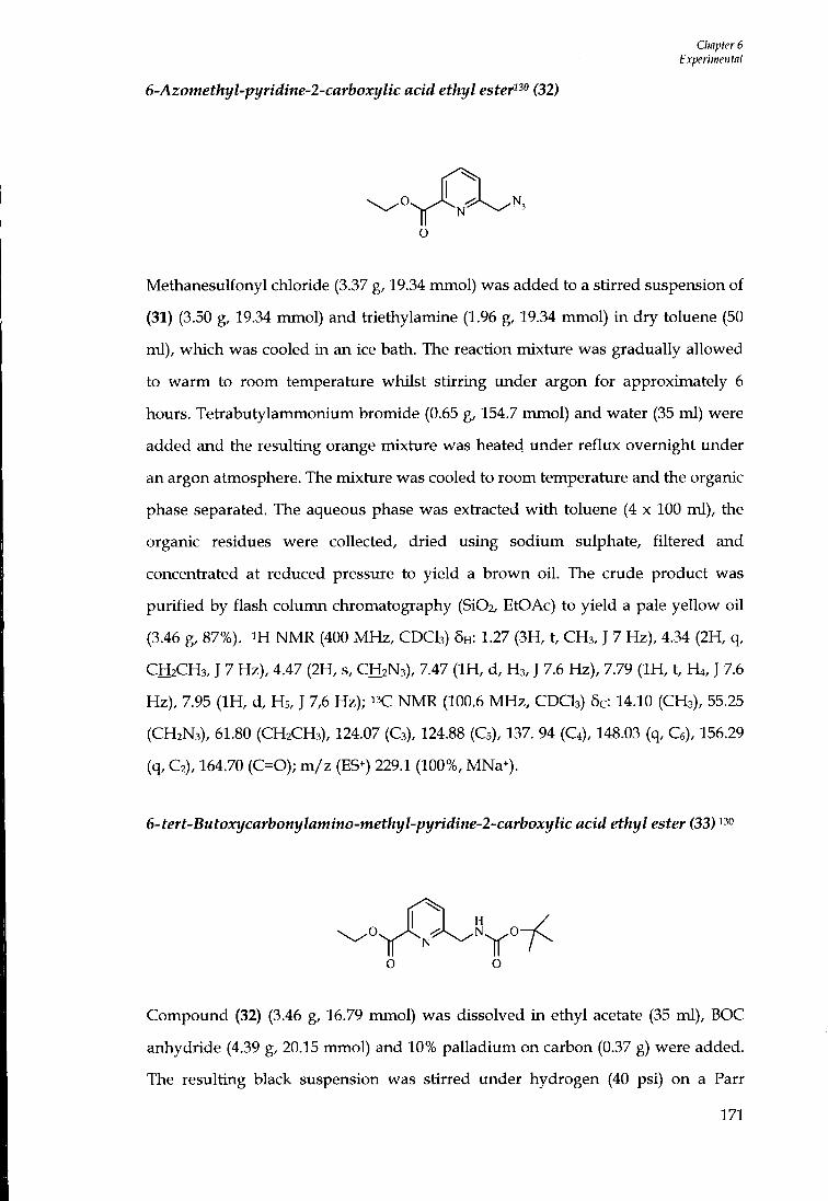
CImpter 6 Experimental
6-Azomethyl-pyridine-2-carboxylic acid ethyl ester"" (32)
Methanesulfonyl chloride (3.37 g, 19.34 mmol) was added to a stirred suspension of
(31) (3.50 g, 19.34 mmol) and tiiethylamine (1.96 g, 19.34 mmol) i n dry toluene (50
ml), which was cooled in an ice bath. The reaction mixture was gradually allowed
to warm to room temperature whilst stirring under argon for approximately 6
hours. Tetiabutylammonium bromide (0.65 g, 154.7 mmol) and water (35 ml) were
added and the resulting orange mixture was heated under reflux overnight imder
an argon atmosphere. The mixture was cooled to room temperature and the organic
phase separated. The aqueous phase was extiacted w i t h toluene (4 x 100 ml), the
organic residues were collected, dried using sodium sulphate, filtered and
concentiated at reduced pressure to yield a brown oi l . The crude product was
purif ied by flash column chromatography (Si02, EtOAc) to yield a pale yellow oil
(3.46 g, 87%). i H N M R (400 MHz, CDCI3) 5H: 1.27 (3H, t, CH3, J 7 Hz), 4.34 (2H, q,
CH2CH3, J 7 Hz), 4.47 (2H, s, CH2N3), 7.47 ( I H , d, H3, J 7.6 Hz), 7.79 ( I H , t, H4, J 7.6
Hz), 7.95 ( I H , d, Hs, J 7,6 Hz); N M R (100.6 M H z , CDCI3) 5c: 14.10 (CH3), 55.25
(CH2N3), 61.80 (CH2CH3), 124.07 (C3), 124.88 (C5), 137. 94 (C4), 148.03 (q, Ce), 156.29
(q, C2), 164.70 (C=0); m / z (ES+) 229.1 (100%, MNa^).
6-tert-Butoxycarbonylamino-methyl-pyridine-2-carboxylic acid ethyl ester (33)
Compound (32) (3.46 g, 16.79 mmol) was dissolved in ethyl acetate (35 ml) , BOC
anhydride (4.39 g, 20.15 mmol) and 10% palladium on carbon (0.37 g) were added.
The resulting black suspension was stirred under hydrogen (40 psi) on a Parr
171
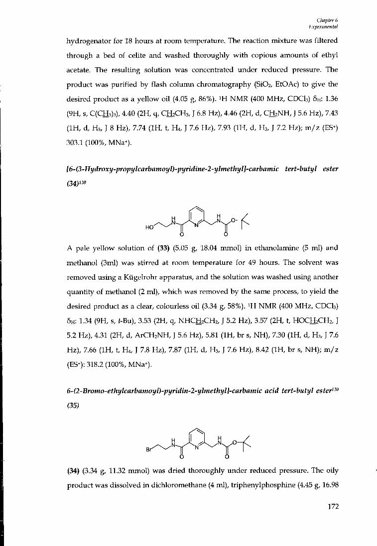
Chapter 6 Experimental
hydrogenator for 18 hours at room temperature. The reaction mixture was filtered through a bed of celite and washed thoroughly w i t h copious amoimts of ethyl acetate. The resulting solution was concentiated imder reduced pressure. The product was purif ied by flash column chromatography (Si02, EtOAc) to give the desired product as a yellow oil (4.05 g, 86%). N M R (400 MHz, CDCI3) 5H: 1.36 (9H, s, C(CH3)3), 4.40 (2H, q, CH2CH3, J 6.8 Hz), 4.46 (2H, d, CH2NH, J 5.6 Hz), 7.43 ( I H , d, Hs, J 8 Hz), 7.74 ( I H , t, H4, J 7.6 Hz), 7.93 ( I H , d, H3, J 7.2 Hz); m / z (ES-) 303.1 (100%, MNa^).
[6-(3-Hydroxy-propylcarbamoyl)-pyridine-2-ylmethyl]-carbamic tert-butyl ester
(34)130
O
A pale yeUow solution of (33) (5.05 g, 18.04 mmol) i n ethanolamine (5 ml) and
methanol (3ml) was stirred at room temperature for 49 hours. The solvent was
removed using a Kiigelrohr apparatus, and the solution was washed using another
quantity of methanol (2 ml), which was removed by the same process, to yield the
desired product as a clear, colourless oi l (3.34 g, 58%). ^H N M R (400 MHz, CDCI3)
6H: 1.34 (9H, s, t-Bu), 3.53 (2H, q, NHCH2CH2, J 5.2 Hz), 3.57 (2H, t, HOCH2CH2, J
5.2 Hz), 4.31 (2H, d, ArCH2NH, J 5.6 Hz), 5.81 ( I H , br s, N H ) , 7.30 ( I H , d, H3, J 7.6
Hz), 7.66 ( I H , t, H4, J 7.8 Hz), 7.87 ( I H , d, H5, J 7.6 Hz), 8.42 ( I H , br s, NH) ; m / z
(ES^): 318.2 (100%, MNa+).
6-(2-Bromo-ethylcarbamoyl)-pyridin-2-ylmethyl]-carbamic acid tert-butyl ester""
(35)
6 O
(34) (3.34 g, 11.32 mmol) was dried thoroughly under reduced pressure. The oily
product was dissolved in dichloromethane (4 ml), triphenylphosphine (4.45 g, 16.98
172

CImpter 6 Experimental
mmol) was added and the mixture cooled in an ice bath. Carbon tetiabromide (4.67
g, 14.13 mmol) was then added and the resulting yellow solution was gradually
warmed to room temperature whilst stirring under argon overnight. The resulting
red solution was concentiated under reduced pressure and purif ied by flash
column chromatography (SiOa, EtOAc) to produce a clear colourless oi l (2.33 g,
58%). ' H NMR (400 MHz, CDCI3) 5H: 1.43 (9H, s, f-Bu), 3.58 (2H, t, H O C H 2 C H 2 N , J
6 Hz), 3.88 (2H, q, N H C H 2 C H 2 , J 6 Hz), 4.49 (2H, d, A R C H 2 N H , J 5.6 Hz), 5.32 ( I H ,
br s, NH) , 7.45 ( I H , d, H 3 , J 7.5 Hz), 7.84 ( I H , t, H4, J 7.8), 8.08 ( I H , d, H5, J 7.6 Hz),
8.45 ( I H , br s, NH) ; ' 'C N M R (75.4 MHz, C D C I 3 ) : 5c 28.50 ( C ( C H 3 ) 3 ) , 32.07 (CHj) ,
41.17 ( C H 2 ) , 45.74 ( C H 2 ) , 120.97 (C4), 124.38 (q, C 2 ) , 138.25 ( C 3 ) , 148.92 (C5), 156.12
(q, Ce), 157.16 (C=0), 164.35 (C=0); m / z (ES^): 380.1 (100%, ^mr MNa^), 382.0
(85%, 8iBr MNa^).
{6-[2-(l,4,7,10-Tetraaza-cyclododec-yl)-ethylcarbamoyll-pyridin-2-ylmethyl}-
carbamic acid tert-hutyl ester"" (36)
H I H
^ J o o '
Under an argon atmosphere, a solution of (35) (1.71 g, 3.271 mmol) i n dry
chloroform (75 ml) was slowly added drop wise to a solution of cyclen (1.79 g, 10.41
mmol) i n dry chloroform (100 ml). The resulting clear solution was stirred at room
temperature for approximately 72 hours. The solution was concentiated at reduced
pressure and the resulting yeUow oi l was dissolved into dichloromethane (60 ml).
The organic phase was washed wi th water (4 x 60 ml), dried using potassium
carbonate and concentiated at reduced pressure to yield a clear oil (1.07 g, 91%). ^H
NMR (400 MHz, C D C I 3 ) 5H: 1.41 (9H, s, C ( C H 3 ) 3 ) , 2.49-2.64 (18H, m, 8 ring C H 2 &
N H C H 2 C H 2 ) , 3.50 (2H, q, N H C H 2 C H 2 , J 5.4 Hz), 4.30 (2H, d, A R C H 2 N H , J 5.6 Hz),
7.32 ( I H , d, H 3 , J 7.6 Hz), 7.71 ( I H , t, H4, J 7.6 Hz), 7.97 ( I H , d, H5, J 7.6 Hz), 8.01
( I H , br, N H ) , 8.35 ( I H , br, NH) ; " C NMR (100.6 M H z , C D C I 3 ) 6c: 24.48 ( C ( C H 3 ) 3 ) ,
37.76 ( C H 2 ) , 44.67 ( C H 2 ) , 45.60 ( C H 2 ) , 46.43 ( C H 2 ) , 51.81 ( C H 2 ) , 54.42 ( C H 2 ) , 78.92
173
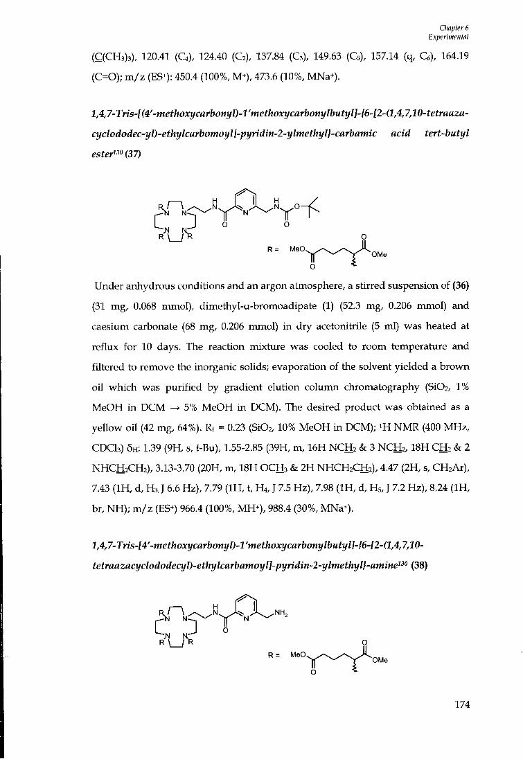
CImpter 6 Experimental
( C ( C H 3 ) 3 ) , 120.41 ( C 4 ) , 124.40 ( C 2 ) , 137.84 ( C 3 ) , 149.63 ( C 5 ) , 157.14 (q, Ce), 164.19 (C=0); m / z (ES^): 450.4 (100%, M^), 473.6 (10%, MNa+).
l,4,7-Tris-l(4'-methoxycarbonyl)-l'methoxycarbonylbutyll-{6-l2-(l,4,7,10-tetraaza-
cyclododec-yl)-ethylcarbomoyl]-pyridin-2-ylmethyl}-carbamic acid tert-butyl
ester^^o(37)
I i H H / 3N in--,
R = MeO^ OMe
O
Under anhydrous conditions and an argon atmosphere, a stirred suspension of (36)
(31 mg, 0.068 mmol), dimethyl-a-bromoadipate (1) (52.3 mg, 0.206 mmol) and
caesium carbonate (68 mg, 0.206 mmol) in dry acetonitrile (5 ml) was heated at
reflux for 10 days. The reaction mixture was cooled to room temperature and
filtered to remove the inorganic solids; evaporation of the solvent yielded a brown
oi l which was purif ied by gradient elution column chromatography (Si02, 1%
MeOH in D C M ^ 5% MeOH in DCM). The desired product was obtained as a
yellow o i l (42 mg, 64%). Rf = 0.23 (Si02, 10% MeOH in DCM); i H N M R (400 M H z ,
C D C I 3 ) 5H: 1.39 (9H, s, t-Bu), 1.55-2.85 (39H, m, 16H N C H 2 & 3 N C H 2 , 1 8 H C H 2 & 2
N H C H 2 C H 2 ) , 3.13-3.70 (20H, m, 18H O C H 3 & 2H N H C H 2 C H 2 ) , 4.47 (2H, s, C H 2 A R ) ,
7.43 ( I H , d, H 3 , J 6.6 Hz), 7.79 ( I H , t, Ht, J 7.5 Hz), 7.98 ( I H , d, H5, J 7.2 Hz), 8.24 ( I H ,
br, N H ) ; m / z (ES^) 966.4 (100%, MH^) , 988.4 (30%, MNa*).
1,4,7-Tris-[4'-methoxycarbonyl)-l 'methoxycarbonylbutyl]-{6-[2-(l,4,7,10-
tetraazacyclododecyl)-ethylcarbamoyl]-pyridin-2-ylmethyl}-amine"'' (38)
R = MeO. ^ ^ J< OMe
R = M e C
O
174
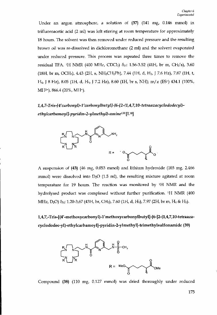
Cliapter 6 Experimental
Under an argon atmosphere, a solution of (37) (141 mg, 0.146 mmol) i n tiifluoroacetic acid (2 ml) was left stirring at room temperature for approximately 18 hours. The solvent was then removed under reduced pressure and the resulting brown oi l was re-dissolved i n dichloromethane (2 ml) and the solvent evaporated imder reduced pressure. This process was repeated three times to remove the residual TFA. ^H N M R (400 MHz, CDCh) 6H: 1.56-3.52 (41H, br m, CH2's), 3.60 (18H, br m , OCH3), 4.43 (2H, s, NH2CH2Ph), 7.44 ( I H , d, H3, J 7.6 Hz), 7.87 ( I H , t, H 4 , J 8 Hz), 8.05 ( I H , d, H 5 , J 7.2 Hz), 8.60 ( I H , br s, N H ) ; m / z (ES^) 434.1 (100%, MH2^), 866.4 (20%, MH+).
l,4,7-Tns-f4'cflrboni//)-l'cflj-boni//bMti/U-/6-f2-(l,4,7,10-tefrafl2flci/c/ot/orfeq//)-
ethylcarbamoyl]-pyridin-2-ylmethyl}-amine^^^ [L^"]
A suspension of (43) (46 mg, 0.053 mmol) and l i th ium hydroxide (103 mg, 2.466
mmol) were dissolved into D2O (1.5 ml), the resulting mixture agitated at room
temperature for 19 hours. The reaction was monitored by ^H N M R and the
hydrolysed product was complexed without further purification. ^H N M R (400
MHz, D2O) 5H: 1.20-3,67 (43H, br, CH2), 7.60 ( I H , d, H3), 7.97 (2H, br m, H 4 & H 5 ) .
l,4,7,-Tris-[(4'-methoxycarbonyI)-l'methoxycarbonylbutyl]-{6-[2-(l,4,7,10-tetraaza-
cyclododec-yI)-ethyIcarbamoyl]-pyridin-2-yImethyl}-trimethylsulfonamide (39)
O
Compound (38) (110 mg, 0.127 mmol) was dried thoroughly under reduced
175
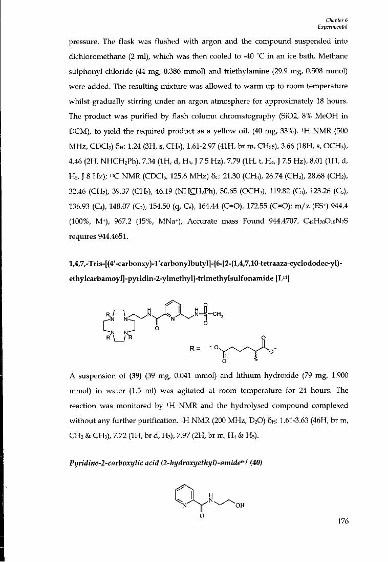
Cliapter 6 Experimental
pressure. The flask was flushed w i t h argon and the compovmd suspended into dichloromethane (2 ml), which was then cooled to -40 °C i n an ice bath. Methane sulphonyl chloride (44 mg, 0.386 mmol) and triethylamine (29.9 mg, 0.508 mmol) were added. The resulting mixture was allowed to warm up to room temperature whilst gradually stirring tmder an argon atmosphere for approximately 18 hours. The product was purif ied by flash column chromatography (Si02, 8% MeOH in DCM), to yield the required product as a yellow oi l . (40 mg, 33%). ^H N M R (500 MHz, CDCI3) 5H: 1.24 (3H, s, CH3), 1.61-2.97 (41H, br m, CH2S), 3.66 (18H, s, O C H 3 ) , 4.46 (2H, NHCH2Ph), 7.34 ( I H , d, H3, J 7.5 Hz), 7.79 ( I H , t, H4, J 7.5 Hz), 8.01 ( I H , d, Hs, J 8 Hz); 13C N M R (CDCI3,125.6 MHz) 5c: 21.30 (CH3), 26.74 (CH2), 28.68 (CH2), 32.46 (CH2), 39.37 (CH2), 46.19 (NHCHjPh) , 50.65 (OCH3), 119.82 (C3), 123.26 (Cs), 136.93 (C4), 148.07 (C2), 154.50 (q, Ce), 164.44 (C=0), 172.55 (C=0); m / z (ES^) 944.4 (100%, M+), 967.2 (15%, MNa+); Accurate mass Found 944.4707, C42H70O15N7S requires 944.4651.
lA7,-Tris-[(4'-carbonxy)-l'carbonylbutyI]-{6-[2-(l,4,7,10-tetraaza-cycIododec-yI)-
ethylcarbamoyl]-p5a-idin-2-ylmethyl}-trimethylsulfonamide [L"]
3N N-^
R = - O
o * "
A suspension of (39) (39 mg, 0.041 mmol) and l i th ium hydroxide (79 mg, 1.900
mmol) in water (1.5 ml) was agitated at room temperature for 24 hours. The
reaction was monitored by ^H N M R and the hydrolysed compound complexed
without any further purification. ^H N M R (200 MHz, D2O) 6H: 1.61-3.63 (46H, br m,
CH2 & CH3), 7.72 ( I H , br d, H3), 7.97 (2H, br m, H4 & H5).
Pyridine-2-carboxylic acid (2-hydroxyethyl)-amide"f (40)
O H
176

CImpter 6 Experimental
Picolinic ethyl ester (900 mg, 5.96 mmol) and ethanolamine (5 ml) were stirred together under argon for 24 hours. The ethanolamine solution was removed by distillation under reduced pressure using Kugelrohr apparatus to yield a yellow oi l (700 mg, 66%). m NMR (CDCI3, 200 MHz) 6H: 3.59 ( I H , br, OH) 3.63 (2H, q, CH2NH, J 5.6 Hz), 3.81 (2H, t, CH2OH, J 5.2 Hz), 7.39 ( I H , t, H5, J 6 Hz), 7.80 ( I H , t, H4, J 7.6 Hz) 8.14 ( I H , d, H3, J 5.8 Hz), 8.48 ( I H , br s, N H ) , 8.52 ( I H , d. He, J 4.6 Hz); m / z (ES^) 167.0 (70%, MH*), 189.0 (100%, MNa^).
Pyridine-2-carboxyltc acid (2-bromoethyl)-amide^^" (41)
H
O
Compound (40) (1.30 g, 7.83 mmol) was dried thoroughly under vacuum and then
suspended into dichloromethane (10 ml). Triphenylphosphine (3.1 g, 11.75 mmol)
was added portion-wise to the stirred solution and cooled in an ice salt bath.
Carbon tetiabromide (3.24 g, 9.78 mmol) was added and the mixture left to
gradually warm up overnight whilst stirring under argon. The resulting red
solution was concentiated by reduced pressure and purif ied by flash column
chromatography (Si02, Et02Ac) to isolate the product as a white solid (1.38 g, 72%).
i H NMR (400 MHz, CDCI3) 6H: 3.50 (2H, t, CH2Br, J 6 Hz), 3.82 (2H, q, CH2NH, J 6
Hz), 7.35 ( I H , t, Hs, J 6.4 Hz), 7.75 ( I H , t, H4, J 5.8Hz), 8.16 ( I H , d, H3, J 6.8 Hz), 8.49
(2H, br. He & NH) ; " C NMR (100.6 MHz, CDCI3) 6c: 31.75 (CH2), 41.14 (CH2), 122.19
(C3), 126.12 (C5), 137.32 (C4), 148.16 (Ce), 149.46 (q, C2), 164.42 (C=0); m / z (ES^)
228.9 (10%, M^), 251.0 (100%, 79Br MNa^), 253.0 (100%, s Br MNa^).
Pyridine-2-carboxylic acid [2-(l,4,7,10 tetraazacyclododec-l-yl)-ethyl]-amide (42)
I ^ N i " - ^ II
Under an argon atmosphere, a solution of (41) (228 mg, 0.247 mmol) i n anhydrous
177
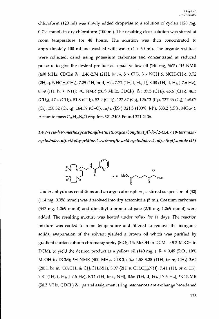
Chapter 6 Experimental
chloroform (120 ml) was slowly added dropwise to a solution of cyclen (128 mg, 0.744 mmol) i n dry chloroform (100 ml). The resulting clear solution was stirred at room temperature for 48 hours. The solution was then concentiated to approximately 100 m l and washed wi th water (4 x 60 ml). The organic residues were collected, dried using potassium carbonate and concentiated at reduced pressure to give the desired product as a pale yellow oi l (140 mg, 56%). ^H N M R (400 MHz, CDCI3) 6H: 2.46-2.74 (21H, br m, 8 x CH2, 3 x N C H & NCH2CH2), 3.52 (2H, q, NHCH2CH2), 7.29 ( I H , br d, H3), 7.72 ( I H , t, H4, J ), 8.08 ( I H , d, H5, J 7.6 Hz), 8.39 ( I H , br s, N H ) ; " C N M R (50.3 MHz , CDCI3) 6c: 37.3 (CH2), 45.6 (CH2), 46.5 (CH2), 47.4 (CH2), 51.8 (CH2), 53.9 (CH2), 122.37 (C3), 126.13 (C5), 137.36 (C4), 148.07 (C2), 150.32 (C6, q), 164.39 (C=0); m / z (ES^) 321.3 (100%, M+), 383.2 (15%, MCu2+); Accurate mass C16H29N6O requires 321.2403 Found 321.2406.
l,4,7-Tns-f(4'-met/ioa:ycflrfaon3/0-l'met/joxycflrbo«i//bMti//J-^6-f2-(l,4,7,10-tetrflfl2fl-
cyclododec-yl)-ethyl-pyridine-2-carboxylic acid cyclododec-l-yl)-ethyl]-amide (43)
o R = Mei
OMe
Under anhydrous conditions and an argon atmosphere, a stirred suspension of (42)
(114 mg, 0.356 mmol) was dissolved into dry acetonitiile (5 ml). Caesium carbonate
(347 mg, 1.069 mmol) and dimethyl-a-bromo adipate (270 mg, 1.069 mmol) were
added. The resulting mixture was heated under reflux for 11 days. The reaction
mixture was cooled to room temperature and filtered to remove the inorganic
solids; evaporation of the solvent yielded a brown oi l which was purif ied by
gradient elution column chromatography (Si02,1% MeOH in D C M —> 8% MeOH in
DCM), to yield the desired product as a yellow oi l (140 m g , ) . Rf = 0.49 (Si02,10%
MeOH in DCM); ^H N M R (400 MHz, CDCI3) 6H: 1.58-3.28 (41H, br m, CH2) 3.62
(20H, br m, CO2CH3 & CH2CH2NH), 3.97 (2H, s, CH2CH2NH), 7.41 ( I H , br d, H3),
7.81 ( I H , t, H4, J 7.6 Hz), 8.14 ( I H , br s, N H ) , 8.56 ( I H , d, H5, J 7.6 Hz); " C N M R
(50.3 M H z , CDCI3) 6c: partial assignment (ring resonances are exchange broadened
178
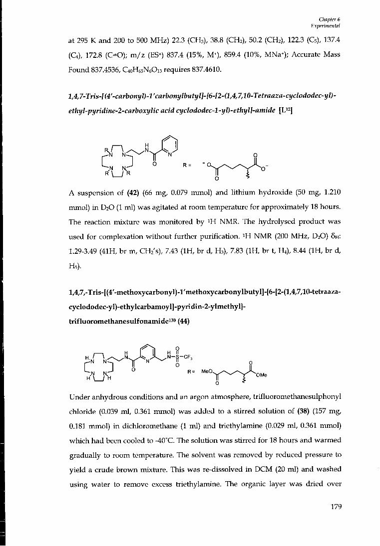
Cliapter 6 Experimental
at 295 K and 200 to 500 MHz) 22.3 (CH3), 38.8 (CH2), 50.2 (CH2), 122.3 (Cs), 137.4
(C4), 172.8 (C=0); m / z (ES^) 837.4 (15%, M+), 859.4 (10%, MNa+); Accurate Mass
Found 837.4536, C40H65N6O13 requires 837.4610.
l,4,7-Tris-l(4'-carbonyl)-l'carbonylbutyl]-{6-[2-(l,4,7,10-Tetraaza-cyclododec-yl)-
ethyl-pyridine-2-carboxylic acid cyclododec-l-yl)-ethyl]-amide [U^
o O R
o
A suspension of (42) (66 mg, 0.079 mmol) and l i th ium hydroxide (50 mg, 1.210
mmol) i n D2O (1 ml) was agitated at room temperature for approximately 18 hours.
The reaction mixture was monitored by ^H NMR. The hydrolysed product was
used for complexation without further purification. i H NMR (200 M H z , D2O) 5H:
1.29-3.49 (41H, br m, CUi's), 7.43 ( I H , br d, H3), 7.83 ( I H , br t, H,) , 8.44 ( I H , br d,
Hs).
l,4,7,-Tris-[(4'-methoxycarbonyI)-l'methoxycarbonylbutyl]-{6-[2-(l,4,7,10-tetraaza-
cyclododec-yI)-ethylcarbamoyl]-pyridin-2-ylmethyI}-
trifluoromethanesulfonamide"o (44)
I—\ H H II
^ - J O Q
R= MeO. OMe
O
Under anhydrous conditions and an argon atmosphere, trifluoromethanesulphonyl
chloride (0.039 ml , 0.361 nunol) was added to a stirred solution of (38) (157 mg,
0.181 mmol) in dichloromethane (1 ml) and tiiethylamine (0.029 ml, 0.361 ntmol)
which had been cooled to -40°C. The solution was stirred for 18 hours and warmed
gradually to room temperature. The solvent was removed by reduced pressure to
yield a crude brown mixture. This was re-dissolved in D C M (20 ml) and washed
using water to remove excess triethylamine. The organic layer was dried over
179
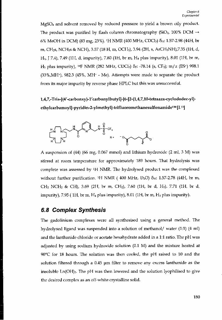
Chapter 6 Experimental
MgS04 and solvent removed by reduced pressure to yield a brown oily product. The product was purif ied by flash column chromatography (Si02, 100% D C M —> 6% MeOH in DCM) (83 mg, 23%). m N M R (400 M H z , CDCI3) 5H: 1.57-2.98 (44H, br m, CH2S, NCH2S & NCH), 3.57 (18 H , m, OCH3), 3.94 (2H, s, ArCH2NH),7.35 ( I H , d, H3, J 7.4), 7.49 ( I H , d, impuri ty) , 7.80 ( I H , br m, H4 plus impuri ty) , 8.01 ( I H , br m, Hs plus impuri ty) , I 'F N M R (282 MHz, CDCI3) 5F: -78.14 (s, CF3); m / z (ES^) 998.1 (33%,MH+), 982.3 (45%, MH+ - Me). Attempts were made to separate the product f r o m its major impuri ty by reverse phase HPLC but this was imsuccessful.
l,4,7,-Tris-[(4'-carbonxy)-l'carbonyIbutyI]-{6-[2-(l,4,7,10-tetraaza-cycIododec-yl)-
ethyIcarbamoyl]-p)Tidin-2-ylmethyI}-trifIuoromethanesuIfonamidei^<'[Li3]
I \ H H II
f" T 'i 9
A suspension of (44) (66 mg, 0.067 mmol) and l i th ium hydroxide (2 ml , 3 M) was
stirred at room temperature for approximately 189 hours. That hydrolysis was
complete was assessed by ^H NMR. The hydrolysed product was the complexed
without further purification. ^H N M R ( 400 MHz, D2O) 5H: 1.57-2.78 (44H, br m,
CH2 NCH2 & CH), 3.69 (2H, br m, CH2), 7.60 ( I H , br d, H 3 ) , 7.71 ( I H , br d,
impuri ty) , 7.95 ( I H , br m, H4 plus impuri ty) , 8.01 ( I H , br m, H5 plus impuri ty) .
6.8 Complex Synthesis
The gadolinium complexes were all synthesised using a general method. The
hydrolysed ligand was suspended into a solution of methanol/ water (1:1) (4 ml)
and the lantharude chloride or acetate hexahydrate added i n a 1:1 ratio. The p H was
adjusted by using soditun hydroxide solution (0.1 M) and the mixture heated at
90°C for 18 hours. The solution was then cooled, the p H raised to 10 and the
solution filtered through a 0.45 ]im filter to remove any excess lanthanide as the
insoluble Ln(OH)3. The p H was then lowered and the solution lyophUised to give
the desired complex as an off-white crystalline solid.
180
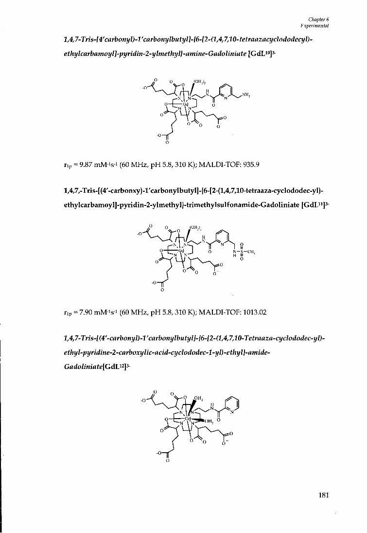
Chapter 6 Experimental
l,4,7-Tris-[4'carbonyl)-l'carbonylbutyl]-{6-[2-(l,4,7,10-tetraazacyclododecyl)-
ethylcarbamoyl]-pyridin-2-ylmethyl}-amine-Gadoliniate [GdW]^
o-b--94 J O
rip = 9.87 mM-is-i (60 MHz, p H 5.8, 310 K); MALDl-TOF: 935.9
l,4,7,-Tris-[(4'-carbonxy)-l'carbonylbutyl]-{6-[2-(l,4,7,10-tetraaza-cycIododec-yl)-
ethylcarbamoyI]-pyridin-2-ylmethyl}-trimethyIsuIfonamide-GadoIiniate [GdDi]^-
-0
^ \ N - ^ H II N - S - C H ,
rip = 7.90 mM-is-i (60 MHz, p H 5.8,310 K); MALDl-TOF: 1013.02
14,7-Tris-[(4'-carbonyl)-rcarbonylbutyl]-{6-[2-(l,4,7,10-Tetraaza-cyclododec-yl)-
ethyl-pyridine-2-carboxylic-acid-cyclododec-l-yl)-ethyl]-amide-
Gadoliniate[GdU^^
181

Chapter 6 Experimental
rip: 7.80 mM-is-i (60 MHz, pH 5.6, 310 K); MALDI-TOF
6.9 Chapter 5 Experimental Disodium-S44-(4-Ethoxybenzyl)-3,6,9-tris[(carboxy-KO)methyl]-3,6,9-
triazaundecandioata)(5-)-K?W,W,N^, K^O^, 0 " J europiate-(2-) (Europium-EOB-
DTPA) [EuLH
(S)-EOB-DTPA (100 mg, 0.177 mol) and europium chloride hexahydrate were
suspended in a mixture of water-methanol (2 ml, 50/50 v /v) , and the pH of
solution was adjusted to 5.5. The resulting mixture was heated under reflux for 1
hour. The mixture was allowed to cool to room temperature, and the solid colloidal
waste was removed by filtration through a 0.45 ]im syringe filter. The pH of the
filtrate was increased to 7 using sodium hydroxide solution (0.1 M). The solution
was concentrated by reduced pressure to a volume of approximately 1 ml. This
solution was then dropped onto ethanol (5 ml); the resulting white precipitate was
heated vmder reflux for two hours, then cooled to room temperature. The turbid
solution was centiifuged, supernatant solution was discarded and the white solid
washed with cold dry ethanol and dried under reduced pressure. NMR analysis
confirmed the presence of free europium (III) salts. The solid was dissolved in
water (5 ml) and the pH increased to 10.0 using sodium hydroxide solution (0.1 M)
for 30 minutes. The soUd colloidal waste was then removed by filtration through
0.45 | im filter. The pH of the resulting clear solution was decreased to 5.5 using
hydrochloric acid (0.1 M). The solvent was removed under reduced pressure to
yield a white solid product (58 mg, 50%). Semi- preparative HPLC: Hypersil ODS 5
|im, column length 25 cm, diameter 16 mm; mobile phase, 2.5 g of NH4HCO3 in 940
iTil of distilled water and 60 ml of C H 3 C N at 295 K; flow rate 10 ml min-1. tR (major
isomer): 15.6 min; 19. 7 min (minor isomer), m/z (ES ) 674 ("lEuLH), 676 (i53EuLH),
182
CImpter 6 Experimental
386.8, 387.8 (doubly charged). IR (KBr, cm-1) 3440, 2990, 2980, 2920, 1607, 1509,
1476,1401,1325,1276,1241,1182,1115,1089, 991, 973, 926. NMR (200 MHz, D2O,
pD 6.0 major isomer. Figure 5. 8 Chapter 5 for numbering scheme): 29.1 ( IH, brs,
H8ax), 28.0 ( IH, brs, H5ax), 13.8 ( IH, brs, H8eq), 10.9 (2H, s, H17), 8.9 (2H, s, H18),
6.8 ( IH, s, Heq), 5.6 ( IH, s, H5eq), 5.3 ( IH, s, H14), 5.1 (2H, s, H15), 2.2 (3H, brs,
CH3), -0.9 (3H, brs, H4ax, H'ac-2, H'ac-1), -2.7 ( IH, brs, H'14), -7.7 ( IH, brs, H'ac-3),
-8.1 (IH, brs, Hac-2), -10.0 ( IH, brs, H'ac-4), -10.5 ( IH, brs, Hac-3), -11.1 ( IH, brs,
Hac-4), -13.9 and -14.0 (2H, brs, Hac-1 + H'ac-5), -16.8 ( IH, brs, Hac-5).
183

References
References
1. p. Caravan, J.J. Ellison, T.J. McMurry, R.B. Lauffer, Chem.Rev., 1999, 99, 2293-2352.
2. R. B. Lauffer, Chem. Rev., 1987,87, 901-927.
3. A. E. Merbach, E. Toth, Die Cheniishy of Contrast Agents in Medical Magnetic Resonance Imaging, Wiley, New York, 2001.
4. S. Aime, M. Botta, M. Fasano, E . Terreno, Chem. Soc. Rev., 1998, 27,19-29.
5. M. Woods, Z. Kovacs, S. Zhang, A. D. Sherry, Angew. Chem. Int. Ed., 2003, 5889-5892.
6. A. Bianchi, L . Calabi, F. Corana, S. Fontana, P. Losi, A. Maiocchi, L . Paleari, B. Valtancoli, Coordination. Chem. Rev., 2000, 204, 309-393.
7. S. Aime, L . Barbero, M. Botta, Mag. Resn. Imag., 1991,9, 843-847.
8. W. Krause, Topics in Current Chemistry, Contrast Agents I - Magnetic Resonance Imaging, Vol. 221, Springer, Berlin, 2002.
9. . E. Toth, L . Burai, A. E . Merbach, CoordinaHon.Chem. Rev., 2001, 216-217,363-382.
10. Y. Okuhata, Ad. Drug Delivery Review, 1999,37,121-137.
11. D. Dischino, E. J. Delaney, J. E . Emswiler, G. T. Gaughan, J. S. Prasad, S. K. Srivasta, M. Tweedle, Inorg. Chem., 1991, 30,1265-1269.
12. X. Zhang, C. A. Chang, H. G. Brittain, J. M. Garrison, J. Telser, M. F. Tweedle, Inorg. Chem., 1992,31, 5597-5600.
13. J. Platzek, P. Blaszkiewicz, H. Gries, P. Luger, G. Michl, A. Miiller-Fahrnow, B. Radiichel D. Siilzle, Inorg. Chem., 1997,36, 6086-6093.
14. S. I. Kang, R. S. Ranganathan, J. E . Emswiler, K. Kumar, J. Z. Gougoutas, M. F. Malley, M. F. Tweddle, Inorg. Chem., 1993,32, 2912-2918.
15. S. Aime, A. Barge, M. Botta, J. A. K. Howard, R. Kataky, K. P. Lowe, J. M. Moloney, D. Parker, A. S. deSousa, Chem. Commun., 1999,1047-1048.
16. J. I. Bruce, R. S. Dickins, L . J. Govenlock, T. Gunnlaugsson, S. Lopinski, M. P. Lowe, D. Parker, R. D. Peacock, J. J. B. Perry, S. Aime, M. Botta, /. Am. Chem. Soc, 2000,122, 9674-9684.
17. R. M. Supkowski, W. D. Horrocks, J. Inorg. Chem., 1999,38, 5616-5619.
18. R. S. Dickins, T. Gunnlaugsson, D. Parker, R. D. Peacock, Chem. Commun., 1998,1643-1644.
19. S. Aime, E . Gianolio, E. Terreno, G. B. Giovenzana, R. PagUarin, M. Sisti, G Palmisano, M. Botta, M. P. Lowe, D. Parker, SBIC, 2000,5,488-497.
20. S. Aime, M. Botta, S. G.Crich, G. Giovenzana, G. Jommi, R. Pagliarin, M. Sisti, /. Chem. Soc, Chem. Commun., 1995,1885-1886.
21. H. Stetter, W. Frank, R. Mertens, Tetrahedron, 1981, 37, 767-772.
184
Refireiices
22. S. Aime, M. Botta, S. G. Crich, G. B. Giovenzana, G. Jommi, R. Pagliarin, M. Sisti, Inorg. Chem., 1997,36, 2992-3000.
23. W. D. Kim, G. E. Kiefer, F. Maton, K. McMUlan, R. N. MuUer, A. D. Sherry, Inorg. Chem., 1995,34, 2233-2243.
24. R. Hovland, C. Gl0gard, A. J.Aasen, J. Klaveness, Org. Biomol. Chem., 2003,1, 644-647.
25. S. Aime, M. Botta, S. G. Crich, G. Giovenzana, R. Pagliarin, M. Sisti, E . Terreno, Magn. Reson. Chem., 1998,36, S200-S209.
26. S. Aime, M. Botta, L . Frullano, S. G. Crich, G. Giovenzana, R. Pagliarin, G. Palmisano, F. R. Sirtori, M. Sisti, /. Med. Chem., 2000,43,4017-4024.
27. Y. Bretonniere, M. Mazzanti, J. Pecaut, F. A. Dunand, A. E. Merbach, Chem. Commun., 2001, 621-622.
28. J. Xu, S. J. Franklin, D. W. Whisenhunt, K. N. Raymond, /. Am. Chem. Soc, 1995,117, 7245-7246.
29. D. M. J. Doble, M. Melchior, B. O'SuUivan, C. Siering, J. Xu, V.C. Pierre, K. N. Raymond, Inorg. Chem. 2003,42,4930-4937.
30. K. Micskei, L . Hehn, E. Brucher, A.E. Merbach, Inorg. Chem, 1993, 32, 3844-3850.
31. S. M. Cohen, J. Xu, E. Radkov, K. N. Raymond, M. Botta, A. Barge, S. Aime, Inorg. Chem., 2000,39, 5747-5756.
32. D. M. J. Doble, M. Melchior, B. O'Sullivan, C. Siering, J. Xu, V. C. Pierre, K. N. Raymond, Inorg. Chem., 2003,42, 4930-4937.
33. S. Hajela, M. Botta, S. Giraudo, J. Xu, K. N. Raymond, S. Aime, /. Am. Chem. Soc., 2000, 122,11228-11229.
34. D. M. J. Doble, M. Botta, J. Wang, S. Aime, A. Barge, K. N. Raymond, /. Am. Chem. Soc, 2001,123,10758-10759.
35. S. Aime, A. Barge, M. Botta, J. A. K. Howard, R. Kataky, M. P. Lowe, J. M. Moloney, D. Parker, A. S. DeSousa, CItem. Commun., 1999,1047-1048.
36. S. Aime, M. Botta, S. G. Crich, G. B. Giovenzana, R. Pagliarin, M. Piccinini, M. Sisti, E . Terreno, /. Bio. Inorg. Chem., 1997, 2, 70-479.
37. D. Messeri, M. P. Lowe, D. Parker, M. Botta, Chem. Commun., 2001,2742-2743.
38. D. Messeri, Ph. D. Thesis, Targeted and High Relaxivity Contrast Agents, University of Durham, 2002.
39. A. Borel, L . Helm, A. E . Merbach, Chem. Eur. J., 2001, 7, 600-610.
40. C. J. Broan, E. Cole, K. J. Jankowski, D. Parker, K. Pulukkody, B. A. Boyce, N. R. A. Beeley, K. Millar, A. T. Millican, Synthesis, 1992, 63-68.
41. J. W. Chen, R. L . Belford, R. B. Clarkson, /. Phys. Chem., 1996,100, 8093.
42. P. Lebduskova, J. Kotek, P. Hermann, L . V. Elst, R. N. Muller, I. Lukes, J. A. Peters, Bioconjugate Chem. 2004,15,881-889.
185
References
43. X. L i , S. Zhang, P. Zhao, Z. Kovacs, A. D. J. Sherry, Inorg. Chem., 2001, 40, 6572-6579.
44. J. Ren, A. D. J. Sherry, /. Magn. Reson., 1996, B i l l , 178-186.
45.1. Lazar, A. D. Sherry, R. Ramasamy, E . Brucher, R. Kiraly, Inorg. Chem, 1991, 30, 5016-5019.
46. J. Rohovec, P. Vojtisek, P. Hermann, J. Ludvik, I. Luke§, /. Chem. Soc, Daltoii Trans., 2000, 141-148.
47. S. Aime, L . FruUano, S. G. Crich, Angeiv. Chem. Int. Ed., 2002,41,1017-1019.
48. K. P. Pulukkody, T. J. Norman, D. Parker, L . Royle, C . J. Broan, /. Chem. Soc. Perkin Trans., 1993, 605-620.
49. S. Aime, A. S. Batsanov, M. Botta, R. S. Dickins, S. Faulkner, C. E . Foster, A. Harrison, J. A. K. Howard, J. M. Moloney, T. J. Norman, D. Parker, L . Royle, J. A. G. Williams, J. Chem. Soc, Dalton Trans., 1997,3623-3636.
50. L . Burai, R. Kiraly, I. Lazar, E . Briicher, Eur. ]. Inorg. Chem., 2001,813-820.
51. S. Aime, A. S. Batsanov, M. Botta, R. S. Dickins, S. Faulkner, C. E . Foster, A. Harrison, J. A. K. Howard, J. M. Moloney, T. J. Norman, D. Parker, L . Royle, J. A. G. Williams, J. Chem. Soc, Dalton Trans., 1997, 3623-3636.
52. C.Bianchini, G. Giambastiani, F. Laschi, P. Mariani, A. Vacca, F. Vizza, P. Zanello, Org. Biomol. Chem., 2003,1, 879-886.
53. J. Kotek, P. Lebdugkova, P. Hermann, L . V. Elst, R. N. MuUer, C. F. G. C. Geraldes, T. Maschmeyer, I. Lukes, J. A. Peters, Chem. Eiir. ]., 2003,9, 5899-5915.
54. L . V. Elst, S. Laurent, H. M. Bintoma, R. N. MuUer, Mag. Res. Mat. Physics Biology Medicine, 2001,12,135-140.
55. E . Toth, D. Pubanz, S. Vauthey, L . Helm, A. E . Merbach, Chem. Eiir. }., 1996, 2,1606-1615.
56. H . Kobayashi, S. Kawamoto, R. A. Star, T. A. Waldmann, M. W. Brechbiel, P. L . Choyke, Bioconjugate Chem., 2003,14,1044-1047.
57. L . Lattuada, G. Lux, Tetrahedron Letters, 2003,44, 3893-3895.
58. P. Lebduskova, J. Kotek, P. Hermann, L . V. Elst, R. N. MuUer, I. Lukes, J. A. Peters, Bioconjugate Chem., 2004,15, 881-889.
59. T. N. Parac-Vogt, K. Kimpe, K. Binnemans, Journals of Alloys and Compounds, 2004,374, 325-329.
60. N. Fatin-Rouge, E. Toth, R. Meuli, J. G. Bunzli, Journal of Alloys and Compounds, 2004,298-302.
61. S. W. A. Bligh, A. H. M. S. Chowdhury, D. Kennedy, C. Luchinat, G. Parigi, Magn. Res. Med., 1999,41, 767-773.
62. E . Toth, F. Connac, L . Helm, K. Adzamli, A. E. Merbach, }BIC, 1998,3, 606-613.
63. F. A. Dunand, A. Borel, A. E . Merbach, /. Am.Chem. Soc, 2002,124, 710-716.
186

References
64. S. Aime, M. Botta, M. Fasano, E. Terreno, Acc. Chein. Res., 1999, 32,941-949.
65. U. Cosentino, D. Pitea, G. Moro, V. Barone, A. Villa, R. N. Muller, F. Botteman, Theor. Chem. Ace, 2004, 111, 204-209.
66. E . Toth, S. Vauthey, D. Pubanz, A. E . Merbach, Inorg. Chem., 1996,35, 3375-3379.
67. E . Toth, O. M. Ni Dhubhghaill, G. Besson, L . Helm, A. E . Merbach, Magn. Reson. Chem., 1999,37, 701-708.
68. K. Micskei, L . Helm, E . Briicher, A. E . Merbach, Inorg. Chem., 1993, 32, 3844-3850.
69. M. P. Lowe, Aust. /. Chem., 2002,55, 551-556.
70. D. Parker, Chem. Soc. Rev., 2004,33,156-65.
71. S. Laurent, F. Botteman, L . V. Elst, R. N. Muller, Magma, 2004,16,235-245.
72. R. Ruloff, E. Toth, R. Scopelliti, R. Triplet, H . Handel, A. E . Merbach, Chem. Commiin., 2002, 2630-2631.
73. S. Aime, M. Botta, J. I. Bruce, V. Mainero, D. Parker, E . Terreno, Chem. Commun., 2001, 115-116.
74. A. Barge, M. Botta, D. Parker, H. Puschmann, Chem. Commun., 2003,1386-1387.
75. E . Toth, F, Connac, L. Helm, K. Adzamli, A. E . Merbach, /. Bio. Inorg. Chem., 1998, 3, 606-613.
76. S. Zhang, K. Wu, A. D. Sherry, Angeu) Chem. Int. Ed., 1999,38, 3192-3194.
77. M. Woods, G. E . Kiefer, S. Bott, A. Castillo-Muzquiz, C. Eshelbrenner, L . Michaudet, K. McMillan, S. D. K. Mudigunda, D. Ogrin, G. Tircso, S. Zhang, P. Zhao, A. D. Sherry, /. Am. Chem. Soc, 2004,126,9248-9256.
78. M. P. Lowe, D. Parker, O Reany, S. Aime, M. Botta, G. Gastellano, E . Gianolio, R. Pagharin, /. Am. Chem. Soc., 2001,123, 7601-7609.
79. N. Raghunand, C. Howison, A.D. Sherry, S. Zhang, J. Gillies, Mag. Res. Med. 2003, 49, 249-257.
80. S. Aime, E. GianoUo, E . Terreno, M. Botta, Angew. Chem. Int. Ed., 2000, 39, 747-750.
81. W. L i , S. E . Eraser, T. J. Meade, /. Am. Chem. Soc, 1999,121,1413-1414.
82. K. Hanaoka, K. Kikuchi, Y. Urano, T. Nagano, /. Chem. Soc, 2001, 2,1840-1843.
83. T. J. McMurry, A. L . Nivorozhkin, A. F. Kolodziej, P. Caravan, M. T. Greenfield, R. B. Lauffer, Angew. Chem. bid. Ed., 2001,40, 2903-2906.
84. R. A. Moats, S. E . Eraser, T. J. Meade, Angeio Chem. Int. Ed. Engl., 1997, 36, 726-728.
85. L. Pascolo, F. CupeUi, P. L . Anelli, V. Lorusso, M. Visigalli, F. Uggeri, C . Tiribelh, Biochemical & Biophysical Res. Comm., 1999,257, 746-752.
187

References
86. F. Uggeri, S. Aime, P. L . Anelli, M. Botta, M. Brocchetta, C. De Haen, G. Ermondi, M. Grandi, P. Paoli, Inorg. Chem, 34,1995, 633-642.
87. H . Schmitt-Willich, M. Brehm, C. L . J. Ewers, G. Michl, A. Miiller-Fahrnow, O. Petrov, J. Platzek, B. Radiichel, D. Sulzle, Itiorg. Chem., 1999,3,1134-1144.
88. M. A. Schiffler, Biomaterials Seminar, 2001,1,1-7.
89. F. M. Cavagna, M. Dapr^, P. M. CasteUi, F. Maggioni, M. A. Kirchin, Eur. Radiol, 1997, 7, (suppl. 5), S222-224.
90. T. Zhang, A. Matsumura, T. Yamamoto, F. Yoshida, T. Nose, N. Shimojo, Am. ]. Neuroradiol, 2002,23,15-18.
91. G. Bombieri, R. Artali, /. Alloys and Compounds, 2002,344, 9-16.
92. T. Peters, All about Albumin: biochemistry, genetics and medical applications. Academic Press, San Diego, 1996.
93. S. Curry, H. Mandelkow, P. Brick, N. Franks, Nature Structural Biology, 1998,5,9, 827-835.
94. X. M. He, D. C. Carter, Nature, 1992, 209-215.
95. S. Curry, P. Brick, N. P. Franks, Biochimica et Biophysica Acta, 1999,131-140.
96.1. Petitpas, A. A. Bhattacharya, S. Twine, M. East, S. Curry, /. Biochem., 2001, 276, 22804-22809.
97. S. Baroni, M. Mattu, A. Vannini, R. CipoUone, S. Aime, P. Ascenzi, M. Fasano, Eur. J. Biochem, 2001,265, 6214-6220.
98. A. Sulkowska, /. Molecular Structure, 2002, 227-232.
99. M. Botta, S. Quici, G. Pozzi, G. Marzanni, R. Pagharin, S. Barra, S. G. Crich, Org. Biomol. Chem., 2004,2, 570-577.
100. S. Aime, M. Botto, M. Fasano, S. G. Crich, E . Terreno, SBJC, 1996,1, 312-319.
101. P. Caravan, N. J. Cloutier, M. T. Greenfield, S. A. McDermid, S. U. Dunham, J. W. M. Bulte, J. C . Amedio, Jr., R. J. Looby, R. M. Supkowski, W. D. Horrocks, T. J. McMurry, R. B. Lauffer, /. Am. Chem. Soc, 2002,124, 3152-3162.
102. P. Caravan, C. Commuzzi, W. Crooks, T. J. McMurry, G. R. Choppin, S. R. Woulfe, Inorg. Chem., 2001,40, 2170-2176.
103. K. Adzamli, D. A. Yablonskiy, M. R. Chicoine, E . K. Won, K. P. Galen, M. C. Zahner, T. A. Woolsey, J. J. H . Ackerman, Mag. Reson. Med, 2003,49, 586-590.
104. K. AdzamU, M. SpiUer, S. Koenig, Acad Radiol, 2002,9 (suppl 1), S11-S16.
105. K. Adzamh, L . V. Elst, S.Laurent, R. N. MuUer, Mag. Res. Mat. Phy. Bio. Med., 2001,12, 92-95.
106. S. Aime, M. Chiaussa, G. Digilio, E . Gianolio, E . Terreno, Bio. Inorg. Chem., 1999, 4, 766-774.
188

References
107. P. Caravan, M. T. Greenfield, X. L i , A. D. Sherry, Inorg. Chem., 2001,40, 6580-6587.
108. D. Parker, J.A.G. Wilhams, Metal Ions in Biological Systems - Tlie Lanthanides and their interrelations with Biosystems, Volume 40, Eds. A. Sigel, H. Sigel, Marcel Dekker, New York. 2003.
109. D. Parker, J. A. G. Wilhams, /. Chem. Soc. Dalton Trans, 1996,3613-3628.
110. D. Parker, Coord. Chem. Rev., 2000, 205,109-130.
111. A. Beeby, I. M. Clarkson, R. S. Dickins, S. Faulkner, D. Parker, L . Royle, A. S. de Sousa, J. A. G. Williams, M. Woods, /. Chem. Soc, Perkin Trans. 2,1999,493-503.
112. M. P. Lowe, D. Parker, Chem. Commun. 2000, 707-708.
113. W. D. Horrocks, Jr., D. R. Sudnick, /. Am. Chem. Soc, 1979,101,334-340.
114. M. V. Rekharsky, Y. Inove, Cltem. Rev, 1998,1875-1917.
115. S. Aime, M. Botta, L . FruUano, S. G. Crich,G. B. Giovenzana, R. Pagliarin, G. Palmisano, M.Sisti, Eur. J. Chem, 1999,5, 4,1253-1260.
116. A. A. Bogdanov Jr, K. Licha, Molecular Imaging: An Essential Tool in Preclinical Research, Diagnostic Imaging, and Therapy, Ernst Schering Reasearch Foundation, Springer, New York, 2005.
117. Y. Bretonniere, M. J. Cann, D. Parker, R. Slater, Org. Biomol. Chem., 2004, 2 (11), 1624-1632.
118. P. Atkinson, Y. Bretonniere, D. Parker, Chem. Commun., 2004,4,438-439.
119. F. Uggeri, S. Aime, P. L. Anelli, M. Botta, M. Brocchetta, C. De Haen, G. Ermondi, M. Grandi, P. Paoli, Inorg. Chem, 34,1995, 633-642.
120. T. I. Richardson, S. D. Rychnovsky, /. Am. Chem. Soc, 1997,119,12360-12361.
121. U. Schmidt, C. Braun, H. Sutoris, Syntliesis, 1996, 223-229.
122. P. Y. Bruice, Organic Chemistry, 3rd Edition, Prentice Hall Inc., 2001, 786-787.
123. A. K.Bhattacharya, G. Thyagarajan, Clmn. Rev., 1981, 81, 415-430.
124. J. March, Advanced Organic Chemistry - Reactions, Mechanisms and Structure, 3rd Edition, John Wiley and Sons, New York, 530-531.
125. W. Yang, C. M. Giandomenico, M. Sartori, D. A. Moore, Tetrahedron Letters, 2003, 44, 2481-2483.
126. T.J. Norman, Ph. D Thesis, Radioimmunotherapy with Yttrium Macrocylces, University of Durham, September 1994.
127. D. Parker, R. S. Dickins, H. Puschmann, C. Crossland, J. A. K. Howard, Chem. Rev., 2002,102,1977-2010.
128. A. J. Stewart, C. A. Bhndauer, S. Berezenko, D. Sleep, P. J. Sadler, PNAS, 2003, 7,1-, 3701-3706.
189
References
129. J. Masuoka, J. Hegenauer, B. R. Van Dyke, P. Saltman, /. Biol. Chem., 1993, 21533-21537.
130. A. Congreve, Responsive Lanthardde Complexes for Metal Ion Sensing, Ph. D. Thesis, University of Durham, 2004.
131. C. A. Hunter, D. H . Purvis, Angew. Chem. Int. Ed., 1992, 31, 6, 792-795.
132. A. Congreve, R. Kataky, M. Knell, D. Parker, H . Puschmann, K. Senanayake, L . Wyhe, New J. Chem., 2003, 27, 98-106.
133. R. Lencioni, D. Cioni, L , Crocetti, C. D. Pina, C. Bartolozzi, Journal ofHepatology., 2004, 40,162-171.
134. K. P. Pulukkody, T. Norman, D. Parker, L . Royle, /. Chem. Soc. Perkin Trans, 1993, 605-612.
135. P. Reimer, E . J. Rummeny, H . E . Daldrup, T. Hesse, T. Balzer, B. Tombach, P. E . Peters, Eur. Radiol, 1997, 7, 275-280.
136. P. Reimer, E . J. Rummeny, K. Shamsi, T. Balzer, H. E . Daldrup, B. Tanbach, T. Hesse, T. Berns, P. E . Peters, Radiologij, 1996,199,177-183.
137. N. C. Thompson, D. Parker, H. Schmitt-WiUich, D. Siilzle, G. Muller, J. P. Riehl, Dalton Trans., 2004,1892-1895.
138. E . N. Rizkalla, G. R. Choppin, Inorg. Chem, 1993,32, 582-586.
139. L . V. Elst, F.Chapelle, S. Laurent, R. N. Muller, /. Biol. Inorg. Chem., 2001, 6,196-200.
140. H. Schmitt-Willich, M. Brehm, C. L. J. Ewers, G. Michl, A. Muller-Fahrnow, O. Petrov, J. Platzek, B. Radiichel, D. Siilzle, Inorg. Chem., 1999,38,1134-1144.
141. J. P. Riehl, F. S. Richardson, Methods Enzymol, 1993, 226, 539.
142. J. I. Bruce, D. Parker, S. Lopinski, R. D. Peacock, Chirality., 2002,14, 562-567.
143. S. Aime, M. Botta, G. Ermondi, Inorg. Chem., 1992, 31, 4291-4299.
144. S. Hoeft, K. Roth, Cliem. Ber., 1993,126, 869-873.
145. S. Aime, A. Barge, J. I. Bruce, M. Botta, J. A. K. Howard, J. M. Moloney, D. Parker, A. S. de Sousa, M. Woods, /. Am. Chem. Soc, 1999,121, 5762-5771.
146. Circular Dichroism - Principles and Applications, Eds. N. Berova, K. Nakanishi, R. W. Woody, Wiley-VCH, New York, 2000.
147. G. Bobba, Interaction of Chiral Lanthanide Complexes with Nucleic Acids, Ph.D. Thesis, University of Durham, 2002.
148. S. D. Belair, C. L . Maupm, M. W. Logue, J. P. Riehl, /. Luminescence., 2000, 86, 61-66.
149. G. Bobba, R. S. Dickins, S. D. Kean, C. E . Mathieu, D. Parker, R. D. Peacock, G. Siligardi, M. J. Smith, J. A. G. Williams, C. F. G. C. Geraldes, /. Chem Soc, Perkin Trans. 2., 2001,1729-1737
150. L. Govenlock, J. A. K. Howard, J. M. Moloney, D. Parker, R. D. Peacock, G. Siligardi, /. Chem. Soc, Perkin Trans. 2., 1999, 2415-2418.
190

References
151. J. P. Riehl, F. S. Richardson, Chem. Rev., 1986, 86,1-16.
152. H. G. Brittain, Chirality., 1996, 8, 357-363.
191

Appendix
Appendix
192
Appendix
Lecture Courses Attended 2001/2002 Fluorine Cherrustry
Separation Methods
Supramolecular Chemistry
Practical Spectroscopy
Conferences Attended RSC Dalton Division, Coordination Chemistry in Action, University of Edinburgh,
6'i' November 2002.
RSC UK Macrocycles and Supramolecular Chemistiy Group Meeting, University of
York, 18-19* December 2002.
RSC Perkin Group Meeting, University of York, 8* April 2002.
Biomolecular Probes Mini-Symposium, University of Durham, 16* January 2003.
Seminars Attended
2001 October 9 Dr Roy S Lehrle, Birmingham
Forensics, Fakes and Failures
October 30 Professor Steve Howdle, Nottingham University
A thousand and one uses for CO2- from coffee to concrete to bone
October 31 Dr Colin Raston, School of Chemistry, Uni. of Leeds
Towards benign supramolecular chemistiy: synthesis - self
organization
November 6 Dr Cliff Ludman, Durham University
Explosions - a demonstiation lecture
November 14 Professor John Goodby, Departinent of Chemistiy, University of HuU
193

Appendix
Supermolecular liquid crystals - multipodes and dendrimers
November 21 Dr Roy Copely, GlaxoSmithKline
Crystallography in the Pharmaceutical Industry
December 5 Dr Mike Eaton, Celltech
Drugs of the Future
2002 January 22 Dr Ian Fallis, University of Cardiff
Size is Everything
January 29 Dr Paul Monks, University of Leicester
Ozone - the good, the bad and the ugly
January 30 Dr Peter Hore, PCL, University of Oxford
Chemistry in a spin: effects of magnetic fields on chemical reactions
February 13 Dr Helen AspinaU, Department of Chemistry, University of
Liverpool
Defining effective chiral binding sites at lanthanides
February 26 Dr Mike Griffin, Forensic Science Service, Metropolitan PoUce
Smack, Crack, Speed and Weed: A forensic chemists tale
March 12 Professor David Williams, Cardiff
Beer and Health: 7000 years of history
May 8 Professor Paul Madden, PCL, University of Oxford
Covalent Effects in "Ionic" Systems
October 8 Dr Stella Peace, Unilever
194

Appendix
The Chemistry Behind your Cuppa
October 15 Professor Mike Zaworotko from the University of South Florida
Suparamolecular Synthesis of Fimctional Molecules & Materials
November 12 Dr Dave Alker, Pfizer
The Discovery of a New Medicine
November 13 Professor Geoffrey Lawrance, Newcastle University, Australia
Designer Ligands: Macrocyclic and alicyclic moleculas for metal
complexation and biocatalysis
December 3 Dr Ian Sage, Qinetiq
Tough Plastics for a tough world
2003 January 22 Dr David Procter, Department of Chemistry, University of Glasgow
New Strategies and Methods for Organic Synthesis
February 11 Dr John Emsley, University of Cambridge
False Alarms: Chemistry & the Media
February 19 Professor Tony Ryan, Department of Chemistry, University, of
Sheffield
Introducing Soft Nanotechnology
March 4 Professor Richard Taylor, University of York
Adventures in Natural Product Synthesis"
October 15 Professor Huw Davies, Department of Chemistry, University of
Buffalo
Applications of Catalytic Asyrrunetric C-H Activation to Synthesis
195
Appendix
October 21 Dr Roger New^ton, Maybridge pic
The Pharmaceutical Industry - Does it have a Future?
October 28 Dr Dave Alker, Pfizer
Careers in the Pharmaceutical Industry
December 2 Dr Bill MacDonald, DuPont
'Flexible Electionics - What's it all About?!
January 28 Dr Lesley Yellowlees, Department of Chemistry, Edinburgh
University
Electrochemical and spectroelectrochemical studies of transition
metals
February 11 Professor D Parker FRS, Department of Chemistry, University of
Durham
Chiral Lanthanide Complexes: Structure, dynamics and fimction
February 17 Professor A P De Silva, Queens University of Belfast
Designer Molecules for Photonic Signalling
March 16 Dr Chris Clarke, Unilever
The Science Of Ice Cream
July 2 Professor Sir Harry Kroto, Nobel Laureate in Chemistry (1996)
2010: a NanoSpace Odyssey
196

Appendix
Publications NMR and chiroptical examination of the diastereoisomers of (S)-Eu-EOB-DTPA.
N. C. Thompson, D. Parker, H. Schmitt-WilUch, D. Sulzle, G. Muller, J. P. Riehl,
Dalton Trans., 2004,1892-1895.
197
mm