duplication of superficial femoral artery: imaging
TRANSCRIPT
CASE REPORT Open Access
Duplication of superficial femoral artery:imaging findings and literature reviewSung Hyun Yu1, Jung Han Hwang1* , Jeong Ho Kim1, Suyoung Park1, Ki Hyun Lee1 and Sang Tae Choi2
Abstract
Background: Duplication of the superficial femoral artery (SFA) is an extremely rare anatomic variation, with fewcase studies reported. We report one case of the duplicated SFA, discovered by both ultrasonography (US) andcomputed tomography angiography (CTA). We also reviewed literatures concerning 6 cases of the duplicated SFA(including our present case), and summarized the clinical and imaging features of the anatomic variation.
Case presentation: A 55-year-old woman presented to our hospital with an intermittent cramp in the lateralaspect of the right leg. The patient underwent Doppler US examination on bilateral lower extremity arteries andveins to examine potential vascular abnormality. Incidentally, US discovered the duplicated left SFA and CTA ofbilateral lower extremities revealed the anatomic orientation, course, length, diameter and distance of theduplicated left SFA. It was revealed to be divided into two trunks with similar luminal diameter and courses parallel.They reunited at distal thigh level. The findings of US and CTA examination did not correspond with the symptomof the patient, and the patient was discharged.
Conclusion: We report a rare case of the duplicated SFA diagnosed with the combinations of US and CTAexamination, which served as valuable imaging methods to detect and diagnose the vascular anatomic variation inlower extremities.
Keywords: Anatomic variation, Diagnostic imaging, Lower extremity, Case reports
BackgroundThe precise knowledge of vascular anatomy and its varia-tions is crucial as the treatment with endovascular tech-niques has increased. Variations of the femoral artery arerarely reported, especially for the superficial femoral artery(SFA) [1–3]. Few cases of the duplicated SFA have beenreported with limited combinations of imaging modalities,such as computed tomography angiography (CTA), con-ventional angiography, ultrasonography (US), or magneticresonance angiography (MRA) [2, 4–7]. We report a caseof the duplicated SFA, which was diagnosed using bothCTA and US, with a brief literature review.
Case presentationA 55-year-old woman presented to our hospital withnonspecific knee pain in the lateral aspect of the rightleg. She had no symptoms on the left leg. She had hyper-tension, diabetes mellitus, and dyslipidemia, and she wason a treatment for intervertebral disc herniation in an-other hospital. Her physical examination was nonspe-cific, and the right and left ankle-brachial pressure indexwas 1.06 and 0.88, respectively.The patient underwent Doppler US examination on
bilateral lower extremity arteries and veins to examinepotential vascular abnormality. There was no abnormal-ity in vessels of the right side. The left SFA was revealedto be divided into two trunks with similar luminal diam-eter and courses parallel (Fig. 1). They reunited at distalthigh level. No other abnormalities or diseases in vesselsof the left side were identified by US examination.
© The Author(s). 2020 Open Access This article is licensed under a Creative Commons Attribution 4.0 International License,which permits use, sharing, adaptation, distribution and reproduction in any medium or format, as long as you giveappropriate credit to the original author(s) and the source, provide a link to the Creative Commons licence, and indicate ifchanges were made. The images or other third party material in this article are included in the article's Creative Commonslicence, unless indicated otherwise in a credit line to the material. If material is not included in the article's Creative Commonslicence and your intended use is not permitted by statutory regulation or exceeds the permitted use, you will need to obtainpermission directly from the copyright holder. To view a copy of this licence, visit http://creativecommons.org/licenses/by/4.0/.The Creative Commons Public Domain Dedication waiver (http://creativecommons.org/publicdomain/zero/1.0/) applies to thedata made available in this article, unless otherwise stated in a credit line to the data.
* Correspondence: [email protected] of Radiology, Gil Medical Center, Gachon University College ofMedicine, 21, Namdong-daero 774beon-gil, Namdong-gu, Incheon 21565,Republic of KoreaFull list of author information is available at the end of the article
Yu et al. BMC Medical Imaging (2020) 20:99 https://doi.org/10.1186/s12880-020-00500-4

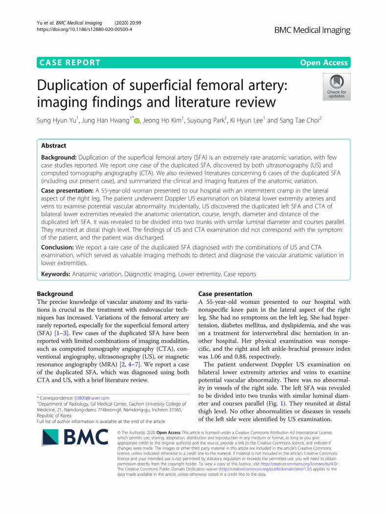
For further evaluation, CTA of bilateral lower extrem-ities was performed. The left SFA appeared to originatefrom left common femoral artery at the same level ofthe contralateral side. It appeared to run as a single ves-sel, 4 cm long, then split into two branches, medial andlateral ones. The luminal diameter of the medial one ofthe SFA was 5.3 mm, whereas that of lateral one was 4.4mm, measured in each proximal portion (Fig. 1). Boththen traveled 14 cm distal along anterior side of the leftsuperficial femoral vein. They merged at distal thighlevel to form single vessel and ran 4 cm distal to enterthe adductor hiatus. The anatomic orientation was wellvisualized in three-dimensional volume rendering andmaximum intensity projection images (Fig. 2). Therewas no evidence of atherosclerotic stenosis or other dis-eases on the bilateral lower extremity arteries. The find-ings of US and CTA examination did not correspondwith the symptom of the patient, and the patient wasdischarged.
Discussion and conclusionVariations of the femoral artery are rarely reported, in-cluding aplasia and hypoplasia of the SFA with associ-ated persistent sciatic artery, duplication or hypoplasiaof the deep femoral artery, trifurcation of femoral artery,and duplication of the SFA [1, 2]. To date, only a few
cases have been reported on the duplicated SFA withone or two imaging modalities [2, 4–7], and some re-ports and textbooks discuss embryologic basis for ana-tomic variant of lower extremities [2, 8–12].A literature search on PubMed database was per-
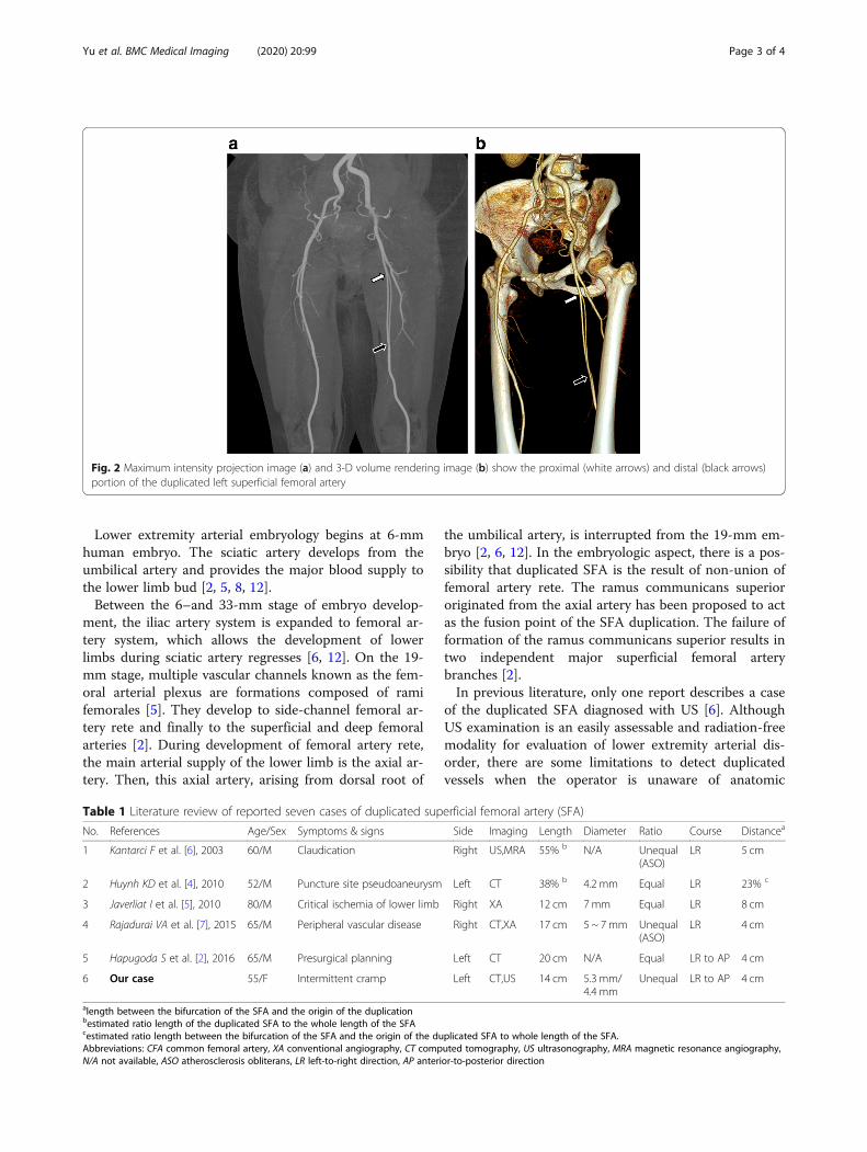
formed to review the previous reports concerning theduplicated SFA. The detailed analysis is summarized onTable 1. Including our report, all reported cases of theduplicated SFA have single vessel segments at proximaland distal portions of the duplicated segment. Further-more, all the reported cases have the duplicated SFAvariation in unilateral side of lower extremity [2, 4, 5, 7].Duplication of SFA occurred in the right side (3 cases)and left side (2 cases), like our case [2, 4–7]. The vascu-lar diameters of each duplicated SFA are equal in thethree case reports [2, 4, 5], whereas unequal in two [6,7]. In our case, the diameter of the duplicated SFA is un-equal without atherosclerosis obliterans or other vascu-lar disease. In most of the cases, duplicated SFA werediscovered incidentally except two cases with intermit-tent claudication. Similarly, our patient had a nonspecificknee pain in the lateral aspect of the right leg, but shehad no symptoms associated with duplicated SFA. As faras the gender information, previous reports only exam-ined male patients. However, our case described the caseof a female patient with duplicated SFA.
Fig. 1 Gray scale (a) and color Doppler ultrasonography (b) show that the left superficial femoral artery is divided into two trunks (arrows) withsimilar luminal diameter and courses parallel. The axial image of arterial phase CTA of bilateral lower extremity (c) shows the duplicated leftsuperficial femoral artery (arrows). The luminal diameter of the medial one is slightly larger than that of the lateral one. Note normal rightsuperficial femoral artery
Yu et al. BMC Medical Imaging (2020) 20:99 Page 2 of 4
Lower extremity arterial embryology begins at 6-mmhuman embryo. The sciatic artery develops from theumbilical artery and provides the major blood supply tothe lower limb bud [2, 5, 8, 12].Between the 6–and 33-mm stage of embryo develop-
ment, the iliac artery system is expanded to femoral ar-tery system, which allows the development of lowerlimbs during sciatic artery regresses [6, 12]. On the 19-mm stage, multiple vascular channels known as the fem-oral arterial plexus are formations composed of ramifemorales [5]. They develop to side-channel femoral ar-tery rete and finally to the superficial and deep femoralarteries [2]. During development of femoral artery rete,the main arterial supply of the lower limb is the axial ar-tery. Then, this axial artery, arising from dorsal root of
the umbilical artery, is interrupted from the 19-mm em-bryo [2, 6, 12]. In the embryologic aspect, there is a pos-sibility that duplicated SFA is the result of non-union offemoral artery rete. The ramus communicans superiororiginated from the axial artery has been proposed to actas the fusion point of the SFA duplication. The failure offormation of the ramus communicans superior results intwo independent major superficial femoral arterybranches [2].In previous literature, only one report describes a case
of the duplicated SFA diagnosed with US [6]. AlthoughUS examination is an easily assessable and radiation-freemodality for evaluation of lower extremity arterial dis-order, there are some limitations to detect duplicatedvessels when the operator is unaware of anatomic
Table 1 Literature review of reported seven cases of duplicated superficial femoral artery (SFA)
No. References Age/Sex Symptoms & signs Side Imaging Length Diameter Ratio Course Distancea
1 Kantarci F et al. [6], 2003 60/M Claudication Right US,MRA 55% b N/A Unequal(ASO)
LR 5 cm
2 Huynh KD et al. [4], 2010 52/M Puncture site pseudoaneurysm Left CT 38% b 4.2 mm Equal LR 23% c
3 Javerliat I et al. [5], 2010 80/M Critical ischemia of lower limb Right XA 12 cm 7mm Equal LR 8 cm
4 Rajadurai VA et al. [7], 2015 65/M Peripheral vascular disease Right CT,XA 17 cm 5 ~ 7mm Unequal(ASO)
LR 4 cm
5 Hapugoda S et al. [2], 2016 65/M Presurgical planning Left CT 20 cm N/A Equal LR to AP 4 cm
6 Our case 55/F Intermittent cramp Left CT,US 14 cm 5.3 mm/4.4 mm
Unequal LR to AP 4 cm
alength between the bifurcation of the SFA and the origin of the duplicationbestimated ratio length of the duplicated SFA to the whole length of the SFAcestimated ratio length between the bifurcation of the SFA and the origin of the duplicated SFA to whole length of the SFA.Abbreviations: CFA common femoral artery, XA conventional angiography, CT computed tomography, US ultrasonography, MRA magnetic resonance angiography,N/A not available, ASO atherosclerosis obliterans, LR left-to-right direction, AP anterior-to-posterior direction
Fig. 2 Maximum intensity projection image (a) and 3-D volume rendering image (b) show the proximal (white arrows) and distal (black arrows)portion of the duplicated left superficial femoral artery
Yu et al. BMC Medical Imaging (2020) 20:99 Page 3 of 4

variation. For example, initial US examination of othertwo previous case reports [7] have not necessarily re-vealed the duplicated SFA. Since anatomic variations offemoral artery, including the duplicated SFA, are notcommon and may not be easily discovered by US exam-ination only, CTA may provide a better implementalanalysis of the vascular system and its surrounding tis-sues [5]. In our case, the duplicated SFA was initiallyidentified at US examination and confidently diagnosedwith CTA.In summary, we report an extremely rare case of the
duplicated SFA diagnosed with the combination of USand CTA examination.
AbbreviationsSFA: Superficial femoral artery; US: Ultrasonography; CTA: Computedtomography angiography; MRA: Magnetic resonance angiography
AcknowledgementsNot applicable.
Authors’ contributionsYSH: Acquisition and analysis of the work, Drafted the work. HJH: Conceptionof the work and substantively revised it. KJH: Design of the work andsubstantively revised it. LKH: Writing – review & editing. PS: Writing – review& editing. CST: Writing – review & editing. All authors have checked theauthorship to a submitted version and agreed to the author list andcontributions.
FundingNot applicable.
Availability of data and materialsAll data generated or analyzed during this study are included in thispublished article.
Ethics approval and consent to participateNot applicable.
Consent for publicationThe patient provided written informed consent for publication of this casereport and accompanying images.
Competing interestsThe authors declare that they have no competing interests.
Author details1Department of Radiology, Gil Medical Center, Gachon University College ofMedicine, 21, Namdong-daero 774beon-gil, Namdong-gu, Incheon 21565,Republic of Korea. 2Department of Surgery, Gil Medical Center, GachonUniversity College of Medicine, 21, Namdong-daero 774beon-gil,Namdong-gu, Incheon 21565, Republic of Korea.
Received: 20 May 2020 Accepted: 19 August 2020
References1. Savithri P. A rare variation of trifurcation of right femoral artery. Int J Anat
Var. 2013;6(1):4–6.2. Hapugoda S, Hsu CC, Kwan GN, Watkins TW, Rophael JA. Duplication of the
superficial femoral artery: comprehensive review of imaging literature andinsight into embryology. Acta Radiol Open. 2016;5(7):1–4.
3. Nwaejike N, Saravanan P, Tang A. Duplicated Common Femoral Artery CanBe Safely Cannulated for Femorofemoral Cardiopulmonary Bypass. Ann VascSurg. 2014;28(1):262 e13-. e15.
4. Huynh KD, Alfaqawi M, Paaske WP. Duplication of the superficial femoralartery. Vascular. 2010;18(5):292–3.
5. Javerliat I, Rouanet A, Bourguignon T, Long A, Lermusiaux P. Duplication ofsuperficial femoral artery: an uncommon variation of the lower limb arterialsystem. Ann Vasc Surg. 2010;24(3):415 e1–3.
6. Kantarci F, Mihmanli I, Aksoy H, Barutca H, Gurses B, Kaynak K. Duplicationof the superficial femoral artery diagnosed primarily on the basis of colorDoppler ultrasonography. J Ultrasound Med. 2003;22(6):641–3.
7. Rajadurai VA, Sieunarine K. Superficial femoral artery duplication. J VascInterv Radiol. 2015;26(9):1323.
8. Senior HD. An interpretation of the recorded arterial anomalies of thehuman leg and foot. J Anat. 1919;53(Pt 2–3):130–71.
9. Bergman RA. Compendium of human anatomic variation: text, atlas, andworld literature: Urban & Schwarzenberg; 1988.
10. Tubbs RS, Shoja MM, Loukas M. Bergman's comprehensive encyclopedia ofhuman anatomic variation: john Wiley & sons; 2016.
11. Lippert H, Pabst R. Arterial variations in man: classification and frequency:springer; 1985.
12. Sahin B, Bilgiç SJS. Two rare arterial variations of the deep femoral artery inthe newborn. Surg Radiologic Anatomy. 1998;20(3):233–5.
Publisher’s NoteSpringer Nature remains neutral with regard to jurisdictional claims inpublished maps and institutional affiliations.
Yu et al. BMC Medical Imaging (2020) 20:99 Page 4 of 4