dual met and smo negative modulators overcome resistance ... · †oncologia medica, dipartimento...
TRANSCRIPT
Dual MET and SMO Negative Modulators Overcome Resistance toEGFR Inhibitors in Human Nonsmall Cell Lung CancerFloriana Morgillo,*,† Giorgio Amendola,‡ Carminia Maria Della Corte,† Chiara Giacomelli,§
Lorenzo Botta,∥ Salvatore Di Maro,‡ Anna Messere,‡ Vincenza Ciaramella,† Sabrina Taliani,§
Luciana Marinelli,∥ Maria Letizia Trincavelli,§ Claudia Martini,§ Ettore Novellino,∥
Fortunato Ciardiello,† and Sandro Cosconati*,‡
†Oncologia Medica, Dipartimento Medico-Chirurgico di Internistica Clinica e Sperimentale “F. Magrassi e A. Lanzara”, Universita della Campania “Luigi Vanvitelli”, Via Pansini 6, 80131 Naples, Italy‡DiSTABiF, Universita della Campania “Luigi Vanvitelli”, Via Vivaldi 43, 81100 Caserta, Italy§Dipartimento di Farmacia, Universita di Pisa, Via Bonanno 6, 56126 Pisa, Italy∥Dipartimento di Farmacia, Universita di Napoli “Federico II”, Via D. Montesano 49, 80131 Naples, Italy
*S Supporting Information
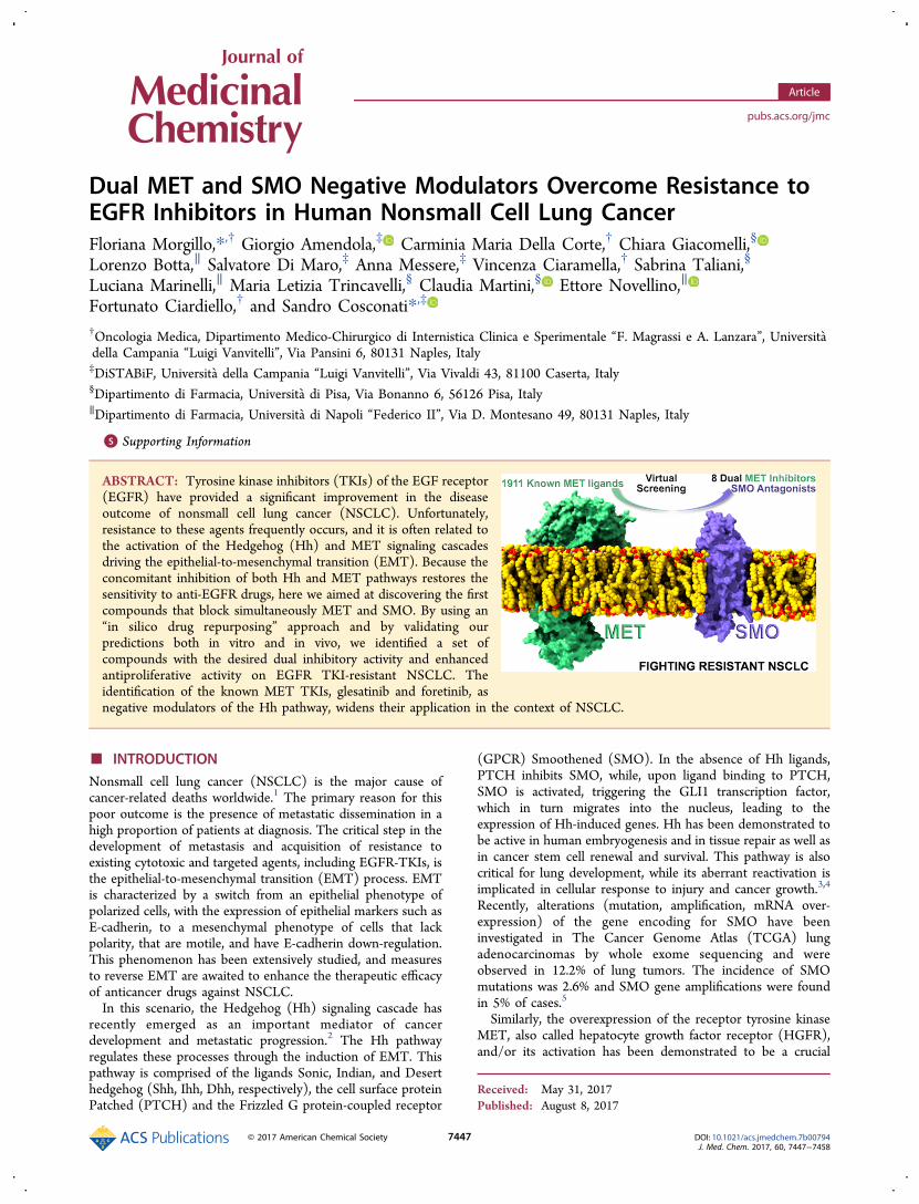
ABSTRACT: Tyrosine kinase inhibitors (TKIs) of the EGF receptor(EGFR) have provided a significant improvement in the diseaseoutcome of nonsmall cell lung cancer (NSCLC). Unfortunately,resistance to these agents frequently occurs, and it is often related tothe activation of the Hedgehog (Hh) and MET signaling cascadesdriving the epithelial-to-mesenchymal transition (EMT). Because theconcomitant inhibition of both Hh and MET pathways restores thesensitivity to anti-EGFR drugs, here we aimed at discovering the firstcompounds that block simultaneously MET and SMO. By using an“in silico drug repurposing” approach and by validating ourpredictions both in vitro and in vivo, we identified a set ofcompounds with the desired dual inhibitory activity and enhancedantiproliferative activity on EGFR TKI-resistant NSCLC. Theidentification of the known MET TKIs, glesatinib and foretinib, asnegative modulators of the Hh pathway, widens their application in the context of NSCLC.
■ INTRODUCTIONNonsmall cell lung cancer (NSCLC) is the major cause ofcancer-related deaths worldwide.1 The primary reason for thispoor outcome is the presence of metastatic dissemination in ahigh proportion of patients at diagnosis. The critical step in thedevelopment of metastasis and acquisition of resistance toexisting cytotoxic and targeted agents, including EGFR-TKIs, isthe epithelial-to-mesenchymal transition (EMT) process. EMTis characterized by a switch from an epithelial phenotype ofpolarized cells, with the expression of epithelial markers such asE-cadherin, to a mesenchymal phenotype of cells that lackpolarity, that are motile, and have E-cadherin down-regulation.This phenomenon has been extensively studied, and measuresto reverse EMT are awaited to enhance the therapeutic efficacyof anticancer drugs against NSCLC.In this scenario, the Hedgehog (Hh) signaling cascade has
recently emerged as an important mediator of cancerdevelopment and metastatic progression.2 The Hh pathwayregulates these processes through the induction of EMT. Thispathway is comprised of the ligands Sonic, Indian, and Deserthedgehog (Shh, Ihh, Dhh, respectively), the cell surface proteinPatched (PTCH) and the Frizzled G protein-coupled receptor
(GPCR) Smoothened (SMO). In the absence of Hh ligands,PTCH inhibits SMO, while, upon ligand binding to PTCH,SMO is activated, triggering the GLI1 transcription factor,which in turn migrates into the nucleus, leading to theexpression of Hh-induced genes. Hh has been demonstrated tobe active in human embryogenesis and in tissue repair as well asin cancer stem cell renewal and survival. This pathway is alsocritical for lung development, while its aberrant reactivation isimplicated in cellular response to injury and cancer growth.3,4
Recently, alterations (mutation, amplification, mRNA over-expression) of the gene encoding for SMO have beeninvestigated in The Cancer Genome Atlas (TCGA) lungadenocarcinomas by whole exome sequencing and wereobserved in 12.2% of lung tumors. The incidence of SMOmutations was 2.6% and SMO gene amplifications were foundin 5% of cases.5
Similarly, the overexpression of the receptor tyrosine kinaseMET, also called hepatocyte growth factor receptor (HGFR),and/or its activation has been demonstrated to be a crucial
Received: May 31, 2017Published: August 8, 2017
Article
pubs.acs.org/jmc
© 2017 American Chemical Society 7447 DOI: 10.1021/acs.jmedchem.7b00794J. Med. Chem. 2017, 60, 7447−7458
mediator of the EMT process and has been implicated inresistance to chemotherapy and to anti-EGFR TKIs. SeveralMET TKIs inhibitors have been evaluated in phase II/IIIclinical studies in NSCLC patients, with controversial results.6
Most probably, blocking MET alone is not sufficient to revertthe resistant phenotype as this latter is implicated in severalintracellular interactions and the best way to overcomeresistance is a combined approach, where the concomitantinhibition of MET and Hh pathways is performed.In this respect, we have recently demonstrated the
occurrence of SMO gene amplification, MET activation, anda functional interaction of these two signaling pathways in amodel of EGFR-mutated TKI-resistant NSCLC cells.7 In thesame cell model, inhibition of SMO in combination with METinhibition significantly reduced cancer cell proliferation,induced apoptosis, blocked the invasive and migratory behavior,and induced the complete regression of 100% of tumorsxenografted in nude mice.7 Moreover, blockade of Hh pathwayreverted EMT and was also associated with enhanced tumorsensitivity to cytotoxic agents in EGFR-wild-type NSCLCmodels.7 Consistently, recent data demonstrated that aberrant
activation of the Hh pathway represented also a commonfeature, along with EMT, in an in vivo model of acquiredresistance to EGFR-inhibitors obtained with a sequence of first-generation (erlotinib), second-generation (afatinib), and third-generation (osimertinib) EGFR TKIs.8
The synergistic interaction of Hh and MET pathwaysstrongly supports the rationale for a combined therapy inorder to overcome resistance to EGFR TKIs. On the otherhand, despite the established pharmacological significance ofcombination therapies, several advantages can be envisagedwith the employment of rationally discovered compounds thatare able to simultaneously hit two different pharmacologicaltargets9 such as a better description of the pharmacokineticprofile compared to combination therapy, diminished risks ofdrug−drug interactions, and a simplified dosing scheduling. Onthe other hand, multitarget compounds might indeed display adegree of target promiscuity resulting in unexpected adverseeffects that could lead to late attrition in the drug discoverypipeline. To this end, repurposing of known drugs, with analready described toxicological profile, might indeed offer anattractive opportunity in the search of multitarget ligands.10
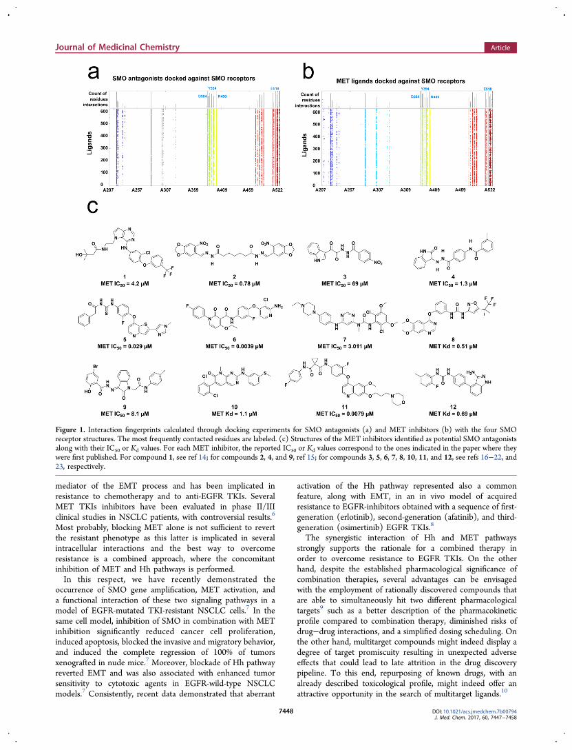
Figure 1. Interaction fingerprints calculated through docking experiments for SMO antagonists (a) and MET inhibitors (b) with the four SMOreceptor structures. The most frequently contacted residues are labeled. (c) Structures of the MET inhibitors identified as potential SMO antagonistsalong with their IC50 or Kd values. For each MET inhibitor, the reported IC50 or Kd values correspond to the ones indicated in the paper where theywere first published. For compound 1, see ref 14; for compounds 2, 4, and 9, ref 15; for compounds 3, 5, 6, 7, 8, 10, 11, and 12, see refs 16−22, and23, respectively.
Journal of Medicinal Chemistry Article
DOI: 10.1021/acs.jmedchem.7b00794J. Med. Chem. 2017, 60, 7447−7458
7448

In this context, the aim of the present study was to rationallydiscover a set of antiproliferative compounds able tosimultaneously block MET and SMO receptors. To this end,we wanted to merge the advantages of the drug repurposingstrategy,10 which searches for novel indications, mechanism ofaction, and/or pharmacological targets of already existing drugs,with the predictive power of theoretical docking-based virtualscreening (VS). Through this “in silico drug repurposingapproach”, we selected, among 1911 MET-inhibitors, a set of12 promising compounds for their potential dual inhibitoryactivity against MET and SMO, which were validated in in vitroand in vivo models of resistance to anti-EGFR TKIs.This strategy allowed us to identify two compounds that are
currently in clinical trials, as new inhibitors of the Hh pathway,in addition to the already known AXL and MET inhibitoryproperties. The potent antitumor activity in vitro and in vivo ina NSCLC model of EGFR-acquired resistance potentiallyexpands the indications for these two drugs.
■ RESULTSIn Silico Identification of Known MET Inhibitors as
New SMO Binders. To identify MET inhibitors, which mightshow interesting affinities at the SMO receptor and vice versa,we took advantage of a public domain repository of compoundstructures and activity data, the BindingDB database.11 Thus, aset of 1911 known selective, as well as unselective METinhibitors, was virtually docked against the available SMOreceptor X-ray structures by following a protocol that werecently devised to optimize the performances of structure-based VS against SMO receptor.12 By following this protocol,we were able to rank the inspected compounds for theirpredicted affinity against the SMO receptor.A critical step of a VS campaign is the postdocking selection
of the compounds to experimentally test. To this end, we firstdecided to in silico characterize the theoretical interactionpattern established by known SMO antagonists and thepublished structures for this receptor. Therefore, as happenedfor the MET inhibitors, a collection of 412 SMO antagonistswas also obtained by interrogating the BindingDB database.11
Thus, by following the above-mentioned docking protocol forthese latter compounds, the binding pose within the SMOreceptor was calculated. These results were subsequentlyanalyzed through an interaction fingerprints routine (seeMaterials and Methods), thereby allowing us to detect whichSMO residues are predicted to be more frequently contacted bythe known antagonists for this receptor. According to thisinspection, the SMO D384, Y394, R400, and E518 residues arethe most frequently contacted ones. Interestingly, alsomutagenesis experiments outlined the importance of theabove-mentioned residues for SMO antagonist binding.13 Thesame interaction pattern was also observed for the dockedMET inhibitors within the SMO receptor, thereby substantiat-ing our initial design hypothesis. Parts a and b of Figure 1depict the interaction fingerprints obtained for the two dockingexperiments (SMO antagonists against SMO receptor andMET inhibitors against the same receptor, Figure 1a,b,respectively). In addition, analysis of the X-ray antagonist/SMO structures also outlined that N219, F484, and W281receptor residues provide additional anchoring points forcocrystallized antagonists.Thus, in our theoretical model, we further filtered out the
docked MET inhibitors for which no favorable contacts withthe above-mentioned SMO residues were predicted. Of the
remaining 421 compounds, 25 were commercially available and,of the latter, we decided to select only those whose METinhibitory potency and or affinity were reported to be at least inthe midmicromolar regimen. This additional filter allowedselecting 12 inhibitors that were finally purchased fromdifferent vendors and then checked for compound compositionand purity (see Supporting Information). The selectedinhibitors along with their reported inhibitory activity and/oraffinity against MET are reported in Figure 1c.
Selected MET Inhibitors Bind and Antagonize SMOReceptor. The affinity of the selected compounds towardSMO protein was evaluated by radioligand binding competitionstudies using [3H]-cyclopamine ((2′R,3S,3′R,3′aS,6′S,6aS,-6bS,7′aR,11aS,11bR)-1,2,3,3′a,4,4′,5′,6,6′,6a,6b,7,7′,7′a,8,11,-11a,11b-octadecahydro-3′,6′,10,11b-tetramethyl-spiro[9H-benzo[a]fluorene-9,2′(3′H)-furo[3,2-b]pyridin]-3-ol, 13, Sup-porting Information, Chart S1).24 For this purpose, we used theEGFR exon 19 deletion mutant (delE746-A750) HCC827-GRhuman NSCLC cell line made resistant to gefitinib in vitro,which we previously characterized for SMO and METexpression.7 The HCC827-GR cell line presents SMO geneamplification and MET pathway overexpression and activation.7
In particular, NGS analysis of this cell line showed a 47% allelicfrequency of V404 M mutation in the SMO gene as comparedto the parental HCC827, which showed an allelic frequency of0.1%, indicating the selection of an SMO mutated resistantclone during acquisition of resistance to gefitinib. The affinitydissociation constant (Kd) of [3H]-13 in HCC827-GR cellsoverexpressing the SMO receptor was 36.9 ± 8.9 nM and aBmax of 1567 ± 61 fmol/mg, as obtained by saturation bindingstudies (Figure 2).
The SMO antagonist vismodegib (2-chloro-N-[4-chloro-3-(2-pyridinyl)phenyl]-4-(methylsulfonyl)benzamide, 14,25 Sup-porting Information, Chart S1) showed a Ki value of 12.2 ± 1.7nM in accordance with literature data.26 Some of thecompounds (compounds 1, 2, 4, and 6) were not able tocompletely displace the radiolabeled ligand when tested at a 10μM concentration. On the contrary, compounds 3, 5, and 7−12
Figure 2. SMO binding analysis. Saturation assay of [3H]-13 bindingto HCC827-GR cells. Cells were incubated 4 h at RT in binding buffercontaining increasing concentrations of [3H]-13. Nonspecific bindingwas determined in the presence of 25 μM of compound 14. Scatchardplot analysis of the specific binding. Data are means ± SEM (n = 3) ofa representative experiment over three independent experiments. Theability of each of the 12 potential dual inhibitors to displace 25 nM[3H]-13 specific binding is reported in Table 1.
Journal of Medicinal Chemistry Article
DOI: 10.1021/acs.jmedchem.7b00794J. Med. Chem. 2017, 60, 7447−7458
7449

demonstrated the ability to compete with the [3H]-13 for theSMO binding site with an affinity in the nanomolar range(Table 1). In particular, compounds 5 and 11 were
demonstrated to be comparably efficient in inhibiting METand binding SMO. For these latter inhibitors, we also
demonstrated that a similar affinity was recorded for the wild-type SMO receptor (Figure 2b), indicating that the V404 MSMO mutation does not affect drug affinity. Figure 3 reportsthe binding mode of compounds 3, 5, and 7−12 in the SMOreceptor (see Figures S1−S8 in Supporting Information for the2D diagrams of the calculated complexes).Because GLI1 is a SMO regulated transcription factor,27 we
tested the functional significance of treatment with METinhibitors that demonstrated the highest ability to displace 13from binding to the SMO receptor on GLI1 activity by using aGLI1-responsive promoter within a luciferase reporterexpression vector. In particular, we performed a dose-dependent analysis of luciferase activity in HCC827-GR cellline (Figure 3). Treatment with 2 μM (Figure 4a) or 60 μM(Figure 4b) of each compound resulted, respectively, in a 25−50% and in a 10−100% decrease in GLI1-responsive promotercompared with the HCC827-GR untreated cells (p < 0.001).These data demonstrate that the tested MET inhibitors have
a SMO antagonist activity in a concentration-dependentmanner. Furthermore, to test if the effect of the selectedcompounds on GLI1 activity was mediated by SMOantagonism, we analyzed the ability of the SMO agonist SAG(100 nM) [N-methyl-N′-(3-pyridinylbenzyl)-N′-(3-chlorobenzo[b]thiophene-2-carbonyl)-1,4 diaminocyclohex-ane]28 to revert this effect. Interestingly, in the presence ofSAG, the induced inhibition of GLI1 activity was completelyreverted in almost all treatments at a 2 μM concentration of
Table 1. Experimental Binding Affinity of Compounds 1−14towards V404M Mutant SMO Receptor in HCC827-GRCells and of Compounds 5 and 11 on WT SMO Receptor inHEK293T Cellsa
compd Ki V404 M SMO (nM) Ki wt SMO (nM)
1 nd2 nd3 168.1 ± 21.9 nd4 nd5 53.1 ± 9.9 41.7 ± 8.26 nd7 46.5 ± 6.3 nd8 187.8 ± 36.9 nd9 50.4 ± 11.8 nd10 46.2 ± 11.7 nd11 66.8 ± 7.0 59.7 ± 9.612 87.6 ± 12.4 nd13 51.0 ± 8.4 nd14 12.2 ± 1.7 nd
and: not determined.
Figure 3. Predicted binding pose of compound 3, 5, and 7−12 in the SMO X-ray structure. Compounds are represented as green sticks, whilereceptor as orange sticks and transparent white ribbons. H-bonds are represented as dashed yellow lines.
Journal of Medicinal Chemistry Article
DOI: 10.1021/acs.jmedchem.7b00794J. Med. Chem. 2017, 60, 7447−7458
7450
compounds 3, 5, and 7−12 (p < 0.01) (Figure 4a) and partiallyreverted at a 60 μM concentration (Figure 4b), confirming thatthe selected MET inhibitors antagonize the SMO receptorfunction.Dual MET/SMO Inhibitors Are Potent Antiproliferative
Agents in EGFR-TKI Resistant Human NSCLC. We thenselected, among the 12 potential dual inhibitors, compounds 5and 11 as the most active MET inhibitors with the most potentactivity also on SMO. Moreover, we also kept compounds 6(significant inhibitory activity against MET and no binding atSMO receptor) and 9 (weak MET inhibitor and nanomolaraffinity for SMO) as negative controls. Of interest, compound 5was identified as glesatinib (N-[(3-fluoro-4-{[2-(5-{[(2-methoxyethyl)amino]methyl}pyridin-2-yl)thieno[3,2-b]-pyridin-7-yl]oxy}phenyl)carbamothioyl]-2-(4-fluorophenyl)-acetamide),17 MGCD265,54 Mirati Therapeutics), a tyrosinekinase inhibitor of MET and AXL, and compound 11 asforetinib (N1′-[3-fluoro-4-[[6-methoxy-7-(3-morpholinopro-poxy)-4-quinoly l]oxy]phenyl]-N1-(4-fluorophenyl)-cyc lopropane-1 ,1-d icarboxamide , XL880, Exe l ix i s ,GSK1363089, GlaxoSmithKline),22 known as a MET andVEGFR2 inhibitor, with activity against AXL.We analyzed the activity of 5 and 11 in the EGFR-mutated
HCC827-GR NSCLC cells with the already describedamplification of SMO and overexpression of MET anddisplaying a typical mesenchymal behavior.7 In this model, asalready demonstrated, activation of AXL (phospho-AXL),which is a known pathway responsible for the acquisition ofresistance to anti-EGFR TKIs and mediator of EMT,29 andanother signaling pathway potentially activated as a resistance
mechanism, was not significantly high as compared to sensitivecell models.7
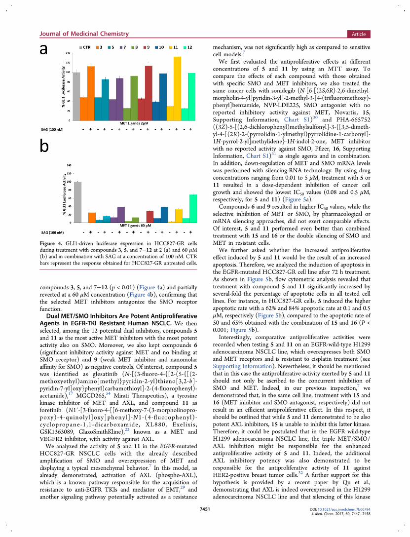
We first evaluated the antiproliferative effects at differentconcentrations of 5 and 11 by using an MTT assay. Tocompare the effects of each compound with those obtainedwith specific SMO and MET inhibitors, we also treated thesame cancer cells with sonidegib (N-[6-[(2S,6R)-2,6-dimethyl-morpholin-4-yl]pyridin-3-yl]-2-methyl-3-[4-(trifluoromethoxy)-phenyl]benzamide, NVP-LDE225, SMO antagonist with noreported inhibitory activity against MET, Novartis, 15,Supporting Information, Chart S1)30 and PHA-665752((3Z)-5-[(2,6-dichlorophenyl)methylsulfonyl]-3-{[3,5-dimeth-yl-4-[(2R)-2-(pyrrolidin-1-ylmethyl)pyrrolidine-1-carbonyl]-1H-pyrrol-2-yl]methylidene}-1H-indol-2-one, MET inhibitorwith no reported activity against SMO, Pfizer, 16, SupportingInformation, Chart S1)31 as single agents and in combination.In addition, down-regulation of MET and SMO mRNA levelswas performed with silencing-RNA technology. By using drugconcentrations ranging from 0.01 to 5 μM, treatment with 5 or11 resulted in a dose-dependent inhibition of cancer cellgrowth and showed the lowest IC50 values (0.08 and 0.5 μM,respectively, for 5 and 11) (Figure 5a).Compounds 6 and 9 resulted in higher IC50 values, while the
selective inhibition of MET or SMO, by pharmacological ormRNA silencing approaches, did not exert comparable effects.Of interest, 5 and 11 performed even better than combinedtreatment with 15 and 16 or the double silencing of SMO andMET in resistant cells.We further asked whether the increased antiproliferative
effect induced by 5 and 11 would be the result of an increasedapoptosis. Therefore, we analyzed the induction of apoptosis inthe EGFR-mutated HCC827-GR cell line after 72 h treatment.As shown in Figure 5b, flow cytometric analysis revealed thattreatment with compound 5 and 11 significantly increased byseveral-fold the percentage of apoptotic cells in all tested celllines. For instance, in HCC827-GR cells, 5 induced the higherapoptotic rate with a 62% and 84% apoptotic rate at 0.1 and 0.5μM, respectively (Figure 5b), compared to the apoptotic rate of50 and 65% obtained with the combination of 15 and 16 (P <0.001; Figure 5b).Interestingly, comparative antiproliferative activities were
recorded when testing 5 and 11 on an EGFR-wild-type H1299adenocarcinoma NSCLC line, which overexpresses both SMOand MET receptors and is resistant to cisplatin treatment (seeSupporting Information). Nevertheless, it should be mentionedthat in this case the antiproliferative activity exerted by 5 and 11should not only be ascribed to the concurrent inhibition ofSMO and MET. Indeed, in our previous inspection,7 wedemonstrated that, in the same cell line, treatment with 15 and16 (MET inhibitor and SMO antagonist, respectively) did notresult in an efficient antiproliferative effect. In this respect, itshould be outlined that while 5 and 11 demonstrated to be alsopotent AXL inhibitors, 15 is unable to inhibit this latter kinase.Therefore, it could be postulated that in the EGFR wild-typeH1299 adenocarcinoma NSCLC line, the triple MET/SMO/AXL inhibition might be responsible for the enhancedantiproliferative activity of 5 and 11. Indeed, the additionalAXL inhibitory potency was also demonstrated to beresponsible for the antiproliferative activity of 11 againstHER2-positive breast tumor cells.32 A further support for thishypothesis is provided by a recent paper by Qu et al.,demonstrating that AXL is indeed overexpressed in the H1299adenocarcinoma NSCLC line and that silencing of this kinase
Figure 4. GLI1-driven luciferase expression in HCC827-GR cellsduring treatment with compounds 3, 5, and 7−12 at 2 (a) and 60 μM(b) and in combination with SAG at a concentration of 100 nM. CTRbars represent the response obtained for HCC827-GR untreated cells.
Journal of Medicinal Chemistry Article
DOI: 10.1021/acs.jmedchem.7b00794J. Med. Chem. 2017, 60, 7447−7458
7451
inhibited cell proliferation and migration.33 This might alsoexplain why in the HCC827-GR cell lines 5 and 11 are slightlymore active than the concurrent treatment with 15 and 16(Figure 5).To study the influence of 5 and 11 on the activation/
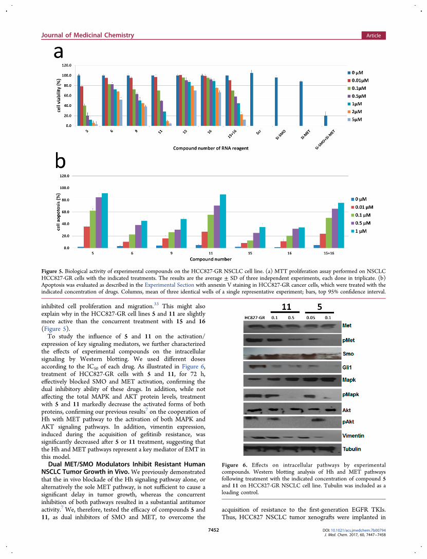
expression of key signaling mediators, we further characterizedthe effects of experimental compounds on the intracellularsignaling by Western blotting. We used different dosesaccording to the IC50 of each drug. As illustrated in Figure 6,treatment of HCC827-GR cells with 5 and 11, for 72 h,effectively blocked SMO and MET activation, confirming thedual inhibitory ability of these drugs. In addition, while notaffecting the total MAPK and AKT protein levels, treatmentwith 5 and 11 markedly decrease the activated forms of bothproteins, confirming our previous results7 on the cooperation ofHh with MET pathway to the activation of both MAPK andAKT signaling pathways. In addition, vimentin expression,induced during the acquisition of gefitinib resistance, wassignificantly decreased after 5 or 11 treatment, suggesting thatthe Hh and MET pathways represent a key mediator of EMT inthis model.Dual MET/SMO Modulators Inhibit Resistant Human
NSCLC Tumor Growth in Vivo. We previously demonstratedthat the in vivo blockade of the Hh signaling pathway alone, oralternatively the sole MET pathway, is not sufficient to cause asignificant delay in tumor growth, whereas the concurrentinhibition of both pathways resulted in a substantial antitumoractivity.7 We, therefore, tested the efficacy of compounds 5 and11, as dual inhibitors of SMO and MET, to overcome the
acquisition of resistance to the first-generation EGFR TKIs.Thus, HCC827 NSCLC tumor xenografts were implanted in
Figure 5. Biological activity of experimental compounds on the HCC827-GR NSCLC cell line. (a) MTT proliferation assay performed on NSCLCHCC827-GR cells with the indicated treatments. The results are the average ± SD of three independent experiments, each done in triplicate. (b)Apoptosis was evaluated as described in the Experimental Section with annexin V staining in HCC827-GR cancer cells, which were treated with theindicated concentration of drugs. Columns, mean of three identical wells of a single representative experiment; bars, top 95% confidence interval.
Figure 6. Effects on intracellular pathways by experimentalcompounds. Western blotting analysis of Hh and MET pathwaysfollowing treatment with the indicated concentration of compound 5and 11 on HCC827-GR NSCLC cell line. Tubulin was included as aloading control.
Journal of Medicinal Chemistry Article
DOI: 10.1021/acs.jmedchem.7b00794J. Med. Chem. 2017, 60, 7447−7458
7452
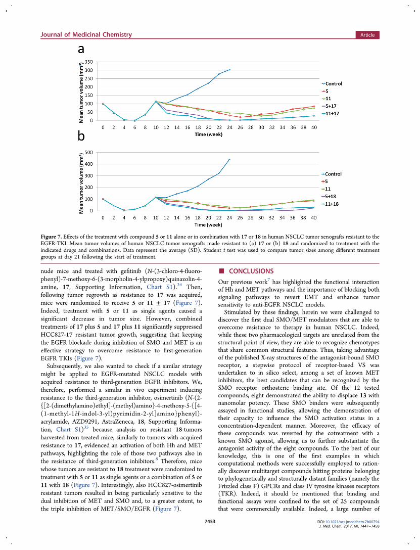
nude mice and treated with gefitinib (N-(3-chloro-4-fluoro-phenyl)-7-methoxy-6-(3-morpholin-4-ylpropoxy)quinazolin-4-amine, 17, Supporting Information, Chart S1).34 Then,following tumor regrowth as resistance to 17 was acquired,mice were randomized to receive 5 or 11 ± 17 (Figure 7).Indeed, treatment with 5 or 11 as single agents caused asignificant decrease in tumor size. However, combinedtreatments of 17 plus 5 and 17 plus 11 significantly suppressedHCC827-17 resistant tumor growth, suggesting that keepingthe EGFR blockade during inhibition of SMO and MET is aneffective strategy to overcome resistance to first-generationEGFR TKIs (Figure 7).Subsequently, we also wanted to check if a similar strategy
might be applied to EGFR-mutated NSCLC models withacquired resistance to third-generation EGFR inhibitors. We,therefore, performed a similar in vivo experiment inducingresistance to the third-generation inhibitor, osimertinib (N-(2-{[2-(dimethylamino)ethyl]-(methyl)amino}-4-methoxy-5-{[4-(1-methyl-1H-indol-3-yl)pyrimidin-2-yl]amino}phenyl)-acrylamide, AZD9291, AstraZeneca, 18, Supporting Informa-tion, Chart S1)35 because analysis on resistant 18-tumorsharvested from treated mice, similarly to tumors with acquiredresistance to 17, evidenced an activation of both Hh and METpathways, highlighting the role of those two pathways also inthe resistance of third-generation inhibitors.8 Therefore, micewhose tumors are resistant to 18 treatment were randomized totreatment with 5 or 11 as single agents or a combination of 5 or11 with 18 (Figure 7). Interestingly, also HCC827-osimertinibresistant tumors resulted in being particularly sensitive to thedual inhibition of MET and SMO and, to a greater extent, tothe triple inhibition of MET/SMO/EGFR (Figure 7).
■ CONCLUSIONS
Our previous work7 has highlighted the functional interactionof Hh and MET pathways and the importance of blocking bothsignaling pathways to revert EMT and enhance tumorsensitivity to anti-EGFR NSCLC models.Stimulated by these findings, herein we were challenged to
discover the first dual SMO/MET modulators that are able toovercome resistance to therapy in human NSCLC. Indeed,while these two pharmacological targets are unrelated from thestructural point of view, they are able to recognize chemotypesthat share common structural features. Thus, taking advantageof the published X-ray structures of the antagonist-bound SMOreceptor, a stepwise protocol of receptor-based VS wasundertaken to in silico select, among a set of known METinhibitors, the best candidates that can be recognized by theSMO receptor orthosteric binding site. Of the 12 testedcompounds, eight demonstrated the ability to displace 13 withnanomolar potency. These SMO binders were subsequentlyassayed in functional studies, allowing the demonstration oftheir capacity to influence the SMO activation status in aconcentration-dependent manner. Moreover, the efficacy ofthese compounds was reverted by the cotreatment with aknown SMO agonist, allowing us to further substantiate theantagonist activity of the eight compounds. To the best of ourknowledge, this is one of the first examples in whichcomputational methods were successfully employed to ration-ally discover multitarget compounds hitting proteins belongingto phylogenetically and structurally distant families (namely theFrizzled class F) GPCRs and class IV tyrosine kinases receptors(TKR). Indeed, it should be mentioned that binding andfunctional assays were confined to the set of 25 compoundsthat were commercially available. Indeed, a large number of
Figure 7. Effects of the treatment with compound 5 or 11 alone or in combination with 17 or 18 in human NSCLC tumor xenografts resistant to theEGFR-TKI. Mean tumor volumes of human NSCLC tumor xenografts made resistant to (a) 17 or (b) 18 and randomized to treatment with theindicated drugs and combinations. Data represent the average (SD). Student t test was used to compare tumor sizes among different treatmentgroups at day 21 following the start of treatment.
Journal of Medicinal Chemistry Article
DOI: 10.1021/acs.jmedchem.7b00794J. Med. Chem. 2017, 60, 7447−7458
7453

MET inhibitors have been published and screened through ourtheoretical protocol that would deserve further experimentaltesting as SMO antagonists but were not considered in thepresent study because of their unavailability on the market. Inthis context, this study should also stimulate the biologicalevaluation of these latter compounds.Prompted by these encouraging data, cell-based experiments
were performed in NSCLC cell lines made resistant to theEGFR-TKI 17. Strikingly, in this model, 5 and 11 were evenmore effective in achieving an antiproliferative effect than thecombination treatment with a pure MET inhibitor and theSMO antagonist as well as the double silencing of the twotarget proteins. This result might be explained by the ability of5 and 11 to inhibit AXL TKI. In this respect, both drugs werealso the most active apoptosis inducers. When tested in vivo, 5and 11 alone significantly decreased the tumor size of humanNSCLC xenografts resistant to the EGFR-TKI, and this effectwas potentiated by the coadministration with 17. Thisdemonstrates that, at least in our model, the triple inhibitionof SMO, MET, and EGFR is indeed the most effective strategyto circumvent drug resistance to EGFR TKIs. The same datawere also achieved by cotreatment with the selected drugs andthird-generation EGFR TKIs, 18.Of note, our findings allow unraveling previous results
achieved for 5 and 11. In particular, it has been recentlyreported that 5, when used in combination with erlotinib(EGFR-TKI) in a gastric cancer model, impairs the glycolysispathway also by inhibiting the 6-phosphofructo-2-kinase/fructose-2,6-bisphosphatase 3 (PFKFB3) enzyme, therebysuggesting a novel mechanism of action for this drug.36 Inaddition, it has been recently reported that the Hh signalingpromotes glucose utilization and glycolysis, thereby acceleratingcell proliferation in breast cancer cells, by positively modulatingthe 6-phosphofructo-2-kinase/fructose-2,6-bisphosphatase 3(PFKFB3) phosphorylation.37 In this scenario, our datareconcile the negative modulation of cancer glycolysis exertedby 5 with its antagonist activity on SMO receptor.On the other hand, 11 has been demonstrated to efficiently
decrease tumor cell proliferation and to induce apoptosis inHh-driven medulloblastomas. These data were first explainedwith the ability of this drug to inhibit MET that was proposedto be a marker of the aforementioned tumor.38 Herein wesuggest that the efficient antiproliferative activity displayed by11 should also be ascribed to a direct interaction with the SMOreceptor.In conclusion, in this work, the tandem application of
predictive theoretical studies and in-depth in vitro and in vivoevaluation allowed us to identify from a large number of METinhibitors those that can negatively modulate the SMO receptorand achieve superior antiproliferative effects in a model ofresistant NSCLC tumor. In this context, the identification of 5and 11 as negative modulators of the Hh pathway widens therange of action of these drugs in the context of NSCLCimproving the already known AXL and MET targeting activity.
■ EXPERIMENTAL SECTIONMolecular Modeling. Proteins and ligands setup: The structure of
the SMO receptor in complex with the antagonists (4-benzyl-piperazin-1-yl)-(3,5-dimethyl-1-phenyl-1H-pyrazol-4-ylmethylene)-amine39 (SANT-1, PDB 4N4W),40 2-(6-(4-(4-benzylphthalazin-1-yl)piperazin-1-yl)pyridin-3-yl)propan-2-ol41 (ANTA XV, PDB4QIM),40 4-fluoro-N-methyl-N-(1-(4-(1-methyl-1H-pyrazol-5-yl)-phthalazin-1-yl)piperidin-4-yl)-2-(trifluoromethyl)benzamide42
(LY2940680, PDB 4JKV),43 and 13 (PDB 4O9R)44 were downloadedfrom the Protein Data Bank and prepared for docking calculationsusing the “Protein Preparation Wizard” panel of Schrodingermolecular modeling package.45 In particular, using the “pre-processand analyze structure” tool, the bond orders and disulfide bonds wereassigned, all the hydrogen atoms were added, and water moleculeswere deleted. Using Epik 2.0, a prediction of the side chains heterogroups ionization and tautomeric states was performed.46 Anoptimization of the hydrogen-bonding network was performed usingthe “H-bond assignment” tool. Finally, using the “impref utility”, thepositions of the hydrogen atoms were optimized by keeping all theheavy atoms in place.
A database of known MET ligands (1911 compounds) wassubsequently prepared by interrogating the BindingDB database.11
Ligands were prepared for docking using LigPrep.47 Hydrogen atomswere added, ionizations states were generated, and tautomers weregenerated to prepare the required structures. All structures were thensubjected to minimization using the OPLS-2005 force field. The samepreparation was attained for the database of the known SMOantagonists downloaded from the BindingDB site.11
Docking Calculations. According to the results of our very recentretrospective study, the employment of the AutoDock Vina dockingsoftware48 when used against the ensemble of all the four receptorsgave the best results in terms of enrichment factors (EF) whenconsidering the lowest percentages of the ranked database (25%).12
AutoDock Vina performances and accuracy are assured by aLamarckian algorithm and an empirical binding free energy forcefield. Before the actual docking calculations could be run, both thedatabase of ligands and the receptors had to be converted to the pdbqtformat. The latter is very similar to a standard pdb but it includes “Q”(partial charges) and “T” atom types. Preparing the structures involvesensuring that its atoms are assigned the correct atom types, addingGasteiger charges if necessary, merging nonpolar hydrogens, detectingaromatic carbons, and setting up the “torsion tree” in the case ofligands. Thus, the python scripts prepare_receptor4.py and prepar-e_ligand4.py, part of MGLTools,49−51 were employed by applying thestandard settings. After the preparation, the docking was performedusing the default settings of Vina, setting a box whose center anddimensions were identical to the one used in our previous work.12
Results of these calculations produced four different rankings based onthe predicted binding free energy (herein referred to as ΔGVINA), onefor each docking attained on the four receptor structures, which weresubsequently unified using a parallel selection method. This selectionmethod resulted in a ranking of the screened compounds in whichΔGVINA values ranged from −6.5 to −14.8 kcal/mol.
Interaction fingerprints and interaction matrices of the SMO ligandsagainst SMO receptors and MET ligands against the same receptorswere calculated by employing a routine of the Schroedinger 2015release. The pose filter routine of the same release was used to countthe number of interactions predicted for the MET compounds againstD384, Y394, R400, E518, N219, F484, and W281 residues.
The fingerprint-based algorithm was used to cluster the 511 METligands in order to further refine them into 421 compounds. Thecanvas similarity and clustering, which is also part of the Schrodingersuite, was employed by applying the following settings: the eighthatom typing scheme for the fingerprinting (atoms distinguished byring size, aromaticity, HB acceptor/donor, ionization potential,whether terminal, whether halogen; bonds distinguished by bondorder), Tanimoto as the similarity metric, and average as linkage as theclustering method. The same method (i.e., canvas similarity using theeighth atom typing scheme for the fingerprinting and Tanimoto as thesimilarity metric) was also used to evaluate the structural similaritybetween each of the 12 final compounds and any of the SMOantagonist reported in the BindingDB database.11 Table S2 inSupporting Information reports the Tanimoto similarity indexbetween the 12 compounds and the most similar SMO antagonist.These values range from 7.8% to 32%, demonstrating that the selectedligands are structurally unrelated to the SMO antagonists described sofar.
Journal of Medicinal Chemistry Article
DOI: 10.1021/acs.jmedchem.7b00794J. Med. Chem. 2017, 60, 7447−7458
7454
All of the pictures were rendered with the UCSF Chimera packagefrom the Resource for Biocomputing, Visualization, and Informatics atthe University of California, San Francisco.37
Prior to biological evaluation, the 12 compounds were screened forPan Assay Interference Compounds using the PAINS-Remover webserver (http://www.cbligand.org), which is a data analysis toolsimplemented for removal of PAINS compounds. All compoundspassed this filtering with the exception of compound 7 and 9.Compound 7 features an anil_di_alk_A substructure that is suggestedto interfere with AlphaScreen technology, possibly through efficientquenching of singlet oxygen.52 In our case, we performed a radioliganddisplacement binding assay so no interference is expected. Compound9 features an imine_one_isatin substructure, which was found tointerfere sometimes with the AlphaScreen technology “for reasonsunknown”,52 as stated by the authors. Also in this case, the testedligand should not interfere with the implemented radioliganddisplacement binding assay. On the basis of NMR and MS analysis,all tested compounds have a purity ranging from 96 to 98%.Cell Lines and Drugs. The human NSCLC HCC827 cell line was
provided by American Type Culture Collection (ATCC, Manassas,VA, USA) and maintained in RPMI-1640 (Sigma-Aldrich, Saint Louis,MO, USA) medium supplemented with 10% fetal bovine serum (FBS;Life Technologies, Gaithersburg, MD) in a humidified atmospherewith 5% CO2. The identity of all cell lines was confirmed by STRprofiling (Promega, Madison, WV, USA) on an ad hoc basis prior toperforming experiments. HCC827-GR (gefitinib resistant) cell linewas obtained in vitro, as previously described.7 Briefly, over a period of12 months, HCC827 cells were continuously exposed to increasingdoses of 17, starting from an IC50 dose that represented the dose toinhibit the growth of 50% of cells. HCC827-GR cells were maintainedin culture with the maximum 17 dose that allowed cellularproliferation. The identity of cells was verified by STR profiling(Promega) on an ad hoc basis prior to performing experiments andrepeated for all cell lines after a majority of the experiments wereperformed. Drugs gefitinib and sonidegib were purchased from SelleckChemicals (Selleckchem, Houston, TX, USA). 18 was generouslyprovided by Astra Zeneca.SMO Radioligand Binding Assays. Prior to binding experiments,
all purchased compounds were analyzed to confirm the samplecomposition by LC/MS and 1H NMR experiments. The bindingexperiments were performed as previously described with fewmodifications.26 Briefly, HCC827-GR cells expressing V404 M SMOreceptors were grown in 24-well plates (100000 cells/well) and fixedwith 4% (v/v) formaldehyde/PBS for 20 min at room temperature(RT). Cells were subsequently incubated for 4 h at RT in bindingbuffer (Hanks Balanced Salt Solution (HBSS) without Ca2+ and Mg2+
with glucose 1 g/L). Saturation binding experiments were performedusing different [3H]-13 (American Radiolabeled Chemicals, Inc. Art.1473 a.s. 20 Ci/mmol) concentrations ranging from 0.1 to 500 nM; incompetition binding experiments cells were incubated with 25 nM[3H]-13 and different concentrations of the compounds. Nonspecificbinding was determined in the presence of 25 μM of compound 14.Incubations were terminated by rapid washing with 1 mL of bindingbuffer for three times. Then, the bound [3H]-13 was extracted in 200μL of 0.1 N NaOH for 12 h and neutralized with 200 μL of 0.1 N HCl.The amount of [3H]-13 in the extracts was measured using ascintillation counter. For the active compounds, the IC50 values weredetermined and Ki values were derived in accordance with theequation of Cheng and Prusoff.The affinity of the compounds for the wild-type SMO was
performed as previously described with few modifications.26 Briefly, 15μg of HEK293T-WT SMO membranes (Multispan Inc., Cod.MC1442; Kd = 20 nM; Bmax = 6.2 pmol/mg protein) were incubatedfor 4 h at RT in binding buffer (HEPES 50 mM, MgCl2 5 mM andBSA 0.02%) containing 25 nM [3H]-13 (American RadiolabeledChemicals, Inc. Art. 1473 a.s. 20 Ci/mmol) and differentconcentrations of the compounds. Nonspecific binding wasdetermined in the presence of 25 μM of compound 14. The boundradioactivity was separated by rapid filtration through GF/C glass fiberfilters presoaked for 2 h in 0.3% polyethylenimine pH 13.0 and washed
three times with 4 mL of ice-cold phosphate-buffered saline with0.01% Triton X-100, pH 7.0. Radioactivity was measured by liquidscintillation spectrometry. Dose−response curves are reported inSupporting Information, Figures S8 and S9.
Luciferase Assay. Here, we performed GLI1 luciferase assay totest the influence of selected compounds on GLI1 activity in HCC827-GR cells by using the Dual-Luciferase Assay system (Promega),following the manufacturer’s protocol. For each experiment, a total of5 × 105 cells were seeded in FBS-free medium in 24-well plates andtransfection started when cells reach a confluency of 50%. Wecotransfected the cells with the GLI1-Luc reporter plasmid (400 ng/well), kindly provided by Dr. Mariolina Castellone53 (University ofNaples Federico II) and pRL-TK, Renilla reporter, encoding theRenilla luciferase (Promega), to normalize the results. In GLI1reporter vector, the transcription of Firefly luciferase gene is under thecontrol of a promoter region specific for the transcription factor GLI1,so GLI1 luciferase activity correlates with the binding of GLI1 to itspromoter region; in control reporter, Renilla luciferase activitydepends on a constitutive promoter (thymidine kinase). Thetransfection was done in triplicate using Lipofectamine 2000 (providedby Thermo Fisher Scientific) as transfection reagent according tomanufacturer’ instructions. The medium of transfection was replacedafter 8 h with culture medium, containing the indicated MET ligandsor without the SMO agonist SAG (100 nM) [N-methyl-N′-(3-pyridinylbenzyl)-N′-(3-chlorobenzo[b]thiophene-2-carbonyl)-1,4-dia-minocyclohexane], purchased from Sigma-Aldrich.
Firefly luciferase is a protein from Photinus pyralis that reacts with itssubstrate (beetle luciferin, ATP, magnesium, and molecular oxygen),-creating luminescence, measured by the luminometer Autolumat LB953 (EG&G, Berthold, Bad Wildbad, Germany). According to theprotocol of the kit Dual-Luciferase Reporter Assay System (Promega),after about 48 h from transfection, we removed the medium andwashed once with PBS. Then, we added 100 μL of lysis buffer 1× ineach well, shaking for 20 min at RT, and finally scraped.
For the measurement of GLI1 luciferase activity, we mixed bypipetting 30 μL of lysated cells and 30 μL of Luciferase Assay Reagent,containing its substrate for reaction. After the reading at luminometerand the record of data, we added 30 μL of Stop and Glo Reagent,which blocked GLI1 induced Firefly luciferase signal and containedRenilla luciferase substrate and repeated the luminometer measure-ment to obtain the control values. For the analysis of data, wecalculated the average values of GLI1 luciferase signal, normalized toRenilla luciferase values, and represented the results as fold changewith respect to control cells and cells transfected with the emptyvector; results were the average of three independent experiments.
Protein Expression Analysis. Protein lysates were obtained byhomogenization in RIPA lysis buffer (0.1% sodium dodecyl sulfate(SDS), 0.5% deoxycholate, 1% Nonidet, 100 mmol/L NaCl, 10 mmol/L Tris-HCl (pH 7.4), 0.5 mmol/L dithiothreitol, and 0.5%phenylmethylsulfonyl fluoride, protease inhibitor cocktail (Hoff-mann-La Roche), and clarification by centrifugation at 14000 rpmfor 10 min a 4 °C. Cancer cells were lysed with Tween-20 lysis buffer(50 mmol/L HEPES, pH 7.4, 150 mmol/L NaCl, 0.1% Tween-20,10% glycerol, 2.5 mmol/L EGTA, 1 mmol/L EDTA, 1 mmol/L DTT,1 mmol/L phenylmethylsulfonyl fluoride, and 10 μg/mL of leupeptinand aprotinin). Protein lysates containing comparable amounts ofproteins, estimated by a modified Bradford assay (Bio-Rad), weresubjected to Western blot analysis as previously described.7
Immunocomplexes were detected with the enhanced chemilumines-cence kit ECL plus, by Thermo Fisher Scientific (Rockford, IL).Desired proteins were probed with corresponding antibodies. Primaryantibodies for Western blot analysis against p-MAPK44/42 (Thr202/Tyr204), MAPK44/42, p-AKT (Ser473), AKT, p-MET (Tyr1234/1235), MET, SMO, and GLI1 were obtained from Cell SignalingTechnology; monoclonal anti-α-tubulin antibody (T8203) from SigmaChemical Co. The following secondary antibodies from Bio-Rad wereused: goat antirabbit IgG and rabbit antimouse IgG. Immunoreactiveproteins were visualized by enhanced chemiluminescence (ECL plus;Thermo Fisher Scientific). Each experiment was done in triplicate.
Journal of Medicinal Chemistry Article
DOI: 10.1021/acs.jmedchem.7b00794J. Med. Chem. 2017, 60, 7447−7458
7455

Cell Proliferation Assays. Cancer cells were seeded in 96-multiwell plates and were treated with different doses of indicateddrugs for 72 h. Cell proliferation was measured with the MTT assay, aspreviously described.7 IC50 values were determined by interpolationfrom the dose−response curves. Results represent the median of threeseparate experiments, each performed in quadruplicate. Synergism wascalculated with ComboSyn software, ComboSyn Inc., Paramus, NK.07652 USA.RNA Silencing. The small inhibitor duplex RNAs (siRNA) (ON-
target plus SMARTpool) siSMO, siMET, and siCONTROL Non-targeting Pool (no. D-001206-13-05), used as a negative (scrambled)control, were provided from Dharmacon (Lafayette, CO). Cells weretransfected with 100 nM siRNAs using Dharmafect reagent followingmanufacturer’s instructions. The day before transfection, the cells wereplated in 35 mm dishes at 40% of confluence in medium supplementedwith 5% FBS without antibiotics. Where necessary, cells were treatedwith different compounds, as previously described; 24 h beforeharvesting and cell proliferation or Western blot analysis were thenperformed.Assessment of Apoptosis. Apoptosis was detected by flow
cytometry via the examination of altered plasma membranephospholipid packing by lipophilic dye annexin V as describedelsewhere.7 Briefly, treated cells were harvested by trypsin, washedtwice with PBS, and were then resuspended in binding buffer at aconcentration of 1 × 10⁶ cells/mL according to the manufacturer’sinstruction. Thereafter, 5 μL of annexin V-FITC and 5 μL ofpropidium iodide were added into 100 μL of cell suspension andincubated for 30 min at room temperature in the dark. After adding400 μL of binding buffer, labeled cells were counted by flow cytometrywithin 30 min. All early apoptotic cells (annexin V, positive; propidiumiodide, negative), necrotic/late apoptotic cells (double positive), aswell as living cells (double negative), were detected by FACSCaliburflow cytometer and subsequently analyzed by Cell Quest software(Becton Dickinson). Argon laser excitation wavelength was 488 nm,whereas emission data were acquired at wavelength 530 nm (FL-1channel) for FITC and 670 nm (FL-3 c3 channel) for propidiumiodide.In Vivo Experiments. The 4−6-week old female balb/c athymic
(nu/nu) mice were purchased from Charles River Laboratories. Theresearch protocol was approved and mice were maintained inaccordance with the Institutional Guidelines of the University ofCampania Luigi Vanvitelli Animal Care and Use Committee. Micewere acclimatized for 1 week before being injected with cancer cellsand injected subcutaneously with 107 HCC827 cells that had beendiluted in 200 μL of Matrigel (Corning Life Sciences, MA, USA), 1:1in the culture medium. For induction of resistance to 17 or 18, whentumors reached a mean volume of 150 mm3, mice were treated withescalating doses of 17 (from 18.7 to 150 mg/kg/day: 3 weeks with18.7 mg/kg/day, 3 weeks with 37.5 mg/kg/day, 3 weeks with 75 mg/kg/day, and last the 3 weeks with 150 mg/kg/day) or 198 (with thesame protocol of escalating doses every 3 weeks: 5 mg/kg/day for thefirst 3 weeks, 10 mg/kg/day for following 3 weeks, 17.5 mg/kg/day forsubsequent 3 weeks, and 25 mg/kg daily for last 3 weeks orally) over 4months to derive 17/18-resistant tumors (defined as >25% regrowthfrom max reduction). At the end of treatment period, resistant tumorswere randomized into one of the following four arms: control, 5, 11,17/18 plus 5, and 17/18 plus 11. 17 and 18 were continued atmaximum dose reached before resistance, and 5 and 11 were used assingle agents and in combination at fixed doses of 15 and 20 mg/kg/day orally, respectively. Body weight and tumor volume weremonitored on alternate days. Tumor volume was measured usingthe formula π/6 larger diameter × (smaller diameter).2 Character-ization of the pharmacokinetics of 11 have been already reported byFaria et al., demonstrating that this compound reaches the maximumplasma concentration after 5 h from oral administration.38 Character-ization of the pharmacokinetics of 5 demonstrated that this drugreaches a plasma concentration of 0.8 μM when orally administered inmice (20 mg/kg).
■ ASSOCIATED CONTENT*S Supporting InformationThe Supporting Information is available free of charge on theACS Publications website at DOI: 10.1021/acs.jmed-chem.7b00794.
Binding affinities, Tanimoto similarity index, ligandinteractions, and compound characterization (PDF)Molecular formula strings and some data (CSV)Coordinates of the calculated SMO/ligand complex of 3with 4N4W (PDB)Coordinates of the calculated SMO/ligand complex of 5with 4O9R (PDB)Coordinates of the calculated SMO/ligand complex of 6with 4O9R (PDB)Coordinates of the calculated SMO/ligand complex of 7with 4N4W (PDB)Coordinates of the calculated SMO/ligand complex of 8with 4O9R (PDB)Coordinates of the calculated SMO/ligand complex of11 with 4JKV (PDB)Coordinates of the calculated SMO/ligand complex of 9with 4JKV (PDB)Coordinates of the calculated SMO/ligand complex of12 with 4N4W (PDB)Coordinates of the calculated SMO/ligand complex of10 with 4O9R (PDB)
■ AUTHOR INFORMATIONCorresponding Authors*For S.C.: phone, +39 0823 274789; E-mail, [email protected].*For F.M.: phone, +39 081 5666732; E-mail, [email protected] Amendola: 0000-0003-4271-5031Chiara Giacomelli: 0000-0002-6244-602XClaudia Martini: 0000-0001-9379-3027Ettore Novellino: 0000-0002-2181-2142Sandro Cosconati: 0000-0002-8900-0968Author ContributionsThe manuscript was written through contributions of allauthors. All authors have given approval to the final version ofthe manuscript. F.M, G.A., and C.M.D.C. contributed equallyto this work.NotesThe authors declare no competing financial interest.
■ ACKNOWLEDGMENTSThis work was supported by Associazione Italiana per la Ricercasul Cancro (AIRC)-Project MFAG 2013-N.14392 to F.M., byProgetti di Rilevante Interesse Nazionale (PRIN) 2012 (grantno. 2012C5YJSK_003 to L.M. and 2012CTAYSY_002 to S.C.),and by Regione Campania under POR Campania FESR 2007−2013-O.O.2.1 (FarmaBioNet) to A.M. and S.C.
■ ABBREVIATIONS USEDTKI, tyrosine kinase inhibitors; EGFR, EGF receptor; NSCLC,nonsmall cell lung cancer; EMT, epithelial-to-mesenchymaltransition; Hh, Hedgehog; SMO, smoothened; TCGA, theCancer Genome Atlas; HGFR, hepatocyte growth factorreceptor; GR, gefitinib resistant
Journal of Medicinal Chemistry Article
DOI: 10.1021/acs.jmedchem.7b00794J. Med. Chem. 2017, 60, 7447−7458
7456
■ REFERENCES(1) Jemal, A.; Siegel, R.; Ward, E.; Hao, Y.; Xu, J.; Thun, M. J. CancerStatistics, 2009. Ca-Cancer J. Clin. 2009, 59, 225−249.(2) Yang, L.; Xie, G.; Fan, Q.; Xie, J. Activation of the hedgehog-signaling pathway in human cancer and the clinical implications.Oncogene 2010, 29, 469−481.(3) Yuan, Z.; Goetz, J. A.; Singh, S.; Ogden, S. K.; Petty, W. J.; Black,C. C.; Memoli, V. A.; Dmitrovsky, E.; Robbins, D. J. Frequentrequirement of hedgehog signaling in non-small cell lung carcinoma.Oncogene 2006, 26, 1046−1055.(4) Ahmad, A.; Maitah, M. I. Y.; Ginnebaugh, K. R.; Li, Y.; Bao, B.;Gadgeel, S. M.; Sarkar, F. H. Inhibition of hedgehog signalingsensitizes NSCLC cells to standard therapies through modulation ofEMT-regulating miRNAs. J. Hematol. Oncol. 2013, 6, 77.(5) Tsao, A.; Byers, L. A.; Diao, L.; Wang, J.; Weinstein, J. N.; Meric-Bernstam, F.; Aldape, K.; Heymach, J. V. P2.06-032-SMO mutationsoccur in non-small cell lung cancer (NSCLC) and may respond tohedgehog inhibitors. 15th World Conference on Lung Cancer, 2013,Sydney, Australia, 2013.(6) Reungwetwattana, T.; Liang, Y.; Zhu, V.; Ou, S. I. The race totarget MET exon 14 skipping alterations in non-small cell lung cancer:the why, the how, the who, the unknown, and the inevitable. LungCancer. 2017, 103, 27−37.(7) Della Corte, C. M.; Bellevicine, C.; Vicidomini, G.; Vitagliano, D.;Malapelle, U.; Accardo, M.; Fabozzi, A.; Fiorelli, A.; Fasano, M.;Papaccio, F.; Martinelli, E.; Troiani, T.; Troncone, G.; Santini, M.;Bianco, R.; Ciardiello, F.; Morgillo, F. SMO gene amplification andactivation of the hedgehog pathway as novel mechanisms of resistanceto anti-epidermal growth factor receptor drugs in human lung cancer.Clin. Cancer Res. 2015, 21, 4686−4697.(8) Della Corte, C. M.; Malapelle, U.; Vigliar, E.; Pepe, F.; Troncone,G.; Ciaramella, V.; Troiani, T.; Martinelli, E.; Belli, V.; Ciardiello, F.;Morgillo, F. Efficacy of continuous EGFR-inhibition and role ofhedgehog in EGFR acquired resistance in human lung cancer cells withactivating mutation of EGFR. Oncotarget 2017, 8, 23020−23032.(9) Anighoro, A.; Bajorath, J.; Rastelli, G. Polypharmacology:challenges and opportunities in drug discovery. J. Med. Chem. 2014,57, 7874−7887.(10) Nosengo, N. Can you teach old drugs new tricks? Nature 2016,534, 314−316.(11) Liu, T.; Lin, Y.; Wen, X.; Jorissen, R. N.; Gilson, M. K.BindingDB: a web-accessible database of experimentally determinedprotein−ligand binding affinities. Nucleic Acids Res. 2007, 35, 198−201.(12) Amendola, G.; Di Maio, D.; La Pietra, V.; Cosconati, S. Bestmatching protein conformations and docking programs for a virtualscreening campaign against SMO receptor. Mol. Inf. 2016, 35, 340−349.(13) Wang, C.; Wu, H.; Evron, T.; Vardy, E.; Han, G. W.; Huang, X.P.; Hufeisen, S. J.; Mangano, T. J.; Urban, D. J.; Katritch, V.; Cherezov,V.; Caron, M. G.; Roth, B. L.; Stevens, R. C. Structural basis forSmoothened receptor modulation and chemoresistance to anticancerdrugs. Nat. Commun. 2014, 5, 4355.(14) Ishikawa, T.; Seto, M.; Banno, H.; Kawakita, Y.; Oorui, M.;Taniguchi, T.; Ohta, Y.; Tamura, T.; Nakayama, A.; Miki, H.;Kamiguchi, H.; Tanaka, T.; Habuka, N.; Sogabe, S.; Yano, J.;Aertgeerts, K.; Kamiyama, K. Design and synthesis of novel humanepidermal growth factor receptor 2 (HER2)/epidermal growth factorreceptor (EGFR) dual inhibitors bearing a pyrrolo[3,2-d]pyrimidinescaffold. J. Med. Chem. 2011, 54, 8030−8050.(15) Liang, Z.; Zhang, D.; Ai, J.; Chen, L.; Wang, H.; Kong, X.;Zheng, M.; Liu, H.; Luo, C.; Geng, M.; Jiang, H.; Chen, K.Identification and synthesis of N′-(2-oxoindolin-3-ylidene)hydrazidederivatives against c-Met kinase. Bioorg. Med. Chem. Lett. 2011, 21,3749−3754.(16) Schmidt, S.; Preu, L.; Lemcke, T.; Totzke, F.; Schachtele, C.;Kubbutat, M. H.; Kunick, C. Dual IGF-1R/SRC inhibitors based on aN′-aroyl-2-(1H-indol-3-yl)-2-oxoacetohydrazide structure. Eur. J. Med.Chem. 2011, 46, 2759−2769.
(17) Claridge, S.; Raeppel, F.; Granger, M.-C.; Bernstein, N.;Saavedra, O.; Zhan, L.; Llewellyn, D.; Wahhab, A.; Deziel, R.; Rahil, J.;Beaulieu, N.; Nguyen, H.; Dupont, I.; Barsalou, A.; Beaulieu, C.;Chute, I.; Gravel, S.; Robert, M.-F.; Lefebvre, S.; Dubay, M.; Pascal, R.;Gillespie, J.; Jin, Z.; Wang, J.; Besterman, J. M.; Macleod, A. R.;Vaisburg, A. Discovery of a novel and potent series of thieno[3,2-b]pyridine-based inhibitors of c-Met and VEGFR2 tyrosine kinases.Bioorg. Med. Chem. Lett. 2008, 18, 2793−2798.(18) Schroeder, G. M.; An, Y.; Cai, Z.-W.; Chen, X.-T.; Clark, C.;Cornelius, L. A. M.; Dai, J.; Gullo-Brown, J.; Gupta, A.; Henley, B.;Hunt, J. T.; Jeyaseelan, R.; Kamath, A.; Kim, K.; Lippy, J.; Lombardo,L. J.; Manne, V.; Oppenheimer, S.; Sack, J. S.; Schmidt, R. J.; Shen, G.;Stefanski, K.; Tokarski, J. S.; Trainor, G. L.; Wautlet, B. S.; Wei, D.;Williams, D. K.; Zhang, Y.; Zhang, Y.; Fargnoli, J.; Borzilleri, R. M.Discovery of N-(4-(2-amino-3-chloropyridin-4-yloxy)-3-fluorophen-yl)-4-ethoxy-1-(4-fluorophenyl)-2-oxo-1,2-dihydropyridine-3-carboxa-mide (BMS-777607), a selective and orally efficacious inhibitor of theMet kinase superfamily. J. Med. Chem. 2009, 52, 1251−1254.(19) Guagnano, V.; Furet, P.; Spanka, C.; Bordas, V.; Le Douget, M.;Stamm, C.; Brueggen, J.; Jensen, M. R.; Schnell, C.; Schmid, H.;Wartmann, M.; Berghausen, J.; Drueckes, P.; Zimmerlin, A.; Bussiere,D.; Murray, J.; Porta, D. G. Discovery of 3-(2,6-dichloro-3,5-dimethoxy-phenyl)-1-{6-[4-(4-ethyl-piperazin-1-yl)-phenylamino]-pyrimidin-4-yl}-1-methyl-urea (NVP-BGJ398), a potent and selectiveinhibitor of the fibroblast growth factor receptor family of receptortyrosine kinase. J. Med. Chem. 2011, 54, 7066−7083.(20) Rowbottom, M. W.; Faraoni, R.; Chao, Q.; Campbell, B. T.; Lai,A. G.; Setti, E.; Ezawa, M.; Sprankle, K. G.; Abraham, S.; Tran, L.;Struss, B.; Gibney, M.; Armstrong, R. C.; Gunawardane, R. N.;Nepomuceno, R. R.; Valenta, I.; Hua, H.; Gardner, M. F.; Cramer, M.D.; Gitnick, D.; Insko, D. E.; Apuy, J. L.; Jones-Bolin, S.; Ghose, A. K.;Herbertz, T.; Ator, M. A.; Dorsey, B. D.; Ruggeri, B.; Williams, M.;Bhagwat, S.; James, J.; Holladay, M. W. Identification of 1-(3-(6,7-dimethoxyquinazolin-4-yloxy)phenyl)-3-(5-(1,1,1-trifluoro-2-methyl-propan-2-yl)isoxazol-3-yl)urea hydrochloride (CEP-32496), a highlypotent and orally efficacious inhibitor of V-RAF murine sarcoma viraloncogene homologue b1 (BRAF) V600E. J. Med. Chem. 2012, 55,1082−1105.(21) Davis, M. I.; Hunt, J. P.; Herrgard, S.; Ciceri, P.; Wodicka, L. M.;Pallares, G.; Hocker, M.; Treiber, D. K.; Zarrinkar, P. P.Comprehensive analysis of kinase inhibitor selectivity. Nat. Biotechnol.2011, 29, 1046−1051.(22) Qian, F.; Engst, S.; Yamaguchi, K.; Yu, P.; Won, K.-A.; Mock, L.;Lou, T.; Tan, J.; Li, C.; Tam, D.; Lougheed, J.; Yakes, F. M.; Bentzien,F.; Xu, W.; Zaks, T.; Wooster, R.; Greshock, J.; Joly, A. H. Inhibitionof tumor cell growth, invasion, and metastasis by EXEL-2880 (XL880,GSK1363089), a novel inhibitor of HGF and VEGF receptor tyrosinekinases. Cancer Res. 2009, 69, 8009−8016.(23) Albert, D. H.; Tapang, P.; Magoc, T. J.; Pease, L. J.; Reuter, D.R.; Wei, R. Q.; Li, J.; Guo, J.; Bousquet, P. F.; Ghoreishi-Haack, N. S.;Wang, B.; Bukofzer, G. T.; Wang, Y. C.; Stavropoulos, J. A.; Hartandi,K.; Niquette, A. L.; Soni, N.; Johnson, E. F.; McCall, J. O.; Bouska, J.J.; Luo, Y.; Donawho, C. K.; Dai, Y.; Marcotte, P. A.; Glaser, K. B.;Michaelides, M. R.; Davidsen, S. K. Preclinical activity of ABT-869, amultitargeted receptor tyrosine kinase inhibitor. Mol. Cancer. Ther.2006, 5, 995−1006.(24) Chen, J. K.; Taipale, J.; Cooper, M. K.; Beachy, P. A. Inhibitionof hedgehog signaling by direct binding of cyclopamine toSmoothened. Genes Dev. 2002, 16, 2743−2274.(25) Gunzner, J.; Sutherlin, D.; Stanley, M.; Bao, L.; Castanedo, G.;Lalonde, R.; Wang, S.; Reynolds, M.; Savage, S.; Malesky, K.; Dina, M.Preparation of arylpyridines as inhibitors of hedgehog signalling. PCTInt. Appl. WO 2006028958 A2, March 16, 2006.(26) Wang, J.; Mook, R. A., Jr.; Lu, J.; Gooden, D. M.; Ribeiro, A.;Guo, A.; Barak, L. S.; Lyerly, H. K.; Chen, W. Identification of a novelsmoothened antagonist that potently suppresses hedgehog signaling.Bioorg. Med. Chem. 2012, 20, 6751−6757.
Journal of Medicinal Chemistry Article
DOI: 10.1021/acs.jmedchem.7b00794J. Med. Chem. 2017, 60, 7447−7458
7457
(27) Rahnama, F.; Shimokawa, T.; Lauth, M.; Finta, C.; Kogerman,P.; Teglund, S.; Toftgard, R.; Zaphiropoulos, P. G. Inhibition of GLI1gene activation by Patched1. Biochem. J. 2006, 394, 19−26.(28) Frank-Kamenetsky, M.; Zhang, X. M.; Bottega, S.; Guicherit, O.;Wichterle, H.; Dudek, H.; Bumcrot, D.; Wang, F. Y.; Jones, S.; Shulok,J.; Rubin, L. L.; Porter, J. A. Small-molecule modulators of Hedgehogsignaling: identification and characterization of Smoothened agonistsand antagonists. J. Biol. 2002, 1, 10.(29) Rosell, R.; Wannesson, L. A genetic snapshot of small cell lungcancer. Cancer Discovery 2012, 2, 769−771.(30) Pan, S.; Wu, X.; Jiang, J.; Gao, W.; Wan, Y.; Cheng, D.; Han, D.;Liu, J.; Englund, N. P.; Wang, Y.; Peukert, S.; Miller-Moslin, K.; Yuan,J.; Guo, R.; Matsumoto, M.; Vattay, A.; Jiang, Y.; Tsao, J.; Sun, F.;Pferdekamper, A. C.; Dodd, S.; Tuntland, T.; Maniara, W.; Kelleher, J.F., 3rd; Yao, Y. M.; Warmuth, M.; Williams, J.; Dorsch, M. Discoveryof NVP-LDE225, a potent and selective smoothened antagonist. ACSMed. Chem. Lett. 2010, 1, 130−134.(31) Christensen, J. G.; Schreck, R.; Burrows, J.; Kuruganti, P.; Chan,E.; Le, P.; Chen, J.; Wang, X.; Ruslim, L.; Blake, R.; Lipson, K. E.;Ramphal, J.; Do, S.; Cui, J. J.; Cherrington, J. M.; Mendel, D. B. Aselective small molecule inhibitor of MET kinase inhibits METde-pendent phenotypes in vitro and exhibits cytoreductive antimutoractivity in vivo. Cancer Res. 2003, 63, 7345−7355.(32) Liu, L.; Greger, J.; Shi, H.; Liu, Y.; Greshock, J.; Annan, R.;Halsey, W.; Sathe, G. M.; Martin, A.-M.; Gilmer, T. M. Novelmechanism of lapatinib resistance in HER2-positive breast tumor cells:activation of AXL. Cancer Res. 2009, 69, 6871−6878.(33) Qu, X.; Liu, J.; Zhong, X.; Li, X.; Zhang, Q. Role of AXLexpression in non-small cell lung cancer. Oncol. Lett. 2016, 12, 5085−5091.(34) Xu, T.; Peng, T.; Ren, X.; Zhang, L.; Yu, L.; Luo, J.; Zhang, Z.;Tu, Z.; Tong, L.; Huang, Z.; Lu, X.; Geng, M.; Xie, H.; Ding, J.; Ding,K. C5-substituted pyrido[2,3-d]pyrimidin-7-ones as highly specifickinase inhibitors targeting the clinical resistance-related EGFRT790M
mutant. MedChemComm 2015, 6, 1693−1697.(35) Cross, D. A. E.; Ashton, S. E.; Ghiorghiu, S.; Eberlein, C.;Nebhan, C. A.; Spitzler, P. J.; Orme, J. P.; Finlay, M. R. V.; Ward, R.A.; Mellor, M. J.; Hughes, G.; Rahi, A.; Jacobs, V. N.; Brewer, M. R.;Ichihara, E.; Sun, J.; Jin, H.; Ballard, P.; Al-Kadhimi, K.; Rowlinson, R.;Klinowska, T.; Richmond, G. H. P.; Cantarini, M.; Kim, D. W.;Ranson, M. R.; Pao, W. AZD9291, an irreversible EGFR TKI,overcomes T790M-mediated resistance to EGFR inhibitors in lungcancer. Cancer Discovery 2014, 4, 1046−1061.(36) Bonfils, C.; Beaulieu, N.; Fournel, M.; Ste-Croix, H.; Besterman,J. M.; Maroun, C. R. The combination of MGCD265, a Met/VEGFRinhibitor in clinical development, and erlotinib potently inhibits tumorgrowth by altering multiple pathways including glycolysis. Cancer Res.2012, 72, 1790−1790.(37) Ge, X.; Lyu, P.; Gu, Y.; Li, L.; Li, J.; Wang, Y.; Zhang, L.; Fu, C.;Cao, Z. Sonic hedgehog stimulates glycolysis and proliferation ofbreast cancer cells: modulation of PFKFB3 activation. Biochem.Biophys. Res. Commun. 2015, 464, 862−868.(38) Faria, C. C.; Golbourn, B. J.; Dubuc, A. M.; Remke, M.; Diaz, R.J.; Agnihotri, S.; Luck, A.; Sabha, N.; Olsen, S.; Wu, X.; Garzia, L.;Ramaswamy, V.; Mack, S. C.; Wang, X.; Leadley, M.; Reynaud, D.;Ermini, L.; Post, M.; Northcott, P. A.; Pfister, S. M.; Croul, S. E.; Kool,M.; Korshunov, A.; Smith, C. A.; Taylor, M. D.; Rutka, J. T. Foretinibis effective therapy for metastatic sonic hedgehog medulloblastoma.Cancer Res. 2015, 75, 134−146.(39) Chen, J. K.; Taipale, J.; Young, K. E.; Maiti, T.; Beachy, P. A.Small molecule modulation of smoothened activity. Proc. Natl. Acad.Sci. U. S. A. 2002, 99, 14071−14076.(40) Wang, C.; Wu, H.; Evron, T.; Vardy, E.; Han, G. W.; Huang, X.-P.; Hufeisen, S. J.; Mangano, T. J.; Urban, D. J.; Katritch, V.; Cherezov,V.; Caron, M. G.; Roth, B. L.; Stevens, R. C. Structural basis forSmoothened receptor modulation and chemoresistance to anticancerdrugs. Nat. Commun. 2014, 5, 4355.(41) Miller-Moslin, K.; Peukert, S.; Jain, R. K.; McEwan, M. A.; Karki,R.; Llamas, L.; Yusuff, N.; He, F.; Li, Y.; Sun, Y.; Dai, M.; Perez, L.;
Michael, W.; Sheng, T.; Lei, H.; Zhang, R.; Williams, J.; Bourret, A.;Ramamurthy, A.; Yuan, J.; Guo, R.; Matsumoto, M.; Vattay, A.;Maniara, W.; Amaral, A.; Dorsch, M.; Kelleher, J. F. III.1-Amino-4-benzylphthalazines as orally bioavailable smoothened antagonists withantitumor activity. J. Med. Chem. 2009, 52, 3954−3968.(42) Hipskind, P. A.; Patel, B. K.; Wilson, T. Preparation ofdisubstituted phthalazine derivatives for use as hedgehog pathwayantagonists and useful in treatment of cancer. PCT Int. Appl. WO2010147917 A1, Dec 23, 2010.(43) Wang, C.; Wu, H.; Katritch, V.; Han, G. W.; Huang, X.-P.; Liu,W.; Siu, F. Y.; Roth, B. L.; Cherezov, V.; Stevens, R. C. Structure of thehuman smoothened receptor bound to an antitumour agent. Nature2013, 497, 338−343.(44) Weierstall, U.; James, D.; Wang, C.; White, T. A.; Wang, D.; Liu,W.; Spence, J. C. H.; Doak, R. B.; Nelson, G.; Fromme, P.; Fromme,R.; Grotjohann, I.; Kupitz, C.; Zatsepin, N. A.; Liu, H.; Basu, S.;Wacker, D.; Han, G. W.; Katritch, V.; Boutet, S.; Messerschmidt, M.;Williams, G. J.; Koglin, J. E.; Seibert, M. M.; Klinker, M.; Gati, C.;Shoeman, R. L.; Barty, A.; Chapman, H. N.; Kirian, R. A.; Beyerlein, K.R.; Stevens, R. C.; Li, D.; Shah, S. T. A.; Howe, N.; Caffrey, M.;Cherezov, V. Lipidic cubic phase injector facilitates membrane proteinserial femtosecond crystallography. Nat. Commun. 2014, 5, 3309.(45) Maestro, version 10; Schrodinger, LLC: New York, 2015;http://www.schrodinger.com/.(46) Epik, version 2.2; Schrodinger, LLC: New York, 2011.(47) LigPrep, version 2.5; Schrodinger, LLC: New York, 2011.(48) Trott, O.; Olson, A. J. AutoDock Vina: improving the speed andaccuracy of docking with a new scoring function, efficientoptimization, and multithreading. J. Comput. Chem. 2009, 31, 455−461.(49) Morris, G. M.; Huey, R.; Lindstrom, W.; Sanner, M. F.; Belew,R. K.; Goodsell, D. S.; Olson, A. J. AutoDock4 and AutoDockTools4:automated docking with selective receptor flexibility. J. Comput. Chem.2009, 30, 2785−2791.(50) Sanner, M. F. Python: a programming language for softwareintegration and development. J. Mol. Graph. Model. 1999, 17, 57−61.(51) Pettersen, E. F.; Goddard, T. D.; Huang, C. C.; Couch, G. S.;Greenblatt, D. M.; Meng, E. C.; Ferrin, T. E. UCSF Chimera: avisualization system for exploratory research and analysis. J. Comput.Chem. 2004, 25, 1605−1612.(52) Baell, J. B.; Holloway, G. A. New substructure filters for removalof pan assay interference compounds (pains) from screening librariesand for their exclusion in bioassays. J. Med. Chem. 2010, 53, 2719−2740.(53) Castellone, M. D.; Laukkanen, M. O.; Teramoto, H.; Bellelli, R.;Alì, G.; Fontanini, G.; Santoro, M.; Gutkind, J. S. Cross talk betweenthe bombesin neuropeptide receptor and sonic hedgehog pathways insmall cell lung carcinoma. Oncogene 2015, 34, 1679−1687.(54) Fournel, M.; Dupont, I.; Bonfils, C.; Dubay, M.; Ste-Croix, H.;Beaulieu, C.; Beaulieu, N.; Lemoyne, C.; Wang, J.; Isakovic, L.;Claridge, S.; Saavedra, O.; Raeppel, F.; Raeppel, S.; Mannion, M.;Vaisburg, A.; Martell, R. E.; Besterman, J. M.; Maroun, C. Potentpreclinical antitumor activity of MGCD265, an orally active Met/VEGFR multitargeted kinase inhibitor in phase II clinical develop-ment, in combination with an EGFR Inhibitor. Cancer Res. 2010, 70,3612−3612.
Journal of Medicinal Chemistry Article
DOI: 10.1021/acs.jmedchem.7b00794J. Med. Chem. 2017, 60, 7447−7458
7458