du:30039123 - deakin university
TRANSCRIPT

This is the published version Francis, Paul S. and Adcock, Jacqui L. 2011, Liquid-Phase chemiluminescence detection for HPLC, in Hyphenated and alternative methods of detection in chromatography, CRC Press, Boca Raton, Fla., pp.221-250. Available from Deakin Research Online http://hdl.handle.net/10536/DRO/DU:30039123 Copyright: 2011, Taylor & Francis

8
CONTENTS
Liquid-Phase Chemiluminescence Detection for H PLC
Paul 5. Francis and Jacqui L. Adcock
8.1 Introduction .................................................................................................. 221 8.2 Chemiluminescence Detection for Liquid Chromatography ........................ 223 8.3 Practical Considerations ............................................................................... 224
8.3. l Instrument Components .................................................................... 224 8.3.2 Optimization ..................................................................................... 225
8.4 Commonly Used Reagents and Their Applications ...................................... 225 8.4.1 Luminol and Related Reagents ......................................................... 226 8.4.2 Lucigenin and Acridinium Esters ..................................................... 230 8.4.3 Peroxyoxalate Reagents .................................................................... 232 8.4.4 Tris(2,2'-Bipyridine)Ruthenium(III) ................................................. 238 8.4.5 Potassium Permanganate .................................................................. 241 8.4.6 Other Liquid-Phase Chemiluminescence Reagents ......................... 242
8.5 Concluding Remarks .................................................................................... 244 References .............................................................................................................. 245
8.1 INTRODUCTION
The emission of light from an electronically excited intermediate or product of a chemical reaction is known as chemiluminescence [l-4]. The general process is shown in Scheme 8.1, where species A and B react to form C, with a certain proportion in an electronically excited state (C*) that can subsequently relax to the ground state by emitting a photon. Chemiluminescence is commonly observed in the nearultraviolet, visible and/or near-infrared regions ("'350 to 900 nm), which corresponds to excess chemical energy from 340 to 130 kJ moI-1•
* A+ B ---1' C + other products
* . C ---1' C+ light (8.1)
221
222 Hyphenated and Alternative Methods of Detection in Chromatography
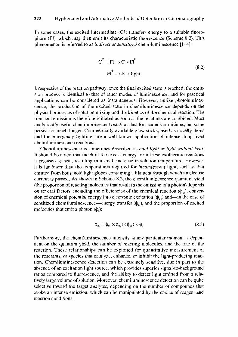
In some cases, the excited intermediate (C*) transfers energy to a suitable fluorophore (Fl), which may then emit its characteristic fluorescence (Scheme 8.2). This phenomenon is referred to as indirect or sensitized chemiluminescence [1-4):
* * C +Fl ~C+Fl
* (8.2)
Fl ~ Fl + light
Irrespective of the reaction pathway, once the final excited state is reached, the emission process is identical to that of other modes of luminescence, and for practical applications can be considered as instantaneous. However, unlike photoluminescence, the production of the excited state in chemiluminescence depends on the physical processes of solution mixing and the kinetics of the chemical reaction. The transient emission is therefore initiated as soon as the reactants are combined. Most analytical1y useful chemiluminescent reactions last for seconds or minutes, but some persist for much longer. Commercially available glow sticks, used as novelty items and for emergency lighting, are a well-known application of intense, long-lived chemiluminescence reactions.
Chemiluminescence is sometimes described as cold light or light without heat. It should be noted that much of the excess energy from these exothermic reactions is released as heat, resulting in a small increase in solution temperature. However, it is far lower than the temperatures required for incandescent light, such as that emitted from household light globes containing a filament through which an electric current is passed. As shown in Scheme 8.3, the chemiluminescence quantum yield (the proportion of reacting molecules that result in the emission of a photon) depends on several factors, including the efficiencies of the chemical reaction «Per), conversion of chemical potential energy into electronic excitation C<PcJ and-in the case of sensitized chemiluminescence-energy transfer (q\1}, and the proportion of excited molecules that emit a photon (cf>1):
(8.3)
Furthermore, the chemiluminescence intensity at any particular moment is dependent on the quantum yield, the number of reacting molecules, and the rate of the reaction. These relationships can be exploited for quantitative measurement of the reactants, or species that catalyze, enhance, or inhibit the light-producing reaction. Chemiluminescence detection can be extremely sensitive, due in part to the absence of an excitation light source, which provides superior signal-to-background ratios compared to fluorescence, and the ability to detect light emitted from a relatively large volume of solution. Moreover, chemiluminescence detection can be quite selective toward the target analytes, depending on the number of compounds that evoke an intense emission, which can be manipulated by the choice of reagent and reaction conditions.

Liquid-Phase Chemiluminescence Detection for HPLC
8.2 CHEMILUMINESCENCE DETECTION FOR LIQUID CHROMATOGRAPHY
223
This subject has been discussed in several previous book chapters [5-8] and reviews [9,10]. We would therefore like this chapter to serve as both an update of contemporary practice in HPLC with chemiluminescence detection, and a practical account of the advantages and pitfalls of this approach.
The most commonly used mode of detection for HPLC is the absorption of ultraviolet (UV) light, which provides reasonable sensitivity for many organic compounds, using reliable and fairly low-cost instrumentation. The downside of the wide applicability is relatively poor selectivity, which must therefore be derived almost entirely from the chromatographic separation and preliminary sample preparation. Furthermore, many compounds of interest in biological or environmental samples are present at concentrations below the limits of detection that can be achieved using UV-absorbance, and therefore the analyst must often turn to more sensitive and selective modes of detection, such as fluorescence or electrochemistry.
Laser-induced fluorescence can provide exceedingly low limits of detection for analytes containing a suitable fluorophore, but with a considerable increase in instrument complexity. On the other hand, chemiluminescence detection is generally superior to electrochemistry or standard fluorescence procedures in terms of sensitivity, selectivity, and analytical working range, and can be performed using simple, robust, inexpensive instrumentation. In general, a chemiluminescence detector consists of a transparent flow cell mounted against a photosensitive device (such as a photomultiplier tube). The column eluate is merged with the chemiluminescence reagent prior to entering the flow cell (see the following section).
Like electrochemical detection, chemiluminescence is dependent on oxidation and reduction processes, but the interactions that initiate the light-producing pathways can be considerably more complex than simple electron transfer. Although rarely exploited to date, chemiluminescence detection can therefore provide a means to rapidly assess the chemical reactivity of sample components eluted from a chromatographic column, which can be particularly useful when coupled with other methods of detection such as UV-absorbance.
The previous discussion on the advantages of chemiluminescence detection prompts the question of what has restricted its use in liquid chromatography (LC). First, chemiluminescence detection requires the column eluate to be merged with a reagent within a suitable detector. The assembly of the flow manifold and daily preparation of reagent solutions increases the total labor requirements, and therefore this approach is less attractive in situations where detection based on the native absorbance or fluorescence of the analytes is appropriate. The complexity of chemiluminescence detection could be considered similar to automated, rapid versions of the pre- or post-column derivatizations used to improve the fluorescence character of analytes.
Second, a large majority of HPLC methods with chemiluminescence detection published in the open literature are based on three well-established reagent classes:

224 Hyphenated and Alternative Methods of Detection in Chromatography
(1) luminol and its derivatives, (2) peroxyoxalate reagents, and (3) ruthenium complexes [5-10]. While a range of other reagents have been explored, in many cases the reaction pathways leading to the emission of light and the influence of the chemical environment are yet to be fully established. Therefore, the application of this mode of detection to new analyte classes has often been based on an imperfect, empirical understanding of the relationship between analyte structure and chemiluminescence intensity with any particular reagent.
The perception that chemiluminescence has limited applicability is reflected in the reluctance of the major HPLC manufacturers to develop chemiluminescence detectors as part of their suite of components. In addition, the lack of purpose-built commercial instrumentation limits the promotion of this approach and the advice and support available from HPLC manufacturers for analysts to develop new procedures.
Nevertheless, a wide range of HPLC procedures incorporating chemiluminescence detection have been developed and successfully applied in areas such as industrial process monitoring, pharmaceutical and biomedical analysis, and forensic science.
8.3 PRACTICAL CONSIDERATIONS
8.3.1 INSTRUMENT COMPONENTS
For chemiluminescence detection, the column eluate must be merged with one or more reagent solutions. Because the pressure within the flow system after the separation column (and UV-absorbance detector, if included) is not high, the additional manifold can be constructed from relatively cheap components (peristaltic pumps, low-pressure polymer fittings), such as those employed for flow-injection analysis. A simple T- or Y-piece fitting can be used to combine solutions (Figure 8.1).
It is crucial that the geometry and dimensions of the flow cell maximize the proportion of light that is emitted when the reacting mixture is in front of the detector. For fast chemiluminescence reactions, this means that the column eluate and reagent solutions should merge at (or as close as possible to) the point of detection. For slower chemiluminescence reactions, the length of tubing between the final T-piece and the detector should be optimized so that the most intense portion of the emission is detected.
1---- -------- --------------- ---- ---1 I 1 HPLC instrument 1 I I
I :
: Mobile phase 1
solvents : I
-- ------------- - -- -- ---- - ----- -- _ I I I I
-------- -- --- ,
: Reagents p
__.. I t--1__;---, I
I I I I
I I I I I CL detection manifold Waste ~----- --- ----- -- ----- ----- --- --'
FIGURE 8.1 An example manifold for HPLC with chemiluminescence detection. Key: q = quaternary pump; v =injection valve; c =column; u = UV-absorbance detector; p =peristaltic pump; d =chemiluminescence detector.

Liquid-Phase Chemiluminescence Detection for HPLC
Column eluate -+-
Chemiluminescence reagent __.. 'f'
PTFEtubing
Coil of glass or polymer tubing
""
• Solution outlet
Signal output HY input
Voltage divider
Photomultiplier tube
FIGURE 8.2 A flow-through chemiluminescence detector.
225
In either case, the "dead volume" of the flow cell should be minimized, to ensure reproducible mixing and rapid washing between injections, and to minimize band broadening.
Unlike fluorescence detection, a light source is not required for chemiluminescence. Wavelength discrimination usually offers no advantage (because different analytes generally lead to the same emission) and should be avoided due to the detrimental effect on sensitivity. One design that satisfies these requirements is a coil of glass or transparent polymer tubing with similar internal diameter to the manifold tubing (0.3-1.0mm) positioned against a photomultiplier tube (Figure 8.2). A light-tight housing is essential. Some chemiluminescence detectors that incorporate this design are now commercially available. Fluorescence detectors (with the excitation source switched off) have also been used for chemiluminescence detection. Although suitable for some applications, they generally provide lower sensitivity, because they are not designed for efficient mixing of column eluate and reagent in front of the photodetector window. Furthermore, the relatively short residence time of the reacting mixture within fluorescence flow cells limits the proportion of the emitted light that can be captured.
8.3.2 OPTIMIZATION
As with any analytical procedure, the optimization of experimental conditions can lead to significant improvements in performance. The optimum conditions (reagent concentration, pH, flow rates, selection of "enhancers") for chemiluminescence detection are often dependent not only on the reagent, but also the class of analyte under investigation. Much of our knowledge on chemiluminescence detection is derived from flow-injection analysis studies using predominantly aqueous conditions, and therefore it is also important to examine the effect of mobile phase solvents on these reactions. For example, acetonitrile quenches the response from chemiluminescence reactions with acidic potassium permanganate, but methanol has been found to be a suitable alternative [11]. Moreover, the optimum pH for separation may not be optimal for detection, but this can be addressed by merging the column eluate with an acid, base, or buffer solution prior to merging with the reagent.
8.4 COMMONLY USED REAGENTS AND THEIR APPLICATIONS
As shown in Figure 8.3, the application of chemiluminescence detection in LC is dominated by a few well-known reagents. The following survey is not exhaustive, but is intended to illustrate the variety of ways that liquid-phase chemiluminescence

226 Hyphenated and Alternative Methods of Detection in Chromatography
Permanganate
Ruthenium complexes
Luminol compounds
Peroxyoxalate reagents
FIGURE 8.3 Relative use of different reagent classes for post-column chemiluminescence detection.
has been applied as a sensitive and selective mode of detection for HPLC. Gas-phase chemiluminescence has also been successfully utilized (e.g., see Refs. [12-18]), but will not be discussed in this chapter.
8.4.1 lUMINOL AND RELATED REAGENTS
The oxidation of luminol (5-amino-2,3-dihydro-l,4-phthaJazinedione) in alkaline solution evokes a blue luminescence (A.max = 425 nm) that emanates from electronically excited 3-aminophthalate (Figure 8.4) [19-21].
A variety of oxidants, such as hydrogen peroxide, periodate, hypochlorite, and permanganate, can be used. The reaction with hydrogen peroxide is slow, but can
0 H20icatalyst or Q¢N oxidant/02
~H Alkaline
NH2 0 solution
Luminol anion
- ~''~(Jlo Y¥o-NH2 0
3~Aminophthalate
emitter
*
~~OH o-
ytN - II N N
NH2 0 NH2 o_
FIGURE 8.4 The chemiluminescent oxidation of luminol in alkaline solution.

Liquid-Phase Chemiluminescence Detection for HPLC 227
be effectively catalyzed by certain transition-metal ions and complexes, such as cobalt(II), copper(II), iron(II) and hexacyanoferrate(III) (ferricyanide), and peroxidase enzymes, which can be used for the highly sensitive detection of these species. For example, cobalt has been detected at picomolar levels after cation-exchange chromatography without pre-concentration [22]. Badocco and coworkers recently described the determination of iron(II), cobalt(II), and manganese(II) using ion chromatography and reported detection limits of 0.50, 0.24, and 375 nM, respectively [23]. This chemistry has also been exploited for the post-column detection of many organic molecules, which mainly fall into four categories: organic hydroperoxides; substrates of enzymatic reactions that produce hydrogen peroxide; compounds derivatized with luminol analogues; and compounds that enhance or inhibit the light-producing pathway.
The determination of organic hydroperoxides using HPLC with luminol chemiluminescence detection has been extensively used to obtain quantitative analytical data on the peroxidation pathways in both laboratory and biological systems [24-28], particularly those involving phosphatidylcholine and cholesterol. Measurement of hydroperoxides has also been employed to assess oxidative stress, antioxidant activity, and free radical-mediated DNA damage [29-33]. The post-column manifold for these applications is simple; the column eluate containing the separated analytes (oxidants) is merged with an aqueous reagent stream containing luminol or isoluminol, a catalyst (cytochrome C, microperoxidase, or other peroxidases), and a buffer (often at pH 10), in a T- or Y-piece prior to the detection flow cell. UV-absorbance detection can be incorporated in series (before the eluate is merged with the chemiluminescence reagent) [24-27], and mass spectroscopy can be used in parallel (by splitting the eluate) [28]. External standards are often used for calibration, but suitable internal standards have been reported [34,35]. Limits of detection for lipid hydroperoxides in blood plasma (including preliminary extraction procedures) have been stated as around 10-3 M [36,37]. Hui and coworkers reported a limit of detection of 1 pmol (i.e., 5 x 10-s M in the 20 µL injection) using synthetic analyte standards [35].
A variety of biomolecules can be sensitively detected by coupling H20rgenerating enzymatic reactions with luminol chemiluminescence (providing detection limits generally between 10-s and 10-6 M). The selectivity of these enzymatic reactions is very high, and this approach has often been applied without chromatographic separation [20]. However, in some cases, more than one concomitant species will form hydrogen peroxide and separation of sample components is required [38-41]. For example, Alam and coworkers determined histamine and N'-methylhistamine in the brain and peripheral tissues of rats, using reversed-phase LC with a post-eolumn reactor containing immobilized diamine oxidase [39]. As with the organic hydroperoxides, the hydrogen peroxide generated in the reactor was detected by subsequent chemiluminescence reaction with luminol and a catalyst, which in this case was potassium hexacyanoferrate(III). The immobilized-enzyme column was stable for more than 1 month of operation (200 samples analyzed).
The highly sensitive detection afforded by luminol reagents has been applied to broad analyte classes through the development of derivatizing agents that contain the phthalazine-1,4-dione chemiluminophore [19,20,42]. In some cases, the

228 Hyphenated and Alternative Methods of Detection in Chromatography
0
H~~~ NyR1 HN _,,,;;, AR N 2
0
0
HN~ R3 H~ 1/ r~ O~N)
0 \ R4
FIGURE 8.5 The reaction of chemiluminogenic reagents, DPH and AMP, with a-dicarbonyl compounds and catecholamines, respectively, to form highly chemiluminescent products.
product is far more chemiluminescent (>100 times) than the unreacted derivatizing agent (which is referred to as a chemiluminogenic reagent). Examples include 4,5-diaminophthalhydrazide (DPH) which forms highly chemiluminescent products with a-keto acids, a-dicarbonyl compounds and aromatic aldehydes, and 6-aminomethylphthalazine-1,4-dione (AMP), which has been used to detect 5-hydroxyindoles and catecholamines (Figure 8.5).
Alternatively, chemiluminescence labeling reagents can be used to bond an already effective chemiluminophore to a target class of analytes. Reagents of this type, such as the examples shown in Figure 8.6, have been developed for carboxylic acids, amines, amino acids, peptides, glycosides, and thiols.
Liquid chromatographic procedures incorporating initial derivatization with phthalazine-1,4-dione based reagents enable the highly sensitive detection of many analytes. Applications include the determination of amphetamines [43] and 3a,5p-tetrahydroaldosterone [44] in urine (using ABEI and DPH, respectively), and amantadine [45] and fatty acids [46] in plasma (using TPB-Suc and PROB). Typical chromatograms for the determination of amantadine in plasma are shown in Figure 8.7.
As with many fluorescence-labeling procedures, the preliminary off-line derivatizations often require considerable time (10-120 min) at elevated temperatures, but some proceed quite quickly at room temperature [47-49]. The post-column chemiluminescence reaction with the derivatized analytes has often been initiated by merging the column eluate with hydrogen peroxide and hexacyanoferrate(III), but electrochemical oxidation has also been reported. Limits of detection are typically between 0.2 and 200 fmol ("' 10-11 to 10-s M). Further details regarding luminol-type derivatization reagents can be found in the review by Yamaguchi and coworkers (19].
Many compounds enhance or inhibit the emission of light from luminol systems, which can also be exploited for quantitative analysis. An early approach was based on the suppression of chemiluminescence due to the interaction between analytes and the catalyst (e.g., Co(II) or Cu(II)). The column eluate is therefore first combined with a stream containing the catalyst, before merging with alkaline luminol and hydrogen peroxide solutions to initiate the chemiluminescence reaction. Limits of

Liquid-Phase Chemiluminescence Detection for HPLC 229
HN I
HN
0
0
HN I
HN
ABEi
0
0 IPO
HN I
HN
HN I
~NH2 HN N
l CH3
NCS
0
0
HN I
HN
0
0
EDA-ABEi
0
H H 0 HN~N~N,
II II NH 2
s 0 PROB
N~OO~ \ ~ /; S O-N
T~~oc 0
FIGURE 8.6 Selected luminol-type chemiluminescence labeling reagents: N-(4-aminobutyl)N-ethylisoluminol (ABEi); 6-[N-(3-propionohydrazino)thioureido]benzo[g]-phthalazinel,4(2H,3H)-dione (PROB); 6-isothiocyanatobenzo[g]-phthalazine-1,4(2H,3H)-dione (IPO); 4-(6,7-dihydro-5,8-dioxothiazolo[4,5-g]phthalazin-2-yl)benzoic acid N-hydroxysuccinimide ester (TPB-Suc ); and 4-[(N-a-ethoxycarbonyldiazoacetyl)aminobutyl]-N-ethyl isoluminol (EDA-ABEi).
detection for primary and secondary amino acids, amines, proteins, catecholamines, catechol, and aminoglycoside antibiotics after ion-exchange, ion-pair, or reversedphase LC [50-53], were typically between I pmol and 2 nmol ("' 10-7 to 10-4 M).
It was later found that reducing agents, such as corticosteroids, phenols, and anilines, can react with radical intermediates of the light-producing pathway of various luminol systems and therefore influence the rate or efficiency of emission [54-57]. For post-column detection, the most common approach involves combining the column eluate stream with one solution containing luminol and base, and another containing hexacyanoferrate(III), but the order in which they are merged varies in different procedures [58-62]. A diverse range of other oxidants and catalysts (e.g., 12, Cu(ll)/H20 2, Bro-, Co(II), and [Cu(HI06) 2]5-) has also been used in procedures based on signal enhancement or inhibition [63-67].
An interesting variant of this approach is the assessment of antioxidant status, where measurement is based on the scavenging of reactive oxygen species, which inhibits the emission of light [68,69]. Appropriate selection of reagents and configuration of the instrument manifold can focus the assessment on specific oxygen species, such as hydrogen peroxide or the superoxide anion radical (produced from

230 Hyphenated and Alternative Methods of Detection in Chromatography
4 4
Cl) 1 2 "' c 0 a. ~ .... .... 0 ...... u Cl.> ...... <I)
0
3 ... _
I I I I I I I l 0 10 20 30 0 10 20 30
(a) Time (min) (b) Time (min)
FIGURE 8.7 Chromatograms for the determination of amantadine in human plasma using reversed-phase chromatographic separation (C18 column) after derivatization with TPB-Suc (80°C, 20min). (a) Drug-free sample without internal standard. (b) Sample taken 2h after single oral administration of amantadine chloride (50mg). Peaks: 1, amantadine; 2, internal standard [l-(1-adamantyl)ethylamine]; 3, an endogenous amino compounds; 4, reagent blank and other endogenous amino compounds. (Reprinted from J. Chromatogr. A, 907, Yoshida, H., Nakao, R., Matsuo, T., Nohta, H., and Yamaguchi, M., 4-(6,7-Dihydro-5,8-dioxothiazolo [4,5-g] phthalazin -2-yl) benzoic acid N-hydrosuccinimide ester as a highly sensitive chemiluminescence derivatization reagent for amines in liquid, 39, Copyright 2001, with permission from Elsevier.)
'
hypoxanthine and xanthine oxidase) [69]. This chemistry has been used to establish the overall antioxidant status of various samples [70], but when coupled with LC, each component of the sample can be individually assessed [68,69]. Figure 8.8 shows the chromatograms for a green tea extract using both UV-absorbance and chemiluminescence detection. In this case, the negative peaks in Figure 8.8b correspond to inhibition of the chemiluminescence signal from the reaction of luminol, hypoxanthine, and xanthine oxidase, therefore indicating which components are most effective scavengers of superoxide ion.
8.4.2 LlJCIGENIN AND ACRIDINllJM ESTERS
The reaction of lucigenin (10,10'-dimethyl-9,9'-bisacridinium nitrate) with hydrogen peroxide in aqueous alkaline solution evokes a blue-green emission from N-methylacridone (Figure 8.9a) [71]. Certain transition metal ions are effective catalysts, but unlike the reaction of luminol with hydrogen peroxide, they are not essential for an intense emission of light with this reagent. Nevertheless, Chen and coworkers demonstrated that the quenching effect of amino acids on the cobalt(II)

Liquid-Phase Chemiluminescence Detection for HPLC
0
l;,,J\,J
(b)
I
2
10
1
3 4 5
20 30
2
231
40
FIGURE 8.8 (a) UV-absorbance (265 nm) and (b) chemiluminescence inhibition (superoxide scavenging) for a green tea extract after reversed-phase chromatographic separation (ODS column). Peaks: 1, epigallocatechin; 2, epigallocatechin gallate; 3, gallocatechin gallate; 4, epicatechin gallate; 5, catechin gallate. (Reprinted from Talanta, 60, Toyo'oka, T., Kashiwazaki, T., and Kato, M., On-line screening methods for antioxidants scavenging superoxide anion radical and hydrogen peroxide by liquid chromatography with indirect chemiluminescence detection, 467, Copyright 2003, with permission from Elsevier.)
(a)
'\eductant
CH3 I N
(b)
FIGURE 8.9 The chemiluminescent reaction of lucigenin in alkaline solution with (a) hydrogen peroxide and (b) a reducing agent and molecular oxygen.

232 Hyphenated and Alternative Methods of Detection in Chromatography
AC-C2-NHS
e I
r-0-CH, O=S=O
MASCI
FIGURE 8.10 Acridinium esters: 4-(2-succinimidyloxycarbonylethyl) phenyl-10-methylacridinium-9-carboxylate fluorosu1fonate (AC-C2-NHS) and 10-methyl-N-(p-tolyl)-N(p-iodoacetamidobenzenesulfonyl)-9-acridinium carboxamide iodide (MASCI), for labeling amino acids [79], and carboxylic acids [80), respectively. (From Zhong, L. and Maloy, J.T., Anal. Chem., 70, 1100, 1998; Steijger, O.M. et al., J. Biolumin. Chemilumin., 13, 31, 1998.)
catalyzed oxidation of lucigenin could be used to detect these species at nanomolar levels without pre-column derivatization [72].
In the presence of dissolved oxygen, lucigenin will also produce light on reaction with a range of reducing agents (Figure 8.9b), which has been exploited for the postcolumn detection of ascorbic acid [73], glucose and other sugars [73], glucuronides (after enzyme-catalyzed hydrolysis to glucuronic acid) [74], and a-hydroxycarbonyl compounds (such as corticosteroids and p-nitrophenacyl esters of carboxylic acids) [75,76].
Like lucigenin, acridinium esters react with hydrogen peroxide in alkaline solution to form N-methylacridone in an electronically excited state. Several labeling reagents for HPLC and immunoassay incorporating acridinium esters have been developed (Figure 8.10) [77-80) and used to detect compounds such as chlorophenols (77], primary amines [79,81], and carboxylic acids [80]. In this approach, the column eluate is merged with base (NaOH or KOH) and hydrogen peroxide streams before entering the detector.
8.4.3 PEROXYOXALATE REAGENTS
Certain diaryloxalates and diaryloxamides produce an intense emission of light when treated with hydrogen peroxide in the presence of a suitable fluorophore (Figure 8.11) [82-85]. Collectively known as peroxyoxalate chemiluminescence, these light-producing reactions have a complex multistep mechanism that has been extensively studied, but is yet to be fully elucidated [83,84]. Among the variety of postulated intermediates, perhaps the most contentious has been 1,2-dioxanedione

Liquid-Phase Chemiluminescence Detection for HPLC 233
OH + H202 01----f° Fluorophore [Fluorophore)* \. \ ..
R R 0-0 2RH 2C02
Intermediates
FIGURE 8.11 A simplified scheme for peroxyoxalate chemiluminescence.
(shown in Figure 8.11), first proposed over four decades ago [86}, and only recently confirmed [87,88]. The reaction is subject to interrelated general-base and nucleophilic catalysis, for which imidazole is particularly effective.
Electronic excitation of the fluorophore is thought to involve: (1) the formation of a charge complex with the dioxanedione intermediate, which accepts an electron to form a radical anion; (2) decomposition of the radical anion into C02 and [C02·1-; and (3) transfer of the electron back to the fluorophore at a higher energy level. For the greatest emission of light, the energy acceptor must therefore have both a low oxidation potential and efficient fluorescence. Polyaromatic hydrocarbons (particularly those with an amino substituent) are among the most effective.
Unlike the other light-producing reactions described in this chapter, peroxyoxalate chemiluminescence is not well suited to the predominantly aqueous conditions of reversed-phase HPLC, due to the limited solubility and stability of the reagents in water. There has been some progress in the development of peroxyoxalate reagents for aqueous solutions [84], but it has not yet reached the point of routine use in HPLC analysis. Coupling HPLC with contemporary peroxyoxalate detection therefore requires merging the column eluate with reagents prepared in organic solvents (such as acetonitrile or ethyl acetate). Efficient mixing is essential for high reproducibility and sensitivity. Acetonitrile is the most commonly used organic modifier for separation. Methanol should be avoided due to its strong quenching effect. The background emission is dependent on the proportion of water in the mobile phase and therefore solvent gradients can produce noisy and sloping baselines, but this can be minimized by a judicious selection of instrumental and reaction conditions [83].
The most commonly used peroxyoxalate reagents are shown in Figure 8. I 2. For any particular application, the best choice of reagent depends on the instrumental approach and the nature of the assay [83,89-92]. For example, DNPO reacts at faster rates than TCPO and produces greater chemiluminescence intensities [93], but this is only an advantage if the detection manifold is configured so that the most intense emission occurs within the flow cell [89,90]. TDPO is much more soluble in acetonitrile and more stable in the presence of hydrogen peroxide [94].
In spite of the complexity of utilizing peroxyoxalate chemiluminescence, it has been successfully employed for the sensitive post-column detection of many compounds. The target analytes normally act as energy acceptors and emitters, either due to their native fluorescence character, or after derivatization with ftuorescencelabeling reagents. To a lesser extent, this chemistry has been used to detect compounds that produce hydrogen peroxide in chemical or photochemical reactions, and (in a few cases [95,96]) compounds that can enhance or inhibit the production of light.

234 Hyphenated and Alternative Methods of Detection in Chromatography
TCPO DNPO
TDPO
FIGURE 8.12 Selected peroxyoxalate reagents: bis(2,4,6-trichlorophenyl)oxalate (TCPO); bis(2,4-dinitrophenyl)oxalate (DNPO); and bis[2-(3,6,9-trioxadecyloxycarbonyl)-4-nitrophenyl] oxalate (TDPO).
For the detection of fluorescent compounds (native or derivatized), a reagent stream containing the diaryloxalate (in ethyl acetate or acetonitrile) is often combined and mixed with a solution of hydrogen peroxide (prepared by diluting the 30% v/v aqueous solution with acetonitrile, ethyl acetate or propan-2-ol) prior to merging with the column eluate. Alternatively, a single reagent stream, containing both the diaryloxalate and hydrogen peroxide can be used, but in this case the stability of the combined reagents should be established during the optimization of conditions [94,97]. Chromatographic separation is typically performed using reversed-phase C18 columns with isocratic mobile phases comprising of 20%-85% acetonitrile and an aqueous solution of imidazole adjusted to a pH between 6 and 8. However, a range of other conditions [98-100), including gradient elution [101-103], have been reported.
This mode of detection has been applied to polycyclic aromatic hydrocarbons (PAHs), particularly those with amino groups, for which the limits of detection are often l-2 orders of magnitude better than those obtainable with fluorescence [104,105]. Perhaps more importantly, this approach has been adapted for the determination of nitro-substituted PAHs in airborne particulates [106-109]. In these procedures, the target analytes are reduced to the corresponding amino species, either off-line prior to analysis (e.g., by refluxing in ethanol in the presence of sodium hydrosulfide [107]) or online, in a reduction column packed with Pt/Rh coated alumina [108,109] or zinc immobilized on glass beads [106].
Many different derivatizing reagents have been used to extend peroxyoxalate chemiluminescence detection to new classes of analytes [84,85]. The most commonly used of these reagents feature the dansyI [i.e., 5-(dimethylamino)-naphthalene-1-sulfonyl] fluorophore, with chloro, hydrazino, or other functionality to aIIow binding with the analytes of interest (Figure 8.13). Applications include the determination of methamphetamine and its metabolites in a single hair or the urine of abusers

liquid-Phase Chemiluminescence Detection for HPlC
Cl I
o=s==o
_,...N....._ H3C CH3
DNS-CI
_,...N, H3C CH3
DNS-H
F
o==S=O I
_..,.N, H3C CH3
DBD-F
235
o=s=o !
...--N-..... H3C CH3
DBD-COCI
FIGURE 8.13 Some of the derivatizing reagents used with peroxyoxalate chemiluminescence detection: 5-(dimethylamino)-naphthalene-1-sulfonyl chloride (DNS-Cl); 5-(dimethylamino)-naphthalene-l-sulfonic hydrazide (DNS-H); 4-(N,N-dimethylaminosulfonyl)-7-fluoro-2, l ,3-benzoxadiazole (DBD-F); and 4-(N,N-di methylaminosul fonyl)-7-(N-chloroformylmethyl-N-methyl)amino-2,1,3-benzoxadiazole (DBD-COCJ).
[101,110,111], ketosteroids in serum [112], and pesticide residues in vegetables and fruit juices [113,114]. Derivatizations generally require relatively long periods of time at elevated temperatures, and are therefore often performed off-line, prior to analysis. However, automated online derivatizations within heated mixing coils [115] or on solid-phase supports [11 lJ have also been developed.
The hydrolysate of DNS-Cl also gives relatively intense chemiluminescence with peroxyoxalate reagents, which can interfere with the detection of analytes at low concentrations [98]. The hydrolysate of 4-(N,N-dimethylam inosulfonyl)-7-fluoro-2, 1,3-benzoxadiazole (DBD-F), a related fluorescence-labeling agent for amines, is less readily formed and is less fluorescent, therefore reducing the potential for interference. DBD-F and its 7-hydrazino and 7-(N-chloroformylmethyl-N-methyl)amino analogues have been used in HPLC procedures with peroxyoxalate chemiluminescence detection, such as the determination of pharmaceuticals in biological fluids [98,116,117], and MDMA (ecstasy) and related compounds in hair (Figure 8.14) (118].
Catecholamines (epinephrine, norepinephrine, and dopamine) have been detected with peroxyoxalate chemiluminescence after derivatization with ethylene diamine or l,2-bis(3-chlorophenyl)ethylenediamine [99,119]. An automated procedure incorporating (1) preliminary ion-exchange extraction, (2) chromatographic separation, (3) post-column fiuorogenic derivatization, and (4) peroxyoxalate chemiluminescence detection has been developed (Figures 8.15 and 8.16) [120].
This approach has been adapted to quantify the 3-0-methyl metabolites, metanephrine, normetanephrine, and 3-methoxytyramine, by incorporating online electrochemical oxidation of the metabolites to the corresponding o-quinones [121,122), and to determine nitrocatecholamines, by introducing a reduction column into the flow manifold [123]. Isoproterenol, 4-methoxytyramine, andN-methyldopamine have been used as internal standards. Limits of detection of0.3-2fmol (rvIO-ll to 10-rnM) for catecholamines and 3-0-methyl metabolites have been reported [100,120,122].

236 Hyphenated and Alternative Methods of Detection in Chromatography
0 10 20 30
(a) Retention time (min)
40 0
(b)
4
3
2 1
10 20 30 40 Retention time (min)
FIGURE 8.14 Chromatograms for the determination of amphetamines in (a) normal (drugfree) hair, and (b) drug abuser's hair. Sample preparation included digestion, extraction (n-heptane), and derivatization with DBD-F. Separation column: Capcellpak C18 UG120. Chemiluminescence reagent: bis(2,4,5-trichloro-6-carbopentoxyphenyl)oxalate (CPPO) and H;P2 in acetonitrile. Peaks: I, amphetamine; 2, ecstasy; 3, methamphetamine; 4, internal standard (l-methyl-3-phenylpropylamine). (With kind permission from Springer Science+ Business Media: Analytical and Bioanalytical Chemistry, A sensitive semi-micro column HPLC method with peroxyoxalate chemiluminescence detection and column switching for determination of MOMA-related compounds in hafr, 387, 2007, 1983, Nakamura, S., Wada, M., Crabtree, B.L., Reeves, P.M., Montgomery, J.H., Byrd, H.J., Harada. S., Kuroda, N., and Nakashima, K., Figures 6a and 7a, Copyright Springer-Verlag 2006.)
Like luminol, peroxyoxalate reagents can be used to detect the hydrogen peroxide generated in post-column immobilized-enzyme reactors, for the determination of substrates such as amino acids [124], choline and acetyJcholine [125,126], polyamines (127,128], and glucose [126]. In general, a solution containing the peroxyoxalate reagent and suitable fluorophore is merged with the exit stream of the immobilizedenzyme reactor. A buffer stream is sometimes added between the separation column and reactor. Limits of detection are typically between 10-s and 10-6 M.
Hydrogen peroxide can also be generated in post-column photochemical reactions, from species such as aliphatic alcohols and molecular oxygen, sensitized by quinones [129]. Because of the catalytic nature of the reaction, each quinone molecule can produce many molecules of hydrogen peroxide as it passes through the reactor, which (when coupled with peroxyoxalate chemiluminescence detection) provides sub-picomole detection limits for species such as 9,10-anthraquinone and menadione (vitamin K3). Originally, the ftuorophore (rubrene) was added to the peroxyoxalate

Liquid-Phase Chemiluminescence Detection for HPLC
f----(}Ff' _____ ()t.i--:
IV*i ! Time/min ! l 0-2 OFF l : 2-5 ON I : 5- OFF I L--------,---------~
I I I I I I
' ' ' ' I I I I I
' I
Eluent
~----------~------------...
Waste
(1)
Precolumn
(iii) {iv)
FL reagent CL reagent (TDPO,Hp2l
(ii)
Column oven
Separation column (ODS column)
Auto sampler
(ED)
Pretreatment buffer
Thermo reactor
Reaction coil
237
Waste
FIGURE 8.15 Flow manifold for the determination of catecholamines in rat plasma. (From Takezawa, K., Tsunoda, M., Murayama, K., Santa, T., and Imai, K., Automatic semimicrocolumn liquid chromatographic determination of catecholamines in rat plasma utilizing peroxyoxalate chemiluminescence reaction, Analyst, 125, 293, 2000. Reproduced by permission of The Royal Society of Chemistry.)
2
2
1
3 4
1 1 4
3
10 20 30 10 20 30 (a) Retention time {min) (b) Retention time (min)
FIGURE 8.16 Chromatograms for the determination of catecholamines in (a) a standard solution and (b) rat plasma. Procedure includes online extraction, separation (Capcell Pak C18 UG120), derivatization (ethylenediamine) and chemiluminescence detection (TDPO and H20 2). Peaks: 1, norepinephrine; 2, epinephrine; 3, dopamine; 4, internal standard (N-methyldopamine). (From Takezawa, K., Tsunoda, M., Murayama, K., Santa, T., and Imai, K., Automatic semi-microcolumn liquid chromatographic determination of catecholamines in rat plasma utilizing peroxyoxalate chemiluminescence reaction, Analyst, 125, 293, 2000. Reproduced by permission of The Royal Society of Chemistry.)

238 Hyphenated and Alternative Methods of Detection in Chromatography
reagent [129], but more recently it has been shown that UV irradiation of quinones can produce both hydrogen peroxide and a highly fluorescent species that can act as the fluorophore in the chemiluminescence detection [130]. This approach has been applied to the determination of vitamin K homologues in human plasma [92]; limits of detection were 32-85 fmol per injection (i.e., 3.2-8.5 nM).
Organic peroxides, which do not normally produce a sufficiently intense emission with peroxyoxalate reagents for quantitative analysis, can also be converted to hydrogen peroxide by online UV irradiation (91,131]. For example, onhne UV irradiation improved the limit of detection for cumene hydroperoxide by five orders of magnitude (to 1 µM) [131]. This simple approach has been applied to the determination of benzoyl peroxide in wheat flour [131], and artemisinin (an antimalarial drug containing an internal endoperoxide) in human serum [91].
8.4.4 TRIS(2,2'-8tPYRIDINE) RUTHENIUM(ll I)
The reduction of tris(2,2'-bipyridine)ruthenium(III) with certain amines (particularly aliphatic tertiary amines), organic acids, conjugated dienes, and various other organic and inorganic compounds evokes an orange luminescence C"-max = 610-620nm) that matches the characteristic photoluminescence of tris(2,2'-bipyridine) ruthenium(II) (132,133].
Tris(2,2'-bipyridine)ruthenium(III) has a limited temporal stability in aqueous solution, but can be formed by oxidizing the stable ruthenium(II) form (Figure 8.17), either shortly before the reagent is merged with the column eluate or within the chemiluminescence detector. There are several ways that this can be achieved; the most appropriate depends on the intended application. The stability of the tris(2, 2'-bipyridine)ruthenium(III) is greater in more acidic solution [134], but the analytedependent optimum pH of the chemiluminescence reaction must also be considered. Solid lead dioxide can be added off-line and then removed by filtration as the solution is aspirated into the detector. Some of the best limits of detection have been obtained by generating tris(2,2'-bipyridine)ruthenium(llI) in this manner [133], but lead dioxide is quite toxic and also breaks down the reagent over several hours.
Online oxidation, using chemical (ceriurn(IV) or permanganate), electrochemical (at an electrode surface), or photochemical (using persulfate) approaches, ensures
Ru(bipy)/+ (stable pre-cursor)
Oxidant
Ru(bipy)i+ (chemiluminescence
reagent)
[Ru(bipy)/+J* (electronically
excited product)
*
FIGURE 8.17 Preparation and chemiluminescent reduction of tris(2,2'-bipyridine) rut hen iu rn(III).
Liquid-Phase Chemiluminescence Detection for HPLC 239
reproducible preparation, but increases the complexity of the flow manifold. When permanganate is used to prepare the reagent, the excess oxidant may also undergo chemiluminescence reactions (see the following section) [135]. Most commonly used organic modifiers, such as acetonitrile and methanol, are compatible with this reagent, although both positive and negative changes in chemiluminescence intensity have been reported.
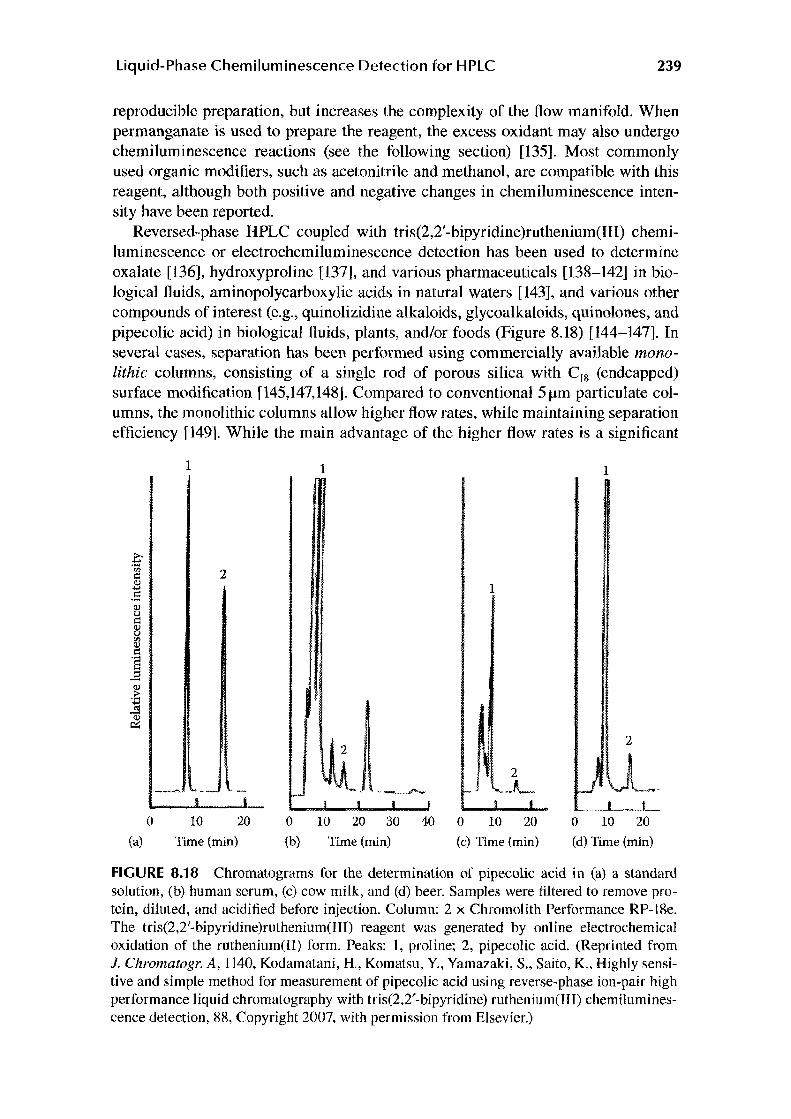
Reversed-phase HPLC coupled with tris(2,2'-bipyridine)ruthenium(lll) chemiluminescence or electrochemiluminescence detection has been used to determine oxalate [136], hydroxyproline [137], and various pharmaceuticals [138-142] in biological fluids, aminopolycarboxylic acids in natural waters [143], and various other compounds of interest (e.g., quinolizidine alkaloids, glycoalkaloids, quinolones, and pipecolic acid) in biological fluids, plants, and/or foods (Figure 8.18) [144-147]. In several cases, separation has been performed using commercially available monolithic columns, consisting of a single rod of porous silica with C18 (endcapped) surface modification [145,147,148]. Compared to conventional 5 µm particulate columns, the monolithic columns allow higher flow rates, while maintaining separation efficiency [149]. While the main advantage of the higher flow rates is a significant
.€ in i::: <ll .... . s <ll u i::: <ll u ,,, <l> i::: '§ E .~ ..... ...\!! ~
0
(a)
1
2
10 20
Time (min)
1
2
0 10 20 30 40
(b) Time (min)
1
0 10 20
(c) Time {min)
1
2
0 10 20
(d) Time (min)
FIGURE 8.18 Chromatograms for the determination of pipecolic acid in (a) a standard solution, (b) human serum, (c) cow milk, and (d) beer. Samples were filtered to remove protein, diluted, and acidified before injection. Column: 2 x Chromolith Performance RP-18e. The tris(2,2'-bipyridine)ruthenium(III) reagent was generated by online electrochemical oxidation of the ruthenium(II) form. Peaks: 1, praline~ 2, pipecolic acid. (Reprinted from 1. Chromatogr. A, 1140, Kodamatani, H., Komatsu, Y., Yamazaki, S., Saito, K., Highly sensitive and simple method for measurement of pipecolic acid using reverse-phase ion-pair high performance liquid chromatography with tris(2,2'-bipyridine) ruthenium(III) chemiluminescence detection, 88, Copyright 2007. with permission from Elsevier.)

240 Hyphenated and Alternative Methods of Detection in Chromatography
reduction in overall run time, they can also improve the sensitivity of fast chemiluminescence reactions, such as those with tris(2,2'-bipyridine)ruthenium(III), due to the more rapid propulsion of the column eluate and reagent mixture into the flowthrough detector [148].
Chemiluminescence detection with this reagent has been extended through the chemical or photochemical modification of compounds that would not otherwise elicit an intense emission. For example, treatment of amino acids with dansyl chloride (DNS-Cl; Figure 8.13) increased the electrogenerated chemiluminescence response with tris(2,2' -bipyridine)ruthenium(II) by three orders of magnitude, due to the greater substitution on the amino nitrogen and the presence of a tertiary amine on the derivatizing agent [150]. Other pre-column derivatizing agents have been specifically designed to add tertiary amines to compounds with alcohol, carboxylic acid, or primary amine functionality (Figure 8.19) [151]. The most sensitive approach (using IDHPIA) provided an on-column detection limit of 0.5 fmol (5 x 10-11 M in the 10 µL injection) for myristic acid [151]. Typical chromatograms for the determination of free fatty acids in human plasma are shown in Figure 8.20.
Divinylsulfone (Figure 8.21), which converts primary amines into l,4-thiazane-1,1-dioxides, has been used in procedures to determine amino acids in serum [152]. The derivatization of carbonyl compounds with methylmalonic acid, and amines with diketene, to form species that produce light with tris(2 ,2' -bipyridi ne )ruthenium(llI) has also been demonstrated [153,154]. Although these procedures provide sensitive detection, the preliminary off-line derivatization of the target analytes increases the overall analysis time by 20-120min.
Automated, post-column derivatization has also been demonstrated. Primary or secondary amines have been converted (online) to tertiary amines using acrylonitrile or epichlorohydrin [155,156]. Limits of detection for mono-, di- and tri-ethanolamine using this approach were 30, 25, and 40pmol, respectively [156]. Post-column chemical or photochemical degradation of diols, amines, amino acids, aromatic compounds, N-nitrosamines, and N-methylcarbamates has been utilized to form species such as oxalate and certain amines that can be more sensitively detected with tris(2,2'-bipyridine)ruthenium(III) [ 157-159].
H2N~o NAPP
FIGURE 8.19 Pre-column derivatizing agents that add a tertiary amine group for detection with tris(2,2'-bipyridine)ruthenium(Ill): 3-(diethylamino)propionic acid (DEAP); N-(3-aminopropyl)pyrrolidine (NAPP); and 3-isobutyl-9, I O-dimethoxy-IJ,4,6,7, 1 lb-hexahydro-2H-pyrido[2, 1-a]isoqui nolin-2-ylamine (IDHPIA).

liquid-Phase Chemiluminescence Detection for HPLC
80
60 1
2 3
20 9 10
0 20 40 (a) Time (min)
80
60
40 1
20
0 (b)
3
8
9
6
5 10
20 40 60
Time (min)
241
FIGURE 8.20 Chromatograms for the determination of fatty acids in (a) a standard solution (2 pmol) and (b) human serum extract (chloroform/n-hexane, 1: 1 v/v). Analytes were derivatized off-line with IDHPIA. Column: Inertsil C8. The tris(2,2'-bipyridine)ruthenium(III) reagent was generated by on-line electrochemical oxidation of the ruthenium(II) form. Peaks: 1, lauric acid; 2, myristoleic acid; 3, myristic acid; 4, R-Iinolenic acid; 5, palmitoleic acid; 6, R-linoleic acid; 7, arachidonic acid; 8, palmitic acid; 9, oleic acid; 10, internal standard (margaric acid); 11, stearic acid. (Reprinted with permission from Morita, H. and Konishi, M., Electrogenerated chemiluminescence derivatization reagent, 3-isobutyl-9,10-dirnethoxy-1,3,4,6,7,llb-hexahydro-2H-pyrido[2,1-a]isoquinolin-2-ylamine, for carboxylic acid in highperformance liquid chromatography using tris(2,2'-bipyridine)ruthenium(JJ). Anal. Chem., 75, 940, 2003. Copyright 2003 American Chemical Society.)
The tris(2,2'-bipyridine)ruthenium(Ill) reagent can be regenerated by oxidation of the reaction product, and there has been considerable effort made to develop stable immobilized reagents, particularly on the surface of electrodes [133]. An electrochemiluminescence sensor with reagent 'immobilized in titania-Nafion nanocomposite films coated on an electrode has been used for the determination of phenothiazine derivatives and erythromycin in human urine [160].
8.4.5 POTASSIUM PERMANGANATE
The reaction of potassium permanganate with a wide variety of organic compounds (particularly phenols, polyphenols, catechols, and indoles) in acidic aqueous solution evokes a broad red emission O'ma)( :::: 734 nm) from an excited manganese(II) species [11,161]. Formaldehyde/formic acid or polyphosphates are commonly used to improve the sensitivity of this reagent (the presence of polyphosphates also shifts the maximum intensity to 689 nm) [162].
Acidic potassium permanganate chemiluminescence is well suited to reversedphase HPLC, including procedures with mobile-phase gradients. A reagent solution

242 Hyphenated and Alternative Methods of Detection in Chromatography
H2C~ R-NH2 -j 502
H2C~ Divinylsulfone
0
R HO
R')==o + CH3
HO
0 0 Methylmalonic acid
0 0
~-NH2 + ===<>==o 0
Diketene
R,N~ H
FIGURE 8.21 Pre-column derivatizing agents that convert amines or carbonyl compounds to other functionality that is suitable for detection with tris(2,2'-bipyridine)ruthenium(III).
containing both permanganate and polyphosphate enhancer (adjusted to pH 2-3) is reasonably stable, and therefore only a single reagent line is required [163]. Enhancement with formaldehyde or formic acid is more problematic, because these species slowly react with permanganate and must be merged with the reagent or column eluate shortly prior to the final confluence point [164,165]. The chemiluminescent reactions with the target analytes are often very rapid, and therefore the time taken to present the reacting mixture to the detection flow cell must be kept as short as possible. Methanol is the most commonly used organic modifier for chromatographic separations coupled with this reagent; acetonitrile should be avoided due its strong quenching effect [11].
Applications include the determination of opiate alkaloids in body fluids and industrial process srreams [166], neurotransmitter metabolites in urine [163], polyhydroxybenzenes in river water [165], adrenergic amines in weight-loss products [167], arbutin and L-ascorbic acid in whitening cosmetics [164], and aJkylthiouracils in human serum [168].
Figure 8.22 shows previously unpublished data from the development of a rapid HPLC procedure for the determination of opiate alkaloids in process streams, using a monolithic column and gradient elution [148]. The superior selectivity of the chemiluminescence detection enabled baseline resolution of both morphine and oripavine within 1.5 min, which could not be achieved with UV-absorbance detection.
8.4.6 OTHER LIQUID-PHASE CHEMILUMINESCENCE REAGENTS
Various phenol [169-171], thiol [172-175], and related compounds [175] have been detected using chemiluminescence reactions with cerium(IV) in acidic aqueous solution. In each system, an enhancer (rhodamine B, rhodamine 60, quinine) is merged

Liquid-Phase Chemiluminescence Detection for HPLC 243
1.2
::ci' 1 M <!)
N .:=l (fj
8 0.8 .... 0 5 <!) 0.6 u c:: (fj
..c .... 0.4 0 <ll
,.0 rlJ
5 0.2
0.5 1 1.5 2
{a) Time (min)
::a-1.2
Q) N
~ 1 M
s .... 0 0.8 5 0 Q) u c:: 0.6 <!) u "'
><10 <!) c:: 0.4 ·s
..=! ·s 0.2 <!) .c u
0 0 0.5 1 1.5 2
(b) Time (min)
FIGURE 8.22 Chromatograms for the determination of morphine (M) and oripavine (0) in an industrial process sample using HPLC with a monolithic column (Chromolith SpeedROD RP-18e) and (a) UV-absorbance or (b) acidic potassium permanganate chemiluminescence detection.
with the column' eluate and the mixture is combined with the oxidant stream shortly prior to entering the detection coil. The dominant light-producing pathway involves the formation of high-energy intermediates that transfer energy to the enhancers, which emit light at their characteristic wavelengths. However, the exact nature of the intermediates is yet to be confirmed. Limits of detection using these systems are typically between 0.01 and 1.0 µM.
Several other oxidant/enhancer combinations have been explored. Tetracyclines can be detected in acidic solution with permanganate, sulfite and P-cyclodextrin [176], or with cerium(IV) and rhodamine B (incorporating photoirradiation of the column eluate) [177]. Polyphenols can be detected in alkaline solution with hydrogen peroxide and l-ethyl-3-(3-dimethylaminopropyl)carbodiimide [178]. Pyrethroid and benzoylurea insecticides have been determined in vegetables by combining· chromatographic separation, post-column irradiation with UV light, and chemiluminescence detection with potassium hexacyanoferrate(III) in alkaline solution. The chemiluminescence signal increased with the percentage of acetonitrile in the reaction medium [179,180].

244 Hyphenated and Alternative Methods of Detection in Chromatography
Like potassium permanganate (described previously), manganese(III) and manganese(IV) oxidants react with many molecules in acidic solution to produce an excited manganese(II) species that emits red light, and some preliminary demonstrations of post-column detection with these alternative manganese oxidants have emerged [181]. "Soluble" manganese(IV) can be formed by dissolving freshly precipitated manganese dioxide in 3.0 M orthophosphoric acid. Manganese(III) can be generated by online electrochemical oxidation of manganese(II). Interestingly, the selectivities of these reagents differ markedly from acidic potassium permanganate [181].
Alkaline hypobromite has been used for the detection of arginine and argininecontaining peptides separated with monolithic column chromatography [182]. Although the limit of detection was only 0.1 µM for arginine and 4µM for bradykinin, the reagent has been found to be selective toward these monosubstituted guanidine species (and urea) in the presence of many common amino acids, peptides, and other low-molecular-weight biological compounds.
The photoirradiation of compounds such as anthracenes, naphthalenes, halogenated biphenyls, and Rose Bengal produces molecular oxygen in its singlet excited state. Although this product itself can emit light (Amax= 634, 703, and 1268 nm) to return to the triplet ground state [183], it can also be detected through the formation of a dioxetane with l,2-diethoxyethylene or ethyl vinyl ether [184]. Thermal degradation (70°C) of the dioxetane in the presence of an efficient fluorophore, such as 9,10-dibromoanthracene or 9,10-dibromoanthracene-2-sulfonate, elicits relatively intense chemiluminescence. This chemistry has been applied to the determination of toxic polychlorinated biphenyls in spiked herring oil samples using reversed-phase and normal-phase LC. Limits of detection were between 2 and 9nM (for normalphase LC) [184].
8.5 CONCLUDING REMARKS
HPLC with chemiluminescence detection has been utilized for the sensitive determination of a wide range of compounds, including many that do not possess a strong chromophore or native ftuorophore. Furthermore, the high selectivity of some chemiluminescence reagents can simplify the analysis of complex samples. This provides the opportunity for more rapid separation, which can enhance the improvements in separation speed obtained through recent innovations in column technology, such as monolithic stationary phases. When a suitable reagent for direct chemiluminescence detection cannot be found, derivatization with chemiluminophores (or fluorophores that can be excited by high-energy reaction intermediates) can provide greater sensitivity than traditional fluorescence-labeling approaches. A large majority of the chemiluminescence detection systems for HPLC published thus far have been based on a few well-es tab I ished reagent classes. The greatest potential for new developments may therefore lie with a more complete understanding of their respective light-producing pathways and the exploration of new chemiluminescence reactions.

Liquid-Phase Chemiluminescence Detection for HPLC 245
REFERENCES
1. Garcia-Campana, A.M.; Baeyens, W.R.G.; Eds.; Chemiluminescence in Analytical Chemistry; Marcel Dekker: New York, 2001.
2. Barnett, N.W.; Francis, P.S.; In: Encyclopedia of Analytical Science, Vol. 1, 2nd edn.; Worsfold, P.J.; Townshend, A.; Poole, C.F.; Eds.; Elsevier: Oxford, U.K., 2005, pp. 506-510.
3. Su, Y.; Chen, H.; Wang, Z.; Lv, Y.; Applied Spectroscopy Reviews, 2007, 42, 139. 4. Francis, P.S.; Hogan, C.F.; In: Advances in Flow Injection Analysis and Related
Techniques, Comprehensive Analytical Chemistry Series, Vol. 54; McKelvie, I.D.; Kolev, S.D.; Eds.; Elsevier: Oxford, U.K., 2008, pp. 343-373.
5. Nieman, T.A.; In: Chemiluminescence and Photochemical Reaction Detection in Chromatography; Birks, J. W.; Ed.; VCH: New York, 1989, pp. 99-123.
6. Nieman, T.A.; In: Luminescence Techniques in Chemical and Biochemical Analysis, Vol. 12; Baeyens, W.R.G.; De Keukeleine, D.; Korkidis, K.; Eds.; Marcel Dekker: New York, 1991,pp. 523-565.
7. Hage, D.S.; In: HPLC Detection: Newer Methods; Patonay, G.; Ed.; VCH: New York, 1992, pp. 57-75.
8. Kuroda, N.; Kai, M.; Nakashima, K.; In: Chemiluminescence in Analytical Chemistry; Garcfa-Campafia, A.M.; Baeyens, W.R.G.; Eds.; Marcel Dekker: New York, 2001, pp. 393-425.
9. Li, F.; Zhang, C.; Guo, X.; Feng, W.; Biomedical Chromatography, 2003, 17, 96. 10. Tsukagoshi, K.; Science and Engineering Review ofDoshisha University, 2005, 45, 168. 11. Adcock, J.L.; Francis, P.S.; Barnett, N.W.; Analytica Chimica Acta, 2007, 601, 36. 12. Howard, A.L.; Thomas, C.L.B.; Taylor, L.T.; Analytical Chemistry, 1994, 66, 1432. 13. Shi, H.;Taylor, L.T.; Fujinari, E.M.;Yan, X.; Journal of Chromatography A, 1997, 779, 307. 14. Yurek, D.A.; Branch, D.L.; Kuo, M.-S.; Journal of Combinatorial Chemistry, 2002, 4, 138. 15. Lane, S.; Boughtflower, B.; Mutton, I.; Paterson, C.; Fan-ant, D.; Taylor, N.; Blaxill, Z.;
Carmody, C.; Borman, P.; Analytical Chemistry, 2005, 77, 4354. 16. Styslo-Zalasik, M.; Li, W.; Journal of Pharmaceutical and Biomedical Analysis, 2005,
37, 529. 17. ldowu, A.D.; Dasgupta, P.K.; Analytical Chemistry, 2007, 79, 9197. 18. Ojanperii, S.; Tuominen, S.; Ojanperii, I.; Journal of Chromatography B, 2007, 856, 239. 19. Yamaguchi, M.; Yoshida, H.; Nohta, H.; Journal of Chromatography A, 2002, 950, l. 20. Marquette, C.A.; Blum, L.; Journal of Analytical and Bioanalytical Chemistry, 2006,
385, 546. 21. Barni, F.; Lewis, S.W.; Berti, A.; Miskelly, G.M.; Lago, G.; Talanta, 2007, 72, 896. 22. Jones, P.; Williams, T.; Ebdon, L.; Analytica Chimica Acta, 1989, 217, 157. 23. Badocco, D.; Pastore, P.; Favaro, G.; Macca, C.; Talanta, 2007, 72, 249. 24. Kumakura, K.; Kitada, M.; Horie, T.; Awazu, S.; Analytical Letters, 1997, 30, 1483. 25. Adachi, J.; Asano, M.; Naito, T.; Ueno, Y.; Tatsuno, Y.; Lipids, 1998, 33, 1235. 26. Nakamura, A.; Ohori, Y.; Watanabe, K.; Sato, Y.; Boger, P.; Wakabayashi, K.; Pesticide
Biochemistry and Physiology, 2000, 66, 206. 27. Adachi, J.; Yoshioka, N.; Funae, R.; Nagasaki, Y.; Naito, T.; Ueno, Y.; Liputs, 2004, 39, 891. 28. Tagiri-Endo, M.; Nakagawa, K.; Sugawara, T.; Ono, K.; Miyazawa, T.; Lipids, 2004,
39, 259. 29. Saeki, R.; Inaba, H.; Suzuki, T.; Miyazawa, T.; Journal of Chromatography, 1992, 606, 187. 30. Miyazawa, T.; Suzuki, T.; Fujimoto, K.; Kinoshita, M.; Journal of Biochemistry [Tokyo],
1993, 114, 588. 31. Park, D.K.; Song, J.H.; Korean Biochemistry Journal, 1994, 27, 473. 32. Henderson, D.E.; Slickman, A.M.; Henderson, S.K.; Journal of Agriculture and Food
Chemistry, 1999, 47, 2563.

246 Hyphenated and Alternative Methods of Detection in Chromatography
33. Adachi, J.; Tomita, M.; Yamakawa, S.; Asano, M.; Naito, T.; Ueno, Y.; Free Radical Research, 2000, 33, 321.
34. Adachi, J.; Kudo, R.; Ueno, Y.; Hunter, R.; Rajendram, R.; Want, E.; Preedy, V.R.; Journal of Nutrition, 2001, 131, 2916.
35. Hui, S.-P.; Chiba, H.; Sakurai, T.; Asakawa, C.; Nagasaka, H.; Murai, T.; Ide, H.; Kurosawa, T.; Journal of Chromatography B, 2007, 857, 158.
36. Miyazawa, T.; Free Radical Biology and Medicine, 1989, 7, 209. 37. Frei, B.; Yamamoto, Y.; Niclas, D.; Ames, B.N.; Analytical Biochemistry, 1988,
175, 120. 38. Kiba, N.; Goto, Y.; Furusawa, M.; Journal of Chromatography B, 1993, 620, 9. 39. Alam, M.K.; Sasaki, M.; Watanabe, T.; Maeyama, K.; Analytical Biochemistry, 1995,
229, 26. 40. Kiba, N.; Oyama, Y.; Kato, A.; Furusawa, M.; Journal of Chromatography A, 1996,
724, 354. 41. Kiba, N.; Saegusa, K.; Furusawa, M.; Journal of Chromatography B, 1997, 689, 393. 42. Ohba, Y.; Kuroda, N.; Nakashima, K.; Analytica Chimica Acta, 2002, 465, 101. 43. Nakashima. K.; Suetsugu, K.; Yoshida, K.; Imai, K.; Akiyama, S.; Analytical Science,
1991, 7, 815. 44. Ishida, J.; Sonezaki, S.; Yamaguchi, M.; Analyst, 1992, 117, 1719. 45. Yoshida, H.; Nakao, R.; Matsuo, T.; Nohta, H.; Yamaguchi, M.; Journal of
Chromatography A, 2001, 907, 39. 46. Yoshida, H.; Ureshino, K.; Ishida, J.; Nohta, H.; Yamaguchi, M.; Analytical Science,
1999, 15, 937. 47. Sano, A.; Nakamura, H.; Analytical Science, 1998, 14, 731. 48. Ishida, J.; Yakabe, T.; Nohta, H.; Yamaguchi, M.; Analytica Chimica Acta, 1997,
346, 175. 49. Yakabe, T.; Ishida, J.: Yoshida, H.; Nohta, H.; Yamaguchi, M.; Analytical Science, 2000,
16, 545. 50. Hara, T.; Toriyama, M.; Ebuchi, T.; Bulletin of the Chemical Society of Japan, 1985, 58, 109. 51. MacDonald, A.; Nieman, T.A.; Analytical Chemistry, 1985, 57, 936. 52. Koerner, P.J.. Jr.; Nieman, T.A.; MikrochimicaActa, 1987, 2, 79. 53. Ci, Y.; Tie, J.; Wang, Q.; Chang, W.; Analytica Chimica Acta, 1992, 269, 109. 54. Kubo, H.; Toriba, A.; Analytica Chimica Acta, 1997, 353, 345. 55. Navas Diaz, A.; Garcia Sanchez, F.; Gonzalez Garcia, J.A.; Journal of Bioluminescence
and Chemiluminescence, 1995, I 0, 175. 56. Navas Diaz, A.; Garcia Sanchez, F.; Gonzalez Garcia, J.A.; Journal of Photochemistry
and Photobiology A, 1998, 113, 27. 57. Navas Diaz, A.; Garcia Sanchez, F.; Gonzalez Garcfa, J.A.; Journal of Bioluminescence
and Chemiluminescence, 1998, 13. 75. 58. Toriba, A.; Kubo, H.; Journal of Liquid Chromatography and Related Technologies,
1997, 20, 2965. 59. Zhou, J.; Cui, H.; Wan, G.; Xu, H.; Pang, Y.; Duan, C.; Food Chemistry, 2004, 88, 613. 60. Vazquez, BJ.; Peas. X.; Lalo, M.; Fente, C.A.; Franco, C.M.; Cepeda, A.; Luminescence,
2005, 20, 197. 61. Huang, C.; Zhou, G.; Peng, H.; Gao, Z.; Analytical Science, 2005, 21, 565. 62. Chen, F.-N.; Zhang, Y.-X.; Zhang, Z.-J.; Chinese Journal of Chemistry, 2007, 25, 942. 63. Serrano, J.M.; Silva, M.; Journal of Chromatography A, 2006, 1117, 176. 64. Zhang, Y.; Zhang, Z.; Sun, Y.; Journal of Chromatography A, 2006, 1129, 34. 65. Zhang, Y.; Zhang, Z.; Sun, Y.; Wei, Y.; Journal of Agriculture and Food Chemistry, 2007,
55, 4949. 66. Nalewajko, E.; Wiszowata, A.: Kojlo, A.; Journal of Pharmaceutical and Biomedical
Analysis, 2007, 43, 1673.

Liquid-Phase Chemiluminescence Detection for HPLC 247
67. Nakashima,K.;Kawaguchi,S.;Akiyama,S.;Schulman,S.G.;BiomedicalChromatography, 1993, 7, 217.
68. Dapkevicius, A.; van Beek, T.A.; Niederlander, H.A.G.; de Groot, A.; Analytical Chemistry, 1999, 71, 736.
69. Toyo'oka, T.; Kashiwazaki, T.; Kato, M.; Talanta, 2003, 60, 467. 70. Magalhaes, L.M.; Segundo, M.A.; Reis, S.; Lima, J.L.F.C.; Analytica Chimica Acta,
2008, 613, 1. 71. Maskiewicz, R.; Sogah, D.; Bruice, T.C.; Journal of the American Chemical Society,
1979, 101, 5347. 72. Chen, G.N.; Xu, X.Q.; Duan, J.P.; Lin, R.E.; Zhang, F.; Analytical Communications,
1996, 33, 99. 73. Veazey, R.L.; Nieman, T.A.; Journal of Chromatography, 1980, 200, 153. 74. Klopf, L.L.; Nieman, T.A.; Analytical Chemistry, 1985, 57, 46. 75. Takeda, M.; Maeda, M.; Tsuji, A; Biomedical Chromatography, 1990, 4, 119. 76. Maeda, M.; Tsuji, A.; Journal of Chromatography, 1986, 352, 213. 77. Novak, T.J.; Grayeski, M.L.; Microchemistry Journal, 1994, 50, 151. 78. Rollag, J.G.; Liu, T.; Hage, D.S.; Journal of Chromatography A, 1997, 765, 145. 79. Zhong, L.; Maloy, J.T.; Analytical Chemistry, 1998, 70, 1100. 80. Steijger, O.M.; Kamminga, D.A.; Lingeman, H.; Brinkman, U.A.T.; Journal of
Bioluminescence and Chemiluminescence, 1998, 13, 31. 81. Nelson, N.C.; Reynolds, M.A.; Arnold, L.J., Jr.; Nonisotopic DNA Probe Techniques;
Academic Press: San Diego, CA, 1992, pp. 275-310. 82. Givens, R.S.; In: Chemiluminescence and Photochemical Reaction Detection in
Chromatography; Birks, J.W.; Ed.; VCH: New York, 1989, pp. 125-147. 83. Hadd, A.G.; Birks, J.W.; In: Selective Detectors; Sievers, R.E.; Ed.; John Wiley & Sons:
New York, 1995, pp. 209-240. 84. Stigbrand, M.; Jonsson, T.; Ponten, E.; lrgum, K.; Bos, R.; In: Chemiluminescence in
Analytical Chemistry; Garcfa-Campafia, A.M.; Baeyens, W.R.G.; Eds.; Marcel Dekker: New York, 2001, pp. 141-173.
85. Tsunoda, M.; Imai, K.; Analytica Chimica Acta, 2005, 541, 13. 86. Rauhut, M.M.; Bollyky, L.J.; Roberts, B.G.; Loy, M.; Whitman, R.H.; Iannotta, A.V.;
Semsel, A.M.; Clarke, R.A.~ Journal of the American Chemical Society, 1967, 89, 6515. 87. Bos, R.; Barnett, N.W.; Dyson, G.A.; Lim, K.F.; Russell, R.A.; Watson, S.P.; Analytica
Chimica Acta. 2004, 502, 141. 88. Tonkin,. S.A.; Bos, R.; Dyson, G.A.; Lim, K.F.; Russell, R.A.; Watson, S.P.; Hindson,
C.M.; Barnett, N.W.; Analytica ChimicaActa, 2008, 614, 173. 89. De Jong, G.J.; Lammers, N.; Spruit,F.J.; Dewaele, C.; Verzele, M.;Analytical Chemistry,
1987, 59, 1458. 90. Weber, A.J.; Grayeski, M.L.; Analytical Chemistry, 1987, 59, 1452. 91. Amponsaa-Karikari, A.; Kishikawa, N.; Ohba, Y.; Nakashima, K.; Kuroda, N.;
Biomedical Chromatography, 2006, 20, 1157. 92. Ahmed, S.; Kishikawa, N.; Nakashima, K.; Kuroda, N.; Analytica Chimica Acta, 2007,
591, 148. 93. Mohan, A.G.; Turro, NJ.; Journal of Chemical Education, 1974, 51, 528. 94. Imai, K.; Matsunaga, Y.; Tsukamoto, Y.; Nishitani, A.; Journal of Chromatography,
1987, 400, 169. 95. Van Zoonen, P.; Bock, H.; Gooijer, C.; Velthorst, N.H.; Frei, R.W.; Analytica Chimica
Acta, 1987, 200, 131. 96. Katayama, M.; Taniguchi, H.; Matsuda, Y.; Akihama, S.; Hara, L; Sato, H.; Kaneko, S.;
Kuroda, Y.; Nozawa, S.; Analytica Chimica Acta, 1995, 303, 333. 97. Imaizumi, N.; Hayakawa, K.; Miyazaki, M.; Imai, K.; Analyst, 1989, 114, 161. 98. Uzu, S.; Imai, K.; Nakashima, K.; Akiyama, S.; Analyst, 1991, 116, 1353.

248 Hyphenated and Alternative Methods of Detection in Chromatography
99. Higashidate, S.; Imai, K.; Analyst, 1992, 117, 1863. 100. Tsunoda, M.; Takezawa, K.; Yanagisawa, T.; Kato, M.; Imai, K.; Biomedical
Chromatography, 2001, 15, 41. 101. Hayakawa, K.; Miyoshi, Y.; Kurimoto, H.; Matsushima, Y.; Takayama, N.; Tanaka, S.;
Miyazaki, M.; Biological and Pharmaceutical Bulletin, 1993, 16, 817. 102. Lin, M.; Huie, C.W.; Journal of Liquid Chromatography and Related Technologies,
1997, 20, 681. 103. Meseguer Lloret, S.; Molins Legua, C.; Verdu Andres, J.; Campfns-Falc6, P.; Journal of
Chromatography A, 2004, l 035, 75. 104. Sigvardson, K.W.; Kennish. J.M.; Birks, J.W.; Analytical Chemistry, 1984, 56, 1096. 105. Hayakawa, K.; Kitamura, R.; Butoh, M.; Imaizumi, N.; Miyazaki, M.; Analytical
Science, 1991, 7, 573. 106. Murahashi, T.; Hayakawa, K.; Analytica ChimicaActa, 1997, 343, 251. 107. Hayakawa, K.; Nakamura, A.; Terai, N.; Kizu, R.; Ando, K.; Chemistry and
Pharmaceutical Bulletin, 1997, 45, 1820. 108. Hayakawa, K.; Noji, K.; Tang, N.; Toriba, A.; Kizu, R.; Sakai, S.; Matsumoto, Y.;
Analytica Chimica Acta, 2001, 445, 205. 109. Hayakawa, K.; Lu, C.; Mizukami, S.; Toriba, A.; Tang, N.; Journal of Chromatography
A, 2006, 1107, 286. 110. Takayama, N.; Tanaka, S.; Hayakawa, K.; Biomedical Chromatography, 1997, I 1, 25. 111. Molins-Legua, C.; Campfns-Falc6, P.; Sevillano-Cabeza, A.; Analyst, 1998, 123, 2871. 112. Appelblad, P.; Jonsson, T.; Backstrom, T.; Irgum, K.; Analytical Chemistry, 1998, 70, 5002. 113. Orejuela, E.; Silva, M.; Journal of Chromatography A, 2003, 1007, 197. 114. Orejuela, E.; Silva, M.; Analytical letters, 2004, 37, 253 l. I 15. Cobo, M.; Silva, M.: Journal of Chromatography A, 1999, 848, 105. 116. Hamachi, Y.; Nakashima, M.N.; Nakashima, K.; Journal of Chromatography B, 1999,
724, 189. 117. Funato, K.; Imai, T.; Nakashima, K.; Otagiri, M.; Journal of Chromatography B, 2001,
757, 229. 118. Nakamura, S.; Wada, M.; Crabtree, B.L.; Reeves, P.M.; Montgomery, J.H.; Byrd, H.J.;
Harada, S.; Kuroda, N.; Nakashima, K.; Analytical and Bioanalytical Chemistry, 2007, 387, l 983.
119. Ragab, G.H.; Nohta, H.; Kai, M.; Ohkura, Y.; Zaitsu, K.; Journal of Pharmaceutical and Biomedical Analysis, l 995, I 3, 645.
120. Takezawa, K.; Tsunoda, M.; Murayama, K.; Santa, T.; Imai, K.;Analyst, 2000, 125, 293. 121. Tsunoda, M.; Takezawa, K.; Santa, T.; Imai, K.; Analytical Biochemistry, 1999, 269, 386. 122. Tsunoda, M.; Nagayama, M.; Funatsu, T.; Hosoda, S.; Imai, K.; Clinica Chimica Acta,
2006, 366, 168. 123. Tsunoda, M.; Uchino, E.; Imai, K.; Hayakawa, K.; Funatsu, T.; Journal of
Chromatography A, 2007, 1164, 162. 124. Jansen, H.; Brinkman, U.A.T.; Frei, R.W.; Journal of Chromatography, 1988, 440, 217. 125. Honda, K.; Miyaguchi, K.; Nishino, H.; Tanaka, H.; Yao, T.; Imai, K.; Analytical
Biochemistry, 1986, 153, 50. 126. Emteborg, M.; Irgum, K.; Gooijer, C.; Brinkman, U.A.T.; Analytica Chimica Acta,
1997, 357, 11 l. 127. Wada, M.; Kuroda, N.; lkenaga, T.; Akiyama, S.; Nakashima, K.; Analytical Science,
1996, 12, 807. 128. Kamei. S.; Ohkubo, A.; Saito, S.; Takagi, S.; Analytical Chemistry, 1989, 61, 1921. 129. Poulsen, J.R.; Birks, J.W.; Analytical Chemistry, 1990, 62, 1242. 130. Ahmed, S.; Fujii, S.; Kishikawa, N.; Ohba, Y.: Nakashima, K.; Kuroda, N.: Journal of
Chromatography A, 2006, 1133, 76.

Liquid-Phase Chemiluminescence Detection for HPLC 249
131. Wada, M.; Inoue, K.; Ihara, A.; Kishikawa, N.; Nakashima, K.; Kuroda, N.; Journal of Chromatography A, 2003, 987, 189.
132. Gerardi, R.D.; Barnett, N.W.; Lewis, S.W.; Analytica ChimicaActa, 1999, 378, 1. 133. Gorman, B.A.; Francis, P.S.; Barnett, N.W.; Analyst, 2006, 131, 616. 134. Gerardi, R.D.; Barnett, N.W.; Jones, P.; Analytica Chimica Acta, 1999, 388, 1. 135. Lenehan, C.E.; Barnett, N.W.; Lewis, S.W.; Essery, K.M.; Australian Journal of
Chemistry, 2004, 57, 1001. 136. Skotty, D.R.; Lee, W.-Y.; Nieman, T.A.; Analytical Chemistry, 1996, 68, 1530. 137. Ikehara, T.; Habu, N.; Nishino, I.; Kamimori, H.; Analytica ChimicaActa, 2005, 536, 129. 138. Holeman, J.A.; Danielson, N.D.; Journal of Chromatographic Science, 1995, 33, 297. 139. Hori, T.; Hashimoto, H.; Konishi, M.; Biomedical Chromatography, 2006, 20, 917. 140. Yoshida, H.; Hidaka, K.; Ishida, J.; Yoshikuni, K.; Nohta, H.; Yamaguchi, M.; Analytica
ChimicaActa, 2000, 413, 137. 141. Chiba, R.; Fukushi, M.; Tanaka, A.; Analytical Science, 1998, 14, 979. 142. Ridlen, J.S.; Skotty, D.R.; Kissinger, P.T.; Nieman, T.A.; Journal of Chromatography B,
1997, 694, 393. 143. Perez-Ruiz, T.; Martinez-Lozano, C.; Garcia, M.D.; Journal of Chromatography A,
2007, 1169, 151. 144. Yi, C.; Li, P.; Tao, Y.; Chen, X.; Microchimica Acta, 2004, 147, 237. 145. Kodamatani, H.; Saito, K.; Niina, N.; Yamazaki, S.; Tanaka, Y.; Journal of
Chromatography A, 2005, 1100, 26. 146. Wan, G.-H.; Cui, H.; Pan, Y.-L.; Zheng, P.; Liu, L.-J.; Journal of Chromatography B,
2006, 843, 1. 147. Kodamatani, H.; Komatsu, Y.; Yamazaki, S.; Saito, K.; Journal of Chromatography A,
2007' 1140, 88. 148. Costin, J.W.; Lewis, S.W.; Purcell, S.D.; Waddell, L.R.; Francis, P.S.; Barnett, N.W.;
Analytica Chimica Acta, 2007, 597, 19. 149. Mistry, K.; Grinberg, N.; Journal of Liquid Chromatography and Related Technologies,
2005, 28, 1055. 150. Lee, W.-Y.; Nieman, T.A.; Journal of Chromatography, 1994, 659, 111. 151. Morita, H.; Konishi, M.; Analytical Chemistry, 2003, 75, 940. 152. Uchikura, K.; Chemical and Phannaceutical Bulletin, 2003, 51, 1092. 153. Uchikura, K.; Chemical letters, 2003, 32, 98. 154. Uchikura, K.; Analytical Science, 2000, 16, 453. 155. Yamazaki, S.; Ban'i, K.; Tanimura, T.; Journal of High Resolution Chromatography,
1999, 22, 487. 156. Niina, N.; Kodamatani, H.; Uozumi, K.; Kokufu, Y.; Saito, K.; Yamazaki, S.; Analytical
Science, 2005, 21, 497. 157. Yokota, K.; Saito, K.; Yamazaki, S.; Muromatsu, A.; Analytical Letters, 2002, 35, 185. 158. Kodamatani, H.; Shimizu, H.; Saito, K.; Yamazaki, S.; Tanaka, Y.; Journal of
Chromatography A, 2006, 1102, 200. 159. Perez-Ruiz, T.; Martinez-Lozano, C.; Garcfa, M.D.; Journal of Chromatography A,
2007, 1164, 174. 160. Choi, H.N.; Cho, S.-H.; Park, Y.-J.; Lee, D.W.; Lee, W.-Y.; Analytica Chimica Acta,
2005, 541, 49. 161. Adcock, J.L.; Francis, P.S.; Smith, T.A.; Barnett, N.W.; Analyst, 2008, 133, 49. 162. Barnett, N.W.; Hindson, B.J.; Jones, P.; Smith, T.A.; Analytica Chimica Acta, 2002,
451, 181. 163. Adcock,J.L.; Barnett, N.W.; Costin,J.W.; Francis, P.S.; Lewis, S.W.; Talanta, 2005, 67, 585. 164. Wei, Y.; Zhang, Z.; Zhang, Y.; Sun, Y.; Chromatographia, 2007, 65, 443. 165. Fan, S.-L.; Zhang, L.-K.; Lin, J.-M.; Talanta, 2006, 68, 646.

250 Hyphenated and Alternative Methods of Detection in Chromatography
166. Francis, P.S.; Adcock, J.L.; Costin, J.W.; Purcell, S.D.; Pfeffer, F.M.; Barnett, N.W.; Journal of Pharmaceutical and Biomedical Analysis, 2008, 48, 508.
167. Slezak, T.; Francis, PS.; Anastos, N.; Barnett, N.W.; Analytica ChimicaActa, 2007, 593, 98. 168. Wei, Y.; Zhang, Z.-J.; Zhang, Y.-T.; Sun, Y.-H.; Journal of Chromatography B, 2007,
854, 239. 169. Zhang, Q.; Cui, H.; Journal of Separation Science, 2005, 28, 1171. 170. Zhang, Q.; Cui, H.; Myint, A.; Lian, M.; Liu, L.; Journal of Chromatography A, 2005,
1095, 94. 171. Zhang, Q.; Lian, M.; Liu, L.; Cui, H.; Analytica ChimicaActa, 2005, 537, 31. 172. Li, H.-N.; Ci, Y.-X.; Huang, L.; Analytical Science, 1997, 13, 821. 173. Zhao, Y.N.; Baeyens, W.RG.; Zhang, X.R.; Calokerinos, A.C.; Nakashima, K.; Van der
Weken, G.; Van Overbeke, A.; Chromatographia, 1997, 44, 31. 174. Zhang, Z.; Baeyens, W.R.G.; Zhang, X.; Zhao, Y.; Van Der Weken, G.; Analytica
ChimicaActa, 1997, 347, 325. 175. Ouyang, J.; Baeyens, W.R.G.; Delanghe, J.; Van Der Weken, G.; Van Daele, W.;
De Keukeleire, D.; Garcia Campana, A.M.; Analytica ChimicaActa, 1999. 386, 257. 176. Wan, G.-H.; Cui, H.; Zheng, H.-S.; Zhou, J.; Liu, L.-J.; Yu, X.-F.; Journal of
Chromatography B, 2005, 824, 57. 177. Santiago Valverde, R.; Sanchez Perez, I.; Franceschelli, F.; Martinez Galera, M.; Gil
Garcia, M.D.; Journal of Chromatography A, 2007, 1167, 85. 178. Ma, L.; Nakazono, M.; Ohba, Y.; Zaitsu, K.; Analytical Science, 2002, 18, 1163. 179. MartfnezGalera, M.; Gil Garcia, M.D.; Santiago Valverde,R.;Jouma/ of Chromatography
A, 2006, 1113, 191. 180. Gil Garcfa, M.D.; Martinez Galera, M.; Santiago Valverde, R.; Analytical and
Bioanalytical Chemistry, 2007, 387, 1973. 181. Brown, A.J.; Francis, P.S.; Adcock, J.L.; Lim, K.F.; Barnett, N.W.; Analytica Chimica
Acta, 2008, 624, 175. 182. Francis, P.S.; Adcock, J.L.; Costin, J.W.; Agg, K.M.; Analytical Biochemistry, 2005,
336, 141. 183. Shakhashiri, B.Z.; Williams, L.G.; Journal of Chemical Education, 1976, 53, 358. 184. Niederlander, H.A.G.; Nuijens, M.J.; Dozy, E.M.; Gooijer, C.; Velthorst, N.H.;Analytica
Chimica Acta, 1994, 297, 349.