dry eye diagnostic system - wave.delegateconnect.co
TRANSCRIPT

Dry eye diagnostic systemComplete examination, Comprehensive evaluation, Precise Diagnosis

Platform for Comprehensive Ocular Surface ExaminationDry eye diagnosis/Anterior Segment Photography/Lens fitting/Patient management/Telemedicine
Guided examination:providing a comprehensive report covering 7 dry eye diagnosis.Non-invasive examination,Quantitative data.
Full-automatic Firefly digital module ,easy operation without parameter settings.High quality optics and built-in yellow filter efficiently increase the accuracy of lens fitting.
Professional 1/1.8-inch sensor and 2.4μm pixel,real-time playing and storage.Smart patient management system,DICOM supported.
0.15mm
Tear Height
NIBUT Lipid Layer
Meibomian GlandsEye Redness
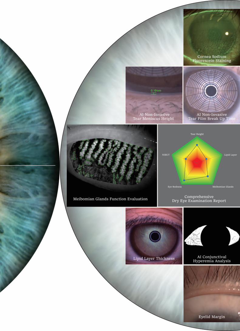
Cornea SodiumFluorescein Staining
AI Non-InvasiveTear Film Break Up Time
AI Non-InvasiveTear Meniscus Height
Meibomian Glands Function Evaluation ComprehensiveDry Eye Examination Report
Lipid Layer Thickness AI ConjunctivalHyperemia Analysis
Eyelid Margin

Full Cornea Dry Eye AnalysisBy Placido ring projection system with visible light,the examination scope is up to 8mm cornea diameter. Examination of the tear film outside of pupil center has the same significance for the diagnosis of Dry Eye.
Automatic Classificationof Meibomian GlandsUnique Built-in infrared lighting system provides a larger scope capture of Meibomian Glands, adjustable depth of field and aperture enables more vivid images.Precise diagnosis of Dry Eye caused by MGD is guaranteed with the help of automatic Meibomian Glands loss classification.
Fully automatic Fireflydigital module
Firefly Digital module is specially designed for anterior segment examination, no parameter settings required(automatic exposure,auto white balance,auto focus),with adjustable depth of field and wide dynamic range,5 Mega Pixels video output , high examination efficiency is allowed.
HD Optical SystemResolution is up to 200 lp/mm, providing more details of the pathologies.
Increase positive rate ofearly corneal epithelial stainingBuilt-in yellow filter along with cobalt-blue filter increases the contrast of Sodium Fluorescein Staining image.
Dry eye diagnostic systemEasy Pathogenic Diagnosis provides guidance for customized treatment.

Cornea Mucous Layer Aqueous Layer Lipid layer
Dry eye classification from the 2007 DEWS Report
Tear film
Due to various causes of Dry Eye Disease,traditional examination is difficult to find out the cause and quantify for the diagnosis.
MediWorks Dry Eye Diagnostic System can provide standardized examination and quantified causes evaluation for Dry Eye Disease.
secreted by the meibomian glandsprevents tear from evaporating and stop sebaceous lipids to enter the tear film.
secreted by the lacrimal glandskeeps the cornea hydrated and supplied with nutrients.
secreted by goblet cellsfights off bacteria and adds stability to the tear film.
Effect of the Environment
Milieu Interieur Low blink rate behavior,VTU, microscopy Wide lid aperture gaze position Aging Low androgen pool Systemic Drugs:antihistamines, beta-blockers, antispasmodics, diuretics,and some psychotropic drugs Milieu Exterieur Low relative humidity High wind velocity Occupational environment
DRY EYE
Aqueous-deficient
SjogrenSyndromeDry Eye
Intrinsic Extrinsic
Non-SjogrenDry Eye
Primary
SecondaryLacrimal
Deficiency
Meibomian OilDeficiency
Vitamin A-Deficiency
Topical DrugsPreservatives
Contact LensWear
Ocular SurfaceDisease
eg,Allergy
Disordersof Lid
Aperture
Low BlinkRate
Drug ActionAccutane
LacrimalGland DuctObstruction
Reflex Block
SystemicDrugs
Evaporative

8mm
AI Non-Invasive Break Up TimeFunctions
MediWorks adopts Placido ring projection system with visible light to do NIBUT examination,the examination scope is up to 8mm cornea diameter which brings much more comprehensive diagnosis outcome.The non-invasive examination avoids the irritation brought by the traditional Cornea Sodium Fluorescein Staining.
AI identifies the break-up area and analyzes NIBUT automatically. Fully automatic analysis system provides efficient quantified evaluation for the overall stability of tear film.It automatically acquires the first break up time, average break up time, break up distribution,break up area percentage curve and time distribution.
After taking one video, it brings out automatis result of NIBUT and Tear Meniscus Height.
Grade 0 Normal, First Rupture Time: 10 s Average Rupture Time: 14 s Grade 1 Warning, First Rupture Time: 6-9 s Average Rupture Time: 7-13 s Grade 2 Dry eye, First Rupture Time: 5 s Average Rupture Time: 7 s
4mm
Visible light Placido ring projection
Infrared Placido ring projection
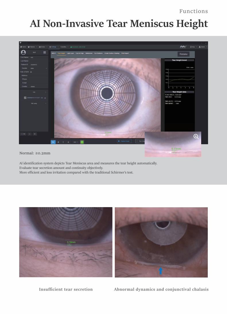
AI identification system depicts Tear Meniscus area and measures the tear height automatically. Evaluate tear secretion amount and continuity objectively.More efficient and less irritation compared with the traditional Schirmer’s test.
AI Non-Invasive Tear Meniscus HeightFunctions
Insufficient tear secretion Abnormal dynamics and conjunctival chalasis
Normal: ≥0.2mm

Automatic classification system provides precise and quantified diagnosis of DES caused by meibomian glands dysfunction.With built-in infrared lighting system,doctors can observe larger image scope of the Meibomian Glands. Adjustable depth of field makes the glands more prominent and distinguishable against the background.
Evaluation of Meibomian Glands FunctionFunctions
Grade 0: No Meibomian Glands LossGrade 1: Meibomian Glands Loss < 1/3 Grade 2: Meibomian Glands Loss 1/3-2/3 Grade 3: Meibomian Glands Loss >2/3
Meibomian glands loss Image of Meibomian Glands under high-magnification
16X

White ring projection system ensures a larger examination area compared to Placido ring.By comparing with the standard grading template and recording the Lipid Layer thickness, it is helpful for judging MGD.
MediWorks professional design of optical system is capable of providing HD digital image that remains clear and sharp even zoom in, meets the examination requirements of the overall shape of eyelid margin and its slight change.
Lipid Layer Thickness
Eyelid Margin
Functions
Grade 1: <15Grade 2: ≈ 15Grade 3: ≈ 30Grade 4: ≈ 30-80Grade 5: ≈ 80Grade 6: ≈ 80-120Grade 7: ≈120-160
1. Normal including (Ophthalmic embolism bright, transparent) 2. Mild including (gland cap crown - glandular prominent) 3. Moderate including (glandular fat plug - disappearance of the marginal mucosa, hyperkeratosis) 4. Severe including (uneven margins, disappearance of the meibomian glands - posterior margin Blunt round, thickening, new blood)
Meibomian Glandsobstruction
(Unit:nm)

Effectively increases positive rate of early corneal epithelial staining.Built-in yellow filter along with cobalt-blue filter makes the corneal sodium fluorescein images more clearly.
The unique AI identification system can identify and calculate percentages of conjunctival congestion and ciliary congestions and evaluate severity of eye congestion.
Cornea Sodium Fluorescein Staining
AI Analysis of Conjunctival HyperemiaFunctions
WithoutBuilt-in yellow filter
Corneal epithelial staining
Normal: ≤2 Abnormal: >2
AI image

Dry Eye Comprehensive Evaluation ReportConvenient Medical Consultation on Dry Eye Syndrome
Patient Management system allows doctors to build and edit medical records. Quickly search the patient case by key words.Doctors can note patients’ situation via the software. This DICOM-supported system enables Mediview to connect with medical systems in hospitals.
Comparison of Patient recordsSupports repeated comparison among medical records to evaluate treatment and guide customized treatment plan.
Smart Patient Management system
Email: [email protected] contact:
We are looking forward to your professional advice for our products and if you are interested in academic or business cooperation with us.
Dry Eye Diagnostic Report
Name: TEST Gender: M Age: 30 Patient ID: MW0001 Diagnostic Type:
Check Date:2020-06-15 14:42:11 OD OS Check Date:
2020-06-15 14:42:11
NIBUTReference value
Grade 0 Healthy, First rupture time: 10Second Average rupture time: 14SecondGrade 1 Warning, First rupture time: 6-9Second Average rupture time: 7-13SecondGrade 2 Dry Eye, First rupture time: 5Second Average rupture time: 7Second
WarningFirst rupture time: 5.59Second
Average rupturetime: 11.83Second
WarningFirst rupture time: 4.56Second
Average rupturetime: 12.75Second
Tear Height
Reference valueHealthy: ≥0.2mm
Healthy0.2 mm
Abnormal0.1 mm
Grade 3
Lipid LayerReference value(Unit:nm)
Grade1: <15 Grade2: ≈15 Grade3: ≈30Grade4: ≈30-80 Grade5: ≈80Grade6: ≈80-120 Grade7: ≈120-160
Abnormal Abnormal
Grade 3
Meibomian Glands
Reference value
Grade 0: No missingGrade 1: Loss of meibomian glands <1/3Grade 2: Loss of meibomian glands 1/3-2/3Grade 3: Loss of meibomian glands >2/3
grade1
grade1
1/2
Dry Eye Diagnostic Report
Name: TEST Gender: M Age: 30 Patient ID: MW0001 Diagnostic Type:
Check Date:2020-06-15 14:42:11 OD OS Check Date:
2020-06-15 14:42:11
Eyelid EdgeReference value
1.Healthy (clear and transparent eyelid plug)2.Mildly included (glandular mouth cap crown-glandularmouth protruding)3.Moderately included (glandular fat plugs ---disappearance of mucous membrane of the eyelidmargin, hyperkeratosis)4.Severe include (irregular eyelid margin, disappearanceof meibomian gland opening --- posterior eyelid marginobtuse, thickened, new blood)
Upper: Healthy
Lower: Healthy
Upper: Mild
Lower: Mild
Eye Redness
Reference value
Healthy: ≤2gradeAbnormal: >2grade
Conjunctival grade: 1.67
Ciliary grade: 1.25
Conjunctival grade: 1.54Ciliary grade: 1.22
Ocular Surface Stainingresult:
result:
Print Date:Doctor: Admin

Specifications
Dry Eye Module
Ø36.2mm, Ø22.3mm, Ø14mm, Ø8.9mm, Ø5.7mm
Microscope
Magnification ChangeMicroscope Type
6.3X, 10X, 16X, 25X, 40X
Eyepieces
Pupillary AdjustmentAngle between Eyepieces
Diopter Adjustment
Total Magnification2700·N lp/mm (200 lp/mm)Optical Resolution
Field of View
Galilean TypeRevolving Drum 5 steps
12.5X10°52mm-80mm-8D~+8D
Slit WidthSlit LengthAperture DiametersSlit AngleSlit InclinationFiltersLampLuminance
0~14mm continuous (slit becomes a circle at 14mm)1~14mm continuous Ø14mm, Ø10mm, Ø5mm, Ø3mm, Ø2mm, Ø1mm, Ø0.2mm0°~180°5°, 10°, 15°, 20°Heat-absorbing filter, ND filter, Red-free filter, Cobalt blue filter,Yellow filter built-in3V LED Module≥150KLX
Slit Illumination
Input VoltageInput FrequencyRated current
100V~240V50Hz/60Hz
Power Supply
Output Voltage 3V LED, Fixation 15V1.2A
Image SensorPhoto ResolutionFormatVideo ResolutionFrame of VideoVideo FormatsExposure ModeTransmission Interface
1/1.8-inch sensor / 2.4μm pixel / 5.0M Pixels2592 x 1944JPEG2592 x 194425fpsMP4 H.264Automatic exposureUSB 3.0 TYPE-C
System SpecificationsDigital Module Automatic exposure/ Automatic white balance / Adjustable depth of field and aperture
DimensionGross weightNet weight
770mm x 470mm x 570mm(L/W/H)23kg17kg
Packaging
PC configuration i5-8500T 8G 1T+128G 2Gdiscrete graphics
Display 1920×1080 23.8inch
PC system Windows 10
System Specifications
Shanghai MediWorks Precision Instruments Co.,Ltd.
*Subject to change in design or speci�cation without advance noti�cation
Add:Building 7, Ming Pu Plaza, No. 3279, San Lu Rd, Min Hang District,Shanghai, 201100,ChinaTel: +86-21-54260421 54260423 Fax:+86-21-54260425Email: [email protected] [email protected]
Cornea Sodium Fluorescein Staining
AI Non-Invasive Tear Break Up Time AI Non-Invasive Tear Meniscus Height
Dry Eye Examination Report
Lipid Layer Thickness
AI Conjunctival Hyperemia Analysis
Meibomian Glands Function Evaluation
Eyelid Margin
AI identify the break-up areaAutomatic first break up timeAutomatic average break up timeVisible light Placido ring projection(23 ring)
AI identification systemAutomatic Non-Invasive Tear Meniscus HeightOptical magnificationElectronic amplification
Template comparison evaluationVisible light White ring projection system
Automatic Meibomian glandsloss classification
AI identification systemAutomatic conjunctival congestion percentagesAutomatic ciliary congestions percentages
Optical magnificationElectronic amplification
Eye surface damage reportBuilt-in yellow filterCobalt blue filter
Automatic analysis report
www.mediworks.biz
Follow Us on