drosophila damaged dna-binding protein 1 is an essential
TRANSCRIPT

Copyright 2004 by the Genetics Society of AmericaDOI: 10.1534/genetics.103.025965
Drosophila Damaged DNA-Binding Protein 1 Is anEssential Factor for Development
Kei-ichi Takata,* Hideki Yoshida,† Masamitsu Yamaguchi‡ and Kengo Sakaguchi*,1
*Department of Applied Biological Science, Faculty of Science and Technology, Tokyo University of Science, Noda-shi, Chiba-ken 278-8510,Japan, †Laboratory of Cell Biology, Department of Bioinformatics, Faculty of Engineering, Soka University,
Hachioji, Tokyo 192-8577, Japan and ‡Department of Applied Biology, Faculty of Textile Sciences,Kyoto Institute of Technology, Matsugasaki, Sakyo-ku, Kyoto 606-8585, Japan
Manuscript received January 6, 2004Accepted for publication February 9, 2004
ABSTRACTThe damaged DNA-binding protein (DDB) complex, thought to recognize (6-4) photoproducts and
other lesions in DNA, has been implicated to have a role in global genomic nucleotide excision repair(NER) and E2F-1-mediated transcription. The complex consists of a heterodimer of p127 (DDB1) andp48 (DDB2), the latter also being known as XPE. We reported previously that in Drosophila expressionof the DDB1 (D-DDB1) gene is controlled by the DRE/DREF system, and external injury to DNA is notessential for D-DDB1 function. In the present study of the function of D-DDB1 in a multicellular system,we prepared transgenic flies, which were knocked down for the D-DDB1 gene due to RNA interference(RNAi), and performed immunocytochemistry to ascertain the distribution of D-DDB1 in the eye imaginaldisc. It was found to be abundant in the anterior of the morphogenetic furrow (MF). Whole-body overex-pression of dsRNA of D-DDB1 in Drosophila using a GAL4-UAS targeted expression system inducedmelanotic tumors and caused complete lethality. When limited to the eye imaginal disc, a severe rougheye phenotype resulted. Correspondingly, all of the D-DDB1 gene knocked-out flies also died. D-DDB1therefore appears to be an essential development-associated factor in a multicellular organism.
THE damaged DNA-binding protein (DDB) com- tants) are selectively defective in global genomic repairplex, which is a heterodimeric protein composed (GGR; Hwang et al. 1999). No mutations of mammalian
of 127-kD (DDB1) and 48-kD (DDB2) subunits, has DDB1 have been described although Zolezzi et al.been shown to recognize many types of DNA lesions (2002) discussed that although DDB1 is not necessarily(Feldberg 1980; Carew and Feldberg 1985; Hirsch- a lethal factor in a unicellular system, the very potentfeld et al. 1990; Keeney et al. 1993; Reardon et al. 1993; defects that result as a consequence of the Schizosaccharo-Payne and Chu 1994). DDB1 can interact with SPT3- myces pombe DDB1 deletion would suggest that mutationsTAFII31-GCN5L acetylase (STAGA) complex (Martinez in the mammalian DDB1 gene are likely to be lethal.et al. 2001) and p300 histone acetyltransferase (Rapic- Because functions of DDB other than directly in DNAOtrin et al. 2002), while DDB2 can interact with CREB- repair have recently been suggested (Nichols et al.binding protein (Datta et al. 2001). DDB might func- 2000; Takata et al. 2002; Zolezzi et al. 2002), the pres-tion as a repair protein to alter chromatin structure and ent study was performed to investigate the propertiesrecruit nucleotide excision repair (NER) factors to DNA of DDB1 in the whole body of Drosophila melanogaster.damage sites. DDB2 is also known as xeroderma pigmento- DDB2 binds to E2F-1, which is a cell cycle regulatorysum complementation group E (XPE) and cells with muta- transcription factor (Hayes et al. 1998). DDB binds totions in this gene are mildly defective in NER of DNA cullin 4A, which is believed to be a ubiquitin-proteindamage (Shiyanov et al. 1999a; Liu et al. 2000). How- isopeptide ligase (type E3; Shiyanov et al. 1999b). Theever, DDB was found not to be required in NER reconsti- apolipoprotein B (apoB) gene regulatory factor-2 (BRF-tution studies in vitro (Aboussekhra and Wood 1995; 2)/human hepatitis B virus X-associated protein-1Mu et al. 1995; Kazantsev et al. 1996) despite the fact (XAP-1)/DDB1 may belong to a new family of transcrip-that damaged DNA-binding activity of DDB is absent in tion factors and control apoB gene transcriptioncells of a subset of XPE patients (Chu and Chang 1988; (Krishnamoorthy et al. 1997). DDB1 also binds to viralKataoka and Fujiwara 1991; Keeney et al. 1992, 1993). transcriptional transactivators, including the hepatitis BIn vivo studies have shown that XPE cells (DDB2 mu- virus X protein (HBVx; Butel et al. 1995). DDB1 has
roles in chromosome segregation and the aberrant nu-clear structures observed in the ddb1� strain in S. pombe
1Corresponding author: Department of Applied Biological Science, (Zolezzi et al. 2002). We found and reported previouslyFaculty of Science and Technology, Tokyo University of Science, Noda-shi, Chiba-ken 278-8510, Japan. E-mail: [email protected] that the expression of the Drosophila -DDB1 (D-DDB1)
Genetics 168: 855–865 (October 2004)
856 K.-i. Takata et al.
lishment of lines carrying GMR-GAL4 was described earlierTABLE 1(Robertson et al. 1988; Takahashi et al. 1999). Enhancer
Transformants carrying the 650-bp D-DDB1 trap line carrying the lacZ marker X63 (inserted in rhomboid),double-strand RNA D120 (inserted in scabrous), and BB02 (inserted in rhomboid)
were obtained from Y. Hiromi. P-element insertion in the 5�exon of D-DDB1, DDB1EY01408, and UAS-P35 were obtained fromChromosomethe Bloomington Indiana Stock Center. mei-9L1 (allele of theP-element plasmid Strain linkageXPF gene) was obtained from the Drosophila Genetic Re-
pUAS-D-DDB1(650)-dsRNA 10 II source Center, Kyoto Institute of Technology. Establishment23 II of lines carrying Collagen-GAL4 (CgGAL4) was kindly provided31 III by Dr. C. R. Dearolf.41 III Ectopic expression of UAS-D-DDB1(650)-dsRNA: Methods
for ectopic expression of D-DDB1(650)-dsRNA were essentiallyas described by Brand and Perrimon (1993):
i. Expression in the whole body: A line carrying heterozygousgene is controlled by the DNA replication-related ele-Act5C-GAL4 on the third chromosome and Act25-GAL4ment (DRE)/DRE-binding factor (DRE/DREF) systemon the second chromosome was crossed with lines carrying
(Takata et al. 2002), which is generally responsible for the homozygous P[UAS-D-DDB1(650)-dsRNA] on the sec-activating the promoters of proliferation-related genes ond chromosome.
ii. Expression in the fat body: A line carrying homozygousfor proliferating cell nuclear antigen (PCNA) (YamaguchiCgGAL4 on the second chromosome was crossed with lineset al. 1995b), the 180-kD and 73-kD subunits of DNAcarrying the homozygous P[UAS-D-DDB1(650)-dsRNA] onpolymerase � (Yamaguchi et al. 1995a; Takahashi et al.the second chromosome.
1996), cyclin A (Ohno et al. 1996), ras (Lightfoot et iii. Expression in the eye imaginal disc: Males carrying ey-al. 1994), raf (Ryu et al. 1997), and D-mtTFA (Takata GAL4 on the second chromosome and GMR-GAL4 on
the X chromosome were crossed with females carryinget al. 2001, 2003). D-DDB1 is thus suggested to havehomozygous P[UAS-D-DDB1(650)-dsRNA] on the secondroles in cell proliferation. We also indicated in a previ-chromosome.ous article that external injury to DNA is not essential
to D-DDB1 function, and that as with UV-irradiation- Northern hybridization: Aliquots of 30 �g of total RNAextracted from living third-instar larvae carrying a single copyinduced transfer of D-DDB1 to the nucleus from theof Act5C-GAL4 (�/�;Act5C-GAL4/�) and single copies ofcytoplasm, during spermatogenesis the D-DDB1 proteinAct5C-GAL4 and UAS-D-DDB1(650)-dsRNA (UAS-D-DDB1transiently shifts from one cell compartment to another (650)-dsRNA/�;Act5C-GAL4/�) were resolved on 1.2% formal-
(Takata et al. 2002). The results indicated that D-DDB1 dehyde-containing agarose gels and transferred onto nylonnot only contributes to the DNA repair system, but also membranes (Hybond-N�; Amersham, Arlington Heights, IL).
After prehybridization, the filters were probed with a 32P-plays roles in cell proliferation and development.labeled D-DDB1 ORF (1774–3423 bp), with a 32P-labeled full-In this report, we provide evidence from analyses oflength PCNA ORF as a negative control or with a 32P-labeledtransgenic flies, knocked down for the D-DDB1 gene by full-length ribosomal protein 49 (RP-49) ORF as a control at 42�
RNAi, and knocked out for the D-DDB1 gene by P-ele- for 16 hr, followed by washing twice with 2� SSPE � 1% SDSment insertion, that D-DDB1 acts as a cell-proliferation at room temperature for 15 min and twice with 1� SSPE �
0.1% SDS at 60� for 20 min. Blots were exposed to Kodakand development-associated factor as well as in DNAX-Omat XAR films and quantified with a NIH imaging ana-repair. Interestingly, we found that a defect in D-DDB1lyzer.caused lethality in our multicellular system. Knock down Western blotting analysis: Western blotting analysis was
of D-DDB1 in the entire eye imaginal disc, but not when carried out by the method of Towbin et al. (1979). A totalposterior to the morphogenetic furrow (MF), further of 60 �g of TCA-precipitated proteins of a homogenate of
Drosophila bodies was separated on 10% SDS-PAGE. Poly-caused a severe rough eye phenotype.clonal antibodies reacting with D-DDB1 were affinity purifiedby D-DDB1 protein-conjugated Sepharose column chromatog-raphy (Takata et al. 2002). DM1A mouse anti-� tubulin mono-MATERIALS AND METHODSclonal antiboby (Accurate Chemical and Scientific, Westbury,NY) was used as a control. Anti-rat IgG and anti-mouse IgGPlasmid construction: The plasmid p5�-D-DDB1(650)-
dsRNA contains the D-DDB1 open reading frame (1–750 bp) conjugated with alkaline phosphatase (Promega, Madison,WI) were used as a secondary antibody. Color was developedand the D-DDB1 ORF (101–750 bp) head to head (3�, 750–1
bp of D-DDB1 ORF and 5�, 101–750 bp of D-DDB1 ORF), in with nitroblue tetrazolium and 5-bromo-4-chloro-3-indolylphosphate as the substrates of alkaline phosphatase.a P-element vector.
Establishment of transgenic flies: P-element-mediated germ- Immunohistochemistry: The polyclonal anti-D-DDB1 anti-body (Takata et al. 2002) used for the immunocytochemicalline transformation was carried out as described earlier
(Spradling and Rubin 1982). F1 transformants were selected study reacts specifically with the D-DDB1 protein in a crudeextract of Drosophila embryos and with that produced inon the basis of white eye color rescue (Robertson et al. 1988).
Established transgenic strains carrying pUAS-D-DDB1(650)- Escherichia coli carrying the expression plasmid for a recombi-nant D-DDB1 (Takata et al. 2002). Third instar larvae weredsRNA and their chromosomal linkages are listed in Table 1.
Fly stocks: Fly stocks were cultured at 25� on standard food. dissected in Drosophila Ringer’s solution and imaginal discswere fixed in 4% paraformaldehyde, PBS for 20 min at roomThe Canton-S fly was used as the wild-type strain. The line
expressed GAL4 under the control of the Act5C gene pro- temperature or at 4�. After washing with PBS, 0.3% TritonX-100 (PBS-T), the samples were blocked with PBS-T con-moter, Act25 gene promoter, and eyeless gene promoter. Estab-

857Drosophila Damaged DNA-Binding Protein 1
taining 10% normal goat serum for 30 min at room tempera- We also found that the D-DDB1 gene is controlled by theture and incubated with a rat anti-D-DDB1 polyclonal antibody DRE/DREF system, which is responsible for activating theat a 1:100 dilution, a rabbit anti-D-RPA70 polyclonal antibody
promoters of proliferation-related genes (Takata et al.at a 1:100 dilution, and mouse anti-�-galactosidase mono-2002), suggesting an importance for development.clonal antibody (Promega) at 1:500 dilution at 4� for 16 hr.
After extensive washing with PBS-T, the imaginal discs were Here we finally succeeded for the first time in knock-incubated with Alexa488-anti-rabbit IgG and Alexa594-anti- ing down D-DDB1 in the Drosophila whole body withrat IgG (Sigma, St. Louis) at a 1:200 dilution or an alkaline the RNAi method. Mutations of DDB2, also termed XPEphosphatase-conjugated goat anti-rabbit IgG (Promega) at
in human, are known to cause human disorders (Chu1:500 dilution for 2 hr at room temperature. After extensiveand Chang 1988; Kataoka and Fujiwara 1991; Keeneywashing with PBS-T, the tissues were also stained with 0.5 �m
TOTO3 (Molecular Probes, Eugene, OR) for 30 min. The et al. 1992; Itoh et al. 1999; Nichols et al. 2000).tissues were washed with PBS and mounted in 90% glycerol, Expression of the 650-bp dsRNA fragment of D-DDB1PBS for confocal microscopic observation (Radiance 2100; in transgenic flies and knock down of D-DDB1 in Dro-Bio-Rad, Richmond, CA) or observation with an Olympus BX-
sophila whole body cause lethality: As shown in Figure50 microscope.1, the 650-bp dsRNA fragment (from 101 to 750 bp ofScanning electron microscopy: Adult flies were anesthetized,
mounted on stages, and observed under a Hitachi S-3000N the ORF) of D-DDB1 was separated by using the D-DDB1scanning electron microscope in the low vacuum mode. gene (from 1 to 100 bp of the ORF) sequence that acts
Histology of adult eyes: Adult eyes were fixed in Bouin’s as a spacer to give a hairpin loop-shaped RNA. Ectopicfixative, embedded in paraffin, sectioned, and stained with
expression of the 650-bp dsRNA fragment of D-DDB1Giemsa solution.(the D-DDB1 ORF is 3420 bp long) in living flies wasLabeling with 5-bromo-2�-deoxyuridine: Detection of cells in
S-phase was performed by a 5-bromo-2�-deoxyuridine (BrdU) performed using the GAL4-mediated expression systemlabeling method as described previously with minor modifica- described in materials and methods. Lines with UAS-tions (Wilder and Perrimon 1995). Third instar larvae cul- D-DDB1(650)-dsRNA transgenes were obtained with pU-tured at 28� were dissected in Grace’s insect medium and then
AST constructs according to standard procedures asincubated in the presence of 20 �g/ml BrdU (Boehringerdescribed by Brand and Perrimon (1993). Four inde-Mannheim, Indianapolis) for 30 min. The samples were fixed
in Carnoy’s fixative (ethanol:acetic acids:chloroform, 6:3:1) pendent lines of germ-line transformants carrying UAS-for 15 min at 25� and further fixed in 80% ethanol, 50 mm D-DDB1(650)-dsRNA were established and used for theglycine buffer, pH 2.0 at 20� for 12 hr. Incorporated BrdU analysis. Established transgenic strains carrying UAS-was visualized using an anti-BrdU antibody and an alkaline D-DDB1(650)-dsRNA wild-type constructs and their chro-phosphatase detection kit (Boehringer).
mosomal linkages are listed in materials and methods.Apoptosis assay: Third instar larvae were dissected in Dro-Transgenic flies carrying UAS-D-DDB1(650)-dsRNA weresophila Ringer’s solution and imaginal discs were fixed in
4.0% paraformaldehyde in PBS for 30 min at room tempera- then crossed with transgenic flies carrying GAL4 cDNAture. After washing with PBS, endogenous peroxidase activity put under the control of the actin-specific enhancer-was blocked by treatment with methanol containing 0.3% promoter (Act5C-GAL4 and Act25-GAL4), of the eyeH2O2 at room temperature for 30 min. The samples were then
imaginal disc-specific promoter (ey-GAL4 and GMR-washed with PBS and permeabilized by incubation in a solutionGAL4). A scheme of the heritable and inducible RNAicontaining 0.1% sodium citrate and 0.1% Triton X-100 on ice
for 2 min. After extensive washing, the apoptosis assay was system is shown in Figure 1. There is no homologycarried out using an in situ cell death detection kit (POD, among the D-DDB1 ORFs (101–750 bp) used for RNAiBoehringer) according to the manufacturer’s recommenda- and other Drosophila genes.tions.
Transgenic flies carrying single copies of Act5C-GAL4and UAS-D-DDB1(650)-dsRNA [UAS-D-DDB1(650)-dsRNA/�;Act5C-GAL4/�] caused a striking phenotype withRESULTS AND DISCUSSIONgeneration of melanotic tumors (Figure 2, A and B),
The DDB complex is thought to recognize (6-4) pho- which are thought to arise as a normal and heritabletoproducts and other lesions in DNA and consists of a response to some form of abnormal development andheterodimer of p127 large subunit (DDB1) and p48 form groups of cells that are recognized by the immunesmall subunit (DDB2) (Feldberg 1980; Carew and system and encapsulated in melanized cuticle (WatsonFeldberg 1985; Hirschfeld et al. 1990; Keeney et al. et al. 1991). All transgenic flies died. Almost all trans-1993; Reardon et al. 1993; Payne and Chu 1994). DDB2 genic flies died by the third instar larvae, after a 1- tohas roles in GGR but not in transcriptional coupled 2-day delay in development. A few transgenic flies livedrepair (TCR). Although DDB1 is evolutionarily con- until early pupal stages (Figure 2B) but none grew toserved from yeast to mammals, its functions remain un- adults. Act25-GAL4/UAS-D-DDB1(650)-dsRNA showed theclear. To generate a deeper comprehension of its nature same phenotype. Note that no phenotypic differenceswe here chose D. melanogaster as an experimental system. were observed among four independent lines carryingIn Drosophila, DDB1 appears to act as a repair factor, UAS-D-DDB1(650)-dsRNA. No melanotic tumors or pre-because D-DDB1 binds to UV-irradiated DNA and a mature deaths were observed in control flies carrying aD-DDB1 knocked down Kc cultured cells generated with single copy of Act5C-GAL4 (�/�;Act5C-GAL4/�) (Fig-
ure 2, C and D). Biological activities of the D-DDB1 genea RNAi method are sensitive to UV (Takata et al. 2002).

858 K.-i. Takata et al.
Figure 1.—Schematic of the heritable and in-ducible RNAi system. Two transgenic fly stocks,GAL4-driver and UAS-D-DDB1(650)-dsRNA, areused in this system. The GAL4-driver fly used inthis experiment has a transgene-conjugating yeasttranscriptional factor GAL4. In this report, weused Act5C-GAL4 and Act25-GAL4 as a GAL4driver to induce D-DDB1 gene silencing in all cellsof the fly, ey-GAL4 and GMR-GAL4 to induceD-DDB1 gene silencing in the eye imaginal disc,and CgGAL4 to induce D-DDB1 gene silencing inthe fat body. The UAS-D-DDB1(650)-dsRNA flyhas a transgene containing the inverted repeat ofthe 650-bp dsRNA fragment (101–750 bp of theORF) of D-DDB1 separated by the D-DDB1 gene(1–100 bp of the ORF) sequence ligated to theUAS promoter, a target of GAL4. In the F1 prog-eny of these flies, the 650-bp dsRNA of the targetgene is expressed to induce the gene silencing.
silencing during development were analyzed with trans- genic animals was analyzed by Northern hybridization withtissue extracts. Living third instar larvae carrying a singlegenic flies of UAS-D-DDB1(650)-dsRNA/UAS-D-DDB1(650)-
dsRNA and Act25-GAL4/Cyo,GFP. UAS-D-DDB1(650)-dsRNA/ copy of Act5C-GAL4 (�/�;Act5C-GAL4/�) and singlecopies of Act5C-GAL4 and UAS-D-DDB1(650)-dsRNAUAS-D-DDB1(650)-dsRNA transgenic flies were then crossed
with Act25-GAL4/Cyo,GFP transgenic flies, and the numbers [UAS-D-DDB1(650)-dsRNA/�;Act5C-GAL4/�] were ho-mogenized, and amounts of D-DDB1 transcripts in theof F1 larvae of Act25-GAL4/UAS-D-DDB1(650)-dsRNA (RNAi
active flies without GFP signal) and UAS-D-DDB1(650)- total RNA extracts were determined with the D-DDB1ORF (1774–3423 bp). As shown in Figure 3, the tran-dsRNA/Cyo,GFP (control flies with GFP signal) were
counted. About 70% of the RNAi active transgenic flies scripts of D-DDB1 were reduced by 92.7% by RNAi com-pared with the amount of RP49 transcripts. As showndied by the third-instar larvae, compared with the con-
trol flies (Figure 2E). When they were crossed with the in Figure 3A, the amount of PCNA was not changedafter inducing the D-DDB1 gene silencing. Like theAct25-GAL4/Cyo,GFP transgenic flies and the wild type
(�/�) flies, there was no significant difference between D-DDB1 gene, PCNA gene is reportedly controlled bythe DRE/DREF system (Yamaguchi et al. 1995a). Thethe number of Act25-GAL4/� flies and Cyo,GFP/� flies.
The reason why the Act25-GAL4/UAS-D-DDB1(650)-dsRNA protein levels of D-DDB1 were also reduced and theamount of RPA30 was not changed by RNAi as com-transgenic flies lived until the larval phase might be be-
cause of maternal loading of the factors (Takata et al. pared with the amount of �-tubulin (Figure 3C). Theresults suggest that 650 bp of D-DDB1-dsRNA acts as a2002). A line-inserted P element in the 5� exon of D-DDB1,
DDB1EY01408, is available (FlyBase ID: FBti0025194). Just specific RNAi effector in vivo and that D-DDB1 is neces-sary for normal development. As shown in Figure 2,the same, none of the DDB1EY01408/DDB1EY01408 flies grew to
adults. Almost all of the DDB1EY01408/DDB1EY01408 flies died the knocked-down flies lived slightly longer than theknocked-out flies. The reason may be that the D-DDB1at the first instar larval stage, caused by melanotic tumors
(Figure 2F). Reverting the lethality by excising the P gene is not completely degraded by the RNAi (Figure3, A and B), and the D-DDB1 protein is tough to de-element in the flies indicates that D-DDB1 must be an
essential factor for development. The melanotic tumor grade. As described previously, Drosophila cultured Kccells knocked down for D-DDB1 did not die completely,develops not only in the RNAi knock-down flies (Figure
2A) but also in the P-element-insertion knock-out flies but became sensitive to UV (Takata et al. 2002). Thereis also a report that a DDB1 knocked-out strain (FZ150)(Figure 2F).
Next, expression of D-DDB1(650)-dsRNA in the trans- of S. pombe does not always suffer mortality, although

859Drosophila Damaged DNA-Binding Protein 1
Figure 2.—(A) Knock down of the D-DDB1 levelby expression of the 650-bp D-DDB1 double-strandRNAs in living flies [UAS-D-DDB1(650)-dsRNA/�;Act5C-GAL4/�] caused melanotic tumors and al-most all flies died in the larval stage. A few transgenicflies lived until early pupal stages (B) but none grewto adults. No melanotic tumors or early lethality wasobserved in control flies carrying a single copy ofAct5C-GAL4 (�/�;Act5C/�) (C and D). (A and C)Third instar larvae; (B and D) pupae. (E) Lethality inthe transgenic flies expressing D-DDB1(650)-dsRNA.UAS-D-DDB1(650)-dsRNA/UAS-D-DDB1(650)-dsRNAtransgenic flies were crossed with Act25-GAL4/Cyo,GFP transgenic flies, and the numbers of F1 larvaeof UAS-D-DDB1(650)-dsRNA/Act25-GAL4 (RNAi ac-tive flies without GFP signal) and UAS-D-DDB1(650)-dsRNA/Cyo,GFP (control flies with GFP signal) werecounted. The values shown are the rate of the num-ber of UAS-D-DDB1(650)-dsRNA/Act25-GAL4 flies di-vided by the number of UAS-D-DDB1(650)-dsRNA/Cyo,GFP flies. (F) The flies knocked out D-DDB1showed melanotic tumors and almost all died at thefirst instar larval stage. (G and H) The DAPI stain-ing images of the fat body from CgGAL4/UAS-D-DDB1(650)-dsRNA and CgGAL4/�. The bottomsof G and H show high-magnification views of thetops. The shape of nuclei in the CgGAL4/UAS-D-DDB1(650)-dsRNA fly (G) was abnormal as com-pared with control (H). Arrows indicate the positionof the melanotic tumor. Bars, 0.5 mm.
aberrant chromosome segregation is caused (Zolezzi et strain lacking DDB1 (Figure 2, G and H). The CgGAL4transgenic fly can express GAL4 in the fat body cells andal. 2002). The nuclei of the fat body cells in transgenic flies
carrying single copies of CgGAL4 and UAS-D-DDB1(650)- the circulating hemocytes (Asha et al. 2003). Aberrantchromosome segregation in the flies lacking D-DDB1dsRNA [CgGAL4/UAS-D-DDB1(650)-dsRNA] were abnor-
mal in shape as well as the phenotype of the S. pombe may result in reduced cell numbers and thus cause le-
860 K.-i. Takata et al.
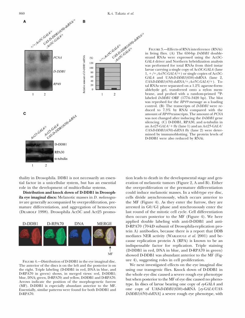
Figure 3.—Effects of RNA interference (RNAi)in living flies. (A) The 650-bp D-DDB1 double-strand RNAs were expressed using the Act5C-GAL4 driver and Northern hybridization analysiswas performed for total RNAs from third instarlarvae carrying a single copy of Act5C-GAL4 (lane1, �/�;Act5C-GAL4/�) or single copies of Act5C-GAL4 and UAS-D-DDB1(650)-dsRNA (lane 2,UAS-D-DDB1(650)-dsRNA/�;Act5C-GAL4/�). To-tal RNAs were separated on a 1.2% agarose-form-aldehyde gel, transferred onto a nylon mem-brane, and probed with a random-primed 32P-labeled D-DDB1 ORF (1774–3420 bp). The blotwas reprobed for the RP49 message as a loadingcontrol. (B) The transcripts of D-DDB1 were re-duced to 7.3% by RNAi compared with theamount of RP49 transcripts. The amount of PCNAwas not changed after inducing the D-DDB1 genesilencing. (C) D-DDB1, RPA30, and �-tubulin inan Act25-GAL4/� fly (lane 1) and an Act25-GAL4/UAS-D-DDB1(650)-dsRNA fly (lane 2) were deter-mined by immunoblotting. The protein levels ofD-DDB1 were also reduced by RNAi.
thality in Drosophila. DDB1 is not necessarily an essen- tion leads to death in the developmental stage and gen-eration of melanotic tumors (Figure 2, A and B). Eithertial factor in a unicellular system, but has an essential
role in the development of multicellular systems. the overproliferation or the premature differentiationcould induce melanotic masses. In a wild-type eye disc,Distribution and knock down of D-DDB1 in Drosoph-
ila eye imaginal discs: Melanotic masses in D. melanogas- cells divide asynchronously, which occurs anterior tothe MF (Figure 4). As they enter the furrow, they areter are generally accompanied by overproliferation, pre-arrested in G0/G1 phase and synchronously enter themature differentiation, and aggregation of hemocyteslast round of the mitotic cell cycle. Cell differentiation(Dearolf 1998). Drosophila Act5C and Act25 promo-then occurs posterior to the MF (Figure 4). We hereapplied double labeling with anti-D-DDB1 and anti-D-RPA70 (70-kD subunit of Drosophila-replication pro-tein A) antibodies, because there is a report that DDBmediates NER activity (Wakasugi et al. 2001) and be-cause replication protein A (RPA) is known to be anindispensable factor for replication. Triple staining(D-DDB1 in red, DNA in blue, and D-RPA70 in green)showed D-DDB1 was abundant anterior to the MF (Fig-ure 4), suggesting roles in cell proliferation.Figure 4.—Distribution of D-DDB1 in the eye imaginal disc.
We next investigated effects on the eye imaginal discThe anterior of the discs is on the left and the posterior is onthe right. Triple labeling (D-DDB1 in red, DNA in blue, and using our transgenic flies. Knock down of D-DDB1 inD-RPA70 in green) shows, in merged views: red, D-DDB1; the whole eye disc caused a severe rough eye phenotypeblue, DNA; green, D-RPA70; and yellow, D-DDB1 and D-RPA70. but when posterior to the MF of eye disc caused no pheno-Arrows indicate the position of the morphogenetic furrow
type. In discs of larvae bearing one copy of ey-GAL4 and(MF). D-DDB1 is especially abundant anterior to the MF.one copy of UAS-D-DDB1(650)-dsRNA [ey-GAL4/UAS-Essentially, similar patterns were found for both D-DDB1 and
D-RPA70. D-DDB1(650)-dsRNA] a severe rough eye phenotype, with

861Drosophila Damaged DNA-Binding Protein 1
Figure 5.—(A) Scanning electron micrographsof adult compound eyes. (Control) Compound eyeof Canton-S. (GMR-GAL4/�;UAS-D-DDB1(650)-dsRNA/�) Knock down of D-DDB1 posterior to themorphogenetic furrow (MF) of the eye disc resultedin a normal eye morphology. (ey-GAL4/UAS-D-DDB1(650)-dsRNA) In contrast, knock down ofD-DDB1 in the whole eye disc caused a severe rougheye phenotype. Negative control flies of ey-GAL4/�and GMR-GAL4/�;�/� have no difference in phe-notype compared with the wild-type (Canton-S)compound eye. Top parts are at 200� and bottomparts are at 800� magnification. (B) Horizontalsections of adult Drosophila eyes. Canton-S (left)and ey-GAL4/UAS-D-DDB1(650)-dsRNA transgenicflies (right) are shown. (C) The D-DDB1 gene silenc-ing in the whole eye disc [ey-GAL4/UAS-D-DDB1(650)-dsRNA] caused a shrunken eye disc pheno-type.
irregular compound eyes and bristles (Figure 5A, right), D-DDB1 gene silencing induces an ectopic S phase poste-rior to the MF, causes apoptosis, and interferes with photo-was noted. Control flies bearing one copy of ey-GAL4
(ey-GAL4/�) exhibited a normal eye morphology. In discs receptor differentiation: Imaginal discs were pulse labeledwith BrdU for 30 min in vitro and stained with an anti-BrdUof larvae bearing one copy of GMR-GAL4 and one copy
of UAS-D-DDB1(650)-dsRNA [GMR-GAL4/�; UAS-D-DDB1 antibody (Figure 6A). No significant difference betweencontrol and D-DDB1 gene-silencing discs was observed in(650)-dsRNA/�], no rough eye phenotype was observed
(Figure 5A, middle). Flies carrying two copies of GMR- the region anterior to the MF. D-DDB1 gene silencinginduced an extra S phase in some cells that are normallyGAL4 and two copies of UAS-D-DDB1(650)-dsRNA [GMR-
GAL4/GMR-GAL4;UAS-D-DDB1(650)-dsRNA/UAS-D-DDB1 postmitotic. In addition, some cells in the region moreposterior to the MF, where commitment to a neuronal(650)-dsRNA] also exhibited normal eye morphology
(data not shown). The normal compound eye has regu- fate occurs and is normally followed by differentiationinto specific cells such as photoreceptors, were labeledlar ommatidia and bristles (Figure 5A, left). Horizontal
sections of eyes of adult flies also showed the D-DDB1 with BrdU (Figure 6A, right).Failure of normal cell cycle progression and distur-gene silencing-induced eye degeneration (Figure 5B).
We next observed effects on the eye imaginal disc of bance of differentiation processes are known to causeapoptosis (Harrington et al. 1994). For example, ittransgenic flies. The eye imaginal disc of ey-GAL4/UAS-
D-DDB1(650)-dsRNA was smaller than that of wild type has been reported that overexpression of dE2F and dDPin eye imaginal discs using a GMR promoter induces(Figure 5C). The results also indicate that D-DDB1 has
a function in cell proliferation so the D-DDB1 gene si- apoptosis and that this counterbalances cells that enteran abnormal S phase (Du et al. 1996). We investigatedlencing caused small eye imaginal disc morphology.

862 K.-i. Takata et al.
Figure 6.—(A) DNA synthesis. Patterns of BrdU incorpora-tion in eye imaginal discs. Left, ey-GAL4/�; right, ey-GAL4/UAS-D-DDB1(650)-dsRNA. The eye disc was stained with ananti-BrdU antibody. Arrows indicate the position of the MF.The anterior of the discs is on the left. The region of an extraS phase in some cells that are normally postmitotic is indicatedby a dotted line. (B) Apoptosis. Detection of apoptotic cellsin eye imaginal discs. Left, ey-GAL4/�; right, ey-GAL4/UAS-D-DDB1(650)-dsRNA. The apoptosis assay was carried out withterminal deoxynucleotidyl transferase. The anterior of thediscs is on the left. Arrows indicate the position of the MF.The apoptotic cells are indicated by a dotted line.
whether D-DDB1 gene silencing can induce apoptosisin eye imaginal disc cells or not. In wild-type discs of
Figure 7.—Immunostaining of eye imaginal discs with anti-third instar larvae, there were very few apoptotic cells �-galactosidase antibody. The anterior of the discs is on the(Figure 6B, left). In contrast, staining of eye imaginal left. Arrows indicate the position of the MF. Wild-type (a and
c) or ey-GAL4,UAS-D-DDB1(650)-dsRNA/� transgenic (b anddiscs from transgenic flies expressing 650 bp dsRNAd) flies were crossed with an enhancer trap line carrying X63of D-DDB1 revealed apoptotic cells to be significantly(inserted in rhomboid), D120 (inserted in scabrous), and BB02increased in the region posterior to the MF (Figure 6B,(inserted in rhomboid), specifically expressing the �-galactosi-
right). The apoptosis by D-DDB1 gene silencing could dase marker in photoreceptor cells of R2/R5/R8, early R8,also produce the morphologically small eye in Figure and late R8, respectively, and F1 larvae were immunohisto-
chemically stained with the anti-�-galactosidase antibody. c5, A and C. Apoptosis seemed to begin in the imaginaland d show high-magnification basal views of the same focaldisc cells in the region where late commitment to photo-planes as a and b, respectively. The differentiated cells arereceptor cells takes place, suggesting that failure ofindicated by a dotted line (a and b in D120 and a and b in
differentiation might induce apoptosis. Therefore, we BB02).analyzed the effect of D-DDB1 gene silencing on photo-receptor specification. In wild-type discs, developmen-tally uncommitted cells are sequentially recruited into order: R8 is generated first, with movement posterior
from the MF, then cells are added pairwise (R2 andclusters that comprise ommatidial precursors. Clusterformation is first observed within the MF, where cells R5, R3 and R4, and R1 and R6), and R7 is the last
photoreceptor to be added to each cluster. We usedare in G1. Cells either leave the cell cycle and differenti-ate or undergo a final synchronous round of cell divi- three enhancer trap lines, X63 (inserted in rhomboid ;
Figure 7, top), D120 (inserted in scabrous ; Figure 7,sion. Overt ommatidial organization starts in the MFwhen cells are grouped into equally spaced concentric middle) and BB02 (inserted in rhomboid ; Figure 7, bot-
tom), specifically expressing the nucleus-localized formaggregates, which convert into preclusters. Photorecep-tor cells have been found to develop in stereotyped of E. coli �-galactosidase marker in photoreceptor cells
863Drosophila Damaged DNA-Binding Protein 1
(R) of R2/R5/R8, early R8, and late R8, respectively.The imaginal discs from F1 larvae generated by matingof enhancer trap lines and 650 bp dsRNA of D-DDB1-expressing transgenic flies [ey-GAL4,UAS-D-DDB1(650)-dsRNA/�;rhomboid-lacZ/�] were immunohistochemi-cally stained with anti-�-galactosidase antibody. In theommatidia of 650 bp dsRNA of D-DDB1-expressing ani-mals, positive nuclei of late R2/R5/R8, early R8, andlate R8 were fewer than in the control case (Figure 7,a and c, top, middle, and bottom), and an abnormalstaining pattern was apparent (Figure 7, b and d, top,middle, and bottom). The results indicate that D-DDB1gene silencing inhibits the differentiation of photore-ceptor cells.
The S. pombe strain lacking ddb1 has slow growthdue to delayed replication progression. Flow-cytometricanalysis shows an extensive heterogeneity in DNA con-tent. A large number of cells specifically displayed DNAcontent intermediate between 1N and 2N, which mayrepresent slow replication (Bondar et al. 2003). Whileit may be due in part to defective chromosomal segrega-tion in the �ddb1 strain, as reported (Zolezzi et al.2002), aberrant S-phase progression also could explainthe heterogeneous DNA distribution. Thus DDB1 ap-pears to have roles in cell proliferation, especially relevantto replication progression and mitosis. Because differentia-tion is correlated with modification of cell cycle processes,D-DDB1 gene silencing may cause inhibition of differentia-
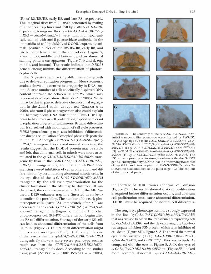
Figure 8.—The sensitivity of the ey-GAL4/UAS-D-DDB1(650)-tion due to accumulation of ectopic S-phase cells posterior dsRNA transgenic flies phenotype was enhanced by UAS-P35.to the MF. Although GMR-GAL4/�;UAS-D-DDB1(650)- (A) wild-type fly (�/�); (B) UAS-D-DDB1(650)-dsRNA/�; (C) ey-
GAL4/UAS-P35; (D) DDB1EY01408/�; (E) ey-GAL4,UAS-D-DDB1(650)-dsRNA/� transgenic flies showed normal phenotype, thedsRNA/�; (F) ey-GAL4,UAS-D-DDB1(650)-dsRNA/�;DDB1EY01408/�;results suggest that the D-DDB1 protein may be stable(G) ey-GAL4,UAS-D-DDB1(650)-dsRNA/ey-GAL4,UAS-D-DDB1(650)-and left, that abnormal cells in mitosis were more accu-dsRNA; (H) ey-GAL4,UAS-D-DDB1(650)-dsRNA/UAS-P35. The
mulated in the ey-GAL4/UAS-D-DDB1(650)-dsRNA trans- P35, anti-apoptotic protein strongly enhances the the D-DDB1genic fly than in the GMR-GAL4/�;UAS-D-DDB1(650)- gene silencing phenotype. Note that the fly carrying two copies
of ey-GAL4 and two copies of UAS-D-DDB1(650)-dsRNAdsRNA/� transgenic fly, and that the D-DDB1 geneshowed no head and died at the pupa stage. (G) The contentsilencing caused inhibition of cell proliferation and dif-of the dissected pupa.ferentiation by accumulating abnormal mitotic cells. In
the eye disc of the ey-GAL4/UAS-D-DDB1(650)-dsRNAtransgenic fly, the cell cycle synchronization for the
the shortage of DDB1 causes abnormal cell divisioncluster formation in the MF may be disturbed. If syn-(Figure 2G). The results showed that cell proliferationchronized, the cells are arrested at G1 in the MF. Weis required before differentiation occurs, and aberrantused a D120 enhancer trap line (inserted in scabrous)cell proliferation must cause abnormal differentiation.to confirm the possibility. The number of the early pho-D-DDB1 must be required for normal cell differentia-toreceptor cells (early R8) immediately after MF wastion.decreased in the ey-GAL4,UAS-D-DDB1(650)-dsRNA/scab-
The rough eye phenotype was more strongly expressedrous-lacZ transgenic fly (Figure 7, middle). The otherin the line [ey-GAL4,UAS-D-DDB1(650)-dsRNA/UAS-P35]photoreceptor cell (R1–R7) differentiation begins afterthat was crossed between the transgenic fly expressing 650the R8 cell differentiation. Shortage of the early R8 cellsbp dsRNA of D-DDB1 and the fly expressing the baculovi-can lead to abnormal differentiation of the cells fromrus caspase inhibitor P35 protein, which is an inhibitor ofR1 to R7 (Figure 7). Failure of all differentiation mightcell death (Figure 8H). Figure 8, A–D, showed the normalinduce apoptosis (Figure 6B, right). This might be oneeyes of the wild-type (�/�), UAS-D-DDB1(650)-dsRNA/�,of the reasons that the ey-GAL4/UAS-D-DDB1(650)-dsRNAey-GAL4/UAS-P35, and DDB1EY01408/� flies, respectively. Astransgenic fly shows a more severe phenotype such ascompared with the eyes in Figure 8, A–D, the eyes ofrough eye than the GMR-GAL4/�;UAS-D-DDB1(650)-ey-GAL4,UAS-D-DDB1(650)-dsRNA/� (Figure 8E) lookeddsRNA/� transgenic fly does. According to the studies
using yeast (Zolezzi et al. 2002; Bondar et al. 2003), more severely abnormal. ey-GAL4,UAS-D-DDB1(650)-

864 K.-i. Takata et al.
dsRNA/�;DDB1EY01408/� (Figure 8F) showed a more se- DDB2 and CSA contain WD40 domains, the other pro-teins with the WD40 domain may also be able to bindvere rough eye phenotype than the eyes in Figure 8E,
meaning that D-DDB1 gene silencing certainly occurs in to DDB1. On the other hand, the D-DDB1 gene washighly conserved in yeast to mammals, and the genesey-GAL4/UAS-D-DDB1(650)-dsRNA. On the other hand, the
eyes of mei-9L1/�;ey-GAL4,UAS-D-DDB1(650)-dsRNA/� of DDB2 and CSA were still not found in the genomeof D. melanogaster.looked same as the eyes of ey-GAL4,UAS-D-DDB1(650)-
dsRNA/� (data not shown). The XPF homolog mei-9 is To conclude, this report documents the first observa-tion of an altered phenotype in a multicellular organismimplicated in nucleotide excision repair. Reduction of
the XPF protein cannot induce the abnormality on the knocked down or knocked out for DDB1. The resultsindicate that, different from the unicellular system case,phenotype by D-DDB1 gene silencing. The results also
indicate that DDB1 has more important roles in the DDB1 is an essential gene in multicellular organisms,playing important roles in development.events other than XPF-related repair. As shown in Figure
8G, when the fly carried two copies of ey-GAL4 and two We are grateful to Fumiko Hirose of Himeji Institute of Technology,copies of UAS-D-DDB1(650)-dsRNA [ey-GAL4,UAS-D-DDB1 Yoshihiro H. Inoue and Kyoko Otsuki of the University of Kyoto
Institute of Technology, Richard D. Wood of University of Pittsburgh(650)-dsRNA/ey-GAL4,UAS-D-DDB1(650)-dsRNA], the flyCancer Institute, Yoko Ogasawara of Tokyo University of Science,lacked the head and died at the pupa stage. Since EyelessKaori Shimanouchi and Shizuka Murakami of our laboratory, andprotein is known to have roles in Drosophila head devel-Malcolm Moore for technical assistance, valuable comments, and help-
opment (Benassayag et al. 2003), D-DDB1 must func- ful discussion. We are also grateful to Yasushi Hiromi of the Nationaltion in brain development. Therefore, we speculate that Institute of Genetics for fly stocks.mouse knocked-out DDB1 may lack the brain and subse-quently die at the early embryo stage. In a mouse knockedout the genes of XPA and CSB (XPA/CSB/), the LITERATURE CITEDcerebellum was remarkably smaller but did not lack the
Aboussekhra, A., and R. D. Wood, 1995 Detection of nucleotidebrain (Murai et al. 2001). These results suggested that excision repair incisions in human fibroblasts by immunostainingthe function of DDB1 was different from the other XP- for PCNA. Exp. Cell Res. 221: 326–332.
Asha, H., I. Nagy, G. Kovacs, D. Stetson, I. Ando et al., 2003 Analy-related proteins. A few reports showed that the abnor-sis of ras-induced overproliferation in Drosophila hemocytes. Ge-mal phenotype occurred when baculovirus P35 proteinnetics 163: 203–215.
and dE2F/dDP were simultaneously expressed (Du et Benassayag, C., S. Plaza, P. Callaerts, J. Clements, Y. Romeo etal., 2003 Evidence for a direct functional antagonism of theal. 1996; Staehling-Hampton et al. 1999). This indi-selector genes proboscipedia and eyeless in Drosophila head de-cated that the majority of cells ectopically entering Svelopment. Development 130: 575–586.
phase as a result of dE2F/dDP expression are eliminated Bondar, T., E. V. Mirkin, D. S. Ucker, W. E. Walden, S. M. Mirkin etal., 2003 S. pombe Ddb1 is functionally linked to the replicationby apoptosis; this phenotype resembles those in Figurecheckpoint pathway. J. Biol. Chem. 278: 37006–37014.8H. The abnormal mitotic cells caused by D-DDB1 gene
Brand, A. H., and N. Perrimon, 1993 Targeted gene expression as asilencing interfere with normal differentiation, and con- means of altering cell fates and generating dominant phenotypes.sequently these abnormal cells are eliminated by apo- Development 118: 401–415.
Butel, J. S., T. H. Lee and B. L. Slagle, 1995 Viral co-factors inptosis. Although coexpression of P35 appears to severelyliver cancer: lessons from hepatitis B virus. Princess Takamatsudecrease the number of cells, the normal cells are few, Symp. 25: 185–198.
and the unusual cells remain without being removed. Carew, J. A., and R. S. Feldberg, 1985 Recognition of a cytosinebase lesion by a human damage-specific DNA binding protein.One of the functions of DDB1 might be not only inNucleic Acids Res. 13: 303–315.DNA repair but also in development. S. pombe DDB1 is Chu, G., and E. Chang, 1988 Xeroderma pigmentosum group E
reportedly linked to the replication checkpoint control cells lack a nuclear factor that binds to damaged DNA. Science242: 564–567.gene cds1, and the S. pombe strain lacking DDB1 grows
Datta, A., S. Bagchi, A. Nag, P. Shiyanov, G. R. Adami et al., 2001slowly because of prolongation of S phase (Bondar et al. The p48 subunit of the damaged-DNA binding protein DDB2003). Aberrant chromosome segregation also occurs in associates with the CBP/p300 family of histone acetyltransferase.
Mutat. Res. 486: 89–97.the S. pombe strain lacking DDB1 (Zolezzi et al. 2002).Dearolf, C. R., 1998 Fruit fly “leukemia”. Biochim. Biophys. ActaDDB1 possibly has a role in chromosome segregation. 1377: M13–M23.
As in S. pombe DDB1, we showed that D-DDB1 has an Du, W., J. E. Xie and N. Dyson, 1996 Ectopic expression of dE2Fand dDP induces cell proliferation and death in the Drosophilaimportant role in cell proliferation. Since cell prolifera-eye. EMBO J. 15: 3684–3692.tion is required before cell differentiation occurs, Dro-
Feldberg, R. S., 1980 On the substrate specificity of a damage-sophila eye differentiation must be closely linked to the specific DNA binding protein from human cells. Nucleic Acids
Res. 8: 1133–1143.cell cycle machinery using D-DDB1.Groisman, R., J. Polanowska, I. Kuraoka, J. Sawada, M. Saijo etRecently, the evidence that not only DDB2 but also
al., 2003 The ubiquitin ligase activity in the DDB2 and CSACSA can bind directly to DDB1 was reported. Further- complexes is differentially regulated by the COP9 signalosome
in response to DNA damage. Cell 113: 357–367.more, like SCF complex and other cullin-based ubiqui-Harrington, E. A., M. R. Bennett, A. Fanidi and G. I. Evan, 1994tin ligases, the DDB1 complex containing DDB2 or CSA
c-Myc-induced apoptosis in fibroblasts is inhibited by specifichas Cul4A and Roc1. DDB1 is part of a cullin-containing cytokines. EMBO J. 13: 3286–3295.
Hayes, S., P. Shiyanov, X. Chen and P. Raychaudhuri, 1998 DDB,E3 ligase complex (Groisman et al. 2003). Since both

865Drosophila Damaged DNA-Binding Protein 1
a putative DNA repair protein, can function as a transcriptional nition protein (UvrA) to the major ultraviolet photoproducts:T[c,s]T, T[t,s]T, T[6–4]T, and T[Dewar]T. J. Biol. Chem. 268:partner of E2F1. Mol. Cell. Biol. 18: 240–249.21301–21308.Hirschfeld, S., A. S. Levine, K. Ozato and M. Protic, 1990 A
Robertson, H. M., C. R. Preston, R. W. Phillis, D. M. Johnson-constitutive damage-specific DNA-binding protein is synthesizedSchlitz, W. K. Benz et al., 1988 A stable genomic source ofat higher levels in UV-irradiated primate cells. Mol. Cell. Biol.P element transposase in Drosophila melanogaster. Genetics 118:10: 2041–2048.461–470.Hwang, B. J., J. M. Ford, P. C. Hanawalt and G. Chu, 1999 Expres-
Ryu, J. R., T. Y. Choi, E. J. Kwon, W. H. Lee, Y. Nishida et al., 1997sion of the p48 xeroderma pigmentosum gene is p53-dependentTranscriptional regulation of the Drosophila-raf proto-oncogeneand is involved in global genomic repair. Proc. Natl. Acad. Sci.by the DNA replication-related element (DRE)/DRE-binding fac-USA 96: 424–428.tor (DREF) system. Nucleic Acids Res. 25: 794–799.Itoh, T., T. Mori, H. Ohkubo and M. Yamaizumi, 1999 A newly
Shiyanov, P., S. A. Hayes, M. Donepudi, A. F. Nichols, S. Linn etidentified patient with clinical xeroderma pigmentosum pheno-al., 1999a The naturally occurring mutants of DDB are impairedtype has a non-sense mutation in the DDB2 gene and incompletein stimulating nuclear import of the p125 subunit and E2F1-repair in (6–4) photoproducts. J. Invest. Dermatol. 113: 251–257.activated transcription. Mol. Cell. Biol. 19: 4935–4943.Kataoka, H., and Y. Fujiwara, 1991 UV damage-specific DNA-bind-
Shiyanov, P., A. Nag and P. Raychaudhuri, 1999b Cullin 4A asso-ing protein in xeroderma pigmentosum complementation groupciates with the UV-damaged DNA-binding protein DDB. J. Biol.E. Biochem. Biophys. Res. Commun. 175: 1139–1143.Chem. 274: 35309–35312.Kazantsev, A., D. Mu, A. F. Nichols, X. Zhao, S. Linn et al., 1996
Spradling, A. C., and G. M. Rubin, 1982 Transposition of clonedFunctional complementation of xeroderma pigmentosum com-P elements into Drosophila germ line chromosomes. Scienceplementation group E by replication protein A in an in vitro218: 341–347.system. Proc. Natl. Acad. Sci. USA 93: 5014–5018.
Staehling-Hampton, K., P. J. Ciampa, A. Brook and N. Dyson, 1999Keeney, S., H. Wein and S. Linn, 1992 Biochemical heterogeneity A genetic screen for modifiers of E2F in Drosophila melanogaster.in xeroderma pigmentosum complementation group E. Mutat. Genetics 153: 275–287.Res. 273: 49–56. Takahashi, Y., M. Yamaguchi, F. Hirose, S. Cotterill, J. KobayashiKeeney, S., G. J. Chang and S. Linn, 1993 Characterization of a et al., 1996 DNA replication-related elements cooperate to en-
human DNA damage binding protein implicated in xeroderma hance promoter activity of the drosophila DNA polymerase alphapigmentosum E. J. Biol. Chem. 268: 21293–21300. 73-kDa subunit gene. J. Biol. Chem. 271: 14541–14547.
Krishnamoorthy, R. R., T. H. Lee, J. S. Butel and H. K. Das, 1997 Takahashi, Y., F. Hirose, A. Matsukage and M. Yamaguchi, 1999Apolipoprotein B gene regulatory factor-2 (BRF-2) is structurally Identification of three conserved regions in the DREF transcrip-and immunologically highly related to hepatitis B virus X associ- tion factors from Drosophila melanogaster and Drosophila virilis.ated protein-1 (XAP-1). Biochemistry 36: 960–969. Nucleic Acids Res. 27: 510–516.
Lightfoot, K., L. Maltby, R. Duarte, R. Veale and O. Segev, 1994 Takata, K., H. Yoshida, F. Hirose, M. Yamaguchi, M. Kai et al.,Conserved cis-elements bind a protein complex that regulates 2001 Drosophila mitochondrial transcription factor A: charac-Drosophila ras2/rop bidirectional expression. Br. J. Cancer 69: terization of its cDNA and expression pattern during develop-264–273. ment. Biochem. Biophys. Res. Commun. 287: 474–483.
Liu, W., A. F. Nichols, J. A. Graham, R. Dualan, A. Abbas et al., Takata, K., G. Ishikawa, F. Hirose and K. Sakaguchi, 2002 Dro-2000 Nuclear transport of human DDB protein induced by ul- sophila damage-specific DNA-binding protein 1 (D-DDB1) is con-traviolet light. J. Biol. Chem. 275: 21429–21434. trolled by the DRE/DREF system. Nucleic Acids Res. 30: 3795–
Martinez, E., V. B. Palhan, A. Tjernberg, E. S. Lymar, A. M. Gamper 3808.et al., 2001 Human STAGA complex is a chromatin-acetylating Takata, K., Y. H. Inoue, F. Hirose, S. Murakami, K. Shimanouchitranscription coactivator that interacts with pre-mRNA splicing et al., 2003 Spatio-temporal expression of Drosophila mitochon-and DNA damage-binding factors in vivo. Mol. Cell. Biol. 21: drial transcription factor A during development. Cell Biol. Int.6782–6795. 27: 361–374.
Mu, D., C. H. Park, T. Matsunaga, D. S. Hsu, J. T. Reardon et al., Towbin, H., T. Staehelin and J. Gordon, 1979 Electrophoretic1995 Reconstitution of human DNA repair excision nuclease transfer of proteins from polyacrylamide gels to nitrocellulose
sheets: procedure and some applications. Proc. Natl. Acad. Sci.in a highly defined system. J. Biol. Chem. 270: 2415–2418.USA 76: 4350–4354.Murai, M., Y. Enokido, N. Inamura, M. Yoshino, Y. Nakatsu et al.,
Wakasugi, M., M. Shimizu, H. Morioka, S. Linn, O. Nikaido et2001 Early postnatal ataxia and abnormal cerebellar develop-al., 2001 Damaged DNA-binding protein DDB stimulates thement in mice lacking Xeroderma pigmentosum group A andexcision of cyclobutane pyrimidine dimers in vitro in concert withCockayne syndrome group B DNA repair genes. Proc. Natl. Acad.XPA and replication protein A. J. Biol. Chem. 276: 15434–15440.Sci. USA 98: 13379–13384.
Watson, K. L., T. K. Johnson and R. E. Denell, 1991 Lethal(1)Nichols, A. F., T. Itoh, J. A. Graham, W. Liu, M. Yamaizumi etaberrant immune response mutations leading to melanotic tumoral., 2000 Human damage-specific DNA-binding protein p48.formation in Drosophila melanogaster. Dev. Genet. 12: 173–187.Characterization of XPE mutations and regulation following UV
Wilder, E. L., and N. Perrimon, 1995 Dual functions of winglessirradiation. J. Biol. Chem. 275: 21422–21428.in the Drosophila leg imaginal disc. Development 121: 477–488.Ohno, K., F. Hirose, K. Sakaguchi, Y. Nishida and A. Matsukage,
Yamaguchi, M., Y. Hayashi, Y. Nishimoto, F. Hirose and A. Matsu-1996 Transcriptional regulation of the Drosophila CycA genekage, 1995a A nucleotide sequence essential for the functionby the DNA replication-related element (DRE) and DRE bindingof DRE, a common promoter element for Drosophila DNa repli-factor (DREF). Nucleic Acids Res. 24: 3942–3946.cation-related genes. J. Biol. Chem. 270: 15808–15814.Payne, A., and G. Chu, 1994 Xeroderma pigmentosum group E Yamaguchi, M., F. Hirose, Y. Nishimoto, T. Naruge, M. Ikeda etbinding factor recognizes a broad spectrum of DNA damage. al., 1995b Expression patterns of DNA replication enzymes and
Mutat. Res. 310: 89–102. the regulatory factor DREF during Drosophila development ana-Rapic-Otrin, V., M. P. Mclenigan, D. C. Bisi, M. Gonzalez and lyzed with specific antibodies. Biol. Cell 85: 147–155.
A. S. Levine, 2002 Sequential binding of UV DNA damage Zolezzi, F., J. Fuss, S. Uzawa and S. Linn, 2002 Characterizationbinding factor and degradation of the p48 subunit as early events of a Schizosaccharomyces pombe strain deleted for a sequenceafter UV irradiation. Nucleic Acids Res. 30: 2588–2598. homologue of the human damaged DNA binding 1 (DDB1) gene.
Reardon, J. T., A. F. Nichols, S. Keeney, C. A. Smith, J. S. Taylor J. Biol. Chem. 277: 41183–41191.et al., 1993 Comparative analysis of binding of human damagedDNA-binding protein (XPE) and Escherichia coli damage recog- Communicating editor: K. V. Anderson
