dr. benaiffer agarwal
DESCRIPTION
Dr. Benaiffer AgarwalTRANSCRIPT

43People’s Journal of Scientific Research Vol. 4(1), Jan. 2011
Treatment of Myofascial Pain Dysfunction Syndrome in an Edentulous Patient– A Case ReportBenaiffer Agrawal *Kirti SomkuwarDepartment of Prosthodontics, People’s Dental Academy, *People’s College of Dental Sciences & Research Centre,Bhopal-462037
Abstract:Pain is a complex phenomenon, causing discomfort, suffering and psychosocial morbidity. Myofascial Pain
Dysfunction Syndrome (MPDS) is attributed to pain & inflammation of the muscles, with no definitive pathogenesiscausing this syndrome. The purpose of this paper is to describe the multidisciplinary approach for the treatment of a 70year old female patient who suffered from myofacial pain syndrome for the last 10 years. Combined with Trans-cutaneouselectrical nerve stimulation (TENS), the construction of a complete denture in order to re-establish the proper verticaldimension leading to decrease in muscular activity thereby eliminating the underlying cause of disease and providing adefinitive treatment for the patient with MPDS.
Key Words: Myofascial pain syndrome, Temporomandibular Joint, Trans – cutaneous electric nerve stimulation.
Introduction:Pain in facial region originating from both
temporomandibular joint and jaw muscles is a commonclinical problem. There are many synonyms for thiscondition including Myofascial Pain DysfunctionSyndrome (MPDS), Mandibular Dys-functionSyndrome and the Temporomandibular JointDysfunction Syndrome (Edmiston & Larsen, 1978).Signs and symptoms of MPDS vary, but generally, thepatient complains of one or more of the following: -Pain in the region of the temporomandibular joints(TMJ), tenderness in the region of one or both joints,temporo-mandibular joint sounds like clicking orcrepitation, restricted jaw opening, disturbed chewingpattern and locking of jaw (Blasberg & Greenburg,2003).Muscle pain is one of the commonest chiefcomplaints of the patient and while it primarily involvesjaw muscles, some times cervical muscles are affectedas well. Headache caused by the muscular tension ofthe jaw muscles is another presenting feature (Lous,1976).The precise cause of MPDS is not fullyunderstood. Postural, emotional and behavioral factorsmay contribute to it (Gerwin, 2004). Consequently,many different therapies including conservative, havebeen advocated for patients with myofascial pain. Anumber of successful treatment outcomes have beenreported, including occlusal splints, physiotherapy,muscle-relaxing appliances, and pharmacological----------------------------------------------------------------------------Corresponding Author: Dr. Benaiffer Agrawal, 38-39, MaheshColony, Eid-Ga-Hills, Convent School Road, Bhopal-462001Phone No .: +91 9425004621E-mail : [email protected]
Case Report
interventions (Al Ani et al, 2005). Inspite of its diverseetiology, occlusal instability has been long consideredan important aetiological factor. In complete denturewearers with mandibular dysfunction, symptoms oftendisappear after improvement of the occlusion (Carlsson,1976).
Trans-cutaneous electrical nerve stimulationhas proven to be useful in many painful syndromes.Based on Wall & Melzack’s Gate Control Theory andlater improved as trans-cutaneous electrical stimulator,TENS has been used very commonly for pain relief inthe last 30 years (Tarhan et al, 1999). It works bydecreasing pain perception and it may be used to controlacute and chronic pain.
Case report:A 70-year-old female patient was referred by
her general dental practitioner to the Department ofProsthodontics, People’s College of Dental Sciencesfor treatment of pain & prosthodontic rehabilitation.The patient complained of pain on the right side of face,difficulty in eating and opening the mouth for the last10 years. The disease was particularly severe for last2 months. She also reported pain while talking or movingher jaw, intense unbearable pain was perceived anteriorto the right ear and radiating to the temple region. Shegave a history of being edentulous for last 3 years andwas a non denture wearer. Pain was described as beingto the score of nine out of ten on the Visual AnalogueScale (VAS).
Pain could be elicited in right joint area duringopening and closing the mouth. Using the flat palpation

44People’s Journal of Scientific Research Vol. 4(1), Jan. 2011
technique, tenderness was recorded over massetricmuscle which could possibly be termed a myofascialtrigger point (Dommerholt, 1995). Based on symptomsand clinical examination the patient was diagnosed tohave Myofascial Pain Dysfunction Syndrome. Inconsultation with an oral physician,a treatment planwas formulated. Patient was put on pharmacotherapywhich included Chlorzoxazone 500 mg, Paracetamol500mg and Diclofenac potassium 50 mg twice daily.Warm fomentation was recommended twice daily for5-10 minutes and physiotherapy in the form of jawexercises. Trans-cutaneous electrical nerve stimulationtherapy was given by placing skin electrodes in theright pre auricular and massetric muscle region. Thepulse width taken was 60 micro-second. At the pulse
Fig. I Showing placement of skin electrode: right pre-auricular andmassetric muscle.
rate of 80 impulses/ sec. it was given for 15 min. onalternate days for 1 month (Fig. I). Prostheticrehabilitation was done, paying attention to theimpression technique and appropriate designing of theocclusal scheme.
A primary impression of the maxillary denturebearing area was made with a low viscosity irreversiblehydrocolloid material (Alginate; DENTSPLY Ltd, UK),to ensure minimal distortion of the displaceable tissues.The final impression was then made using Heavy bodiedaddition-curing polyvinylsiloxane (Zhermack® clinicalpolyvinylsilo-xane impression material; Badia Polesine,Italy) impression material which was loaded on thecustom tray and seated in the patient’s mouth.Subsequently, the material was used for bordermoulding. The area of the custom tray was then filledwith light bodied polyvinylsiloxane impressionmaterial.A wash of light-bodied polyvinylsiloxaneimpression material was also placed over the heavybodied material that had compressed the ‘normal’tissues. This tray was placed in the mouth and allowedto set. Excess material was removed after the materialset. The impression was re-inserted to ensure that itwas retentive and did not rock when pressure wasapplied over the displaceable areas. Propermanipulation ensured so that no overextension occurred.The impression was cast in dental stone, paying carefulattention to preserving the bordered moulded sulcusarea. A heat-cured acrylic transparent base plate wasfabricated to assess the accuracy of fit of denture base. Denture fabrication was then continued in the usualmanner: Face-bow transfer, re-establishing verticaldimension and arrangement of teeth was done on a
Figure II : Showing how facial expression of patient changes with gradual increase of pain and scores given accordingly: No pain 0; pain canbe ignored 1-2; pain interfering with task 3-4; pain interfering with concentration 5-6; interferes with basic needs 7-8 and pain which requirebed rest 9-10 .This pain assessment tool is intended to help patient care providers to assess pain according to individual patient
Treatment of Myofascial Pain Dysfunction Syndrome in an Edentulous Patient--------------- B. Agrawal & K Somkuwar
45People’s Journal of Scientific Research Vol. 4(1), Jan. 2011
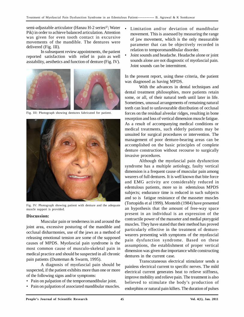
Fig. III: Photograph showing dentures fabricated for patient.
semi-adjustable articulator (Hanau H-2 series@; WaterPik) in order to achieve balanced articulation. Attentionwas given for even tooth contact in excursivemovements of the mandible. The dentures weredelivered (Fig. III).
In subsequent review appointments, the patientreported satisfaction with relief in pain as wellasstability, aesthetics and function of denture (Fig. IV).
Fig. IV: Photograph showing patient with denture and the adequatemuscle support is provided.
• Limitation and/or deviation of mandibularmovement. This is assessed by measuring the rangeof jaw movement, which is the only measurableparameter that can be objectively recorded inrelation to temporomandibular disorder.
• Joint sounds and headache. Headache alone or jointsounds alone are not diagnostic of myofascial pain.Joint sounds can be intermittent.
In the present report, using these criteria, the patientwas diagnosed as having MPDS.
With the advances in dental techniques anddental treatment philosophies, more patients retainsome, or all, of their natural teeth until later in life.Sometimes, unusual arrangements of remaining naturalteeth can lead to unfavourable distribution of occlusalforces on the residual alveolar ridges, resulting in boneresorption and loss of vertical dimension muscle fatigue.As a result of accompanying medical conditions ormedical treatments, such elderly patients may beunsuited for surgical procedures or intervention. Themanagement of poor denture-bearing areas can beaccomplished on the basic principles of completedenture construction without recourse to surgicallyinvasive procedures.
Although the myofascial pain dysfunctionsyndrome has a multiple aetiology, faulty verticaldimension is a frequent cause of muscular pain amongwearers of full dentures. It is well known that bite forceand EMG activity are considerably reduced inedentulous patients, more so in edentulous MPDSsubjects; endurance time is reduced in such subjectsand so is fatigue resistance of the masseter muscles(Tortopidis et al 1999). Monteith (1984) have presentedan hypothesis that the amount of free-way spacepresent in an individual is an expression of thecontractile power of the masseter and medial pterygoidmuscles. They have stated that their method has provedparticularly effective in the treatment of denture-wearers presenting with symptoms of the myofascialpain dysfunction syndrome. Based on theseassumptions, the establishment of proper verticaldimension was given due importance while constructingdentures in the current case.
Transcutaneous electrical stimulator sends apainless electrical current to specific nerves. The mildelectrical current generates heat to relieve stiffness,improve mobility and relieve pain. The treatment is alsobelieved to stimulate the body’s production ofendorphins or natural pain killers. The duration of pulses
Discussion:Muscular pain or tenderness in and around the
joint area, excessive posturing of the mandible andocclusal disharmonies, use of the jaws as a method ofreleasing emotional tension are some of the supposedcauses of MPDS. Myofascial pain syndrome is themost common cause of musculo-skeletal pain inmedical practice and should be suspected in all chronicpain patients (Dunteman & Swarm, 1995).
A diagnosis of myofascial pain should besuspected, if the patient exhibits more than one or moreof the following signs and/or symptoms:• Pain on palpation of the temporomandibular joint.• Pain on palpation of associated mandibular muscles.
Treatment of Myofascial Pain Dysfunction Syndrome in an Edentulous Patient--------------- B. Agrawal & K Somkuwar

46People’s Journal of Scientific Research Vol. 4(1), Jan. 2011
and frequencies can be revised and it is possible tostimulate different types of fibers by chosen stimulationtypes. It is possible to stimulate selectively , , and carrying touch and position sensation and it is possible
to block pain in medulla spinalis level, or to stimulate and C fibers carrying pain and it blocks the pain in
upper levels (Tarhan et al, 1999). In this case TENSproved to be appropriate choice of management.
Optimal function of the postural and facialexpression muscles requires a correct support fromthe natural teeth and the ridge areas or from theadequate designed prostheses. Good muscular controland co-ordination are essential for effective use ofcomplete denture (Jacob et al, 2004). In balancedocclusion, there is equilibrium on both sides of thedenture .The denture base is more stable during variousfunctional movements which will be less likely to abusethe foundation tissue which in turn reduces the boneresorption. This reduces the load transmitted totemporomandibular joints and masticatory apparatus.Balanced Occlusion is required for smooth uninterruptedtooth contact in the dynamics of daily mandibularmovements (Mohl & Drinnan, 2000) and was one ofthe objectives in the presented case.
Conclusion:It is essential that the correct diagnosis be
made before treating a case of MPDS. Merely treatingthe patient symptomatically does not provide long termresults, at the same time injecting trigger points andtender spots and hoping for the best does not providesatisfactory results. The patient should be counseledand trained well with jaw exercises as well asacceptance of the denture. The proper treatment ofMyofascial Pain Syndrome may be one of the mostrewarding if handled correctly.
Bibliography:
1. Al-Ani Z, Gray RJ, Davies SJ, Sloan P, Glenny AM:Stabilization splint therapy for the treatment oftemporomandibular myofascial pain: a systematic review.Journal of Dental Education, 2005;69(11):1242-1250.
2. Blasberg B, Greenberg MS: TemporomandibularDisorders. In: Burket’s Oral Medicine Diagnosis &Treatment. MS Greenberg, M Glick (Eds.); 10th Edn.; BCDecker Inc., Canada, 2003;pp271-306.
3. Carlsson GE:Symptoms of mandibular dysfunction incomplete denture wearers. Journal of Dentistry,1976;4(6):265-270.
4. Dommerholt J, Bron C, Franssen J: Myofascial triggerpoints. An evidence informed review. In: MyofiacialTrigger prints: Pathophysiology and Evidence-InformedDiagnosis & Management. J Domnerhalt, P. Huijibregth(Eds.); Jones & Bartletts Publisher, Canada 2009;pp 17-50.
5. Dunteman ED, Swarm RA: Atypical Facial “Neuralgia”.Anesthesia & Analgesia, 1995;80(1):188-190.
6. Edmiston GF, Laskin DM: Changes in Consistency ofOcclusal Contact in Myofascial Pain-Dysfunction (MPD)Syndrome. Journal of Dental Research, 1978;57(1): 27-30.
7. Gerwin R: Differential diagnosis of trigger points.Journal of Musculoskeletal pain, 2004;12(3 &4):23-28.
8. Jacob RF, Zarb GA, Bolender CL: Waxing and processingthe Dentures, their Insertion and Follow-up. In:Prosthodontic Treatment for Edentulous Patients;Complete Dentures and Implant-Supported Prosthess.G A Zarb, CL Bolender, S E Eckert, AH Fenton,R F Jacob, R Mericske-Stern (Eds.); 12th Edn.; Mosby:An Imprint of Elsevier, St. Louis, 2004;pp389-426.
9. Lous I: The importance of referred pain in myogenicheadache. Headache: The Journal of Head and FacePain, 1976;16(3):119-122.
10. Mohl ND, Drinnan AJ: Anatomy and physiology of theedentulous mouth. In: Essentials of Complete DenturesProsthodontics. S Winkler (Eds.); 2nd Edn.; A.I.T.B.SPublisher & Distributer, Delhi, 2000;pp1-14.
11. Monteith B: The role of the free-way space in thegeneration of muscle pain among denture-wearers.Journal of Oral Rehabilitation, 1984;11(5):483-498.
12.Tarhan C, Inan L, Karaoglan B, Yorgancioglu R:TENS: Treatment in Cervicogenic Headache.Physical Medicine, 1999; 2(2):13-17.
13.Tortopidis D, Lyon M F, Baxendale RH: Bite force,endurance and masseter muscle fatigue in healthyedentulous subjects and those with TMD. Journalof Oral Rehabilitation,1999;26(4):321-328.
Treatment of Myofascial Pain Dysfunction Syndrome in an Edentulous Patient--------------- B. Agrawal & K Somkuwar