dpysl2 is a novel regulator for neural stem cell
TRANSCRIPT

RESEARCH Open Access
DPYSL2 is a novel regulator for neural stemcell differentiation in rats: revealed byPanax notoginseng saponin administrationLiu-Lin Xiong1,2, De-Lu Qiu1, Guang-Hui Xiu1, Mohammed Al-Hawwas2, Ya Jiang3, You-Cui Wang1, Yue Hu1,Li Chen1, Qing-Jie Xia1 and Ting-Hua Wang1,3*
Abstract
Background: The limited neuronal differentiation of the endogenous or grafted neural stem cells (NSCs) after braininjury hampers the clinic usage of NSCs. Panax notoginseng saponins (PNS) were extensively used for their clinicalvalue, such as in controlling blood pressure, blood glucose, and inhibiting neuronal apoptosis and enhancingneuronal protection, but whether or not it exerts an effect in promoting neuronal differentiation of the endogenousNSCs is completely unclear and the potential underlying mechanism requires further exploration.
Methods: Firstly, we determined whether PNS could successfully induce NSCs to differentiate to neurons under theserum condition. Mass spectrometry and quantitative polymerase chain reaction (Q-PCR) were then performed toscreen the differentially expressed proteins (genes) between the PNS + serum and serum control group, upon whichdihydropyrimidinase-like 2 (DPYSL2), a possible candidate, was then selected for the subsequent research. To furtherinvestigate the actual role of DPYSL2 in the NSC differentiation, DPYSL2-expressing lentivirus was employed to obtainDPYSL2 overexpression in NSCs. DPYSL2-knockout rats were constructed to study its effects on hippocampal neuralstem cells. Immunofluorescent staining was performed to identify the differentiation direction of NSCs after 7 daysfrom DPYSL2 transfection, as well as those from DPYSL2-knockout rats.
Results: Seven differentially expressed protein spots were detected by PD Quest, and DPYSL2 was found asone of the key factors of NSC differentiation in a PNS-treated condition. The results of immunostaining furthershowed that mainly Tuj1 and GFAP-positive cells increased in the DPYSL2-overexpressed group, while bothwere depressed in the hippocampal NSCs in the DPYSL2-knockout rat.
Conclusions: The present study revealed that the differentiation direction of NSCs could be enhanced through PNSadministration, and the DPYSL2 is a key regulator in promoting NSC differentiation. These results not only emphasizedthe effect of PNS but also indicated DPYSL2 could be a novel target to enhance the NSC differentiation in futureclinical trials.
Keywords: Neural stem cells, DPYSL2, Panax notoginseng saponins, DPYSL2-knockout
© The Author(s). 2020 Open Access This article is licensed under a Creative Commons Attribution 4.0 International License,which permits use, sharing, adaptation, distribution and reproduction in any medium or format, as long as you giveappropriate credit to the original author(s) and the source, provide a link to the Creative Commons licence, and indicate ifchanges were made. The images or other third party material in this article are included in the article's Creative Commonslicence, unless indicated otherwise in a credit line to the material. If material is not included in the article's Creative Commonslicence and your intended use is not permitted by statutory regulation or exceeds the permitted use, you will need to obtainpermission directly from the copyright holder. To view a copy of this licence, visit http://creativecommons.org/licenses/by/4.0/.The Creative Commons Public Domain Dedication waiver (http://creativecommons.org/publicdomain/zero/1.0/) applies to thedata made available in this article, unless otherwise stated in a credit line to the data.
* Correspondence: [email protected] of Neurological Disease, Translational Neuroscience Center, WestChina Hospital, Sichuan University, Chengdu 610041, China3Institute of Neuroscience, Kunming Medical University, Kunming 650031,ChinaFull list of author information is available at the end of the article
Xiong et al. Stem Cell Research & Therapy (2020) 11:155 https://doi.org/10.1186/s13287-020-01652-4

IntroductionNeural stem cells (NSCs) existing in the subventricle zoneof the brain’s germinal region and the subgranular zone ofthe hippocampus are capable of self-renewing, proliferat-ing, migrating, and differentiating into various types ofcells within the brain and spinal cord tissue [13, 41]. NSCdifferentiation into neurons, astrocytes, and oligodendro-cytes could replace necrotic cells resulting from injuries topromote the structural and functional repair of the brainand spinal cord [50]. However, this self-renewal is not ad-equate for the recovery of neurological function after braininjury [1, 32, 47]. NSCs (autologous or grafted) tend to dif-ferentiate into gliocytes rather than the neurons which aremore valuable in the nervous system’s recovery [39, 51].The development of the NSC therapy is therefore re-stricted, meaning the mechanisms of the NSC differenti-ation into neurons need to be elucidated [15]. So far,many signals and genes were demonstrably involved inthe process of NSC differentiation, such as the bHLHgene, Notch signal, Wnt signal, MAPK signal, and evensome micro-RNAs [5, 12, 45, 46, 54]. However, the effect-ive regulation involving in Chinese medicine for the NSCdifferentiation is complex and demands exploration.Panax notoginseng saponins (PNS) are one of over 200
chemical ingredients isolated from Panax notoginseng(Burk) F.H Chen—the widely used Chinese herbal medi-cine [17, 29]. PNS are clinically used as the Xuesaitonginjection, the Xueshuantong injection, the Lulutong in-jection, the PNS tablet, and the Xuesaitong soft capsule[9, 40, 48]. PNS were demonstrated to have anti-inflammatory and anti-apoptotic effects contributing tothe regulation of nerve cell activity and the secretion ofnerve preservative agent [10, 49, 55]. In addition, recentstudies have reported its preventive and therapeutic ef-fects on the alleviation of neurological dysfunction [21,42, 52]. In Si et al.’s study, the administration of 100 μg/ml PNS in the rat’s embryonic cortical NSC medium hasdemonstrated an increased number of Tuj1, GFAP, andnestin-positive cells after 4 days of culturing. Hence,conclusions were made that PNS could improve the sur-vival status, self-renewal, proliferation, and differenti-ation of the rat’s embryonic cortical NSCs [37].However, little is known about the underlying relatedmolecular mechanism for the key factors that induce theNSCs to differentiate into neurons after PNS administra-tion. Therefore, studying this mechanism would havetremendous value to NSC-based therapy in central ner-vous system diseases.In this study, following the 100-μg/ml PNS treatment of
NSCs, the proliferation and differentiation of the NSCswere observed. To explore the related molecular mechan-ism, mass spectrometry and Q-PCR were employed to de-tect the differential protein and gene expression. Thenbioinformatics analysis was used to research the relatively
important factors, among which DPYSL2 was found tointeract with neuron markers Tubb3 (tubulin, beta 3 classIII) and Numb, thereby functioning in the process ofneurogenesis. Thereafter, overexpressed DPYSL2 in theNSCs revealed that DPYSL2 overexpression could inducethe NSCs to differentiate into neurons, which was indi-cated by the increased number of Tuj1-positive cells.Meanwhile, DPYSL2-knockout rats were used to furtherverify DPYSL2’s function in NSCs, and the process con-trarily demonstrated the diminished level of Tuj1-positivecells. This resulted in the conclusion that the exogenousapplication of DPYSL2 in NSCs could mediate the NSCdifferentiation into neurons, which could provide novel in-sights into NSC-based therapy in the clinical treatment ofneurological disorders.
Material and methodsIsolation of NSCsThe NSCs were isolated from rat hippocampal tissues.Animal experimental protocols were approved by theguidelines of the Institutional Medical Experimental Ani-mal Care Committee of Sichuan University, West ChinaHospital, China. After being disinfected with 75% ethanolfor 2 min and anesthetized by isoflurane inhalation, theneonatal rats (1 day) were sacrificed, and the heads wereimmersed into D-Hanks Solution (Biohao). The hippo-campal tissues of neonatal rats were harvested and werecut into several pieces (about 1 mm3) then suspended with5ml DMEM medium by pipetting. Neural-basal medium(DMEM/F-12, 1:1, Hyclone) was used to maintain the iso-lated cells. The medium was supplemented with 1% N2
(Gibco), 20 μg/L basic fibroblast growth factor (bFGF)(Invitrogen), 2 mmol/L glutamine (Invitrogen), 10,000U/Lpenicillin (Hyclone), and 10mg/L streptomycin (Hyclone).The cells were seeded onto the cell culture flasks, incu-bated with 5% CO2 at 37 °C. The culture medium was halfchanged every other day. The primary NSCs were main-tained for 7 days for the following immunofluorescentstaining and mass spectrum.
Drug administration of the P2 NSCsP2 NSCs were divided into four groups: normal (Nor),serum (Ser), PNS, and PNS + serum (P + S) groups. Tenpercent fetal calf serum (Millipore) was added into theNSCs in the Ser group, 100 μg/ml PNS (Guangxi WuzhouPharmaceutical Group, Z20025652) into the PNS group,and both serum and PNS into the P + S group. Mean-while, NSCs in the Nor group were received an equalquantity of the neural-basal medium. After 7 days, im-munofluorescent staining and mass spectrometry wereperformed to detect the differentiation index and proteinexpression.
Xiong et al. Stem Cell Research & Therapy (2020) 11:155 Page 2 of 16

Immunofluorescent stainingCells preparationThe cells were collected by a scraper and suspended witha phosphate-buffered solution (PBS). The cell suspensionwas applied onto the glass slide treated by polylysine (40g/L) (Sigma). Smears of NSCs were fixed with 4% parafor-maldehyde (Sigma) for 20min at 4 °C, followed bywashing-out with PBS (0.01mol/L) (Sigma) for threetimes (5min each).
Immunofluorescent stainingBriefly, after washing, the cells were incubated with 0.3%TritonX-100 (Hengdailao) at 37 °C for 30 min. Then, 5%goat serum (Millipore) was added to block the nonspe-cific binding site and incubated at 37 °C for 30 min. Sub-sequently, the primary antibodies were added andincubated in the wet box at 4 °C for 20 h. The same vol-ume of 2% goat serum (Invitrogen) was used as thenegative control. The secondary antibody was added andincubated at 37 °C for 1 h. All information of the primaryand secondary antibodies is shown in Table 1. Then, thesections/cells were washed 3 times with 0.01M PBSagain. Afterwards, DAPI was used to stain the nucleus.Inversed fluorescent microscope (Leica, Wetzlar,Germany) was used to observe the outcomes andcapture photos. All the photos were taken in the sameexposure intensity with the same setting of the LeicaMicrosystem. In each group (5 samples), 5 views of eachsample were taken randomly. Then, cell number, area,and the length of the processes were calculated by usingImage-Pro plus 6.0 software (MediaCybernetics, SilverSpring, MD, USA). The final results were shown as theaverage from three observers blinded to the experimen-tal condition.
Second dimension gel electrophoresis (2Delectrophoresis) and matrix-assisted laser desorption/ionization-TOF/TOF mass spectrometry (MALDI-TOF/TOFMS)As the differentiation of NSCs only showed significancebetween the PNS and P + S group, so we just did 2D elec-trophoresis in these two groups. Cells were lysed with 1ml lysis buffer for 10min before 20 μg/ml DNase, 50 μg/ml RNase, and MnCl2 (metered volume with DNase).Then, after 15min at 4 °C, samples were centrifuged at 15,000 r/min for 30min followed by supernatants collection
to new tubes. The protein concentration was determinedby the Bradford Coomassie blue colorimetric assay. There-after, 0.02 g/ml DTT and 0.0025 μl/ml Bio-lyte were addedinto 1.2mg sample solution (volume, 580 μl) and weremixed uniformly and incubated for 1 h at the roomtemperature prior to centrifugation at 25,000 r/min for 10min to separate the protein samples. The supernatant wastransferred into the horizontal plate and one-dimensionalisoelectric focusing (IEF) was performed using IPG strips(17 cm, pH 3–10) with 50 V at low speed for half an hour,250 V at low speed for 1 h, 500 V at rapid speed for 1.5 h,1000 V at rapid speed for 1 h, 4000 V for 3 h, 9000 V for 3h, 9000 V at rapid speed for 50,000 V/h, and 500 V withrapid speed for 30min.After equilibration for 15 min, SDS-PAGE was per-
formed to separate the proteins further. The initial volt-age was 70 V and then changed to 300 V until thebromophenol blue (BPB) crossed the band and thenstopped until the BPB reached the 5 cm from the glass.Finally, the stripe was stained by Coomassie brilliantblue (CBB) G-250. Gel images were scanned fordigitization (American Bio-Rad), and the differential pro-teins were identified by the PD Quest analysis software(Bio-Rad PD Quest Advanced 7.4). “Significant changes”was defined as “more than 1.5-fold changes in expres-sion compared to the serum group,” and only thosespots were gown through matrix-assisted laser desorp-tion/ionization-TOF/TOF mass spectrometry (MALDI-TOF/TOFMS) analysis to identify the protein types.
Quantitative polymerase chain reaction (Q-PCR)Cells were collected from each group and the total RNAwas extracted by using TRIzol reagent (Invitrogen) ac-cording to the manufacturer’s instruction (superfecTRI),followed by reverse transcription to complementaryDNA (cDNA) (TakaRa). Q-PCR was then performed todetermine the gene expression of the differential pro-teins which were identified by 2D electrophoresis andMALDI-TOF/TOFMS. Five genes for 6 proteins werestudied in addition to β-actin that was used as an in-ternal control; the used primers and probes were listedin Table 2. PCR was performed in a DNA thermal cycler(ABI 7300) according to the following standard protocol:90 °C for 5 min, denaturation at 94 °C for 30 s, annealingfor 30 s, and extension at 72 °C for 1 min (total 30 cy-cles). The relative expression levels of mRNA were
Table 1 The information of the antibodies
Primary antibody Species Dilution Company Secondary antibody Species Dilution Company
Nestin Rabbit 1:100 ZSGB-BIO Alexa-594 Anti-rabbit 1:200 ZSGB-BIO
Tuj1 Mouse 1:200 Millipore Alexa-488 Anti-mouse 1:100 ZSGB-BIO
GFAP Rabbit 1:200 Millipore Alexa-594 Anti-rabbit 1:200 ZSGB-BIO
DPYSL2 Rabbit 1:100 Abcam Alexa-594 Anti-rabbit 1:200 ZSGB-BIO
Xiong et al. Stem Cell Research & Therapy (2020) 11:155 Page 3 of 16

calculated as standardization to β-actin by using the2–△△Ct method. Each group had 6 samples.
Lentivirus transductionORF-vector verification by enzyme digest andelectrophoresis and recombinant lentivirus productionGene sequence of DPYSL2/CRMP2 was acquired fromNCBI, and then PCR and electrophoresis were performedto amplify and purify the DPYSL2 gene from rats’ brain.The target DNA fragment was sent to GeneCopoeia Com-pany to construct overexpression recombinant vector(ORF-vector).The lentiviral particles were generated by the standard-
ized protocol using highly purified plasmids andEndoFectin-Lenti™ and TiterBoost™ reagents. Then, lenti-viral stocks were stored at − 80 °C to maintain the activity.
NSC transfection with lentivirusIn this experiment, the cultured NSCs were divided intothe following three groups: Nor, eGFP, and DPYSL2groups. In brief, in the DPYSL2 group, the P2 NSCs weretransfected with the lentivirus and were transfected to upto 20 MOI with 10% polybrene in the 96-well plate; theeGFP group was treated with the same titer of the simpleeGFP-lentivirus and the Nor group was treated with equalquantity of the Neural-basal medium. After 12 h incuba-tion at 37 °C, the neural-basal medium was changed witha fresh one, and then 3- and 7-day photos were taken withthe Inversed Fluorescent Microscope (Leica, Wetzlar,Germany). In addition, the positive cell number in each
group was calculated by using Image-Pro plus 6.0 software(MediaCybernetics, Silver Spring, MD, USA). Five viewsof five samples were randomly considered for each group.At last, the average from three observers blind to the ex-perimental condition was determined as the final results.
Construction of a DPYSL2-knockout ratCRISPR/CAS-mediated genome engineering was appliedto construct DPYSL2-KO rats which were produced andprovided by Cyagen (Guangzhou, China). Two targetswere designed for DPYSL2, and single gRNA was pre-pared by synthesizing two pairs of oligonucleotide chains(CAACGAGTCCTTACGGACAA and CCCTGTACCTGTTAAACGTG). Then, more KO rats were reproducedby mating and were detected by genotyping.
Genotype identificationTail tips were collected and numbered for neonatal rats at7–10 days. Then, Transgen’s genomic DNA extraction kit(ee101-12) was used to extract rats’ genomic DNA and werefurther detected by PCR with the amplification primer:Rat DPYSL2 forward: 5′-ACTGAGCAGGTTCAGTC
CGTGC-3′.Rat DPYSL2 reverse: 5′-ACTTGTGGTGGAAGCTCT
GACTCCC-3′.The PCR amplification reaction system was conducted
with 10 μl PCR master mix: 0.6 μl upstream primers, 0.6 μldownstream, 3 μl DNA template, and 5.8 μl water. Thethermal cycling conditions were performed as initial de-naturation at 94 °C for 5 min; 35 cycles of denaturation at94 °C for 30 s; annealing at 60 °C for 30s, with 1min elong-ation at 72 °C; and followed by final elongation at 72 °Cfor 10min. Subsequently, the agarose gel electrophoresissystem was applied to visualize the final genotype detec-tion under U.V after 55min electrophoresis at 150 V.
Primary hippocampal stem cells culturesThe hippocampal tissues of neonatal rats were harvested fol-lowing the successful construction of DPYSL2-KO rats, andthen they were processed into cellular suspension. After-wards, neural-basal medium (DMEM/F-12, 1:1, Hyclone)was supplemented with 1% N2 (Gibco), 20 μg/l basic fibro-blast growth factor (bFGF) (Invitrogen), 2mmol/l glutamine(Invitrogen), 10,000U/l penicillin (Hyclone), and 10mg/lstreptomycin (Hyclone) and then seeded onto the cell cultureflasks and incubated with 5% CO2 at 37 °C every other day.The primary hippocampal stem cells were kept for immuno-fluorescent staining on the 7th culturing day.
Statistical analysisAll the data were analyzed by using one-way analysis ofvariance (ANOVA) with SPSS 19.0 and presented as themeans ± SD. P < 0.05 was considered statistically signifi-cant, *P < 0.05, **P < 0.01, and ***P < 0.001.
Table 2 The information of the PCR primers and probes
Genes Primer/probe Sequence
CRMP1/FSCN1 Forward GGAGATTTGATAGCTCAGGA
Reverse GACCTTGGTGATCACAG
Probe CCTGGAGATGGGCATCAC
DPYSL2 Forward GAAGGGAACTGTGGTGTAT
Reverse GAGGTCTCCACAGGACAG
Probe CCAACCACTCCAGACTTTCTC
GRP78 Forward GAGTTCTTCAATGGCAAGGAG
Reverse CCTCCCACAGTTTCAATACCA
Probe CTGTCCAGGCTGGTGTCCTC
HSP90/TRA Forward CAAATGCTTCTGATGCTTTAG
Reverse CTCCTCTCTGGTCATTCCTACA
Probe CCTGCTGCATGTCACAGACAC
LDHB Forward CCTCAGATCGTCAAGTACAG
Reverse GCAGCTGCTGGGATGAAT
Probe CCACTGGGTTGGAAACCAC
β-actin Forward GAAGATCAAGATCATTGCTCCT
Reverse TACTCCTGCTTGCTGATCCA
Probe CTGTCCACCTTCCAGCAGA
Xiong et al. Stem Cell Research & Therapy (2020) 11:155 Page 4 of 16
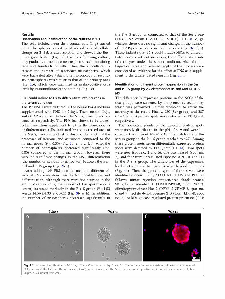
ResultsObservation and identification of the cultured NSCsThe cells isolated from the neonatal rats (1 p) turnedout to be spheres consisting of several tens of cellularclumps on 2–3 days after isolation and showed the fluc-tuant growth state (Fig. 1a). Five days following culture,they gradually turned into neurospheres, each containingtens and hundreds of cells. Then the subculture in-creases the number of secondary neurospheres whichwere harvested after 7 days. The morphology of second-ary neurospheres was similar to that of the primary ones(Fig. 1b), which were identified as nestin-positive cells(red) by immunofluorescence staining (Fig. 1c).
PNS could induce NSCs to differentiate into neurons inthe serum conditionThe P2 NSCs were cultured in the neural basal mediumsupplemented with PNS for 7 days. Then, nestin, Tuj1,and GFAP were used to label the NSCs, neuron, and as-trocytes, respectively. The PNS has shown to be an ex-cellent nutrition supplement to either the neurospheresor differentiated cells, indicated by the increased area ofthe NSCs, neurons, and astrocytes and the length of theprocesses of neurons and astrocytes compared to thenormal group (P < 0.05) (Fig. 2b, e, h, c, f, i). Also, thenumber of neurospheres decreased significantly (P <0.05) compared to the normal group. However, therewere no significant changes in the NSC differentiation(the number of neurons or astrocytes) between the nor-mal and PNS group (Fig. 2h, i).After adding 10% FBS into the medium, different ef-
fects of PNS were shown on the NSC proliferation anddifferentiation. Although there were few neurons in thegroup of serum alone, the number of Tuj1-positive cells(green) increased markedly in the P + S group (9 ± 1.53versus 14.56 ± 4.30, P < 0.05) (Fig. 3b, e, h). In addition,the number of neurospheres decreased significantly in
the P + S group, as compared to that of the Ser group(1.63 ± 0.92 versus 0.38 ± 0.12, P < 0.05) (Fig. 3a, d, g),whereas there were no significant changes in the numberof GFAP-positive cells in both groups (Fig. 3c, f, i).These indicate that PNS could induce NSCs to differen-tiate neurons without increasing the differentiation rateof astrocytes under the serum condition. Also, the en-larged cell area and reduced length of the process wereconsidered as evidence for the effect of PNS as a supple-ment to the differentiated neurons (Fig. 3h, i).
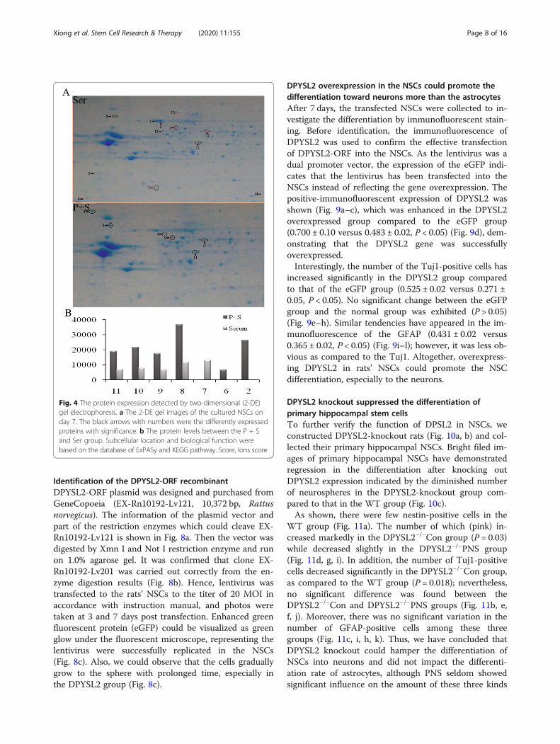
Identification of different protein expression in the Serand P + S group by 2D electrophoresis and MALDI-TOF/MSThe differentially expressed proteins in the NSCs of thetwo groups were screened by the proteomic technologywhich was performed 3 times repeatedly to affirm theaccuracy of the result. Finally, 230 (Ser group) and 287(P + S group) protein spots were detected by PD Quest,respectively.The isoelectric points of the detected protein spots
were mostly distributed in the pH of 4–9 and were lo-cated in the range of 10–90 kDa. The match rate of theserum group to the P + S group reached to 42%. Amongthese protein spots, seven differentially expressed proteinspots were detected by PD Quest (Fig. 4a). Two spotswere new (spot no. 2 and 6), one was missed (spot no.7), and four were unregulated (spot no. 8, 9, 10, and 11)in the P + S group. The differences of the expressionlevels between the two groups were beyond 1.5 times(Fig. 4b). Then the protein types of these seven wereidentified successfully by MALDI-TOF/MS and PMF asfollows: tumor rejection antigen/heat shock protein90 kDa β, member 1 (TRA/HSP90-B, Spot NO.2),dihydropyrimidinase-like 2 (DPYSL2/CRMP-2, spot no.6 and 9), lactate dehydrogenase 2 B chain (LDH-B, spotno. 7), 78 kDa glucose-regulated protein precursor (GRP
Fig. 1 Culture and identification of NSCs. a, b The NSCs culture on days 3 and 7. c The immunofluorescent staining of nestin in the culturedNSCs on day 7. DAPI stained the cell nucleus (blue) and nestin stained the NSCs, which emitted positive red immunofluorescence. Scale bar,50 μm. NSCs, neural stem cells
Xiong et al. Stem Cell Research & Therapy (2020) 11:155 Page 5 of 16

78, spot no. 8), collapsin response mediator protein1(CRMP-1, spot no. 10), and Fscn1 protein (Fscn1, spotno. 11).
DPYSL2 was identified as the key protein in the P + SgroupTwo of these seven protein spots were identified asDPYSL2 (no. 6 and 9). One was new and the other wasunregulated in the P + S group compared to the Sergroup. The PMF of these 2 spots was shown (Fig. 5a, c),and the probability-based Mowse scores were 377 and 681(− 10 × log (P); P, the probability of the observed match isa random event), respectively (Fig. 5b, d). Spot no. 9 ex-hibited higher scores relative to spot no. 6 (Fig. 5c, d). The
coverage of the amino acid sequence in the DPYSL2 was25% for spot no. 6 and 47% for spot no. 9.
The changes of mRNA expression levels of DPYSL2 andCRMP1 in different experimental groupsIn order to validate the result of proteomics, Q-PCR wasperformed to detect the genetic expression of these 6proteins (DPYSL2 has 2 protein spots) in the fourgroups (normal, serum alone, PNS alone, and PNS +serum). The expression of DPYSL2 and CRMP1 alignedwith the protein data (Fig. 6a). Delightedly, the cali-brated CT value of DPYSL2 in the normal group was de-creased to 0.08, in the serum group, and significantlyincreasing in the PNS and P + S groups. In addition,there was a significant increase between the serum and
Fig. 2 The proliferation and differentiation of the cultured NSCs in the normal and PNS groups. a–d, b–e, c–f The immunofluorescent staining ofnestin (red immunofluorescence), Tuj1 (green immunofluorescence), and GFAP (red immunofluorescence) in the normal and PNS group, respectively.DAPI was used to stain the cell nucleus (blue). Scale bar, 25 μm. g–i The representative bar graphs of proliferation and differentiation of NSCs or itsdifferentiated states in the normal and PNS groups. Data were presented as means ± SD. Each group contained 5 samples. *P < 0.05 versus Nor group.Nor, normal group; PNS, Panax notoginseng saponins group; Num, the number of the cells/neuronspheres; S, cell area. S/1000, S/10represents the areavalue divided 1000 and 10, respectively; D, the length of the synapse
Xiong et al. Stem Cell Research & Therapy (2020) 11:155 Page 6 of 16

P + S group (0.056 versus 0.096, P < 0.05). Although theCRMP1 gene expression changed consistently with itsprotein level in the P + S and serum group mostly, itsexpression in each group was low. The expression levelsof the other genes under the study are shown in Fig. 6b.To sum up, we speculated the possible important role ofDPYSL2 in the NSC differentiation.
The possible role of DPYSL2 from bioinformaticspredictionThe known interrelation of the deferential proteins wasbuilt by using GeneMANIA (http://www.genemania.org/)(Fig. 7). Delightedly, we have found that DPYSL2 wasencoded to a family of collapsin response mediator protein
(CRMP) which was associated with microtubule assemblyand synaptic growth [25]. Moreover, DPYSL2 has revealedphysical interaction with Tubb3 (tubulin, beta 3 class III)and Numb. The former is a neuron marker while the lat-ter plays a role in the process of neurogenesis [35, 44].Furthermore, numerous studies found that DPYSL2 playsan important role in the neuronal differentiation and po-larity to further enhance axon outgrowth and guidance[18, 23, 24].Altogether, DPYSL2 expression was enhanced in the P
+ S group compared to that in the serum group at bothgene and protein levels. To sum up, DPYSL2 was se-lected as our target gene for NSC differentiation intoneuron.
Fig. 3 The proliferation and differentiation of the cultured NSCs. a–d, b–e, c–f The immunofluorescent staining of nestin (red immunofluorescence),Tuj1 (green immunofluorescence), and GFAP (red immunofluorescence) in the serum and PNS + serum group. DAPI was used to stain the cell nucleus(blue). Scale bar, 25 μm. g–i The representative bar graphs of the proliferation and differentiation of NSCs or its differentiated states in the serum andPNS + serum groups. Data were presented as means ± SD. Each group contained 5 samples. *P < 0.05 versus Ser group. Ser, Serum group; P + S, PNS+ serum group; Num, the number of the cells/neuronspheres; S, cell area; D, the length of the synapse. S/1000, S/10 represent the area value divided1000 and 10, respectively
Xiong et al. Stem Cell Research & Therapy (2020) 11:155 Page 7 of 16
Identification of the DPYSL2-ORF recombinantDPYSL2-ORF plasmid was designed and purchased fromGeneCopoeia (EX-Rn10192-Lv121, 10,372 bp, Rattusnorvegicus). The information of the plasmid vector andpart of the restriction enzymes which could cleave EX-Rn10192-Lv121 is shown in Fig. 8a. Then the vector wasdigested by Xmn I and Not I restriction enzyme and runon 1.0% agarose gel. It was confirmed that clone EX-Rn10192-Lv201 was carried out correctly from the en-zyme digestion results (Fig. 8b). Hence, lentivirus wastransfected to the rats’ NSCs to the titer of 20 MOI inaccordance with instruction manual, and photos weretaken at 3 and 7 days post transfection. Enhanced greenfluorescent protein (eGFP) could be visualized as greenglow under the fluorescent microscope, representing thelentivirus were successfully replicated in the NSCs(Fig. 8c). Also, we could observe that the cells graduallygrow to the sphere with prolonged time, especially inthe DPYSL2 group (Fig. 8c).
DPYSL2 overexpression in the NSCs could promote thedifferentiation toward neurons more than the astrocytesAfter 7 days, the transfected NSCs were collected to in-vestigate the differentiation by immunofluorescent stain-ing. Before identification, the immunofluorescence ofDPYSL2 was used to confirm the effective transfectionof DPYSL2-ORF into the NSCs. As the lentivirus was adual promoter vector, the expression of the eGFP indi-cates that the lentivirus has been transfected into theNSCs instead of reflecting the gene overexpression. Thepositive-immunofluorescent expression of DPYSL2 wasshown (Fig. 9a–c), which was enhanced in the DPYSL2overexpressed group compared to the eGFP group(0.700 ± 0.10 versus 0.483 ± 0.02, P < 0.05) (Fig. 9d), dem-onstrating that the DPYSL2 gene was successfullyoverexpressed.Interestingly, the number of the Tuj1-positive cells has
increased significantly in the DPYSL2 group comparedto that of the eGFP group (0.525 ± 0.02 versus 0.271 ±0.05, P < 0.05). No significant change between the eGFPgroup and the normal group was exhibited (P > 0.05)(Fig. 9e–h). Similar tendencies have appeared in the im-munofluorescence of the GFAP (0.431 ± 0.02 versus0.365 ± 0.02, P < 0.05) (Fig. 9i–l); however, it was less ob-vious as compared to the Tuj1. Altogether, overexpress-ing DPYSL2 in rats’ NSCs could promote the NSCdifferentiation, especially to the neurons.
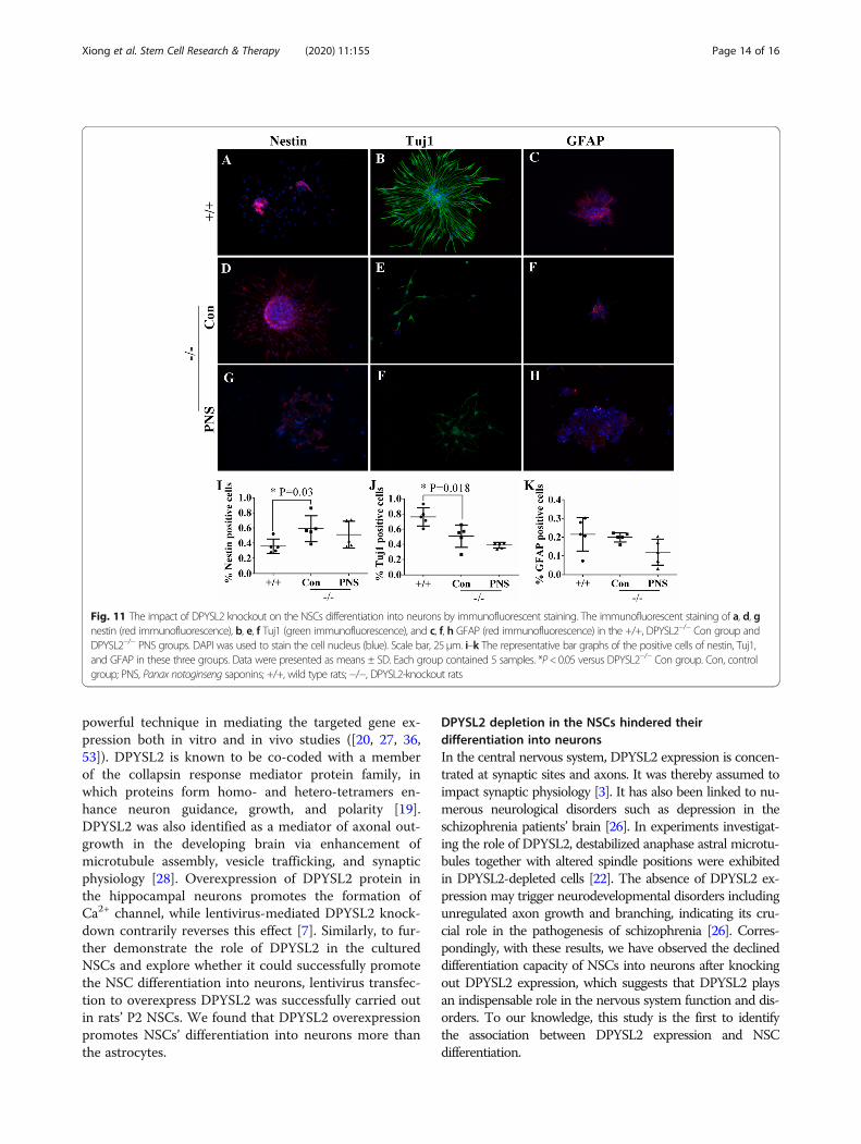
DPYSL2 knockout suppressed the differentiation ofprimary hippocampal stem cellsTo further verify the function of DPSL2 in NSCs, weconstructed DPYSL2-knockout rats (Fig. 10a, b) and col-lected their primary hippocampal NSCs. Bright filed im-ages of primary hippocampal NSCs have demonstratedregression in the differentiation after knocking outDPYSL2 expression indicated by the diminished numberof neurospheres in the DPYSL2-knockout group com-pared to that in the WT group (Fig. 10c).As shown, there were few nestin-positive cells in the
WT group (Fig. 11a). The number of which (pink) in-creased markedly in the DPYSL2−/−Con group (P = 0.03)while decreased slightly in the DPYSL2−/−PNS group(Fig. 11d, g, i). In addition, the number of Tuj1-positivecells decreased significantly in the DPYSL2−/−Con group,as compared to the WT group (P = 0.018); nevertheless,no significant difference was found between theDPYSL2−/−Con and DPYSL2−/−PNS groups (Fig. 11b, e,f, j). Moreover, there was no significant variation in thenumber of GFAP-positive cells among these threegroups (Fig. 11c, i, h, k). Thus, we have concluded thatDPYSL2 knockout could hamper the differentiation ofNSCs into neurons and did not impact the differenti-ation rate of astrocytes, although PNS seldom showedsignificant influence on the amount of these three kinds
Fig. 4 The protein expression detected by two-dimensional (2-DE)gel electrophoresis. a The 2-DE gel images of the cultured NSCs onday 7. The black arrows with numbers were the differently expressedproteins with significance. b The protein levels between the P + Sand Ser group. Subcellular location and biological function werebased on the database of ExPASy and KEGG pathway. Score, Ions score
Xiong et al. Stem Cell Research & Therapy (2020) 11:155 Page 8 of 16

Fig. 5 The information of the DPYSL2/CRMP2 in the two-dimensional (2-DE) gelelectrophoresis. a–c The PMF of the DPYSL2. b–d The Ions scoreof protein spot of DPYSL2 by MASCOT software
Xiong et al. Stem Cell Research & Therapy (2020) 11:155 Page 9 of 16

of cells but exhibited nutritious efficacy to the differenti-ated neurons, revealed by cell area and the reducedlength of the process following of PNS administration(Fig. 11g, f, h).
DiscussionThree main points have emerged from this study. First,PNS induces NSC differentiation into neurons under theserum condition in vitro. The underlying molecularmechanism for DPYSL2 upregulation was indicated byproteomic analysis. Second, overexpressing DPYSL2 inthe rats’ NSCs induces differentiation into the neurons
more than the astrocytes. Third, knocking out DPYSL2hindered the differentiation capability of NSCs into neu-rons. Altogether, these results may provide highly valu-able for NSC-based treatment in nervous systemdiseases.
DPYSL2 was selected as a main mediator for the NSCdifferentiation into neurons under PNS conditionOur results from this study show that PNS enhancesNSC differentiation into neurons in the serum conditionand promotes the growth of the differentiated neurons.For some nervous system diseases or brain injuries, the
Fig. 6 The gene expression level (standardized CT) of the identified proteins in the four groups by Q-PCR. a The results of DPYSL2 and CRMP1 byQ-PCR in the four groups. b The results of other detected genes by Q-PCR in the four groups. Data were presented as means ± SD. *P < 0.05.Each group contained 6 samples
Fig. 7 Correlation analysis of the factors detected by GeneMANIA. Through searching for http://www.genemania.org/, we found that DPYSL2 wasencoded to a family of collapsin response mediator protein (CRMP). Moreover, it has some physical interaction with Tubb3 (tubulin, beta 3 classIII) and Numb
Xiong et al. Stem Cell Research & Therapy (2020) 11:155 Page 10 of 16
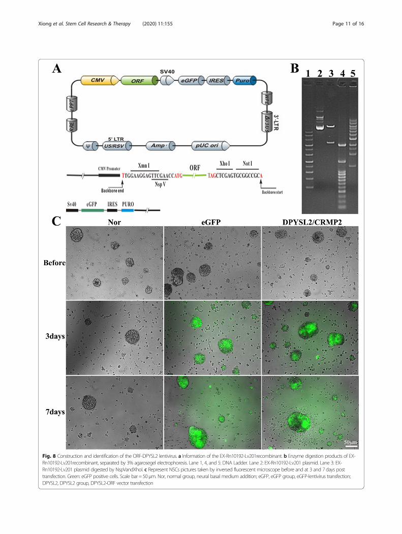
Fig. 8 Construction and identification of the ORF-DPYSL2 lentivirus. a Information of the EX-Rn10192-Lv201recombinant. b Enzyme digestion products of EX-Rn10192-Lv201recombinant, separated by 3% agarosegel electrophoresis. Lane 1, 4, and 5: DNA Ladder. Lane 2: EX-Rn10192-Lv201 plasmid. Lane 3: EX-Rn10192-Lv201 plasmid digested by NspVandXhoI. c Represent NSCs pictures taken by inversed fluorescent microscope before and at 3 and 7 days posttransfection. Green: eGFP positive cells. Scale bar = 50 μm. Nor, normal group, neural basal medium addition; eGFP, eGFP group, eGFP-lentivirus transfection;DPYSL2, DPYSL2 group, DPYSL2-ORF vector transfection
Xiong et al. Stem Cell Research & Therapy (2020) 11:155 Page 11 of 16

neurons’ restoration may play the key role in both func-tional recovery and the prognosis [39, 51]. As a result ofthe limited capacity of self-compensation in the body,exogenous NSC transplantation was used to treat somenervous system diseases. This approach had positive ef-fects in some cases, as previously reported [14, 31]. Todate, however, the limited differentiation of NSCs intoneurons has restricted the treatment by NSC transplant-ation, meaning a mechanism to induce NSC conversionto neurons is in demand. PNS has been used extensivelyin the neurology departments of Chinese hospitals toimprove blood circulation. Some previous researchshowed that PNS administration could upregulate theexpression of neurotrophic factors [11], thereby enhan-cing cerebral neuronal protection after ischemia [33].Moreover, in the Alzheimer’s disease model, PNS exhib-ited an ability to reduce reactive oxygen species andsuppress stress-activated MAPK signaling pathways, ul-timately inhibiting neuronal apoptosis [30]. Mounting
evidence revealed the effect of PNS to increase Tuj1 andGFAP expression in a rat’s hippocampal NSC [37, 38].However, the related molecular mechanism for PNS’ ef-fect on neurons still requires further clarification.With the goal of exploring the possible molecular mecha-
nisms, 2D electrophoresis and Q-PCR were employed in thePNS and PNS + serum group. DPYSL2 stood out from theidentified differentially expressed proteins in these twogroups. Moreover, DPYSL2/CRMP2, a microtubule-bindingphosphoprotein involved in schizophrenia pathogenesis, wasidentified as a key factor involved in the NSC differentiationinto neurons, promoted by PNS administration. CRMP2 is acrucial regulator of cytoskeletal dynamics facilitating axonaloutgrowth and neuronal connectivity [18]. Negative alter-ations of DPYSL2/CRMP-2 were found to exert prominenteffects on neuronal homeostasis and phenotype and may bepartially responsible for the observed neurodegenerationupon chaperone-mediated autophagy impairment in vivo [6].DPYSL2 was previously reported as being a neurite-guiding
Fig. 9 The detection of NSCs differentiation toward neurons and astrocytes after DPYSL2-ORF transfection. a–c, e–g, i–k The immunofluorescentstaining of DPYSL2 (red immunofluorescence), Tuj1 (green immunofluorescence), and GFAP (red immunofluorescence), respectively, in the threegroups (Nor, eGFP, DPYSL2) at 7 days after transfection, DAPI stained the cell nucleus (blue). d, h, l The representative bar graphs for the ratio ofthe positive cells in the three groups. Data were presented as means ± SD. *P < 0.05. Scale bar, 25 μm. Each group contained 5 samples
Xiong et al. Stem Cell Research & Therapy (2020) 11:155 Page 12 of 16

factor, with overexpressing DPYSL2 promoting dendriticgrowth [4]. The hyperphosphorylation of DPYSL2 may in-duce neuronal apoptosis in the development of Alzheimer’sdisease [2, 8]. Additionally, Akama revealed in 2011 thatDPYSLY2 was upregulated in the process of NSCs differ-entiating into neurons by proteomic identification in thecynomolgus monkey [2]. Later, it was emphasized thatpreconditioning with Ginkgo biloba (EGb 761(R)) pro-moted neuroprotection through HO1 and CRMP2 in ratswith brain ischemia [34]. Moreover, DPYSL2 was presentas an early neural differentiation regulator mediated bybone morphogenetic protein 4 (BMP-4), of which the
activation could suppress the expression of DPYSL2 [16].In addition, the high level of BMP-4 could induce theNSCs to remain in a quiescent state [43]. However, someinvestigations highlighted the exact role of DPYSL2 in thedifferentiation of NSCs, which is discussed in the currentstudy.
DPYSL2 overexpression in the NSCs promoted theirdifferentiation into neurons more than the astrocytesRecently, due to the stability, low immunogenicity, andhigh safety profile following infection, lentivirus-transfected gene therapy has become a reliable and
Fig. 10 The impact of DPYSL2 knockout on the NSC differentiation into neurons. a–b DPYSL2-knockout rats plasmid construction and DPYSL2expression identification. c Images of cultured hippocampal NSCs from WT (+/+) and KO (−/−) groups at 3 and 7 day as well as differentiationstatus. +/+, wild type rats; −/−, DPYSL2-knockout rats
Xiong et al. Stem Cell Research & Therapy (2020) 11:155 Page 13 of 16
powerful technique in mediating the targeted gene ex-pression both in vitro and in vivo studies ([20, 27, 36,53]). DPYSL2 is known to be co-coded with a memberof the collapsin response mediator protein family, inwhich proteins form homo- and hetero-tetramers en-hance neuron guidance, growth, and polarity [19].DPYSL2 was also identified as a mediator of axonal out-growth in the developing brain via enhancement ofmicrotubule assembly, vesicle trafficking, and synapticphysiology [28]. Overexpression of DPYSL2 protein inthe hippocampal neurons promotes the formation ofCa2+ channel, while lentivirus-mediated DPYSL2 knock-down contrarily reverses this effect [7]. Similarly, to fur-ther demonstrate the role of DPYSL2 in the culturedNSCs and explore whether it could successfully promotethe NSC differentiation into neurons, lentivirus transfec-tion to overexpress DPYSL2 was successfully carried outin rats’ P2 NSCs. We found that DPYSL2 overexpressionpromotes NSCs’ differentiation into neurons more thanthe astrocytes.
DPYSL2 depletion in the NSCs hindered theirdifferentiation into neuronsIn the central nervous system, DPYSL2 expression is concen-trated at synaptic sites and axons. It was thereby assumed toimpact synaptic physiology [3]. It has also been linked to nu-merous neurological disorders such as depression in theschizophrenia patients’ brain [26]. In experiments investigat-ing the role of DPYSL2, destabilized anaphase astral microtu-bules together with altered spindle positions were exhibitedin DPYSL2-depleted cells [22]. The absence of DPYSL2 ex-pression may trigger neurodevelopmental disorders includingunregulated axon growth and branching, indicating its cru-cial role in the pathogenesis of schizophrenia [26]. Corres-pondingly, with these results, we have observed the declineddifferentiation capacity of NSCs into neurons after knockingout DPYSL2 expression, which suggests that DPYSL2 playsan indispensable role in the nervous system function and dis-orders. To our knowledge, this study is the first to identifythe association between DPYSL2 expression and NSCdifferentiation.
Fig. 11 The impact of DPYSL2 knockout on the NSCs differentiation into neurons by immunofluorescent staining. The immunofluorescent staining of a, d, gnestin (red immunofluorescence), b, e, f Tuj1 (green immunofluorescence), and c, f, h GFAP (red immunofluorescence) in the +/+, DPYSL2−/− Con group andDPYSL2−/− PNS groups. DAPI was used to stain the cell nucleus (blue). Scale bar, 25μm. i–k The representative bar graphs of the positive cells of nestin, Tuj1,and GFAP in these three groups. Data were presented as means ± SD. Each group contained 5 samples. *P<0.05 versus DPYSL2−/− Con group. Con, controlgroup; PNS, Panax notoginseng saponins; +/+, wild type rats; −/−, DPYSL2-knockout rats
Xiong et al. Stem Cell Research & Therapy (2020) 11:155 Page 14 of 16

ConclusionThese findings suggest that PNS promotes NSC differen-tiation into neurons and is associated with DPYSL2overexpression. Taken together with functional evidence,the promotion of NSC differentiation into neuronsclosely correlates with the elevation of DPYSL2 expres-sion, indicating tremendous progress in DPYSL2-basedgene and NSC therapy for clinical nervous diseases.
AbbreviationsPNS: Panax notoginseng saponins; NSCs: Neural stem cells;DPYSL2: Dihydropyrimidinase-like 2; +/+: Wild-type rats; −/−: DPYSL2-knockout rats; Con: Control group; Ser: Serum group; P + S: Panaxnotoginseng saponins and serum group
AcknowledgementsThe authors would like to thank Dr. Xin-Fu Zhou of University of SouthAustralia for the suggestion on the paper.
Authors’ contributionsLLX and THW designed the experiment and gave instructions on the wholeprocess. DLQ and GHX collected and analyzed the obtained data. MA analyzedthe data and participated in the experimental design and manuscript drafting.YCW conducted cell culture, cell transfection, and knockout rats. YJ and YHperformed immunofluorescent staining. QJX and LC performed quantitative Q-PCR. The authors read and approved the final manuscript.
FundingThis study was supported by the grant from the National Natural ScienceFoundation of China (grant nos. 81471268 and 81271358). It was alsosupported by Program Innovative Research Team in Science and Technologyin Yunnan province (2017HC007).
Availability of data and materialsThe datasets analyzed during the current study are available from thecorresponding author on reasonable request.
Ethics approval and consent to participateAnimal care and all experimental protocols were approved by the guidelinesof the Institutional Medical Experimental Animal Care Committee of SichuanUniversity, West China Hospital, China.
Consent for publicationNot applicable.
Competing interestsThe authors declare that they have no competing interests.
Author details1Institute of Neurological Disease, Translational Neuroscience Center, WestChina Hospital, Sichuan University, Chengdu 610041, China. 2School ofPharmacy and Medical Sciences, Division of Health Sciences, University ofSouth Australia, Adelaide, Australia. 3Institute of Neuroscience, KunmingMedical University, Kunming 650031, China.
Received: 13 November 2019 Revised: 4 February 2020Accepted: 13 March 2020
References1. Ahmed AI, Gajavelli S, Spurlock MS, Chieng LO, Bullock MR. Stem cells for
therapy in TBI. J R Army Med Corps. 2015;162(2):98–102.2. Akama K, Horikoshi T, Nakayama T, Otsu M, Imaizumi N, Nakamura M, Toda
T, Inuma M, Hirano H, Kondo Y, Suzuki Y, Inoue N. (2011). Proteomicidentification of differentially expressed genes in neural stem cells andneurons differentiated from embryonic stem cells of cynomolgus monkey(Macacafascicularis) in vitro. BiochimBiophysActa 1814:265–276.
3. American Psychiatric Association. Diagnostic and Statistical Manual of MentalDisorders. 4th ed. Washington: American Psychiatric Press; 1994. p. 886.
4. Arimura N, Hattori A, Kimura T, Nakamuta S, Funahashi Y, Hirotsune S, Furuta K,Urano T, Toyoshima YY, Kaibuchi K. CRMP-2 directly binds to cytoplasmicdynein and interferes with its activity. J Neurochem. 2009;111:380–90.
5. Bond AM, Peng CY, Meyers EA, McGuire T, Ewaleifoh O, Kessler JA. BMPsignaling regulates the tempo of adult hippocampal progenitor maturationat multiple stages of the lineage. Stem Cells. 2014;32:2201–14.
6. Brekk OR, Makridakis M, Mavroeidi P, Vlahou A, Xilouri M, Stefanis L.Impairment of chaperone-mediated autophagy affects neuronalhomeostasis through altered expression of DJ-1 and CRMP-2 proteins. MolCell Neurosci. 2019;95:1–12.
7. Brittain JM, Piekarz AD, Wang Y, et al. An atypical role for collapsin responsemediator protein 2 (CRMP-2) in neurotransmitter release via interaction withpresynaptic voltage-gated calcium channels. J BiolChem. 2009;284:31375–90.
8. Butterfield DA, Perluigi M, Sultana R. Oxidative stress in Alzheimer’s diseasebrain: new insights from redox proteomics. Eur J Pharmacol. 2006;545:39–50.
9. Cai BX, Li XY, Chen JH, Tang YB, Wang GL, Zhou JG, Qui QY, Guan YY.Ginsenoside-Rd, a new voltage-independent Ca2+ entry blocker, reversesbasilar hypertrophic remodeling in stroke-prone renovascular hypertensiverats. Eur J Pharmacol. 2009;606:142–9.
10. Chen X, Zhou M, Li Q, Yang J, Zhang Y, Zhang D, Kong S, Zhou D, He L.Sanchi for acute ischaemicstroke, Cochrane Database Syst Rev. 2008;(4):CD006305.
11. Cui J, Jiang L, Xiang H. Ginsenoside Rb3 exerts antidepressant-like effects inseveral animal models. J Psychopharmacol. 2012;26:697–713.
12. Cui Y, Yin Y, Xiao Z, Zhao Y, Chen B, Yang B, Dai J. LncRNA Neat1 mediatesmiR-124-induced activation of Wnt/beta-catenin signaling in spinal cordneural progenitor cells. Stem Cell Res Ther. 2019;10(1):400.
13. Daynac M, Morizur L, Chicheportiche A, Mouthon MA, Boussin FD. Age-related neurogenesis decline in the subventricular zone is associated withspecific cell cycle regulation changes in activated neural stem cells. Sci Rep.2016;6:21505.
14. Dooley D, Vidal P, Hendrix S. Immunopharmacological intervention forsuccessful neural stem cell therapy: new perspectives in CNS neurogenesisand repair. PharmacolTher. 2014;141:21–31.
15. Esmaeilpour T, Fereydouni E, Dehghani F, Bokkon I, Panjehshahin MR,Csaszar-Nagy N, . . .Salari V. (2020). An experimental investigation ofUltraweakPhoton emission from adult murine neural stem cells. Sci Rep,10(1):463.
16. Fei T, Xia K, Li Z, Zhou B, Zhu S, Chen H, Zhang J, Chen Z, Xiao H, Han JD,Chen YG. Genome-wide mapping of SMAD target genes reveals the role ofBMP signaling in embryonic stem cell fate determination. Genome Res.2010;20:36–44.
17. Fu HZ, Zhong RJ, Zhang DM, Wang D. A new protopanaxadiol-typeginsenoside from the roots of Panax notoginseng. J Asian Nat Prod Res.2013;15:1139–43.
18. Gellert M, Venz S, Mitlohner J, Cott C, Hanschmann EM, Lillig CH.Identification of a dithiol-disulfide switch in collapsin response mediatorprotein 2 (CRMP2) that is toggled in a model of neuronal differentiation. JBiolChem. 2013;288:35117–25.
19. Goshima Y, Nakamura F, Strittmatter P, Strittmatter SM. Collapsin-inducedgrowth cone collapse mediated by an intracellular protein related to UNC-33. Nature. 1995;376:509–14.
20. Honda M, Minami I, Tooi N, Morone N, Nishioka H, Uemura K, Kinoshita A,Heuser JE, Nakatsuji N, Aiba K. The modeling of Alzheimer’s disease by theoverexpression of mutant Presenilin 1 in human embryonic stem cells.BiochemBiophys Res Commun. 2016;469:587–92.
21. Huang JW, Du YQ, Li CJ, Yang JZ, Ma J, Zang YD, et al.Neuroprotectivetriterpenesaponins from the leaves of Panax notoginseng.Nat Prod Res. 2019;22:1–7.
22. Hwayoung L, JaesoonJoo S-SN, Kim JW, Kim H-K, Kwon J-T, Lee H-Y, KimYO, Kim H-J. Changes in Dpysl2 expression are associated with prenatallystressed rat offspring and susceptibility to schizophrenia in humans. Int JMol Med. 2015;35:1574–86.
23. Imperlini E, Orru S, Corbo C, Daniele A, Salvatore F. Altered brain proteinexpression profiles are associated with molecular neurological dysfunctionin the PKU mouse model. J Neurochem. 2014;129:1002–12.
24. Indraswari F, Wong PT, Yap E, Ng YK, Dheen ST. Upregulation of Dpysl2 andSpna2 gene expression in the rat brain after ischemic stroke. NeurochemInt.2009;55:235–42.
25. Khanna R, Wilson SM, Brittain JM, Weimer J, Sultana R, Butterfield A, HensleyK. Opening Pandora’s jar: a primer on the putative roles of CRMP2 in a
Xiong et al. Stem Cell Research & Therapy (2020) 11:155 Page 15 of 16

panoply of neurodegenerative, sensory and motor neuron, and centraldisorders. Future Neurol. 2012;7:749–71.
26. Johnston-Wilson NL, Sims CD, Hofmann JP, et al: Disease-specific alterationsin frontal cortex brain proteins in schizophrenia, bipolar disorder, and majordepressive disorder. The Stanley Neuropathology Consortium. MolPsychiatry 5: 142–149, 2000.
27. Kim DH, Rossi JJ. Strategies for silencing human disease using RNAinterference. Nat Rev Genet. 2007;8:173–84.
28. Lee PR, Brady DL, Shapiro RA, et al. Prenatal stress generates deficits in ratsocial behavior: reversal by oxytocin. Brain Res. 2007;1156:152–67.
29. Li Q, Liang X, Yang Y, Zeng X, Zhong X, Huang C. Panax notoginsengsaponins ameliorate cisplatin-induced mitochondrial injury via the HIF-1alpha/mitochondria/ROS pathway. FEBS Open Bio. 2020;10(1):118–26.
30. Liu JW, Tian SJ, de Barry J, Luu B. Panaxadiol glycosides that induceneuronal differentiation in neurosphere stem cells. J Nat Prod. 2007;70:1329–34.
31. Lu P, Jones LL, Snyder EY, Tuszynski MH. Neural stem cells constitutivelysecrete neurotrophic factors and promote extensive host axonal growthafter spinal cord injury. ExpNeurol. 2003;181:115–29.
32. Liu S, Chen Z. Employing endogenous NSCs to promote recovery of spinalcord injury. Stem Cells Int. 2019;2019:1958631.
33. Luo FC, Wang SD, Qi L, Song JY, Lv T, Bai J. Protective effect of panaxatriolsaponins extracted from Panax notoginseng against MPTP-inducedneurotoxicity in vivo. J Ethnopharmacol. 2011;133:448–53.
34. Nada SE, Shah ZA. Preconditioning with Ginkgo biloba (EGb 761(R))provides neuroprotection through HO1 and CRMP2. Neurobiol Dis. 2012;46:180–9.
35. Nieber F, Hedderich M, Jahn O, Pieler T, Henningfeld KA. NumbL is essentialfor Xenopus primary neurogenesis. BMC DevBiol. 2013;13:36.
36. Qi YH, Yao WL, Zhang CH, Guo YQ. Effect of lentivirus-mediated RNAinterference of APC-Cdh1 expression on spinal cord injury in rats. Genet MolRes. 2014;13:1366–72.
37. Si Y, Zhu J, Huang X, Zhu P, Xie C. Effects of Panax notoginseng saponinsonproliferation and differentiation of rat embryonic cortical neural stem cells. JChin Med Assoc. 2016;79(5):256–63.
38. Si YC, Zhang JP, Xie CE, Zhang LJ, Jiang XN. Effects of Panax notoginsengsaponins on proliferation and differentiation of rat hippocampal neuralstem cells. Am J Chin Med. 2011;39:999–1013.
39. Sirko S, Irmler M, Gascon S, Bek S, Schneider S, Dimou L, Obermann J, DeSouza Paiva D, Poirier F, Beckers J, Hauck SM, Barde YA, Gotz M. Astrocytereactivity after brain injury-: the role of galectins 1 and 3. Glia. 2015;63:2340–61.
40. Song H, Wang P, Liu J, Wang C. Panax notoginseng preparations forunstable angina pectoris: a systematic review and meta-analysis. PhytotherRes. 2017;31:1162–72.
41. Song P, Xia X, Han T, Fang H, Wang Y, Dong F, Shen C. BMSCs promote thedifferentiation of NSCs into oligodendrocytes via mediating Id2 and Oligexpression through BMP/Smad signaling pathway. Biosci Rep. 2018;38(5):BSR20180303.
42. Sun X, Gao RL, Lin XJ, Xu WH, Chen XH. Panax notoginseng saponinsinduced up-regulation, phosphorylation and binding activity of MEK, ERK,AKT, PI-3K protein kinases and GATA transcription factors in hematopoieticcells. Chin J Integr Med. 2013;19:112–8.
43. Sun Y, Fei T, Yang T, Zhang F, Chen YG, Li H, Xu Z. The suppression ofCRMP2 expression by bone morphogenetic protein (BMP)-SMAD gradientsignaling controls multiple stages of neuronal development. J BiolChem.2010;285:39039–50.
44. Tarn WY, Kuo HC, Yu HI, Liu SW, Tseng CT, Dhananjaya D, Hung KY, Tu CC,Chang SH, Huang GJ, Chiu IM. RBM4 promotes neuronal differentiation andneurite outgrowth via modulating Numb isoform expression. MolBiol Cell.2016;27(10):1676–83.
45. Than-Trong E, Ortica-Gatti S, Mella S, Nepal C, Alunni A, Bally-Cuif L. Neuralstem cell quiescence and stemness are molecularly distinct outputs of theNotch3 signalling cascade in the vertebrate adult brain. Development. 2018;145(10):dev161034.
46. Wakabayashi T, Hidaka R, Fujimaki S, Asashima M, Kuwabara T. MicroRNAsand epigenetics in adult neurogenesis. Adv Genet. 2014;86:27–44.
47. Wang Z, Wang Y, Zhao J, Gutkind JS, Srivatsan A, Zhang G, Liao HS, Fu X,Jin A, Tong X, Niu G, Chen X. Polymeric nanovehicle regulatedspatiotemporal real-time imaging of the differentiation dynamics oftransplanted neural stem cells after traumatic brain injury. ACS Nano. 2015;9:6683–95.
48. Wu T, Jia Z, Dong S, Han B, Zhang R, Liang Y, et al. Panax notoginsengsaponins ameliorate leukocyte adherence and cerebrovascular endothelialbarrier breakdown upon ischemia-reperfusion in mice. J Vasc Res. 2019;56(1):1–10.
49. Xie W, Meng X, Zhai Y, Zhou P, Ye T, Wang Z, et al. Panax notoginsengsaponins: a review of its mechanisms of antidepressant or anxiolytic effectsand network analysis on phytochemistry and pharmacology. Molecules.2018;23(4):940.
50. Yang Q, Wu J, Zhao J, Xu T, Zhao Z, Song X, Han P. Circular RNA expressionprofiles during the differentiation of mouse neural stem cells. BMC SystBiol.2018;12(Suppl 8):128.
51. Yang YC, Liu BS, Shen CC, Lin CH, Chiao MT, Cheng HC. Transplantation ofadipose tissue-derived stem cells for treatment of focal cerebral ischemia.CurrNeurovasc Res. 2011;8:1–13.
52. Ying Y, Zhang YL, Ma CJ, Li MQ, Tang CY, Yang YF, . . . Shu XS. (2019).Neuroprotective effects of ginsenoside Rg1 against hyperphosphorylatedtau-induced diabetic retinal neurodegeneration via activation of IRS-1/Akt/GSK3beta signaling. J Agric Food Chem, 67(30), 8348–8360.
53. Younis A., Siddique M.I., Kim C.K., Lim K.B. (2014). RNA Interference (RNAi)Induced Gene Silencing: A Promising Approach of Hi-Tech Plant Breeding.Int J BiolSci. 10:1150–1158.
54. Zhang Z, Xu G, Cai B, Zhang H, Zhu W, Liu X. Genetic variants in microRNAspredict recurrence of ischemic stroke. MolNeurobiol. 2016;54(4):2776–2780.
55. Zhou L, Huang PP, Chen LL, Wang P. Panax notoginseng saponinsameliorate abeta-mediated neurotoxicity in C elegans through antioxidantactivities. Evid Based Complement Alternat Med. 2019;2019:7621043.
Publisher’s NoteSpringer Nature remains neutral with regard to jurisdictional claims inpublished maps and institutional affiliations.
Xiong et al. Stem Cell Research & Therapy (2020) 11:155 Page 16 of 16