downloaded from on january 30, 2019 by ... · 5 raphael p. viscidi 1*, dana e. rollison 2, vernon...
TRANSCRIPT

Age specific-seroprevalence to Merkel cell polyomavirus, BK virus and JC virus 1
2
Running title: Merkel, BK and JC polyomavirus age seroprevalence 3
4
Raphael P. Viscidi1*, Dana E. Rollison2, Vernon K. Sondak3, Barbara Silver1, Jane L. 5
Messina3, Anna R. Giuliano2, William Fulp4, Abidemi Ajidahun2, Daniela Rivanera5 6
1Department of Pediatrics, Johns Hopkins University School of Medicine, Baltimore MD, 7
USA 2Department of Cancer Epidemiology, Moffitt Cancer Center, Tampa, FL, USA 8
3Cutaneous Oncology Program, Moffitt Cancer Center, Tampa, FL, USA 4Department of 9
Biostatistics, Moffitt Cancer Center, Tampa, FL, USA 5Microbiology Unit, Department of 10
Public Health Sciences, University La Sapienza, 00199 Rome, Italy 11
12
Corresponding author: 13
Raphael Viscidi MD 14
Department of Pediatrics 15
The Johns Hopkins Hospital 16
Blalock 1153 17
600 North Wolfe Street 18
Baltimore, MD 21287 19
Phone: 410-614-1494 20
FAX: 410-955-3723 21
Email: [email protected] 22
23
Copyright © 2011, American Society for Microbiology and/or the Listed Authors/Institutions. All Rights Reserved.Clin. Vaccine Immunol. doi:10.1128/CVI.05175-11 CVI Accepts, published online ahead of print on 31 August 2011
on February 15, 2019 by guest
http://cvi.asm.org/
Dow
nloaded from

Abstract 24
We produced capsids of Merkel polyomavirus (MCPyV) in the baculovirus expression 25
system and developed a virus-like particle (VLP) ELISA. To determine age-specifc 26
seroprevalence, serum samples were collected from 947 individuals attending hospital 27
outpatient clinics and ranging in age from 1 to 93 years. To evaluate the association 28
between exposure to MCPyV and Merkel cell cancer (MCC), plasma samples were 29
obtained from 33 MCC cases and 37 controls. MCPyV serorpevalence was 45% in 30
children under 10 years of age, increased to 60% in the next decade of life and peaked 31
at 81% among those 60-69 years of age. Levels of MCPyV capsid antibodies were 32
positively correlated with age (p = 0.007). Virus specificity of MCPyV seroreactivity was 33
supported by competitive inhibition of reactivity by MCPyV VLPs and not by BKPyV 34
VLPs. MCPyV seroprevalence was greater among MCC cases (91%) than controls 35
(68%, age-adjusted p = 0.32); the mean level of MCPyV antibodies was also greater (p 36
= 0.04). The age-specific seroprevalence to MCPyV shares with previously known 37
polyomaviruses, BKPyV and JCPyV, evidence of widespread exposure in human 38
populations beginning early in life. MCPyV age-specific seroprevalence also has unique 39
features. Seroprevalence among children is higher than that to JCPyV but lower than 40
that to BKPyV. Among older adults, MCPyV seroprevalence remains high, while that to 41
BKPyV declines and that to JCPyV continues to rise. In agreement with other studies, 42
we found an association between MCPyV seropositivty and MCC, and higher levels of 43
serum MCPyV capsid antibodies in MCC cases compared to controls. 44
on February 15, 2019 by guest
http://cvi.asm.org/
Dow
nloaded from

Introduction 45
Merkel cell polyomavirus (MCPyV), a new human polyomavirus, was recently 46
discovered by molecular techniques in Merkel cell carcinoma (MCC) (11), a rare and 47
aggressive skin tumor (28,32). Studies from North America and Europe have detected 48
MCPyV DNA by PCR in 69-100% of MCC (1,9,11,13,14,17,25). The virus has also 49
been detected rarely and in low copy number in cutaneous, gastrointestinal and 50
respiratory tract samples from healthy individuals (2,11,15). Little is known about the 51
natural history of MCPyV infection in human populations. Serological assays can reveal 52
the extent of past exposure to a virus and provide insights into its epidemiology. We 53
and others have developed virus like particle (VLP)-based ELISA assays to measure 54
antibodies to various human and animal polyomaviruses (10,27,31). Polyomavirus 55
VLPs are empty viral capsids produced by expression of the major capsid protein gene, 56
VP1, in a eukaryotic expression system. VLPs resemble native virions morphologically 57
and retain their immunological properties, including the ability to bind anti-viral capsid 58
antibodies. We now report the development of a VLP-based ELISA assay to detect 59
antibodies to MCPyV and its application for the comparison of age-specific MCPyV 60
seroprevalence to that of two other human polyomaviruses initially discovered about 61
four decades ago, JC polyomavirus (JCPyV) and BK polyomavirus (BKPyV). We also 62
used the assay to examine the association between prior exposure to MCPyV and MCC 63
in samples from cases and controls. 64
Materials and Methods 65
Study Populations 66
on February 15, 2019 by guest
http://cvi.asm.org/
Dow
nloaded from

For determination of polyomavirus age-specific seroprevalence, serum samples 67
were collected from 947 individuals attending outpatient clinics of the Università degli 68
Studi di Roma “La Sapienza” between Jan 2005 and June 2008. Study participants 69
ranged in age from 1 to 93 years and were comprised of 568 males and 374 females, 70
and five individuals whose gender was unknown. The majority of participants (n=720, 71
76%) were recruited from general medical, pediatric, infectious diseases and surgical 72
clinics. A smaller number were identified through hematology (n=93, 9.8%), 73
transplant/dialysis (n=67, 7.1%), or cystic fibrosis clinics (n=17, 1.8%), or various 74
subspecialty clinics (n=50, 5.1%). All procedures for obtaining serum samples were 75
approved by an institutional medical ethics committee. 76
77
For the evaluation of the association between exposure to MCPyV and MCC, a 78
case-control analysis was conducted using plasma samples obtained from 33 MCC 79
cases and 37 cancer-free controls. MCC cases were comprised of patients diagnosed 80
and/or treated for histologically-confirmed MCC within the Cutaneous Oncology 81
Program at Moffitt Cancer Center in 2006-08, including 25 males and 8 females (ages 82
53-88 years; median age 74 years). Fresh frozen MCC tumor tissues were also 83
available from nine of these patients. Controls were comprised of patients undergoing 84
skin cancer screening exams at Moffitt’s Lifetime Cancer Screening facility and/or the 85
University of South Florida Family Medicine Clinic. The control subjects had no history 86
of any type of skin cancer and were determined to be negative for all types of skin 87
cancer by a nurse practitioner. All study participants provided informed consent, and all 88
on February 15, 2019 by guest
http://cvi.asm.org/
Dow
nloaded from

study procedures were approved by the institutional review board at the University of 89
South Florida. 90
Construction of Merkel cell polyomavirus (MCPyV) virus like particles (VLP) 91
The entire open reading frame (ORF) of the VP1 gene of Merkel cell 92
polyomavirus strain MC 339 (GenBank accession number, EU375804) with a Kozak 93
consensus and unique restrictions sites at each end (EcoR1/Not1) was artificially 94
engineered by PCR-based gene synthesis (GeneScript Inc.) and cloned in a pUC57 95
vector. The VP1 gene was subcloned between the EcoR1/Not1 sites of the pORB 96
baculovirus transfer vector (Orbigen). The transfer vector was co-transfected with linear 97
baculovirus DNA (Diamondback, Sigma) in Spodoptera frugiperda sf9 cells using the 98
Cellfectin reagent (Invitrogen), as suggested by the manufacturer. Five days post 99
transfection, the recovered recombinant baculovirus was further amplified by large scale 100
infection of sf9 cells in TNM-FH/10%FBS. For large-scale production of VLPs, 1 X 10⁸ 101
Trichoplusia ni (High Five) cells (Invitrogen, Carlsbad, Calif.) grown as adherent cultures 102
in a tissue culture plate (245x245 mm; Nunc), were infected with 5 ml of a high-titer 103
recombinant baculovirus stock in 95 mL of Ex-Cell 400 (JRH Biosciences) medium per 104
plate. After 96 h of incubation at 27°C, the cells were harvested, and collected by 105
centrifugation at 2,000 rpm (Sorvall FH18/250 rotor) for 5 minutes. The cell pellet was 106
resuspended in VLP extraction buffer (50 mM Tris pH=7, 150 mM NaCl, 2 mM MgCl2, 107
1mM CaCl2), and the VLPs released by 3 freeze-thaw cycles. The lysate was clarified 108
by centrifugation at 8,000xg for 30 minutes and further delipidated by Freon extraction. 109
The lysate was then loaded onto a cushion of 30% sucrose in VLP buffer and 110
centrifuged in an SW-28 rotor at 27,000 rpm for 4 hours at 4°C. The resulting pellet was 111
on February 15, 2019 by guest
http://cvi.asm.org/
Dow
nloaded from

resuspended in VLP buffer with 0.5 M NaCl, loaded on a discontinuous OptiPrep 112
gradient (26% and 32%), and centrifuged in an SW-40 rotor at 37,000 rpm for 4 hours at 113
16°C. The band collected at the 26/32 interface was diluted 3-fold with VLP buffer, 114
loaded on a discontinuous CsCl gradient (densities of 1.2 and 1.4 gm/ml), and 115
centrifuged in a SW-40 rotor at 37,000 rpm for 4 hours at 4°C. Bands at the 1.2/1.4 116
interface were collected and stored at 4°C. 117
Total particle protein was measured by using the Bio-Rad protein assay kit and 118
immunoglobulin G as a standard. Purity of VLPs was assessed by SDS-PAGE and 119
capsid formation by electron microscopy. For direct visualization of VLPs by electron 120
microscopy, an aliquot of diluted particles was placed on 300-mesh formvar/carbon-121
coated copper grids (Electron Microscopy Sciences, Hatfield, Pennsylvania), allowed to 122
absorb for 5 minutes, washed briefly with dH2O, and air dried. A drop (10 µl) of 2% 123
phosphotungstic acid (pH=7.0) was placed on the grid for 1 minute. The stain was 124
removed, and the grids were allowed to air dry prior to examination by transmission 125
electron microscopy. The microscopy was performed with a JEOL 1200 transmission 126
electron microscope, with micrographs of random sections taken at various 127
magnifications. 128
Virus like particle ELISA assays 129
BK and JC polyomavirus VLPs were produced as previously described (31). For 130
ELISA assays, MCPyV, BKPyV and JCPyV particle proteins were diluted respectively to 131
0.25, 0.20, and 0.50 µg per ml in PBS (pH=7.4), and 100 µl was added to each well of 132
96-well polystyrene flat-bottom PolySorp plates (Nunc, Naperville, IL). The plates were 133
incubated overnight at 4°C. After the antigen solution was removed, each well of the 134
on February 15, 2019 by guest
http://cvi.asm.org/
Dow
nloaded from

plates was blocked for 2 hours at room temperature with 300 µl of 0.5% (wt vol-1) 135
polyvinyl alcohol (PVA), MW 30 000–70 000 (Sigma, St Louis, MO, USA) in BlockerTM 136
Casein in PBS (Pierce). The blocking solution was removed and serum samples, diluted 137
1:200 in blocking solution, were added to the antigen-coated plates. The plates were 138
incubated at 37°C for 1 hour on a microplate shaker and then washed four times with 139
PBS 0.05% Tween 20 in an automatic plate washer (Skanwasher 300; Skatron). Goat 140
anti-human immunoglobulin G conjugated with horseradish peroxidase (HRP) (Southern 141
Biotech, Birmingham AL) was diluted 1:4,000 in 0.5% PVA, 0.025% Tween 20, 0.8% 142
(wt vol-1) polyvinylpyrrolidone, MA 360 000 (Sigma) in PBS, and 100 µl was added to 143
each well. The plates were incubated at 37°C for 30 minutes on a microplate shaker and 144
then washed as described above. Freshly prepared 2,2'-azino-di-(3-ethylbenzthiazoline-145
6-sulfonate) hydrogen peroxide solution (Kirkegaard & Perry, Gaithersburg, Md.) 146
prewarmed to 37°C was added at 100 µl per well. The plates were incubated at room 147
temperature in the dark for 20 minutes. The enzyme reaction was stopped by the 148
addition of 100 µl of 1% sodium dodecyl sulfate to each well of all plates. The plates 149
were read at 405 nm in an automated microtiter plate reader (Molecular Devices, Menlo 150
Park, Calif.) with a reference wavelength of 490 nm. For the age seroprevalence 151
analysis, optical density values were classified as seropositive or seronegative as 152
described in the statistical analysis section. 153
To test for possible serologic cross reactivity, representative serum samples 154
underwent competitive inhibition (“blocking”) assays. Serum samples were diluted 1:800 155
in PVA-Casein buffer containing 0.05 ug/ml of BKPyV or MCPyV VLP protein or buffer 156
alone. After incubation for 1 hr at 37 C, the serum samples were transferred to a 157
on February 15, 2019 by guest
http://cvi.asm.org/
Dow
nloaded from

MCPyV coated microtiter plate and the ELISA assay was completed as described 158
above. Percent inhibition was calculated as 1-OD value of blocking VLP/OD value of 159
buffer control X 100. 160
For the analysis of MCC case and control serum samples, antibody levels were 161
expressed in EIA units. Quantitation of antibodies to MCC and BKV VLPs was 162
performed by incubating serial 2-fold dilutions of test sera and a reference serum in 163
duplicate, beginning with a 1:200 dilution, on VLP coated plates. From dilutions of the 164
reference serum, a standard curve was constructed using a four-parameter equation as 165
implemented in the software package SoftMax Pro. The four-parameter equation is as 166
follows: A+{(B–A)/[1+((C/OD)ˆD)]} where OD=optical density, A=minimum asymptote of 167
the curve, B=maximum asymptote of the curve, C=the midpoint of the curve, and D=the 168
slope of the curve. The highest dilution of the reference serum that gave an OD value 169
greater than twice the mean of PBS controls was arbitrarily assigned as 1 EIA unit. The 170
operational range of the standard curve was from 1 to 4096 EIA units. The EIA units of 171
serum samples at each dilution were calculated by interpolating off the standard curve 172
and multiplying by the dilution factor above 1:200. The titer for individual serum samples 173
was the mean of EIA units of two dilutions of the serum that fell within the operational 174
range of the reference curve (from approximately 5 to 500 EIA units). 175
176
MCV DNA measurement in MCC tumor tissues 177
The presence of MCV DNA was assessed in the nine fresh-frozen MCC tumor 178
tissues using SYBR-Green Real Time-Polymerase Chain Reaction (RT-PCR) for the 179
amplification of the T-antigen region of the MCV genome, primer sequence MCV236F 180
on February 15, 2019 by guest
http://cvi.asm.org/
Dow
nloaded from

(5’- GCA AGC TTT TGG AGA TTG CT-3’; 373R- 5’- TCC AAA GGG TGT TCA ATT CC-181
3’). A standard curve was generated using serial dilutions of T-antigen plasmid, and the 182
same plasmid was used as an MCV-positive control for the assay. MCV-negative 183
controls included water blanks, human male DNA (Promega), and HPV16-positive 184
Caski cells. Each primer was diluted to 0.075 μM, and each purified DNA specimen 185
was diluted to 5 ng/μl. SYBR Green master mix was diluted to a 1X final concentration 186
(AB Applied Biosystems.) RT-PCR was performed with 17.5 ng of purified genomic 187
DNA using SYBR Green in a final reaction volume of 10 μl. Each primer set was 188
evaluated using 40 cycles on the ABI 7900HT real-time analyzer. For the purpose of 189
the current analysis, tumors were considered MCV DNA-positive if the estimated viral 190
copy number was greater than zero. 191
192
Statistical Analyses 193
Previous studies have suggested that exposure to human polyomaviruses occurs 194
early in life. Therefore, very young children represent a population with a low likelihood 195
of exposure. We used serum samples from infants and children less than 10 years of 196
age to derive a cut point for PyV seropositivity. Histograms were constructed for number 197
of serum samples on a continuous scale (y-axis, density) and optical density values on 198
a continuous scale (x-axis). The histograms revealed a bimodal age distribution of 199
seroreactivity to the three viruses, although the valley between the distributions was 200
more evident for BKPyV and JCPyV than for MCPyV (figure 1). Binary cut points for 201
seropositivity were defined as the mean of the lower distribution plus two standard 202
deviations and were equal to 0.367, 0.434 and 0.777 OD units, for JCPyV, BKPyV, and 203
on February 15, 2019 by guest
http://cvi.asm.org/
Dow
nloaded from
MCPyV, respectively. Using these cut points, serology results were dichotomized as 204
antibody positive and negative. 205
For the Italian study population, age-specific seroprevalence was calculated in 206
10-year intervals from <10 years of age to >70 years of age. To examine differences in 207
age-adjusted seroprevalence by gender, logistic regression was used, including 208
seropositivity as the dependent variables, and gender and age as a continuous variable 209
as dependent variables. The p-value corresponding to the gender coefficient was used 210
to determine the statistical significance of gender-associated differences in age-211
adjusted seroprevalence. A similar approach was used to compare age-adjusted 212
seroprevalence between patients recruited from specialty clinics with those recruited 213
from general clinics. 214
To compare MCPyV and BKPyV antibody levels between MCC cases and 215
controls, antibody levels were first log-transformed to achieve a normal distribution. 216
Log-transformed values were then compared between MCC cases and controls using a 217
t-test. Adjustment for age was conducted using logistic regression, with case-control 218
status serving as the dependent variable, and age and log-transformed antibody values 219
serving as continuous independent variables. Using the binary cutpoints defined above, 220
MCPyV and BKPyV seropositivity were first compared between MCC cases and 221
controls using a Fischer’s exact test. Age adjustment was conducted using logistic 222
regression, with seropositivity and age as a continuous independent variables. 223
Results 224
Production of MCPyV virus like particles 225
on February 15, 2019 by guest
http://cvi.asm.org/
Dow
nloaded from

The VP1 gene of MCPyV was expressed in insect cells from a recombinant 226
baculovirus, and the cell lysate was subjected to a protocol we have previously used to 227
prepare VLPs of human polyomaviruses and papillomaviruses. A band was detected at 228
the 26%/32% Optiprep interface where VLPs are normally found and banded in CsCl at 229
the 1.2/1.4 interface, consistent with the expected density of a polyomavirus VLP. On 230
SDS-PAGE, the purified MCPyV particle protein yielded a major band of ~45 kDa, a 231
slightly higher molecular weight than the ~41kDa VP1 proteins of BKPyV and JCPyV 232
(figure 2). For all the VLP preparations, a lighter higher molecular weight band 233
corresponding to a dimer of VP1 was also visible and for JCPyV a faint band the size of 234
a trimer was visible. Analysis of the purified MCPyV preparation by electron microscope 235
showed the presence of fully assembled VLPs with approximate size of 45 nm (figure 236
3). 237
238
Age seroprevalence 239
The age-specific seroprevalences of MCPyV, BKPyV and JCPyV are shown in 240
figure 4 and table 1. For JCPyV, seroprevalence increased with age, the steepest 241
increase observed between children <10 years of age (9.5%) to those ages 10-19 years 242
(50%). JCPyV seroprevalence continued to increase after age 20, peaking at 80% in 243
those >70 years of age. BKPyV seroprevalence among those <10 years of age was 244
62%, and peaked in the second decade of life (79%), then held steady during the third 245
and fourth decades of life and began to gradually decline in those 40-49 years of age 246
(64%), reaching a low point of 55% in those >70 years of age. MCPyV seroprevalence 247
fell between that of BKPyV and JCPyV in those <10 years of age (45%), rose to 60% in 248
on February 15, 2019 by guest
http://cvi.asm.org/
Dow
nloaded from

those 10-19 years of age and eventually surpassed that of both JCPyV and BKPyV by 249
the fifth decade of life (70%). MCPyV seroprevalence peaked in those 60-69 years of 250
age (81%) and fell slightly among those >70 years of age (73%). MCPyV titers 251
expressed in OD values were positively associated with age (Spearman correlation, 252
r=0.103; p = 0.007) and BKPyV titers were negatively associated with age (r=-0.12; p = 253
0.003); age was not associated with JCPyV titers (r=0.047, p = 0.247). Similar age-254
related patterns in PyV seroprevalence were observed in males and females. There 255
were no significant differences in age-adjusted MCPyV, BKPyV or JCPyV 256
seroprevalence among patients attending general medical clinics versus subspecialty 257
clinics (data not shown). 258
259
Specificity of MCPyV seroreactivity 260
There were no correlations between levels of antibodies to MCPyV, JCPyV and 261
BKPyV (r=0.043 MCPyV vs. JCPyV; r=-0.061 MCPyV vs. BKPyV; and r=-0.043 BKPyV 262
vs. JCPyV), indicating little or no cross reactivity between the viruses. The specificity of 263
MCPyV seroreactivity was assessed by competitive inhibition assays. Reactivity of 74 264
serum samples in the MCPyV ELISA was strongly inhibited by pre-incubation with 265
MCPyV VLPs [median percent inhibition 94.8%, interquartile range (IQR) 91.5%-97.4%, 266
minimum inhibition 57.6%] and was minimally inhibited by BKPyV VLPs [median 267
percent inhibition 0.4%, IQR, 0.0%-2.2%, maximum inhibition 10.5%]. 268
269
Seroreactivity of MCC cases and controls 270
on February 15, 2019 by guest
http://cvi.asm.org/
Dow
nloaded from
The prevalence of MCPyV capsid antibodies was greater among MCC cases 271
(91%) as compared to controls (68%, p = 0.02; p=0.32 after age adjustment, table 2). 272
The mean level of MCPyV antibodies was also greater (p = 0.005; p=0.04 after age 273
adjustment). In contrast to the MCPyV results, there were no case-control differences 274
in BKPyV capsid IgG seroprevalence or antibody levels. Of the nine patients for whom 275
MCC tumor tissue was also available, MCPyV DNA was detected in six (67%), with 276
MCPyV copies per cell equivalent ranging from 8.0 to 15.6 (median 10.5). The six DNA-277
positive MCC cases were all MCPyV seropositive as compared to two of three DNA-278
negative MCC cases. The mean level of MCPyV antibodies was greater among the six 279
cases with MCPyV DNA-positive tumors compared to the three MCPyV DNA-negative 280
cases; however, these differences did not reach statistical significance (table 3). In 281
contrast, there were no differences in BKPyV seroprevalence or antibody levels 282
between MCPyV DNA-positive and MCPyV DNA-negative cases. 283
Discussion 284
Using a newly developed virus like particle-based ELISA assays, we determined 285
the age-specific seroprevalence to Merkel cell polyomavirus, a recently discovered 286
human polyomavirus implicated in the etiology of Merkel cell cancer, and compared its 287
seroprevalence to that of the first two human polyomaviruses to be discovered, BKPyV 288
and JCPyV, both isolated in 1971. MCPyV seroprevalence was 45% in children less 289
than 10 years of age and rose steeply during the subsequent decade of life to 60.5%. 290
Serologic evidence of exposure during childhood is characteristic of human 291
polyomaviruses and has been well documented for BKPyV and JCPyV (5,8,12,19,27). 292
In our population, seroprevalence to MCPyV during the first two decades of life was 293
on February 15, 2019 by guest
http://cvi.asm.org/
Dow
nloaded from

lower than that to BKPyV, 79% at 10-19 years of age, and higher than that to JCPyV, 294
50% at 10-19 years of age. Differences in seroprevalence among these viruses may be 295
due to different efficiencies of transmission, differences in the route of transmission, the 296
frequency of perinatal transmission, or the extent of intra versus interfamilial spread of 297
the viruses. The mode of spread of MCPyV is unknown. The virus has been detected 298
in urban sewage, which indicates it may be disseminated through fecal-urine 299
contamination of water and spread by fecal/oral transmission (3). In support of 300
fecal/oral transmission, the virus has been detected in the upper aerodigestive tract, 301
digestive system and saliva (21). MCPyV has also been detected in tonsillar tissue, 302
nasopharyngeal aspirates and nasal swabs, and thus could be spread by the respiratory 303
route (2,15,16). MCC is a cutaneous cancer and MCPyV has been recovered from 304
normal skin of up to 40% of healthy adult volunteers, which would support cutaneous 305
transmission of the virus (13,26). MCPyV was not detected in 535 fetal autopsy 306
samples, and thus vertical transmission from mother to fetus does not occur or is very 307
rare (24). However, this does not exclude the possibility of perinatal horizontal 308
transmission at the time of birth. For BKPyV and JCPyV, we have previously observed 309
serological evidence of possible perinatal infection manifested by a rising IgG titer and 310
IgM seropositivity (4). Serological evidence of exposure to MCPyV in childhood has 311
been reported previously. Chen et al observed a seroprevalence of 35% in children 4-312
13 years of age (7); Kean et al reported a seroprevalence of 34% in subjects under the 313
age of 21 (18); and Tolstov et al described a seroprevalence of 50% in children 15 314
years of age and younger (28). Chen et al. and Tolstov et al. used VLP ELISAs similar 315
in design to our assay, while Kean et al. used an ELISA with N-terminal glutathione-S-316
on February 15, 2019 by guest
http://cvi.asm.org/
Dow
nloaded from

transferase tagged VP1 capsomeres as the solid phase antigen. The higher 317
seroprevalence in our study most likely reflects differences in populations, although 318
technical differences between assays cannot be excluded as an explanation. In our 319
adult population, the seroprevalence ranged from approximately 66 to 81% in subjects 320
20 to greater than 70 years of age, with a trend of rising seroprevalence with increasing 321
age. This finding is consistent with reports in the literature of other adult populations, 322
where seroprevalences have ranged from 46-88% (6,18,23,28,29). The increasing 323
seroprevalence with age suggests that transmission may occur throughout life. 324
Our study and others that have examined age-specific seroprevalence to BKPyV 325
have generally shown that seropositivity is very common in infants and children, 326
reaches peak prevalence in older children or young adults, and declines in older 327
individuals (10,19,27). The peak seroprevalence of 79% observed in adults in the 328
current study is consistent with previous studies that observed peak prevalences 329
ranging from approximately 65% to 95%. The decline in BKPyV seropositivity with age 330
may be due to waning of antibody levels over time and suggests that exogenous or 331
endogenous re-exposure to BKPyV is less common later in life. In contrast, the 332
maintenance of a high seroprevalence to MCPyV even in older individuals suggests 333
there is a source of continued antigenic stimulation for MCPyV. In support of possible 334
age-related differences in antigenic stimulation of BKPyV and MCPyV antibodies, we 335
observed a positive correlation between age and MCPyV antibody levels and a negative 336
correlation between age and BKPyV antibody levels. Although seroprevalence to 337
JCPyV also increased with age there was a null association between age and JCPyV 338
antibody levels. The positive correlation of age and antibody titer for MCPyV may 339
on February 15, 2019 by guest
http://cvi.asm.org/
Dow
nloaded from

reflect unique features of immune surveillance of MCPyV and deserves further study. 340
The age seroprevalence profile that we and others (10,18,19,27) have observed for 341
JCPyV differs from that of MCPyV and BKPyV. Of the three polyomaviruses, 342
seroprevalence to JCPyV was the lowest among children less than 10 years of age 343
(9.5%) and the highest for adults over 70 years of age (80.5%). Similar to MCPyV, the 344
increasing seroprevalence to JCPyV with age suggests that transmission occurs 345
throughout life. A higher seroprevalence in older adults could be interpreted as a cohort 346
effect. While this possibility cannot be entirely excluded for MCPyV, a cohort effect is 347
unlikely to explain the age-related increase in seroprevalence to JCPyV because 348
smaller studies from the 1970s reported similar trends. 349
For viruses within the same family, serological cross reactivity is always possible. 350
We have previously addressed this question for BKPyV and JCPyV using competitive 351
inhibition assays and found no evidence of serological cross reactivity between the 352
major capsid proteins of these two viruses (30). In the present study, competitive 353
inhibition assays with MCPyV and BKPyV support the specificity of the responses 354
although we cannot rule out cross reactivity with other known human polyomavirus. In 355
support of specificity seroreactivity we also found no evidence of a correlation between 356
seroreactivity to MCPyV and that to BKPyV or JCPyV. Kean et al showed that MCPyV 357
seroreactivity cannot be blocked by pre-incubation with soluble VP1 protein of the 358
phylogenetically closely related polyomavirus, lymphotropic polyomavirus. Tolstov et al 359
showed that MCPyV reactivity of 4 serum samples was not blocked by pre-incubation 360
with BKPyV capsids. Pseudovirion neutralization assays for MCPyV also support the 361
species specificity of MCPyV seroreactivity (23, 28). 362
on February 15, 2019 by guest
http://cvi.asm.org/
Dow
nloaded from

Seroepidemiological studies provide important evidence for an etiological role of 363
a virus in a human cancer by demonstrating a higher rate of exposure in cancer cases 364
compared to controls. Similar to other studies, we found a higher seroprevalence to 365
MCPyV in MCC cases compared to controls (6,28), although) the association was 366
attenuated after age adjustment, likely due to the high seroprevalence among controls 367
(68%) and the small sample size. . The lack of association between MCC and BKPyV 368
seropositivity indicates that the tumor does not cause generalized antibody reactivity to 369
polyomaviruses. The findings support an etiological role for MCPyV in MCC but also 370
indicate that other factors play an important role in the development of what is a rare 371
cancer occurring in a very small subset of individuals exposed to a nearly ubiquitous 372
virus. We also found that the level of MCPyV antibodies was higher in MCC cases than 373
controls, even after adjustment for age,, as has been reported in other serological 374
studies of MCC (6,23,28). The high levels in cases are unlikely to be due to antigen 375
stimulation from tumor cells because truncating mutations in the large T antigen gene 376
are expected to block viral replication and production of capsids. However, high levels 377
of antibody could be due to a high viral burden at the time of initial exposure or 378
subsequent reactivation and could be a risk factor for development of MCC. 379
The age-specific seroprevalence to the newly discovered MCPyV has in common 380
with that to previously known polyomaviruses, BKPyV and JCPyV, evidence of 381
widespread exposure in human populations beginning early in life. However, the pattern 382
of MCPyV age-specific seroprevalence also has unique features compared to that of the 383
other two polyomaviruses. Seroprevalence among children is higher than that to JCPyV 384
but lower than that to BKPyV. Among older adults, MCPyV seroprevalence remains 385
on February 15, 2019 by guest
http://cvi.asm.org/
Dow
nloaded from

high while that to BKPyV declines and that to JCPyV continues to rise. Although our 386
study included a small number of subjects, we found an association between MCPyV 387
seropositivty and MCC and higher levels of serum MCPyV capsid antibodies in MCC 388
cases compared to controls, as reported previously by other investigators. 389
390
on February 15, 2019 by guest
http://cvi.asm.org/
Dow
nloaded from
Acknowledgements 391
This study was supported by a generous donation from the Campbell Family to the 392
Moffitt Cancer Center Foundation for support of Merkel cell cancer research, and by 393
NIH grant RO-1 AI 51227. 394
on February 15, 2019 by guest
http://cvi.asm.org/
Dow
nloaded from

Table 1 MCPyV, JCPyV and BKPyV age-specific seroprevalence
MCPyV-
seropositive
JCPyV-
seropositive
BKPyV-
seropositive
Age (years) Total n % n % n %
<10 42 19 45.2 4 9.5 26 61.9
10-19 38 23 60.5 19 50.0 30 78.9
20-29 83 55 66.3 50 60.2 62 74.7
30-39 109 75 68.8 60 55.0 83 76.1
40-49 247 174 70.4 170 68.8 158 64.0
50-59 193 153 79.3 143 74.1 123 63.7
60-69 151 123 81.5 109 72.2 90 59.6
70+ 82 60 73.2 66 80.5 45 54.9
2 samples were excluded because the age of the participants was not available. 395 on February 15, 2019 by guest
http://cvi.asm.org/
Dow
nloaded from

Table 2 Antibodies to MCPyV and BKPyV in MCC cases and controls 396
397
MCC cases controls p-values
Antibodies (Ab) n n (%) seropositive n n (%) seropositive crude1 age-adjusted2
MCPyV capsid IgG 33 30 (90.9) 36 25 (67.6) 0.02 0.32
BKPyV capsid IgG 31 20 (64.5) 37 29 (78.4) 0.28 0.99
n mean units (SD) n mean units (SD) crude3 age-adjusted4
MCPyV capsid IgG 33 1876.0 (4000.6) 36 1521.5 (4889.0) 0.005 0.04
BKPyV capsid IgG 31 215.9 (461.1) 37 199.2 (583.8) 0.93 0.32
1 Fischer’s exact test; 2 logistic regression including seropositivity and age as 398
continuous variables; 3 t-test based on log-transformed antibody levels; 4 logistic 399
regression including log-transformed antibody levels and age as continuous variables 400
SD, Standard deviation 401
402
on February 15, 2019 by guest
http://cvi.asm.org/
Dow
nloaded from

Table 3 Merkel cell polyomavirus (MCPyV) and BK virus (BKPyV) seroreactivity among 403
Merkel cell carcinoma (MCC) cases with and without MCPyV DNA in corresponding 404
tumor tissue 405
MCPyV DNA+ MCC
cases
MCPyV DNA- MCC
cases
Antibodies (Ab) n n (%) seropositive n n (%)
seropositive
p-value
MCPyV capsid IgG 6 6 (100%) 3 2 (67%) 0.331
BKPyV capsid IgG 6 3 (50%) 2 1 (50%) 1.001
mean units (SD) mean units (SD)
MCPyV capsid IgG 6 1040.4 (1210.1) 3 384.3 (493.7) 0.522
BKPyV capsid IgG 6 71.4 (122.9) 2 72.5 (85.6) 0.622
1 Fischer’s exact test; 2 Wilcoxon rank sum test 406
SD, standard deviation 407
408
409
on February 15, 2019 by guest
http://cvi.asm.org/
Dow
nloaded from
410
Reference List 411
412
1. Becker, J. C., R. Houben, S. Ugurel, U. Trefzer, C. Pfohler, and D. Schrama. 413
2009. MC polyomavirus is frequently present in Merkel cell carcinoma of 414
European patients. J.Invest Dermatol. 129:248-250. 415
2. Bialasiewicz, S., S. B. Lambert, D. M. Whiley, M. D. Nissen, and T. P. Sloots. 416
2009. Merkel cell polyomavirus DNA in respiratory specimens from children and 417
adults. Emerg.Infect.Dis. 15:492-494. 418
3. Bofill-Mas, S., J. Rodriguez-Manzano, B. Calgua, A. Carratala, and R. 419
Girones. 2010. Newly described human polyomaviruses Merkel cell, KI and WU 420
are present in urban sewage and may represent potential environmental 421
contaminants. Virol.J. 7:141. 422
4. Boldorini, R., S. Allegrini, U. Miglio, A. Paganotti, N. Cocca, M. Zaffaroni, F. 423
Riboni, G. Monga, and R. Viscidi. 2011. Serological evidence of vertical 424
transmission of JC and BK polyomaviruses in humans. J.Gen.Virol. 92:1044-425
1050. 426
5. Brown, D. W., S. D. Gardner, P. E. Gibson, and A. M. Field. 1984. BK virus 427
specific IgM responses in cord sera, young children and healthy adults detected 428
by RIA. Arch.Virol. 82:149-160. 429
6. Carter, J. J., K. G. Paulson, G. C. Wipf, D. Miranda, M. M. Madeleine, L. G. 430
Johnson, B. D. Lemos, S. Lee, A. H. Warcola, J. G. Iyer, P. Nghiem, and D. 431
on February 15, 2019 by guest
http://cvi.asm.org/
Dow
nloaded from

A. Galloway. 2009. Association of Merkel cell polyomavirus-specific antibodies 432
with Merkel cell carcinoma. J.Natl.Cancer Inst. 101:1510-1522. 433
7. Chen, T., L. Hedman, P. S. Mattila, T. Jartti, O. Ruuskanen, M. Soderlund-434
Venermo, and K. Hedman. 2011. Serological evidence of Merkel cell 435
polyomavirus primary infections in childhood. J.Clin.Virol. 50:125-129. 436
8. Dei, R., F. Marmo, D. Corte, M. G. Sampietro, E. Franceschini, and P. 437
Urbano. 1982. Age-related changes in the prevalence of precipitating antibodies 438
to BK virus in infants and children. J.Med.Microbiol. 15:285-291. 439
9. Duncavage, E. J., B. A. Zehnbauer, and J. D. Pfeifer. 2009. Prevalence of 440
Merkel cell polyomavirus in Merkel cell carcinoma. Mod.Pathol. 22:516-521. 441
10. Egli, A., L. Infanti, A. Dumoulin, A. Buser, J. Samaridis, C. Stebler, R. 442
Gosert, and H. H. Hirsch. 2009. Prevalence of polyomavirus BK and JC 443
infection and replication in 400 healthy blood donors. J.Infect.Dis. 199:837-846. 444
11. Feng, H., M. Shuda, Y. Chang, and P. S. Moore. 2008. Clonal integration of a 445
polyomavirus in human Merkel cell carcinoma. Science 319:1096-1100. 446
12. Flaegstad, T., T. Traavik, and B. E. Kristiansen. 1986. Age-dependent 447
prevalence of BK virus IgG and IgM antibodies measured by enzyme-linked 448
immunosorbent assays (ELISA). J.Hyg.(Lond) 96:523-528. 449
13. Foulongne, V., N. Kluger, O. Dereure, N. Brieu, B. Guillot, and M. Segondy. 450
2008. Merkel cell polyomavirus and Merkel cell carcinoma, France. 451
Emerg.Infect.Dis. 14:1491-1493. 452
14. Garneski, K. M., A. H. Warcola, Q. Feng, N. B. Kiviat, J. H. Leonard, and P. 453
Nghiem. 2009. Merkel cell polyomavirus is more frequently present in North 454
on February 15, 2019 by guest
http://cvi.asm.org/
Dow
nloaded from

American than Australian Merkel cell carcinoma tumors. J.Invest Dermatol. 455
129:246-248. 456
15. Goh, S., C. Lindau, A. Tiveljung-Lindell, and T. Allander. 2009. Merkel cell 457
polyomavirus in respiratory tract secretions. Emerg.Infect.Dis. 15:489-491. 458
16. Kantola, K., M. Sadeghi, A. Lahtinen, M. Koskenvuo, L. M. Aaltonen, M. 459
Mottonen, J. Rahiala, U. Saarinen-Pihkala, P. Riikonen, T. Jartti, O. 460
Ruuskanen, M. Soderlund-Venermo, and K. Hedman. 2009. Merkel cell 461
polyomavirus DNA in tumor-free tonsillar tissues and upper respiratory tract 462
samples: implications for respiratory transmission and latency. J.Clin.Virol. 463
45:292-295. 464
17. Kassem, A., A. Schopflin, C. Diaz, W. Weyers, E. Stickeler, M. Werner, and 465
H. A. Zur. 2008. Frequent detection of Merkel cell polyomavirus in human Merkel 466
cell carcinomas and identification of a unique deletion in the VP1 gene. Cancer 467
Res. 68:5009-5013. 468
18. Kean, J. M., S. Rao, M. Wang, and R. L. Garcea. 2009. Seroepidemiology of 469
human polyomaviruses. PLoS.Pathog. 5:e1000363. 470
19. Knowles, W. A., P. Pipkin, N. Andrews, A. Vyse, P. Minor, D. W. Brown, and 471
E. Miller. 2003. Population-based study of antibody to the human 472
polyomaviruses BKV and JCV and the simian polyomavirus SV40. J.Med.Virol. 473
71:115-123. 474
20. Lemos, B. D., B. E. Storer, J. G. Iyer, J. L. Phillips, C. K. Bichakjian, L. C. 475
Fang, T. M. Johnson, N. J. Liegeois-Kwon, C. C. Otley, K. G. Paulson, M. I. 476
Ross, S. S. Yu, N. C. Zeitouni, D. R. Byrd, V. K. Sondak, J. E. Gershenwald, 477
on February 15, 2019 by guest
http://cvi.asm.org/
Dow
nloaded from

A. J. Sober, and P. Nghiem. 2010. Pathologic nodal evaluation improves 478
prognostic accuracy in Merkel cell carcinoma: analysis of 5823 cases as the 479
basis of the first consensus staging system. J.Am.Acad.Dermatol. 63:751-761. 480
21. Loyo, M., R. Guerrero-Preston, M. Brait, M. O. Hoque, A. Chuang, M. S. Kim, 481
R. Sharma, N. J. Liegeois, W. M. Koch, J. A. Califano, W. H. Westra, and D. 482
Sidransky. 2010. Quantitative detection of Merkel cell virus in human tissues 483
and possible mode of transmission. Int.J.Cancer 126:2991-2996. 484
22. McCardle, T. W., V. K. Sondak, J. Zager, and J. L. Messina. 2010. Merkel cell 485
carcinoma: pathologic findings and prognostic factors. Curr.Probl.Cancer 34:47-486
64. 487
23. Pastrana, D. V., Y. L. Tolstov, J. C. Becker, P. S. Moore, Y. Chang, and C. B. 488
Buck. 2009. Quantitation of human seroresponsiveness to Merkel cell 489
polyomavirus. PLoS.Pathog. 5:e1000578. 490
24. Sadeghi, M., A. Riipinen, E. Vaisanen, T. Chen, K. Kantola, H. M. Surcel, R. 491
Karikoski, H. Taskinen, M. Soderlund-Venermo, and K. Hedman. 2010. 492
Newly discovered KI, WU, and Merkel cell polyomaviruses: no evidence of 493
mother-to-fetus transmission. Virol.J. 7:251. 494
25. Sastre-Garau, X., M. Peter, M. F. Avril, H. Laude, J. Couturier, F. Rozenberg, 495
A. Almeida, F. Boitier, A. Carlotti, B. Couturaud, and N. Dupin. 2009. Merkel 496
cell carcinoma of the skin: pathological and molecular evidence for a causative 497
role of MCV in oncogenesis. J.Pathol. 218:48-56. 498
26. Schowalter, R. M., D. V. Pastrana, K. A. Pumphrey, A. L. Moyer, and C. B. 499
Buck. 2010. Merkel cell polyomavirus and two previously unknown 500
on February 15, 2019 by guest
http://cvi.asm.org/
Dow
nloaded from
polyomaviruses are chronically shed from human skin. Cell Host.Microbe 7:509-501
515. 502
27. Stolt, A., K. Sasnauskas, P. Koskela, M. Lehtinen, and J. Dillner. 2003. 503
Seroepidemiology of the human polyomaviruses. J.Gen.Virol. 84:1499-1504. 504
28. Tolstov, Y. L., D. V. Pastrana, H. Feng, J. C. Becker, F. J. Jenkins, S. 505
Moschos, Y. Chang, C. B. Buck, and P. S. Moore. 2009. Human Merkel cell 506
polyomavirus infection II. MCV is a common human infection that can be 507
detected by conformational capsid epitope immunoassays. Int.J.Cancer 508
125:1250-1256. 509
29. Touze, A., E. Le Bidre, H. Laude, M. J. Fleury, R. Cazal, F. Arnold, A. 510
Carlotti, E. Maubec, F. Aubin, M. F. Avril, F. Rozenberg, M. Tognon, A. 511
Maruani, S. Guyetant, G. Lorette, and P. Coursaget. 2011. High levels of 512
antibodies against merkel cell polyomavirus identify a subset of patients with 513
merkel cell carcinoma with better clinical outcome. J.Clin.Oncol. 29:1612-1619. 514
30. Viscidi, R. P. and B. Clayman. 2006. Serological cross reactivity between 515
polyomavirus capsids. Adv.Exp.Med.Biol. 577:73-84. 516
31. Viscidi, R. P., D. E. Rollison, E. Viscidi, B. Clayman, E. Rubalcaba, R. Daniel, 517
E. O. Major, and K. V. Shah. 2003. Serological cross-reactivities between 518
antibodies to simian virus 40, BK virus, and JC virus assessed by virus-like-519
particle-based enzyme immunoassays. Clin.Diagn.Lab Immunol. 10:278-285 520
521
522
on February 15, 2019 by guest
http://cvi.asm.org/
Dow
nloaded from


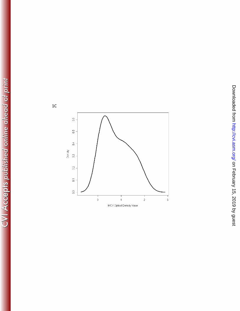

Figure 1 Legend. Histogram of seroreacitivty to JCPyV virus (A), BKPyV virus (B) and Merkel cell polyomavirus, MCPyV, (C) VP1 virus-like particles of samples from 42 children less than 10 years of age.
on February 15, 2019 by guest
http://cvi.asm.org/
Dow
nloaded from

Figure 2 Legend: SDS-PAGE gel of Merkel cell polyomavirus, BK virus and JC virus VP1 virus-like particles. Five μg of gradient purified MCPyV (MC), BKPyV (BK) or JCPyV (JC) VP1 virus like particles, produced in insect cells from recombinant baculoviruses, were subjected to SDS-PAGE. Molecular weight markers (MW) are shown in lane 1, with the size of markers (X103) indicated to the left of the graph.
on February 15, 2019 by guest
http://cvi.asm.org/
Dow
nloaded from

Figure 3 Legend: Electron micrograph of Merkel cell polyomavirus (MCPyV) VP1 virus-likeFigure 3 Legend: Electron micrograph of Merkel cell polyomavirus (MCPyV) VP1 virus-like particles. Electron micrograph of purified MCPyV VP1 virus like particles is shown at ×105,000 magnification. The bars correspond to 100 nm.
on February 15, 2019 by guest
http://cvi.asm.org/
Dow
nloaded from

Figure 4 Legend: Age-specific seroprevalence of Merkel cell polyomavirus, BK virus and JC virus among 945 individuals recruited from hospital-based general and subspecialty outpatient clinics. Serum samples were tested at a 1:200 dilution in virus-like particle based ELISA assays The distribution of reactivity of serum samples fromat a 1:200 dilution in virus like particle based ELISA assays. The distribution of reactivity of serum samples from children less than 10 years of age was used to set cut points for seropositivity and results are displayed as the percent positive in 10-year age groups. Numbers used to construct the graph are shown in Table 1.
on February 15, 2019 by guest
http://cvi.asm.org/
Dow
nloaded from