Your Patient Can Make You Sick
Part 2: Toxoplasmosis
Jonathan M. Chapman, DVM, MPH, CPHSmall Animal Veterinarian Intern
VCA Arboretum View Animal HospitalVeterinary Technician Educational Seminar
January 12, 2016
Presentation outline
• What is toxoplasmosis?• Etiology and pathogenesis• Clinical findings• Diagnosis• Treatment• Zoonotic risk• Prevention• Summary and recommendations
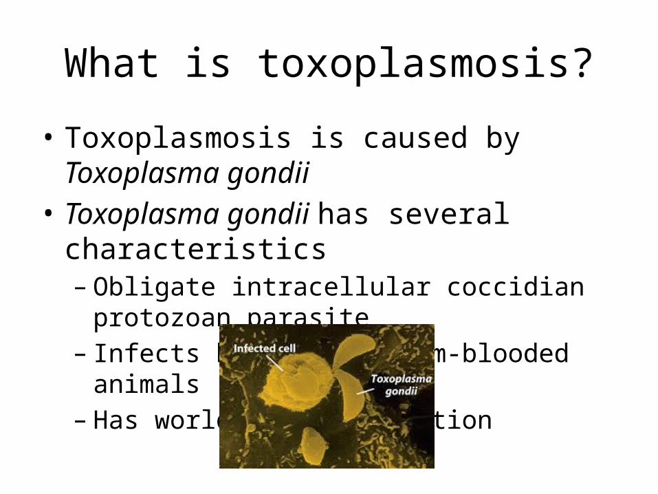
What is toxoplasmosis?
• Toxoplasmosis is caused by Toxoplasma gondii • Toxoplasma gondii has several characteristics– Obligate intracellular coccidian protozoan parasite– Infects humans and warm-blooded animals– Has worldwide distribution

Etiology and pathogenesis• Felids are the only definitive hosts of T. gondii
– Wild and domestic cats serve as the main reservoir of infection– Approximately 30% of cats are serologically positive for infection
• There are three infectious stages of T. gondii– Tachyzoites (rapidly multiplying form)– Bradyzoites (tissue cyst form)– Sporozoites (in oocysts)
• T. gondii is transmitted in several ways– Consumption of infectious oocysts from cat feces– Consumption of tissue cysts in infected meat– Transplacental transfer of tachyzoites from mother to fetus
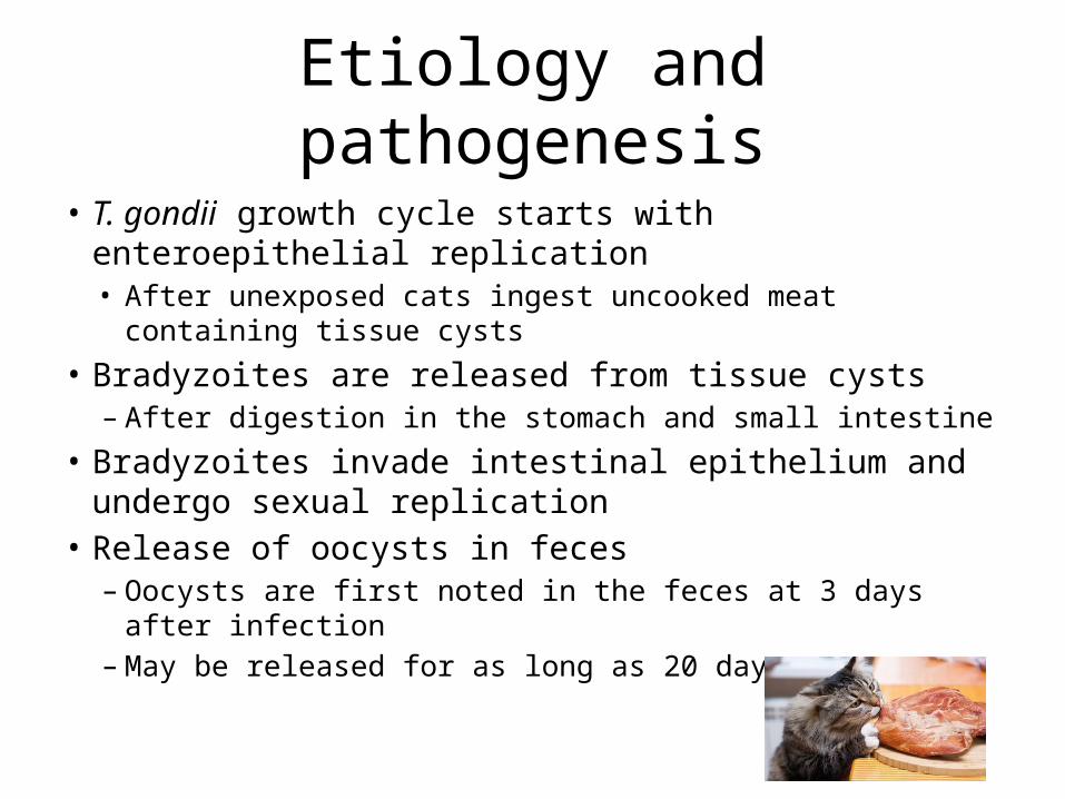
Etiology and pathogenesis• T. gondii growth cycle starts with enteroepithelial
replication• After unexposed cats ingest uncooked meat containing tissue
cysts • Bradyzoites are released from tissue cysts
– After digestion in the stomach and small intestine• Bradyzoites invade intestinal epithelium and undergo
sexual replication• Release of oocysts in feces
– Oocysts are first noted in the feces at 3 days after infection– May be released for as long as 20 days
Etiology and pathogenesis• Oocysts sporulate and become infectious outside the cat within 1–5 days
– Remain viable in the environment for several months• Depends on various factors
– Aeration– Environmental temperature
– Cats usually shed oocysts only once in their lifetime• Develop immunity to T gondii after the initial infection
• Oocysts are consumed– Uncooked meat containing tissue cysts (carnivores)– Food or water contaminated with cat feces containing oocysts (all warm-
blooded animals)• T. gondii initiates extraintestinal replication• Bradyzoites followed by sporozoites are released and infect intestinal
epithelium
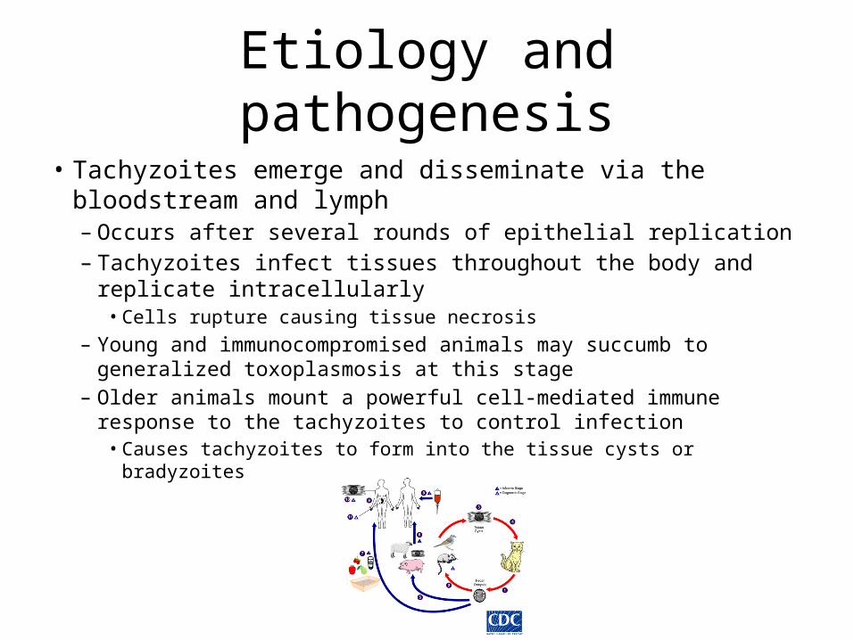
Etiology and pathogenesis• Tachyzoites emerge and disseminate via the bloodstream and
lymph– Occurs after several rounds of epithelial replication– Tachyzoites infect tissues throughout the body and replicate
intracellularly• Cells rupture causing tissue necrosis
– Young and immunocompromised animals may succumb to generalized toxoplasmosis at this stage
– Older animals mount a powerful cell-mediated immune response to the tachyzoites to control infection• Causes tachyzoites to form into the tissue cysts or bradyzoites

Etiology and pathogenesis
• Tissue cysts are usually seen in neurons, but also seen in other tissues– Individual cysts are microscopic• Up to 70 µm in diameter
– May enclose hundreds of bradyzoites in a strong and thin cyst wall
– May remain viable in the host for many years or for the life of the host

Clinical findings• General clinical signs of T. gondii infection include:
– Pyrexia– Lethargy– Anorexia– Weight loss– Vomiting– Diarrhea– Icterus– Coughing– Dyspnea– Ocular discharge– Photophobia– Uveitis– Ataxia– Seizures– Death
Clinical findings• The tachyzoite stage causes tissue damage and results in clinical signs• Clinical signs depend on several tachyzoite characteristics
– The number of tachyzoites released– The organs damaged by the tachyzoites– The ability of the host immune system to limit tachyzoite spread
• Adult immunocompetent animals control tachyzoite spread efficiently– Toxoplasmosis is usually a subclinical illness
• Tachyzoites spread systemically in young animals, especially puppies, kittens, (and piglets) causing illness
• Tachyzoites may also spread to a fetus– Necrosis in multiple fetal organs
• Acute generalized toxoplasmosis can develop– Immunocompromised adult animals
• Example: Cats infected with feline immunodeficiency virus

Diagnosis• Standard bloodwork, urinalysis, and thoracic and abdominal radiographs will not
provide a definitive diagnosis– The following may be noted:
• CBC– Leukopenia mainly characterized by a lymphopenia– Neutropenia with a degenerative left shift– Leukocytosis may occur during recovery
• Chemistry– Increased ALT– Increased ALP– Hypoalbuminemia– Hyperbilirubinemia– Mild hypocalcemia
• Urinalysis– Mild proteinuria– Bilirubinuria
• Thoracic radiographs– Interstitial to alveolar lung pattern– Pleural effusion
• Abdominal radiographs– Abdominal effusion– Hepathomegaly
Diagnosis• Definitive diagnosis requires biologic, serologic, or histologic methods
– Clinical signs of toxoplasmosis are nonspecific and are not sufficient for a definite diagnosis• Antemortem diagnosis
– Indirect hemagglutination assay– Indirect fluorescent antibody assay– Latex agglutination test– ELISA
• IgM antibodies appear faster after infection than IgG antibodies– Increased IgM titers (>1:256) are consistent with recent infection– IgG antibodies appear by the fourth week after infection– IgM antibodies generally do not persist past 3 months after infection– IgG antibodies remain increased for years during subclinical infection– IgG titers must be measured in paired sera from the acute and convalescent stages (approx. 3–4
wk apart)• Infection is indicated by an at least 4-fold increase in the titer
• CSF and aqueous humor may be analyzed – Presence of tachyzoites or anti-T. gondii antibodies indicates infection

Diagnosis
• Postmortem diagnosis– Tissue impression smears• Tachyzoites may be noted
– Microscopic examination of tissue sections • Tachyzoites or bradyzoites may be noted
• T. gondii is morphologically similar to other protozoan parasites– Sarcocystis species– Neospora caninum
Diagnosis
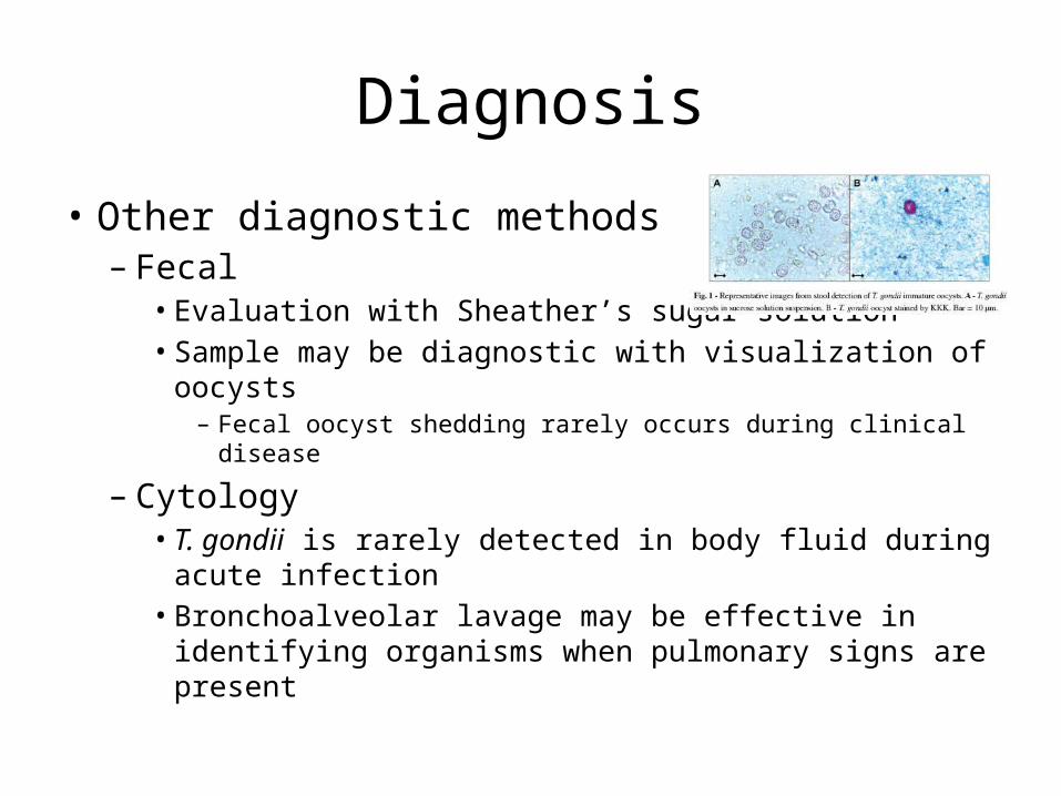
• Other diagnostic methods– Fecal• Evaluation with Sheather’s sugar solution• Sample may be diagnostic with visualization of oocysts
– Fecal oocyst shedding rarely occurs during clinical disease
– Cytology• T. gondii is rarely detected in body fluid during acute
infection• Bronchoalveolar lavage may be effective in identifying
organisms when pulmonary signs are present
Treatment• Treatment for toxoplasmosis consists of oral medication and supportive care
– However, treatment is seldom warranted in animals• Clindamycin
– 12.5 mg/kg PO BID x 14-28 days– Treatment of choice for dogs and cats
• Pyrimethamine and sulfadiazine– Pyrimethamine : 0.25 – 0.5 mg/kg PO BID x 14-28 days– Sulfadiazine: 30 mg/kg PO BID x 14-28 days– Widely used for treatment of toxoplasmosis– Drugs act synergistically– Beneficial if given in the acute stage of the disease
• Active multiplication of the parasite– Usually will not eradicate infection– Little effect on the bradyzoite stage

Treatment
• Other drugs used less commonly– Diaminodiphenylsulfone (dapsone)• 1 mg/kg PO q8h x 10 days• Do not use in cats
– Can cause hemolytic anemia or neurotoxicity
– Atovaquone• 13.3 mg/kg PO q8h x 10-21 days with a fatty meal
– Spiramycin • 12.5-23.4 mg/kg PO q24h x 5-10 days
Zoonotic risk• T. gondii is an important zoonotic agent• As much as 60% of the human population in some areas of the world have
serum IgG titers to T. gondii– Exposure most likely occurs after ingestion of T. gondii
• Undercooked meat• Oocysts from cat feces
– These people are likely to be persistently infected• Toxoplasmosis is a major concern for people with immune system
dysfunction– Usually presents as meningoencephalitis
• Results from the emergence of T. gondii from tissue cysts located in the brain
• Toxoplasmosis is also a concern for pregnant women– Tachyzoites can migrate transplacentally and cause birth defects in human fetuses
Prevention• The stages of T. gondii in meat are killed by contact with
soap and water– Wash hands thoroughly with soap and water– Wash all surfaces in contact with meat (Ex. Cutting boards, sink
tops, knives, etc.)• T. gondii organisms in meat can also be killed by exposure
to extreme cold or heat or by gamma irradiation– Heat meat throughout to 67°C (152.6°F) or cool to −13°C (8.6°F)– Avoid tasting meat while cooking or while seasoning – Tissue cysts are killed by exposure to 0.5 kilorads of gamma
irradiation
Prevention• Wear gloves and wash hands thoroughly after working with soil or
cat litter• Thoroughly wash vegetables or other foods in contact with soil• Pregnant women should avoid contact with possible contaminants
– Cat litter– Soil– Raw meat
• Pet cats should be fed only dry or canned cat foods or properly cooked foods
• A cat litter box should be emptied daily– Preferably not by a pregnant woman
• No vaccine is available for toxoplasmosis in animals or humans

Summary and recommendations
• Zoonotic disease• Mild to severe clinical signs occur including death• Definitive diagnosis requires advanced testing• Treatment is not always necessary• Disease prevention is important• Clean cat litterboxes every day• Always properly handle meat and other contaminants• Pregnant women, youth, and immunocompromised
individuals are most at risk

Acknowledgements
• Joao Felipe de Brito Galvao, MV, MS, DACVIM• Kathleen Van Lanen, DVM, DACVECC• Elizabeth Norberg, CVT, BS• The entire staff at VCA Arboretum View
Animal Hospital

Any questions?

References• Centers for Disease Control and Prevention. "Toxoplasmosis.“
Parasites - Toxoplasmosis (Toxoplasma Infection). Centers for Disease Control and Prevention, 10 Jan. 2013. Web. 08 Jan. 2016.
• Lunn, Katharine F., BVMS, MS, PhD, MRCVS, DACVIM. "Overview of Toxoplasmosis." Merck Veterinary Manual. Merck Sharp & Dohme Corp., Apr. 2015. Web. 22 Dec. 2015.
• Plumb, Donald C. Plumb's Veterinary Drug Handbook. 8th ed. Hoboken, NJ: Wiley-Blackwell, 2015. Print.
• Tilley, Lawrence P. “Toxoplasmosis." Blackwell's Five-minute Veterinary Consult: Canine and Feline. Oxford: Wiley-Blackwell, 2011. 1242-43. Print.


