
Chem 2601/2011
Molecular Imaging Lecture 3 and 4: Introduction to Nuclear imaging and Radiochemistry
Dr. Erik Årstad, KLB room 2.11 ([email protected])
1

Overview (lecture 3 and 4):
1) The principles of Nuclear imaging
2) Nuclear imaging techniques
3) Instrumentation
4) Introduction to radioactivity
5) Production of radionuclides
6) Radiochemistry
2
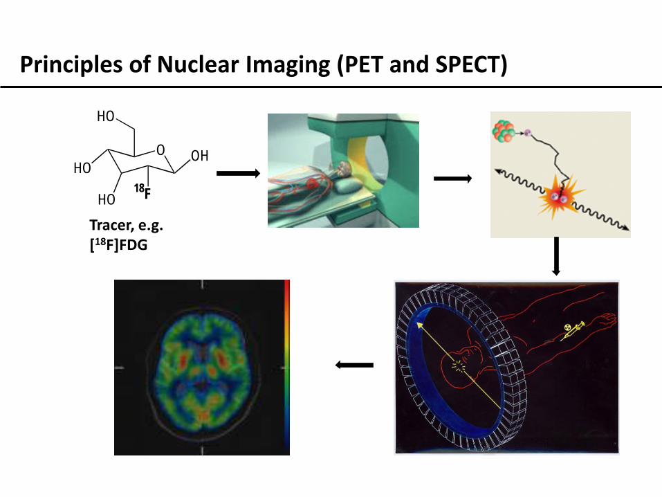
Principles of Nuclear Imaging (PET and SPECT)
O
HO
HO
OH
HO
18F
Tracer, e.g. [18F]FDG
4
Nuclear Imaging techniques: 1) Positron emission tomography (PET)
2) Single Photon Emission Computed Tomography (SPECT)
3) Autoradiography

- Positron: the antimatter equivalent of an electron
- Positrons are emitted from certain radioactive substances
- Positrons and electrons annihilates to produce two gamma rays
Positron
Electron
Gamma ray (511 KeV)
Gamma ray (511 KeV)
- A chemical is labelled with a radioactive isotope (positron emitter)
- Positrons annihilate in surrounding tissue
- The resulting gamma rays are emitted from the subject

Generates 3D maps of radioactivity concentration - tomographic

Tracer labelled with gamma (single photon) emitting radionuclide
9
Autoradiography (imaging in vitro):
Contact exposure of radioactive samples (e.g. 20 μm tissue section on X-ray film) Lower resolution than fluorescence microscopy, but quantitative Requires low energy beta emission
Burton et al. (2009), TOXICOLOGICAL SCIENCES, 111(1): 131–139. http://www.nationaldiagnostics.com
10
Instrumentation: 1) Detector principles
2) Principle of PET scanners
3) Principle of SPECT scanners
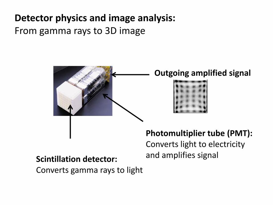
Detector physics and image analysis: From gamma rays to 3D image
Scintillation detector: Converts gamma rays to light
Photomultiplier tube (PMT): Converts light to electricity and amplifies signal
Outgoing amplified signal
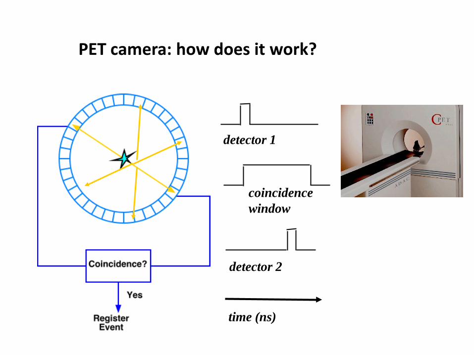
PET camera: how does it work?
detector 1
detector 2
coincidence window
time (ns)
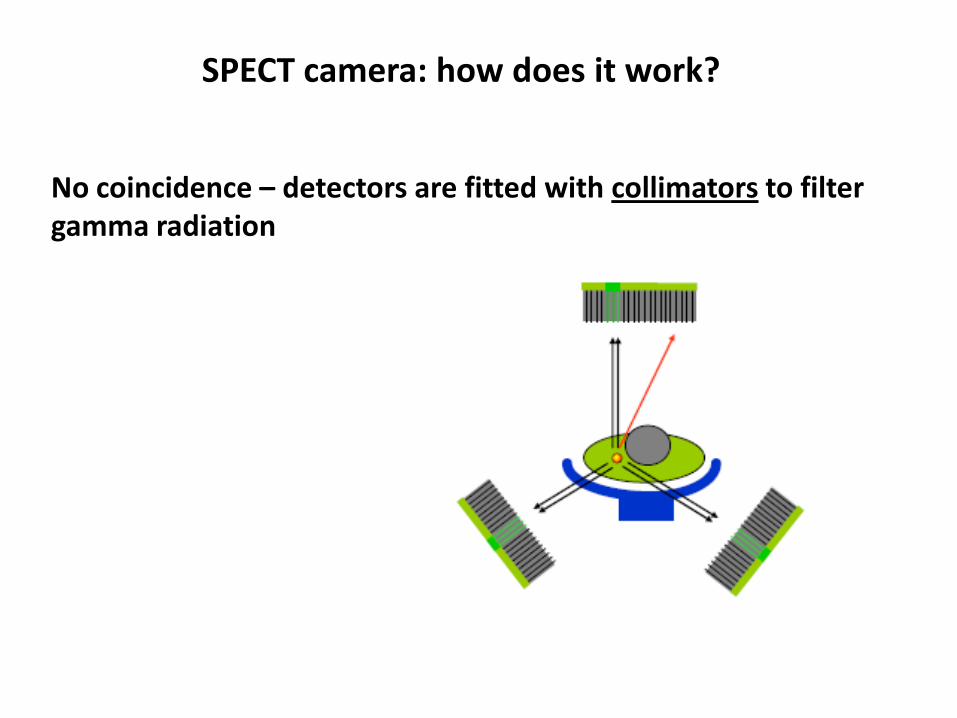
SPECT camera: how does it work?
No coincidence – detectors are fitted with collimators to filter gamma radiation
14
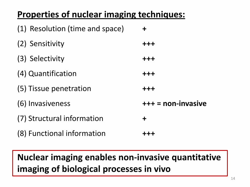
Properties of nuclear imaging techniques: (1) Resolution (time and space) +
(2) Sensitivity +++
(3) Selectivity +++
(4) Quantification +++
(5) Tissue penetration +++
(6) Invasiveness +++ = non-invasive
(7) Structural information +
(8) Functional information +++
Nuclear imaging enables non-invasive quantitative imaging of biological processes in vivo

SPECT vs. PET
SPECT PET Resolution 12-15mm 4-7mm Sensitivity
(gamma detection) 0.03% 3.0%
dual radionuclides yes no Radionuclides Gamma emitters
half-life > 6 hours Positron emitters
half-life < 2h Sensitivity
(Target concentration)
10-13 molar 10-14 molar
Cost $$ $$$ (£500-2000/scan)
Radioactivity: 1) Introduction to radioactivity
2) Ionizing radiation
3) Half-life and radioactive decay
4) Specific activity
5) Attenuation
16

17
What is radioactivity?

A brief introduction to Radioactivity:
1895: Roentgen discovered X-rays 1896: Henri Becquerel discovered rays from uranium
1897: Marie Curie named the rays ‘radioactivity’ 1898: Marie and Pierre Curie discovered Polonium and Radium 2012: > 2500 radioactive nuclides are known!

19
Definitions: A nuclide (from nucleus) is an atomic species characterized by the specific constitution of its nucleus, i.e., by its number of protons Z and its number of neutrons N. A radionuclide is any radioactive nuclide. Isotopes are atoms from the same element (i.e. same proton number) but different number of neutrons.
18F- Mass number = N + Z
Proton (Z) number
Charge

20
Radioactivity is defined as the process in which unstable atomic nuclei spontaneously emit ionizing radiation
Types of ionizing radiation:
Alpha particles = He nucleus
Beta particles = electron
Positrons = antimatter of electrons
Gamma rays = highly energetic photons
E = mc2

21
The units of radioactivity: Historical units: Ci (curie) = 3.7 × 10¹⁰ disintegrations per second (= 1 g of 226Ra) mCi = 37 x 106 disintegrations per second
SI units: Bq (Becquerel) = 1 disintegration per second KBq = 1 x 10³ Bq MBq = 1 x 10⁶ Bq GBq = 1 x 10⁹ Bq Conversion factor: 1 mCi = 37 MBq
22
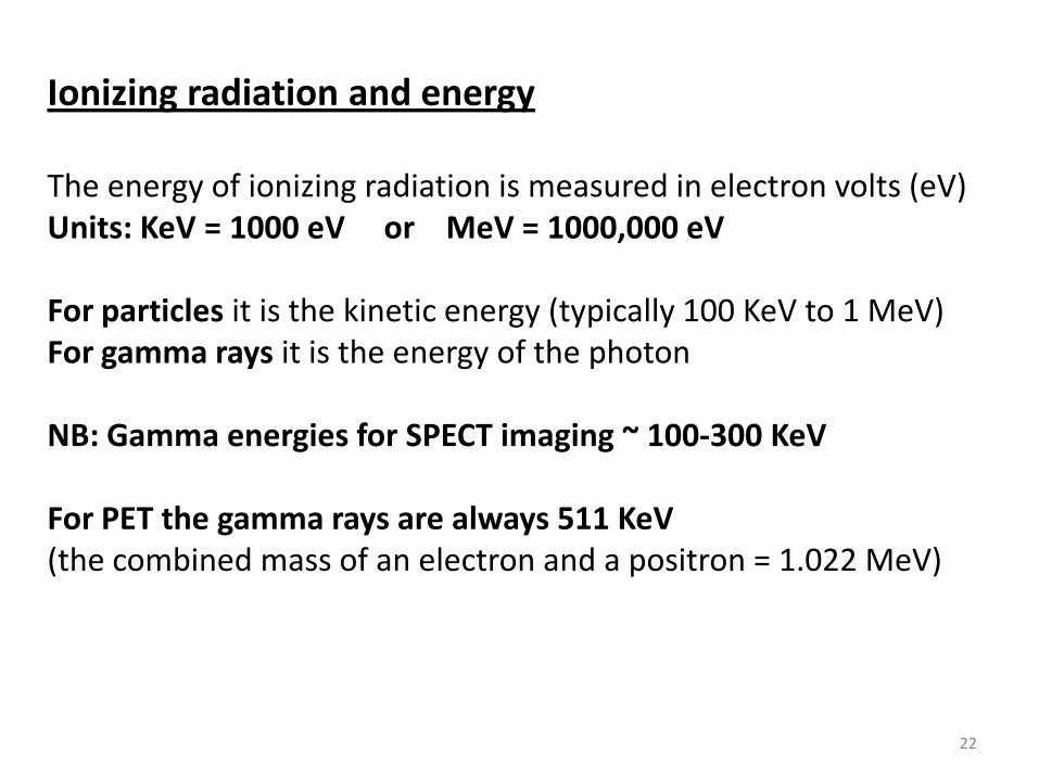
Ionizing radiation and energy The energy of ionizing radiation is measured in electron volts (eV) Units: KeV = 1000 eV or MeV = 1000,000 eV For particles it is the kinetic energy (typically 100 KeV to 1 MeV) For gamma rays it is the energy of the photon NB: Gamma energies for SPECT imaging ~ 100-300 KeV For PET the gamma rays are always 511 KeV (the combined mass of an electron and a positron = 1.022 MeV)
23
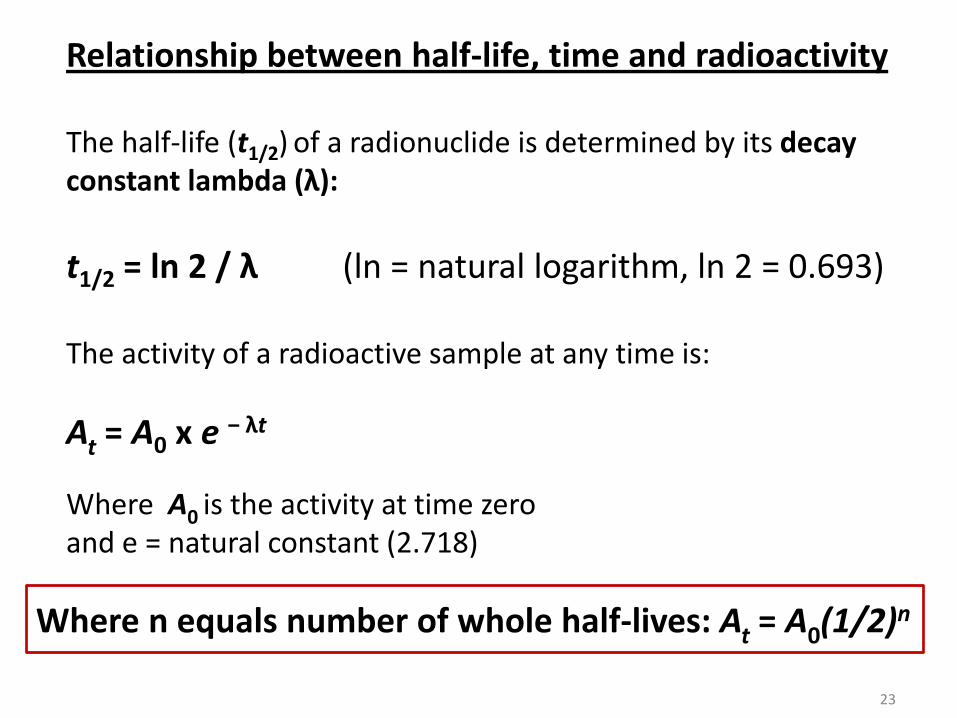
At = A0 x e − λt
t1/2 = ln 2 / λ (ln = natural logarithm, ln 2 = 0.693)
Where n equals number of whole half-lives: At = A0(1/2)n
Relationship between half-life, time and radioactivity
The activity of a radioactive sample at any time is:
The half-life (t1/2) of a radionuclide is determined by its decay constant lambda (λ):
Where A0 is the activity at time zero and e = natural constant (2.718)

24
Half-life and radioactive decay over time

Question: Carbon-11 has a half-life of 20 min. The synthesis of a tracer takes 40 min and it takes another 20 min to analyse the product before injection to a subject. The radiochemical yield is 20%. How much of the initial activity is available for injection?
26
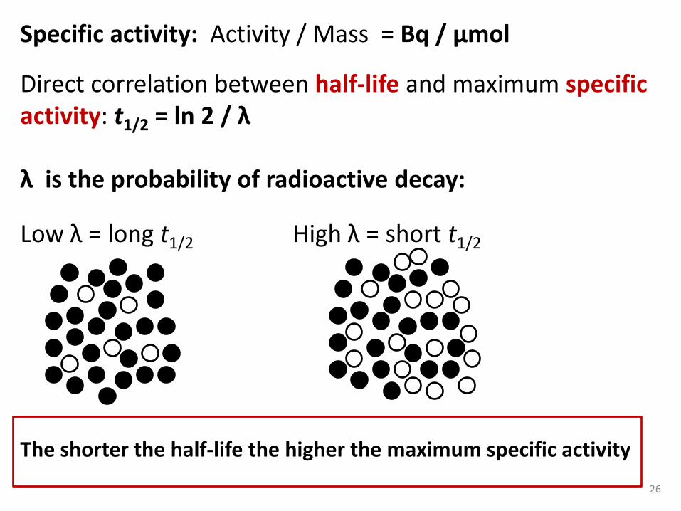
Specific activity: Activity / Mass = Bq / µmol
Direct correlation between half-life and maximum specific activity: t1/2 = ln 2 / λ λ is the probability of radioactive decay:
Low λ = long t1/2 High λ = short t1/2 The shorter the half-life the higher the maximum specific activity

27
Samples exclusively made up of molecules containing the radioactive nuclide are carrier-free (c.f.). Samples without addition of non-radioactive carrier but containing naturally occurring isotopic dilutions are non-carrier-added (n.c.a.). Samples diluted with non-labelled molecules are carrier-added (c.a.).
Specific activity – important terms:
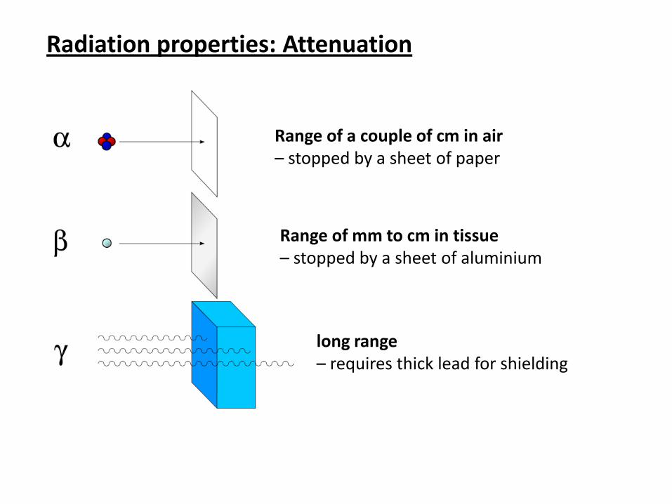
Radiation properties: Attenuation
Range of a couple of cm in air – stopped by a sheet of paper
Range of mm to cm in tissue – stopped by a sheet of aluminium
long range – requires thick lead for shielding

29
Attenuation: effect of matter
http://en.wikibooks.org/wiki/Basic_Physics_of_Nuclear_Medicine/Attenuation_of_Gamma-Rays
Gamma ray intensity I0 Gamma ray intensity Ix
0 X
Δ I = I0 – Ix, where Δ I is proportional to Z3 Doubling the atomic number leads to 8 fold increase in attenuation!

30
Attenuation: effect of matter AND energy
http://en.wikibooks.org/wiki/Basic_Physics_of_Nuclear_Medicine/Attenuation_of_Gamma-Rays
Half value (in cm) for gamma rays:
Biological tissues: “Different stopping power of radiation” e.g. lungs vs. bones

Attenuation of positrons in tissue:
NB: this defines the maximum theoretical resolution of PET!
32
Nuclear imaging is quantitative, because: Radioactive decay is determined by the half-life Radioactive decay is unaffected by the environment The interactions of ionizing radiation with matter follows clear physical rules and can be accounted for

Question: What would happen if a subject is injected with a PET tracer but scanned with SPECT camera? What would happen if a subject is injected with a SPECT tracer but scanned with PET camera?

Radiochemistry: 1) Production of radionuclides
2) Labelling with 11C
3) Labelling with 18F
4) Labelling with 123I
5) Labelling with 3H
34

35
Radiochemistry and production of radionuclides Examples for PET: 11C (t1/2 20.4 min) and 18F (t1/2 110 min) Example for SPECT: 123I (t1/2 13.1 h) Example for autoradiography: 3H (t1/2 12 years)

Reaction: Product: half-life: Decay mode:
16O (p,α) 13N 10 min β+ (positron)
14N (p,α) 11C 20 min β+ (positron) 14N (d,n) 15O 2 min β+ (positron) 18O (p,n) 18F 110 min β+ (positron) 124Te(p,2n) 123I 13.1 h γ (gamma) 99mTc 6.01 h γ (gamma) 68Ga 68 min β+ (positron)
82Rb 1.26 min β+ (positron)
Generator based radionuclides:
Production of radionuclides with a cyclotron
36

37
Production of radionuclides: formation of 3H
6Li + n 4He + 3H (half-life 12 years, beta emitter)
Nuclear reactor provides neutron flux:
All radionuclides for biomedical research are either produced by a cyclotron, or in nuclear reactors (directly or indirectly).
38
Radiochemistry – general principles: Fast reactions
High yields
Reliable and reproducible reactions
Few side products
Simple purification Introduce the radionuclide as late in the synthesis
as possible!

39
Radiochemistry – important definitions: Radiochemical yield (r.c.y): the efficiency of a labelling reaction measured as the proportion of radioactivity that has been transferred from a reagent to a product. Radiochemical yield can be decay corrected or non-decay corrected: Decay-corrected: the amount of activity in the product is corrected for the decay that has occured during the synthesis before calculation of radiochemical yield Non-decay-corrected: there is no correction for decay, so the radiochemical yield is simply the amount of radioactivity in the product divided on the amount of radioactivity in the reagent.

Radiochemical yield - example: Carbon-11 has a half-life of 20 min. You start the synthesis with 2 GBq of 11CO2. After 40 min you obtain 200 MBq of a tracer. The non-decay corrected radiochemical yield is: 0.2 GBq/2 GBq = 10% The decay-corrected radiochemical yield is: Decay correction: 0.2 GBq/(0.5 x 0.5) = 0.8 GBq 0.8 GBq/2 GBq = 40%

41
Radiochemistry: labelling with carbon-11 Advantages: Enables isotopic labelling, i.e. replacement of 12C with 11C in the molecule. The biological fate of the molecule is unchanged
Very versatile labelling chemistry – it’s carbon! Disadvantages: Short half-life (20.4 min) Only available on sites with a in-house cyclotron Alternative for biochemical applications: 14C (half-life 5730 years, beta emitter)

42
Radiochemistry: Labelling with carbon-11
14N (p,α) 11C
Cyclotron:
H2 11C H4
O2 11C O2
I2 11C H3I
ROH (alcohols) 11C H3OR
RNH2 (amines) 11C H3NRH
ArOH (phenols) 11C H3OAr
Sn2 reactions:
RSH (thiols) 11C H3SR
RCONH2 (amides) RC(O)NH11C H3
RMgX , SOCl2 (Grignard reagents)
R11C (O)Cl
R’NH2 (amines)
R11C (O)NHR’

43
Radiochemistry: Labelling with carbon-11 [11C ]Methionine: [11C ]Methionine
[11C ]Way 100635:
11C H3I
Base
11C O2
H2N C
HS
O
OHH2N C
H311CS
O
OH
MgBr CO
OMgBrCO
Cl SOCl2
ON
NN
C
N
O
H3C
RNHR’
[11C ]WAY 100635
Typical specific activities: 40-200 GBq/µmol
Sources of 12C: atmospheric CO2

44
Radiochemistry: labelling with fluorine-18 Advantages: Near ideal half-life (110 min) and low positron energy
Small size makes it suitable for labelling of small molecules Fluoride can reduce metabolism of tracers Tracers can be transported for imaging at centres without a cyclotron Disadvantages: Limited reactivity, need to protect OH and NH groups!
Limited chemistry – fluoride is the most electronegative of all elements
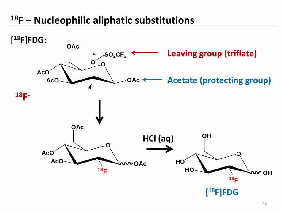
18F – Nucleophilic aliphatic substitutions
18F-
Leaving group (triflate)
Acetate (protecting group)
OAcO
AcO OAc
OAc
18F
OAcO
AcO
O
OAc
OAcSO2CF3
OHO
HO
OH
18FOH
HCl (aq)
[18F]FDG:
[18F]FDG 45

[18F]Fluoride – Nucleophilic aliphatic substitutions
N
N
O
OO
DMTrO
18F
DMBn
MeCN, K2CO3, Kryptofix 100 ºC, 10 min,
Deprotection
38% RCY
N
NH
O
OO
HO
18F
[18F]FLT 13% RCY
Grierson and Shields, Nuclear Medicine & Biology 2000, Vol. 27; 143–156
Protecting group
Protecting group
18F-
Leaving group = nosylate O2N
SO
OO
R
46
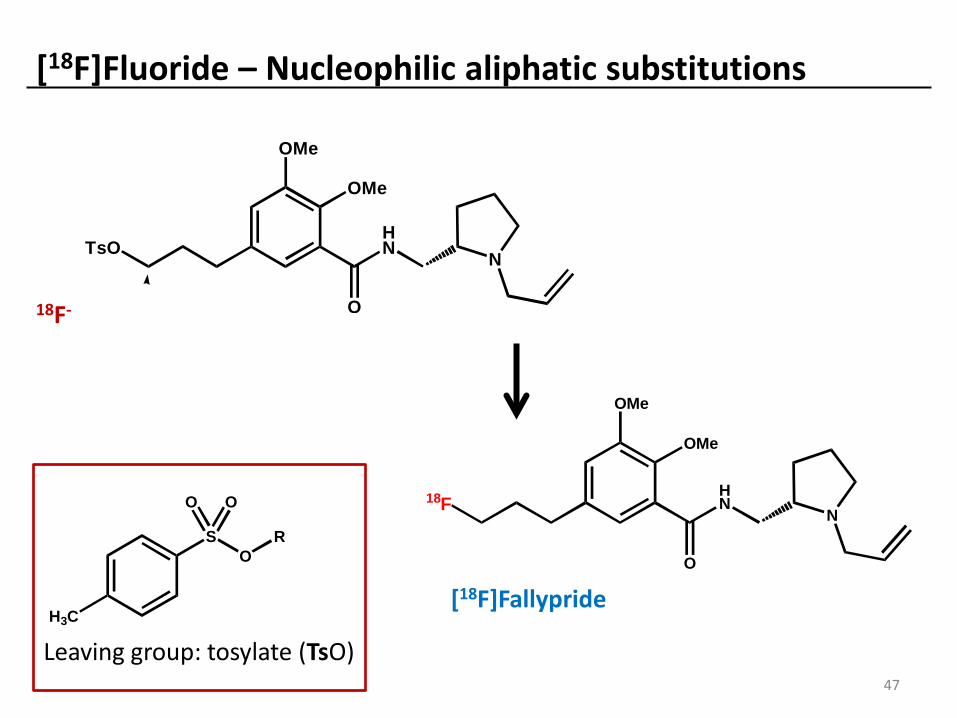
[18F]Fluoride – Nucleophilic aliphatic substitutions
[18F]Fallypride
TsOHN
OMe
OMe
O
N
18F-
Leaving group: tosylate (TsO)
H3C
SO
OO
R
18FHN
OMe
OMe
O
N
47
48
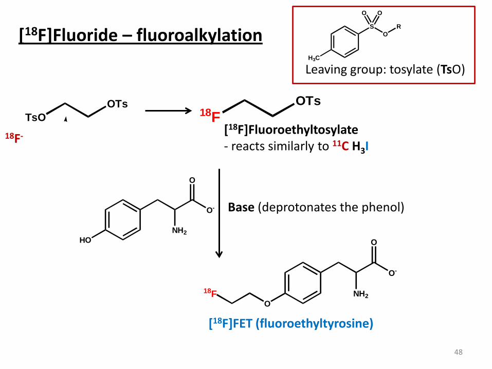
[18F]Fluoride – fluoroalkylation
18F-
Leaving group: tosylate (TsO)
18FOTs
[18F]Fluoroethyltosylate - reacts similarly to 11C H3I
HONH2
O-
O
ONH2
O-
O
18F
TsOOTs
Base (deprotonates the phenol)
[18F]FET (fluoroethyltyrosine)
H3C
SO
OO
R
49
Radiochemistry: labelling with iodine (123,124,125,131I) Advantages: Easy labelling chemistry Many isotopes available (autoradiography, SPECT and PET)
Range of half-lives from 13 h to 60 days
Disadvantages: Limited metabolic stability
Large size (similar to benzene!)
Limited labelling chemistry

Direct labelling of peptides/proteins with iodine:
123I- “ 123I+ “ Oxidant
HO
O
NHR
HN R
OTyrosine residue
HO
O
NHR
HN R
O
123I
Advantages: High yields Simple chemistry Disadvantages: Low metabolic stability No control of labelling positions (in large proteins)
50

Labelling of trialkyl tin compounds with iodine (aryl and alkene):
Advantages: Site specific labelling Good yields Disadvantages: Only suitable for small molecules (or indirect labelling)
51
123I
OH
OMe O
HN
N
SnMe3
OH
OMe O
HN
N
123I-, H2O2
Dilute acid
[123I]IBZM

52
Radiochemistry: labelling with tritium (3H or T) Advantages: Can label almost any organic compound
Ideal for isotopic labelling – replacing H with tritium
Widely used basic research Disadvantages: Only possible in specialised labs (custom service)
Only suitable for in vitro applications

53
N
N
T2, Pd/C
N
N
T
T
Labelling with 3H (also known as tritium - T):
Reduction of alkenes, alkynes and other saturated bonds with 3H2 gas:
Maximum specific activity for labelling with 3H ~ 2-3 GBq/µmol
Alkylation with C3H3I – equivalent to 11C H3I and suitable for the same reactions!

54
Exercise 3: How would you label this molecule?
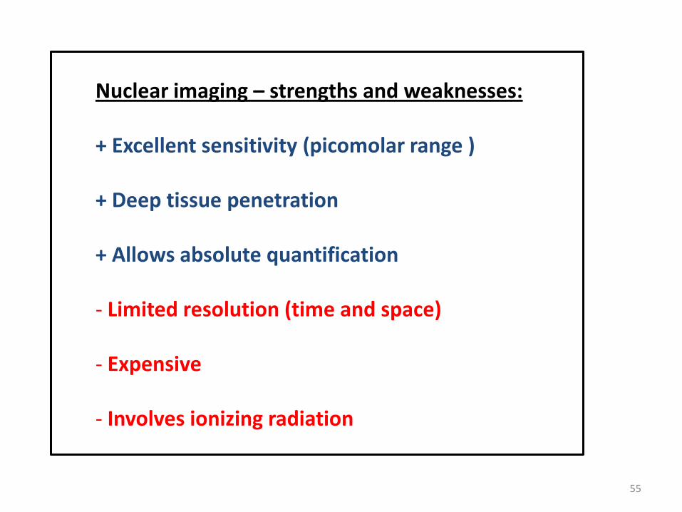
Nuclear imaging – strengths and weaknesses: + Excellent sensitivity (picomolar range ) + Deep tissue penetration + Allows absolute quantification - Limited resolution (time and space)
- Expensive
- Involves ionizing radiation
55

Synopsis: PET and SPECT principles
PET - Positron emitters - short half-life (11C, 18F) < 2 h - Detects annihilation gamma rays - Coincidence detection for location - Higher resolution - Better for quantification - More expensive
SPECT - Gamma emitters - Longer half-life (123I) > 6 h - Detects gammas emitted directly - Collimators for location - Can use multiple radionuclides - Lower resolution - Lower cost
Synopsis: autoradiography Detects beta particles by X-ray film. Suitable radionuclides have long half-lives – 2 weeks to many years! The best results are achieved with low energy beta emitters, as energetic particles are not fully stopped by the imaging medium.

57
Synopsis: Radioactivity
Radioactive decay:
-There main modes of decay are: alpha, beta, positron and gamma emission
- Radioactivity is measured in Bq = 1 disintegration per second
- Activity is defined by the half-life and the number of a radionuclide
- Specific Activity is the activity per mass (in Bq / µmol)
Interactions of ionizing radiation with matter:
-Each type of radiation interacts with matter in a unique way
- Particles are rapidly stopped by matter and travel only short distances
- Attenuation of gamma rays depends strongly on the gamma energy and atomic number of the absorbing matter
NB: Radioactivity can readily be quantified as the activity level is unaffected by the environment and the interactions of ionizing radiation with matter can be accounted for.

58
Synopsis: Radiochemistry
Production of radionuclides:
Most PET radionuclides are produced with particle bombardment in a cyclotron
Some radionuclides are produced in high flux nuclear reactors
Radiochemistry:
Reactions should be fast, efficient and reliable
Radionuclides typically introduced late in the synthesis
Chemistry of carbon-11, fluorine-18, iodine-123 and tritium
Carbon-11 converted to CH3I or CO2
Carbon-11 labelling typically with methylation or Grignard reactions
Fluorine-18 typically introduced by Sn2 nucleophilic reactions
Iodine-123 introduced by addition to tyrosine or reaction with trialkytin groups
Tritium typically introduced by reduction of unsaturated bonds
Learning outcomes - you should understand: - The principles of radioactivity - The decay modes - The interaction of radiation with matter - The principles for imaging with PET, SPECT and autoradiography - How the decay mode, half-life and energy range of the radiation effect the suitability for imaging -The advantages and disadvantages of nuclear imaging
- The chemistry of common radionuclides for PET, SPECT and autoradiography
59

Assessment – you should be able to apply your knowledge of radioactivity and nuclear imaging to explain underlying principles, solve practical problems and provide rationale explanations related to: - The principles of radioactivity - The decay modes - The interaction of radiation with matter - The principles for imaging with PET, SPECT and autoradiography - How the decay mode, half-life and energy range of the radiation effect the suitability for imaging -The advantages and disadvantages of nuclear imaging
- Labelling reactions with common radionuclides for PET, SPECT and autoradiography
60
Useful websites and background reading: Radioactivity: http://en.wikipedia.org/wiki/Radioactive_decay Interaction of gamma rays with matter: http://en.wikibooks.org/wiki/Basic_Physics_of_Nuclear_Medicine/Attenuation_of_Gamma-Rays Sensitivity of nuclear imaging: John V. Fragiono, Journal of Clinical Oncology, 26(24): 4012-4021 Autoradiography: http://www.nationaldiagnostics.com Burton et al. (2009), TOXICOLOGICAL SCIENCES, 111(1): 131–139. Langstrom et al. (2007), Mol Imaging Biol, 9(4): 161-175. Charon et al. (1998), Nuclear Medicine & Biology, 25:699–704.
61


