
Neuroscience 224 (2012) 135–144
TREADMILL TRAINING STIMULATES BRAIN-DERIVEDNEUROTROPHIC FACTOR mRNA EXPRESSION IN MOTOR NEURONSOF THE LUMBAR SPINAL CORD IN SPINALLY TRANSECTED RATS
M. S. JOSEPH, a N. J. K. TILLAKARATNE b ANDR. D. DE LEON c*
aDepartment of Biological Science, California State University, Los
Angeles, CA, United States
bDepartment of Integrative Biology and Physiology, Brain
Research Institute, University of California, Los Angeles,
CA, United StatescSchool of Kinesiology and Nutritional Science, California
State University, Los Angeles, CA, United States
Abstract—Brain-derived neurotrophic factor (BDNF)
induces plasticity within the lumbar spinal circuits thereby
improving locomotor recovery in spinal cord-injured
animals. We examined whether lumbar spinal cord motor
neurons and other ventral horn cells of spinally transected
(ST) rats were stimulated to produce BDNF mRNA in
response to treadmill training. Rats received complete
spinal cord transections as neonates (n= 20) and one
month later, received four weeks of either a low (100 steps/
training session; n= 10) or high (1000 steps/training ses-
sion; n= 10) amount of robotic-assisted treadmill training.
Using combined non-radioactive in situ hybridization and
immunohistochemical techniques, we found BDNF mRNA
expression in heat shock protein 27-labeled motor neurons
and in non-motor neuron cells was greater after 1000
steps/training session compared to the 100 steps/training
session and was similar to BDNF mRNA labeling in
untrained Intact rats. In addition, there were significantly
more motor neurons that contained BDNF mRNA labeling
within processes in the ST rats that received the higher
amount of treadmill training. These findings suggested that
motor neurons and other ventral horn cells in ST rats syn-
thesized BDNF in response to treadmill training. The find-
ings support a mechanism by which postsynaptic release
of BDNF from motor neurons contributed to synaptic plas-
ticity. � 2012 IBRO. Published by Elsevier Ltd. All rights
reserved.
Key words: body weight-supported treadmill training, plas-
ticity, neurotrophins, locomotion.
0306-4522/12 $36.00 � 2012 IBRO. Published by Elsevier Ltd. All rights reservehttp://dx.doi.org/10.1016/j.neuroscience.2012.08.024
*Corresponding author. Address: Department of Kinesiology andNutritional Science, California State University, Los Angeles, 5151State University Drive, Los Angeles, CA 90032-8162, United States.Tel: +1-323-343-4855; fax: +1-323-343-6024.
E-mail address: [email protected] (R. D. de Leon).Abbreviations: BDNF, brain-derived neurotrophic factor; DIG,Digoxigenin; HSP27, heat shock protein 27; ISH, in situ hybridization;mRNA, messenger ribonucleic acid; ST, spinally transected; ST100,spinally transected + treadmill training for 100 steps/session; ST1000,spinally transected + treadmill training for 1000 steps/session.
135
INTRODUCTION
The neurotrophin, brain-derived neurotrophic factor
(BDNF), improves the ability of the lumbar spinal cord to
generate locomotion in spinal cord-injured animals
(Boyce et al., 2007, 2012; Ying et al., 2008). The
mechanisms are unknown, but it is believed that BDNF
triggers changes in the spinal circuits by raising the
excitability of spinal neurons (Boyce et al., 2012) and
strengthening synaptic connections within the circuitry
(Ying et al., 2005). The beneficial effects can be
produced by the delivery of exogenous BDNF to the
injured spinal cord (Boyce et al., 2012) but BDNF levels
are also increased by hindlimb exercise (Hutchinson
et al., 2004; Ying et al., 2005; Macias et al., 2009;
Sandrow-Feinberg et al., 2009; Cote et al., 2011; de
Leon et al., 2011). Treadmill training in particular is
effective in raising BDNF levels (Hutchinson et al., 2004).
Thus, stimulating the production of endogenous BDNF
promotes plasticity within the spinal circuits and leads to
improved locomotor recovery after spinal cord injury.
Interestingly, the cells that synthesize and release
BDNF in spinal cord-injured animals are unknown. We
and others have shown that treadmill training increased
BDNF protein expression in motor neurons of spinally
transected (ST) rats (Macias et al., 2009; de Leon
et al., 2011). These findings suggest that BDNF was
secreted by motor neurons in response to treadmill
training. However, it was not clear whether the motor
neurons themselves synthesized BDNF or if the BDNF
was derived from other cells such as muscle fibers
(Gomez-Pinilla et al., 2002). Large increases in BDNF
mRNA levels in hindlimb muscles were stimulated by a
single bout of hindlimb exercise in ST rats (Dupont-
Versteegden et al., 2004), suggesting target hindlimb
muscles synthesized BDNF then retrogradely
transported BDNF to motor neurons (Koliatsos et al.,
1993). In order to understand the possible contribution
of spinal neurons to BDNF levels in the lumbar spinal
cord, it was necessary to demonstrate that synthesis of
BDNF mRNA occurred in spinal neurons. Only one
study to date has examined cellular expression of
BDNF mRNA expression in spinal cord-injured animals
(Keeler et al., 2012). In this study, passive cycling of
the hindlimbs was sufficient to raise BDNF mRNA
levels in motor neurons, raising the possibility that
activity during treadmill training would have a similar
effect. In addition to motor neurons, other cells in the
ventral horn, e.g. interneurons, glial cells, may produce
d.
136 M. S. Joseph et al. / Neuroscience 224 (2012) 135–144
BDNF that influenced enhanced plasticity, but their
contribution to the BDNF pool has not yet been
examined in exercised spinal cord-injured animals.
In the present study, we examined whether treadmill
training in ST rats influenced motor neuronal expression
of BDNF mRNA. We used in situ hybridization (ISH) and
immunohistochemical techniques (Tillakaratne et al.,
2002) to study the expression of BDNF mRNA in heat
shock protein 27 (HSP27)-labeled motor neurons and
other ventral horn cells in rats that received a complete
mid-thoracic spinal cord transection at five days of age.
Previous studies have shown that BDNF mRNA is
targeted to dendrites of postsynaptic neurons and locally
translated into protein (An et al., 2008; Chiaruttini et al.,
2009). Thus, we included an analysis of BDNF mRNA in
motor neuron processes. A robotic treadmill system was
used to train the ST rats to perform either 100 or 1000
steps/training session and the kinematic data from this
study have previously been reported (Cha et al., 2007).
Here, we hypothesize that imposing a higher amount of
treadmill training would result in a greater synthesis of
BDNF by motor neurons and affect its subcellular
localization. The findings were consistent with this
hypothesis and support a mechanism in which
postsynaptic release of BDNF from motor neurons
contributed to plasticity within the lumbar spinal circuits
controlling locomotion.
EXPERIMENTAL PROCEDURES
Experimental design
Twenty female Sprague–Dawley rats received a complete mid-
thoracic spinal transection at five days of age. After weaning
(21 days old), a robotic device was used to assess the ability of
the rats to perform hindlimb stepping on a treadmill. The rats
were distributed into two experimental groups that were
balanced according to their locomotor performance during the
baseline tests. One group (n= 10) received daily treadmill
training that consisted of 100 steps/session while the other
group (n= 10) performed 1000 steps/training session and
these rats will be referred to as the spinally transected +
treadmill training for 100 steps/session (ST100) and spinally
transected + treadmill training for 1000 steps/session (ST1000)
rats respectively. Training was performed five days/week for
four weeks. The animals were perfused with 4%
paraformaldehyde, the spinal cords were removed and
processed for the histology, ISH and immunohistochemical
experiments. All procedures with rats were carried out in
accordance with NIH guidelines and the protocols were
approved by the Institutional Animal Care and Use Committee
at California State University, Los Angeles.
Spinal cord transection
The spinal cords of the rats were transected at a mid-thoracic
level as previously described (Cha et al., 2007). Briefly, the
pups were anesthetized using isoflurane (1%). A dorsal mid-
line skin incision was made over the mid-thoracic vertebra and
the overlying fascia and muscles were retracted to expose the
dorsal surface of the vertebrae. A partial laminectomy was
performed at the mid-thoracic level to expose the spinal cord.
The spinal cord was then lifted with a curved probe and
completely transected. Afterwards, the skin incision was closed
with sutures. The entire surgical procedure took about 10–
15 min.
Following surgery, the rats were allowed to recover in a warm
incubator. The temperature was maintained at 37 �C. The
neonatal pups were placed in the incubator until fully alert (10–
30 min) and then returned to the mothers. After the rats were
weaned (21 days old), the rats were housed in spacious cages,
2–3 rats per cage. The bladders and colons of the rats were
checked daily.
Robotic-assisted treadmill training
A commercially available robotic device (Rodent Robot 3000,
Robomedica Inc.) was used to train treadmill stepping in the
rats. It consisted of two robotic arms that were attached to the
ankles of the rat, a motorized body weight support system and
a treadmill (Cha et al., 2007). A thin, padded strip of neoprene
was placed around the rat’s ankle. A metal clip at the end of
the robotic arm held the two ends of the neoprene strip
together to form a loop around the ankle. A soft vest was
placed over the shoulders of the rat and was attached to a
mechanical arm, which raised the rat’s body above the
treadmill and controlled the amount of weight exerted on the
hindlimbs. The robotic device was used to count the number of
steps performed by the rats as previously described (Heng and
de Leon, 2009). Briefly, a step was detected whenever the
robotic arm was displaced by 1 mm in the horizontal direction.
A training session was completed when the total number of
steps performed by both hindlimbs was 100 or 1000 steps.
Tissue preparation
Beginning two hours after the last training session, the rats were
anesthetized with isoflurane (1%) followed by intracardiac
perfusion with 4% paraformaldehyde in Sorensen phosphate
buffer (Tillakaratne et al., 2002). The spinal cord was dissected
and was post-fixed in 30% sucrose solution for a period of 48–
72 h for cryoprotection and then embedded in Tissue-Tek
compound, as spinal cord blocks. Transverse sections (18-lmthick) of the spinal cords were cut using a cryostat and
collected as free-floating sections in PBS. After washing in
PBS, adjacent sections were processed for ISH and
immunohistochemistry. The tissue sections used to compare
the experimental groups were processed simultaneously. To
minimize tissue damage that may occur with tissue handling,
free-floating sections were processed in net-wells (75-lmmesh; Costar, Cambridge, MA). Spinal cord sections in net-
wells were transferred sequentially to net-well trays containing
appropriate solutions. Incubation with cRNA probes, antibodies,
and ribonuclease A (Sigma, St. Louis, MO) and color reactions
were performed in 24-well plates.
Non-radioactive ISH and immunohistochemistry
In order to localize BDNF mRNA in the spinal cord, ISH using
Digoxigenin (DIG)-labeled RNA probes was performed. DIG-
labeled BDNF riboprobe was prepared from a cDNA template
coding for the full-length rat BDNF gene (kindly provided by Dr.
Amelia Russo-Neustadt). A pBluescript KS 700 bp fragment,
linearized with XbaI, and transcribed with T3 RNA polymerase,
generated the anti-sense cRNA. Linearization with HindIII andtranscription with T7 RNA polymerase generated the sense
cRNA. For non-radioactive ISH, a mix of unlabeled and DIG
labeled Uracil (Roche Applied Sciences, Indianapolis, IN) was
used as described previously (Tillakaratne et al., 2002). The
concentration of DIG-labeled ribo-probe was then quantified
using known amounts of a control labeled probe provided in the
RNA detection kit (Roche Applied Sciences, Indianapolis, IN).

M. S. Joseph et al. / Neuroscience 224 (2012) 135–144 137
Following the quantification of the probe, the proper working
concentration was optimized for the spinal cord tissues. The rat
hippocampus was used as a positive control.
Five spinal cord sections from each rat representing lumbar
segments L2–L5 were selected for ISH. Each hybridization well
contained 20 lg labeled probe/100 ll hybridization solution
consisting of 50% dextran sulfate, 250 lg/ll salmon sperm
DNA, 50% formamide, 5� hybridization salt 1� Denhardts
solution, and DEPC water as described before (Tillakaratne
et al., 2002). Hybridization was carried out overnight in a 52 �Cincubator. Antibody against dig conjugated to a peroxidase
(anti-DIG-POD) (Roche Applied Scientific, Indianapolis, IN) was
used to detect DIG-labeled hybrids. Next, the Tyramide Signal
Amplification (TSA) assay (PerkinElmer, Waltham, MA) with
Cyanin 3 was used to amplify the fluorescent signal in ISH.
Following the BDNF mRNA labeling using non-radioactive
ISH, the motor neurons were labeled using an antibody against
HSP27 (Santa Cruz Biotech, Santa Cruz, CA). A previous
study has shown that HSP27 is expressed in motor neurons
and can be reliably used to identify them (Plumier et al., 1997).
The immunohistochemistry (IHC) process begins with three
rounds of washes in PBS and into one hour of blocking with
3% normal donkey serum. The tissue was then transferred into
wells containing rabbit anti-HSP27 1:500 diluted in PBS into a
96-well-plate and incubated overnight at 4 �C with slow
continuous shaking. After 16–18 h the spinal cord sections was
transferred into net-wells and washed in PBS three times. The
sections were then transferred to wells in a 24-well plate
containing the secondary antibody (anti rabbit IgG, conjugated
with Fluorescein-isothiocynate FITC, 1:500 (Jackson
ImmunoResearch Lab, West Grove, PA) diluted in PBS and
incubated for one hour at room temperature. Following the
incubation, sections were again washed in PBS three times and
mounted on microscope slides and cover-slipped with Vectashield
mounting media with DAPI (Vector Laboratories, Burlingame, CA)
for visualization and protection from photo bleaching.
Image analysis
A semi-quantitative analysis was performed to measure BDNF
mRNA in motor neurons and other surrounding cells in the
ventral horn. Three-five spinal cord sections from each animal
were analyzed. Microscopic images were acquired under
uniform conditions for all spinal cord sections using C-Imaging
software (Compix Inc., PA) under Leica DMLA microscope
equipped with a Hamamatsu Digital color camera. A region of
interest (ROI) was drawn around the ventral horn using the
dorsal edge of the central canal as the vertical border. Objects
(cells) with BDNF mRNA label (red) and/or HSP27 label
(green) were identified based on intensity values relative to a
threshold level. Motor neurons were differentiated from non-
motor neuron objects based on size of the object and HSP27
label. After background labeling was subtracted from the
images, the mean red (intensity per pixel) value corresponding
to BDNF mRNA label was subsequently measured within each
identified motor neuron and non-motor neuron object. Motor
neurons with processes expressing BDNF mRNA were
identified by carefully inspecting the HSP27-positive processes
emanating from the soma. A motor neuron process was
considered to contain BDNF mRNA only if BDNF mRNA label
(red) could be unambiguously detected within the outline of the
HSP27-positive (green) process. For each rat, the total number
of motor neurons (Fig. 3A), total number of non-motor neuron
objects (Fig. 5A) and total number of motor neurons with BDNF
mRNA labeled processes were calculated by summing the
number of identified objects found across tissue sections. The
intensity values for motor neurons (Fig. 3B) and for non-motor
neuron objects (Fig. 5B) were averaged across tissue sections
for each rat.
Statistical analyses
Group means for the number of motor neurons, number of non-
motor neurons and intensity per tissue section were calculated
by averaging the values from individual rats in each group.
One-way ANOVA with Tukey post hoc test was used to
determine significant differences between the Intact, ST100 and
ST1000 group means. Histograms of motor neuron BDNF
mRNA label intensity were constructed for each group (Fig. 4).
The group distribution of intensity values in motor neurons
contained within a spinal cord section was calculated by
averaging the within-tissue-section distributions from individual
rats. The interquartile range of intensities was calculated by
subtracting the intensities corresponding to the upper and lower
quartiles. Correlations between BDNF mRNA labeling and
stepping performance were analyzed using Pearson’s
correlation with significance test. For each group, the average
intensity of BDNF mRNA label for a rat was plotted against the
average area of the step cycle for the rat and the Pearson
correlation (r) was calculated. All statistical analyses were
performed using SPSS (17.0 for Windows Software).
RESULTS
Cellular expression of BDNF mRNA in the ventralhorn
Fig. 1 shows an example of BDNF mRNA and HSP27
labeling in the ventral horn of a representative ST rat
(see red and green labeling respectively in Fig. 1).
HSP27-positive motor neurons expressed BDNF mRNA
label (see yellow-colored cells in Fig. 1A) but there were
also numerous smaller cells (i.e. non motor neurons)
that expressed BDNF mRNA label and they were found
primarily in Lamina VII and VIII (see arrows in Fig. 1A).
This pattern of HSP27 and BDNF mRNA labeling was
observed in the Intact rats as well as the ST rats that
received 100 or 1000 steps of training (see Fig. 2).
BDNF mRNA labeling filled most of the cell body of
motor neurons (see Fig. 1B–D), however, BDNF mRNA
labeling was also observed in processes emanating
from the motor neuron cell body (see arrowheads in
Fig. 1E–G).
Comparison of motor neuronal expression of BDNFmRNA between the Intact, ST100 and ST1000 rats
Fig. 2 shows BDNF mRNA labeling in the ventral horn of
representative Intact, ST100 and ST1000 rats. HSP27-
positive motor neurons were observed in the ventral
horn of Intact, ST100 and ST1000 rats (see green cells
in Fig. 2A–C). BDNF mRNA labeling in HSP27-positive
motor neurons was greater in the Intact and ST1000
rats than in the ST100 rats (see yellow-orange cells in
Fig. 2D–F). The amount of motor neuronal BDNF
mRNA labeling in the ST1000 rats was similar to the
amount observed in the Intact rats (compare Fig. 2D
and F). Semiquantitative analysis of BDNF mRNA label
intensity in HSP27-positive motor neurons was
performed. On average, the number of motor neurons
analyzed in each rat was 94.4 ± 13.4, 109 ± 19.2,
111.5 ± 21.1 for the Intact, ST100 and ST1000 groups
respectively and there was no significant difference in
these values between the groups based on an analysis
of variance (F[2,26] = 0.285, p= 0.755; Fig. 3A).

Fig. 1. Expression of BDNF mRNA in ventral horn cells of a representative ST rat. (A) BDNF mRNA labeling (red) in HSP27-positive motor neurons
(green) and other ventral horn cells. Overlap of BDNF mRNA and HSP27 label (yellow-orange) indicates HSP27 positive motor neurons expressing
BDNF mRNA. Arrows indicate non-motor neuron ventral horn cells that express BDNF mRNA. A dashed line indicates the edge of the ventral horn.
One motor neuron (see lower box in A) is shown in (B–D) to demonstrate BDNF mRNA labeling in the soma. A motor neuron with a long process is
shown in (E–G) to demonstrate BDNF mRNA labeling in the process (see arrow heads). Two motor neurons (see upper box in A) are shown in (H–J)
to demonstrate high and low intensities of BDNF mRNA labeling. Scale bars = 100 lm in (A) and 50 lm in (J). (For interpretation of the references
to color in this figure legend, the reader is referred to the web version of this article.)
138 M. S. Joseph et al. / Neuroscience 224 (2012) 135–144

Fig. 2. Expression of BDNF mRNA in the ventral horn. HSP27-labled motor neurons (green) are shown in the ventral horns of transverse spinal
cord sections of representative Intact (A), ST100 (B) and ST 1000 (C) rats. The same sections are shown in (D–F) with both BDNF mRNA labeling
(red) and HSP-27 label (green). Colocalization of BDNF mRNA label and HSP27 label (yellow-orange) are motor neurons expressing BDNF. Arrows
indicate motor neurons with processes that contain BDNF mRNA label. Scale bar = 100 lm in (C). (For interpretation of the references to color in
this figure legend, the reader is referred to the web version of this article.)
Fig. 3. Plots summarizing analyses of BDNF mRNA label in motor neurons. Average number of HSP27 positive motor neurons per rat (A), average
intensity of BDNF mRNA labeling in motor neurons (B), average percentage of motor neurons with processes (C) and average percentage of motor
neurons with processes that contained BDNF mRNA staining (D) in the Intact, ST100 and ST1000 groups is shown. Both the mean intensity of
BDNF mRNA in motor neurons and % of motor neurons with BDNF mRNA labeling in ST100 are significantly lower than Intact and ST1000 groups
(B, D). ⁄ and ⁄⁄ indicate significant difference at p< 0.05 and p < 0.01 levels respectively. Average values ± standard errors are shown for n= 10
per each group.
M. S. Joseph et al. / Neuroscience 224 (2012) 135–144 139
However, there was a significant difference in the intensity
of BDNF mRNA label in motor neurons between groups
(F[2,26] = 5.912, p= 0.008; Fig. 3B). Post-hoc
comparisons revealed that the intensity of BDNF mRNA
label in motor neurons was significantly greater in the
ST1000 and Intact rats relative to the ST100 rats
(Fig. 3B). No differences between the Intact and
ST1000 groups were found.
A comparison of BDNF mRNA label in motor neuronal
processes between the groups was next performed. Only

140 M. S. Joseph et al. / Neuroscience 224 (2012) 135–144
motor neurons with HSP27-positive processes were
included in these analyses (e.g. see arrows Fig. 2). The
percentage of motor neurons with HSP27-positive
processes was 73.7 ± 2.5%, 77.5 ± 2.8% and
76.3 ± 3.3% in the Intact, ST100 and ST1000 groups
respectively and these values were not significantly
different between the groups (F[2,26] = 0.451,
p= 0.642; Fig. 3C). However, there was a significant
difference in the percentage of motor neurons that
contained BDNF mRNA label in processes
(F[2,26] = 10.918, p= 0.001; Fig. 3D). Specifically,
78.8 ± 3.6% and 59.1 ± 9.9% of motor neurons in
Intact and ST1000 rats respectively had BDNF mRNA
labeling in processes. In contrast, only 28 ± 8.1% of
motor neurons in ST100 rats had BDNF mRNA labeling
in processes and this value was significantly less than
Intact and ST1000 values based on post hoc
comparisons (Fig. 3D).
The expression of BDNF mRNA labeling varied in
motor neurons found in the same spinal cord section.
For example, although most motor neurons expressed
BDNF mRNA label, there were some motor neurons
that had very low levels of BDNF mRNA labeling (see
Fig. 1H–J). Fig. 4 shows histograms of the intensity of
BDNF mRNA labeling in motor neurons found in the
same spinal cord section. There was a range of BDNF
mRNA labeling intensities in the Intact, ST100 and
ST1000 rats (Fig. 4). The mean interquartile ranges
(Intact: 29.2, ST100: 21.0, and ST1000: 28.1) were not
significantly different between the groups.
Expression of BDNF mRNA in non-motor neuronalcells
Other cells in the ventral horn besides motor neurons
expressed BDNF mRNA (see arrows in Fig. 1A). The
intensity of BDNF mRNA labeling in these non-motor
neuronal cells was significantly different between groups
based on analysis of variance (F[2,26] = 4.456,
p= 0.023). Post-hoc comparisons revealed a significant
difference between the Intact and ST100 rats (Fig. 5B).
No significant differences in the number of non-motor
neuronal cells expressing BDNF mRNA were found
between the groups (F[2,26] = 0.513, p= 0.605;
Fig. 5A).
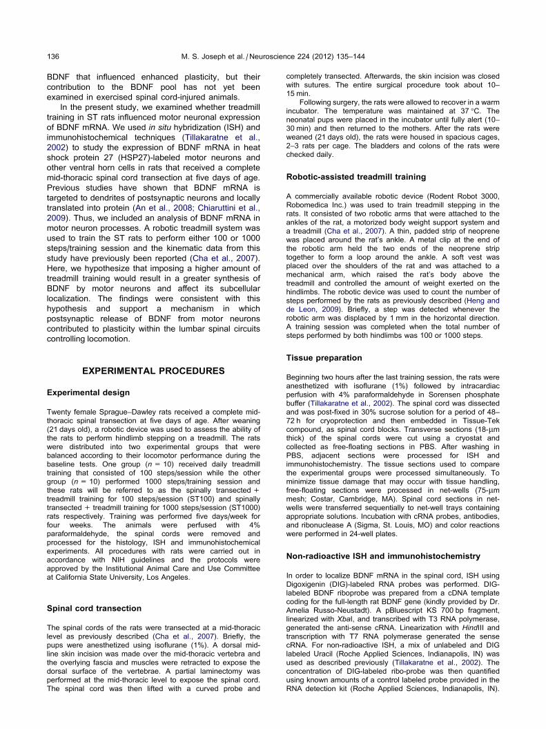
BDNF mRNA in the ventral horn cells was notcorrelated with stepping ability
The ST1000 rats recovered significantly better stepping
than the ST100 rats and these behavioral and kinematic
data have been reported elsewhere (Cha et al., 2007).
Here, we examined the correlation between BDNF
mRNA labeling and step area. In the ST1000 rats, there
was a positive correlation between the BDNF mRNA
label in motor neurons and step area but this correlation
was not statistically significant (Fig. 6A; p= 0.353). In
contrast, BDNF mRNA label in motor neurons of the
ST100 rats was negatively correlated with step area, but
this correlation was also not statistically significant
(Fig. 6B; p= 0.359). For non-motor neurons, BDNF
mRNA labeling was positively correlated with step area
in the ST1000 (Fig. 6C) rats but negatively correlated
with step area in the ST100 rats (Fig. 6D). These
correlations were also not statistically significant
(p= 0.611 for ST1000 and p= 0.378 for ST100).
DISCUSSION
In summary, we found that the expression of BDNF
mRNA in the motor neurons and non-motor neurons in
the ventral horn of the lumbar spinal cord was greater in
ST rats that received a high amount of treadmill training
relative to a low amount and resembled the normal
expression. BDNF mRNA labeling was present in motor
neuron processes, but the high amount of training
resulted in a greater number of motor neurons
containing BDNF mRNA label in processes than the low
amount of training. BDNF mRNA expression was not
correlated with stepping performance in the ST rats.
BDNF synthesis by motor neurons was enhanced bytreadmill training following spinal cord injury
Previous studies have shown that motor neurons in the
lumbar spinal cord expressed BDNF mRNA (Buck et al.,
2000) and that treadmill training increased motor
neuronal expression of BDNF mRNA in Intact rats
(Macias et al., 2007). The present results expand on
these findings by demonstrating that the effect of
treadmill training on BDNF mRNA expression occurred
even in animals that had a complete spinal cord injury.
Our findings were consistent with a recent report that
hindlimb cycling exercise in ST rats increased BDNF
mRNA expression in motor neurons (Keeler et al.,
2012). Whether changes in BDNF protein expression
were coincident with the observed BDNF mRNA
expression cannot be determined from the present
study. However, we and others have shown that BDNF
protein in motor neurons was increased by treadmill
training in ST rats (Macias et al., 2009; de Leon et al.,
2011). Taken together, these findings suggested that
motor neurons in the lumbar spinal cord were stimulated
by treadmill exercise to synthesize BDNF. In addition to
muscle-derived BDNF, BDNF synthesized by motor
neurons may provide another source of BDNF used to
enhance the plasticity after spinal cord injury.
Treadmill training in ST animals improved hindlimb
function in part by influencing synaptic inputs to motor
neurons (Tillakaratne et al., 2002; Cote et al., 2003,
2011; Cote and Gossard, 2004; Macias et al., 2009; de
Leon et al., 2011; Ichiyama et al., 2011). In other
systems, activity-dependent release of BDNF from the
dendrites of postsynaptic neurons influenced synaptic
plasticity (for review see Kuczewski et al. (2012)). A
similar phenomenon may be occurring with synapses
onto motor neurons. Under this scenario, BDNF
secreted by motor neuron dendrites affected synaptic
inputs onto the motor neurons themselves or perhaps
modulated other, nearby synapses. In the present study,
BDNF mRNA labeling was observed in motor neuron
processes (presumably, some of these were dendrites)
and the expression in these processes was augmented
by training. Other studies have reported BDNF protein to

Fig. 4. Histograms of BDNF mRNA label intensity in motor neurons found within the same spinal cord section for Intact (A), ST100 (B) and ST1000
(C). The frequency was calculated as a percentage of total motor neurons within the spinal cord section. The data are the averages ± standard
errors from n= 10 for each group. See Experimental procedures for details.
Fig. 5. Plots summarizing analyses of BDNF mRNA label in non-motor neurons cells in the ventral horn. Average number of BDNF mRNA labeled
objects per rat (A) and average intensity of BDNF mRNA staining in non-motor neurons (B) in the Intact, ST100 and ST1000 groups is shown. Mean
intensity of BDNF mRNA in non-motor neuron cells in ST100 is significantly lower (p< 0.05) than the Intact group (B). Average values are shown
±standard errors for n= 10 per each group.
M. S. Joseph et al. / Neuroscience 224 (2012) 135–144 141
Fig. 6. Correlation of BDNF mRNA labeling and area of step cycle trajectory. Step area was correlated with BDNF mRNA labeling in the motor
neurons (A, B) and non-motor neurons (C, D). White squares and black triangles are data from ST1000 and ST100 rats, respectively. The line is the
linear regression. The Pearson correlation coefficient (r) is shown.
142 M. S. Joseph et al. / Neuroscience 224 (2012) 135–144
cluster within motor neuron dendrites following treadmill
training (Skup et al., 2002; Macias et al., 2005). These
findings are consistent with targeting of BDNF mRNA to
the dendritic compartment and local translation into
BDNF protein (An et al., 2008; Chiaruttini et al., 2009).
Recent findings suggested that another neurotrophin,
NT-3, enhanced motor neuronal inputs within the lumbar
spinal circuitry. Viral-delivery of NT-3 resulted in
enhanced excitatory synaptic potentials recorded from
ankle extensor motor neurons in ST rats (Boyce et al.,
2012) and a similar result was reported in Intact rats
(Petruska et al., 2010). Interestingly, delivery of BDNF
did not affect excitatory synaptic potentials. Based on
electrophysiological data and c-fos labeling in the spinal
cord, the effect of BDNF was to raise the level of
excitation in motor neuron and interneurons, some of
which may have been involved in central pattern
generation (Boyce et al., 2012). Endogenous BDNF
derived from motor neurons may have a similar effect
on neuronal excitability but clearly further studies are
necessary to examine the role of exercise-induced
BDNF on spinal plasticity.
One important question was how much treadmill
exercise was necessary for stimulating BDNF synthesis
by spinal neurons? Based on the present findings, a
higher amount of treadmill training (1000 steps/session)
was better than the lower amount (100 steps/session).
Compared to previous studies of BDNF mRNA, the
exercise regimen in the present study can be
considered low intensity. Intact rats in other studies ran
on the treadmill for 30–80 min/session (Gomez-Pinilla
et al., 2001; Macias et al., 2007). In the present study,
the treadmill speed was kept low (i.e. walking speed) to
facilitate stepping in the ST rat and the rats were trained
for shorter durations (approximately 5 and 20 min for the
100 and 1000 rats respectively). The number of
treadmill training sessions was another factor
determining BDNF mRNA expression. In Intact rats, a
single treadmill training session was not sufficient for
increasing BDNF mRNA (Gomez-Pinilla et al., 2001).
Only when rats underwent five or more treadmill training
sessions was there an increase in BDNF mRNA
(Gomez-Pinilla et al., 2001). The rats in the present
study underwent a total of 20 training sessions. Taken
together, these findings suggested that imposing low
intensity treadmill training may be a sufficient stimulus
for BDNF synthesis if it is continued over a long period
of time. This finding has implications for body weight-
supported treadmill training which is typically carried out
at slower treadmill speeds for individuals with spinal
cord injury.
We chose to use a group that received a low amount
of exercise rather than an untrained ST control as has
been commonly used in previous studies. Our rationale
was that BDNF mRNA levels are influenced by non-
specific factors related to the robotic training protocol, in
particular restraint of the rats during training and robotic
linkage stimulating the ankle. Hippocampal BDNF
mRNA levels were influenced by restraint alone (Smith
et al., 1995). Since the ST100 group received robotic
training, effects due to restraint of the rats or stimulation
from the robotic linkages on BDNF mRNA were taken
into account. It is possible that imposing a low amount
of training was sufficient to stimulate BDNF mRNA
M. S. Joseph et al. / Neuroscience 224 (2012) 135–144 143
expression but this cannot be determined in the present
study. If BDNF synthesis was activity-dependent, then
one would expect that the low amount of training would
result in greater BDNF mRNA expression than in the
absence of training. We previously showed that training
ST rats to perform 100 step/training session did not
significantly raise BDNF protein expression in motor
neurons suggesting the low amount of training did not
generate enough activity to stimulate BDNF protein (de
Leon et al., 2011). This finding suggested that there
may be a threshold level of activity that was necessary
to induce BDNF protein and perhaps mRNA expression.
Cellular expression of BDNF mRNA was notassociated with improved locomotor function
The positive but insignificant correlation between BDNF
mRNA expression in motor neurons and the kinematic
variable, step area suggested that the increased BDNF
synthesis by motor neurons was not associated with
improved locomotor recovery. We previously showed
that BDNF protein expression in the ventral horn was
raised by treadmill training in ST rats there was a
significant correlation between the overall expression of
BDNF protein in the ventral horn region and locomotor
recovery (de Leon et al., 2011). Other studies have
reported that the overall levels of BDNF protein and
mRNA in the lumbar spinal cord were significantly
correlated with stepping recovery in exercised spinal
cord-injured rats (Ying et al., 2005).
One explanation for the lack of correlation to
locomotor recovery may be related to the variability in
BDNF mRNA expression that was observed in the
ventral horn. BDNF mRNA expression in motor neurons
ranged from low to high in the ST and Intact rats
(Fig. 4). This conclusion was based on the intensity of
labeling observed in motor neurons found within the
same tissue section thus, the variability was not due to
other factors that potentially influenced intensity values
across different tissue sections. The repetitive use of
specific spinal pathways during treadmill training would
be expected to result in BDNF synthesis by specific
motor pools. We performed a broad analysis of motor
neurons and this may have reduced the strength of
correlations to locomotor recovery particularly if
localized motor pools were recruited during training. The
idea that treadmill training differentially modulated
hindlimb motor pools is supported by previous findings
(Tillakaratne et al., 2002; Khristy et al., 2009). For
example, treadmill training in ST rats increased in
GABA receptor expression in ankle extensor motor
pools but decreased receptor expression in flexor motor
pools (Khristy et al., 2009). Further studies are
necessary to explore the differential cellular expression
of BDNF expression by integrating retrograde labeling of
motor pools.
Other cells in the ventral horn, particularly in Lamina
VII and VIII that were not motor neurons also expressed
BDNF mRNA. The expression of BDNF mRNA in these
cells was affected by training and similar to motor
neurons, the expression of BDNF mRNA in these cells
was not significantly correlated with stepping recovery.
Unfortunately, other markers for neurons and glial cells
were not used in the present study, thus, the identity of
these cells is unknown. Some of these cells may have
been interneurons that were involved in the generation
of stepping and like motor neurons, training may have
induced activity in these interneurons thereby
stimulating them to synthesize BDNF. Glial cells cannot
be ruled out though since microglia have been shown to
express BDNF in spinal cord-injured rats (Dougherty
et al., 2000). In any case, the present findings
suggested that in addition to motor neurons, other
ventral horn cells may contribute to the pool of BDNF
within the spinal cord circuitry.
Clinical implications
Body weight-supported treadmill training has been shown
to be effective in improving locomotor function in spinal
cord-injured patients (Behrman et al., 2005).
Understanding BDNF’s role in spinal plasticity may lead
to new interventions that enhance the effectiveness of
this form of therapy. Past studies have successfully
delivered BDNF and NT-3 to the lumbar spinal cord to
improve locomotor recovery in spinal cord-injured
animals (Boyce et al., 2007, 2012). While more studies
are necessary, the present findings indicated that spinal
plasticity may be under the control of endogenously
produced neurotrophins such as BDNF. BDNF from
spinal cord cells and muscle fibers may trigger plasticity
within the locomotor-generating circuitry of the spinal
cord. If this hypothesis is true, the implications are that
individuals with spinal cord injury may benefit from
activity-based therapies that optimally stimulate the
synthesis and release of endogenous BDNF.
Acknowledgements—This work was supported by NIH Grant
R01NS055911 and an American Recovery and Reinvestment
Act (ARRA) supplement.
REFERENCES
An JJ, Gharami K, Liao GY, Woo NH, Lau AG, Vanevski F, Torre ER,
Jones KR, Feng Y, Lu B, Xu B (2008) Distinct role of long 30 UTR
BDNF mRNA in spine morphology and synaptic plasticity in
hippocampal neurons. Cell 134:175–187.
Behrman AK, Lawless-Dixon AR, Davis SB, Bowden MG, Nair P,
Phadke C, Hannold EM, Plummer P, Harkema SJ (2005)
Locomotor training progression and outcomes after incomplete
spinal cord injury. Phys Ther 85:1356–1371.
Boyce VS, Park J, Gage FH, Mendell LM (2012) Differential effects of
brain-derived neurotrophic factor and neurotrophin-3 on hindlimb
function in paraplegic rats. Eur J Neurosci 35:221–232.
Boyce VS, Tumolo M, Fischer I, Murray M, Lemay MA (2007)
Neurotrophic factors promote and enhance locomotor recovery in
untrained spinalized cats. J Neurophysiol 98:1988–1996.
Buck CR, Seburn KL, Cope TC (2000) Neurotrophin expression by
spinal motoneurons in adult and developing rats. J Comp Neurol
416:309–318.
Cha J, Heng C, Reinkensmeyer DJ, Roy RR, Edgerton VR, de Leon
RD (2007) Locomotor ability in spinal rats is dependent on the
amount of activity imposed on the hindlimbs during treadmill
training. J Neurotrauma 24:1000–1012.
Chiaruttini C, Vicario A, Li Z, Baj G, Braiuca P, Wu Y, Lee FS,
Gardossi L, Baraban JM, Tongiorgi E (2009) Dendritic trafficking
144 M. S. Joseph et al. / Neuroscience 224 (2012) 135–144
of BDNF mRNA is mediated by translin and blocked by the G196A
(Val66Met) mutation. Proc Natl Acad Sci U S A 106:
16481–16486.
Cote MP, Azzam GA, Lemay MA, Zhukareva V, Houle JD (2011)
Activity-dependent increase in neurotrophic factors is associated
with an enhanced modulation of spinal reflexes after spinal cord
injury. J Neurotrauma 28:299–309.
Cote MP, Gossard JP (2004) Step training-dependent plasticity in
spinal cutaneous pathways. J Neurosci 24:11317–11327.
Cote MP, Menard A, Gossard JP (2003) Spinal cats on the treadmill:
changes in load pathways. J Neurosci 23:2789–2796.
de Leon R, See PA, Chow CH (2011) Differential effects of low versus
high amounts of weight supported treadmill training in spinal rats.
J Neurotrauma 28:1021–1033.
Dougherty KD, Dreyfus CF, Black IB (2000) Brain-derived
neurotrophic factor in astrocytes, oligodendrocytes, and
microglia/macrophages after spinal cord injury. Neurobiol Dis
7:574–585.
Dupont-Versteegden EE, Houle JD, Dennis RA, Zhang J, Knox M,
Wagoner G, Peterson CA (2004) Exercise-induced gene
expression in soleus muscle is dependent on time after spinal
cord injury in rats. Muscle Nerve 29:73–81.
Gomez-Pinilla F, Ying Z, Opazo P, Roy RR, Edgerton VR (2001)
Differential regulation by exercise of BDNF and NT-3 in rat spinal
cord and skeletal muscle. Eur J Neurosci 13:1078–1084.
Gomez-Pinilla F, Ying Z, Roy RR, Molteni R, Edgerton VR (2002)
Voluntary exercise induces a BDNF-mediated mechanism that
promotes neuroplasticity. J Neurophysiol 88:2187–2195.
Heng C, de Leon RD (2009) Treadmill training enhances the recovery
of normal stepping patterns in spinal cord contused rats. Exp
Neurol 216:139–147.
Hutchinson KJ, Gomez-Pinilla F, Crowe MJ, Ying Z, Basso DM
(2004) Three exercise paradigms differentially improve sensory
recovery after spinal cord contusion in rats. Brain 127:1403–1414.
Ichiyama RM, Broman J, Roy RR, Zhong H, Edgerton VR, Havton LA
(2011) Locomotor training maintains normal inhibitory influence
on both alpha- and gamma-motoneurons after neonatal spinal
cord transection. J Neurosci 31:26–33.
Keeler BE, Liu G, Siegfried RN, Zhukareva V, Murray M, Houle JD
(2012) Acute and prolonged hindlimb exercise elicits different
gene expression in motoneurons than sensory neurons after
spinal cord injury. Brain Res 1438:8–21.
Khristy W, Ali NJ, Bravo AB, de Leon R, Roy RR, Zhong H, London
NJ, Edgerton VR, Tillakaratne NJ (2009) Changes in GABA(A)
receptor subunit gamma 2 in extensor and flexor motoneurons
and astrocytes after spinal cord transection and motor training.
Brain Res 1273:9–17.
Koliatsos VE, Clatterbuck RE, Winslow JW, Cayouette MH, Price DL
(1993) Evidence that brain-derived neurotrophic factor is a trophic
factor for motor neurons in vivo. Neuron 10:359–367.
Kuczewski N, Porcher C, Gaiarsa JL (2012) Activity-dependent
dendritic secretion of brain-derived neurotrophic factor modulates
synaptic plasticity. Eur J Neurosci 32:1239–1244.
Macias M, Dwornik A, Skup M, Czarkowska-Bauch J (2005) Confocal
visualization of the effect of short-term locomotor exercise on
BDNF and TrkB distribution in the lumbar spinal cord of the rat:
the enhancement of BDNF in dendrites? Acta Neurobiol Exp
(Wars) 65:177–182.
Macias M, Dwornik A, Ziemlinska E, Fehr S, Schachner M,
Czarkowska-Bauch J, Skup M (2007) Locomotor exercise alters
expression of pro-brain-derived neurotrophic factor, brain-derived
neurotrophic factor and its receptor TrkB in the spinal cord of adult
rats. Eur J Neurosci 25:2425–2444.
Macias M, Nowicka D, Czupryn A, Sulejczak D, Skup M, Skangiel-
Kramska J, Czarkowska-Bauch J (2009) Exercise-induced motor
improvement after complete spinal cord transection and its
relation to expression of brain-derived neurotrophic factor and
presynaptic markers. BMC Neurosci 10:144.
Petruska JC, Kitay B, Boyce VS, Kaspar BK, Pearse DD, Gage FH,
Mendell LM (2010) Intramuscular AAV delivery of NT-3 alters
synaptic transmission to motoneurons in adult rats. Eur
J Neurosci 32:997–1005.
Plumier JC, Hopkins DA, Robertson HA, Currie RW (1997)
Constitutive expression of the 27-kDa heat shock protein
(Hsp27) in sensory and motor neurons of the rat nervous
system. J Comp Neurol 384:409–428.
Sandrow-Feinberg HR, Izzi J, Shumsky JS, Zhukareva V, Houle JD
(2009) Forced exercise as a rehabilitation strategy after unilateral
cervical spinal cord contusion injury. J Neurotrauma 26:721–731.
Skup M, Dwornik A, Macias M, Sulejczak D, Wiater M, Czarkowska-
Bauch J (2002) Long-term locomotor training up-regulates
TrkB(FL) receptor-like proteins, brain-derived neurotrophic
factor, and neurotrophin 4 with different topographies of
expression in oligodendroglia and neurons in the spinal cord.
Exp Neurol 176:289–307.
Smith MA, Makino S, Kvetnansky R, Post RM (1995) Stress and
glucocorticoids affect the expression of brain-derived neurotrophic
factor and neurotrophin-3 mRNAs in the hippocampus. J Neurosci
15:1768–1777.
Tillakaratne NJ, de Leon RD, Hoang TX, Roy RR, Edgerton VR,
Tobin AJ (2002) Use-dependent modulation of inhibitory capacity
in the feline lumbar spinal cord. J Neurosci 22:3130–3143.
Ying Z, Roy RR, Edgerton VR, Gomez-Pinilla F (2005) Exercise
restores levels of neurotrophins and synaptic plasticity following
spinal cord injury. Exp Neurol 193:411–419.
Ying Z, Roy RR, Zhong H, Zdunowski S, Edgerton VR, Gomez-Pinilla
F (2008) BDNF-exercise interactions in the recovery of
symmetrical stepping after a cervical hemisection in rats.
Neuroscience 155:1070–1078.
(Accepted 14 August 2012)(Available online 21 August 2012)


