CLINICAL STUDY
The superiority of conservative resection and adjuvant radiationfor craniopharyngiomas
Adam Schoenfeld • Melike Pekmezci • Michael J. Barnes • Tarik Tihan •
Nalin Gupta • Kathleen R. Lamborn • Anu Banerjee • Sabine Mueller •
Susan Chang • Mitchel S. Berger • Daphne Haas-Kogan
Received: 26 October 2011 / Accepted: 25 January 2012 / Published online: 15 February 2012
� Springer Science+Business Media, LLC. 2012
Abstract The purpose of this study is to evaluate the roles
of resection extent and adjuvant radiation in the treatment of
craniopharyngiomas. We reviewed the records of 122
patients ages 11–52 years who received primary treatment
for craniopharyngioma between 1980 and 2009 at the
University of California, San Francisco (UCSF). Primary
endpoints were progression free survival (PFS) and overall
survival (OS). Secondary endpoints were development of
panhypopituitarism, diabetes insipidus (DI), and visual field
defects. Of 122 patients, 30 (24%) were treated with gross
total resection (GTR) without radiation therapy (RT), 3 (3%)
with GTR ? RT, 41 (33.6%) with subtotal resection (STR)
without RT, and 48 (39.3%) with STR ? RT. Median age at
diagnosis was 30 years, with 46 patients 18 years or
younger. Median follow-up for all patients was 56.4 months
(interquartile range 18.9–144.2 months) and 47 months
(interquartile range 12.3–121.8 months) for the 60 patients
without progression. Fifty six patients progressed, 10 have
died, 6 without progression. Median PFS was 61.1 months
for all patients. PFS rate at 2 years was 61.5% (95% CI:
52.1–70.9). OS rate at 10 years was 91.1% (95% CI
84.3–97.9). There was no significant difference in PFS and
OS between patients treated with GTR vs. STR ? XRT
(PFS; p = 0.544, OS; p = 0.735), but STR alone resulted in
significantly shortened PFS compared to STR ? RT or GTR
(p \ 0.001 for both). STR was associated with significantly
shortened OS compared to STR ? RT (p = 0.050) and
trended to shorter OS compared to GTR (p = 0.066). GTR
was associated with significantly greater risk of developing
DI (56.3 vs. 13.3% with STR ? XRT, p \ 0.001) and pan-
hypopituitarism (54.8 vs. 26.7% with STR ? XRT, p =
0.014). In conclusion, for patients with craniopharyngioma,
STR ? RT may provide superior clinical outcome, achiev-
ing better disease control than STR and limiting side effects
associated with aggressive surgical resection.
Keywords Craniopharyngioma � Surgical resection �Radiation therapy � Adult � Pediatric
Introduction
Craniopharyngioma is a neuroepithelial tumor that com-
prises 6–9% of pediatric brain tumors and 1–4% of adult
brain tumors [1–4]. The peak incidence is between ages
5 to 14 in childhood, although a second peak occurs during
adulthood between 50 to 74 years [1]. Craniopharyngioma
is thought to originate from squamous epithelial remnants
of Rathke’s pouch, an embryonic structure that ultimately
forms the anterior pituitary gland [2]. The tumor is most
A. Schoenfeld � D. Haas-Kogan (&)
Department of Radiation Oncology, University of California,
San Francisco (UCSF), 1600 Divisadero St. Suite H1031,
San Francisco, CA 94143-1708, USA
e-mail: [email protected]
M. Pekmezci � M. J. Barnes � T. Tihan
Department of Pathology, University of California,
San Francisco (UCSF), San Francisco, CA, USA
N. Gupta � K. R. Lamborn � S. Chang � M. S. Berger �D. Haas-Kogan
Department of Neurosurgery, University of California,
San Francisco (UCSF), San Francisco, CA, USA
N. Gupta � A. Banerjee
Department of Pediatrics, University of California,
San Francisco (UCSF), San Francisco, CA, USA
S. Mueller
Department of Neurology, University of California,
San Francisco (UCSF), San Francisco, CA, USA
123
J Neurooncol (2012) 108:133–139
DOI 10.1007/s11060-012-0806-7
often located in the infrasellar/suprasellar region of the
brain and is frequently closely associated with adjacent
structures such as the hypothalamus, pituitary gland, optic
chiasm and carotid artery [3, 4].
The optimal treatment strategy for craniopharyngioma is
controversial. Historically, gross total resection has been
the preferred treatment approach, but the tumor’s proximity
to vital structures may lead to high rates of hypothalamic-
pituitary and/or optic impairment [5–8]. Alternative
approaches such as subtotal resection followed by adjuvant
radiation therapy may have comparable long-term outcomes,
while limiting side effects [5, 9–14]. Specifically, Yang et al.
[5, 11–14], have shown no significant difference in tumor
control rates between patients who received STR ? RT vs.
GTR and, a number of other studies have demonstrated a
significantly higher risk of neurologic, ophthalmic, and
endocrine side effects associated with GTR. Conversely,
radiation therapy may lead to long-term problems such as
vasculopathies, intellectual deficits, and secondary tumors
[2, 11, 15, 16]. The issue of long-term toxicity caused by
radiation is especially critical in the pediatric population,
[11, 15–18].
The current study evaluates a historical cohort of
patients treated at the University of California, San Fran-
cisco and seeks to examine the efficacy of different treat-
ment approaches in the management of craniopharyngioma
in both adult and pediatric patients.
Materials and methods
Existing pathology, neurosurgery, and radiation oncology
databases were searched using keywords designed to
retrieve all patients with craniopharyngioma treated at
University of California, San Francisco between 1980 and
2009. Patients were excluded if: (1) initial surgery was not
performed at UCSF; (2) initial treatment was unknown; (3)
there was no confirmatory pathology; and/or (4) lost to
follow-up within two weeks after initial treatment. The
medical records were reviewed and the following data were
collected: demographic information, treatment, treatment-
related morbidity, and outcome. This study was approved
by the UCSF Institutional Review Board.
The degree of resection was determined by reviewing
the operative notes and post-operative imaging. All com-
plete resections and near-total resections defined radio-
logically were classified as GTR, with all other surgical
procedures, including biopsy with cyst aspiration, consid-
ered as STR. The histologic diagnosis was confirmed by a
neuropathologist at UCSF. If progression was not docu-
mented, they were assumed to be progression-free upon the
last day of documented contact. Patients were followed
clinically and with imaging studies (MRI and/or CT). The
imaging studies were performed during follow-up period at
the discretion of the treating physician and were not done at
uniform intervals for all patients. The development of en-
docrinopathies and visual field defects were documented in
UCSF medical records or laboratory studies during the
follow-up period after primary treatment.
SPSS version 18.0 was used for all statistical analyses.
Progression Free Survival was defined as time between ini-
tial surgery and recurrence or death. If the patient was alive
with no documented recurrence, the patient was censored for
PFS at date of last follow-up. Overall Survival was defined as
the time between initial surgery and death. Patients alive
were censored at last known follow-up. Curves for PFS and
OS were generated using the Kaplan–Meier method. A Cox
model was used to assess the association between primary
treatment and outcome while allowing for adjustment of
other potential prognostic factors including age, gender,
histology, and decade of diagnosis. Two-tailed Pearson’s
chi-square and Fisher exact tests were used to evaluate the
relationship between the primary treatment, age, endocrin-
opathies, visual field defects, and histology.
Results
Patient and treatment characteristics
We identified 225 patients in our search, 122 of whom met
our inclusion criteria. Reasons for non-eligibility included
initial surgery was performed at an outside institution, initial
treatment was unknown, no confirmatory pathology from a
UCSF pathology review, and loss to follow-up within
2 weeks of initial treatment (see ‘‘Methods and materials’’
section for details). Patient characteristics are shown in
Table 1. The median age at diagnosis was 30 years (inter-
quartile range 11–52 years), with 47 patients age 18 or under
at diagnosis. Sixty seven (55%) patients were male. There
was a significant association between histology and age
group (p = 0.023). Papillary histology was only detected in
patients older than 18, whereas adamantinomatous histology
was present in patients of all ages. GTR was performed in 33
patients (27%), 3 of whom received adjuvant RT (3%).
Among the 89 patients that underwent STR, adjuvant radi-
ation treatment was given in 48 cases (54%). Extent of
resection and adjuvant radiation, according to age group and
decade of diagnosis, are shown in Tables 2 and 3. Treatment
was significantly associated with age group (p \ 0.001).
Among patients 3 years old or younger, STR ? RT was not
performed and STR only was performed in seven of 8
patients. In patients between the ages 3 and 18, 48% had a
GTR, 26% of patients had STR without RT and 26% of
patients had STR with RT whereas in patients older than 18,
17% had a GTR, 32% had STR without RT and 51% of
134 J Neurooncol (2012) 108:133–139
123
patients had STR with RT. Treatment was not impacted by
histological diagnosis (p = 0.631).
Over the duration of this historical cohort, five neuro-
surgeons performed these surgeries. In general, GTR was
done only if minimal morbidity could be achieved.
Otherwise, the general philosophy was subtotal resection
with XRT. Thirty three surgeries were done through
transsphenoidal approaches, while the rest were performed
via craniotomies.
Treatment outcomes
Fifty-six patients progressed, 10 died, 6 without known
progression. The median follow-up for the 60 patients
without progression was 46 months (interquartile range
12.5–119.9 months) and for all 122 patients was
56.4 months (interquartile range 18.5–142.6 months). Sixty
patients (48.8%) were followed for a minimum of 5 years, 36
patients (29.3%) were followed for a minimum of 10 years
and 13 patients (10.6%) patients were followed for a mini-
mum of 15 years. Patients undergoing GTR had similar
follow-up to those undergoing STR and STR ? XRT.
Median follow-up was 83.3 months (Interquartile range
24–169 months) for GTR, 46.3 months (Interquartile range
7–121 months) for STR, and 56.4 months (Interquartile
range 27–144 months) for STR ? XRT.
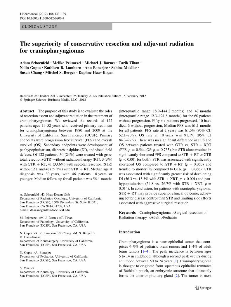
The median PFS was 61.1 months (95% CI 24.3–97.9) and
PFS rate at 2 years was 61.5% (95% CI 52.1–70.9). OS rate at
10 years was 91.1% (95% CI 84.3–97.9). Kaplan meier
curves for PFS and OS are shown in Fig. 1a, b, respectively.
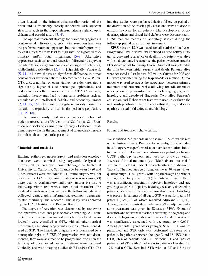
PFS and OS for patients who received GTR, STR and
STR ? RT are shown in Fig. 2a, b. The PFS at 2 years for
patients who received GTR, STR, and STR ? XRT were
75.2% (95% CI 59.3–91.1), 36.2% (95% CI 20.1–52.3),
and 73.3% (95% CI 60.4–86.2), respectively. The OS at
10 years for those who received GTR, STR, and
STR ? XRT were 96.2% (95 CI 88.8–100), 80.8% (95 CI
64.3–97.3), and 95.8% (95 CI 87.8–100), respectively.
There was no significant difference in PFS or endocri-
nopathy with regards to treatment approach. The trans-
sphenoidal approach, however, was associated with
improved OS (p = 0.006).
The seven deaths that occurred in patients who were
treated with STR were due to: a hemorrhagic infarct into
the basal ganglia and frontal lobe 5 days after surgery;
malignant melanoma 90 months after initial treatment;
disseminated intravascular coagulation that occurred fol-
lowing salvage surgery 3.2 months after initial treatment;
and four cases due to disease progression (9.7, 56.0, 125.1,
and 291.2 months, respectively, after initial treatment). In
patients treated with STR ? XRT, two deaths were docu-
mented; one due to progression of disease 60 months after
initial treatment and one from leukemia 151.4 months after
initial treatment. In patients treated with GTR, there was
one death due to a subarachnoid hemorrhage 22.3 months
after initial surgery.
Univariate analysis is shown in Table 4. STR without
XRT was significantly associated with a shortened
Table 1 Patient characteristics and treatment details
Median age (interquartile range) in years 30 (11–52)
Median follow-up (interquartile range) in months 56.4 (18.9–144.2)
Sex M:F 67:55
Histology
Adamantinomatous 79
Papillary 15
NOS 28
Primary therapy
GTR alone 30
GTR ? XRT 3
STR alone 37
STR ? XRT 46
Biopsy or cyst aspiration 6
Decade of diagnosis
1980s 28
1990s 49
2000s 45
Table 2 Extent of resection/adjuvant radiation by age group
Number
B3 years
(n = 8)
3 years \ Age B 18
(n = 39)
[18 years
(n = 75)
GTR 1 19 13
STR 7 10 24
STR ? XRT 0 10 38
Table 3 Extent of resection/adjuvant radiation and endocrinopathy
by decade of diagnoses
Number (total)
1980s 1990s 2000s
GTR 6 19 8
Developed DI 4 (6) 10 (18) 4 (8)
Developed panhypopituitarism 4 (6) 11 (17) 6 (8)
Developed worsening visual defect 2 (4) 4 (11) 1 (3)
STR 7 15 19
Developed DI 2 (7) 7 (13) 2 (19)
Developed panhypopituitarism 1 (7) 7 (13) 4 (19)
Developed worsening visual defect 3 (5) 2 (7) 1 (11)
STR ? XRT 15 15 18
Developed DI 2 (14) 0 (13) 4 (18)
Developed panhypopituitarism 5 (14) 1 (13) 6 (18)
Developed worsening visual defect 1 (7) 1 (4) 3 (14)
J Neurooncol (2012) 108:133–139 135
123
progression free survival in comparison to STR ? XRT
(PFS; HR = 4.152, p \ 0.001). There were no differences in
PFS or OS between GTR and STR ? XRT (PFS;
HR = 1.240, p = 0.544. OS; HR = 0.659, p = 0.735).
Increasing age at diagnosis as a continuous variable was also
associated with decreased OS (HR = 1.35, p = 0.051), but
not with PFS. Gender, histological diagnosis, and decade of
diagnosis did not significantly affect PFS or OS. For PFS,
where sufficient data was available, and adjusting for gender,
histological diagnosis, age and decade of diagnosis, multi-
variate analysis was performed and confirmed treatment type
as the only factor significantly associated with PFS.
A B
Fig. 1 a Progression-free survival. b Overall survival for all patients (n = 122)
ASTR+RT
GTRSTR
B
STR+RT GTRSTR
Fig. 2 a Progression-free survival. b Overall survival according to treatment group. Gross total (GTR) resection: solid line. (STR) Subtotal
resection plus adjuvant radiation (RT): dashed line. STR without RT: dotted line
136 J Neurooncol (2012) 108:133–139
123

Toxicities of treatment
One hundred and fifteen patients had endocrine data
available. A pre-existing diagnosis of panhypopituitarism
or diabetes insipidus (DI) prior to treatment in six and 16
patients, led to their exclusion from our evaluations of
treatment toxicity. Seven additional patients were excluded
from both analyses because there was insufficient data to
determine their endocrine status.
For the GTR cohort, 26 and 29 patients, respectively,
were assessable for DI and panhypopituitarism; for the
STR without XRT cohort 37 were assessable for DI and
panhypopituitarism; and for the STR ? XRT cohort 44 and
43 patients, respectively, were assessable for DI and pan-
hypopituitarism. Of patients treated with GTR, 18 devel-
oped DI and 17 developed panhypopituitarism; in the STR
without XRT cohort 11 developed DI and 12 developed
panhypopituitarism; and in the STR ? XRT cohort six
developed DI and 12 developed panhypopituitarism.
A significantly higher percentage of patients treated with
GTR developed DI and panhypopituitarism than patients
who received STR or STR ? XRT [(DI two-tailed Fisher’s
exact test: GTR vs. STR p = 0.002, GTR vs. STR ? XRT
p \ 0.001, STR vs. STR ? XRT p = 0.100); (panhypo-
pituitarism two-tailed Fisher’s exact test: GTR vs. STR
p = 0.046, GTR vs. STR ? XRT p = 0.014, STR vs.
STR ? XRT p = 0.634)].
Data were available on the visual status of 98 patients
before and after surgical intervention. Thirty three patients
did not have visual defects at baseline and only one
(a patient who had a GTR) of these patients developed a
worsening deficit after surgery. Of 17 GTR patients who
had an initial deficit, 35.3, 29.4, and 35.3% were better, the
same, and worse post treatment, respectively. Of 25
STR ? XRT patients who had an initial deficit, 60.0, 20.0,
and 20.0% were better, the same, and worse post treatment,
respectively. Of 23 STR only patients who had an initial
deficit, 47.8, 26.1, and 26.1% were better, the same, and
worse post treatment, respectively. These differences were
not statistically significant.
Other major adverse events included one case of a
thalamic infarct post surgery and one case of a subdural
hematoma, both occurring in patients who underwent GTR.
In the STR ? XRT group, one patient developed a left
MCA infarct 2 years after treatment, one patient developed
a left parietal infarct 7 years after treatment, and
one patient had multiple strokes after treatment during a
follow-up of 16.5 years. Additionally, 1 patient in STR ?
XRT developed parathyroid cancer 16 years after STR ?
XRT.
Discussion
In this report, we describe the UCSF experience for the
treatment of craniopharyngiomas between 1980 and 2009.
The disease control rates for patients undergoing GTR and
STR ? XRT were comparable and both were better than
STR without adjuvant radiation. However, patients who
underwent GTR had an increased rate of long-term endo-
crine deficits compared to those undergoing STR and
STR ? XRT.
Previously reported series of craniopharyngioma
patients report conflicting results. Yang et al. [9] reviewed
442 patients who underwent tumor resection with a mean
follow-up of 54 months, and found no significant differ-
ence in PFS and OS between GTR and STR ? XRT.
Stripp et al. [5] reported significantly better tumor control
in patients treated with STR ? XRT or GTR, compared to
those who received STR only. Other studies maintain better
Table 4 Univariate analysis of progression-free survival and overall survival
Parameter Progression free survival Overall survival
Hazards ratio 95% CI p value Hazards ratio 95% CI p value
Treatment \0.001 0.047
GTR vs. STR ? RT 1.240 0.619–2.485 0.544 0.659 0.059–7.398 0.735
STR vs. STR ? RT 4.152 2.264–7.614 \0.001 4.880 1.00–23.740 0.050
GTR vs. STR 0.299 0.159–0.560 \0.001 0.135 0.16–1.137 0.066
Age (years) 0.999 0.987–1.012 0.901 1.031 1.000–1.064 0.051
Gender (M/F) 1.102 0.665–1.826 0.706 1.319 0.371–4.689 0.669
Decade of diagnosis 0.403 0.160
1980s vs. 2000s 0.649 0.323–1.306 0.226 0.168 0.023–1.234 0.080
1990s vs. 2000s 0.952 0.523–1.733 0.873 0.257 0.048–1.394 0.115
Histology 0.231 0.583
Adamantinomatous vs. NOS 1.474 0.794–2.737 0.219 3.012 0.377–24.099 0.299
Papillary vs. NOS 0.728 0.237–2.235 0.579 0.000 0 0.988
J Neurooncol (2012) 108:133–139 137
123

outcomes for patients following GTR only [19, 20]. These
inconsistencies among studies may be due to the variable
nature of treatment selection at different institutions.
Patients with less aggressive tumors may be dispropor-
tionately selected for GTR in some studies, accounting for
the better GTR outcomes.
Data on treatment with STR only for craniopharyngioma
consistently demonstrates that this treatment strategy is
associated with poor outcomes. Yang et al. [9], showed a
significantly decreased PFS and a trend towards decreased
OS in STR only vs. GTR. Stripp et al. [5], found that the
majority (78%) of patients who received STRs had tumor
recurrences within a year if they did not receive XRT
whereas patients who received postoperative radiation had
a local control rate of 84% at 10 years. Our results are
relatively similar in that 61% of patients who had STR
without radiation progressed within 1 year. Karavitaki
et al. [19] also found that patients who received
STR ? XRT had markedly better PFS rates than those who
received STR only (77% PFS rate at 10 years for
STR ? XRT vs. 38% for STR only).
Data on toxicities among treatment approaches are also
conflicting. Many studies have shown lower toxicity rates
after STR compared with GTR, whereas others maintain no
difference in toxicity among treatment approaches. Stripp
et al. [5] report a significantly increased risk of DI when
GTR is performed rather than STR. Merchant et al. [11, 14]
and Thomsett et al. further report that GTR may be asso-
ciated with higher rates of a number of endocrine distur-
bances including hypothyroidism, hypogonadism, and
growth hormone insufficiency as well as neurologic and
ophthalmic side effects. In contrast, Karavitaki et al. [19]
and Weiner et al.[20] report no association between
endocrine or neurologic side effects and treatment strategy.
Studies that report comparable toxicities in STR as com-
pared to GTR may have included a large number of more
extensive resections in the STR group, which could explain
the lack of association between toxicity and treatment
strategy in these studies.
Our study evaluates a relatively large cohort of patients
treated at a single institution with long follow-up over a
period of 30 years. All patients had pathological confir-
mation by a UCSF neuro-pathologist, and regular follow up
was documented. However, the inherent constraints of a
retrospective study limit the conclusions we can draw from
our findings with variability in surgery, radiation, and
follow-up. Even longer follow-up may be especially
important to assess the impact of long-term toxicity in
patients treated at a young age. Furthermore, endocrinop-
athy from XRT may increase over time whereas endocri-
nopathy from surgical resection would likely occur closer
to surgery. Therefore, the observed difference between
GTR and STR ? XRT may in actuality be less pro-
nounced. Further studies with long follow-up of irradiated
patients are necessary to evaluate endocrinopathy devel-
opment over time.
We were not able to analyze the efficacy of other
treatment modalities currently being used such as stereo-
tactic radiosurgery and fractionated stereotactic radiother-
apy since this is not the primary treatment philosophy at
our institution. Minniti et al. [21] recently reviewed data in
eight published studies for patients who received stereo-
tactic radiosurgery and fractionated stereotactic radiother-
apy for craniopharyngiomas (252 patients with a median
follow-up of 57 months) and demonstrated the potential
efficacy of these as primary treatment modalities. The
study reported a control rate of 69% with no differences
between children and adult patients in late toxicities
(neurological and endocrine) ranging from 0–34%. Still,
further prospective studies with long follow-up are needed
to directly compare efficacy of stereotactic radiosurgery
and fractionated stereotactic radiotherapy to GTR and
STR ? XRT.
Finally, our evaluation of toxicities following treatment
was limited to the toxicities that were readily verifiable
from the data available for our patients. Notably, toxicities
associated with radiation, including vasculopathies and
secondary tumors, did not occur commonly despite the
long-term follow-up in this study. A total of five clinically
significant vasculopathies occurred, two in the GTR group
and three in the STR ? XRT group. Only one secondary
tumor occurred in the entire cohort–a parathyroid cancer
that developed 16 years after radiation. Neuro-cognitive
effects that may be associated with radiation therapy were
not evaluated in this study.
We highlight and confirm the shortcomings of STR
alone as primary treatment for craniopharyngioma. Patients
who received STR only were at significantly increased risk
of recurrence and death in comparison to patients who
received STR ? XRT or GTR. Moreover, our study dem-
onstrated that patients who received GTR developed
endocrine dysfunction at a significantly higher rate than
those who received STR ? XRT, with equivalent long-
term efficacy, supporting SRT ? XRT as an appealing
treatment option. Further studies with longer follow-up are
necessary to assess the long-term outcomes and morbidities
associated with craniopharyngioma treatment, especially in
the pediatric subpopulation.
Acknowledgments This research was supported in part by NIH
Brain Tumor SPORE grant P50 CA097257 (DHK, KL, MSB), Nancy
and Stephen Grand Philanthropic Fund (DHK), and The V Foundation
(DHK).
Conflict of interest No actual or potential conflicts exist.
138 J Neurooncol (2012) 108:133–139
123
References
1. Bunin GR, Surawicz TS, Witman PA et al (1998) The descriptive
epidemiology of craniopharyngioma. J Neurosurg 89:547–551
2. Moore K, Couldwell WT (2000) Craniopharyngioma. In: Bern-
stein M, Berger MS (eds) Neuro-oncology: the essentials. Thi-
eme, New York
3. Harwood-Nash DC (1994) Neuroimaging of childhood cranio-
pharyngioma. Pediatr Neurosurg 21:2–10
4. Karavitaki N, Cudlip S, Adams CB et al (2006) Craniopharyn-
giomas. Endocr Rev 27:371–397
5. Stripp DC, Maity A, Janss AJ et al (2004) Surgery with or
without radiation therapy in the management of craniopharyn-
giomas in children and young adults. Int J Radiat Oncol Biol Phys
58:714–720
6. De Vile CJ, Grant DB, Kendall BE et al (1996) Management of
childhood craniopharyngioma: can the morbidity of radical sur-
gery be predicted? J Neurosurg 85:73–81
7. Honegger J, Buchfelder M, Fahlbusch R (1999) Surgical treat-
ment of craniopharyngiomas: endocrinological results. J Neuro-
surg 90:251–257
8. Kalapurakal JA, Goldman S, Hsieh YC et al (2000) Clinical
outcome in children with recurrent craniopharyngioma after pri-
mary surgery. Cancer J 6:388–393
9. Yang I, Sughrue ME, Rutkowski MJ et al (2010) Craniopha-
ryngioma: a comparison of tumor control with various treatment
strategies. Neurosurg Focus 28:E5
10. Lin LL, El Naqa I, Leonard JR et al (2008) Long-term outcome in
children treated for craniopharyngioma with and without radio-
therapy. J Neurosurg Pediatr 1:126–130
11. Merchant TE, Kiehna EN, Sanford RA et al (2002) Craniopha-
ryngioma: the St. Jude Children’s Research Hospital experience
1984–2001. Int J Radiat Oncol Biol Phys 53:533–542
12. Hetelekidis S, Barnes PD, Tao ML et al (1993) 20-year experi-
ence in childhood craniopharyngioma. Int J Radiat Oncol Biol
Phys 27:189–195
13. Scott RM, Hetelekidis S, Barnes PD et al (1994) Surgery, radi-
ation, and combination therapy in the treatment of childhood
craniopharyngioma—a 20 year experience. Pediatr Neurosurg
21:75–81
14. Thomsett MJ, Conte FA, Kaplan SL et al (1980) Endocrine and
neurologic outcome in childhood craniopharyngioma: review of
effect of treatment in 42 patients. J Pediatr 97:728–735
15. Kiehna EN, Merchant TE (2010) Radiation therapy for pediatric
craniopharyngioma. Neurosurg Focus 28:E10
16. Einhaus SL, Sanford RA (1999) Craniopharyngiomas. In: Al-
bright L, Pollack I, Adelson D (eds) Principles and practice of
pediatric neurosurgery. Thieme, New York
17. Liu AK, Bagrosky B, Fenton LZ et al (2009) Vascular abnor-
malities in pediatric craniopharyngioma patients treated with
radiation therapy. Pediatric Blood Cancer 52:227–230
18. Merchant TE, Kiehna EN, Kun LE et al (2006) Phase II trial of
conformal radiation therapy for pediatric patients with cranio-
pharyngioma and correlation of surgical factors and radiation
dosimetry with change in cognitive function. J Neurosurg 104:
94–102
19. Karavitaki N, Brufani C, Warner JT et al (2005) Craniopharyn-
giomas in children and adults: systematic analysis of 121 cases
with long-term follow-up. Clin Endocrinol 62:397–409
20. Weiner HL, Wisoff JH, Rosenberg ME et al (1994) Cranio-
pharyngiomas: a clinicopathological analysis of factors predictive
of recurrence and functional outcome. Neurosurgery 35:1001–
1011
21. Minniti G, Esposito V, Amichetti M et al (2009) The role of
fractionated radiotherapy and radiosurgery in the management of
patients with craniopharyngioma. Neurosurg Rev 32(2):125–132
J Neurooncol (2012) 108:133–139 139
123


