
Signs and Symptoms of the Musculoskeletal and
Connective Tissue System Disorders (2016-2017, Spring Semester)
LECTURE IN INTERNAL MEDICINE PROPAEDEUTICS
M. Yabluchansky L. Bogun, L.Martymianova, O. Bychkova, N. Lysenko
Kharkiv V.N. Karazin National University Medical School’ Internal Medicine Dept.

Plan of the lecture
• Musculoskeletal and Connective Tissue System (MS)
• Spectrum of the Musculoskeletal and Connective Tissue System Disorders (MSDs)
• MSDs’ Methods of Examination
• MSDs’ Signs and Symptoms
• Glossary of Terms referred to the MSDs

Musculoskeletal System (MS)
The musculoskeletal (locomotors) system gives the ability to move using the skeletal (skeleton) and muscular portions
http://www.mkdosteopathy.com/wp-content/uploads/2012/09/musculoskeletal-concerns.jpg

MS: Skeleton and bones
• The skeleton provides the shape for the body, support and protection, allows body movement
• The skeleton is composed of bones supported by ligaments, tendons, muscles and cartilage
• An adult skeleton consists of 206 bones
• The skeleton stores minerals and lipids (99% of the body's calcium is found in the bones, the bones also store energy reserves as lipids in yellow marrow), produce blood cells in the red marrow, which fills the internal cavities of many bones

MS: Skeleton and bones
http://classconnection.s3.amazonaws.com/257/flashcards/769257/png/axial_skeleton1318356986539.png

MS: Muscles 1
• There are skeletal, cardiac, and smooth types of muscles
• Only skeletal and smooth muscles are part of the MS and only the skeletal muscles can move the body
• Skeletal muscles are attached to bones and arranged in opposing groups around joints
• Skeletal muscles are innervated by nerves, which cause the muscles to contract
MS: Muscles 2
• Skeletal muscles contraction is stimulated by the motor neuron sending a message to the muscles from the somatic nervous system
• Skeletal muscles maintain body temperature
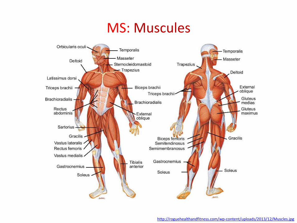
MS: Muscules
http://roguehealthandfitness.com/wp-content/uploads/2013/12/Muscles.jpg
MS: Tendons 1
• A tendon is a tough, flexible band of fibrous connective tissue that connects muscles to bones
• The extra-cellular connective tissue between muscle fibers binds to tendons at the distal and proximal ends, and the tendon binds to the periosteum of individual the sites of muscle's beginning and insertion

MS: Tendons 2
• As muscles contract, tendons transmit the forces to the relatively rigid bones, pulling on them and causing movement
• Tendons can stretch substantially, allowing them to function as springs during locomotion, thereby saving energy

MS: Joints 1
• Joints connect individual bones and may allow bones to move against each other to cause movement:
–Diarthroses which allow extensive mobility between two or more articular heads
http://www.hawaiianshirtray.com/anatomy-physiology/skeletal-system-bones-joints-connective-tissue/

MS: Joints 2
• Joints connect individual bones and may allow bones to move against each other to cause movement:
– False joints or synarthroses, joints that are immovable, that allow little or no movement and are predominantly fibrous
http://www.hawaiianshirtray.com/anatomy-physiology/skeletal-system-bones-joints-connective-tissue/

MS: Joints 3
• Joints connect individual bones and may allow bones to move against each other to cause movement:
– Synovial joints are lubricated by a solution called synovial fluid that is produced by the synovial membranes
http://www.hawaiianshirtray.com/anatomy-physiology/skeletal-system-bones-joints-connective-tissue/

MS: Ligaments
A ligament is a small band of dense, white, fibrous elastic tissue, and connects the ends of bones together in order to form a joint
http://www.humankinetics.com/AcuCustom/Sitename/DAM/086/249art1_Main .png

MS: Bursa
A bursa is a fluid-filled sac made of fibrous tissue and lined with synovial membrane, it provides a cushion between bones and tendons and/or muscles around a joint
http://www.easynotecards.com/uploads/97/40/_53022d86_13d85ffda7d__8000_00004741.jpg

MS: Cartilage 1
• Cartilage is a type of connective tissue. There are three types of cartilage: hyaline cartilage, elastic cartilage, and fibrocartilage
–Hyaline cartilage is the most common type of cartilage, it provides stiff but somewhat flexible support
– Elastic cartilage provides support but can tolerate distortion without damage and return to its original shape

MS: Cartilage 2
• Cartilage is a type of connective tissue. There are three types of cartilage: hyaline cartilage, elastic cartilage, and fibrocartilage
– Fibrocartilage resists compression, prevents bone-to-bone contact, and limits relative movement

MS: Cartilage
http://akopc.com//wp-content/uploads/2012/11/Cartilage1.jpg

Spectrum of MS Disorders (MSDs) 1
• MSDs are heritable and acquired degenerative and inflammatory conditions that cause pain and impair normal activities in the body's joints, ligaments, muscles, nerves, tendons, and structures that support limbs, neck and back
• MSDs can arise from a sudden, or they can arise from making the same motions repeatedly repetitive strain, or from repeated exposure to force, vibration, or awkward posture
http://www.chegg.com/homework-help/definitions/endocrine-system-13

Spectrum of MS Disorders (MSDs) 2
• Abrasions, contusions, and fractures that occur from sudden physical contact with objects that might occur in an accident are not considered MSDs
http://www.chegg.com/homework-help/definitions/endocrine-system-13

MSDs: Degenerative 1
Heritable
• Marfan syndrome (causing abnormal fibrillin)
• Osteogenesis imperfecta (brittle bone disease, caused by insufficient production of normal collagen)
• Ehlers-Danlos syndrome (defect in the synthesis of collagen, affecting joints, heart valves, organ walls, arterial walls, atc)

MSDs: Degenerative 2
Acquired
• Osteoporosis
• Others

MSDs: Other
Scurvy – caused by a dietary deficiency in vitamin C, leading to abnormal collagen
http://cdn.vitaminsestore.com/wp-content/uploads/2012/05/3.jpg

MSDs: Acquired
Autoimmune
• Systemic Lupus Erythematosus (SLE)
• Rheumatoid Arthritis
• Scleroderma
• Sjögren's syndrome (Sjögren's disease, a chronic, slowly progressing inability to secrete saliva and tears)
• Psoriatic arthritis
• Others

MSDs: Diagnosis • Since degenerative or inflammatory process
involves soft tissue, there are often no visible signs of injury
• Assessments are based on self-reports by people as to whether or not they are experiencing pain
• A popular measure of MSDs is the Nordic Questionnaire that has a picture of the body with various areas labeled and asks the individuals to indicate in which areas they have experienced pain, and in which areas the pain has interfered with normal activity
http://en.wikipedia.org/wiki/Musculoskeletal_disorder

MSDs: History of Presenting Complaint 1
• Character- What is the pain like?
• Location- Where does it start? Ask specifically (joint distribution)
• Onset- When and how did it come on? (acute vs. gradual/insidious)
• Radiation- Does the pain move/travel anywhere? Deep pain can be poorly localized.
• Intensity- Scale of 1 to 10; effect on ADL and IADLs. Is it getting better, worse, the same?
http://www.sharinginhealth.ca/clinical_evaluation/musculoskeletal_exam.html
MSDs: History of Presenting Complaint 2
• Duration- How long as it been there?
• Pattern: intermittent, migratory, or additive
• Events associated: Falls, morning stiffness/swelling/redness, joint clicking or locking, muscle pain/cramping, wasting, limitation of movement/weakness, numbness/tingling, fevers/chills/night sweats/wt loss, trauma, job vs sports vs repetitive movements?
• Frequency- New vs recurrent pain
http://www.sharinginhealth.ca/clinical_evaluation/musculoskeletal_exam.html

MSDs: History of Presenting Complaint 3
• Palliative factors- what makes the pain better? (rest/activity/pain meds/heat or cold?)
• Provocative factors- As above, but what makes pain worse?
http://www.sharinginhealth.ca/clinical_evaluation/musculoskeletal_exam.html
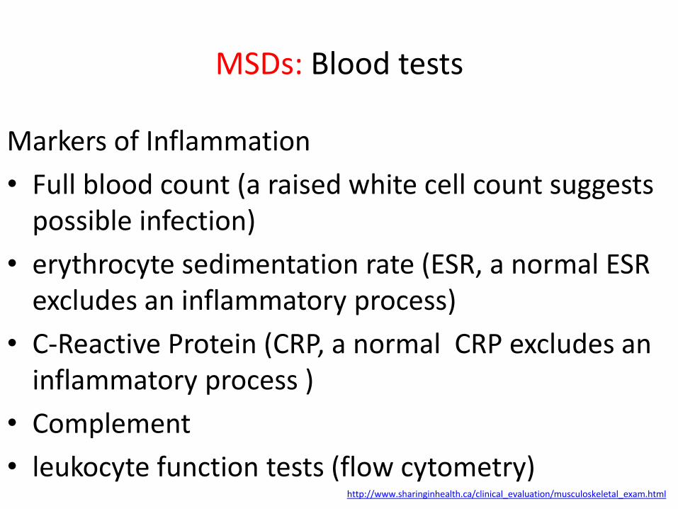
MSDs: Blood tests
Markers of Inflammation
• Full blood count (a raised white cell count suggests possible infection)
• erythrocyte sedimentation rate (ESR, a normal ESR excludes an inflammatory process)
• C-Reactive Protein (CRP, a normal CRP excludes an inflammatory process )
• Complement
• leukocyte function tests (flow cytometry) http://www.sharinginhealth.ca/clinical_evaluation/musculoskeletal_exam.html
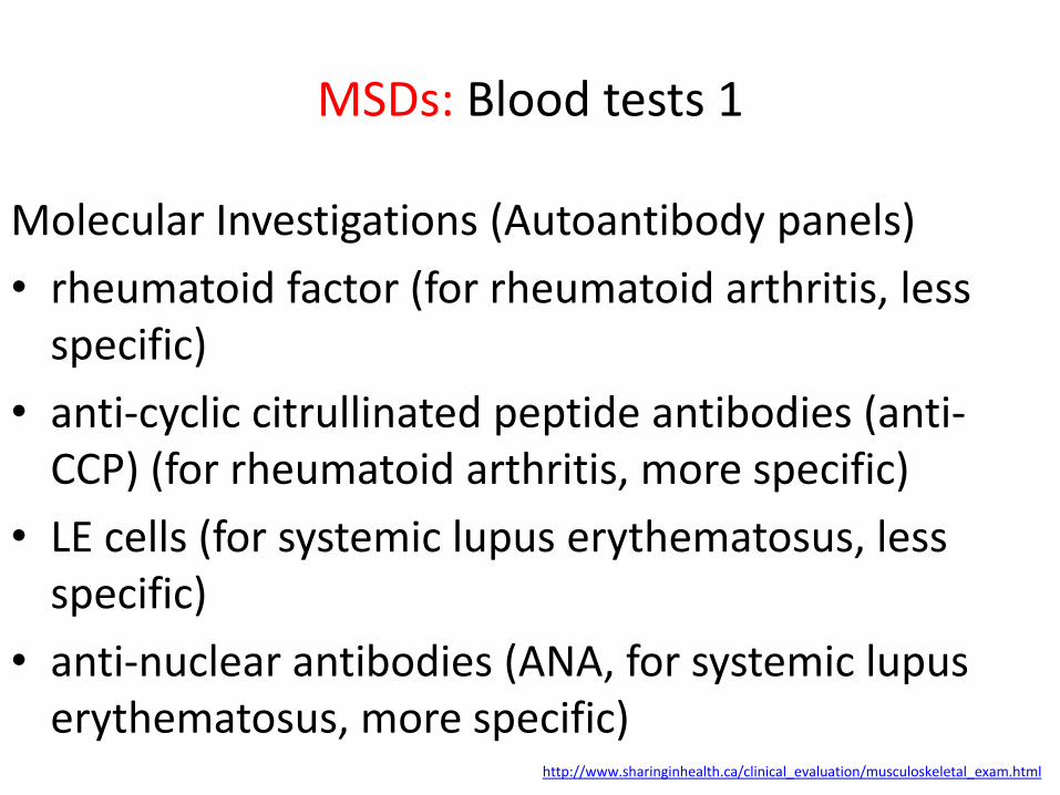
MSDs: Blood tests 1
Molecular Investigations (Autoantibody panels)
• rheumatoid factor (for rheumatoid arthritis, less specific)
• anti-cyclic citrullinated peptide antibodies (anti-CCP) (for rheumatoid arthritis, more specific)
• LE cells (for systemic lupus erythematosus, less specific)
• anti-nuclear antibodies (ANA, for systemic lupus erythematosus, more specific)
http://www.sharinginhealth.ca/clinical_evaluation/musculoskeletal_exam.html

MSDs: Blood tests 2
Molecular Investigations (Autoantibody panels)
• anti-double-stranded DNA antibody (anti-dsDNA) for systemic lupus erythematosus, confirmation of diagnosis of SLE in patients who have a positive test for ANA)
• anti-neutrophil cytoplasmic antibody (ANCA) - Wegener's, microvascular polyangiitis
• extractable nuclear antigens (ENA , for systemic lupus erythematosus)
http://www.sharinginhealth.ca/clinical_evaluation/musculoskeletal_exam.html

MSDs: Blood tests 3
Molecular Investigations (Autoantibody panels)
• anti-phospholipid antibodies (APLA , for systemic lupus erythematosus)
• gene HLA-B27 (increased risk for development of spondyloarthropathy)
http://www.sharinginhealth.ca/clinical_evaluation/musculoskeletal_exam.html

MSDs: Nerve and muscle tests • Electromyography (neuromuscular junction)
• Nerve Conduction Studies (neuromuscular junction)
– If nerve conduction is slow, the cause may be a disorder that affects one nerve, or the cause may be a disorder that affects nerves throughout the body (diabetes)
– If the muscle’s response is progressively weaker after repeated stimulation, a problem with the neuromuscular junction (as occurs in myasthenia gravis) may be the cause
http://www.merckmanuals.com/home/brain-spinal-cord-and-nerve-disorders/symptoms-and-diagnosis-of-brain-spinal-cord-and-nerve-disorders/tests-for-brain-spinal-cord-and-nerve-disorders#v734272
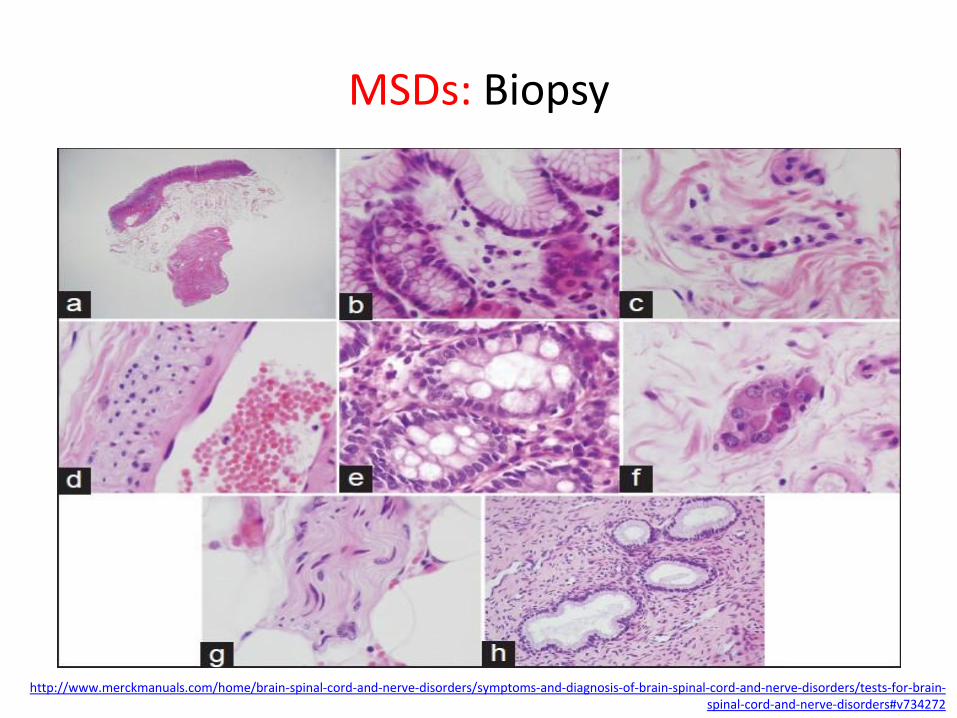
MSDs: Biopsy
http://www.merckmanuals.com/home/brain-spinal-cord-and-nerve-disorders/symptoms-and-diagnosis-of-brain-spinal-cord-and-nerve-disorders/tests-for-brain-spinal-cord-and-nerve-disorders#v734272

MSDs: Imaging tests
• CT— Computed Tomography
• MRI— Magnetic Resonance Imaging
• Angiography
• PET— Radionuclide Scanning
• Doppler ultrasonography
http://www.merckmanuals.com/home/brain-spinal-cord-and-nerve-disorders/symptoms-and-diagnosis-of-brain-spinal-cord-and-nerve-disorders/tests-for-brain-spinal-cord-and-nerve-disorders#v734272

MSDs: Dual-energy X-ray absorptiometry (DXA)
http://www.svmedicalimaging.com/images/GE-Dexa-AdvanceTB.jpg

MSDs: Dual-energy X-ray absorptiometry (DXA)
The most accurate way to evaluate bone density at the lower spine, hip, wrist, or entire body, which is necessary when screening for or diagnosing osteopenia or osteoporosis
http://dxline.info/img/new_ail/dual-energy-x-ray-absorptiometry_3.jpg

MSDs: Synovial Fluid Analysis
Synovial fluid should be examined in all pts with arthritis and whenever joint infection is probable:
• cell count and differential
• crystal examination for sodium urate and calcium phosphate dehydrate (CPPD)
• bacteria (culture and sensitivity, gram stain)
–500-1000 = non-inflammatory (OA)
–1000-10,000 = inflammatory
normal synovial fluid is colourless or straw-
coloured and has <200 WBC/mm3

MSDs: Synovial Fluid Analysis
http://ocw.tufts.edu/data/19/301911/301928_xlarge.jpg

MSDs: Arthroscopy During arthroscopy, doctors can take a piece of tissue (such as joint cartilage or the joint capsule) for analysis (biopsy), and, if necessary, do surgery to correct the condition
http://i.ytimg.com/vi/KgU_dOeQLQM/maxresdefault.jpg http://www.merckmanuals.com/home/brain-spinal-cord-and-nerve-disorders/symptoms-and-diagnosis-of-brain-spinal-cord-and-nerve-disorders/tests-for-brain-spinal-cord-and-nerve-disorders#v734272

Marfan syndrome
Definition
• An inherited genetic autosomal dominant disorder that affects the body’s connective tissue and is caused by the misfolding of fibrillin-1, a glycoprotein which forms elastic fibers in connective tissue and contributes to cell signaling activity by binding to and sequestering transforming growth factor beta (TGF-β)
• Named after the French pediatrician Antoine Marfan, who described the condition in 1896

Marfan syndrome
Features may include
• Tall and slender build
• Disproportionately long arms, legs, fingers and toes
• A breastbone that protrudes outward or dips inward
• A high, arched palate and crowded teeth
• Heart murmurs
• Extreme nearsightedness
• An abnormally curved spine
• Flat feet

Marfan syndrome
https://c2.staticflickr.com/4/3091/3201874900_7665d26194.jpg https://carlacerqueirawrites.files.wordpress.com/2011/11/01-curve-spine-awareness1.jpg http://healthsciencedegree.info/wp-content/uploads/2014/10/marfan-syndrome-heart.jpg
http://www.kellogg.umich.edu/theeyeshaveit/congenital/images/marfans-syndrome.jpg
Marfan feet
Marfan hand
Marfan scoliosis Marfan heart
Marfan eyes

Marfan syndrome
Diagnosis: in the absence of family history
• Aortic Root Dilatation Z score ≥ 2 AND Ectopia Lentis = Marfan syndrome
• Aortic Root Dilatation Z score ≥ 2 AND FBN1 = Marfan syndrome
• Aortic Root Dilatation Z score ≥ 2 AND Systemic Score ≥ 7pts = Marfan syndrome
• Ectopia lentis AND FBN1 with known Aortic Root Dilatation = Marfan syndrome

Marfan syndrome
Diagnosis: in the presence of family history
• Ectopia lentis AND Family History of Marfan syndrome (as defined above) = Marfan syndrome
• A systemic score ≥ 7 points AND Family History of Marfan syndrome (as defined above) = Marfan syndrome
• Aortic Root Dilatation Z score ≥ 2 above 20 yrs. old, ≥ 3 below 20 yrs. old) + Family History of Marfan syndrome (as defined above) = Marfan syndrome
http://www.marfan.org/dx/rules

Marfan syndrome
Complications
• Cardiovascular: aortic aneurysm, aortic dissection, valve malformations
• Eye: lens dislocation, retinal problems, early-onset glaucoma or cataracts
• Skeletal: abnormal curves in the spine, such as scoliosis; foot pain; and low back pain are common with Marfan syndrome
• Pulmonary: pneumothorax , sleep apnea, obstructive lung disease

Marfan syndrome The Mystery of Akhenaten
• Akhenaten is one of the most famous pharaohs of ancient Egypt
• In sculptures Akhenaten is shown as having a long, slender neck, a long face with a sharp chin, narrow, almond-shaped eyes, full lips, long arms and fingers, rounded thighs and buttocks, a soft belly, and enlarged breasts
• Maybe Akhenaten had a Marfan Syndrome
http://en.wikipedia.org/wiki/Marfan_syndrome http://www.ancient-code.com/wp-content/uploads/2013/02/akhenaten1.jpg

Osteoporosis
Definition
• Osteoporosis (porous bones) is characterized by a decrease in bone mass and density which can lead to an increased risk of fracture
• Osteoporosis is a silent disease!
http://en.wikipedia.org/wiki/Osteoporosis http://www.painmanagementworks.com/wordpress/wp-content/uploads/2014/05/osteoporosis-bone.jpg

Osteoporosis
Risk factors
• Getting older
• Being small and thin
• Having a family history of osteoporosis
• Taking certain medicines
• Being a white or Asian woman
• Having osteopenia, which is low bone density
http://www.nlm.nih.gov/medlineplus/osteoporosis.html http://4.bp.blogspot.com/-2G-Ap0mBsi0/Um7jjPq7GpI/AAAAAAAAUuk/2jtXTyHM2dE/s640/D%C3%ADa+Mundial+de+la+Osteoporosis+20+de+Octubre.png

Osteoporosis
Criteria
• Osteoporosis is defined by the World Health Organization (WHO) as a bone mineral density of 2.5 standard deviations or more below the mean peak bone mass (average of young, healthy adults) as measured by dual-energy X-ray absorptiometry
http://en.wikipedia.org/wiki/Osteoporosis

Osteoporosis
Types
• Primary type 1 (in women after menopause )
• Primary type 2 (senile osteoporosis occurs after age 75 and is seen in both females and males at a ratio of 2:1)
• Secondary (chronic predisposing medical problems or disease (cancer with metastasis to the bone, multiple myeloma, Cushing's disease) or prolonged use of medications such as glucocorticoids)
http://en.wikipedia.org/wiki/Osteoporosis
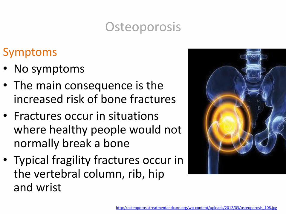
Osteoporosis
Symptoms
• No symptoms
• The main consequence is the increased risk of bone fractures
• Fractures occur in situations where healthy people would not normally break a bone
• Typical fragility fractures occur in the vertebral column, rib, hip and wrist
http://osteoporosistreatmentandcure.org/wp-content/uploads/2012/03/osteoporosis_108.jpg

Osteoporosis
The WHO Fracture Risk Assessment Tool (FRAX®)
• Postmenopausal women or men age 50 and older
• People with low bone density (osteopenia)
• People who have not taken an osteoporosis medicine
http://cdns.medindia.net/health-images/world-osteoporosis-day-10.jpg
Osteoporosis
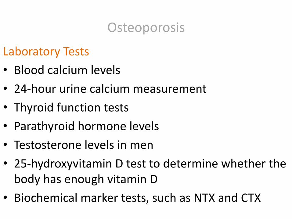
Laboratory Tests
• Blood calcium levels
• 24-hour urine calcium measurement
• Thyroid function tests
• Parathyroid hormone levels
• Testosterone levels in men
• 25-hydroxyvitamin D test to determine whether the body has enough vitamin D
• Biochemical marker tests, such as NTX and CTX

Osteoporosis Dual-energy X-ray: osteoporosis (WHO guidelines) is diagnosed when the bone mineral density is less than 2.5 standard deviations below that of a young (30–40-year-old), healthy adult women rreference population (T-score)
Category T-score range % young women
Normal T-score ≥ −1.0 85%
Osteopenia −2.5 < T-score < −1.0 14%
Osteoporosis T-score ≤ −2.5 0.6%
Severe osteoporosis
T-score ≤ −2.5 with fragility fracture
http://en.wikipedia.org/wiki/Osteoporosis

Osteoporosis
Conventional radiography (in conjunction with CT or MRI)
• detecting complications of osteopenia (reduced bone mass; preosteoporosis), such as fractures
• differential diagnosis of osteopenia
• follow-up examinations in specific clinical settings, such as soft tissue calcifications, secondary hyperparathyroidism, or osteomalacia in renal osteodystrophy
http://en.wikipedia.org/wiki/Osteoporosis
Systemic Lupus Erythematosus
Definition
• Systemic lupus erythematosus (SLE) is a chronic systemic autoimmune connective tissue disease that can affect almost any organ system, including joints, skin, kidneys, blood cells, brain, heart and lungs, has protean manifestations and follows a relapsing and remitting course, ranging from indolent to fulminant
• More than 90% of cases of SLE occur in women, frequently starting at childbearing age

Systemic Lupus Erythematosus
http://medicscientist.com/wp-content/uploads/2012/01/SystemicLupusErythematosusSLE.jpg
Systemic Lupus Erythematosus
Causes 1
• Genetics: no single causal gene has been identified, multiple genes appear to influence a person's chance of developing lupus when triggered by environmental factors, HLA class I, class II, and class III genes are associated with SLE, but only classes I and II contribute independently to increased risk of SLE

Systemic Lupus Erythematosus
Causes 2
• Drug reactions: drug-induced SLE is a (generally) reversible condition that usually occurs in people being treated for a long-term illness, more than 38 medications can cause this condition (procainamide, isoniazid, hydralazine, quinidine, and phenytoin)

Systemic Lupus Erythematosus
http://www.fidanoski.ca/sle/08-main01-a.jpg
Butterfly rash

Systemic Lupus Erythematosus Signs and symptoms 1
• Constitutional (eg, fatigue, fever, arthralgia, weight changes)
• Musculoskeletal (eg, arthralgia, arthropathy, myalgia, frank arthritis, avascular necrosis)
• Cutaneous (eg, malar rash, photosensitivity, discoid lupus)
• Renal (eg, acute or chronic renal failure, acute nephritic disease)
• Neuropsychiatric (eg, seizure, psychosis)

Systemic Lupus Erythematosus Signs and symptoms 2
• Pulmonary (eg, pleurisy, pleural effusion, pneumonitis, pulmonary hypertension, interstitial lung disease)
• Gastrointestinal (eg, nausea, dyspepsia, abdominal pain)
• Cardiac (eg, pericarditis, myocarditis)
• Hematologic (eg, multiple cytopenias such as leukopenia, lymphopenia, anemia, or thrombocytopenia)

Systemic Lupus Erythematosus
http://images.rheumatology.org/image_dir/album75693/md_00-06-0034.jpg
Butterfly rash

Systemic Lupus Erythematosus
Other cutaneous manifestations
• Raynaud phenomenon
• Livedo reticularis
• Panniculitis (lupus profundus)
• Bullous lesions
• Vasculitic purpura
• Telangiectasias
• Urticaria

Systemic Lupus Erythematosus
http://thehealthscience.com/thsattachs/930938/111919333189133.jpg
Raynaud phenomenon

Systemic Lupus Erythematosus
• CBC (complete blood count) with differential
• Serum creatinine
• Urinalysis with microscopy
• ESR or CRP results
• Complement levels
• Liver function tests
• Creatine kinase assay
• Spot protein/spot creatinine ratio
• Autoantibody tests
• Electrolytes
• Liver enzymes
Laboratory tests

Systemic Lupus Erythematosus
http://mydermpath.com/images/dermpath/diagnosis/839dbd1d84e31576c71c33eb4e056ba3.jpg
Bullous lesions

Systemic Lupus Erythematosus
Lupus band test
• Skin biopsy, with direct immunofluorescence
• The minimum criteria for positivity are:
• In sun-exposed skin: presence of a band of deposits of IgM along the epidermal basement membrane in 50% of the biopsy, intermediate (2+) intensity or more
• In sun-protected skin : presence of interrupted (i.e. less than 50%) deposits of IgM along the epidermal basement membrane, intermediate (2+) intensity or more
http://en.wikipedia.org/wiki/Lupus_band_test

Systemic Lupus Erythematosus
Vasculitic purpura
http://en.wikipedia.org/wiki/Lupus_band_test

Systemic Lupus Erythematosus
Imaging studies
• Joint radiography
• Chest radiography and chest CT scanning
• Echocardiography
• Brain MRI/ MRA
Procedures
• Cardiac MRI
• Arthrocentesis
• Lumbar puncture
• Renal biopsy

Systemic Lupus Erythematosus
http://images.rheumatology.org/image_dir/album75695/md_99-08-0024.tif.jpg
Arthropathy
Systemic Lupus Erythematosus
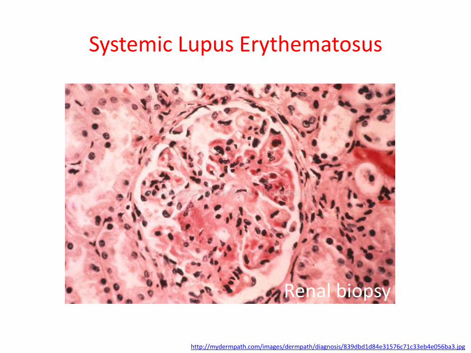
http://mydermpath.com/images/dermpath/diagnosis/839dbd1d84e31576c71c33eb4e056ba3.jpg
Renal biopsy

Systemic Lupus Erythematosus
American College of Rheumatology (ACR) criteria
• Serositis
• Oral ulcers
• Arthritis
• Photosensitivity
• Blood disorders
• Renal involvement
• Antinuclear antibodies
• Immunologic phenomena (eg, dsDNA; anti-Smith [Sm] antibodies)
• Neurologic disorder
• Malar rash
• Discoid rash

Systemic Lupus Erythematosus
http://members.shaw.ca/tiderington/handsImage.jpg
Discoid rash

Rheumatoid Arthritis
Definition
• A chronic systemic inflammatory disorder that typically affects the small joints of hands and feet, causing a painful swelling that eventually results in bone erosion and joint deformity, which lead to loss of function, and can affect multiple organs of the body (the lungs, heart, eyes atc.)
https://www.rheumatology.org/Practice/Clinical/Patients/Diseases_And_Conditions/Rheumatoid_Arthritis/

Rheumatoid Arthritis
http://physioworks.com.au/rheumatoid%20arthritis1.jpg?bc_t=2i7STXCmpaaLa6JskfT3mw
Rheumatoid Arthritis
Causes
• Specific agent or agents are not yet known
• Half of the risk is believed to be genetic (the inherited tissue type major histocompatibility complex (MHC) antigen HLA-DRB1 (most specifically the shared epitope alleles, including *0401 and *0404), and the genes PTPN22 and PADI4—hence family history is an important risk factor)
• Infectious agents, such as viruses and bacteria, may trigger RA in people with an inherited tendency to develop the disease
http://en.wikipedia.org/wiki/Rheumatoid_arthritis http://www.webmd.com/rheumatoid-arthritis/guide/rheumatoid-arthritis-symptoms

Rheumatoid Arthritis
http://annschenkel.com/wp-content/uploads/2014/03/RA-joint-01.jpg

Rheumatoid Arthritis
Symptoms that affect the joints and the skin
• Tender, warm, swollen joints
• Morning stiffness that may last for hours
• Pain
• Firm bumps of tissue under the skin on arms (rheumatoid nodules)
Early rheumatoid arthritis (RA) tends to affect smaller joints first. As the RA, symptoms spread to the wrists, knees,
ankles, elbows, hips and shoulders. In most cases, symptoms occur in the same joints on both sides of the body.
http://www.mayoclinic.org/diseases-conditions/rheumatoid-arthritis/basics/symptoms/con-20014868 https://www.rheumatology.org/Practice/Clinical/Patients/Diseases_And_Conditions/Rheumatoid_Arthritis/

Rheumatoid Arthritis
http://api.ning.com/files/*JdnwHQL9f7-gHJp9YDdEdmDVHr9qnqq4VAZD8l4PiTE3yFtAn-KAmMy9MUOvzZ1iqFCeVqQwminDWXv6CD2J7AxlbidS1CS/md_99050020.tif.jpg
Rheumatoid nodules

Rheumatoid Arthritis
The most commonly affected joints
• The proximal interphalangeal (PIP) and metacarpophalangeal (MCP) joints of the hands (middle and base joints of the finger)
• The wrists, especially the ulnar-styloid articulation
• The shoulders, Elbows, Knees, Ankles
• Metatarsophalangeal (MTP) joints (in the toes)
http://www.medicalnewstoday.com/info/rheumatoid-arthritis/signs-of-rheumatoid-arthritis.php

Rheumatoid Arthritis
http://www.bodybuilding.com/fun/images/2009/manage_rheumatoid_arthritis_b.jpg

Rheumatoid Arthritis
Symptoms that affect the entire body
• Fatigue
• Malaise (feeling ill)
• Loss of appetite, which can lead to weight loss
• Muscle aches
• The lung involvement
• The heart involvement
• The tear glands, and the salivary glands involvement
• Affect a joint in voice box or larynx (cricoarytenoid joint)
• Anemia: approximately 80% of patients are anemic
http://www.webmd.com/rheumatoid-arthritis/guide/rheumatoid-arthritis-symptoms

Rheumatoid Arthritis
http://f.tqn.com/y/arthritis/1/S/X/N/183873142.jpg
The lung involvement

Rheumatoid Arthritis
Blood tests
• Rheumatoid factor (RF)
• Anti-citrullinated protein antibody (ACPA)
• Anti-cyclic citrullinated peptide (anti-CCP)
• Antibodies against mutated citrullinated Vimentin (Anti-MCV)
• C-reactive protein
• Erythrocyte sedimentation rate (ESR)
• A serological point-of-care test (POCT) combines the detection of rheumatoid factor and anti-MCV
• Full blood count
• Kidney function
• Liver enzymes https://www.rheumatology.org/Practice/Clinical/Patients/Diseases_And_Conditions/Rheumatoid_Arthritis/

Rheumatoid Arthritis
The name “rheumatoid factor” is a bit misleading - in patients in the very early stages of rheumatoid
arthritis, only about half (50%) will be positive for it http://rheuminfo.com/wp-content/uploads/2014/02/Rheumatoid-factor.jpg

Rheumatoid Arthritis
Imaging
• X-rays
• Magnetic resonance imaging (MRI)
• Ultrasound (high-frequency transducers > 10 MHz),
• Color Doppler and power Doppler ultrasound
https://www.rheumatology.org/Practice/Clinical/Patients/Diseases_And_Conditions/Rheumatoid_Arthritis/

Rheumatoid Arthritis
http://media1.break.com/breakstudios/2012/2/9/arthritis.jpg

Rheumatoid Arthritis
http://www.medgadget.com/wp-content/uploads/2012/02/Siemens-Biograph-mCT-scan.jpg

Rheumatoid Arthritis
http://www.hopkinsarthritis.org/wp-content/uploads/2011/04/slide59.jpg
MRI

Rheumatoid Arthritis
http://www.nature.com/nrrheum/journal/v9/n4/images/nrrheum.2013.39-f1.jpg
The power Doppler signal is located at areas of synovial proliferation

Rheumatoid Arthritis
The 2010 ACR / EULAR Rheumatoid Arthritis Classification Criteria 1
• Joint involvement: 1 large joint - 0 points, 2–10 large joints - 1 point, 1–3 small joints - 2 points, 4–10 small joints - 3 points, more than 10 joints - 5 points
https://www.rheumatology.org/Practice/Clinical/Patients/Diseases_And_Conditions/Rheumatoid_Arthritis/

Rheumatoid Arthritis
The 2010 ACR / EULAR Rheumatoid Arthritis Classification Criteria 2
• Serological parameters (rheumatoid factor – RF, anti-citrullinated protein antibody - ACPA): negative RF and negative ACPA - 0 points, low-positive RF or low-positive ACPA - 2 points, high-positive RF or high-positive ACPA - 3 points
https://www.rheumatology.org/Practice/Clinical/Patients/Diseases_And_Conditions/Rheumatoid_Arthritis/

Rheumatoid Arthritis
The 2010 ACR / EULAR Rheumatoid Arthritis Classification Criteria 3
• Acute phase reactants: elevated erythrocyte sedimentation rate (ERS) - 1 point, or elevated c-reactive protein (CRP) - 1 point for symptoms lasting six weeks or longer
A score of 6 or greater unequivocally classifies a person with a diagnosis of rheumatoid arthritis
https://www.rheumatology.org/Practice/Clinical/Patients/Diseases_And_Conditions/Rheumatoid_Arthritis/

Rheumatoid Arthritis
Clinical practice criteria
• Two or more swollen joints
• Morning stiffness lasting more than one hour for at least six weeks
• The detection of rheumatoid factors or autoantibodies against ACPA such as autoantibodies to mutated citrullinated vimentin can confirm the suspicion of RA
• A negative autoantibody result does not exclude a diagnosis of RA
http://en.wikipedia.org/wiki/Rheumatoid_arthritis


