Evidence Synthesis
Number 214 Screening for Glaucoma in Adults A Systematic Review for the US Preventive Services Task Force Prepared for
Agency for Healthcare Research and Quality
US Department of Health and Human Services
5600 Fishers Lane
Rockville MD 20857
wwwahrqgov
Contract No HHSA-290-2015-00011-I Task Order No HHSA29032008T
Prepared by
Pacific Northwest Evidence-Based Practice Center
Oregon Health amp Science University
Mail Code BICC
3181 SW Sam Jackson Park Road
Portland OR 97239
wwwohsueduepc
Investigators
Roger Chou MD
Shelley Selph MD MPH
Ian Blazina MPH
Christina Bougatsos MPH
Rebecca Jungbauer DrPH
Rongwei Fu PhD
Sara Grusing BA
Daniel Jonas MD MPH
Shandiz Tehrani MD PhD
AHRQ Publication No 21-05286-EF-1
October 2021
Screening for Glaucoma ii Pacific Northwest EPC
This report is based on research conducted by the Pacific Northwest Evidence-based Practice
Center (EPC) under contract to the Agency for Healthcare Research and Quality (AHRQ)
Rockville MD (Contract No HHSA-290-2015-00011-I Task Order No HHSA29032008T)
The findings and conclusions in this document are those of the authors who are responsible for
its contents and do not necessarily represent the views of AHRQ Therefore no statement in this
report should be construed as an official position of AHRQ or of the US Department of Health
and Human Services
The information in this report is intended to help healthcare decisionmakersmdashpatients and
clinicians health system leaders and policymakers among othersmdashmake well-informed
decisions and thereby improve the quality of healthcare services This report is not intended to be
a substitute for the application of clinical judgment Anyone who makes decisions concerning the
provision of clinical care should consider this report in the same way as any medical reference
and in conjunction with all other pertinent information (ie in the context of available resources
and circumstances presented by individual patients)
The final report may be used in whole or in part as the basis for development of clinical practice
guidelines and other quality enhancement tools or as a basis for reimbursement and coverage
policies AHRQ or US Department of Health and Human Services endorsement of such
derivative products may not be stated or implied
None of the investigators has any affiliations or financial involvement that conflicts with the
material presented in this report
Acknowledgments The authors thank research librarian Tracy Dana MLS for conducting the searches AHRQ
Medical Officer Justin Mills MD MPH as well as the US Preventive Services Task Force
Screening for Glaucoma iii Pacific Northwest EPC
Table of Contents Chapter 1 Introduction and Background 1
Purpose 1
Condition Background 1
Condition Definition 1
Prevalence and Burden of DiseaseIllness 2
Etiology and Natural History 2
Risk Factors 3
Rationale for ScreeningScreening Strategies 3
InterventionsTreatment 3
Current Clinical PracticeRecommendations of Other Groups 4
Chapter 2 Methods 5
Key Questions and Analytic Framework 5
Search Strategies 6
Study Selection 6
Data Abstraction and Quality Rating 7
Data Synthesis and Analysis 8
USPSTF Involvement 9
Expert Review and Public Comment 9
Chapter 3 Results 10 Key Question 1 What are the effects of screening for OAG versus no screening on a) IOP
visual field loss visual acuity or optic nerve damage or b) visual impairment quality of
life or function 10
Summary 10
Evidence 10
Key Question 2 What are the harms of screening for OAG versus no screening 11
Summary 11
Evidence 11
Key Question 3 What are the effects of referral to an eye health provider versus no referral on
a) IOP visual field loss visual acuity or optic nerve damage or b) visual impairment
quality of life or function 12
Key Question 4 What is the accuracy of screening for diagnosis of OAG 12
Summary 12
Evidence 13
Key Question 5 What is the accuracy of instruments for identifying patients at higher risk of
OAG 19
Summary 19
Evidence 19
Key Question 6 What are the effects of medical treatments for OAG versus placebo or no
treatments on a) IOP visual field loss visual acuity or optic nerve damage or b) visual
impairment quality of life or function 20
Summary 20
Evidence 20
Key Question 7 What are the harms of medical treatments for OAG versus placebo or no
treatments 23
Screening for Glaucoma iv Pacific Northwest EPC
Summary 23
Evidence 23
Key Question 8 What are the effects of newly FDA-approved medical treatments
(latanoprostene bunod and netarsudil) versus older medical treatments on a) IOP visual
field loss visual acuity or optic nerve damage or b) visual impairment quality of life or
function 24
Summary 24
Evidence 24
Key Question 9 What are the harms of newly FDA-approved medical treatments versus older
medical treatments 25
Summary 25
Evidence 26
Key Question 10 What are the effects of laser trabeculoplasty for OAG versus no
trabeculoplasty or medical treatment on a) IOP visual field loss visual acuity or optic
nerve damage or b) visual impairment quality of life or function 26
Summary 26
Evidence 27
Key Question 11 What are the harms of laser trabeculoplasty for OAG versus no
trabeculoplasty or medical treatment 28
Summary 28
Evidence 28
Contextual Question 1 What is the association between changes in IOP visual field loss
visual acuity or optic nerve damage following treatment for OAG and improvement in visual
impairment quality of life or function and what is the association between changes in IOP
and visual field loss 30
Chapter 4 Discussion 33 Summary of Review Findings 33
Limitations 35
Emerging IssuesNext Steps 35
Relevance for Priority Populations 36
Future Research 36
Conclusions 37
References 38
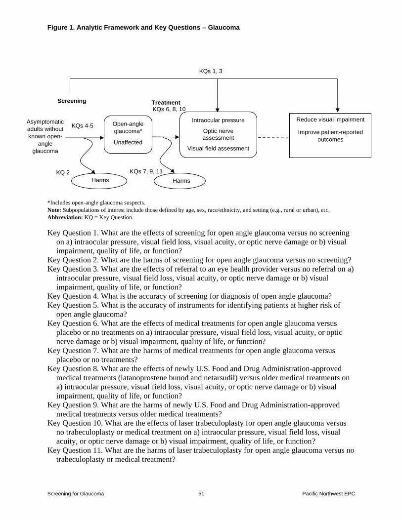
Figures Figure 1 Analytic Framework and Key Questions
Figure 2 Glaucoma vs Control Spectral Domain-OCT Sensitivity and Specificity for Retinal
Nerve Fiber Layer Thickness
Figure 3 Glaucoma vs Control Spectral Domain-OCT Retinal Nerve Fiber Layer Thickness
Figure 4 AUROC Spectral Domain-OCT Retinal Nerve Fiber Layer Thickness by
Comparison
Figure 5 AUROC Curves Spectral Domain-OCT Retinal Nerve Fiber Layer Thickness
Figure 6 Glaucoma vs Control Spectral Domain-OCT Sensitivity and Specificity for
Ganglion Cell Complex Thickness
Figure 7 Glaucoma vs Control Spectral Domain-OCT Ganglion Cell Complex Thickness
Figure 8 Ganglion Cell Analysis
Screening for Glaucoma v Pacific Northwest EPC
Figure 9 Ganglion Cell Analysis by Control Group
Figure 10 Glaucoma vs Control Humphrey Field Analyzer Visual Field
Figure 11 Glaucoma vs Control Visual Field Sensitivity and Specificity
Figure 12 Glaucoma vs Control AUROC Humphrey Field Analyzer Visual Field Mean
Deviation
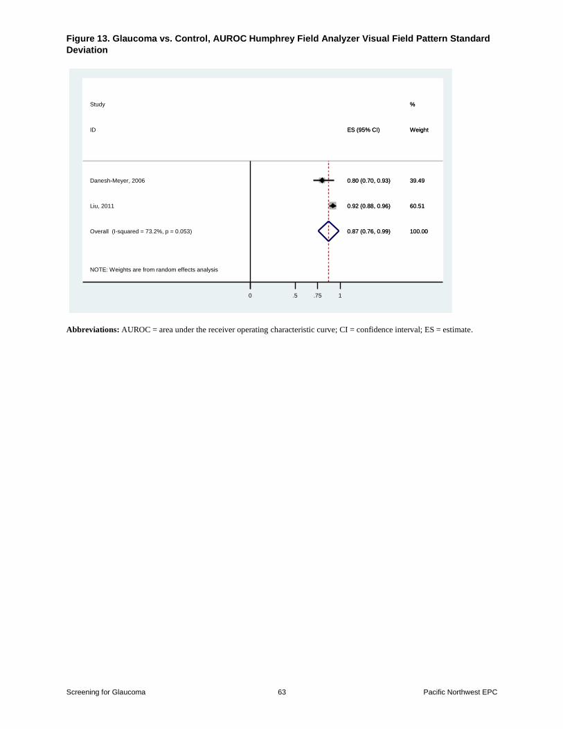
Figure 13 Glaucoma vs Control AUROC Humphrey Field Analyzer Visual Field Pattern
Standard Deviation
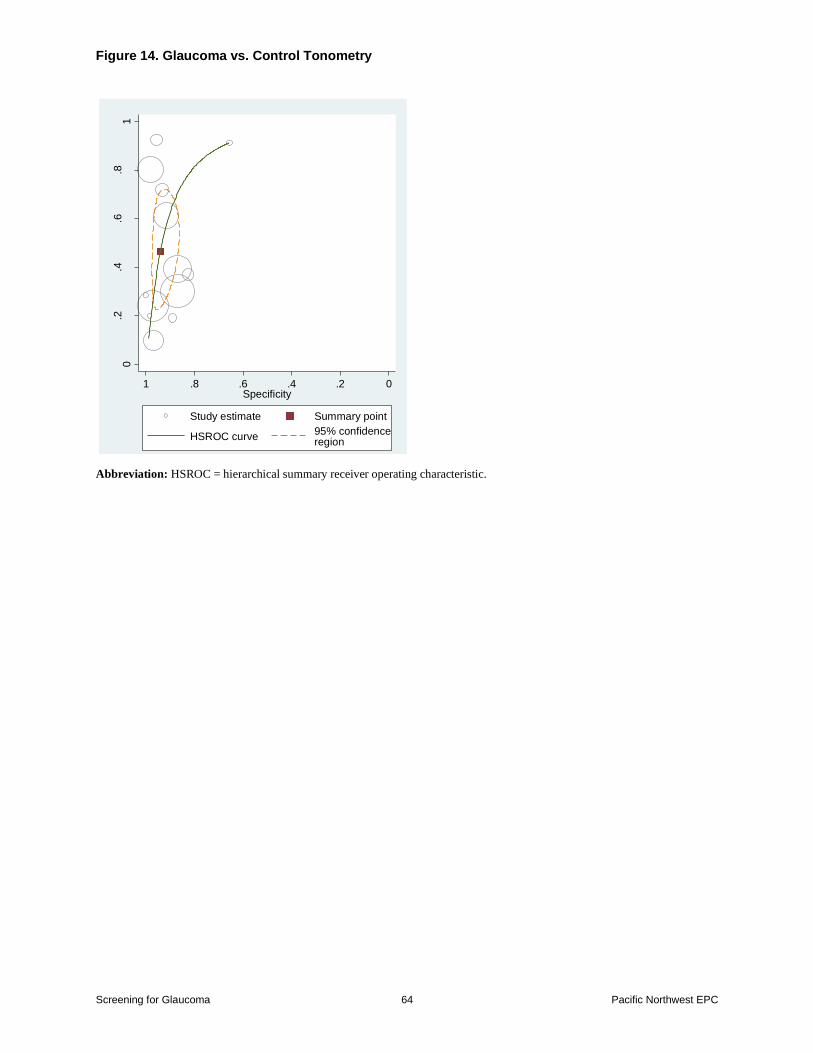
Figure 14 Glaucoma vs Control Tonometry
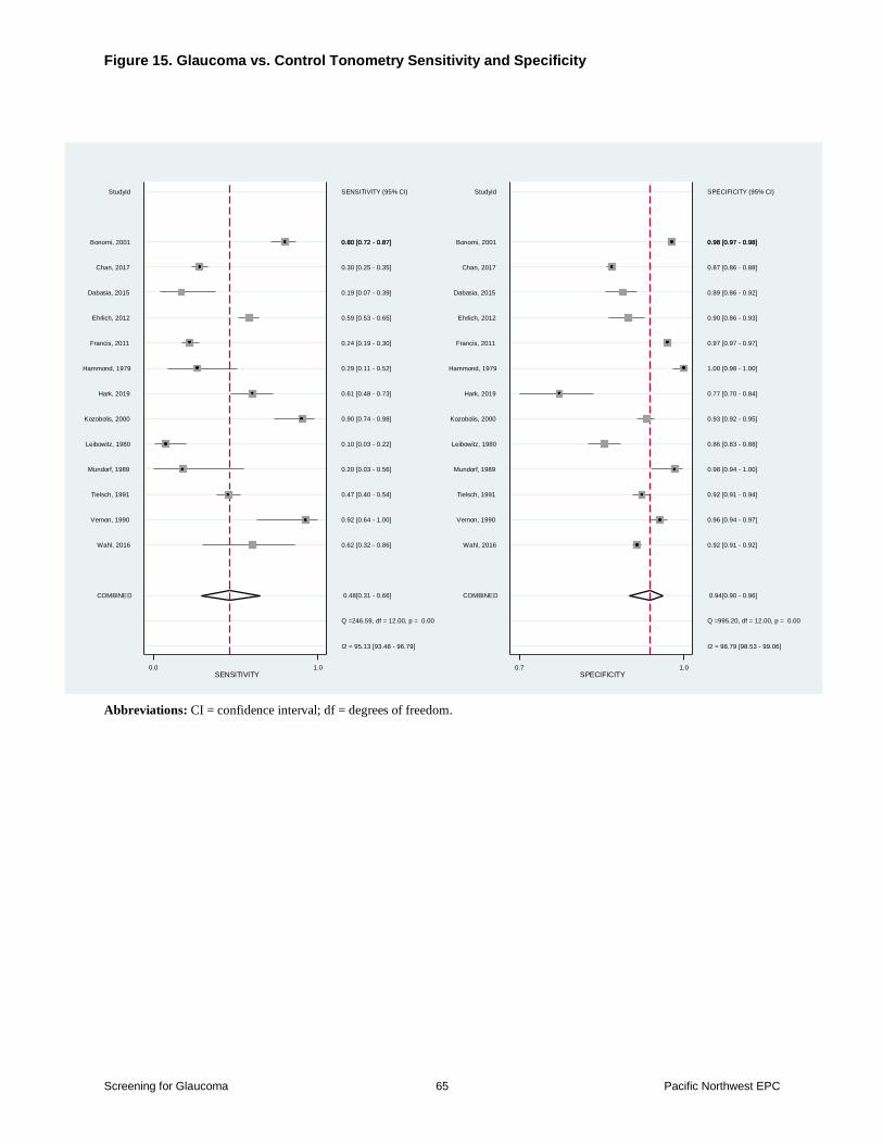
Figure 15 Glaucoma vs Control Tonometry Sensitivity and Specificity
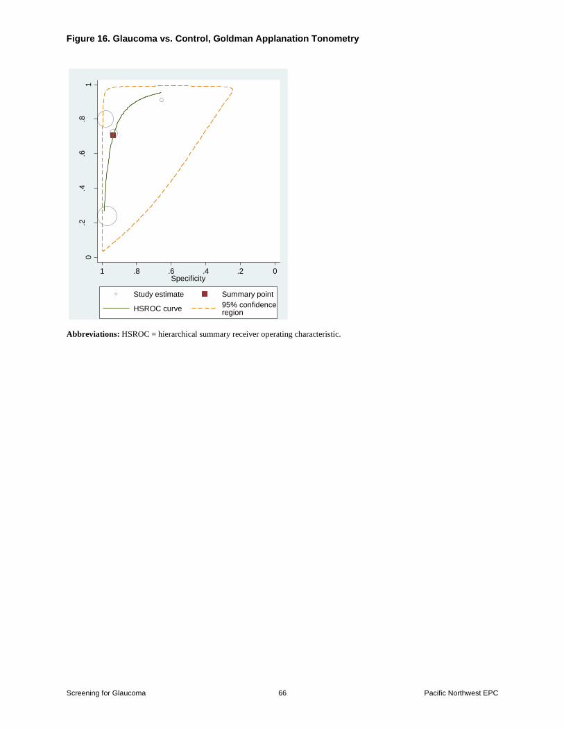
Figure 16 Glaucoma vs Control Goldman Applanation Tonometry
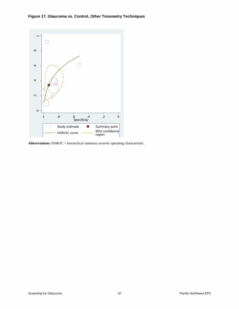
Figure 17 Glaucoma vs Control Other Tonometry Techniques
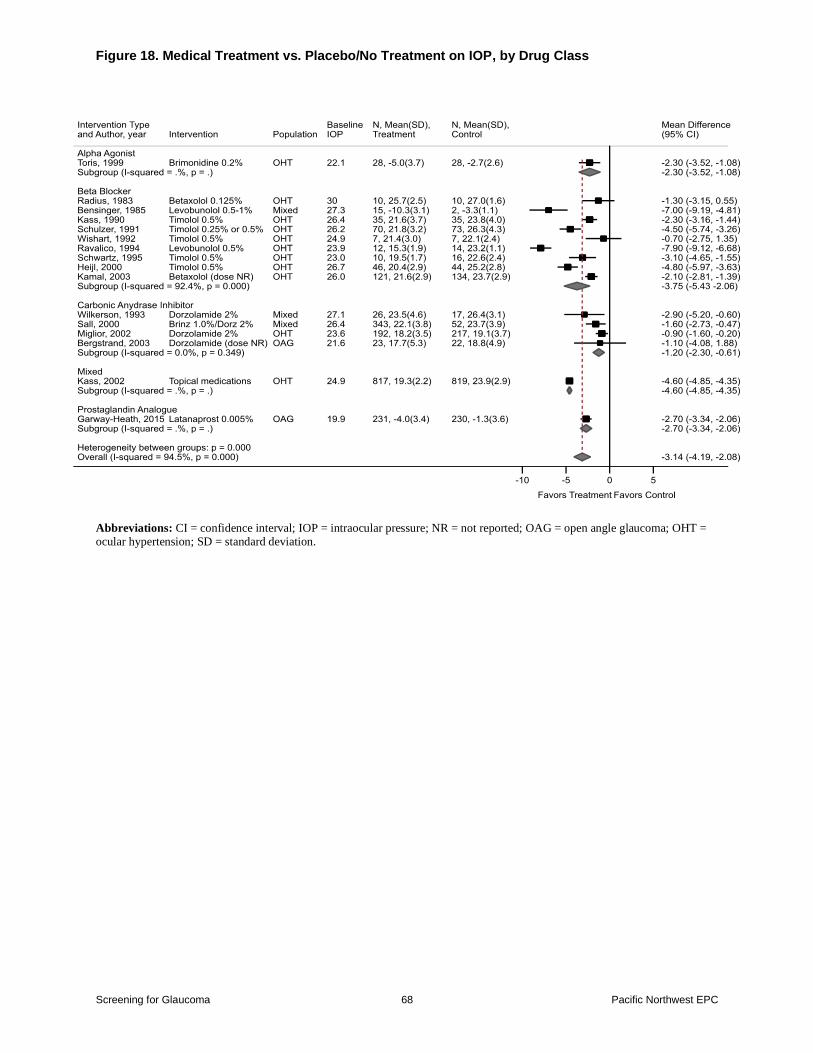
Figure 18 Medical Treatment vs PlaceboNo Treatment on IOP by Drug Class
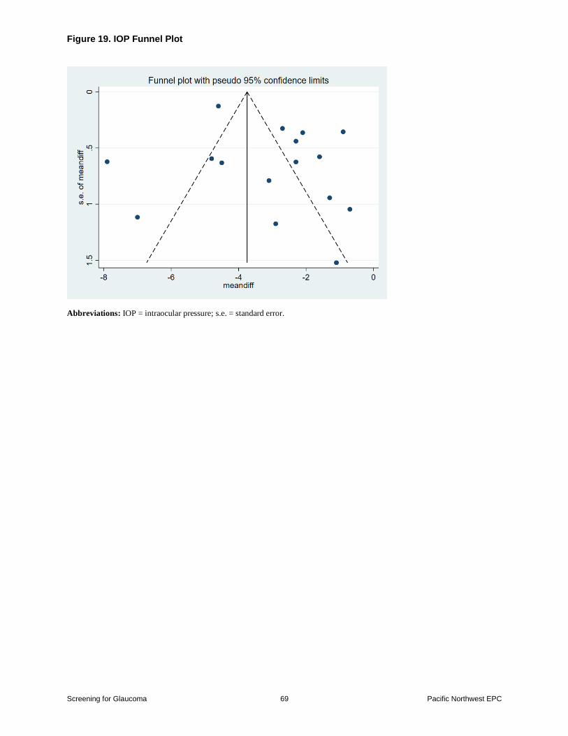
Figure 19 IOP Funnel Plot
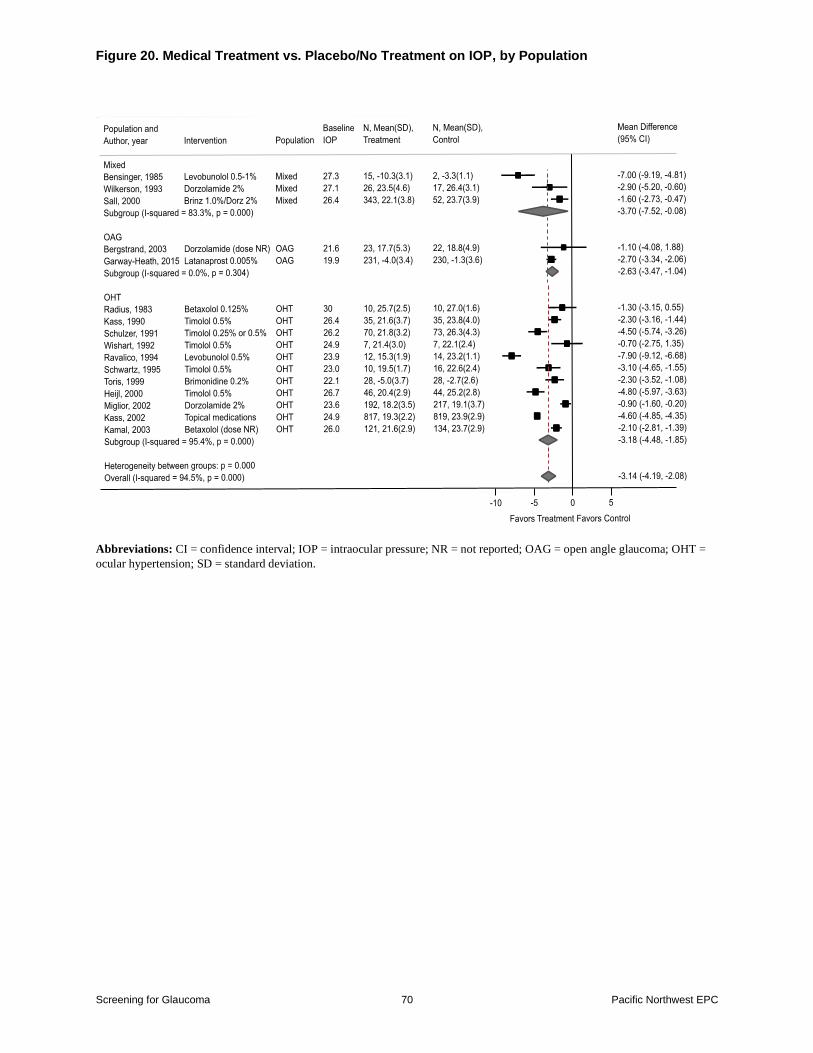
Figure 20 Medical Treatment vs PlaceboNo Treatment on IOP by Population
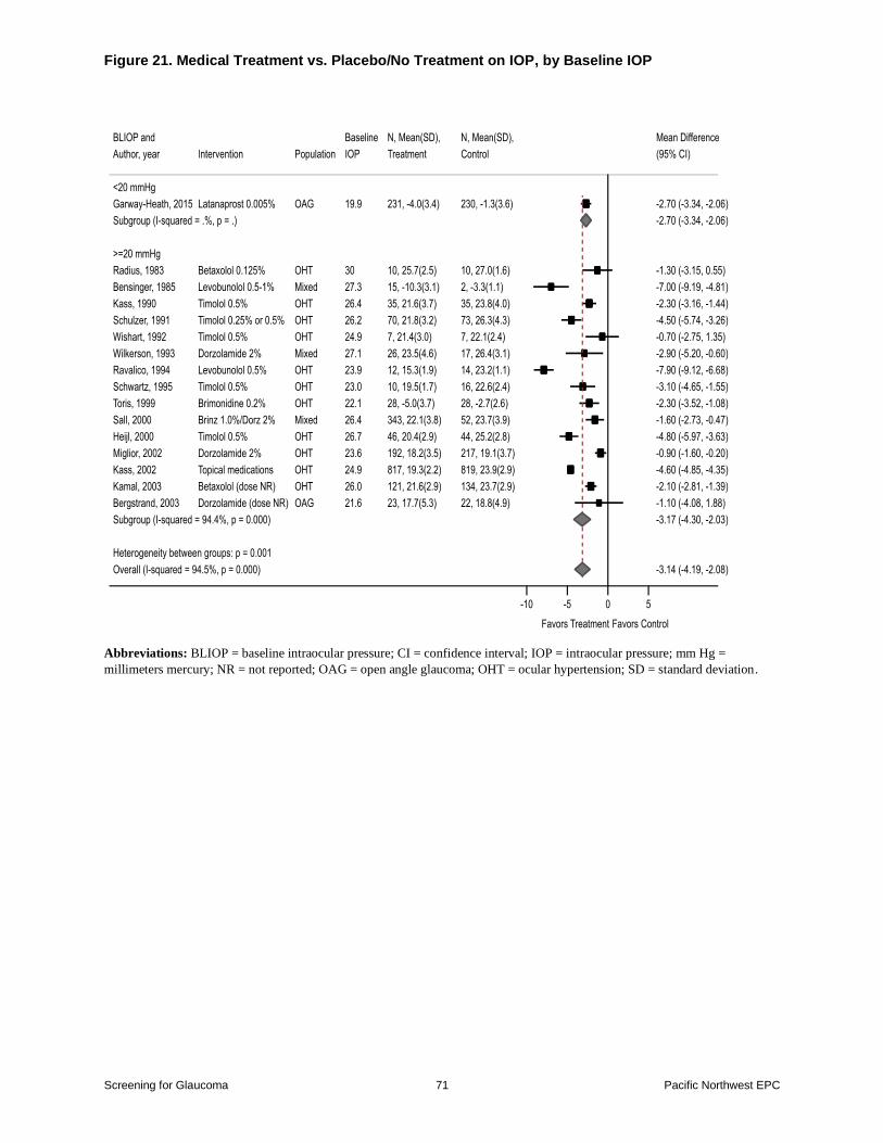
Figure 21 Medical Treatment vs PlaceboNo Treatment on IOP by Baseline IOP
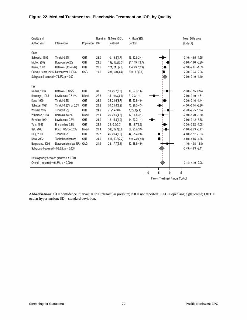
Figure 22 Medical Treatment vs PlaceboNo Treatment on IOP by Quality
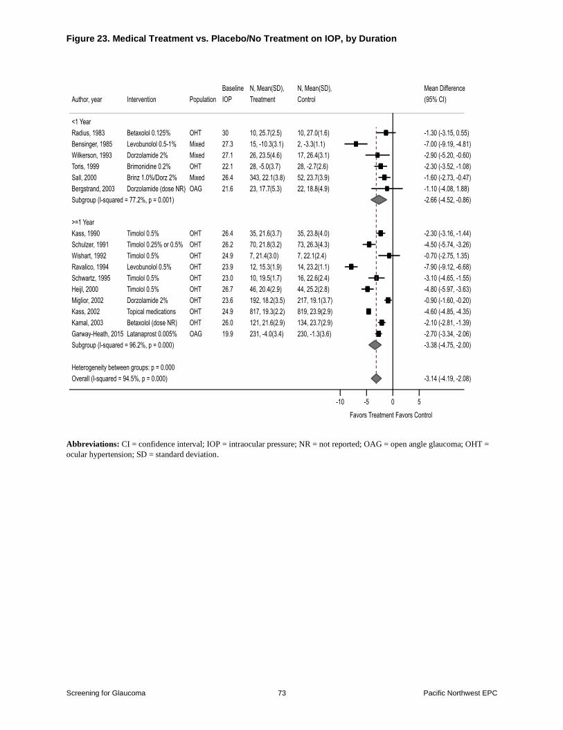
Figure 23 Medical Treatment vs PlaceboNo Treatment on IOP by Duration
Figure 24 Medical Treatment vs PlaceboNo Treatment on Progression to Glaucoma
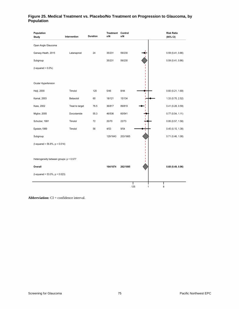
Figure 25 Medical Treatment vs PlaceboNo Treatment on Progression to Glaucoma by
Population
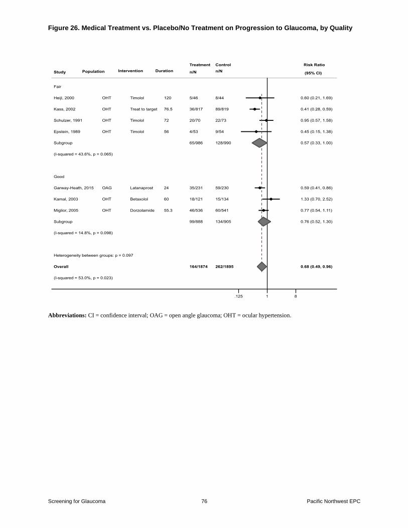
Figure 26 Medical Treatment vs PlaceboNo Treatment on Progression to Glaucoma by
Quality
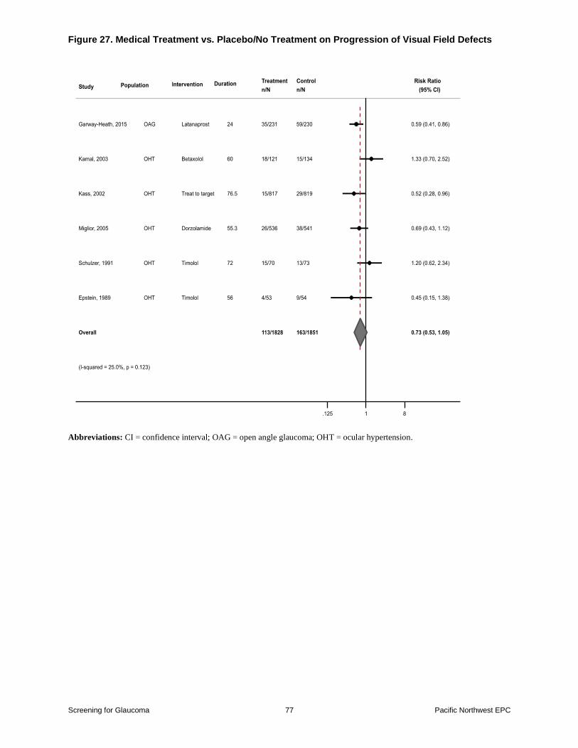
Figure 27 Medical Treatment vs PlaceboNo Treatment on Progression of Visual Field
Defects
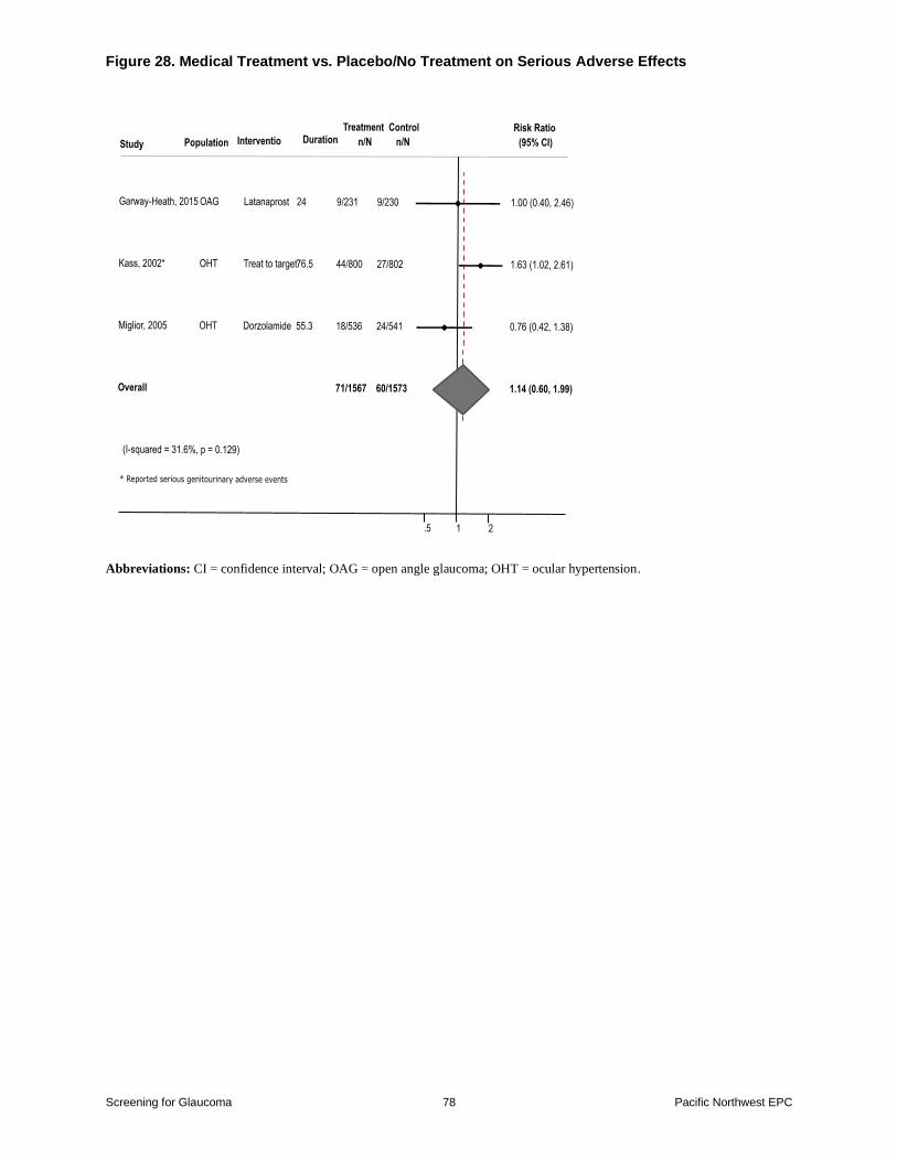
Figure 28 Medical Treatment vs PlaceboNo Treatment on Serious Adverse Effects
Figure 29 Medical Treatment vs PlaceboNo Treatment on IOP Withdrawals Due to Adverse
Effects
Tables
Table 1 Diagnostic Accuracy Pooled Analyses
Table 2 Sensitivity and Specificity Spectral Domain-OCT Retinal Nerve Fiber Layer
Thickness
Table 3 Sensitivity and Specificity Spectral Domain-OCT Ganglion Cell Complex Thickness
Table 4 Glaucoma vs Control Spectral Domain-OCT Cup-to-Disc Ratio
Table 5 Glaucoma vs Control Optic Disc Photography Cup-to-Disc Ratio
Table 6 OphthalmoscopyBiomicroscopyStereoscopy Cup-to-Disc Ratio
Table 7 Glaucoma vs Control Humphrey Field Analyzer Sensitivity and Specificity
Table 8 Glaucoma vs Control Tonometry Sensitivity and Specificity
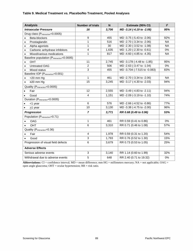
Table 9 Medical Treatment vs PlaceboNo Treatment Pooled Analyses
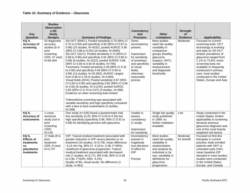
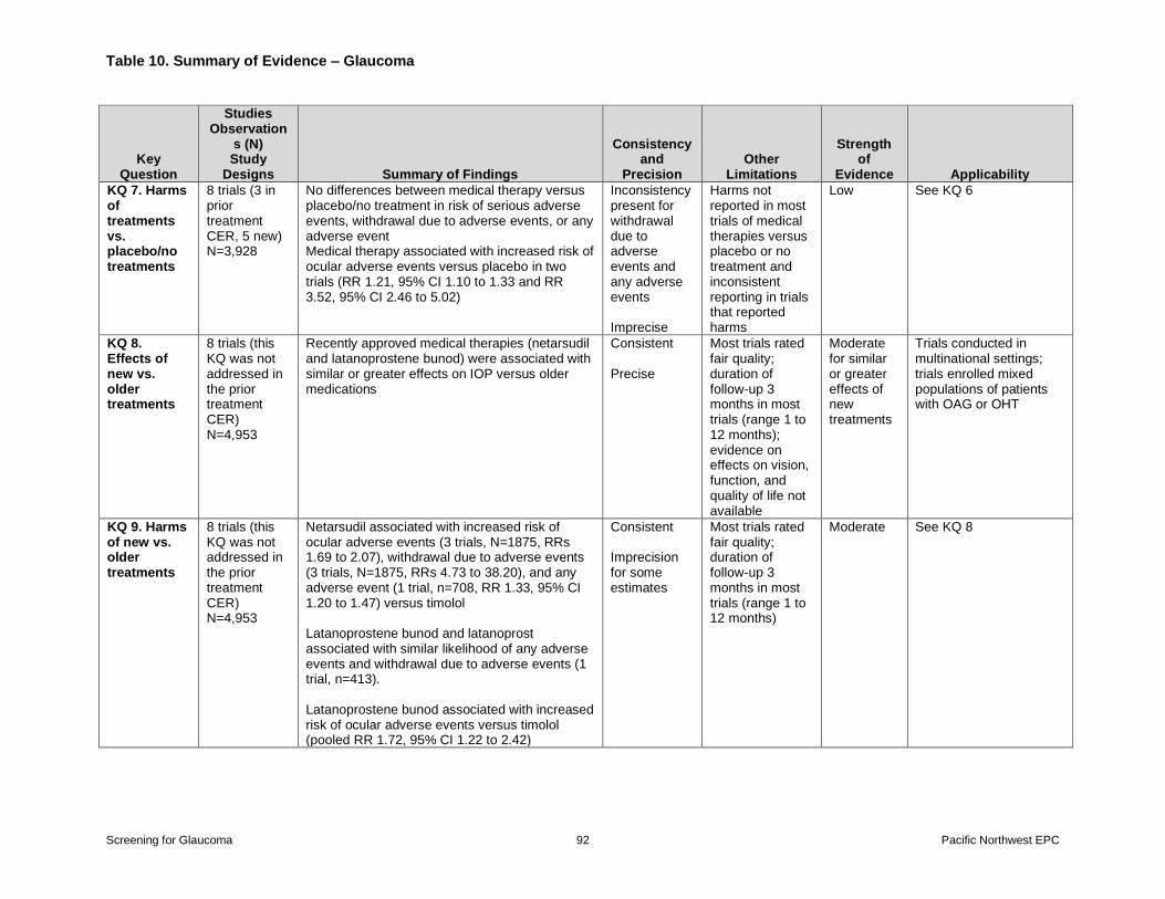
Table 10 Summary of Evidence
Screening for Glaucoma vi Pacific Northwest EPC
Appendixes Appendix A Detailed Methods
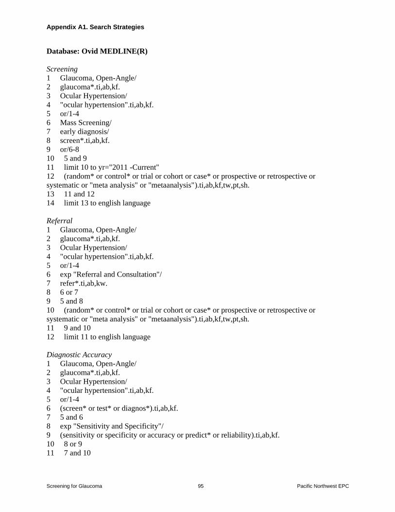
Appendix A1 Search Strategies Appendix A2 Inclusion and Exclusion Criteria
Appendix A3 Literature Flow Diagram
Appendix A4 Included Studies
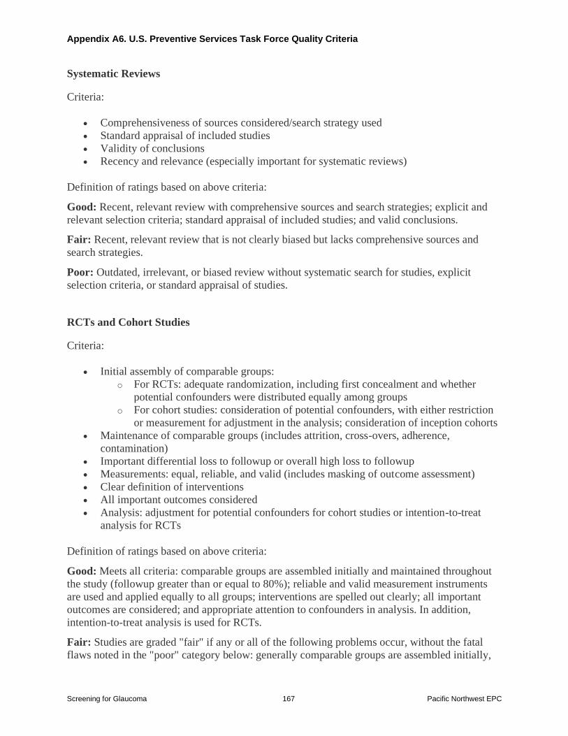
Appendix A5 Excluded Studies Appendix A6 US Preventive Services Task Force Quality Rating Criteria
Appendix A7 Reviewers of the Draft Report
Appendix B Evidence Tables and Quality Tables
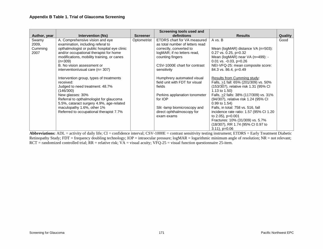
Appendix B Table 1 Trial of Glaucoma Screening
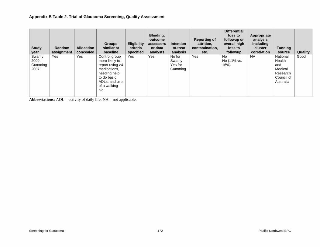
Appendix B Table 2 Trial of Glaucoma Screening Quality Assessment
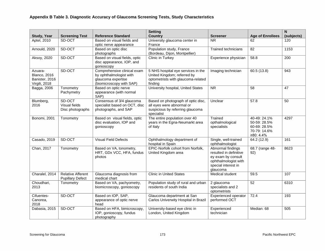
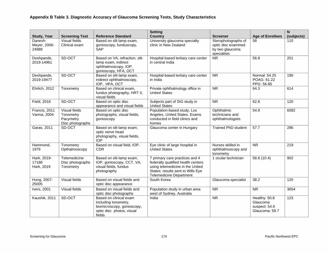
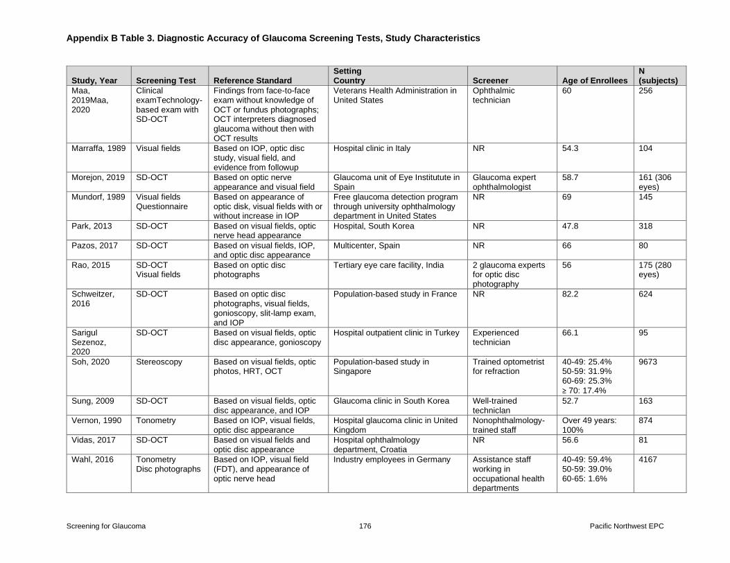
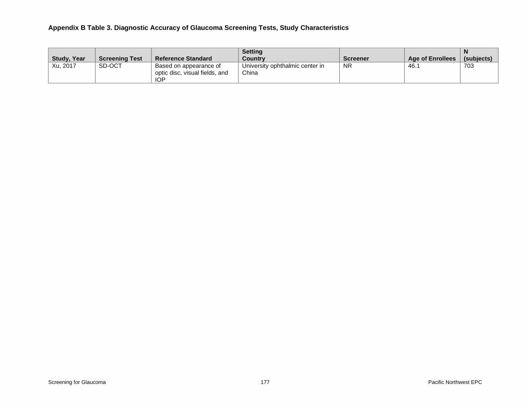
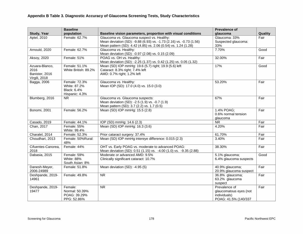
Appendix B Table 3 Diagnostic Accuracy of Glaucoma Screening Tests Study
Characteristics
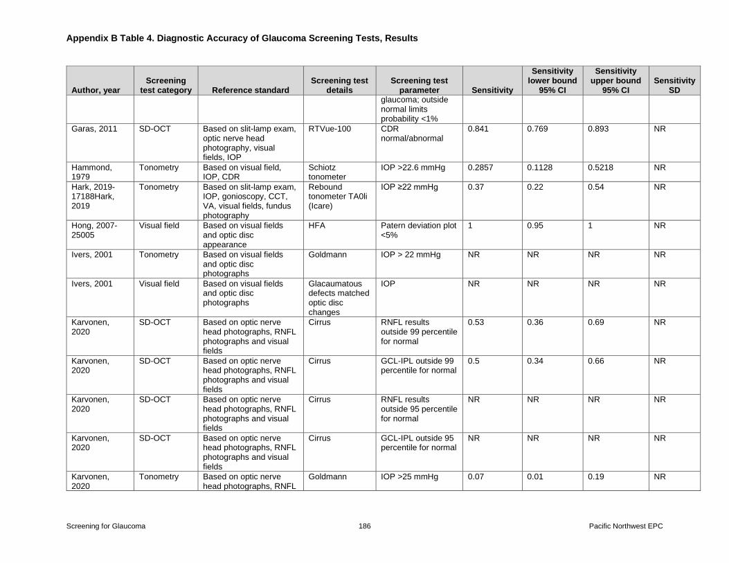
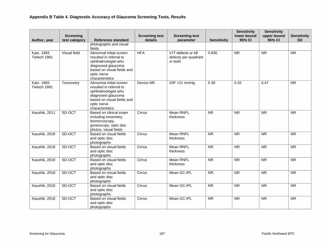
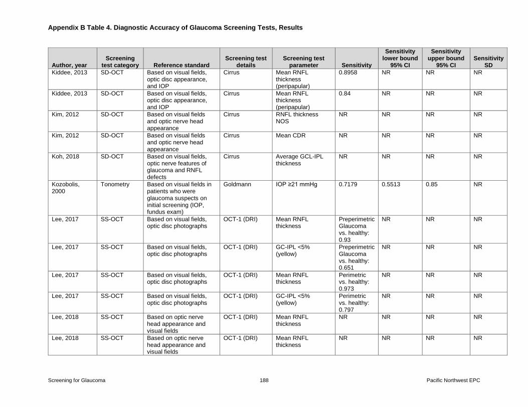
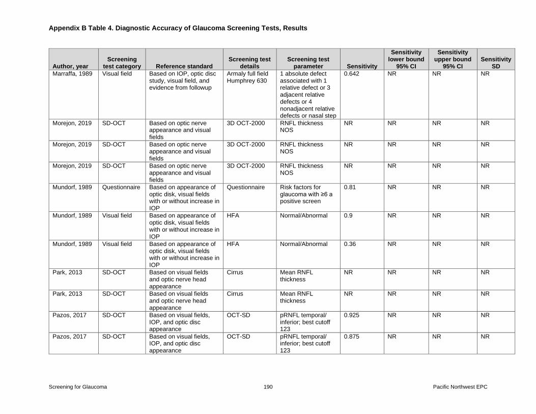
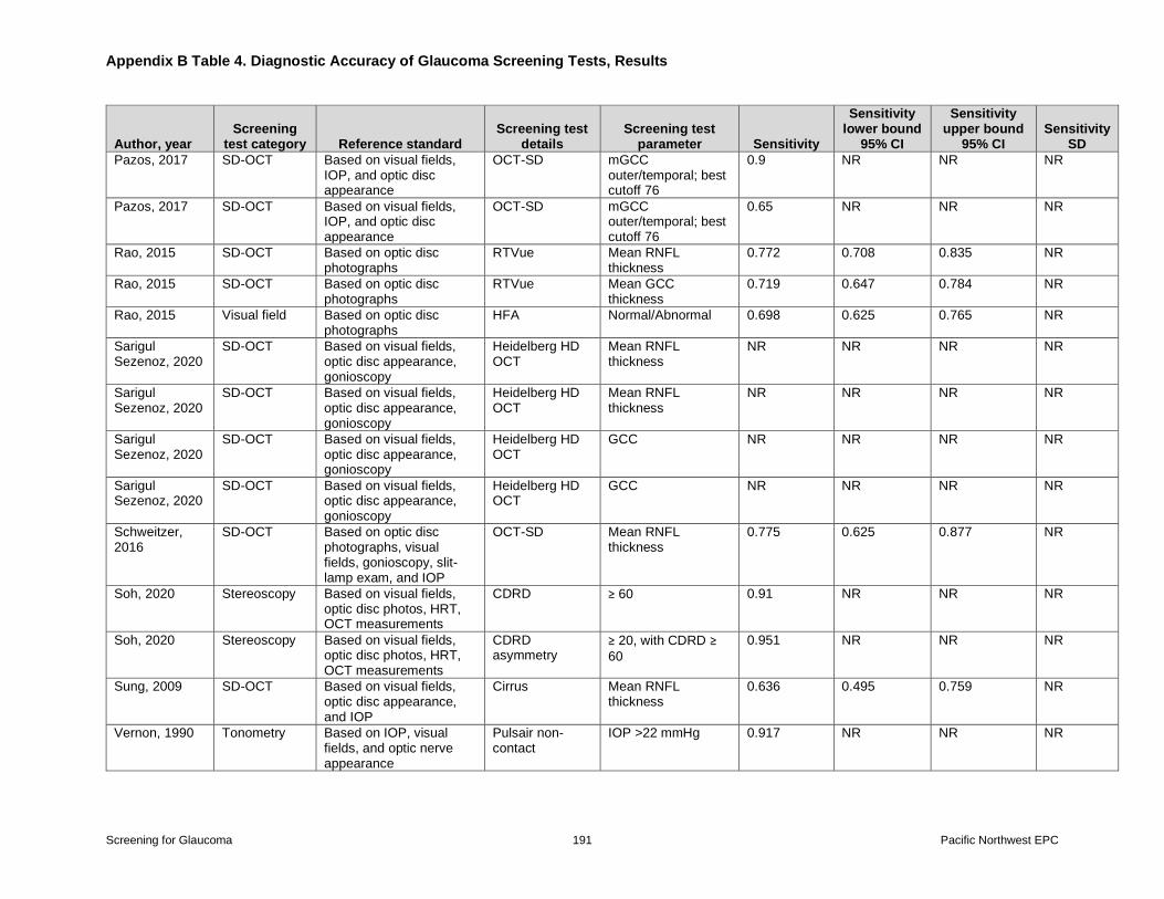
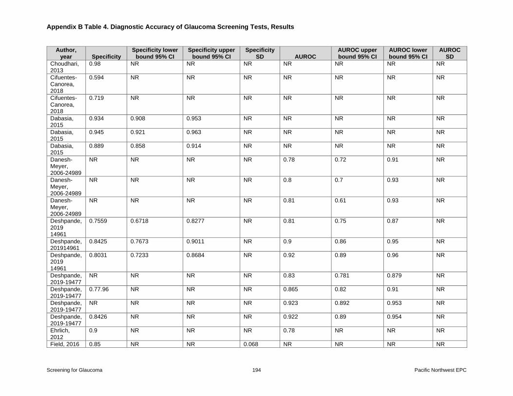
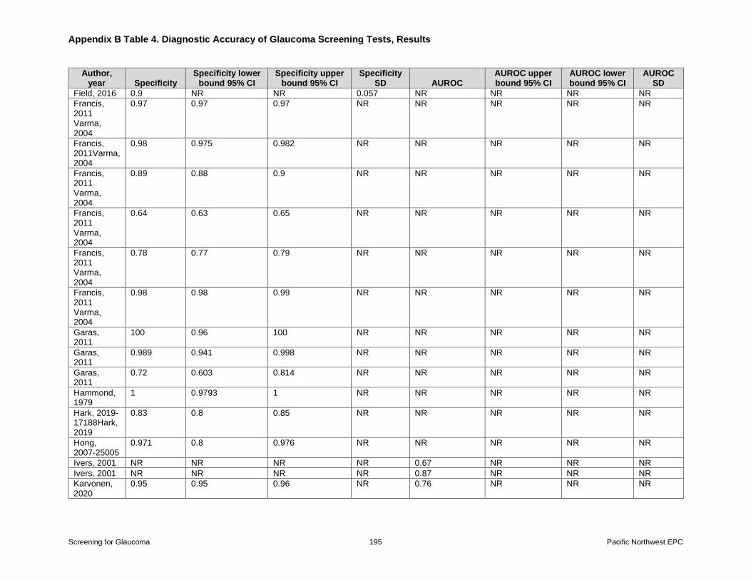
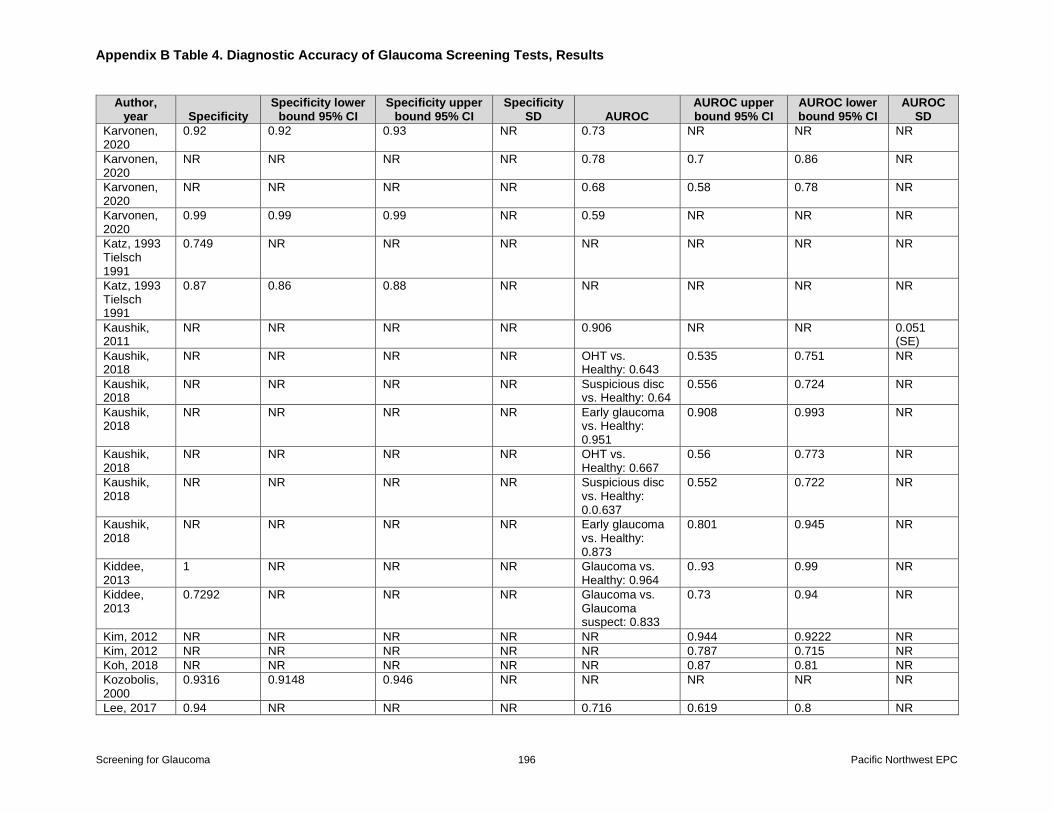
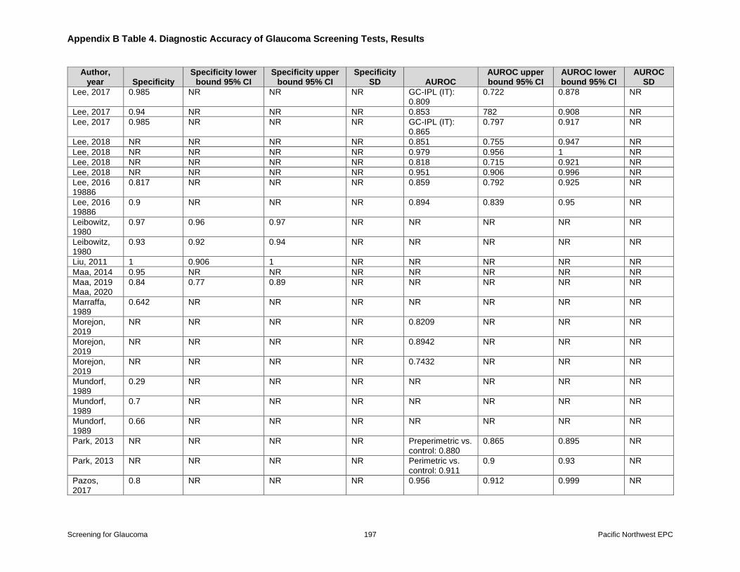
Appendix B Table 4 Diagnostic Accuracy of Glaucoma Screening Tests Results
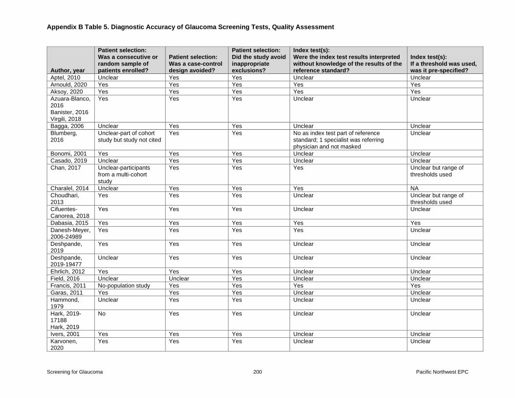
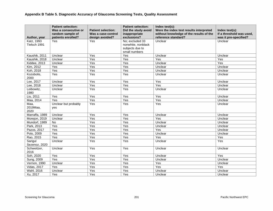
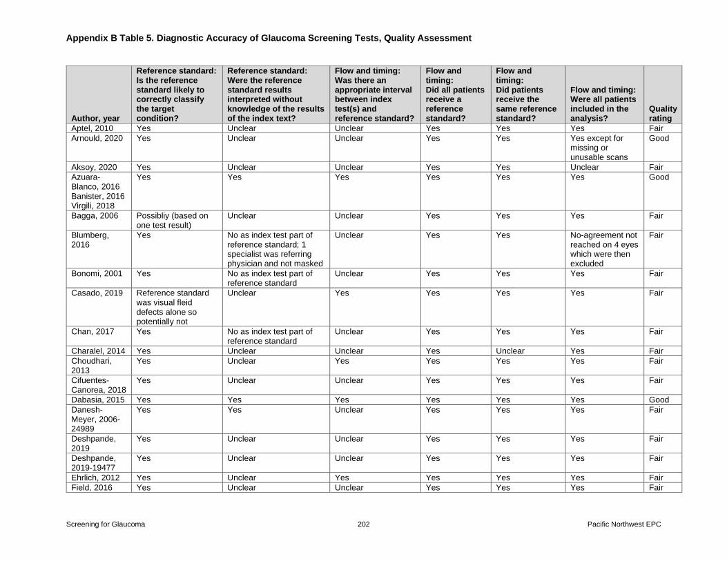
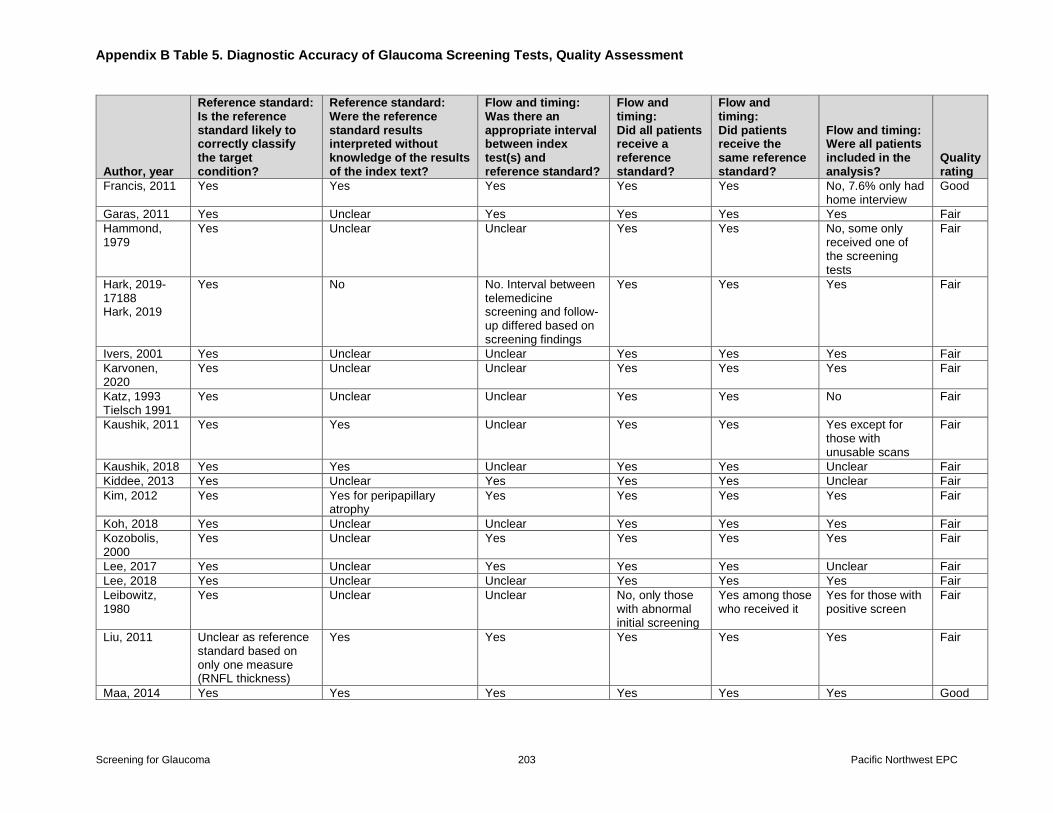
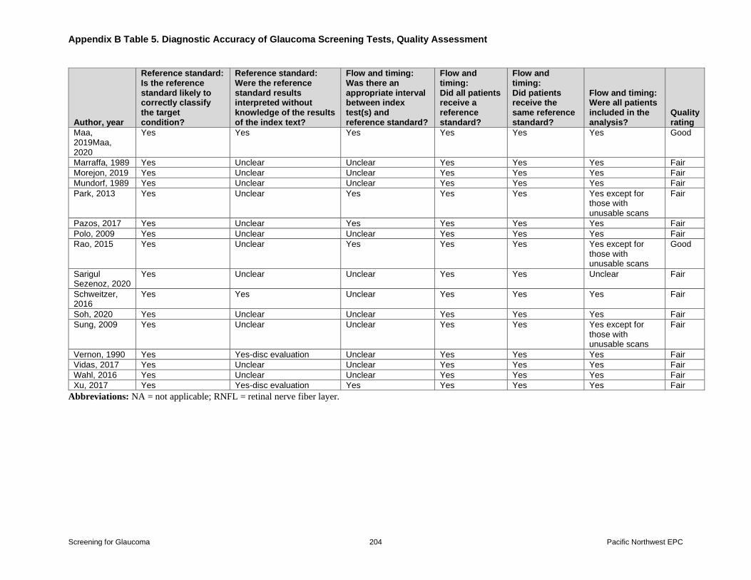
Appendix B Table 5 Diagnostic Accuracy of Glaucoma Screening Tests Quality
Assessment
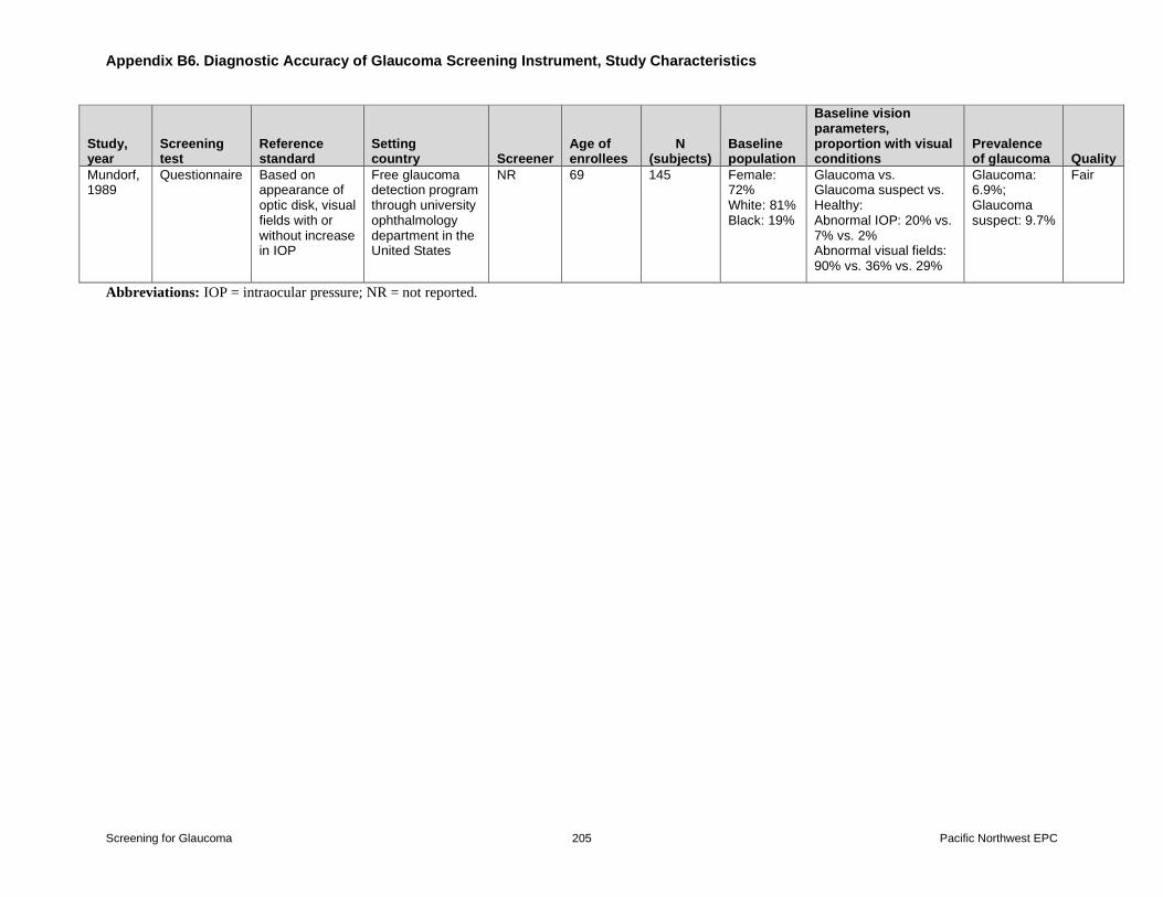
Appendix B Table 6 Diagnostic Accuracy of Glaucoma Screening Instrument Study
Characteristics
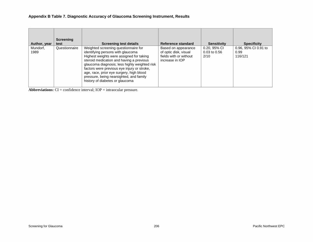
Appendix B Table 7 Diagnostic Accuracy of Glaucoma Screening Instrument Results
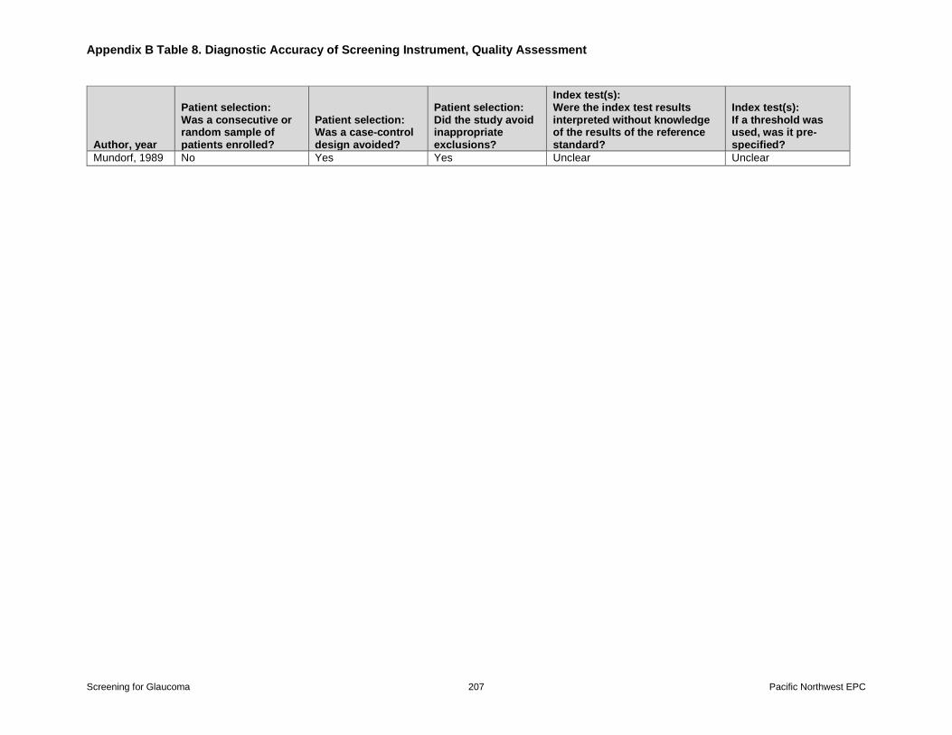
Appendix B Table 8 Diagnostic Accuracy of Glaucoma Screening Instrument Quality
Assessment
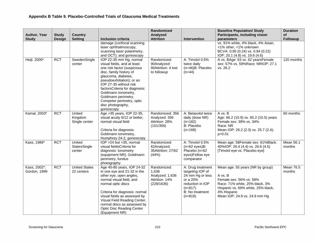
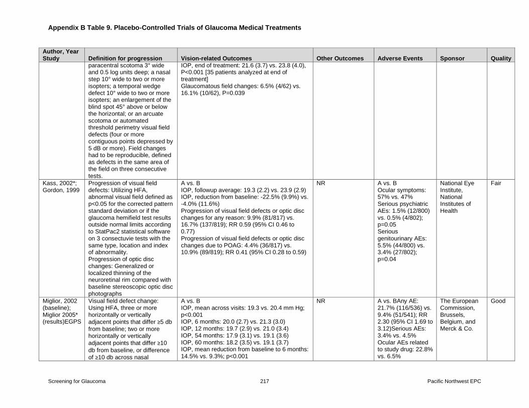
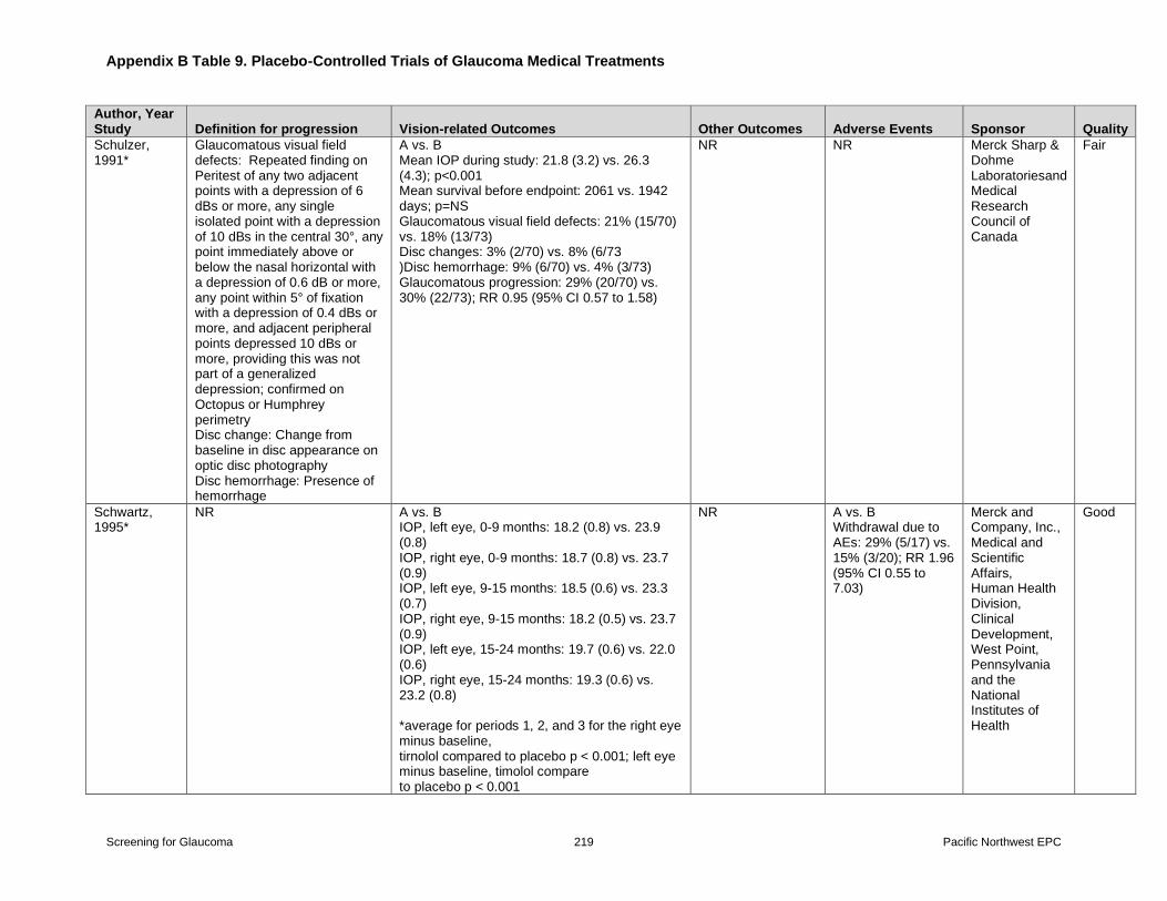
Appendix B Table 9 Placebo-controlled Trials of Glaucoma Medical Treatments
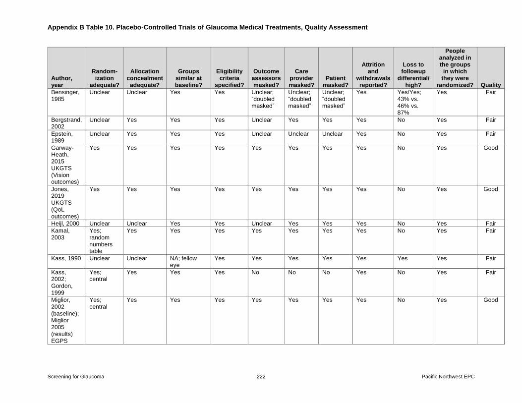
Appendix B Table 10 Placebo-controlled Trials of Glaucoma Medical Treatments Quality
Assessment
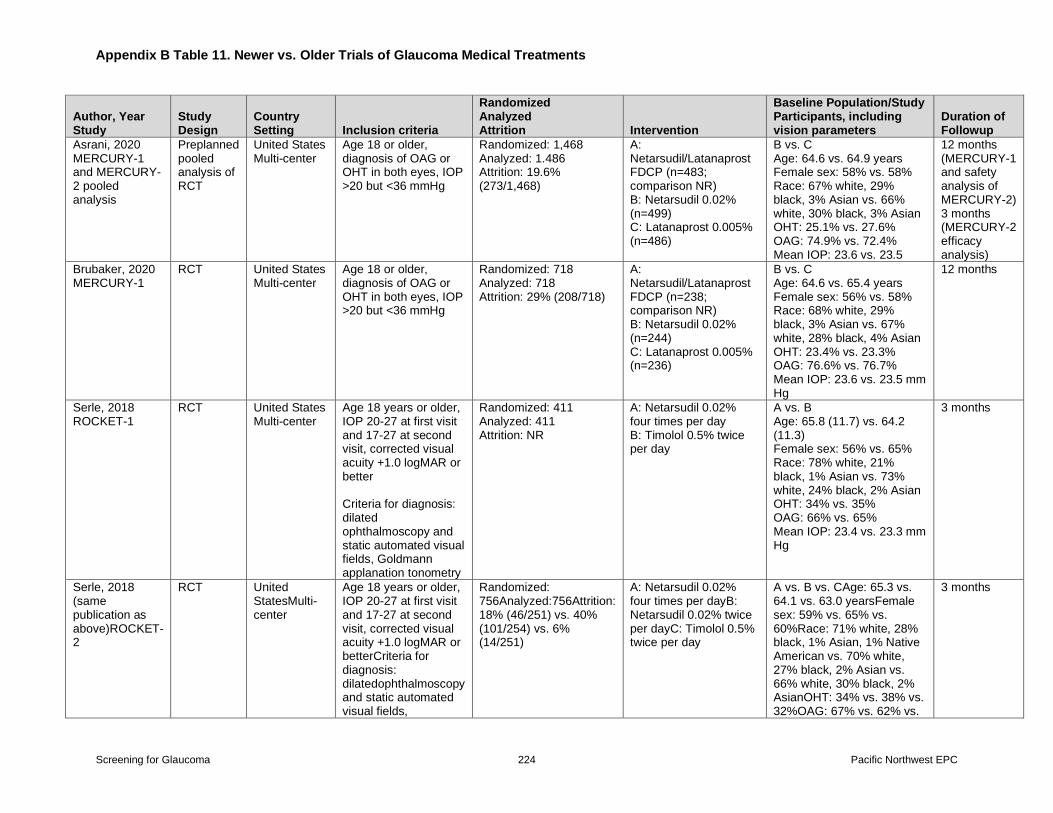
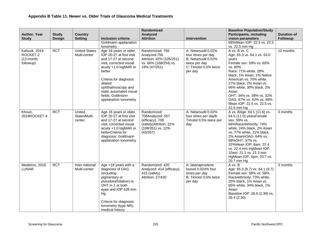
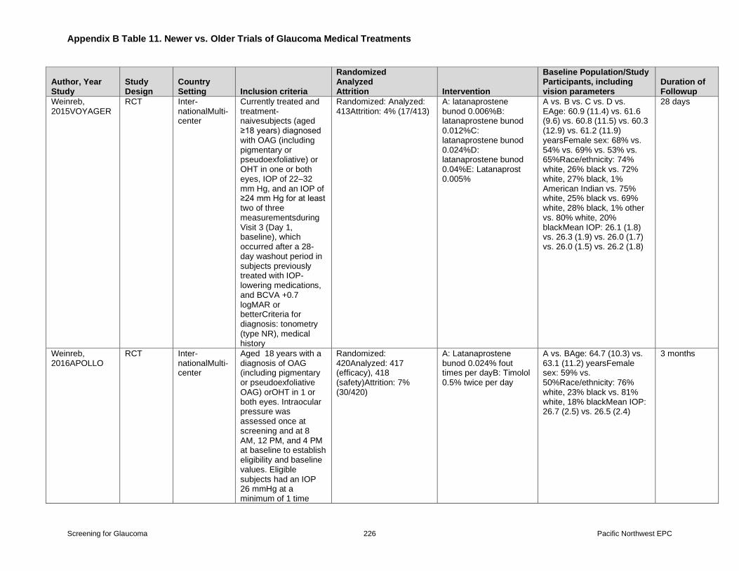
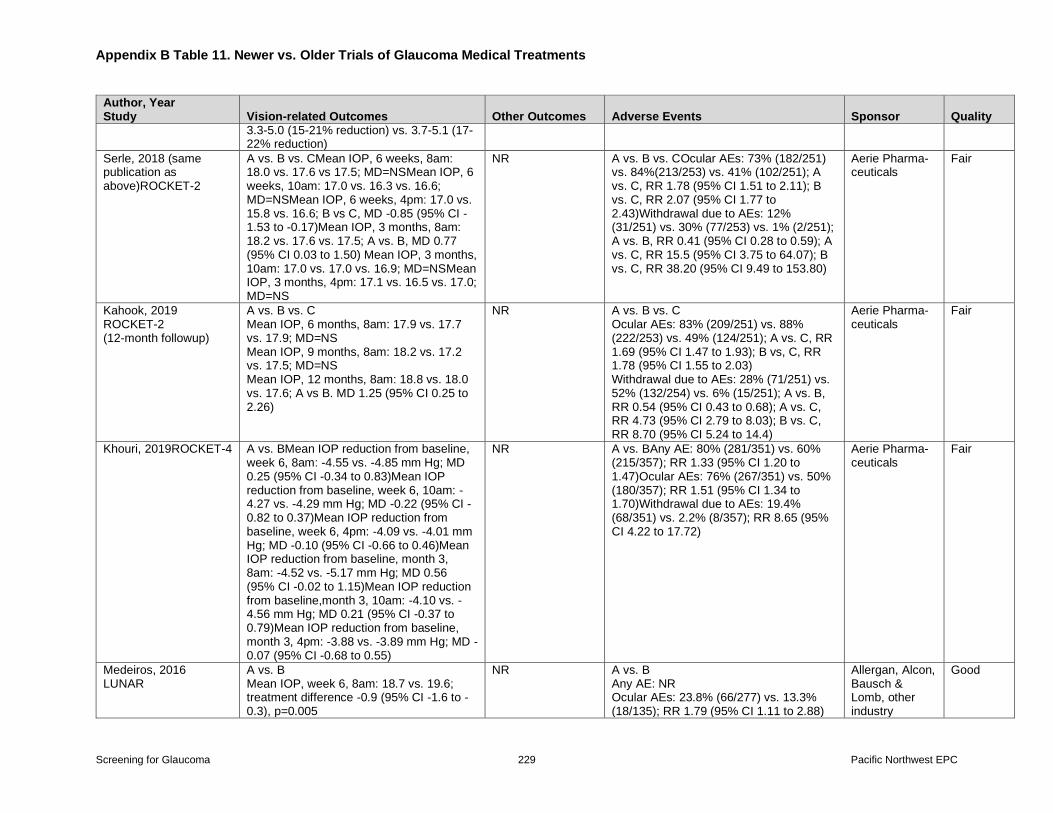
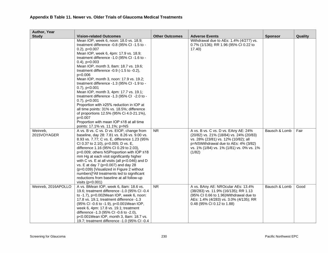
Appendix B Table 11 Newer vs Older Trials of Glaucoma Medical Treatments
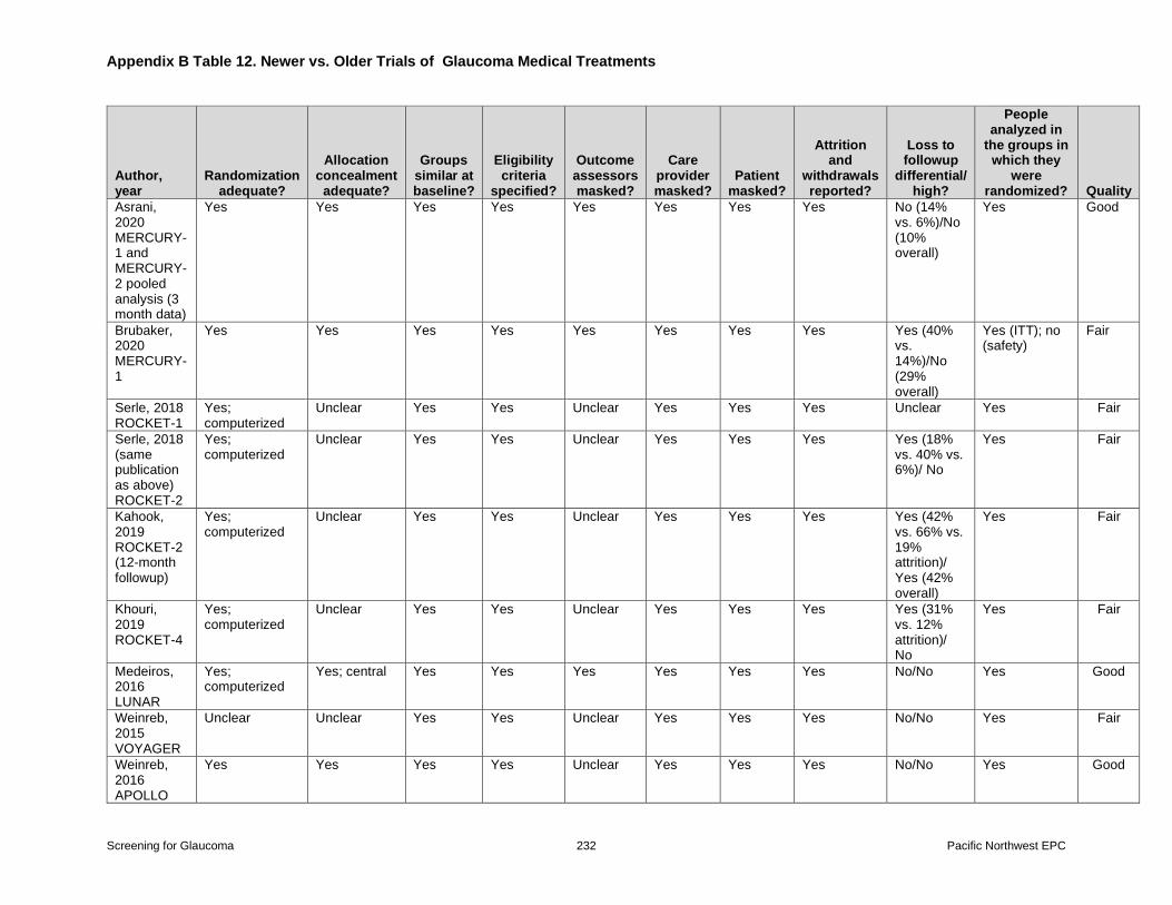
Appendix B Table 12 Newer vs Older Trials of Glaucoma Medical Treatments Quality
Assessment
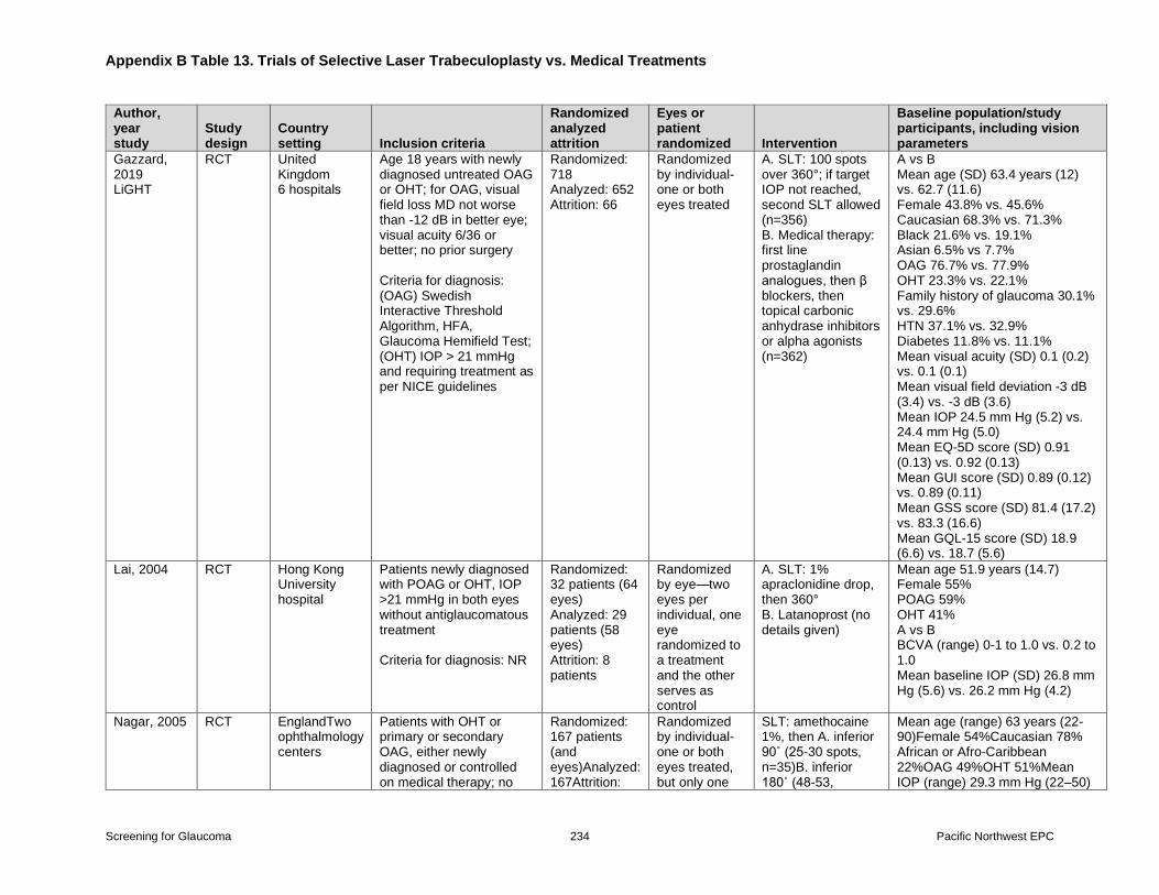
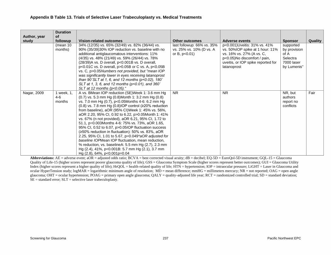
Appendix B Table 13 Trials of Selective Laser Trabeculoplasty vs Medical Treatments
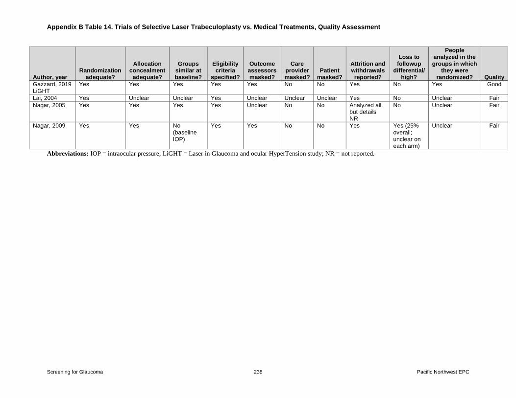
Appendix B Table 14 Trials of Selective Laser Trabeculoplasty vs Medical Treatments
Quality Assessment
Screening for Glaucoma vii Pacific Northwest EPC
Structured Abstract Background In 2013 the United States Preventive Services Task Force (USPSTF) concluded
that the evidence was insufficient to assess the balance of benefits and harms of screening for
primary open angle glaucoma in adults (I Statement) Although the USPSTF found that
treatment of increased intraocular pressure (IOP) and early glaucoma reduces progression of
visual field defects it found inadequate evidence on the effects of treatment on the development
of impaired vision or quality of life There was no direct evidence on benefits and harms of
glaucoma screening versus no screening
Purpose To systematically review the evidence on screening and treatment of glaucoma for
populations and settings relevant to primary care in the United States
Data Sources We searched the Cochrane Central Register of Controlled Trials Cochrane
Database of Systematic Reviews and MEDLINE (through February 9 2021) reviewed the
studies in the prior reports and manually reviewed reference lists
Study Selection Randomized controlled trials (RCTs) of screening and referral studies on
diagnostic accuracy of currently utilized screening tests (optical coherence tomography [OCT]
optic disc photography ophthalmoscopy and biomicroscopy pachymetry tonometry and visual
fields) and RCTs of medical therapy versus placebo or no treatment recently approved medical
therapies versus older therapies and selective laser trabeculoplasty versus medical therapy
Data Extraction One investigator abstracted data and a second checked accuracy Two
investigators independently assessed study quality using methods developed by the USPSTF
Data Synthesis (Results) A total of 83 studies (N=76807) were included in this review (30
trials and 53 diagnostic accuracy studies) Sixteen studies were carried forward from the prior
review and 67 studies were new
One RCT (n=616) found vision screening (including components for glaucoma) by an
optometrist was associated with no difference in visual acuity or vision-related quality of life
compared with no screening but greater risk of falls (likelihood of at least 1 fall 65 vs 50
relative risk [RR] 131 95 confidence interval [CI] 113 to 150) No study evaluated effects of
referral to an eye health provider versus no referral on vision or other health outcomes Evidence
on accuracy of screening tests for identifying persons with glaucoma was most robust for
spectral domain-OCT retinal nerve fiber layer thickness (15 studies N=4242 sensitivity 079
95 CI 075 to 083 and specificity 092 95 CI 087 to 096) area under the receiver operating
characteristic curve (16 studies N=4060) 090 95 CI 086 to 093 and spectral domain-OCT
ganglion cell analysis (nine studies N=1522 sensitivity 074 95 CI 068 to 080 and
specificity 091 95 CI 080 to 096) tonometry (13 studies N=32892 sensitivity 048 95
CI 031 to 066 and specificity 094 95 CI 090 to 096) and the Humphrey Visual Field
Analyzer (six studies N=11244 sensitivity 087 95 CI 069 to 095 and specificity 082 95
CI 066 to 092) Evidence on other screening tests (swept source-OCT optic disc photography
ophthalmoscopy and biomicroscopy and pachymetry) was limited A pilot study and followup
found telemedicine screening in primary care associated with variable sensitivity for identifying
Screening for Glaucoma viii Pacific Northwest EPC
persons with glaucoma but high specificity Evidence on the accuracy of instruments for
identifying patients at higher risk of glaucoma was limited to one study that was of limited
applicability to screening because prior diagnosis of glaucoma was one of the key risk factors
Medical therapy for ocular hypertension and untreated glaucoma was associated with greater
reduction in IOP (16 trials N=3706 mean difference -314 millimeters mercury [mm Hg] 95
CI -419 to -208) decreased likelihood of glaucoma progression (7 trials N=3771 RR 068
95 CI 049 to 096 absolute risk difference -42) and increased risk of ocular adverse events
(2 trials RR 121 95 CI 110 to 133 and RR 352 95 CI 246 to 502) versus placebo or no
treatment One trial (n=461) found no differences between medical therapy versus placebo or no
treatment in visual acuity quality of life or function Recently approved medical therapies for
glaucoma (netarsudil and latanoprostene bunod) were associated with similar or slightly greater
reduction in IOP versus older therapies (6 trials N=3128) but increased risk of adverse events
Selective laser trabeculoplasty and medical therapy were associated with similar effects on IOP
visual acuity visual fields quality of life and adverse events (4 trials N=957)
Limitations Excluded non-English language studies statistical heterogeneity in pooled analyses
on effects of medical therapy versus placebo or no treatment on IOP though inconsistency was
in the magnitude (not direction) of benefit evidence on effects of treatment on visual
impairment quality of life and function remains very limited excluded case-control studies of
diagnostic accuracy evaluation of publication bias limited by small numbers of studies and
statistical heterogeneity most head-to-head comparisons excluded
Conclusions Direct evidence on glaucoma screening versus no screening is limited and showed
no benefits on vision-related quality of life or function and increased risk of falls Screening
tests (OCT visual field assessment) can identify persons with OAG with reasonable accuracy
Treatment for ocular hypertension or untreated OAG is associated with reduction in IOP and
reduced risk of glaucoma progression based on visual fields or optic nerve changes but limited
evidence on the association with visual outcome quality of life and function indicates no clear
effects
Screening for Glaucoma 1 Pacific Northwest EPC
Chapter 1 Introduction and Background
Purpose
This review will be used by the US Preventive Services Task Force (USPSTF) to update its
2013 recommendation on screening for primary open-angle glaucoma (POAG) in adults1 In
2013 the USPSTF concluded that the evidence was insufficient to assess the balance of benefits
and harms of screening for POAG in adults (I statement) The USPSTF came to this conclusion
because it found no direct evidence on the benefits of screening inadequate evidence on the
effects of treatment of increased intraocular pressure (IOP) or early asymptomatic POAG on the
development of impaired vision or quality of life and potential risk of overdiagnosis and
overtreatment The USPSTF found convincing evidence that treatment of increased IOP and
early glaucoma reduces the number of persons who develop small clinically unnoticeable visual
field defects and that treatment of early asymptomatic POAG decreases the number of persons
whose visual field defects worsen however these were considered intermediate outcomes The
prior USPSTF recommendation was based on comparative effectiveness reviews (CER) of
screening2 and treatment34 for glaucoma in this report these are referred to as the ldquoprior
screening CERrdquo and the ldquoprior treatment CERrdquo
Condition Background
Condition Definition
Open-angle glaucoma (OAG) is a chronic progressive neurodegenerative disease of the optic
nerve characterized by structural optic disc andor retinal nerve fiber layer thinning with
associated visual field defects (some authorities consider typical optic nerve changes or visual
field defects to be sufficient to diagnosis glaucoma)5 ldquoOpenrdquo refers to an open anterior chamber
angle on gonioscopy this is in contrast to ldquoclosedrdquo or narrow-angle glaucoma which has a
different presentation and treatment and is outside the scope of this review POAG the focus of
this review is characterized by the absence of other known secondary causes such as
neovascularization trauma uveitis or steroid use OAG is generally bilateral but can be
asymmetric The onset of POAG is often in mid to late adulthood Although there is an
association between elevated IOP (typically defined as ge21 mm Hg) and OAG up to 40 percent
of patients with OAG do not have elevated IOP6-8
ldquoGlaucoma suspectrdquo is a nonspecific term describing individuals who do not meet criteria for
glaucoma but have findings or risk factors associated with developing OAG5 Criteria for
glaucoma suspect include a consistently elevated IOP a suspicious appearance of the optic nerve a strong family history of OAG or visual field abnormalities consistent with glaucoma
ldquoOcular hypertensionrdquo refers to the presence of elevated IOP without glaucomatous changes of
the optic nerve or visual fields9 It can be difficult to distinguish a glaucoma suspect from a
patient with early OAG and prospective followup and repeat diagnostic testing are often
necessary to make the distinction
Screening for Glaucoma 2 Pacific Northwest EPC
Prevalence and Burden of DiseaseIllness
Glaucoma is the second leading cause of irreversible blindness in the United States (US) and
the leading cause in Black and Latino persons810 Earlier stages of glaucoma can also impact
quality of life and function including ability to drive and risk of motor vehicle crashes11 Age-
stratified data indicate a decrease in glaucoma related blindness (incidence within 10 years of
diagnosis 87 per 100000 for persons diagnosed in 1965 to 1980 and 55 per 100000 for persons
diagnosed in 1981 to 2000)12 The degree to which the observed trend is related to improved
treatmentmanagement earlier diagnosis or other factors is unclear The prevalence of POAG in
the US is estimated at about 2 percent based on optic nerve fundus photography assessment of
participants in the 2005 to 2008 National Health and Nutrition Examination Survey13 In 2011
an estimated 271 million persons had OAG this number was projected to reach 37 million in
2020 and 43 million in 20251415 The number of persons with glaucoma increases with age
from an estimated 025 million persons 40 to 49 years of age to 128 million persons 70 to 79
years of age In the US Black and Latino persons a threefold or higher prevalence of OAG
relative to non-Latino White persons8131617 In the US the proportion of persons 40 years and
older with ocular hypertension is estimated at 45 percent in non-Latino White and 35 percent in
Latino persons1416 Data on glaucoma suspect prevalence (not limited to ocular hypertension) are
lacking
Etiology and Natural History
The etiology of OAG is likely multifactorial and includes genetic factors18 and age-related
neurodegeneration of the optic nerve19 The degree of IOP elevation correlates with the rapidity
of OAG progression though the susceptibility of individuals to IOP-related optic nerve damage
varies5 As noted above a substantial proportion of patients with OAG have an IOP within the
normal range and some patients with elevated IOP do not develop glaucoma2021 In the Ocular
Hypertension Treatment Study (OHTS) 95 percent of untreated glaucoma suspects with
elevated IOP progressed to glaucoma after 5 years21 and 295 percent after 20 years22
Other factors hypothesized to contribute to the optic nerve damage seen in OAG include a
deficient blood supply to the optic nerve inadequate structural support for the neurons that
comprise the optic nerve and insufficient supplies of neurotrophins The typical natural history
of OAG is of gradual often insidious loss of retinal ganglion cells and corresponding loss of
peripheral andor central vision potentially progressing to blindness The vision loss is generally
irreversible A study of newly diagnosed OAG glaucoma patients in Olmsted County Minnesota
found that after 20 years 27 percent were blind in one eye and 9 percent in both eyes23
However the rate of progression varies Visual field loss is often detectable before visual acuity
loss which usually occurs late in patients with glaucoma While treatment strategies (currently
all based on IOP lowering) can slow the progression of glaucomatous vision loss some patients
continue to lose vision despite apparently adequate IOP lowering24
Screening for Glaucoma 3 Pacific Northwest EPC
Risk Factors
A number of risk factors have been identified for OAG including older age25-27 Black or Latino
raceethnicity8162528 family history2629 higher IOP825 thinner central cornea25 optic disc
hemorrhage30 large optic disc cup-to-disc ratio25 and lower ocular perfusion pressure (as
determined by systemic blood pressure and IOP)31
Rationale for ScreeningScreening Strategies
Untreated glaucoma can lead to irreversible vision loss or blindness Early or mild glaucoma
damage to the optic nerve may be asymptomatic and mild visual loss may not be perceived as
warranting medical evaluation Visual field loss from OAG is often not perceived by patients32
and 50 percent or more of patients with OAG are unaware that they have glaucoma8172733
Therefore screening could identify patients with asymptomatic or mild OAG who could benefit
from early treatment to prevent further visual loss Screening could also identify patients who are
glaucoma suspects and might benefit from treatments or monitoring to prevent progression to
OAG andor vision loss21
Screening for glaucoma is based on a number of tests including tonometry (for IOP)
ophthalmoscopy on dilated eye examination (for evaluation of the optic nerve) perimetry (visual
field test) gonioscopy (to measure the angle in the eye where the iris meets the cornea)
pachymetry (to measure the thickness of the cornea) and visual acuity testing34 Imaging tests
such as optical coherence testing (OCT which uses low-coherence light to image the retina) and
optic disc photography (to view the optic nerve head andor retina) can supplement the clinical
examination A challenge in screening for glaucoma in primary care settings is that with the
exception of visual acuity and certain tonometry tests primary care clinicians lack training or
equipment to perform much of the glaucoma clinical examination which is typically performed
in an eye specialty setting As previously described tonometry and visual acuity testing lack
sensitivity for glaucoma because a significant proportion of patients have normal IOP and visual
acuity changes are a late finding In addition diagnostic criteria for glaucoma lack consensus and
are difficult to standardize
InterventionsTreatment
The only known modifiable risk factor for glaucoma is IOP Therefore all current glaucoma
treatments aim to lower IOP even in persons with non-elevated IOP An optimal target IOP has
not been identified and the IOP target is typically individualized though the American Academy
of Ophthalmology (AAO) suggests a reduction in IOP of 25 percent from baseline as a
reasonable initial goal in most patients Current IOP lowering strategies include topical
medicated drops (prostaglandin analogs beta-blockers alpha-adrenergic agonists carbonic
anhydrase inhibitors Rho kinase inhibitors nitric oxide donators and less frequently cholinergic
agents)2135 oral agents (carbonic anhydrase inhibitors hyperosmotic agents) laser
trabeculoplasty3637 laser cyclophotocoagulation3839 and incisional surgery (ie trabeculectomy
glaucoma drainage device implantation and angle-based surgeries)4041 The AAO recommends
Screening for Glaucoma 4 Pacific Northwest EPC
medications or laser trabeculoplasty as initial therapy in most patients5 Topical prostaglandins
are currently the most commonly used initial medication for OAG Selective laser
trabeculoplasty (SLT) using a frequency-doubled neodynmiumyttrium-aluminum-garnet laser
produces less thermal damage to the trabecular network compared with argon laser
trabeculoplasty and is the most commonly used laser trabeculoplasty technique Surgery is
usually reserved for patients with severe visual field loss at baseline or patients with advanced
OAG who do not respond to medications or laser trabeculoplasty due to complications
associated with surgery In patients who are glaucoma suspects the AAO recommends a shared
decision making approach based on the risk of developing glaucoma to determine whether to
initiate treatment5 For persons with ocular hypertension a risk calculator is available to estimate
the risk of developing glaucoma in persons with ocular hypertension42
New developments in treatment for glaucoma since the prior USPSTF recommendation include
the approval by the US Food and Drug Administration (FDA) of two new medications for OAG
and ocular hypertension latanoprostene bunod43 (a nitric oxide-donating medication) and
netarsudil44 (a Rho kinase inhibitor) These are the first new medications approved for glaucoma
since 1996 Unlike the majority of medications for OAG that decrease IOP by reducing aqueous
production these medications increase aqueous outflow The development of newer minimally-
invasive surgical procedures for treatment of OAG is ongoing45
Current Clinical PracticeRecommendations of Other Groups
The AAO recommends a baseline comprehensive eye evaluation at age 40 In persons without
risk factors for ocular disease the AAO recommends examinations every 2 to 4 years for persons
40 to 54 years of age every 1 to 3 years for persons 55 to 64 years of age and every 1 to 2 years
in persons 65 years of age or older5 In persons at higher risk for ocular disease the AAO
recommends that decisions regarding when to initiate eye evaluations and the frequency of
periodic examinations be based on the risks but does not provide specific guidance For
glaucoma evaluation the AAO describes a number of components of the comprehensive eye
examination including visual acuity measurement pupil examination anterior segment
examination IOP measurement gonioscopy optic nerve hypoplasia and retinal nerve fiber layer
examination and fundus examination5 Diagnostic tests include central corneal thickness
measurement visual field evaluation and optic nerve hypoplasia and retinal nerve fiber layer
imaging
The American Academy of Family Physicians supports the USPSTF recommendation on
glaucoma screening46
Data on the frequency of glaucoma screening in primary care settings are not available though it
is unlikely to be high due to a lack of training and specialized equipment Data are also not
available on the proportion of patients in primary care settings referred for glaucoma screening
An area of ongoing interest is use of telemedicine to facilitate glaucoma screening in primary
care settings47 and use of artificial intelligence for screening diagnosis and classification of
glaucoma48
Screening for Glaucoma 5 Pacific Northwest EPC
Chapter 2 Methods
Key Questions and Analytic Framework
The scope and key questions (KQs) were developed by the Evidence-based Practice Center
(EPC) investigators USPSTF members and Agency for Healthcare Research and Quality
(AHRQ) Medical Officers using the methods developed by the USPSTF49 The analytic
framework and KQs that guided the review are shown in Figure 1 In the KQs ldquoOAGrdquo refers to
POAG patients and glaucoma suspects Eleven KQs were developed for this review
Key Questions
Key Question 1 What are the effects of screening for OAG versus no screening on a) IOP
visual field loss visual acuity or optic nerve damage or b) visual impairment quality of
life or function
Key Question 2 What are the harms of screening for OAG versus no screening
Key Question 3 What are the effects of referral to an eye health provider versus no referral
on a) IOP visual field loss visual acuity or optic nerve damage or b) visual impairment
quality of life or function
Key Question 4 What is the accuracy of screening for diagnosis of OAG
Key Question 5 What is the accuracy of instruments for identifying patients at higher risk of
OAG
Key Question 6 What are the effects of medical treatments for OAG versus placebo or no
treatments on a) IOP visual field loss visual acuity or optic nerve damage or b) visual
impairment quality of life or function
Key Question 7 What are the harms of medical treatments for OAG versus placebo or no
treatments
Key Question 8 What are the effects of newly FDA-approved medical treatments
(latanoprostene bunod and netarsudil) versus older medical treatments on a) IOP visual
field loss visual acuity or optic nerve damage or b) visual impairment quality of life or
function
Key Question 9 What are the harms of newly FDA-approved medical treatments versus
older medical treatments
Key Question 10 What are the effects of laser trabeculoplasty for OAG versus no
trabeculoplasty or medical treatment on a) IOP visual field loss visual acuity or optic
nerve damage or b) visual impairment quality of life or function
Key Question 11 What are the harms of laser trabeculoplasty for OAG versus no
trabeculoplasty or medical treatment
The KQs focus on areas most relevant to inform recommendation on screening in primary care
settings and are informed by evidence gaps identified in the prior reviews23 KQs on the effects
of screening versus no screening on intermediate outcomes health outcomes and harms were
carried forward from the prior reviews A KQ on the effects of referral to an eye health provider
versus no referral was added because diagnosis of glaucoma is often based on a comprehensive
Screening for Glaucoma 6 Pacific Northwest EPC
eye examination by an eye health provider A KQ on the accuracy of screening for diagnosis of
OAG was also carried forward We added a KQ on the accuracy of risk prediction instruments to
identify persons with OAG Regarding therapies the prior treatment CER3 included many head-
to-head comparisons In order to focus on the comparisons of most relevance for informing
recommendations on screening we included a KQ focusing on the effectiveness of first-line
medical therapies versus placebo or no therapy We also included KQs of newly FDA-approved
therapies versus older medical therapies and SLT versus first-line therapies or no SLT as trials
comparing these therapies versus placebo or no treatment are lacking
Contextual Question
One Contextual Question was also requested by the USPSTF to help inform the report
Contextual Questions are not reviewed using systematic review methodology
Contextual Question 1 What is the association between changes in IOP visual field loss
visual acuity or optic nerve damage following treatment for OAG and improvement in
visual impairment quality of life or function and what is the association between changes
in IOP and visual field loss
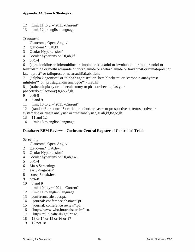
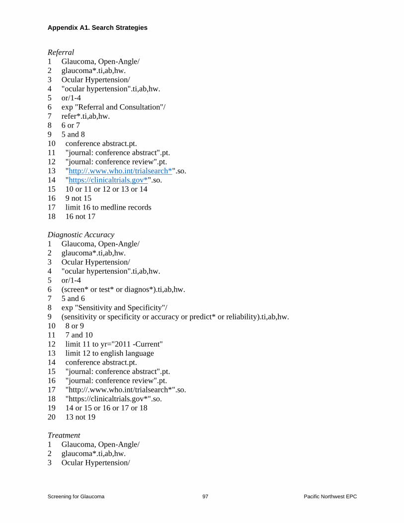
Search Strategies
A research librarian searched the Cochrane Central Register of Controlled Trials Cochrane
Database of Systematic Reviews and MEDLINE (January 2011 to February 9 2021) for
relevant studies and systematic reviews The search relied primarily on the previous systematic
review for the USPSTF to identify potentially relevant studies published before 2011 (we
reassessed all articles included in that systematic review using the eligibility criteria) Search
strategies are available in Appendix A1 To supplement electronic searches we reviewed
reference lists of relevant articles
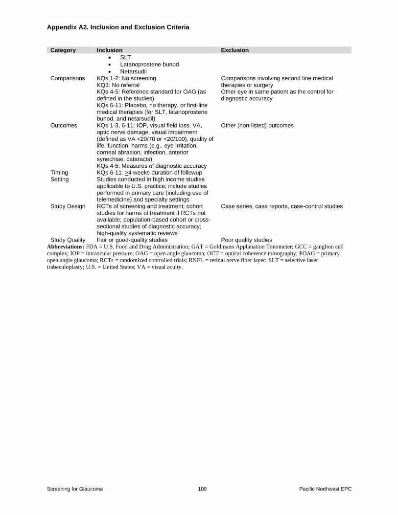
Study Selection
At least two reviewers independently evaluated each study to determine eligibility We selected
studies on the basis of inclusion and exclusion criteria developed for each KQ (Appendix A2)
Articles were selected for full text review if they were about OAG or glaucoma suspect in adults
40 years of age or older were relevant to a KQ and met the per-defined inclusion criteria We
excluded studies of patients with narrow-angle glaucoma secondary OAG (including exfoliation
glaucoma) or advanced glaucoma (eg with severely impaired vision) We restricted inclusion to
English-language articles and excluded studies published only as abstracts Studies of non-
human subjects were also excluded and studies had to report original data
For screening we included studies on a complete eye examination (as defined in the studies)
various components of a complete eye examination (ophthalmoscopy perimetry tonometry
pachymetry evaluation for afferent pupillary defect) and imaging tests (optic disc photography
Screening for Glaucoma 7 Pacific Northwest EPC
optical coherence testing [OCT] and fundus photography) We excluded screening tests that are
considered outdated or no longer used such as the water drinking test the Heidelberg Retina
Tomograph scanning laser polarimetry and older OCT technology (time-domain OCT) For
treatment we included first line medical treatments (defined as prostaglandin analogues beta-
blockers alpha2 agonists and carbonic anhydrase inhibitors) SLT and newly FDA-approved
medical treatments (latanoprostene bunod and netarsudil) We excluded studies of combination
treatment and trabeculectomy which are not considered first line therapy for ocular hypertension
or early glaucoma and outdated therapies (eg argon laser trabeculoplasty) The comparison for
screening was no screening and the main comparison for treatment was placebo or no treatment
We also included head to head trials that compared latanoprostene bunod or netarsudil versus
first-line medical therapies For screening referral and treatment outcomes were IOP visual
field loss visual acuity optic nerve damage visual impairment (defined as visual acuity lt2070
or lt20100) quality of life function and harms (eg eye irritation corneal abrasion infection
anterior synechiae cataracts) reported at least four weeks after initiating the intervention We
included randomized trials of screening and treatment and cohort and cross-sectional studies on
screening test diagnostic accuracy We excluded diagnostic accuracy studies that used a case-
control design due to potential spectrum bias50 Telemedicine studies of screening were included
if they were conducted in primary care settings Studies on imaging test diagnostic accuracy that
utilized artificial intelligence to analyze images were included if they evaluated a clinical cohort
(eg did not analyze images in a databank) did not use a case-control design reported
validation testing and utilized algorithms available for widespread use Studies on screening
accuracy were not restricted by clinical setting although results from primary care settings were
highlighted if available This report utilized primary studies and systematic reviews were used to
identify potentially eligible studies In accordance with USPSTF methods studies rated poor
quality (see below) were excluded
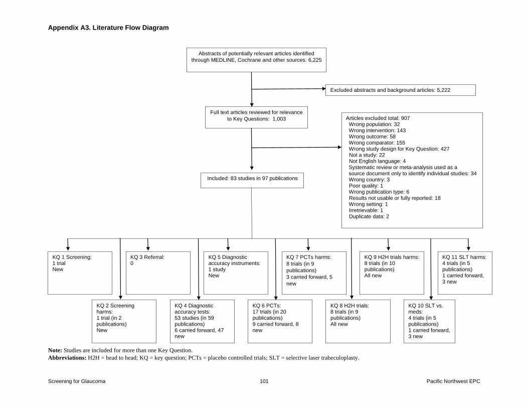
The selection of literature is summarized in the literature flow diagram (Appendix A3)
Appendix A4 lists the included studies and Appendix A5 lists the excluded studies with
reasons for exclusion
Data Abstraction and Quality Rating
For studies meeting inclusion criteria we created data abstraction forms to summarize
characteristics of study populations interventions comparators outcomes study designs
settings and methods One investigator conducted data abstraction which was reviewed for
completeness and accuracy by another team member
Predefined criteria were used to assess the quality of individual studies by using criteria
developed by the USPSTF Studies were rated as ldquogoodrdquo ldquofairrdquo or ldquopoorrdquo per USPSTF criteria
depending on the seriousness of methodological shortcomings (Appendix A6)49 For each study
quality assessment was performed by two team members Disagreements were resolved by
consensus
Screening for Glaucoma 8 Pacific Northwest EPC
Data Synthesis and Analysis
We performed a random effects meta-analysis using the profile likelihood model to summarize
the effects of first-line medical treatments versus placebo or no treatment on likelihood of
glaucoma progression (based on progression of visual field loss with or without optic nerve
changes) serious adverse events and withdrawal due to adverse events (dichotomous outcomes)
and mean IOP (continuous outcome) For mean IOP we used adjusted differences when
reported otherwise the difference in followup IOP was utilized when available followed by the
difference in change from baseline Further we used the differences based on per-individual data
when available For trials that randomized each individual to a treatment but reported a per-eye
analysis (ie two eyes per individual) we averaged the mean IOP between the two eyes and
calculated the standard deviation (SD) for the mean IOP by assuming a correlation of 05
between an individualrsquos eyes For trials in which one eye in each individual was randomized to
the medical treatment and the other eye received the control treatment we used the mean
difference based on the within-subject comparison If the SD for the within-subject mean
difference was not reported it was calculated based on the reported SD for each treatment group
again assuming a correlation of 05 When the SD for the followup IOP was not reported it was
imputed using the average coefficient of variation from other included trials We combined arms
of comparable interventions within the same study in the primary analysis so each study was
represented once in a meta-analysis in order to avoid overweighting We stratified analyses by
the type of medical treatment (alpha agonist prostaglandin analogue beta-blocker carbonic
anhydrase inhibitor or mixed) and conducted prespecified study-level subgroup analyses on the
following factors glaucoma status (OAG ocular hypertension or mixed) quality (good or fair)
mean IOP (lt20 vs gt20 mm Hg) and duration of follow-up (lt1 year vs gt1 year) For glaucoma
progression we conducted a sensitivity analysis restricted to trials that defined progression based
on visual field loss (excluding optic nerve changes) Statistical heterogeneity was assessed using
the Cochran Q-test and I2 statistic51 When at least 10 studies were available for meta-analysis
we tested for small sample effects using graphical (funnel plot) and statistical (Eggerrsquos test)
methods All meta-analyses were conducted using Stata 142 or StataSE 161 (Statacorp
College Station Texas)
For diagnostic accuracy we performed a random effects meta-analysis to summarize sensitivity
and specificity of screening tests to distinguish glaucomatous eyes from eyes without glaucoma
(healthy eyes glaucoma suspect or ocular hypertension) We used a bivariate model to account
for the correlation between sensitivity and specificity to produce summary values for sensitivity
and specificity with corresponding 95 percent confidence intervals (CI) and summary receiver
operating characteristic (ROC) curves For the bivariate model at least four studies were needed
to pool We created paired sensitivity and specificity forest plots and summary ROC figures that
also show the summary sensitivity and specificity with 95 percent confidence regions We
restricted the meta-analysis to studies that used one eye per individual studies that used both
eyes were not included in the meta-analysis because they did not report the correlation between
eyes and inclusion would result in overweighting When studies reported a range of testing
cutoffs we used data based on the most commonly used cutoff (eg IOP gt21 mm Hg) or closest
to it For one study52 that reported sensitivities across multiple specificities without reporting a
cutoff we used the sensitivity and specificity pair with the fewest misclassifications We also
conducted meta-analysis using the Dersimonian-Laird method to summarize discrimination
Screening for Glaucoma 9 Pacific Northwest EPC
based on the area under the receiver operating characteristic (AUROC) curve as reported in
individual studies Stratified analyses were conducted based on the type of control (healthy eye
glaucoma suspect or ocular hypertension) and study quality (good or fair) We also conducted
sensitivity analyses on factors related to specific imaging tests studies of retinal nerve fiber layer
on spectral domain-OCT that measured retinal nerve fiber layer based on the mean thickness
studies of ganglion cell complex on spectral domain-OCT that utilized measures of the retinal
nerve fiber layer inner plexiform layer and the ganglion cell layer and studies that measured
IOP using Goldmann tonometry Statistical heterogeneity was assessed using the I2 however
this value is often high and difficult to interpret in diagnostic accuracy studies because it does not
account for variability in estimates due to threshold effects
For all KQs the overall strength of evidence was determined using the approach described in the
USPSTF Procedure Manual49 The strength of evidence was rated ldquohighrdquo ldquomoderaterdquo ldquolowrdquo or
ldquoinsufficientrdquo based on study quality consistency of results between studies precision of
estimates study limitations and risk of reporting bias49 Additionally the applicability of the
findings to US primary care populations and settings was assessed Discrepancies were resolved
through consensus discussion
USPSTF Involvement
This review was funded by AHRQ AHRQ staff and USPSTF members participated in
developing the scope of the work and reviewed draft reports but the authors are solely
responsible for the content
Expert Review and Public Comment
The draft research plan was posted for public comment from February 13 2020 to March 11
2010 The comments were reviewed and no changes to the scope or Key Questions were
required though some edits were made for clarity The eligibility criteria table (Appendix A2)
was revised to clarify included and excluded diagnostic tests for glaucoma tests that are no
longer used were excluded A final research plan was posted on the USPSTFrsquos Web site on June
11 2020
A draft version of this report has been reviewed by content experts and representatives of Federal
partners (Appendix A7) USPSTF members and AHRQ Medical Officers and edits were made
for clarity The draft will be posted for public comment prior to finalization
Screening for Glaucoma 10 Pacific Northwest EPC
Chapter 3 Results
A total of 6225 new references from electronic database searches and manual searches of
recently published studies were reviewed and 1003 full-text papers were evaluated for inclusion
We included a total of 83 studies (in 97 publications) Sixty-seven studies were newly identified
as part of this update and 16 were carried forward from the previous USPSTF reviews Included
studies and quality ratings are described in Appendix B
Key Question 1 What are the effects of screening for OAG
versus no screening on a) IOP visual field loss visual acuity or optic nerve damage or b) visual impairment quality of life
or function
Summary
bull One trial (N=616) of frail elderly persons found no difference between vision screening
including components for glaucoma versus no screening on vision outcomes (mean
logarithm of the minimum angle of resolution [logMAR] distance visual acuity scores
027 vs 025 p=032 and mean logMAR near visual acuity scores -001 vs -003
p=026) and vision-related quality of life (National Eye Institute Visual Function
Questionnaire-25 [NEI-VFQ-25] mean composite scores 843 vs 864 p=049) after 1
year
Evidence
The prior screening CER included no trials of screening versus no screening2 We identified one
good-quality trial (n=616) conducted in Australia of vision screening by an optometrist versus no
screening that included components relevant for diagnosis of glaucoma (IOP direct
ophthalmoscopy and visual field) as well as other visual testing (visual acuity contrast
sensitivity and slit lamp examination Appendix B Table 1)53 In the screened group
interventions for screen-positive persons included referral to an ophthalmologist or public
hospital eye clinic andor an occupational therapist (for home modifications mobility training or
a cane) those in the control group received no vision assessment or intervention The mean age
was 81 years and 68 percent were female race and ethnicity were not reported Thirty-one
percent of participants needed help with activities of daily living at baseline and 52 percent were
taking more than four medications At baseline 46 percent of participants had experienced a fall
in the past year At baseline mean visual acuity was 022 logMAR (Snellen 2030) the mean
NEI-VFQ-25 score was 855 (scale 0 to 100 higher is better) 63 percent had cataracts 39
percent had undergone cataract surgery and 98 percent wore glasses Fourteen percent of
patients had glaucoma at baseline and 50 percent self-reported vision as ldquogoodrdquo In addition to
appropriate randomization the trial blinded outcome assessors and data analysts and attrition
was low (11 screening arm and 16 control arm Appendix B Table 2)
Screening for Glaucoma 11 Pacific Northwest EPC
Nearly half (487) of the patients in the screening arm were judged to need treatment though
only 55 percent of patients judged to need treatment were referred for glaucoma management
Other interventions were new glasses (298) referral for cataract surgery (49) referral for
age-related macular degeneration (AMD) (16) and referral to an occupational therapist
(77)
At 1 year there were no differences in vision parameters or vision-related quality of life Mean
distance visual acuity was 027 vs 025 logMAR (p=032) mean near visual acuity -001 vs -
003 logMAR (p=026) and NEI-VFQ-25 mean composite scores were 843 vs 864 (p=049)
Nearly three-quarters of patients in the control group reported having seen an eye care
professional in the 12 months prior to study which could have attenuated potential benefits of
screening
Key Question 2 What are the harms of screening for OAG
versus no screening
Summary
bull One trial (n=616) found screening associated with an increased risk for falls versus no
screening (incidence rate ratio 157 95 CI 120 to 205 and risk of one or more falls
65 vs 50 RR 131 95 CI 113 to 150) screening was associated with increased
risk for fractures that was not statistically significant (relative risk [RR] 174 95 CI
097 to 311)
Evidence
No trial in the prior screening CER evaluated harms of screening versus no screening2 A
previously described trial53 of vision screening (including components for identification of
glaucoma) versus no screening in frail elderly reported risk of falls and fracture (Appendix B
Table 1)54 In the trial 46 percent of patients had fallen in the past year Although the trial
hypothesized that screening would reduce the risk of falls screening was associated with
increased incidence of falls (758 vs 516 falls incidence rate ratio 157 95 CI 120 to 205)
risk of one or more falls (65 vs 50 RR 131 95 CI 113 to 150) and risk of two or more
falls (38 vs 31 RR 124 95 CI 099 to 154) versus no screening Screening was also
associated with increased risk of fracture though the difference was just above the threshold for
statistical significance (10 vs 57 RR 174 95 CI 097 to 311 p=006)
Screening for Glaucoma 12 Pacific Northwest EPC
Key Question 3 What are the effects of referral to an eye health provider versus no referral on a) IOP visual field loss visual acuity or optic nerve damage or b) visual impairment
quality of life or function
No eligible study evaluated effects of referral to an eye health provider for glaucoma versus no
referral
Key Question 4 What is the accuracy of screening for diagnosis of OAG
Summary
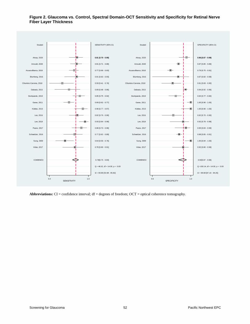
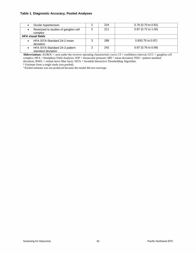
bull Retinal nerve fiber layer thickness on spectral domain-OCT was associated with a pooled
sensitivity of 079 (95 CI 075 to 083) and specificity of 092 (95 CI 087 to 096) for
distinguishing between glaucomatous eyes and controls based on 15 studies (N=4242)
the pooled AUROC curve was 090 (95 CI 086 to 093) based on 16 studies
(N=4060)
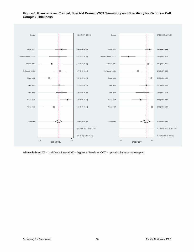
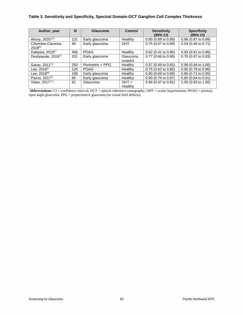
bull Ganglion cell complex thickness on spectral domain -OCT was associated with a pooled
sensitivity of 074 (95 CI 068 to 080) and specificity of 091 (95 CI 080 to 096) for
distinguishing between glaucomatous eyes and controls based on 9 studies (N=1522)
the pooled AUROC curve was 088 (95 CI 084 to 092) based on 6 studies (N=765)
bull Tonometry was associated with a pooled sensitivity of 048 (95 CI 031 to 066) and
specificity of 094 (95 CI 090 to 096) based on 13 studies (N=32892)
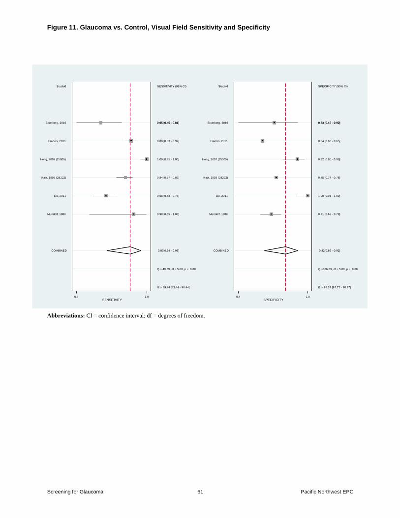
bull The Humphrey Visual Field Analyzer was associated with a pooled sensitivity of 087
(95 CI 069 to 095) and specificity of 082 (95 CI 066 to 092) for distinguishing
between glaucomatous eyes and controls based on 6 studies (N=11244)
bull Evidence on diagnostic accuracy was limited for other screening tests cup-to-disc ratio
on spectral domain-OCT swept source-OCT optic disc photography
ophthalmoscopybiomicroscopystereoscopy pachymetry and afferent papillary defect
bull One pilot study (n=56) and a followup study (n=256) found a telemedicine screening
intervention performed in a primary care setting had variable sensitivity but high
specificity for identifying persons with glaucoma compared with a face-to-face
evaluation by an ophthalmologist
Evidence
The prior screening CER2 included a systematic review55 and 83 additional studies on the
diagnostic accuracy of tests for glaucoma Since the prior screening CER several diagnostic tests
have been superseded by newer technologies and are not included in this review For example
Screening for Glaucoma 13 Pacific Northwest EPC
for imaging the optic nerve and retinal structures OCT has superseded Heidelberg retina
tomography and scanning laser polarimetry for evaluating visual field loss the Humphrey Field
Analyzer has superseded frequency doubling technology In addition the prior screening CER
included case-control studies which were excluded from this review and had an emphasis on
comparative diagnostic accuracy which was not the focus of this review The prior screening
CER concluded that it was unclear whether any one test or combination of tests was suitable for
glaucoma screening in the general population due to the lack of a definitive diagnostic reference
standard for glaucoma and heterogeneity in the design and conduct of the studies
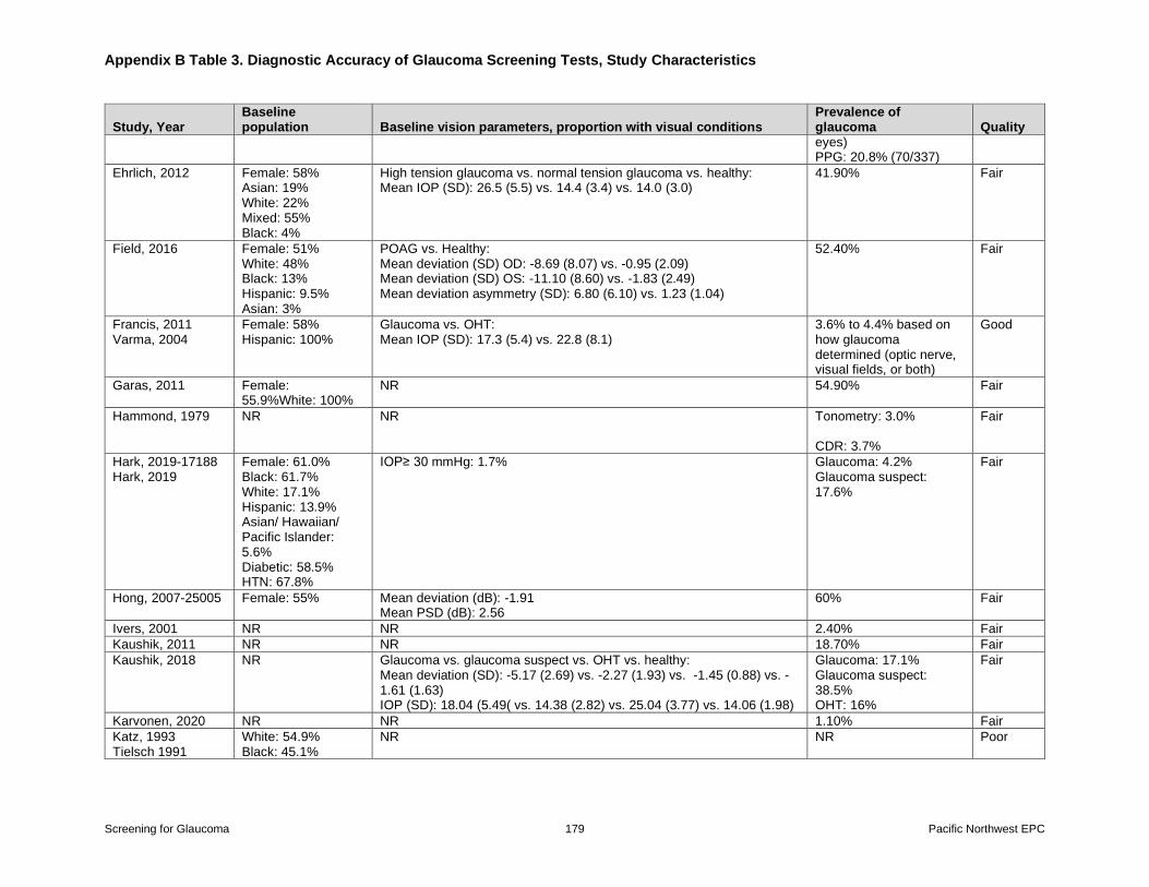
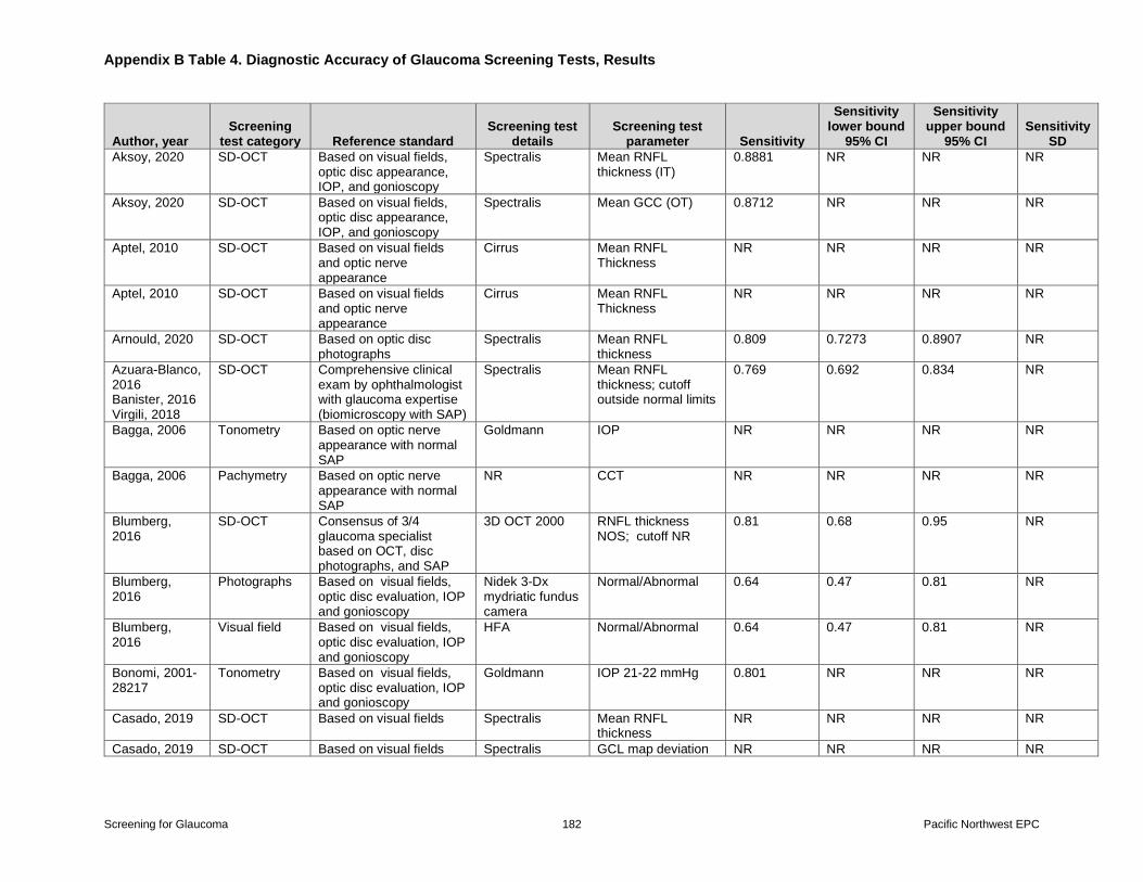
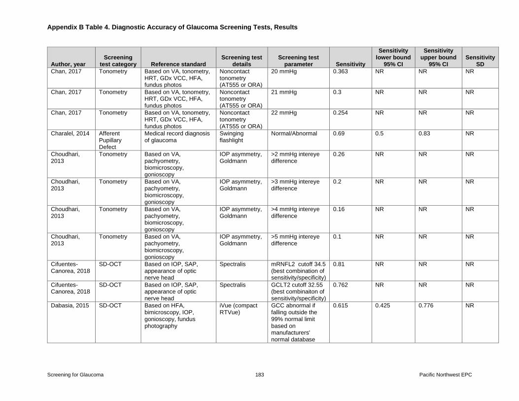
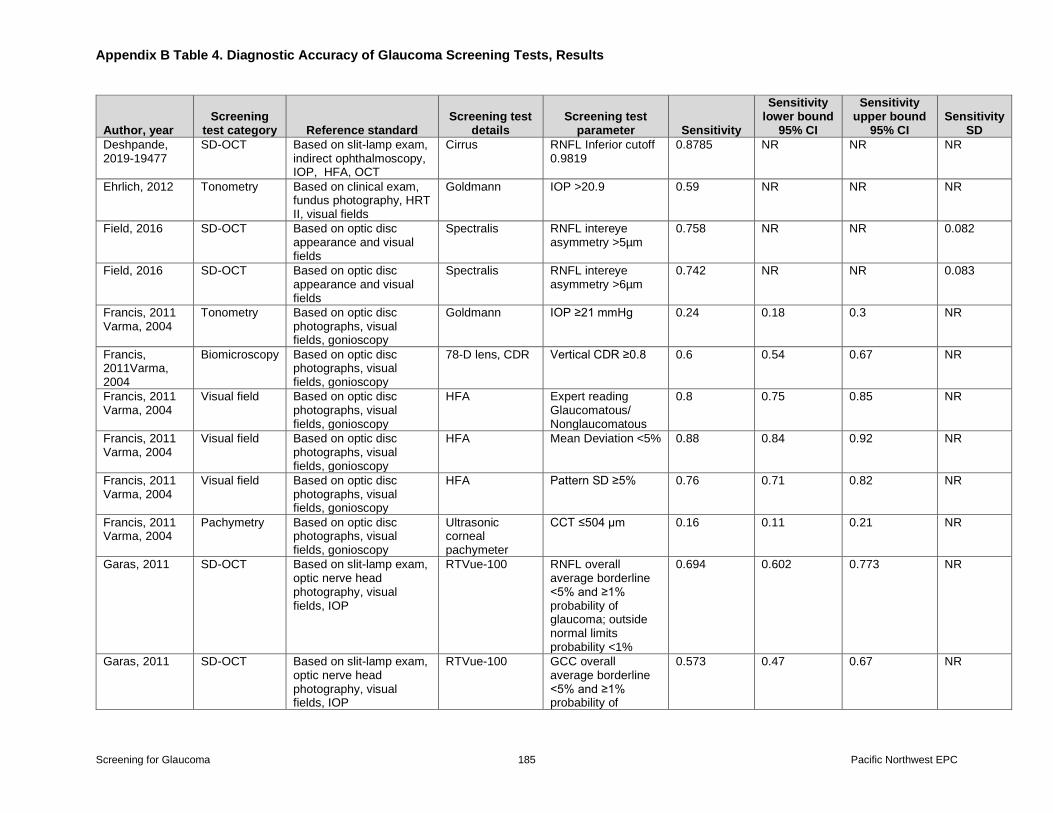
This review includes 53 diagnostic accuracy studies (sample sizes 46 to 8623 N=65464 in 59
publications) (Table 1 Appendix B Tables 3-4)165256-112 The largest groups of studies
evaluated spectral domain-OCT (k=29 N=11434) and tonometry (k=17 N=49742) followed
by visual fields (k=10 N=11633) ophthalmoscopybiomicroscopystereopscopy (k=3
N=17519) optic disc photography (k=4 N=3133) pachymetry (k=2 N=6129) telemedicine
(k=2 N=308) and afferent pupillary defect (k=1 N=107) Most studies evaluated more than one
test of diagnostic accuracy Forty-six studies included a single eye per participant in the analysis
and six studies6980859495108 allowed two eyes per participant (the number of eyes analyzed was
unclear in one study106) In most studies the reference standard was based on findings related to
ophthalmic structure (eg appearance of optic disc) as well as function (eg visual fields)
though exact criteria differed (Appendix B Table 3)
Mean age ranged from 382 years to 822 years (median 58 years) The proportion of females
enrolled ranged from 133 to 723 percent (median 55) in studies that reported gender Twelve
studies reported raceethnicity Two studies restricted enrollment to Asian persons110111 one
study restricted enrollment to Latino persons73 and one study restricted enrollment to White
persons74 In the other studies the proportion of White participants ranged from 17 to 99 percent
In two of the studies the majority of participants (61 and 62) were Black7792 Studies were
conducted in Western Europe (N=16) the US (N=13) and Asia (N=18) two studies were
conducted in Turkey and one study each was conducted in Hungary Australia New Zealand
and Croatia The prevalence of glaucoma ranged from 1180 to 73684 percent Seven studies were
rated good-quality5767739193103108 and the remainder were rated fair-quality (Appendix B Table
5) Methodological limitations in the fair-quality studies included lack of blinding and uncertain
interval between index and reference tests
Tests of Ophthalmic Structure The diagnosis of glaucoma is typically made by using tests of both ophthalmic structure and
function together Tests of eye structure include OCT optic disc photography and clinical
examination with an ophthalmoscope or slit-lamp (biomicroscopy) Thirty studies (N=11618)
evaluated OCT (Appendix B Tables 3-4) Four studies were rated good-quality
(N=2575)5767103108 and 27 were rated fair-quality (N=8859)5256606266697072748083-
8587889597101102104-107109111112 (Appendix B Table 5)
Screening for Glaucoma 14 Pacific Northwest EPC
Optical Coherence Tomography
There are three types of OCT time domain spectral domain and swept source Time domain-
OCT represents the earliest technology and became commercially available in 1996113 Time
domain-OCTs have a movable reference light and can produce 400 axial scans of the eye per
second Time domain-OCT was not included in this review as it has been superseded by spectral
domain-OCT which entered the market in 2006 uses a fixed reference light and can produce
50000 axial scans per second resulting in images with greater resolution113 Swept source is the
latest OCT technology and is even faster than spectral domain-OCT (200000 or more axial
scans per second) but is not yet in widespread use The primary parameters used on OCT are the
thickness of the retinal nerve fiber layer and the ganglion cell complex
The prior screening CER included 48 studies of OCT Based on average retinal nerve fiber layer
estimates on OCT sensitivity ranged from 24 to 96 percent and specificity ranged from 66 to
100 percent Many studies (k=34) in the prior screening CER used time domain-OCT and are not
included in this review Two studies of spectral domain-OCT were carried forward from the
prior USPSTF report (k=2 n=283)56111 and we identified 27 new studies (N=14199) Twenty-
nine studies (N=14482) evaluated spectral domain-OCT52565760626667697072748083-
8587889597101-105107-109112 and 3 studies (n=120 145 and not reported) assessed swept source-
OCT8788106
Retinal Nerve Fiber Layer Thickness
Retinal nerve fiber layer thickness on spectral domain-OCT was associated with a pooled
sensitivity of 079 (95 CI 075 to 083) and specificity of 092 (95 CI 087 to 096) for
diagnosing eyes with glaucoma versus no glaucoma (healthy eyes glaucoma suspect andor
ocular hypertension) based on 15 studies (N=4242)52576066677074878897102103105111112 (Table
2 Figures 2-3) Pooled estimates were similar when the analysis was limited to studies in which
the control group was healthy eyes (9 studies N=2404 sensitivity 081 95 CI 074 to 086 and
specificity 096 95 CI 089 to 099)5267748788102103105111 Pooled estimates were also similar
when the analysis was limited to studies that measured retinal nerve fiber layer based on the
mean overall thickness as opposed to mean inferior74 mean outerinferior6687 or mean
temporalinferior retinal nerve fiber layer thickness52102 (12 studies N=3819 sensitivity 079
95 CI 074 to 084 and specificity 090 95 CI 085 to 093)5760666770878897103105111112 and
when results were limited to the 12 fair quality studies (N=1880 sensitivity 080 95 CI 074
to 085 specificity 094 95 CI 088 to 097)5260667074878897102105111112 In three good-quality
studies (N=2400)5767103 sensitivity ranged from 069 to 081 and specificity ranged from 079
to 094 One study (n=129) also reported accuracy of retinal nerve fiber layer thickness for
diagnosing ocular hypertension versus healthy eyes (sensitivity 008 5 CI 0005 to 063
specificity 100 95 CI 096 to 100)74
Five studies on diagnostic accuracy of retinal nerve fiber layer thickness on spectral domain-
OCT were not pooled because they were based on the inter-eye retinal nerve fiber layer thickness
asymmetry72 or because they evaluated more than one eye per participant698085108 Details of
these studies are shown in Appendix B Tables 3-4
Screening for Glaucoma 15 Pacific Northwest EPC
Retinal nerve fiber layer on spectral domain-OCT was associated with high discrimination for
distinguishing glaucomatous eyes from non-glaucoma with a pooled AUROC curve of 090
(95 CI 086 to 093 I2=96) based on 16 studies (N=4060)5256576066708384878897102-105107
(Figure 4) Discrimination was similar for the two good-quality studies (N=1944 pooled
AUROC 087 95 CI 080 to 094 I2=86)57103 and 14 fair-quality studies (N=2116 pooled
AUROC 090 95 CI 086 to 094 I2=97)52566066708384878897102105107 All studies reported
an AUROC greater than or equal to 083 with the exception of two studies that reported an
AUROC of 07866102 Results were similar in a sensitivity analysis restricted to studies that
utilized overall mean retinal nerve fiber layer thickness (12 studies N=3634 AUROC 092
95 CI 089 to 094 I2=80) and in an analysis stratified according to whether the non-
glaucoma group was healthy eyes (10 studies N=2262 AUROC 092 95 CI 089 to 094
I2=84)5256848788102-105107 glaucoma suspects (4 studies N=496 AUROC 090 95 CI 086
to 094 I2=51)6070104105 or ocular hypertension (3 studies N=319 AUROC 080 95 CI 071
to 089 I2=87)66102104 (Figure 5)
Three studies (N=364) of retinal nerve fiber layer reported a pooled AUROC of 076 (95 063
to 090 I2=83) for discrimination of glaucoma suspect from healthy eyes5683104 One study
(n=122) of retinal nerve fiber layer reported an AUROC of 064 (95 CI 054 to 075) for
discrimination of ocular hypertension from healthy eyes104 Four other studies reported
discrimination of retinal nerve fiber layer but were not pooled due to inadequate data
(N=1335)67101109112 or because they enrolled more than one eye in some participants
(N=659)626995 Details are shown in Appendix B Tables 3-4
Ganglion Cell Complex
The ganglion cell complex is composed of three thickness areas which can be imaged using
OCT the retinal nerve fiber layer the inner plexiform layer and the ganglion cell layer
Ganglion cell complex on spectral domain-OCT was associated with a pooled sensitivity of 074
(95 CI 068 to 080) and pooled specificity of 091 (95 CI 080 to 096) for identifying
individuals with glaucoma based on nine studies (N=1522)52666770748788102112 (Table 3
Figures 6-7) Estimates were similar when three studies6670112 in which persons with ocular
hypertension or glaucoma suspects were excluded from the analysis (6 studies N=1145
sensitivity 076 95 CI 066 to 083 and specificity 092 95 CI 086 to 096) Estimates were
also similar when studies that reported only the ganglion cell layer or inner plexiform
layer66708788 were excluded (5 studies N=998 pooled sensitivity 073 95 CI 060 to 083 and
pooled specificity 095 95 CI 087 to 098) One good-quality study (n=456) reported
sensitivity of 062 (95 CI 041 to 080) and specificity of 093 (95 CI 091 to 096)67 in eight
fair-quality studies (N=542) pooled sensitivity was 075 (95 CI 068 to 081) and pooled
specificity was 091 (95 CI 078 to 097)526670748788102112
Six fair-quality studies found ganglion cell complex ganglion cell layer and inner plexiform
layer associated with high discrimination for distinguishing glaucoma from non-glaucoma
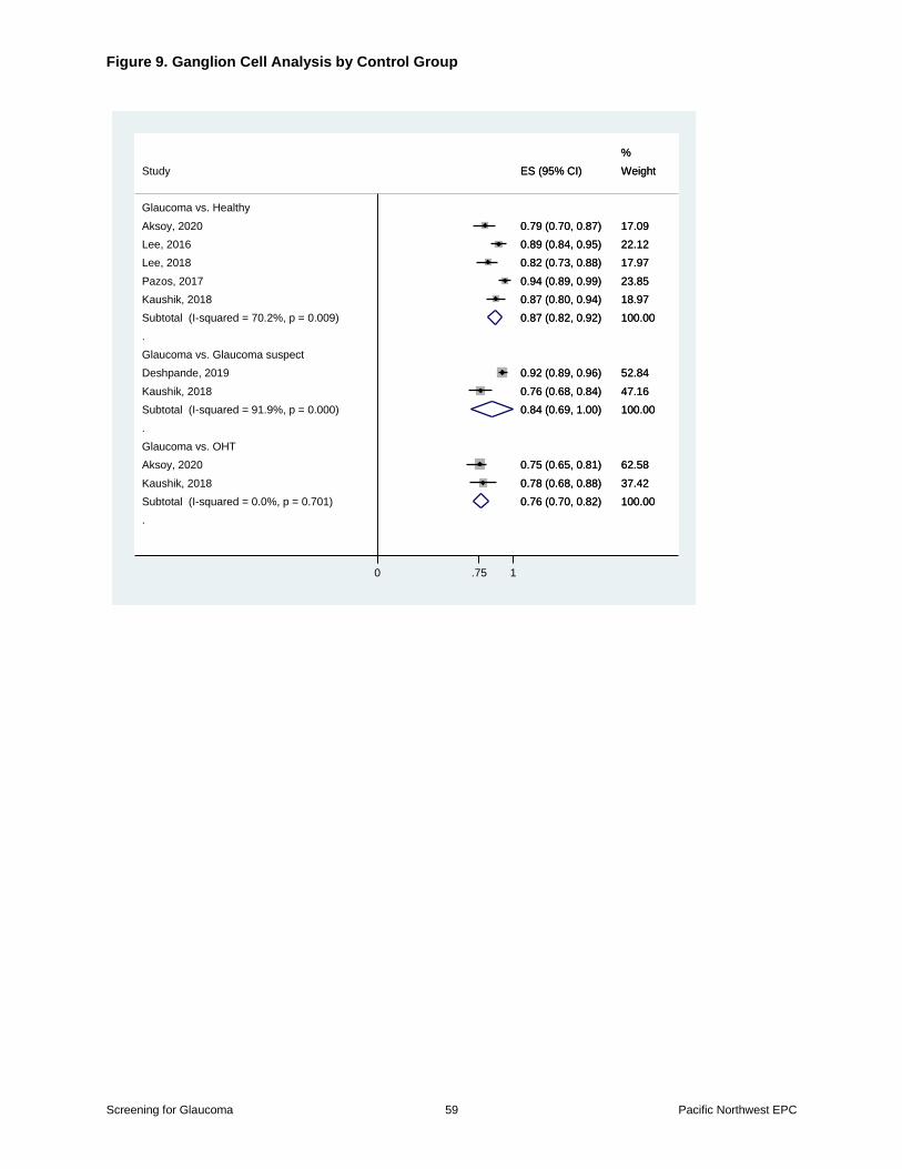
(N=765 AUROC 088 95 CI 084 to 092 I2=68)52698788102104 (Figure 8) Results were
similar when the non-glaucoma groups were stratified as healthy eyes (5 studies N=564
AUROC 087 95 CI 082 to 092 I2=70)528788102104 glaucoma-suspect eyes (2 studies
N=354 AUROC 084 95 CI 069 to 100 I2=92)69104 or eyes with ocular hypertension (2
Screening for Glaucoma 16 Pacific Northwest EPC
studies N=224 AUROC 076 95 CI 070 to 082 I2=0)102104 (Figure 9) Results were also
similar when the analysis was restricted to two studies that assessed the ganglion cell complex
(N=211 AUROC 087 95 CI 073 to 100 I2=89)52102 Five studies could not be pooled due
to inadequate data (eg reported sensitivity specificity andor AUROC without confidence
intervals only reported odds ratios)66108109112 or did not report standard AUROC67 Four studies
were not pooled because they enrolled more than one eye in some participants62698085 Details
are provided in Appendix B Tables 3-4
Cup-to-Disc Ratio
One study (N=286) found the cup-to-disc ratio on spectral domain-OCT associated with
sensitivity of 084 (95 CI 077 to 089) and specificity of 072 (95 CI 060 to 081) for
identifying persons with glaucoma versus healthy eyes The cup-to-disc ratio threshold for a
positive test was not specified74
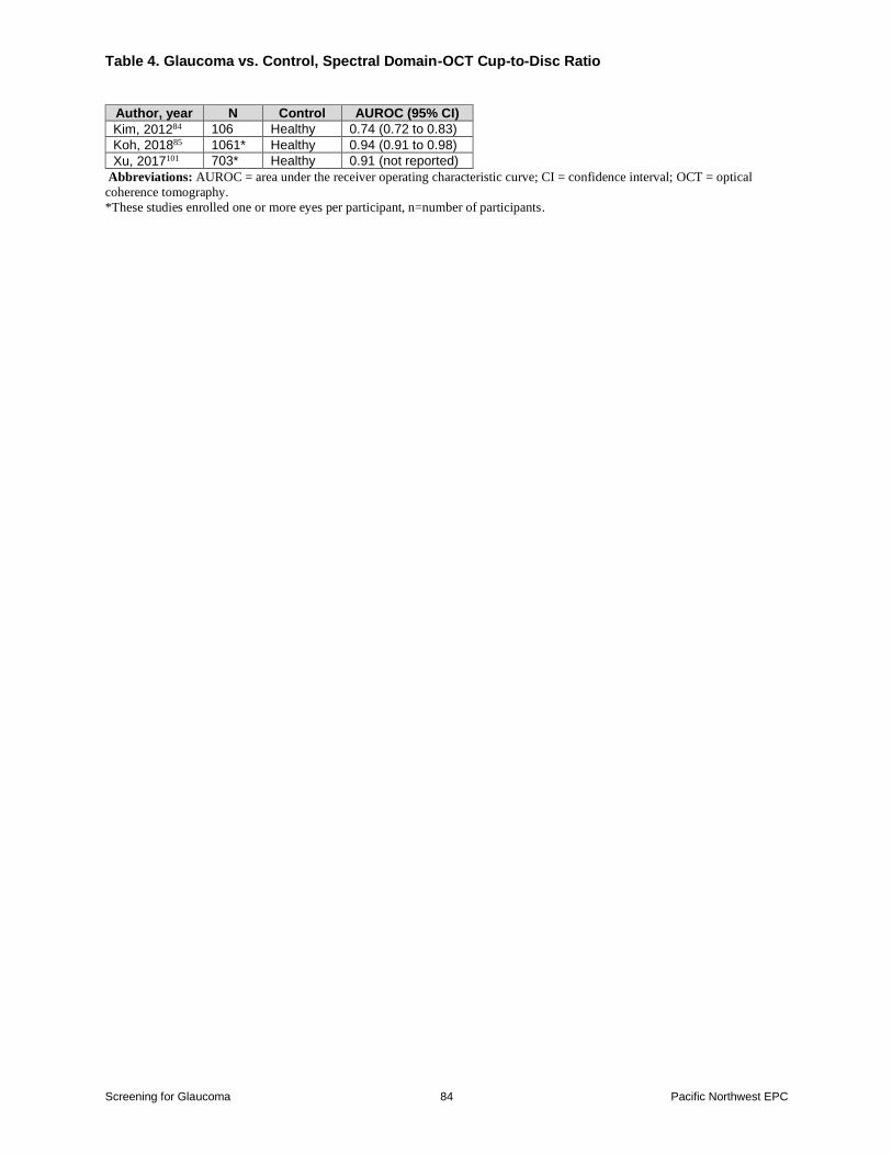
Three studies (n=1870) found the spectral domain-OCT vertical cup-to-disc ratio associated
with an AUROC that ranged from 074 to 0948485101 (Table 4) These studies were not pooled
because they enrolled more than one eye in some participants85101 and one did not report SD101
Swept SourcendashOCT
Swept source-OCT utilizes a longer wavelength than spectral domain-OCT to visualize deeper
structures and is faster than spectral domain-OCT
Two studies (reported in 3 publications N=266) assessed the diagnostic accuracy of swept-
source OCT using retinal nerve fiber layer thickness8788106 One study found wide-field retinal
nerve fiber layer thickness map associated with sensitivity of 095 (95 CI 090 to 098) and
specificity of 089 (95 CI 075 to 097) for distinguishing between participants with glaucoma
versus healthy eyes88 The other study reported an AUROC for distinguishing persons with
glaucoma from those with healthy eyes of 085 (95 CI 078 to 092) for the retinal nerve fiber
layer outerinferior sector and 083 (95 CI 075 to 090) for the outertemporal sector of the
ganglion cell inner plexiform layer87 Another article (n not reported 184 eyes)106 reported an
AUROC of 085 95 CI 078 to 091 for discriminating between early perimetric glaucoma and
healthy eyes of 085 (95 CI 078 to 091) for the retinal nerve fiber layer thickness and 087
(95 CI 080 to 092) for the ganglion cell inner plexiform layer (inferior temporal) but
appeared to nearly completely
Optic Disc Photography
Four studies (N=3133) reported diagnostic accuracy of cup-to-disc ratio on optic disc
photography separate from OCT60687789 One study (n=2631) screened participants with
indirect ophthalmoscopy as well as disc photographs to assess the optic disc89 (Table 5)
Two studies reported similar discrimination of cup-to-disc ratio on optic disc photography with
AUROCs of 085 (95 CI 074 to 096) and 081 (95 CI 074 to 092)6068 In one of these
studies sensitivity was 064 (95 CI 045 to 081) and specificity was 073 (95 CI 045 to
Screening for Glaucoma 17 Pacific Northwest EPC
092) for distinguishing persons with glaucoma from glaucoma suspects the cup-to-disc ratio
threshold was not reported60 Two studies did not report discrimination in one study sensitivity
was 018 (95 CI 009 to 031) and specificity was 067 (95 CI 062 to 071) based on a cup-
to-disc ratio threshold of 0489 In the other study sensitivity was 071 (95 CI 054 to 085) and
specificity was 049 (044 to 055) for distinguishing between glaucoma and nonglaucoma based
on a cup-to-disc ratio of 065 for average-sized or large discs and 05 for small discs77
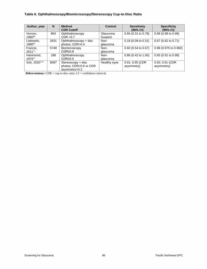
Ophthalmoscopy Biomicroscopy and Stereoscopy
Five studies reported accuracy of cup-to-disc ratio on ophthalmoscopy biomicroscopy and
stereoscopy (N=17519)73758998110 (Table 6) Studies were not pooled because the methods used
to determine cup-to-disc ratio as well as the cutoffs to define a positive screen varied Although
specificity was high in all studies sensitivity varied widely (range 018 to 092)
Pachymetry
Two studies (N=6129) reported the diagnostic accuracy of corneal thickness on pachymetry5873
One study (n=6082) reported a sensitivity of 016 (95 CI 011 to 021) and specificity of 091
(95 CI 090 to 092) for distinguishing between glaucoma and non-glaucoma within a Latino
population using a central corneal thickness of less than or equal to 504microm73 The other study
(n=47) reported an AUROC of 055 (standard error [SE] 008) for pachymetry sensitivity and
specificity were not reported58
Tests of Ophthalmic Function Tests of optic nerve function include measurements of IOP through tonometry and visual field
assessment
Visual Fields
The Humphrey Field Analyzer has superseded frequency doubling technology as standard of
care for the assessment of visual fields Although the Humphrey Field Analyzer was often used
as part of the reference standard for the diagnosis of glaucoma ten studies (N=11633) reported
diagnostic accuracy of the Humphrey Field Analyzer against a reference
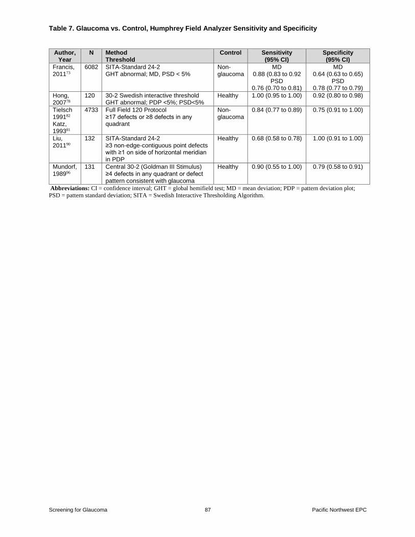
standard606873787981909496108 In these studies Humphrey Field Analyzer methods varied five
studies used the Swedish Interactive Threshold Algorithm-Standard 24-260687390108 two use the
76-point 30 degree suprathreshold7879 and one study each used the Full Field 120 Protocol81 the
630 Armaly Full Field Test94 and the Central 30-2 (Goldmann III stimulus)96
The Humphrey Field Analyzer was associated with pooled sensitivity of 087 (95 CI 069 to
095) and pooled specificity of 082 (95 CI 066 to 092) based on six studies
(N=11244)607378819096 (Figures 10-11) There were too few studies of specific Humphrey Field
Analyzer methods to conduct a meaningful analysis stratified by method (Table 7) One good-
quality study (N=6082)73 reported a sensitivity of 088 (95 CI 083 to 092) and specificity of
064 (95 CI 063 to 065) using SITA-Standard 24-2
Screening for Glaucoma 18 Pacific Northwest EPC
The mean deviation on the SITA-Standard 24-2 was associated with a pooled AUROC (083
95 CI 070 to 097 I2=88) based on three studies (N=288)606890 (Figure 12) and the pattern
standard deviation was associated with an AUROC of 087 (95 CI 076 to 099) based on two
studies (N=242)6890 (Figure 13) One other study found the 76-point 30 degree suprathreshold
associated with an AUROC of 087 (CI not reported)79
One study (n=175 280 eyes) found the Humphrey Field Analyzer Swedish Interactive
Threshold associated with sensitivity of 070 (95 CI 063 to 077) and specificity of 095 (95
CI 089 to 098)108 Another study (n=104 182 eyes) found the Humphrey Field Analyzer
Armaly full field test associated with a sensitivity of 064 (95 CI 056 to 072) and specificity
of 064 (95 CI 048 to 078)94 Because these studies included more than one eye of some
participants they were not included in pooled analysis
Afferent Pupillary Defect
One study (N=107) tested afferent pupillary defect using the swinging flashlight test64
Sensitivity was 067 (95 CI 054 to 078) and specificity was 083 (95 CI 067 to 092)
Sensitivity and specificity were similar when 40 participants without prior cataract surgery were
excluded from the analysis (069 95 CI 050 to 083 089 95 CI 072 to 096 respectively)
Tests of IOP Measurement Tonometry
Seventeen studies (n=49742) evaluated the accuracy of tonometry for identifying
glaucoma58616365677173757779808286899698100 Tonometry was associated with a pooled
sensitivity of 048 (95 CI 031 to 066) and pooled specificity of 094 (95 CI 090 to 096) for
diagnosing glaucoma from non-glaucomatous or healthy eyes based on thirteen studies
(N=32892)616367717375778286899698100 (Figure 14-15 Table 8) The IOP cutoff was 21 to 22
mm Hg in all studies except for two which used cutoffs of 22675 and 25 mm Hg80 Results were
similar when one study100 that evaluated diagnostic accuracy for probable glaucoma versus not
probable glaucoma was excluded from the analysis (12 studies N=28726 pooled sensitivity
047 95 CI 029 to 066 and pooled specificity 094 95 CI 090 to 097) When stratified by
tonometry method sensitivity was higher for Goldmann tonometry (4 studies N=11690
sensitivity 066 95 CI 036 to 087) 61717386 than for other methods (9 studies N=21202
sensitivity 039 95 CI 022 to 058)61717386 (Table 8) However the sensitivity estimate for
Goldmann tonometry was imprecise Specificity was similar regardless of tonometry technique
Results were also similar when the analysis was limited to fair quality studies (11 studies
N=26305 pooled sensitivity 054 95 CI 034 to 072 and specificity 094 95 CI 089 to
097)61637175778186899698100 Only two studies were rated good-quality6773 (sensitivity 024
95 CI 019 to 030 and 019 95 CI 007 to 039 and specificity 097 95 CI 097 to 097 and
089 95 CI 086 to 092) One study that included more than one eye per individual (n=3039
eyes=6060) reported a sensitivity of 007 (95 CI 001 to 019) and specificity of 099 (95 CI
099 to 099) for glaucoma versus non-glaucoma based on an IOP threshold of gt25 mm Hg
using a rebound tonometer80 Two studies (N=418) found tonometry associated with low
Screening for Glaucoma 19 Pacific Northwest EPC
sensitivity (001 95 CI 000 to 005 and 027 95 CI 020 to 036) and high specificity (098
95 CI 094 to 100 and 081 95 CI 073 to 088) for distinguishing glaucoma suspects versus
healthy controls7796
In three studies (N=4684) discrimination of tonometry based on the AUROC ranged from 066
to 078587179 All three studies used Goldmann applanation tonometry (Figures 16-17)
One study (N=6310) that evaluated intereye IOP asymmetry on tonometry65 was not pooled
details are shown in Appendix B Tables 3-4
Other Telemedicine Screening
Two studies examined the diagnostic accuracy of a telemedicine screening intervention called
Technology-based Eye Care Services used in the Veteran Affairs Healthcare System91-93 The
first was a small pilot study (n=52) where screening was conducted in primary care clinics and
consisted of distance auto-refraction visual acuity tonometry pachymetry and a pupil exam for
depth reactivity afferent papillary defect and fundus A blinded ophthalmologist reviewed
screening findings and made recommendations for the participant These recommendations were
compared with the diagnosis and recommendations of a physician who conducted a face-to-face
exam which was considered the reference standard In the pilot study the technology-based
exam was associated with sensitivity of 064 (95 CI 035 to 087) and specificity of 095 (95
CI 082 to 099)
A subsequent larger (n=256) followup study followed a similar protocol as the pilot study93
Most participants were male (87) and Black (61) and over a quarter of participants had a
history of eye trauma (28) or a family history of eye diagnosis or blindness (25) Persons had
no known ocular disease those with ldquoglaucoma suspectrdquo history and documented visual field
changes or prior treatment were excluded Two ophthalmologists reviewed the screening
findings and accuracy was compared against a face-to-face exam On the face-to-face exam
266 (68256) were diagnosed with glaucoma or glaucoma suspect other conditions diagnosed
were cataracts referred for surgery (39) macular degeneration (23) diabetic retinopathy
(31) and other diagnoses resulting in referral (438) Compared with a face-to-face exam
the sensitivity of the technology-based exam to identify persons with glaucoma varied between
readers (072 95 CI 060 to 082 and 047 95 CI 035 to 06) though specificity was high
with both readers (091 95 CI 087 to 095 and 097 95 CI 094 to 099) The addition of
spectral domain-OCT to the screening protocol did not improve diagnostic accuracy92
Key Question 5 What is the accuracy of instruments for
identifying patients at higher risk of OAG
Summary
Screening for Glaucoma 20 Pacific Northwest EPC
bull One cross-sectional study (n=145) that was not in the prior CER found a questionnaire
associated with low sensitivity (020 95 CI 003 to 056) but high specificity
(specificity 096 95 CI 091 to 099) for identifying persons with glaucoma
Evidence
One fair-quality cross-sectional study (n=145) not in the prior screening CER2 reported the
diagnostic accuracy of a weighted screening questionnaire for identifying persons with glaucoma
(Appendix B Tables 6-8)96 In the instrument the highest weights were assigned for taking
steroid medication and having a previous glaucoma diagnosis less highly weighted risk factors
were previous eye injury or stroke age race prior eye surgery high blood pressure being
nearsighted and family history of diabetes or glaucoma Two out of ten participants with
glaucoma were correctly identified as having glaucoma based on the questionnaire alone
(sensitivity 020 95 CI 003 to 056) and 116 out of 121 correctly identified as not having
glaucoma (specificity 096 95 CI 091 to 099) The study was conducted in the US but
applicability to screening is likely limited because previous glaucoma diagnosis was one of the
most heavily weighted risk factors
Key Question 6 What are the effects of medical treatments for OAG versus placebo or no treatments on a) IOP visual field loss visual acuity or optic nerve damage or b) visual
impairment quality of life or function
Summary
bull Treatment was associated with greater reduction in IOP versus placebo or no treatment
(16 trials N=3706 mean difference -314 mm Hg 95 CI -419 to -208 I2=95) there
was an interaction between drug class and effects of medical therapy on IOP (p for
interaction lt00005) though estimates favored treatment for all drug classes
bull Treatment with topical therapy decreased risk of glaucoma progression versus placebo or
no treatment at 24 to 120 months (7 trials N=3771 RR 068 95 CI 049 to 096
I2=53 absolute risk difference (ARD) -48 95 CI -85 to -10)
bull Evidence on effects of medical therapy on quality of life was very limited with one trial
(n=461) reporting no differences between latanoprost and placebo in general or vision-
related quality of life measured using the EuroQol-5D (EQ-5D 17 vs 17 p=098)
Short Form Health Survey-36 (SF-36 48 vs 50 p=094) Glaucoma Quality of Life-15
(GQL-15 27 vs 32 p=066) or Glaucoma Activity Limitation-9 (GAL-9 30 vs 32
p=087) scales at 24 months
Evidence
The prior treatment CER3 primarily focused on head-to-head comparisons of glaucoma
Screening for Glaucoma 21 Pacific Northwest EPC
treatment but included a systematic review that found medical (topical) therapy for ocular
hypertension associated with reduced risk of onset of visual field defects versus placebo or no
treatment (10 trials N=3648 odds ratio [OR] 062 95 CI 047 to 081)114 For this review we
included 13 placebo-controlled trials and four trials of medical therapy versus no treatment21115-
117 in patients with OAG or ocular hypertension Nine trials21116-123 were in the systematic review
utilized in the prior treatment CER and we identified eight additional trials35115124-129 including
the UK Glaucoma Treatment Study35 (UKGTS) which reported effects on quality of life and
visual acuity in addition to IOP and visual field progression (Appendix B Table 9)
Across trials sample sizes ranged from 20 to 1636 participants (N=4665) Mean age ranged
from 55 to 74 years and 34 to 75 percent of participants were female In 10 trials that reported
raceethnicity the proportion of White participants ranged from 68 to 100 percent Two trials
enrolled patients with untreated newly diagnosed OAG (excluding advanced disease and
pigment dispersion)35126 three trials enrolled mixed populations with OAG or ocular
hypertension (proportion with OAG 40 76 and not reported)124125129 and 12 trials enrolled
patients with ocular hypertension (elevated IOP but normal visual fields often also normal optic
discs) OAG or ocular hypertension was diagnosed using a variety of tests including perimetry
tonometry gonioscopy and visualization of the optic nerve by ophthalmoscopic examination
andor imaging only the UKGTS35 utilized OCT (primarily time domain-OCT) as part of the
diagnostic evaluation Mean baseline IOP ranged from 196 to 273 mm Hg mean baseline IOP
was lt22 mm Hg in the two trials35126 of patients with early untreated OAG and ge22 mm Hg in
the other trials Treatment was a beta-blocker in 10 trials (timorol in six trials116-119127
levobunolol in two trials115125 and betaxolol in two trials120127) a carbonic anhydrase inhibitor in
five trials (dorzolamide in four trials122124126129130 and brinzolamide in one trial124) a
prostaglandin analogue (latanaprost) in one trial35 and an alpha agonist (brimonidine) in one
trial128 One trial did not evaluate a specific drug but allowed various topical therapies with a
target IOP le24 mm Hg or ge20 percent IOP reduction21 The duration of followup ranged from
15 months126127 to 120 months119 with followup gt1 year in 10 trials One study was
multinational122130 and the others were conducted in the US21118121123-125127-129 UK35117120
Sweden119126 Italy115 and Canada116
In 12 trials2135116118-120122124-127129130 randomization and analysis was per individual (two of
which enrolled only one eye120126) one trial123 randomized by individual but reported a per-eye
analysis and three trials117121128 randomized one eye in each individual (with the other eye
serving as the control) one trial115 reported a per-eye analysis but randomization by eye or
individual was unclear Four trials were rated good-quality35120123130 and 12 were rated fair-
quality21115-119121124125127-129 (Appendix B Table 10) Methodological limitations in the fair-
quality trials included unclear reporting of randomization allocation concealment and blinding
methods and high attrition in some studies
Intraocular Pressure
Mean IOP was the most commonly reported outcome reported in all but two trials2135115-117119-
121123125-130 Overall treatment was associated with greater reduction in IOP versus placebo or no
treatment (16 studies N=3706 mean difference -314 mm Hg 95 CI -419 to -208 I2=95)
(Figure 18) Statistical heterogeneity was substantial though inconsistency was in the magnitude
Screening for Glaucoma 22 Pacific Northwest EPC
of effect but not the direction of effect with all trials reporting effects on IOP that favored
treatment (range -070 to -700 mm Hg) A funnel plot did not indicate small sample effects
(Eggerrsquos test p=016) (Figure 19) but results were difficult to interpret due to statistical
heterogeneity There was an interaction between drug class and effects of medical therapy on
IOP (p for interaction lt00005) though estimates favored treatment for all drug classes Beta
blockers were associated with a pooled mean difference of -375 mm Hg (95 CI -543 to -206
I2=92) based on nine trials (N=455) prostaglandin analogues with a mean difference of -270
mm Hg (95 CI -334 to -206) based on one trial (n=516) alpha agonists with a mean
difference of -230 mm Hg (95 CI -352 to -108) based on one trial (n=30) and carbonic
anhydrase inhibitors with a mean difference of -120 mm Hg (95 CI -230 to -061) based on
four trials (N=1635) One trial (n=1636) found treatment to target using various medications
associated with a mean difference of -460 mm Hg (95 CI -485 to -435) Estimates also
consistently favored medical therapy with differences ranging from -2 to -4 mm Hg when
analyses were stratified according to ocular hypertension untreated OAG or mixed status at
baseline (Figure 20) baseline IOP (lt20 mm Hg vs ge20 mm Hg) (Figure 21) and quality (fair
vs good) (Figure 22) or duration (lt1 year vs gt1 year) (Figure 23) though statistically
significant interactions were present (Table 9) In the OHTS effects of medication versus
placebo on IOP were almost identical in Black persons and persons of other races131
Progression of Glaucomatous Changes
Nine trials reported effects of topical medical therapies versus placebo or no treatment on risk of
glaucoma progression (Appendix B Table 9)2135116-122130 Glaucoma progression was defined as
progression of visual field defects35118 progression to a glaucoma diagnosis (among those with
ocular hypertension based on visual field defects or optic disc changes)116119120 or progression
of visual field defects or optic disc changes21122130 Definitions and measurement methods for
progression of visual field loss varied but were based on the development of focal or
reproducible visual field defects or by the development of any reproducible visual field defect
(Appendix B Table 9) No trial reported the proportion of patients with overall visual field loss
exceeding a minimum clinically important threshold such as a change in mean deviation of gt3 to
5 decibels (dB a measure of light intensity when testing visual fields)132
Treatment with topical therapy decreased risk of glaucoma progression versus placebo or no
treatment (seven trials N=3771 RR 068 95 CI 049 to 096 I2=53 ARD -48 95 CI -
85 to -10 Figure 24) at 24 to 120 months Estimates were similar in the new UKGTS
trial133 which evaluated patients with untreated OAG (n=461 RR 059 95 CI 041 to 086)
and trials included in the prior treatment CER of patients with ocular hypertension (six trials
N=3310 RR 071 95 CI 046 to 108 I2=57) (Figure 25) Results were also similar in fair-
quality studies (four trials N=1978 RR 057 95 CI 033 to 100 I2=44) and good-quality
studies (three trials N=1793 RR 076 95 CI 052 to 130 I2=15 Figure 26) There was no
interaction between medication type (p for interaction=030) or study quality (p for
interaction=036) and effects on risk of glaucoma progression Two trials117121 (ns=34 and 62)
were not included in the meta-analysis because they randomized one eye in each individual and
reported a per-eye analysis but both found treatment associated with decreased risk of
progression (RR 063 95 CI 017 to 239 and RR 038 95 CI 013 to 116) An analysis
restricted to progression of visual field defects (excluding optic disc changes as a criterion for
Screening for Glaucoma 23 Pacific Northwest EPC
progression) produced similar results (six trials N=3679 RR 073 95 CI 053 to 105
I2=25) Figure 27)2135116118120122 In the UKGTS latanaprost was associated with a small and
non-statistically significant difference in overall visual field loss measured by the mean
deviation (-023 vs 014 dB p=007) there was no difference in visual acuity (-001 vs -002
logMAR p=09)35 In the OHTS there was no interaction between race and risk of progression
from ocular hypertension to OAG (HR 050 95 CI 028 to 090 for Black patients and HR
036 95 CI 023 to 057 for other races p for interaction=040)131
Quality of Life
Evidence on effects of medical therapy on quality of life was very limited In the UKGTS
(n=461)134 there were no differences between latanoprost and placebo in general or vision-
related quality of life measured using the EQ-5D (17 vs 17 p=098) SF-36 (48 vs 50
p=094) GQL-15 (27 vs 32 p=066) or GAL-9 scales (30 vs 32 p=087) at 24 months
followup
Key Question 7 What are the harms of medical treatments
for OAG versus placebo or no treatments
Summary
bull There were no significant differences in risk of serious adverse events (three trials
N=3140 RR 114 95 CI 060 to 199 I2=32) withdrawal due to adverse events (five
trials N=648 RR 240 95 CI 071 to 1932 I2=0) or any adverse event (two trials
N=1538 RR 156 95 CI 059 to 403 I2=82)
bull Two trials found treatment associated with increased risk of ocular adverse events
(primarily itching irritation tearing dryness or taste issues) versus placebo (RR 121
95 CI 110 to 133 in a trial of various treatments and RR 352 95 CI 246 to 502 in
a trial of dorzolamide)
Evidence
Eight trials (in nine publications) of medical treatments versus placebo or no treatment reported
harms (Appendix B Table 9)2135118122-125129130 There were no statistically significant
differences in risk of serious adverse events (three trials N=3140 RR 114 95 CI 060 to
199 I2=32 Figure 28)2135122130 withdrawal due to adverse events (five trials N=648 RR
240 95 CI 071 to 1932 I2=0 Figure 29)35118123125129 or any adverse event (two trials
N=1538 RR 156 95 CI 059 to 403 I2=82)35122130 However estimates were imprecise
and the estimate for any adverse event was based on two trials with substantial statistical
heterogeneity (RR 104 95 CI 073 to 147 in a trial of latanaprost35 and RR 230 95 CI 169
to 312 in a trial of dorzolamide122130) Two trials found treatment associated with increased risk
of ocular adverse events versus placebo (RR 121 95 CI 110 to 133 in a trial of various
treatments122130 and RR 352 95 CI 246 to 50221 in a trial of dorzolamide) The most
common ocular adverse events were localized itching irritation tearing dryness or taste issues
Screening for Glaucoma 24 Pacific Northwest EPC
Because of extreme statistical heterogeneity (I2=94) the pooled estimate was unreliable and
not reported The OHTS found no interaction between race and likelihood of experiencing one or
more serious adverse events (p for interaction=016) or any adverse event (p for
interaction=058) with medical treatment versus placebo131
Key Question 8 What are the effects of newly FDA-approved medical treatments (latanoprostene bunod and netarsudil)
versus older medical treatments on a) IOP visual field loss visual acuity or optic nerve damage or b) visual impairment
quality of life or function
Summary
bull Three trials (N=1875) found netarsudil to be noninferior to timolol for IOP lowering at
3 to 12 months
bull A pooled analysis of two trials trials (N=985) found netarsudil and latanaprost to be
associated with similar effects on IOP (mean diference 03 mm Hg) and likelihood of
IOP le18 mm Hg 574 vs 655) at 12 months
bull One trial (N=413) found latanoprostene bunod 0024 or 0040 percent associated with
slightly greater effects on IOP (difference 12 mm) versus latanoprost at short-term (1
month) followup
bull Two trials (N=840) found latanoprostene bunod associated with slightly greater IOP
reduction versus timolol at 3 months (difference -10 to -13 mm Hg)
bull A pooled analysis of two trials (N=840) found latanoprostene bunod associated with
increased likelihood of IOP le18 (202 vs 112 p=0001) and IOP reduction ge25
percent (229 vs 190 plt0001) at 3 months
Evidence
Eight trials evaluated the effects of latanaprostene bunod or netarsudil compared to an older
glaucoma medication (Appendix B Table 11)135-143 The Rho Kinase Elevated IOP Treatment
(ROCKET) trials compared netarsudil with timolol (ROCKET-1 and ROCKET-2138 and
ROCKET-4 136 trials) the VOYAGER study compared different doses of latanaprostene bunod
versus latanaprost140 the LUNAR137 and APOLLO139141 trials (acronyms not defined) compared
latanaprostene bunod versus timolol and the MERCURY-1143144 and MERCURY-2142 trials
(acronym not defined) compared netarsudil versus latanaprost
Sample sizes ranged from 411 to 985 (N=4953) The mean age ranged from 61 to 66 years and
50 to 68 percent of participants were female All trials enrolled mixed populations of patients
with OAG or ocular hypertension The proportion of patients with OAG in the ROCKET trials
ranged from 62 to 68 percent and in the MERCURY-1 and MERCURY-2 trials ranged from 72
to 77 percent the three trials of latanaprostene bunod did not report the proportion of patients
with OAG Mean baseline IOP ranged from 207 to 267 mm Hg The duration of follow-up was
Screening for Glaucoma 25 Pacific Northwest EPC
3 months in all trials except for 3 which had 1140 or 12 month followup138142 All trials were
multinational except for the MERCURY trials which were conducted in the United States Three
trials (LUNAR APOLLO and MERCURY-2)137141142 were rated good-quality and five trials
were rated fair-quality135136138140143 Methodological limitations in the fair-quality trials
included unclear reporting of randomization allocation concealment and blinding of outcome
assessors the ROCKET and MERCURY-1 trials also had high and differential attrition
(Appendix B Table 12)135136138 All of the trials focused on the outcome of IOPThe ROCKET
trials (N=1875) found netarsudil to be noninferior to timolol in mean IOP reduction at 3 months
(three trials) and 12 months (one trial)135 The MERCURY-1 trial (N=480) found netarsudil and
latanaprost associated with similar reduction in IOP (mean difference 03 mm Hg for netarsudil
vs latanaprost) and likelihood of achieving IOP le18 mm Hg RR (574 vs 655 RR 073
95 CI 061 to 088) at 12 months
The short-term (1 month) VOYAGER trial (n=413) found latanaprostene bunod 0024 and
004 associated with greater reduction in IOP versus latanaprost (mean differences 123 mm
Hg 95 CI 037 to 210 004 and 116 mm Hg 95 CI 029 to 203 respectively) and
increased likelihood of IOP le18 (68 vs 64 vs 47 plt005 for both latanaprostene bunod
doses versus latanaprost)140 Two trials (LUNAR and APOLLO N=840) found latanaprostene
bunod associated with greater IOP reductions versus timolol137141 In a pre-planned pooled
analysis mean differences in IOP ranged from -10 to -13 mm Hg at 3 months (plt0001)139
Latanaprostene was also associated with a higher likelihood of IOP le18 at week 2 week 6 and 3
month timepoints (202 vs 112 p=0001) and IOP reduction ge25 percent at all timepoints
(29 vs 190 plt0001)
Key Question 9 What are the harms of newly FDA-approved
medical treatments versus older medical treatments
Summary
bull Netarsudil was associated with increased risk of ocular adverse events (three trials
N=1875 RRs ranged from 169 to 207) withdrawal due to adverse events (three
trials N=1875 RRs ranged from 473 to 3820) and any adverse event (one trial
n=708 80 vs 60 RR 133 95 CI 120 to 147) versus timolol
bull Netarsudil also associated with increased risk of any adverse event (one trial N=718
RR 145 95 CI 127 to 166) ocular adverse events (one trial n=718 RR 176
95 CI 150 to 207) and withdrawal due to adverse events (one trial n=718 RR
1282 95 CI 471 to 3485) versus latanaprost at 12 months
bull One trial (n=413) found latanaprostene bunod and latanaprost associated with similar
likelihood of any adverse events and withdrawals due to adverse events
bull Two trials (N=840) found latanaprostene bunod associated with increased risk of
ocular adverse events versus timolol (pooled RR 172 95 CI 122 to 242)
estimates for withdrawal due to adverse events were imprecise
Evidence
Screening for Glaucoma 26 Pacific Northwest EPC
Eight head-to-head trials and two meta-analyses reported adverse events associated with newly
approved glaucoma medications versus older medications (Appendix B Table 11)135-144 In the
ROCKET trials (k=3 N=1875) netarsudil was associated with increased risk of ocular adverse
events versus timolol135136138 The most common ocular adverse events were conjunctival
redness or hemorrhage corneal deposits (cornea verticillata typically asymptomatic and without
effects on vision) blurry vision tearing and itching The proportion of patients with ocular
adverse events ranged from 73 to 88 percent with netarsudil and from 41 to 50 percent with
timolol RRs ranged from 151 to 207 at 3 to 12 months (ARDs ranged from 26 to 38)
Netarsudil was also associated with increased likelihood of withdrawal due to adverse events
(RRs ranged from 473 to 3820 ARDs ranged from 8 to 34) though estimates were
imprecise One trial found netarsudil associated with increased likelihood of any adverse event
versus timolol (80 vs 60 RR 133 95 CI 120 to 147)136
Netarsudil was also associated with increased risk of any adverse event (RR 145 95 CI 127
to 166) and ocular adverse events (RR 176 95 CI 150 to 207) at 12 months versus
latanaprost Netarsudil was also associated with increased risk of withdrawal due to adverse
events at 3 months (two trials N=1486 RR 740 95 CI 294 to 1865) and 12 months (one
trial N=718 RR 1282 95 CI 471 to 3485)
One short-term (1 month) trial (n=413) found no differences between latanaprostene bunod
versus latanaprost in risk of any adverse event or withdrawal due to adverse events140 Two trials
(N=840) found latanaprostene bunod associated with increased risk of ocular adverse events
versus timolol (pooled RR 172 95 CI 122 to 242)139 Estimates for withdrawal due to
adverse events were imprecise (RR 196 95 CI 022 to 1740 and RR 048 95 CI 012 to
188)
Key Question 10 What are the effects of laser
trabeculoplasty for OAG versus no trabeculoplasty or medical treatment on a) IOP visual field loss visual acuity
or optic nerve damage or b) visual impairment quality of life or function
Summary
bull The large (n=718) good-quality Laser in Glaucoma and Ocular Hypertension (LiGHT)
trial reported SLT and medical therapy were associated with similar effects on IOP
visual acuity visual field general quality of life and glaucoma-specific utility
symptoms and quality of life at 3 years
bull Three smaller fair-quality trials found SLT and medical therapy similar for IOP at 4 to
12 months and 5 years the trials did not evaluate other ocular and health outcomes
Evidence
Screening for Glaucoma 27 Pacific Northwest EPC
The prior treatment CER3 included two trials145146 (N=220) of SLT versus medical therapy One
trial145 was carried forward for this update but the other146 was ineligible because a high
proportion of patients had capsular glaucoma and use of an outdated intervention (argon laser)
Three additional trials (in four publications N=925) not included in the prior treatment CER of
SLT versus a topical prostaglandin analogue were added for this update (Appendix B Table
13)37147-149
The largest study was the LiGHT trial which enrolled 718 participants with OAG or ocular
hypertension and visual acuity ~20120 or better37147 In the medical therapy arm of LiGHT
patients received stepped therapy with prostaglandins as initial therapy followed by beta
blockers topical carbonic anhydrase inhibitors or alpha agonists Patients were excluded if they
had prior surgery or were currently on or had prior exposure to glaucoma medical therapy Forty-
five percent of patients were female and mean age was 63 years Seventy percent of patients
were White 20 percent Black and 71 percent Asian Mean baseline IOP was 245 mm Hg the
majority of randomized patients were diagnosed with OAG (773) versus ocular hypertension
(227) Of those with OAG disease severity was most commonly assessed as mild (886)
followed by moderate (201) and severe (104)
The sample sizes in three smaller trials (including the trial in the prior treatment CER)145 ranged
from 32 to 167 participants 48 percent to 55 percent were female145148149 Mean ages ranged
from 52 years to 66 years and mean baseline IOP ranged from 228 mm Hg to 293 mm Hg The
proportion of patients with OAG ranged from 43 to 59 percent and the proportion with ocular
hypertension ranged from 41 to 57 percent All studies excluded patients with prior laser or
glaucoma surgery In two trials145148 patients were randomized to 360deg SLT in the other trial149
patients were randomized to 90deg 180deg or 360deg SLT The duration of followup ranged from 4
months to 5 years Three trials were conducted in the United Kingdom (UK)37147-149 and one in
Hong Kong145
One randomized controlled trial (RCT)37147 was rated good-quality the remaining three were
rated fair-quality (Appendix B Table 14)145148149 Methodological limitations in the fair-quality
trials included unclear reporting of randomization allocation concealment and blinding
methods
Intraocular Pressure
The good-quality large (n=718) LiGHT trial found 360deg SLT and medical therapy associated
with very similar mean IOP at 3 years (166 [SD 362] vs 163 [SD 387] mm Hg)37147 Among
those randomized to SLT 741 percent received one treatment per eye 257 percent received two
treatments and 02 percent received three treatments Medical therapy was associated with more
treatment escalations than SLT SLT and medical therapy were also associated with very similar
proportions of eyes at target IOP based on Canadian Target IOP Workshop criteria (950 vs
931) Among those who achieved target IOP at 3 years 782 percent of eyes randomized to
SLT did not require medication versus 3 percent of patients randomized to medical therapy37147
Raceethnicity was not a predictor of response to SLT (22 of participants were Black 65
were Asian and 68 were White)36
Screening for Glaucoma 28 Pacific Northwest EPC
Three smaller trials (n=32 40 and 167) reported results for IOP that were consistent with
LiGHT145148149 One trial148 (n=40) found 360deg SLT and medical therapy associated with similar
mean reduction from baseline in IOP at 4 to 6 months (62 [SE 08] vs 78 [SE 08] mm Hg
pgt005) Similar results were reported in a trial145 (n=32) assessing mean IOP reduction in SLT
versus medical therapy at 5 years (86 [SD 67] vs 87 [SD 66] mm Hg pgt005) Two trials
found 360deg SLT and medical therapy associated with similar likelihood of ge20 percent reduction
in IOP from baseline at 4 to 6 months (75 vs 73 adjusted OR 165 95 CI 052 to 607)148
or 12 months (82 vs 90)149 In one trial (n=167)149 there was no difference between 360deg
SLT versus medical therapy in likelihood of achieving ge20 percent IOP reduction (82 vs
90) However 90deg and 180deg SLT were both associated with decreased likelihood of ge20
percent IOP reduction versus medical therapy (34 vs 65 vs 90 respectively plt0001 for
90deg vs medical therapy and plt001 for 180deg vs medical therapy)149
Other Ocular Outcomes
The LiGHT trial found 360deg SLT and medical therapy associated with similar mean visual acuity
at 3 years (007 [SD 018] vs 008 [SD 017] logMAR)37147 SLT and medical therapy were also
associated with similar visual field mean deviation (-321 [SD 376] vs -319 [SD 392] dB)
Effects on visual acuity and visual fields were similar when patients stratified according to
whether they had ocular hypertension or mild moderate or severe OAG at baseline The other
trials did not report visual acuity or other visual outcomes
Quality of Life and Function
The LiGHT trial found SLT and medical therapy associated with similar quality of life at 3
years as measured using the EQshy5D-5 (090 [SD 016] vs 089 [SD 018] adjusted mean
difference 002 95 CI -000 to 003)37147 SLT and medical therapy were also associated with
very similar glaucoma-specific utility symptoms and quality of life based on the Glaucoma
Utility Index (GUI 089 [SD 013] vs 089 [SD 013]) Glaucoma Symptom Scale (GSS 831
[SD 177] vs 833 [SD 173]) and the GQL-15 (198 [SD 72] vs 198 [78])37147 The other
trials did not report visual acuity or other visual outcomes
Key Question 11 What are the harms of laser trabeculoplasty
for OAG versus no trabeculoplasty or medical treatment
Summary
bull The LiGHT trial reported similar adverse and serious adverse event rates between SLT
and medical therapy evidence on harms from three smaller trials was limited
Evidence
Screening for Glaucoma 29 Pacific Northwest EPC
The LiGHT trial found no differences between SLT and medical therapy in patients experiencing
any adverse event (733 vs 718) or any serious adverse event (180 vs 188) (Appendix
B Table 13)37147 SLT and medical therapy were associated with similar risk of ocular adverse
events such as ocular irritation retinal hemorrhage or floaters (52 vs 61) and serious ocular
adverse events such as trauma central retinal artery occlusion or choroidal neovascularization
(22 vs 17) In the SLT group 344 percent of patients experienced SLT-related transient
adverse events including discomfort transient blurred vision transient photophobia and
hyperemia Evidence on harms of SLT versus medical therapy from other trials was limited due
to suboptimal reporting and imprecision145148149
Contextual Question 1 What is the association between changes in IOP visual field loss visual acuity or optic nerve damage following treatment for OAG and improvement in visual impairment quality of life or function and what is the association between changes in IOP and visual field loss As described in the prior treatment CER3 evidence is available on the association between
decreased IOP following treatment and decreased visual field loss However evidence on the
association between changes in intermediate outcomes (IOP visual field loss visual acuity or
optic nerve damage) following treatment for OAG and improvement in health outcomes remains
limited Other information that may aid in interpreting intermediate outcomes include standards
for classifying the severity of impaired visual acuity limited evidence is available on minimum
clinically important differences for visual acuity and visual field loss
As described in the Results there is direct evidence that treatment for OAG or ocular
hypertension versus placebo or no treatment is associated with decreased risk of progression of
(variably defined) visual field loss (see KQ 6) Studies have also evaluated the association
between the degree of IOP lowering following treatment for POAG or ocular hypertension and
decreased visual field loss In these analyses greater IOP reduction or lower IOP has consistently
been associated with reduced likelihood of visual field or glaucoma progression Several studies
have focused on cohorts of patients enrolled in randomized trials of glaucoma treatment One
analysis evaluated patients (n=738) in the Advanced Glaucoma Intervention Study (AGIS)
which enrolled patients with OAG that could not be adequately controlled by medications alone
Patients had baseline visual acuity of 2080 or better and the minimum Visual Field Defect Score
ranged from 1 to 16 (0 to 20 scale 20 indicates insufficient vision to count fingers at 30 cm)
Patient eyes were randomized to argon laser trabeculoplasty or trabeculectomy150 The analysis
found higher average IOP associated with greater visual field loss at 24 60 and 84 months with
a 1 mm Hg increase in average IOP associated with an increase in the Visual Field Defect Score
of 008 to 018 after adjusting for age intervention sequence age diabetes gender baseline
IOP and baseline Visual Field Defect Score
Subsequent studies also found an association between degree of IOP lowering following
treatment and decreased visual field loss in less advanced glaucoma The Early Manifest
Glaucoma Trial (EMGT) enrolled persons (n=255) with previously untreated glaucoma151
Screening for Glaucoma 30 Pacific Northwest EPC
Patients with advanced visual field defects visual acuity worse than 05 (2040) or mean IOP
greater than 30 mm Hg were excluded Patients were randomized to argon laser trabeculoplasty
plus topical betaxolol or no immediate treatment The outcome was glaucoma progression
defined as a composite outcomes based on perimetric criteria for visual field loss or photographic
criteria for optic disc progression At 6 years greater decrease in IOP from baseline to 3 months
was associated with decreased risk of glaucoma progression (per 1 mm Hg hazard ratio [HR]
090 95 CI 086 to 094) and higher mean IOP at follow-up was associated with increased risk
of glaucoma progression (per 1 mm Hg HR 113 95 CI 107 to 119) after adjusting for
baseline IOP presence of exfoliation baseline visual field loss and age Similar results were
found at longer (up to 11 years) followup152 The European Glaucoma Prevention Study (EGPS)
which randomized persons (n=1077) with ocular hypertension to dorzolamide or placebo found
greater reduction in mean followup IOP associated with decreased risk of progression to OAG
during 5-year followup (per 1 mm Hg HR 089 95 CI 080 to 098) after adjusting for
treatment arms and baseline predictive factors153 Greater increase in mean followup IOP (per 1
mm Hg HR 112 95 CI 103 to 122) and area under the curve of IOP (mm Hg per year HR
109 95 CI 106 to 112) were associated with increased risk of progression to OAG A
limitation of the analyses from EGPS and EMGT is that they did not exclude patients
randomized to placebo or no immediate treatment potentially reducing the directness of findings
to treated patients though both reported estimates adjusted for treatment arm Long-term (12 to
15 year) analyses from cohort studies of treated patients with normal tension glaucoma also
found an association between greater IOP reduction and risk of glaucoma progression154155
The prior treatment CER found no direct evidence on the association between improvements in
intermediate outcomes (IOP visual fields visual acuity or optic nerve damage) following
treatment for OAG or ocular hypertension and improvement in visual impairment quality of life
or function3 However the prior treatment CER noted that cross-sectional studies not meeting
inclusion criteria indicated an association between more severe visual field loss with more visual
impairment and worse patient reported outcomes Although such studies can evaluate
correlations between intermediate and health outcomes they cannot demonstrate causality or the
association between changes in IOP following treatment and subsequent outcomes due to the
lack of longitudinal followup
As in the prior treatment CER we identified no studies on the association between
improvements in intermediate outcomes and health outcomes Studies published since the prior
USPSTF review were consistent with previous findings with regard to the association between
greater visual field loss and reduced vision-related quality of life or function but were cross-
sectional or did not evaluate the association between treatment-related changed in visual fields
and function156-158
We identified no studies on the association between improvements in optic nerve damage
following treatment and health outcomes The association between increased optic nerve damage
and greater visual field loss has been described in numerous articles but this represents an
association between two intermediate outcomes159-164
Evidence on minimum clinically important differences for visual field loss is limited One
longitudinal study found a mean deviation gt5 dB visual field loss or gt3 dB visual field gain
Screening for Glaucoma 31 Pacific Northwest EPC
associated with clinically meaningful losses or gains in vision-specific quality of life (defined as
a change of gt5 points on the 0 to 100 composite NEI-VFQ)132 Effects of visual field changes
varied according to baseline vision status with similar levels of visual field change associated
with greater impact on quality of life in persons with pre-existing vision loss
As noted in the 2016 USPSTF review on screening for impaired visual acuity standards for
classifying severity of impaired visual acuity are available For example visual acuity of 2070
or better is classified as mild or no impairment by the World Health Organization165 The
International Council of Ophthalmology uses a slightly lower (2063 or better) threshold for
mildly impaired visual acuity166 However effects of even mildly impaired visual acuity are
variable and can have a significant impact on quality of life The best-corrected visual acuity
acceptable for driving in most US states is 2040167 Therefore even relatively small changes in
even ldquomildrdquo impaired visual acuity could have a clinically important impact depending on
baseline visual acuity and type of work or other activities in which an individual is engaged
As described in the 2016 USPSTF review on screening for impaired visual acuity168 minimum
clinically important differences for visual acuity have been described Although definitions for a
clinically important change in visual acuity vary across studies a difference of at least 15 letters
(equivalent to three lines on the Early Treatment Diabetic Retinopathy Study [ETDRS])
representing a doubling of the visual angle is a commonly reported outcome in studies assessing
visual acuity and has been used to indicate a clinically meaningful difference169170 This
threshold is based primarily on studies that evaluate effects of changes in visual acuity on vision-
related function Studies using the NEI-VFQ to assess vision-related function though not
necessarily in patients with glaucoma found a difference of 4 to 10 points to be clinically
meaningful to patients corresponding to a 10- to 15-letter change in visual acuity170-173
Screening for Glaucoma 32 Pacific Northwest EPC
Chapter 4 Discussion
Summary of Review Findings
Table 10 summarizes the evidence reviewed for this update The prior screening CER2 that
informed the 2012 USPSTF recommendation included no studies of glaucoma screening versus
no screening We identified one trial of frail elderly persons not included in the prior screening
CER that found no difference between vision screening by an optometrist (including components
relevant for diagnosis of glaucoma) versus no screening on vision outcomes or vision-related
quality of life53 In this trial vision screening was not specific for glaucoma imaging was not
utilized as part of screening and the proportion of patients referred for glaucoma management
was small (55 of those judged to need treatment) In addition most patients in the no
screening arm had seen an eye care professional in the prior year which could have attenuated
potential benefits of screening Unexpectedly the trial found screening associated with an
increased risk of falls (number needed to screen 67 for 1 additional person falling) with a non-
statistically significant increased risk of fractures The reason for the increase in falls was
unclear but could be related to difficulty adapting to large corrections in vision or use of
multifocal lenses No study evaluated outcomes associated with referral to an eye health provider
versus no referral
For diagnostic accuracy our review found spectral domain-OCT and visual field assessment
using the Humphrey Automated Field Analyzer to be associated with reasonable accuracy for
identifying persons with glaucoma compared with a comprehensive eye exam Although visual
field assessment generally requires referral to an eye specialty setting OCT could be ordered
from a primary care clinic and has potential as a standalone screening test Swept source-OCT a
newer OCT technology with increased scan speed and resolution compared with spectral
domain-OCT appears to offer improved visualization of ocular structures but evidence on
diagnostic accuracy for glaucoma is currently limited174 Tonometry for measurement of IOP
was associated with high specificity but low sensitivity indicating that it is insufficient as a
standalone screening test The low sensitivity of tonometry is consistent with data indicating that
a significant proportion of patients with glaucoma have normal IOP The gold standard for
tonometry is the Goldmann Applanation Tonometer which is not widely used in primary care
settings Evidence on other screening tests including swept source-OCT optic disc photography
ophthalmoscopy and biomicroscopy and pachymetry was more limited Our review differs from
and expands upon the prior screening CER2 which included case-control studies included
screening tests no longer in use focused on head-to-head comparisons of screening modalities
did not perform meta-analysis and did not include more recent studies on spectral domain-OCT
A persistent challenge in interpreting studies on diagnostic accuracy of screening is the
variability and lack of standardization in the reference standard
An ophthalmic telemedicine screening program was associated with inconsistent sensitivity but
high specificity for identifying persons with glaucoma compared with a face-to-face
evaluation91-93 Although telemedicine screening was performed in a Veterans Affairs primary
care setting potential barriers to more widespread implementation include the need to install
specialized instruments and utilize trained technicians One study evaluated the accuracy of a
Screening for Glaucoma 33 Pacific Northwest EPC
risk assessment instrument for identifying persons with glaucoma but reported low sensitivity
and was conducted over 30 years ago with no subsequent validation96 The risk assessment
instrument also may be of limited applicability to screening because one of the primary risk
factors was a previous glaucoma diagnosis
Our findings on the effectiveness of medications for early OAG and ocular hypertension are
generally consistent with the prior treatment CER3 which found moderate evidence that medical
and surgical treatments can lower IOP and reduce risk of progression by visual field and optic
nerve criteria but no studies on effects of treatments on visual impairment or patient reported
outcomes Our review differs from the prior treatment CER by focusing on comparisons
involving treatment versus placebo or no treatment and conducting meta-analysis We included
several new trials conducted since the prior treatment CER that evaluated effects on visual acuity
and vision-related function or quality of life Most notable were most the addition of the large
UKGTS35 which compared latanoprost versus placebo in persons with untreated glaucoma and
LiGHT37147 which compared SLT versus topical medications Our meta-analysis found topical
medications associated with decreased risk of glaucoma progression defined by visual field or
optic disc changes with a number needed to treat of 208 to prevent one case of progression over
2 to 10 years Results were similar when the analysis was restricted to trials that defined
glaucoma progression based on visual field changes (excluding optic disc changes) However
these results are difficult to interpret because methods for defining visual field changes varied
and were based on the development of focal visual field deficits or any visual field loss no trial
evaluated the proportion of patients who met a minimum clinically important different
threshold132 for overall visual field loss In the UKGTS there were no differences between
medications versus placebo in mean visual field loss or visual acuity35
Medications were also associated with greater reduction in IOP compared with placebo or no
medication with a pooled difference of about 3 mm Hg Although there was statistical
heterogeneity in the IOP meta-analysis the inconsistency was in the magnitude rather than
direction of effects as results favored therapy in all studies and for all specific classes and
medications Across individual studies and in various stratified analyses effects of medications
on mean IOP generally ranged from 2 to 3 mm Hg Data on harms of topical medical therapies
was limited but did not indicate an increased risk of serious adverse events though non-serious
ocular adverse events (eg redness irritation itching burning tearing) were more common
Newly approved topical medications for glaucoma (netarsudil and latanaprost bunod) were
associated with similar or greater IOP reducing effects versus older medications but increased
risk of adverse events New data on effects of medical treatment for glaucoma on quality of life
was available from the UKGTS134 which found no differences between latanoprost versus
placebo in general or vision-related quality of life at 2 years For SLT versus medical therapy
LiGHT found effects of SLT and medical therapy associated with similar effects on IOP visual
acuity visual field and quality of life with no differences in serious adverse events or ocular
adverse events37147 Results of smaller trials of SLT versus medical therapy were consistent with
LiGHT Our findings regarding treatment are most applicable to patients with ocular
hypertension or early untreated OAG the populations typically enrolled in the trials
As in the prior treatment CER interpretation of effects of treatment on IOP and glaucoma
progression is a challenge because of a lack of evidence on the association between
Screening for Glaucoma 34 Pacific Northwest EPC
improvements in these and other intermediate outcomes (eg optic nerve damage) following
treatment for OAG or ocular hypertension and improvement in visual impairment quality of life
or function Although cross-sectional studies indicate an association between more severe visual
field loss and grater visual impairment and patient reported outcomes such studies cannot
demonstrate causality and do not evaluated the association between changes in IOP following
treatment and subsequent outcomes
Limitations
Our evidence review has some limitations First we excluded non-English language studies
which could introduce language bias Although we identified one randomized trial of medical
therapy published in Japanese it was small (n=16) and would not impact conclusions175 Second
there was statistical heterogeneity in pooled analyses on effects of medical therapy versus
placebo or no treatment on IOP However as described above inconsistency was in the
magnitude but not direction of effect which favored medical therapy across studies In addition
differences in IOP lowering effects were small generally ranging from 1 to 2 mm Hg Because
of anticipated heterogeneity we utilized a random effects model for pooling Third statistical
heterogeneity was present in pooled analyses of sensitivity and specificity However standard
methods for measuring statistical heterogeneity do not account for the variability in estimates
related to threshold effects Despite the statistical heterogeneity results were robust in stratified
and sensitivity analyses Fourth direct evidence on benefits and harms of screening versus no
screening and effects of treatment versus no treatment for ocular hypertension or early OAG on
visual impairment quality of life and function remains very limited though the UKGTS study
found no effects on quality of life or function Fifth we excluded case-control studies of
diagnostic accuracy which reduced the evidence available for evaluating screening tests but
reduced potential spectrum bias Sixth evaluations of publication bias through graphical or
statistical methods was limited by small numbers of studies or statistical heterogeneity However
we did not identify unpublished studies likely to impact findings Seventh unlike the prior
screening and treatment CERs23 we excluded most head-to-head comparisons which might
provide indirect evidence regarding the diagnostic accuracy of screening tests and outcomes of
treatment However we included trials of newly FDA-approved medications versus older
medications and SLT versus medical therapy because the new medications and SLT are
considered first-line treatments and placebo- and sham-controlled trials are not available
Emerging IssuesNext Steps
Latanoprostene bunod (a nitric oxide-donating medication) was FDA-approved in 2017 and
netarsudil (a Rho kinase inhibitor) was approved in 2019 These medications are the first in their
respective classes for treatment of glaucoma and decrease IOP primarily by increasing outflow
(rather than reducing aqueous production) Although some trials of latanoprostene bunod versus
netarsudil are available additional studies with longer-term follow-up are needed to verify
benefits and harms The development of newer minimally-invasive surgical procedures for
treatment of OAG is ongoing45 including angle-based surgeries (Kahook dual blade and
gonioscopy-assisted transluminal trabeculotomy) micro-shunting surgeries (Xen gel stent) and
Screening for Glaucoma 35 Pacific Northwest EPC
micropulse laser therapy
With regard to screening tests OCT technology has evolved rapidly with respect to scanning
speed and resolution which may lead to improvements in diagnostic accuracy (eg with use of
swept source-OCT)176 Although OCT may have some potential as a standalone screening test
for glaucoma177 evidence on the effects of screening with OCT are not available and evidence
on effects of treatments in persons diagnosed using OCT is limited An area of high interest and
a rapidly expanding evidence base is the use of artificial intelligence to analyze and categorize
data from OCT and other screening tests48 potentially improving diagnostic accuracy and
facilitating implementation of screening However we did not identify studies utilizing artificial
intelligence that were eligible for our review because they utilized (non-clinical) imaging
databanks used a case-control design lacked validation testing or did not evaluate algorithms
available for widespread use The FDA has published a proposed regulatory framework to
evaluate artificial intelligence and machine learning as a medical device178 as yet no artificial
intelligence technologies have been approved for diagnosis of glaucoma
Relevance for Priority Populations
Glaucoma disproportionately impacts Black persons and to a lesser extent Latino persons
relative to non-Latino White persons Black persons have the highest prevalence of glaucoma a
higher rate of glaucoma progression and blindness and earlier presentation of glaucoma17179
Race-related disparities have been reported in glaucoma management and adherence to care180-
182 and disparities exist with regard to access to care Evidence on how race or ethnicity impacts
effectiveness of treatment is limited In the OHTS in which 25 percent of participants were
Black there was no interaction between race and effects of medical treatment versus placebo on
IOP likelihood of progression from ocular hypertension to OAG or adverse events131 Although
Black participants were at increased risk of progression to OAG in univariate analysis race was
not a predictor when analyses adjusted for other demographic factors markers of glaucoma
severity and comorbidities In LiGHT in which 22 percent of participant were Black 65
percent were Asian and 68 percent were White raceethnicity was not a predictor of response to
SLT36 In UKGTS approximately 5 percent of participants were Black and 31 percent were
Asian no analysis performed on the interaction between raceethnicity and effects of latanoprost
versus placebo35 Two trials that did not meet inclusion criteria because they evaluated surgery
(one trial also enrolled persons with advanced glaucoma) found that Black participants had
worse outcomes than White participants who had surgery first183184
Future Research
Important gaps remain in the evidence on screening for glaucoma Additional trials of screening
versus no screening that utilize contemporary screening and diagnostic modalities (eg spectral
domain-OCT or swept source-OCT) and include vision-related outcome function and quality of
life would provide direct evidence on effects of screening Research is also needed to determine
optimal screening approaches such as strategies that target higher-risk populations versus
screening of all adults Research on the accuracy of instruments for identifying persons at
Screening for Glaucoma 36 Pacific Northwest EPC
increased risk of glaucoma would be useful for informing screening strategies Studies are
needed to verify the diagnostic accuracy of current screening tests when applied to screened
populations Studies are needed to better understand the utility of artificial intelligence to aid in
the analysis and interpretation of screening tests using validated algorithms in clinical cohorts of
patients that are available for use in clinical practice (ideally FDA-approved) Telehealth
approaches to screening that can be implemented in primary care settings could potentially
facilitate access and are particularly relevant in the post-COVID-19 era Studies on the effects of
referral to glaucoma screening from primary care versus no referral are lacking and would help
clarify outcomes associated with referral Research is needed to better understand the long-term
effects of treatment on visual impairment quality of life and function to understand how effects
of treatment vary by raceethnicity and to verify that benefits of treatment are retained in
persons diagnosed with OAG using newer imaging methods Longer-term studies of the recently
approved medications netarsudil and latanoprostene bunod would help clarify benefits and harms
relative to older first-line therapies
Conclusions
Direct evidence on glaucoma screening versus no screening is limited and showed no benefits on
vision-related quality of life or function and increased risk of falls Screening tests (OCT visual
field assessment) can identify persons with OAG with reasonable accuracy Treatment for ocular
hypertension or untreated OAG is associated with reduction in IOP and reduced risk of glaucoma
progression based on visual fields or optic nerve changes but limited evidence on the association
with visual outcome quality of life and function indicates no clear effects
Screening for Glaucoma 37 Pacific Northwest EPC
References 1 Moyer VA Screening for glaucoma US Preventive Services Task Force
recommendation statement Ann Intern Med 2013 Oct 1159(7)484-9 doi
1073260003-4819-159-6-201309170-00686 PMID 24325017
2 Ervin A-M Boland M Myrowitz E et al Screening for Glaucoma Comparative
Effectiveness Comparative Effectiveness Review No 59 (Prepared by the Johns
Hopkins University Evidence-based Practice Center under Contract No 290-2007-
10061) AHRQ Publication No 12-EHC037-EF Rockville MD Agency for Healthcare
Research and Quality April 2012 2013doi 1073260003-4819-159-6-201309170-
00686
3 Boland MV Ervin AM Friedman D et al Treatment for glaucoma comparative
effectiveness Comparative effectiveness review no 60 AHRQ publication No 12-
EHC038-EF Treatment for Glaucoma Comparative Effectiveness 2012
4 Boland MV Ervin AM Friedman DS et al Comparative effectiveness of treatments for
open-angle glaucoma a systematic review for the US Preventive Services Task Force
Ann Intern Med 2013 Feb 19158(4)271-9 doi 1073260003-4819-158-4-201302190-
00008 PMID 23420235
5 American Academy of Ophthalmology Primary open-angle glaucoma suspect preferred
practice pattern((R)) guidelines Ophthalmology 2016 Jan123(1)P112-51 doi
101016jophtha201510055 PMID 26581560
6 Dielemans I Vingerling JR Wolfs RC et al The prevalence of primary open-angle
glaucoma in a population-based study in The Netherlands The Rotterdam Study
Ophthalmology 1994 Nov101(11)1851-5 doi 101016s0161-6420(94)31090-6
PMID 7800368
7 Mitchell P Smith W Attebo K et al Prevalence of open-angle glaucoma in Australia
The Blue Mountains Eye Study Ophthalmology 1996 Oct103(10)1661-9 doi
101016s0161-6420(96)30449-1 PMID 8874440
8 Sommer A Tielsch JM Katz J et al Relationship between intraocular pressure and
primary open angle glaucoma among white and black Americans The Baltimore Eye
Survey Arch Ophthalmol 1991109(8)1090-5 doi
101001archopht199101080080050026
9 Bathija R Gupta N Zangwill L et al Changing definition of glaucoma J Glaucoma
1998 Jun7(3)165-9 PMID 9627855
10 Rodriguez J Sanchez R Munoz B et al Causes of blindness and visual impairment in a
population-based sample of US Hispanics Ophthalmology 2002 Apr109(4)737-43
doi 101016s0161-6420(01)01008-9 PMID 11927431
11 Kwon M Huisingh C Rhodes LA et al Association between Glaucoma and At-fault
Motor Vehicle Collision Involvement among Older Drivers A Population-based Study
Ophthalmology 2016 Jan123(1)109-16 doi 101016jophtha201508043 PMID
26459997
12 Malihi M Moura Filho ER Hodge DO et al Long-term trends in glaucoma-related
blindness in Olmsted County Minnesota Ophthalmology 2014 Jan121(1)134-41 doi
101016jophtha201309003 PMID 24823760
Screening for Glaucoma 38 Pacific Northwest EPC
13 Gupta P Zhao D Guallar E et al Prevalence of glaucoma in the United States The
2005-2008 National Health and Nutrition Examination Survey Invest Ophthalmol Vis
Sci 2016 May 157(6)2905-13 doi 101167iovs15-18469 PMID 27168366
14 Klein BE Klein R Linton KL Intraocular pressure in an American community The
Beaver Dam Eye Study Invest Ophthalmol Vis Sci 1992 Jun33(7)2224-8 PMID
1607232
15 Vajaranant TS Wu S Torres M et al The changing face of primary open-angle
glaucoma in the United States demographic and geographic changes from 2011 to 2050
Am J Ophthalmol 2012 Aug154(2)303-14e3 doi 101016jajo201202024 PMID
22541661
16 Varma R Ying-Lai M Francis BA et al Prevalence of open-angle glaucoma and ocular
hypertension in Latinos the Los Angeles Latino Eye Study Ophthalmology 2004
Aug111(8)1439-48 doi 101016jophtha200401025 PMID 15288969
17 Tielsch JM Sommer A Katz J et al Racial variations in the prevalence of primary open-
angle glaucoma The Baltimore Eye Survey Jama 1991 Jul 17266(3)369-74 PMID
2056646
18 Stone EM Fingert JH Alward WL et al Identification of a gene that causes primary
open angle glaucoma Science 1997 Jan 31275(5300)668-70 doi
101126science2755300668 PMID 9005853
19 Kwon YH Fingert JH Kuehn MH et al Primary open-angle glaucoma N Engl J Med
2009 Mar 12360(11)1113-24 doi 101056NEJMra0804630 PMID 19279343
20 Heijl A Leske MC Bengtsson B et al Reduction of intraocular pressure and glaucoma
progression results from the Early Manifest Glaucoma Trial Arch Ophthalmol
2002120(10)1268-79 doi 101001archopht120101268
21 Kass MA Heuer DK Higginbotham EJ et al The Ocular Hypertension Treatment
Study a randomized trial determines that topical ocular hypotensive medication delays or
prevents the onset of primary open-angle glaucoma Arch Ophthalmol 2002120(6)701-
13 doi 101001archopht1206701
22 Kass MA Heuer DK Higginbotham EJ et al Assessment of Cumulative Incidence and
Severity of Primary Open-Angle Glaucoma Among Participants in the Ocular
Hypertension Treatment Study After 20 Years of Follow-up JAMA Ophthalmol 2021
Apr 15139(5)1-9 doi 101001jamaophthalmol20210341 PMID 33856434
23 Hattenhauer MG Johnson DH Ing HH et al The probability of blindness from open-
angle glaucoma Ophthalmology 1998 Nov105(11)2099-104 doi 101016s0161-
6420(98)91133-2 PMID 9818612
24 Alward W Feldman F Cashwell LF Comparison of glaucomatous progression between
untreated patients with normal-tension glaucoma and patients with therapeutically
reduced intraocular pressures Collaborative Normal-Tension Glaucoma Study Group
Am J Ophthalmol 1998 Oct126(4)487-97 doi 101016s0002-9394(98)00223-2
PMID 9780093
25 Gordon MO Beiser JA Brandt JD et al The Ocular Hypertension Treatment Study
baseline factors that predict the onset of primary open-angle glaucoma Arch Ophthalmol
2002 Jun120(6)714-20 discussion 829-30 doi 101001archopht1206714 PMID
12049575
Screening for Glaucoma 39 Pacific Northwest EPC
26 Le A Mukesh BN McCarty CA et al Risk factors associated with the incidence of
open-angle glaucoma the visual impairment project Invest Ophthalmol Vis Sci 2003
Sep44(9)3783-9 doi 101167iovs03-0077 PMID 12939292
27 Quigley HA West SK Rodriguez J et al The prevalence of glaucoma in a population-
based study of Hispanic subjects proyecto VER Arch Ophthalmol 2001
Dec119(12)1819-26 doi 101001archopht119121819 PMID 11735794
28 Brandt JD Beiser JA Kass MA et al Central corneal thickness in the Ocular
Hypertension Treatment Study (OHTS) Ophthalmology 2001 Oct108(10)1779-88 doi
101016s0161-6420(01)00760-6 PMID 11581049
29 McMonnies CW Glaucoma history and risk factors J Optom 201710(2)71-8
30 Diehl DL Quigley HA Miller NR et al Prevalence and significance of optic disc
hemorrhage in a longitudinal study of glaucoma Arch Ophthalmol 1990
Apr108(4)545-50 doi 101001archopht199001070060093056 PMID 2322157
31 Tielsch JM Katz J Sommer A et al Hypertension perfusion pressure and primary
open-angle glaucoma A population-based assessment Arch Ophthalmol 1995
Feb113(2)216-21 doi 101001archopht199501100020100038 PMID 7864755
32 Crabb DP Smith ND Glen FC et al How does glaucoma look patient perception of
visual field loss Ophthalmology 2013 Jun120(6)1120-6 doi
101016jophtha201211043 PMID 23415421
33 Sponsel WE Shoemaker J Trigo Y et al Frequency of sustained glaucomatous-type
visual field loss and associated optic nerve cupping in Beaver Dam Wisconsin Clin Exp
Ophthalmol 2001 Dec29(6)352-8 PMID 11778803
34 American Academy of Ophthalmology Primary Open-Angle Glaucoma 2015
httpswwwaaojournalorgarticleS0161-6420(15)01276-2pdf Accessed September 9
2020
35 Garway-Heath DF Crabb DP Bunce C et al Latanoprost for open-angle glaucoma
(UKGTS) a randomised multicentre placebo-controlled trial Lancet 2015 Apr
4385(9975)1295-304 doi 101016s0140-6736(14)62111-5 PMID 25533656
36 Garg A Vickerstaff V Nathwani N et al Primary selective laser trabeculoplasty for
open-angle glaucoma and ocular hypertension clinical outcomes predictors of success
and safety from the laser in glaucoma and ocular hypertension trial Ophthalmology
2019 Sep126(9)1238-48 doi 101016jophtha201904012 PMID 31028768
37 Gazzard G Konstantakopoulou E Garway-Heath D et al Selective laser trabeculoplasty
versus eye drops for first-line treatment of ocular hypertension and glaucoma (LiGHT) a
multicentre randomised controlled trial Lancet 2019 Apr 13393(10180)1505-16 doi
101016s0140-6736(18)32213-x PMID 30862377
38 Hasan S Theilig T Unterlauft JD Comparing the efficacy of trabeculectomy and diode
laser cyclophotocoagulation in primary open-angle glaucoma Int Ophthalmol 2019 Mar
4doi 101007s10792-019-01093-w PMID 30830546
39 Shah P Bhakta A Vanner EA et al Safety and efficacy of diode laser transscleral
cyclophotocoagulation in eyes with good visual acuity J Glaucoma 2018
Oct27(10)874-9 doi 101097ijg0000000000001057 PMID 30113509
40 Budenz DL Barton K Gedde SJ et al Five-year treatment outcomes in the Ahmed
Baerveldt comparison study Ophthalmology 2015 Feb122(2)308-16 doi
101016jophtha201408043 PMID 25439606
Screening for Glaucoma 40 Pacific Northwest EPC
41 Gedde SJ Feuer WJ Shi W et al Treatment outcomes in the primary tube versus
trabeculectomy study after 1 year of follow-up Ophthalmology 2018 May125(5)650-
63 doi 101016jophtha201802003 PMID 29477688
42 Washington University School of Medicine in St Louis Ocular Hypertension Treatment
Study (OHTS) Risk Calculator 2019 httpsohtswustledurisk Accessed September
16 2020
43 Hoy SM Latanoprostene bunod ophthalmic solution 0024 a review in open-angle
glaucoma and ocular hypertension Drugs 2018 May78(7)773-80 doi 101007s40265-
018-0914-6 PMID 29761382
44 Kopczynski CC Heah T Netarsudil ophthalmic solution 002 for the treatment of
patients with open-angle glaucoma or ocular hypertension Drugs Today 2018
Aug54(8)467-78 doi 101358dot20185482849627 PMID 30209441
45 Bar-David L Blumenthal EZ Evolution of glaucoma surgery in the last 25 years
Rambam Maimonides Med J 2018 Jul 309(3)doi 105041rmmj10345 PMID
30089089
46 American Academy of Family Physicians Glaucoma 2019
httpswwwaafporgpatient-careclinical-recommendationsallglaucomahtml Accessed
September 16 2020
47 Hark LA Katz LJ Myers JS et al Philadelphia telemedicine glaucoma detection and
follow-up study methods and screening results Am J Ophthalmol 2017 Sep181114-24
doi 101016jajo201706024 PMID 28673747
48 Zheng C Johnson TV Garg A et al Artificial intelligence in glaucoma Curr Opin
Ophthalmol 2019 Mar30(2)97-103 doi 101097icu0000000000000552 PMID
30562242
49 U S Preventive Services Task Force Procedure Manual 2018
httpswwwuspreventiveservicestaskforceorgPageNameprocedure-manual Accessed
September 16 2020
50 Whiting PF Rutjes AW Westwood ME et al QUADAS-2 a revised tool for the quality
assessment of diagnostic accuracy studies Ann Intern Med 2011 Oct 18155(8)529-36
doi 1073260003-4819-155-8-201110180-00009 PMID 22007046
51 Higgins JP Thompson SG Quantifying heterogeneity in a meta-analysis Stat Med 2002
Jun 1521(11)1539-58 doi 101002sim1186 PMID 12111919
52 Pazos M Dyrda AA Biarnes M et al Diagnostic accuracy of spectralis SD OCT
automated macular layers segmentation to discriminate normal from early glaucomatous
eyes Ophthalmology 2017 08124(8)1218-28 doi 101016jophtha201703044
PMID 28461015
53 Swamy B Cumming RG Ivers R et al Vision screening for frail older people a
randomised trial Br J Ophthalmol 2009 Jun93(6)736-41 doi
101136bjo2007134650 PMID 18614568
54 Cumming RG Ivers R Clemson L et al Improving vision to prevent falls in frail older
people a randomized trial J Am Geriatr Soc 200755(2)175-81
55 Burr JM Mowatt G Hernandez R et al The clinical effectiveness and cost-effectiveness
of screening for open angle glaucoma a systematic review and economic evaluation
Health Technol Assess 2007 Oct11(41)iii-iv ix-x 1-190 doi 103310hta11410
PMID 17927922
Screening for Glaucoma 41 Pacific Northwest EPC
56 Aptel F Sayous R Fortoul V et al Structure-function relationships using spectral-
domain optical coherence tomography comparison with scanning laser polarimetry Am
J Ophthalmol 2010 Dec150(6)825-33 doi 101016jajo201006011 PMID
20851372
57 Azuara-Blanco A Banister K Boachie C et al Automated imaging technologies for the
diagnosis of glaucoma a comparative diagnostic study for the evaluation of the
diagnostic accuracy performance as triage tests and cost-effectiveness (GATE study)
Health Technol Assess 2016 Jan20(8)1-168 doi 103310hta20080 PMID 26822760
58 Bagga H Feuer WJ Greenfield DS Detection of psychophysical and structural injury in
eyes with glaucomatous optic neuropathy and normal standard automated perimetry
Arch Ophthalmol 2006 Feb124(2)169-76 doi 101001archopht1242169 PMID
16476885
59 Banister K Boachie C Bourne R et al Can automated imaging for optic disc and retinal
nerve fiber layer analysis aid glaucoma detection Ophthalmology 2016
May123(5)930-8 doi 101016jophtha201601041 PMID 27016459
60 Blumberg DM De Moraes CG Liebmann JM et al Technology and the glaucoma
suspect Invest Ophthalmol Vis Sci 2016 07 0157(9)OCT80-5 doi 101167iovs15-
18931 PMID 27409509
61 Bonomi L Marchini G Marraffa M et al The relationship between intraocular pressure
and glaucoma in a defined population Data from the Egna-Neumarkt Glaucoma Study
Ophthalmologica 2001 Jan-Feb215(1)34-8 doi 101159000050823 PMID
11125267
62 Casado A Cervero A Lopez-de-Eguileta A et al Topographic correlation and
asymmetry analysis of ganglion cell layer thinning and the retinal nerve fiber layer with
localized visual field defects PLoS One 201914(9)e0222347 doi
101371journalpone0222347 PMID 31509597
63 Chan MPY Broadway DC Khawaja AP et al Glaucoma and intraocular pressure in
EPIC-Norfolk Eye Study cross sectional study Bmj 2017 Sep 13358j3889 doi
101136bmjj3889 PMID 28903935
64 Charalel RA Lin HS Singh K Glaucoma screening using relative afferent pupillary
defect J Glaucoma 2014 Mar23(3)169-73 doi 101097IJG0b013e31826a9742
PMID 23296370
65 Choudhari NS George R Baskaran M et al Can intraocular pressure asymmetry
indicate undiagnosed primary glaucoma The Chennai Glaucoma Study J Glaucoma
2013 Jan22(1)31-5 doi 101097IJG0b013e31822af25f PMID 21878819
66 Cifuentes-Canorea P Ruiz-Medrano J Gutierrez-Bonet R et al Analysis of inner and
outer retinal layers using spectral domain optical coherence tomography automated
segmentation software in ocular hypertensive and glaucoma patients PLoS One
201813(4)e0196112 doi 101371journalpone0196112 PMID 29672563
67 Dabasia PL Fidalgo BR Edgar DF et al Diagnostic accuracy of technologies for
glaucoma case-finding in a community setting Ophthalmology 2015 Dec122(12)2407-
15 doi 101016jophtha201508019 PMID 26411836
68 Danesh-Meyer HV Gaskin BJ Jayusundera T et al Comparison of disc damage
likelihood scale cup to disc ratio and Heidelberg retina tomograph in the diagnosis of
glaucoma Br J Ophthalmol 2006 Apr90(4)437-41 doi 101136bjo2005077131
PMID 16547323
Screening for Glaucoma 42 Pacific Northwest EPC
69 Deshpande G Gupta R Bawankule P et al Structural evaluation of preperimetric and
perimetric glaucoma Indian J Ophthalmol 2019 1167(11)1843-9 doi
104103ijoIJO_1955_18 PMID 31638046
70 Deshpande GA Bawankule PK Raje DV et al Linear discriminant score for
differentiating early primary open angle glaucoma from glaucoma suspects Indian J
Ophthalmol 2019 Jan67(1)75-81 doi 104103ijoIJO_678_18 PMID 30574897
71 Ehrlich JR Radcliffe NM Shimmyo M Goldmann applanation tonometry compared
with corneal-compensated intraocular pressure in the evaluation of primary open-angle
glaucoma BMC ophthalmol 2012 Sep 251252 doi 1011861471-2415-12-52 PMID
23009074
72 Field MG Alasil T Baniasadi N et al Facilitating glaucoma diagnosis with intereye
retinal nerve fiber layer asymmetry using spectral-domain optical coherence tomography
J Glaucoma 2016 Feb25(2)167-76 doi 101097IJG0000000000000080 PMID
24921896
73 Francis BA Varma R Vigen C et al Population and high-risk group screening for
glaucoma the Los Angeles Latino Eye Study Invest Ophthalmol Vis Sci 2011 Aug
0552(9)6257-64 doi 101167iovs09-5126 PMID 21245400
74 Garas A Vargha P Hollo G Diagnostic accuracy of nerve fibre layer macular thickness
and optic disc measurements made with the RTVue-100 optical coherence tomograph to
detect glaucoma Eye 2011 Jan25(1)57-65 doi 101038eye2010139 PMID
20930859
75 Hammond EA Begley PK Screening for glaucoma a comparison of ophthalmoscopy
and tonometry Nurs Res 1979 Nov-Dec28(6)371-2 PMID 258807
76 Hark LA Myers JS Ines A et al Philadelphia telemedicine glaucoma detection and
follow-up study confirmation between eye screening and comprehensive eye
examination diagnoses Br J Ophthalmol 2019 Dec103(12)1820-6 doi
101136bjophthalmol-2018-313451 PMID 30770354
77 Hark LA Myers JS Pasquale LR et al Philadelphia telemedicine glaucoma detection
and follow-up study intraocular pressure measurements found in a population at high
risk for glaucoma J Glaucoma 201928(4)294-301 PMID 30946709
78 Hong S Ahn H Ha SJ et al Early glaucoma detection using the Humphrey Matrix
Perimeter GDx VCC Stratus OCT and retinal nerve fiber layer photography
Ophthalmology 2007 Feb114(2)210-5 doi 101016jophtha200609021 PMID
17270671
79 Ivers RQ Optom B Macaskill P et al Sensitivity and specificity of tests to detect eye
disease in an older population Ophthalmology 2001 Initial Search 12508108(5)968-
75 PMID 11320029
80 Karvonen E Stoor K Luodonpaa M et al Diagnostic performance of modern imaging
instruments in glaucoma screening Br J Ophthalmol 2020 Jan 16104(10)1399-405
doi 101136bjophthalmol-2019-314795 PMID 31949097
81 Katz J Tielsch JM Quigley HA et al Automated suprathreshold screening for
glaucoma the Baltimore Eye Survey Invest Ophthalmol Vis Sci 1993 Nov34(12)3271-
7 PMID 8225862
82 Tielsch JM Katz J Singh K et al A population-based evaluation of glaucoma screening
the Baltimore Eye Survey Am J Epidemiol 1991 Nov 15134(10)1102-10 doi
101093oxfordjournalsajea116013 PMID 1746520
Screening for Glaucoma 43 Pacific Northwest EPC
83 Kaushik S Singh Pandav S Ichhpujani P et al Retinal nerve fiber layer measurement
and diagnostic capability of spectral-domain versus time-domain optical coherence
tomography Eur J Ophthalmol 2011 Sep-Oct21(5)566-72 doi
105301EJO20116289 PMID 21279977
84 Kim SY Park HY Park CK The effects of peripapillary atrophy on the diagnostic ability
of stratus and cirrus OCT in the analysis of optic nerve head parameters and disc size
Invest Ophthalmol Vis Sci 2012 Jul 0353(8)4475-84 doi 101167iovs12-9682
PMID 22618588
85 Koh V Tham YC Cheung CY et al Diagnostic accuracy of macular ganglion cell-inner
plexiform layer thickness for glaucoma detection in a population-based study
comparison with optic nerve head imaging parameters PLoS One 201813(6)e0199134
doi 101371journalpone0199134 PMID 29944673
86 Kozobolis VP Detorakis ET Tsilimbaris M et al Crete Greece glaucoma study J
Glaucoma 2000 Apr9(2)143-9 doi 10109700061198-200004000-00003 PMID
10782623
87 Lee KM Lee EJ Kim TW et al Comparison of the abilities of SD-OCT and SS-OCT in
evaluating the thickness of the macular inner retinal layer for glaucoma diagnosis PLoS
One 201611(1)e0147964 doi 101371journalpone0147964 PMID 26812064
88 Lee WJ Oh S Kim YK et al Comparison of glaucoma-diagnostic ability between wide-
field swept-source OCT retinal nerve fiber layer maps and spectral-domain OCT Eye
2018 0932(9)1483-92 doi 101038s41433-018-0104-5 PMID 29789659
89 Leibowitz HM Krueger DE Maunder LR et al The Framingham Eye Study monograph
an ophthalmological and epidemiological study of cataract glaucoma diabetic
retinopathy macular degeneration and visual acuity in a general population of 2631
adults 1973-1975 Surv Ophthalmol 1980 May-Jun24(Suppl)335-610 PMID
7444756
90 Liu S Lam S Weinreb RN et al Comparison of standard automated perimetry
frequency-doubling technology perimetry and short-wavelength automated perimetry for
detection of glaucoma Invest Ophthalmol Vis Sci 2011 Sep 2152(10)7325-31 doi
101167iovs11-7795 PMID 21810975
91 Maa AY Evans C DeLaune WR et al A novel tele-eye protocol for ocular disease
detection and access to eye care services Telemed J E Health 2014 Apr20(4)318-23
doi 101089tmj20130185 PMID 24527668
92 Maa AY McCord S Lu X et al The impact of OCT on diagnostic accuracy of the
technology-based eye care services protocol Part II of the Technology-Based Eye Care
Services Compare Trial Ophthalmology 2020 Oct 31127(4)544-9 doi
101016jophtha201910025 PMID 31791664
93 Maa AY Medert CM Lu X et al Diagnostic accuracy of technology-based eye care
services The Technology-Based Eye Care Services Compare Trial Part I
Ophthalmology 2020 Aug 13127(1)38-44 doi 101016jophtha201907026 PMID
31522900
94 Marraffa M Marchini G Albertini R et al Comparison of different screening methods
for the detection of visual field defects in early glaucoma Int Ophthalmol 1989
Jan13(1-2)43-5 doi 101007bf02028636 PMID 2744954
Screening for Glaucoma 44 Pacific Northwest EPC
95 Morejon A Mayo-Iscar A Martin R et al Development of a new algorithm based on
FDT Matrix perimetry and SD-OCT to improve early glaucoma detection in primary
care Clin Ophthalmol 20191333-42 doi 102147OPTHS177581 PMID 30643378
96 Mundorf TK Zimmerman TJ Nardin GF et al Automated perimetry tonometry and
questionnaire in glaucoma screening Am J Ophthalmol 1989 Nov 15108(5)505-8 doi
1010160002-9394(89)90425-x PMID 2683793
97 Schweitzer C Korobelnik JF Le Goff M et al Diagnostic performance of peripapillary
retinal nerve fiber layer thickness for detection of glaucoma in an elderly population the
ALIENOR Study Invest Ophthalmol Vis Sci 2016 Nov 0157(14)5882-91 doi
101167iovs16-20104 PMID 27802518
98 Vernon SA Henry DJ Cater L et al Screening for glaucoma in the community by non-
ophthalmologically trained staff using semi automated equipment Eye 19904(Pt 1)89-
97 PMID 2182352
99 Virgili G Michelessi M Cook J et al Diagnostic accuracy of optical coherence
tomography for diagnosing glaucoma secondary analyses of the GATE study Br J
Ophthalmol 2018 05102(5)604-10 doi 101136bjophthalmol-2017-310642 PMID
28855198
100 Wahl J Barleon L Morfeld P et al The Evonik-Mainz Eye Care-Study (EMECS)
development of an expert system for glaucoma risk detection in a working population
PLoS One 201611(8)e0158824 doi 101371journalpone0158824 PMID 27479301
101 Xu X Xiao H Guo X et al Diagnostic ability of macular ganglion cell-inner plexiform
layer thickness in glaucoma suspects Medicine (Baltimore) 2017 Dec96(51)e9182 doi
101097MD0000000000009182 PMID 29390457
102 Aksoy FE Altan C Yilmaz BS et al A comparative evaluation of segmental analysis of
macular layers in patients with early glaucoma ocular hypertension and healthy eyes J
Fr Ophtalmol 2020 Aug 2143(9)869-78 doi 101016jjfo201912020 PMID
32839014
103 Arnould L De Lazzer A Seydou A et al Diagnostic ability of spectral-domain optical
coherence tomography peripapillary retinal nerve fiber layer thickness to discriminate
glaucoma patients from controls in an elderly population (the MONTRACHET study)
Acta Ophthalmol 2020 Apr 2498(8)e1009-e16 doi 101111aos14448 PMID
32333503
104 Kaushik S Kataria P Jain V et al Evaluation of macular ganglion cell analysis
compared to retinal nerve fiber layer thickness for preperimetric glaucoma diagnosis
Indian J Ophthalmol 2018 0466(4)511-6 doi 104103ijoIJO_1039_17 PMID
29582810
105 Kiddee W Tantisarasart T Wangsupadilok B Performance of optical coherence
tomography for distinguishing between normal eyes glaucoma suspect and glaucomatous
eyes J Med Assoc Thai 2013 Jun96(6)689-95 PMID 23951826
106 Lee WJ Na KI Kim YK et al Diagnostic ability of wide-field retinal nerve fiber layer
maps using swept-source optical coherence tomography for detection of preperimetric
and early perimetric glaucoma J Glaucoma 2017 Jun26(6)577-85 doi
101097IJG0000000000000662 PMID 28368998
107 Park HY Park CK Structure-function relationship and diagnostic value of RNFL area
Index compared with circumpapillary RNFL thickness by spectral-domain OCT J
Screening for Glaucoma 45 Pacific Northwest EPC
Glaucoma 2013 Feb22(2)88-97 doi 101097IJG0b013e318231202f PMID
23232911
108 Rao HL Yadav RK Addepalli UK et al Comparing spectral-domain optical coherence
tomography and standard automated perimetry to diagnose glaucomatous optic
neuropathy J Glaucoma 2015 Jun-Jul24(5)e69-74 doi
101097IJG0000000000000048 PMID 25144210
109 Sarigul Sezenoz A Gur Gungor S Akman A et al The diagnostic ability of ganglion cell
complex thickness-to-total retinal thickness ratio in glaucoma in a caucasian population
Turk J Ophthalmol 2020 Mar 0550(1)26-30 doi 104274tjogalenos201919577
PMID 32167260
110 Soh ZD Chee ML Thakur S et al Asian-specific vertical cup-to-disc ratio cut-off for
glaucoma screening an evidence-based recommendation from a multi-ethnic Asian
population Clin Experiment Ophthalmol 2020 Jul 3048(9)1210-8 doi
101111ceo13836 PMID 32734654
111 Sung KR Kim DY Park SB et al Comparison of retinal nerve fiber layer thickness
measured by Cirrus HD and Stratus optical coherence tomography Ophthalmology 2009
Jul116(7)1264-70 70e1 doi 101016jophtha200812045 PMID 19427696
112 Vidas S Popovic-Suic S Novak Laus K et al Analysis of ganglion cell complex and
retinal nerve fiber layer thickness in glaucoma diagnosis Acta Clin 2017 Sep56(3)382-
90 doi 1020471acc2017560304 PMID 29479903
113 Fujimoto J Swanson E The development commercialization and impact of optical
coherence tomography Invest Ophthalmol Vis Sci 2016 Jul 157(9)Oct1-oct13 doi
101167iovs16-19963 PMID 27409459
114 Vass C Hirn C Sycha T et al Medical interventions for primary open angle glaucoma
and ocular hypertension Cochrane Database Syst Rev 2007(4)doi
10100214651858CD003167pub3
115 Ravalico G Salvetat L Toffoli G et al Ocular hypertension a follow-up study in treated
and untreated patients New Trends in Ophthalmology 19949(2)97-101
116 Schulzer M Drance SM Douglas GR A comparison of treated and untreated glaucoma
suspects Ophthalmology 1991 Mar98(3)301-7 doi 101016s0161-6420(91)32296-6
PMID 2023749
117 Wishart PK Batterbury M Ocular hypertension correlation of anterior chamber angle
width and risk of progression to glaucoma Eye (Lond) 19926 ( Pt 3)248-56 doi
101038eye199248 PMID 1446756
118 Epstein DL Krug JH Jr Hertzmark E et al A long-term clinical trial of timolol therapy
versus no treatment in the management of glaucoma suspects Ophthalmology 1989
Oct96(10)1460-7 doi 101016s0161-6420(89)32688-1 PMID 2685707
119 Heijl A Bengtsson B Long-term effects of timolol therapy in ocular hypertension a
double-masked randomised trial Graefes Arch Clin Exp Ophthalmol 2000
Nov238(11)877-83 doi 101007s004170000189 PMID 11148810
120 Kamal D Garway-Heath D Ruben S et al Results of the betaxolol versus placebo
treatment trial in ocular hypertension Graefes Arch Clin Exp Ophthalmol 2003
Mar241(3)196-203 doi 101007s00417-002-0614-4 PMID 12644943
121 Kass MA Gordon MO Hoff MR et al Topical timolol administration reduces the
incidence of glaucomatous damage in ocular hypertensive individuals A randomized
Screening for Glaucoma 46 Pacific Northwest EPC
double-masked long-term clinical trial Arch Ophthalmol 1989 Nov107(11)1590-8
doi 101001archopht198901070020668025 PMID 2818278
122 Miglior S Results of the European Glaucoma Prevention Study Ophthalmology
2005112(3)366-75 doi 101016jophtha200411030
123 Schwartz B Lavin P Takamoto T et al Decrease of optic disc cupping and pallor of
ocular hypertensives with timolol therapy Acta Ophthalmol Scand Suppl 1995(215)5-
21 doi 101111j1600-04201995tb00588x PMID 8846250
124 Sall K The efficacy and safety of brinzolamide 1 ophthalmic suspension (Azopt) as a
primary therapy in patients with open-angle glaucoma or ocular hypertension
Brinzolamide Primary Therapy Study Group Surv Ophthalmol 2000 Jan44 Suppl
2S155-62 doi 101016s0039-6257(99)00107-1 PMID 10665518
125 Bensinger RE Keates EU Gofman JD et al Levobunolol a three-month efficacy study
in the treatment of glaucoma and ocular hypertension Arch Ophthalmol
1985103(3)375-8
126 Bergstrand IC Heijl A Harris A Dorzolamide and ocular blood flow in previously
untreated glaucoma patients a controlled double‐masked study Acta Ophthalmol Scand
200280(2)176-82
127 Radius RL Use of betaxolol in the reduction of elevated intraocular pressure Arch
Ophthalmol 1983101(6)898-900
128 Toris CB Camras CB Yablonski ME Acute versus chronic effects of brimonidine on
aqueous humor dynamics in ocular hypertensive patients Am J Ophthalmol
1999128(1)8-14
129 Wilkerson M Cyrlin M Lippa EA et al Four-week safety and efficacy study of
dorzolamide a novel active topical carbonic anhydrase inhibitor Arch Ophthalmol 1993
Oct111(10)1343-50 PMID 8216014
130 Miglior S Zeyen T Pfeiffer N et al The European glaucoma prevention study design
and baseline description of the participants Ophthalmology 2002 Sep109(9)1612-21
doi 101016s0161-6420(02)01167-3 PMID 12208707
131 Higginbotham EJ Gordon MO Beiser JA et al The ocular hypertension treatment study
topical medication delays or prevents primary open-angle glaucoma in African American
individuals Arch Ophthalmol 2004 Jun122(6)813-20 doi 101001archopht1226813
PMID 15197055
132 Patino CM Varma R Azen SP et al The impact of change in visual field on health-
related quality of life the los angeles latino eye study Ophthalmology 2011
Jul118(7)1310-7 doi 101016jophtha201012018 PMID 21458074
133 Founti P Bunce C Khawaja AP et al Risk Factors for Visual Field Deterioration in the
United Kingdom Glaucoma Treatment Study Ophthalmology 2020 Jun
12127(12)1642-51 doi 101016jophtha202006009 PMID 32540325
134 Jones L Garway-Heath DF Azuara-Blanco A et al Are patient self-reported outcome
measures sensitive enough to be used as end points in clinical trials Evidence from the
United Kingdom Glaucoma Treatment Study Ophthalmology 2019 May126(5)682-9
doi 101016jophtha201809034 PMID 30273622
135 Kahook MY Serle JB Mah FS et al Long-term safety and ocular hypotensive efficacy
evaluation of netarsudil ophthalmic solution rho kinase elevated IOP treatment trial
(ROCKET-2) Am J Ophthalmol 2019 04200130-7 doi 101016jajo201901003
PMID 30653957
Screening for Glaucoma 47 Pacific Northwest EPC
136 Khouri AS Serle JB Bacharach J et al Once-daily netarsudil versus twice-daily timolol
in patients with elevated intraocular pressure the randomized phase 3 ROCKET-4 study
Am J Ophthalmol 2019 Aug20497-104 doi 101016jajo201903002 PMID
30862500
137 Medeiros FA Martin KR Peace J et al Comparison of latanoprostene bunod 0024
and timolol maleate 05 in open-angle glaucoma or ocular hypertension the LUNAR
Study Am J Ophthalmol 2016 Aug168250-9 doi 101016jajo201605012 PMID
27210275
138 Serle JB Katz LJ McLaurin E et al Two phase 3 clinical trials comparing the safety and
efficacy of netarsudil to timolol in patients with elevated intraocular pressure rho kinase
elevated IOP treatment trial 1 and 2 (ROCKET-1 and ROCKET-2) Am J Ophthalmol
2018 Feb186116-27 doi 101016jajo201711019 PMID 29199013
139 Weinreb RN Liebmann JM Martin KR et al Latanoprostene bunod 0024 in subjects
with open-angle glaucoma or ocular hypertension pooled phase 3 study findings J
Glaucoma 2018 Jan27(1)7-15 doi 101097ijg0000000000000831 PMID 29194198
140 Weinreb RN Ong T Scassellati Sforzolini B et al A randomised controlled comparison
of latanoprostene bunod and latanoprost 0005 in the treatment of ocular hypertension
and open angle glaucoma the VOYAGER study Br J Ophthalmol 2015 Jun99(6)738-
45 doi 101136bjophthalmol-2014-305908 PMID 25488946
141 Weinreb RN Scassellati Sforzolini B Vittitow J et al Latanoprostene bunod 0024
versus timolol maleate 05 in subjects with open-angle glaucoma or ocular
hypertension the APOLLO Study Ophthalmology 2016 May123(5)965-73 doi
101016jophtha201601019 PMID 26875002
142 Asrani S Bacharach J Holland E et al Fixed-dose combination of netarsudil and
latanoprost in ocular hypertension and open-angle glaucoma pooled efficacysafety
analysis of phase 3 MERCURY-1 and -2 Adv Ther 2020 0437(4)1620-31 doi
101007s12325-020-01277-2 PMID 32166538
143 Brubaker JW Teymoorian S Lewis RA et al One year of netarsudil and latanoprost
fixed-dose combination for elevated intraocular pressure Phase 3 randomized
MERCURY-1 study Ophthalmol Glaucoma 2020 Jun 043(5)327-38 doi
101016jogla202005008 PMID 32768361
144 Asrani S Robin AL Serle JB et al NetarsudilLatanoprost fixed-dose combination for
elevated intraocular pressure three-month data from a randomized phase 3 trial Am J
Ophthalmol 2019 Nov207248-57 doi 101016jajo201906016 PMID 31229466
145 Lai JS Chua JK Tham CC et al Five-year follow up of selective laser trabeculoplasty in
Chinese eyes Clin Exp Ophthalmol 2004 Aug32(4)368-72 doi 101111j1442-
9071200400839x PMID 15281969
146 Tuulonen A Koponen J Alanko HI et al Laser trabeculoplasty versus medication
treatment as primary therapy for glaucoma Acta Ophthalmol (Copenh) 1989
Jun67(3)275-80 doi 101111j1755-37681989tb01871x PMID 2669435
147 Gazzard G Konstantakopoulou E Garway-Heath D et al Selective laser trabeculoplasty
versus drops for newly diagnosed ocular hypertension and glaucoma the LiGHT RCT
Health Technol Assess 2019 Jun23(31)1-102 doi 103310hta23310 PMID
31264958
Screening for Glaucoma 48 Pacific Northwest EPC
148 Nagar M Luhishi E Shah N Intraocular pressure control and fluctuation the effect of
treatment with selective laser trabeculoplasty Br J Ophthalmol 2009 Apr93(4)497-501
doi 101136bjo2008148510 PMID 19106150
149 Nagar M Ogunyomade A OBrart DP et al A randomised prospective study comparing
selective laser trabeculoplasty with latanoprost for the control of intraocular pressure in
ocular hypertension and open angle glaucoma Br J Ophthalmol 2005 Nov89(11)1413-
7 doi 101136bjo2004052795 PMID 16234442
150 AGIS Investigators The Advanced Glaucoma Intervention Study (AGIS) 7 The
relationship between control of intraocular pressure and visual field deteriorationThe
AGIS Investigators Am J Ophthalmol 2000 Oct130(4)429-40 doi 101016s0002-
9394(00)00538-9 PMID 11024415
151 Leske MC Heijl A Hussein M et al Factors for glaucoma progression and the effect of
treatment the early manifest glaucoma trial Arch Ophthalmol 2003 Jan121(1)48-56
doi 101001archopht121148 PMID 12523884
152 Leske MC Heijl A Hyman L et al Predictors of long-term progression in the early
manifest glaucoma trial Ophthalmology 2007 Nov114(11)1965-72 doi
101016jophtha200703016 PMID 17628686
153 Miglior S Torri V Zeyen T et al Intercurrent factors associated with the development of
open-angle glaucoma in the European glaucoma prevention study Am J Ophthalmol
2007 Aug144(2)266-75 PMID 17543874
154 Kim M Kim DM Park KH et al Intraocular pressure reduction with topical medications
and progression of normal-tension glaucoma a 12-year mean follow-up study Acta
Ophthalmol 2013 Jun91(4)e270-5 doi 101111aos12082 PMID 23406253
155 Oie S Ishida K Yamamoto T Impact of intraocular pressure reduction on visual field
progression in normal-tension glaucoma followed up over 15 years Jpn J Ophthalmol
2017 Jul61(4)314-23 doi 101007s10384-017-0519-8 PMID 28550431
156 Rulli E Quaranta L Riva I et al Visual field loss and vision-related quality of life in the
Italian Primary Open Angle Glaucoma Study Sci Rep 2018 Jan 128(1)619 doi
101038s41598-017-19113-z PMID 29330448
157 Jammal AA Ogata NG Daga FB et al What Is the amount of visual field loss associated
with disability in glaucoma Am J Ophthalmol 2019 Jan19745-52 doi
101016jajo201809002 PMID 30236774
158 Qiu M Wang SY Singh K et al Association between visual field defects and quality of
life in the United States Ophthalmology 2014 Mar121(3)733-40 doi
101016jophtha201309043 PMID 24342021
159 Bayer A Harasymowycz P Henderer JD et al Validity of a new disk grading scale for
estimating glaucomatous damage correlation with visual field damage Am J
Ophthalmol 2002 Jun133(6)758-63 doi 101016s0002-9394(02)01422-8 PMID
12036666
160 Guthauser U Flammer J Niesel P The relationship between the visual field and the optic
nerve head in glaucomas Graefes Arch Clin Exp Ophthalmol 1987225(2)129-32 doi
101007bf02160344 PMID 3582996
161 Miglior S Brigatti L Lonati C et al Correlation between the progression of optic disc
and visual field changes in glaucoma Curr Eye Res 1996 Feb15(2)145-9 doi
10310902713689608997407 PMID 8670722
Screening for Glaucoma 49 Pacific Northwest EPC
162 Lee KH Park KH Kim DM et al Relationship between optic nerve head parameters of
Heidelberg Retina Tomograph and visual field defects in primary open-angle glaucoma
Korean J Ophthalmol 1996 Jun10(1)24-8 doi 103341kjo199610124 PMID
8755198
163 Hitchings RA Anderton S Identification of glaucomatous visual field defects from
examination of monocular photographs of the optic disc Br J Ophthalmol 1983
Dec67(12)822-5 doi 101136bjo6712822 PMID 6671098
164 Keltner JL Johnson CA Anderson DR et al The association between glaucomatous
visual fields and optic nerve head features in the Ocular Hypertension Treatment Study
Ophthalmology 2006 Sep113(9)1603-12 PMID 16949445
165 World Health Organization Global data on visual impairments 2010 World Health
Organization 2010 httpwwwwhointblindnessGLOBALDATAFINALforwebpdf
Accessed 16 January 2015
166 Colenbrander A Visual standards aspects and ranges of vision loss with emphasis on
population surveys International Council of Ophthalmology Sydney Australia 2002
httpwwwicophorgdownloadsvisualstandardsreportpdf Accessed September 16
2020
167 Summary of visual acuity and bioptic telescope requirements by state Macular
Degeneration Support Grandview MO 2014
httpwwwmdsupportorglibrarysummarychartpdf
168 Chou R Dana T Bougatsos C et al US Preventive Services Task Force Evidence
Syntheses formerly Systematic Evidence Reviews Screening for Impaired Visual
Acuity in Older Adults A Systematic Review to Update the 2009 US Preventive
Services Task Force Recommendation Rockville (MD) Agency for Healthcare Research
and Quality (US) 2016
169 Suner IJ Kokame GT Yu E et al Responsiveness of NEI VFQ-25 to changes in visual
acuity in neovascular AMD validation studies from two phase 3 clinical trials Invest
Ophthalmol Vis Sci 2009 Aug50(8)3629-35 doi 101167iovs08-3225 PMID
19255158
170 Lindblad AS Clemons TE Responsiveness of the National Eye Institute Visual Function
Questionnaire to progression to advanced age-related macular degeneration vision loss
and lens opacity AREDS Report no 14 Arch Ophthalmol 2005 Sep123(9)1207-14
PMID 16157800
171 Chang TS Bressler NM Fine JT et al Improved vision-related function after
ranibizumab treatment of neovascular age-related macular degeneration results of a
randomized clinical trial Arch Ophthalmol 2007 Nov125(11)1460-9 PMID
17998507
172 Clemons TE Chew EY Peto T et al Responsiveness and the minimal clinically
important difference for the NEI VFQ-25 in patients with Macular Telangectasia Type 2
(MacTel Type 2) Invest Ophthalmol Vis Sci 201556(7)1360
173 Gillespie BW Musch DC Niziol LM et al Estimating minimally important differences
for two vision-specific quality of life measures Invest Ophthalmol Vis Sci 2014 Jun
0655(7)4206-12 doi 101167iovs13-13683 PMID 24906863
174 Alibhai AY Or C Witkin AJ Swept source optical coherence tomography a review
Curr Ophthalmol Rep 20186(1)7-16
Screening for Glaucoma 50 Pacific Northwest EPC
175 Kitazawa Y The effect of timolol on topographic features of the optic disk in ocular
hypertension Chibret International Journal of Ophthalmology 19907(1)14-7
176 Kostanyan T Wollstein G Schuman JS New developments in optical coherence
tomography Curr Opin Ophthalmol 201526(2)110-5 doi
101097ICU0000000000000133 PMID 25594766
177 Bengtsson B Andersson S Heijl A Performance of time-domain and spectral-domain
optical coherence tomography for glaucoma screening Acta Ophthalmol 2012
Jun90(4)310-5 doi 101111j1755-3768201001977x PMID 20946342
178 US Food and Drug Administration Proposed Regulatory Framework for Modifications
to Artificial IntelligenceMachine Learning (AIML) - Based Software as a Medical
Device (SaMD) httpswwwfdagovmedia122535download Accessed September 11
2020
179 Sommer A Tielsch JM Katz J et al Racial differences in the cause-specific prevalence
of blindness in east Baltimore N Engl J Med 1991 Nov 14325(20)1412-7 doi
101056nejm199111143252004 PMID 1922252
180 Elam AR Andrews C Musch DC et al Large disparities in receipt of glaucoma care
between enrollees in medicaid and those with commercial health insurance
Ophthalmology 2017124(10)1442-8 doi 101016jophtha201705003 PMID
28583710
181 Murakami Y Lee BW Duncan M et al Racial and ethnic disparities in adherence to
glaucoma follow-up visits in a county hospital population Arch Ophthalmol 2011
Jul129(7)872-8 doi 101001archophthalmol2011163 PMID 21746977
182 Stein JD Talwar N Laverne AM et al Racial disparities in the use of ancillary testing to
evaluate individuals with open-angle glaucoma Arch Ophthalmol 2012
Dec130(12)1579-88 doi 101001archophthalmol20121325 PMID 23229700
183 The Advanced Glaucoma Intervention Study (AGIS) 4 Comparison of treatment
outcomes within race Seven-year results Ophthalmology 1998 Jul105(7)1146-64 doi
101016s0161-6420(98)97013-0 PMID 9663215
184 Lichter PR Musch DC Gillespie BW et al Interim clinical outcomes in the
collaborative initial glaucoma treatment study comparing initial treatment randomized to
medications or surgery Ophthalmology 2001 Nov108(11)1943-53 doi
101016s0161-6420(01)00873-9 PMID 11713061
Figure 1 Analytic Framework and Key Questions ndash Glaucoma
Screening for Glaucoma 51 Pacific Northwest EPC
Includes open-angle glaucoma suspects
Note Subpopulations of interest include those defined by age sex raceethnicity and setting (eg rural or urban) etc
Abbreviation KQ = Key Question
Key Question 1 What are the effects of screening for open angle glaucoma versus no screening
on a) intraocular pressure visual field loss visual acuity or optic nerve damage or b) visual impairment quality of life or function
Key Question 2 What are the harms of screening for open angle glaucoma versus no screening
Key Question 3 What are the effects of referral to an eye health provider versus no referral on a)
intraocular pressure visual field loss visual acuity or optic nerve damage or b) visual
impairment quality of life or function
Key Question 4 What is the accuracy of screening for diagnosis of open angle glaucoma
Key Question 5 What is the accuracy of instruments for identifying patients at higher risk of
open angle glaucoma
Key Question 6 What are the effects of medical treatments for open angle glaucoma versus
placebo or no treatments on a) intraocular pressure visual field loss visual acuity or optic
nerve damage or b) visual impairment quality of life or function
Key Question 7 What are the harms of medical treatments for open angle glaucoma versus
placebo or no treatments
Key Question 8 What are the effects of newly US Food and Drug Administration-approved
medical treatments (latanoprostene bunod and netarsudil) versus older medical treatments on
a) intraocular pressure visual field loss visual acuity or optic nerve damage or b) visual
impairment quality of life or function
Key Question 9 What are the harms of newly US Food and Drug Administration-approved
medical treatments versus older medical treatments
Key Question 10 What are the effects of laser trabeculoplasty for open angle glaucoma versus
no trabeculoplasty or medical treatment on a) intraocular pressure visual field loss visual
acuity or optic nerve damage or b) visual impairment quality of life or function
Key Question 11 What are the harms of laser trabeculoplasty for open angle glaucoma versus no
trabeculoplasty or medical treatment
Reduce visual impairment
Improve patient-reported
outcomes
Screening
Asymptomatic
adults without
known open-
angle
glaucoma
KQs 4-5
Treatment
KQ 2
Open-angle
glaucoma
Unaffected
KQs 1 3
KQs 7 9 11
Harms Harms
Intraocular pressure
Optic nerve assessment
Visual field assessment
KQs 6 8 10
Figure 2 Glaucoma vs Control Spectral Domain-OCT Sensitivity and Specificity for Retinal Nerve Fiber Layer Thickness
Screening for Glaucoma 52 Pacific Northwest EPC
Abbreviations CI = confidence interval df = degrees of freedom OCT = optical coherence tomography
SENSITIVITY (95 CI)
Q = 4652 df = 1400 p = 000
I2 = 6990 [5398 - 8582]
079[075 - 083]
078 [060 - 091]
064 [050 - 076]
077 [062 - 089]
088 [073 - 096]
093 [084 - 098]
082 [070 - 090]
090 [077 - 097]
069 [062 - 077]
085 [075 - 092]
069 [048 - 086]
059 [041 - 076]
081 [063 - 093]
077 [069 - 083]
081 [071 - 088]
081 [070 - 090]081 [070 - 090]
StudyId
COMBINED
Vidas 2017
Sung 2009
Schweitzer 2016
Pazos 2017
Lee 2018
Lee 2016
Kiddee 2013
Garas 2011
Deshpande 2019
Dabasia 2015
Cifuentes-Canorea 2018
Blumberg 2016
Azuara-Blanco 2016
Arnould 2020
Aksoy 2020
04 10SENSITIVITY
SPECIFICITY (95 CI)
Q =15014 df = 1400 p = 000
I2 = 9068 [8710 - 9425]
092[087 - 096]
092 [080 - 098]
100 [094 - 100]
088 [085 - 091]
095 [083 - 099]
092 [078 - 098]
082 [070 - 090]
100 [090 - 100]
100 [096 - 100]
084 [077 - 090]
094 [092 - 096]
081 [069 - 090]
087 [060 - 098]
079 [075 - 081]
087 [085 - 089]
096 [087 - 099]096 [087 - 099]
StudyId
COMBINED
Vidas 2017
Sung 2009
Schweitzer 2016
Pazos 2017
Lee 2018
Lee 2016
Kiddee 2013
Garas 2011
Deshpande 2019
Dabasia 2015
Cifuentes-Canorea 2018
Blumberg 2016
Azuara-Blanco 2016
Arnould 2020
Aksoy 2020
06 10SPECIFICITY
Figure 3 Glaucoma vs Control Spectral Domain-OCT Retinal Nerve Fiber Layer Thickness
Screening for Glaucoma 53 Pacific Northwest EPC
Abbreviations HSROC = hierarchical summary receiver operating characteristic OCT = optical coherence tomography
02
46
81
Sen
sitiv
ity
024681Specificity
Study estimate Summary point
HSROC curve95 confidenceregion
Sensitivity 078 (074 082) Specificity 091 (086 095)
Figure 4 AUROC Spectral Domain-OCT Retinal Nerve Fiber Layer Thickness by Comparison
Screening for Glaucoma 54 Pacific Northwest EPC
Abbreviations AUROC = area under the receiver operating characteristic curve CI = confidence interval ES = estimate OCT
= optical coherence tomography
Overall (I-squared = 963 p = 0000)
Kaushik 2011
Arnould 2020
Pazos 2017
Study
Aptel 2010
Kim 2012
Schweitzer 2016
Park 2013
Kaushik 2018
Kiddee 2013
Aksoy 2020
Blumberg 2016
Deshpande 2019
Azuara-Blanco 2016
Lee 2016
Lee 2018
Cifuentes-Canorea 2018
090 (086 093)
091 (081 100)
090 (087 094)
096 (091 100)
ES (95 CI)
095 (087 100)
094 (092 098)
089 (085 094)
089 (088 090)
095 (091 099)
096 (093 099)
078 (070 086)
095 (090 100)
090 (086 095)
083 (079 087)
086 (079 093)
091 (084 096)
078 (077 079)
10000
469
666
642
Weight
579
681
623
705
645
675
533
623
638
651
571
597
703
0 75 1
Figure 5 AUROC Curves Spectral Domain-OCT Retinal Nerve Fiber Layer Thickness
Screening for Glaucoma 55 Pacific Northwest EPC
Abbreviations AUROC = area under the receiver operating characteristic curve CI = confidence interval ES = estimate OCT
= optical coherence tomography
Glaucoma vs Healthy
Aksoy 2020
Aptel 2010
Arnould 2020
Kaushik 2018
Kiddee 2013
Kim 2012
Lee 2016
Lee 2018
Park 2013
Pazos 2017
Subtotal (I-squared = 841 p = 0000)
Glaucoma vs Glaucoma suspectOHT
Azuara-Blanco 2016
Subtotal (I-squared = p = )
Glaucoma vs Glaucoma suspect
Blumberg 2016
Deshpande 2019
Kaushik 2018
Kiddee 2013
Subtotal (I-squared = 511 p = 0105)
Glaucoma vs OHT
Cifuentes-Canorea 2018
Kaushik 2018
Aksoy 2020
Subtotal (I-squared = 874 p = 0000)
Glaucoma vs Glaucoma suspectHealthy
Kaushik 2011
Subtotal (I-squared = p = )
Glaucoma vs Not glaucoma
Schweitzer 2016
Subtotal (I-squared = p = )
Study
078 (070 086)
095 (087 100)
090 (087 094)
095 (091 099)
096 (093 099)
094 (092 098)
086 (079 093)
091 (084 096)
089 (088 090)
096 (091 100)
092 (089 094)
083 (079 087)
083 (079 087)
095 (090 100)
090 (086 095)
088 (082 094)
083 (074 093)
090 (086 094)
078 (077 079)
090 (084 096)
071 (063 080)
080 (071 089)
091 (081 100)
091 (081 100)
089 (085 094)
089 (084 094)
ES (95 CI)
644
781
1140
1035
1187
1221
754
841
1375
1022
10000
10000
10000
2910
3164
2540
1386
10000
3927
3258
2814
10000
10000
10000
10000
10000
Weight
0 75 1
Figure 6 Glaucoma vs Control Spectral Domain-OCT Sensitivity and Specificity for Ganglion Cell Complex Thickness
Screening for Glaucoma 56 Pacific Northwest EPC
Abbreviations CI = confidence interval df = degrees of freedom OCT = optical coherence tomography
SENSITIVITY (95 CI)
Q = 3259 df = 800 p = 000
I2 = 7545 [5937 - 9154]
074[068 - 080]
066 [047 - 081]
090 [076 - 097]
080 [069 - 089]
075 [062 - 085]
057 [049 - 065]
077 [066 - 086]
062 [041 - 080]
075 [057 - 089]
080 [068 - 089]080 [068 - 089]
StudyId
COMBINED
Vidas 2017
Pazos 2017
Lee 2018
Lee 2016
Garas 2011
Deshpande 2019b
Dabasia 2015
Cifuentes-Canorea 2018
Aksoy 2020
04 10SENSITIVITY
SPECIFICITY (95 CI)
Q =10919 df = 800 p = 000
I2 = 9267 [8923 - 9612]
091[080 - 096]
100 [093 - 100]
080 [064 - 091]
086 [071 - 095]
090 [079 - 096]
099 [094 - 100]
076 [067 - 083]
093 [091 - 096]
059 [046 - 071]
096 [087 - 099]096 [087 - 099]
StudyId
COMBINED
Vidas 2017
Pazos 2017
Lee 2018
Lee 2016
Garas 2011
Deshpande 2019b
Dabasia 2015
Cifuentes-Canorea 2018
Aksoy 2020
05 10SPECIFICITY
Figure 7 Glaucoma vs Control Spectral Domain-OCT Ganglion Cell Complex Thickness
Screening for Glaucoma 57 Pacific Northwest EPC
Abbreviations HSROC = hierarchical summary receiver operating characteristic OCT = optical coherence tomography
02
46
81
Sen
sitiv
ity
024681Specificity
Study estimate Summary point
HSROC curve95 confidenceregion
Sensitivity 072 (064 078) Specificity 091 (079 096)
Figure 8 Ganglion Cell Analysis
Screening for Glaucoma 58 Pacific Northwest EPC
Overall (I-squared = 680 p = 0008)
Pazos 2017
Lee 2016
Aksoy 2020
Study
Deshpande 2019
Lee 2018
Kaushik 2018
088 (084 092)
094 (089 099)
089 (084 095)
079 (070 087)
ES (95 CI)
092 (089 096)
082 (073 088)
087 (080 094)
10000
1953
1769
1279
Weight
2185
1360
1454
088 (084 092)
094 (089 099)
089 (084 095)
079 (070 087)
ES (95 CI)
092 (089 096)
082 (073 088)
087 (080 094)
10000
1953
1769
1279
Weight
2185
1360
1454
0 75 1
Figure 9 Ganglion Cell Analysis by Control Group
Screening for Glaucoma 59 Pacific Northwest EPC
Glaucoma vs Healthy
Aksoy 2020
Lee 2016
Lee 2018
Pazos 2017
Kaushik 2018
Subtotal (I-squared = 702 p = 0009)
Glaucoma vs Glaucoma suspect
Deshpande 2019
Kaushik 2018
Subtotal (I-squared = 919 p = 0000)
Glaucoma vs OHT
Aksoy 2020
Kaushik 2018
Subtotal (I-squared = 00 p = 0701)
Study
079 (070 087)
089 (084 095)
082 (073 088)
094 (089 099)
087 (080 094)
087 (082 092)
092 (089 096)
076 (068 084)
084 (069 100)
075 (065 081)
078 (068 088)
076 (070 082)
ES (95 CI)
1709
2212
1797
2385
1897
10000
5284
4716
10000
6258
3742
10000
Weight
079 (070 087)
089 (084 095)
082 (073 088)
094 (089 099)
087 (080 094)
087 (082 092)
092 (089 096)
076 (068 084)
084 (069 100)
075 (065 081)
078 (068 088)
076 (070 082)
ES (95 CI)
1709
2212
1797
2385
1897
10000
5284
4716
10000
6258
3742
10000
Weight
0 75 1
Figure 10 Glaucoma vs Control Humphrey Field Analyzer Visual Field
Screening for Glaucoma 60 Pacific Northwest EPC
Abbreviation HSROC = hierarchical summary receiver operating characteristic
02
46
81
Se
nsitiv
ity
024681Specificity
Study estimate Summary point
HSROC curve95 confidenceregion
Sensitivity 087 (073 094) Specificity 078 (057 091)
Figure 11 Glaucoma vs Control Visual Field Sensitivity and Specificity
Screening for Glaucoma 61 Pacific Northwest EPC
Abbreviations CI = confidence interval df = degrees of freedom
SENSITIVITY (95 CI)
Q = 4969 df = 500 p = 000
I2 = 8994 [8344 - 9644]
087[069 - 095]
090 [055 - 100]
068 [058 - 078]
084 [077 - 089]
100 [095 - 100]
088 [083 - 092]
065 [045 - 081]065 [045 - 081]
StudyId
COMBINED
Mundorf 1989
Liu 2011
Katz 1993 (28222)
Hong 2007 (25005)
Francis 2011
Blumberg 2016
05 10SENSITIVITY
SPECIFICITY (95 CI)
Q =30683 df = 500 p = 000
I2 = 9837 [9777 - 9897]
082[066 - 092]
071 [062 - 079]
100 [091 - 100]
075 [074 - 076]
092 [080 - 098]
064 [063 - 065]
073 [045 - 092]073 [045 - 092]
StudyId
COMBINED
Mundorf 1989
Liu 2011
Katz 1993 (28222)
Hong 2007 (25005)
Francis 2011
Blumberg 2016
04 10SPECIFICITY
Figure 12 Glaucoma vs Control AUROC Humphrey Field Analyzer Visual Field Mean Deviation
Screening for Glaucoma 62 Pacific Northwest EPC
Abbreviations AUROC = area under the receiver operating characteristic curve CI = confidence interval ES = estimate
NOTE Weights are from random effects analysis
Overall (I-squared = 876 p = 0000)
Study
ID
Liu 2011
Blumberg 2016
Danesh-Meyer 2006 (24989)
083 (070 097)
ES (95 CI)
094 (089 097)
076 (065 087)
078 (072 091)
10000
Weight
3736
3045
3219
083 (070 097)
ES (95 CI)
094 (089 097)
076 (065 087)
078 (072 091)
10000
Weight
3736
3045
3219
00 5 75 1
Figure 13 Glaucoma vs Control AUROC Humphrey Field Analyzer Visual Field Pattern Standard
Deviation
Screening for Glaucoma 63 Pacific Northwest EPC
Abbreviations AUROC = area under the receiver operating characteristic curve CI = confidence interval ES = estimate
NOTE Weights are from random effects analysis
Overall (I-squared = 732 p = 0053)
Liu 2011
Danesh-Meyer 2006
Study
ID
087 (076 099)
092 (088 096)
080 (070 093)
ES (95 CI)
10000
6051
3949
Weight
087 (076 099)
092 (088 096)
080 (070 093)
ES (95 CI)
10000
6051
3949
Weight
0 5 75 1
Figure 14 Glaucoma vs Control Tonometry
Screening for Glaucoma 64 Pacific Northwest EPC
Abbreviation HSROC = hierarchical summary receiver operating characteristic
02
46
81
Se
nsitiv
ity
024681Specificity
Study estimate Summary point
HSROC curve95 confidenceregion
Sensitivity 046 (029 065) Specificity 094 (089 097)
Figure 15 Glaucoma vs Control Tonometry Sensitivity and Specificity
Screening for Glaucoma 65 Pacific Northwest EPC
Abbreviations CI = confidence interval df = degrees of freedom
SENSITIVITY (95 CI)
Q =24659 df = 1200 p = 000
I2 = 9513 [9348 - 9679]
048[031 - 066]
062 [032 - 086]
092 [064 - 100]
047 [040 - 054]
020 [003 - 056]
010 [003 - 022]
090 [074 - 098]
061 [048 - 073]
029 [011 - 052]
024 [019 - 030]
059 [053 - 065]
019 [007 - 039]
030 [025 - 035]
080 [072 - 087]080 [072 - 087]
StudyId
COMBINED
Wahl 2016
Vernon 1990
Tielsch 1991
Mundorf 1989
Leibowitz 1980
Kozobolis 2000
Hark 2019
Hammond 1979
Francis 2011
Ehrlich 2012
Dabasia 2015
Chan 2017
Bonomi 2001
00 10SENSITIVITY
SPECIFICITY (95 CI)
Q =99520 df = 1200 p = 000
I2 = 9879 [9853 - 9906]
094[090 - 096]
092 [091 - 092]
096 [094 - 097]
092 [091 - 094]
098 [094 - 100]
086 [083 - 088]
093 [092 - 095]
077 [070 - 084]
100 [098 - 100]
097 [097 - 097]
090 [086 - 093]
089 [086 - 092]
087 [086 - 088]
098 [097 - 098]098 [097 - 098]
StudyId
COMBINED
Wahl 2016
Vernon 1990
Tielsch 1991
Mundorf 1989
Leibowitz 1980
Kozobolis 2000
Hark 2019
Hammond 1979
Francis 2011
Ehrlich 2012
Dabasia 2015
Chan 2017
Bonomi 2001
07 10SPECIFICITY
Figure 16 Glaucoma vs Control Goldman Applanation Tonometry
Screening for Glaucoma 66 Pacific Northwest EPC
Abbreviations HSROC = hierarchical summary receiver operating characteristic
02
46
81
Se
nsitiv
ity
024681Specificity
Study estimate Summary point
HSROC curve95 confidenceregion
Sensitivity 071 (039 to 090) Specificity 093 (081 to 098)
Figure 17 Glaucoma vs Control Other Tonometry Techniques
Screening for Glaucoma 67 Pacific Northwest EPC
Abbreviations HSROC = hierarchical summary receiver operating characteristic
02
46
81
Se
nsitiv
ity
024681Specificity
Study estimate Summary point
HSROC curve95 confidenceregion
Sensitivity 034 (021 051) Specificity 093 (083 097)
Figure 18 Medical Treatment vs PlaceboNo Treatment on IOP by Drug Class
Screening for Glaucoma 68 Pacific Northwest EPC
Abbreviations CI = confidence interval IOP = intraocular pressure NR = not reported OAG = open angle glaucoma OHT = ocular hypertension SD = standard deviation
Figure 19 IOP Funnel Plot
Screening for Glaucoma 69 Pacific Northwest EPC
Abbreviations IOP = intraocular pressure se = standard error
Figure 20 Medical Treatment vs PlaceboNo Treatment on IOP by Population
Screening for Glaucoma 70 Pacific Northwest EPC
Abbreviations CI = confidence interval IOP = intraocular pressure NR = not reported OAG = open angle glaucoma OHT =
ocular hypertension SD = standard deviation
Figure 21 Medical Treatment vs PlaceboNo Treatment on IOP by Baseline IOP
Screening for Glaucoma 71 Pacific Northwest EPC
Abbreviations BLIOP = baseline intraocular pressure CI = confidence interval IOP = intraocular pressure mm Hg =
millimeters mercury NR = not reported OAG = open angle glaucoma OHT = ocular hypertension SD = standard deviation
Figure 22 Medical Treatment vs PlaceboNo Treatment on IOP by Quality
Screening for Glaucoma 72 Pacific Northwest EPC
Abbreviations CI = confidence interval IOP = intraocular pressure NR = not reported OAG = open angle glaucoma OHT =
ocular hypertension SD = standard deviation
Figure 23 Medical Treatment vs PlaceboNo Treatment on IOP by Duration
Screening for Glaucoma 73 Pacific Northwest EPC
Abbreviations CI = confidence interval IOP = intraocular pressure NR = not reported OAG = open angle glaucoma OHT =
ocular hypertension SD = standard deviation
Figure 24 Medical Treatment vs PlaceboNo Treatment on Progression to Glaucoma
Screening for Glaucoma 74 Pacific Northwest EPC
Abbreviations CI = confidence interval OAG = open angle glaucoma OHT = ocular hypertension
Figure 25 Medical Treatment vs PlaceboNo Treatment on Progression to Glaucoma by Population
Screening for Glaucoma 75 Pacific Northwest EPC
Abbreviation CI = confidence interval
Figure 26 Medical Treatment vs PlaceboNo Treatment on Progression to Glaucoma by Quality
Screening for Glaucoma 76 Pacific Northwest EPC
Abbreviations CI = confidence interval OAG = open angle glaucoma OHT = ocular hypertension
Figure 27 Medical Treatment vs PlaceboNo Treatment on Progression of Visual Field Defects
Screening for Glaucoma 77 Pacific Northwest EPC
Abbreviations CI = confidence interval OAG = open angle glaucoma OHT = ocular hypertension
Figure 28 Medical Treatment vs PlaceboNo Treatment on Serious Adverse Effects
Screening for Glaucoma 78 Pacific Northwest EPC
Abbreviations CI = confidence interval OAG = open angle glaucoma OHT = ocular hypertension
Figure 29 Medical Treatment vs PlaceboNo Treatment on IOP Withdrawals Due to Adverse
Screening for Glaucoma 79 Pacific Northwest EPC
Abbreviations CI = confidence interval IOP = intraocular pressure OAG = open angle glaucoma OHT = ocular hypertension
Table 1 Diagnostic Accuracy Pooled Analyses
Screening for Glaucoma 80 Pacific Northwest EPC
Sensitivity and Specificity
Pooled analysis trials N Sensitivity (95 CI) Specificity (95 CI)
RNFL thickness 15 4242 079 (075 to 083) 092 (087 to 096)
bull Healthy eye controls 9 2404 081 (074 to 086) 096 (089 to 099)
bull Glaucoma suspect controls 3 1130 Range 077 to 085 Range 079 to 087
bull Ocular hypertension + healthy controls 1 81 078 (060 to 091) 092 (080 to 098)
bull Ocular hypertension controls 2 228 059 and 080 081 and 096
bull Not glaucoma 1 532 077 (062 to 089) 088 (085 to 091)
bull Restricted to overall mean RNFL 12 3819 079 (074 to 084) 090 (085 to 093)
bull Good quality 3 2400 Range 065 to 081 Range 079 to 090
bull Fair quality 12 1880 080 (074 to 085) 094 (088 to 097)
GCC thickness 9 1522 074 (068 to 080) 091 (080 to 096)
bull Healthy eye controls 6 1145 076 (066 to 083) 092 (086 to 096)
bull Glaucoma suspect controls 1 201 077 (066 to 086) 076 (067 to 083)
bull Ocular hypertension controls 1 95 075 (057 to 089) 059 (046 to 071)
bull Healthy + ocular hypertension controls 1 81 066 (047 to 081) 100 (093 to 100)
bull Restricted to studies that utilized inner plexiform layer or ganglion cell layer
5 998 073 (060 to 083) 095 (087 to 098)
bull Good quality 1 456 062 (041 to 080) 093 (091 to 096)
bull Fair quality 8 542 075 (068 to 081) 091 (078 to 097)
Intraocular pressure 13 32892 048 (031 to 066) 094 (090 to 096)
bull Healthy or non-glaucoma controls 12 28726 047 (029 to 066) 094 (090 to 097)
bull Probable glaucoma vs not probable glaucoma
1 4166 061 (056 to 067) 092 (091 to 092)
bull Goldmann tonometry 4 11690 066 (036 to 087)
bull Other tonometry methods 9 21202 039 (022 to 058)
bull Good quality 2 6587 024 and 019 097 and 089
bull Fair quality 11 26305 054 (034 to 072) 094 (089 to 097)
HFA visual fields 6 11244 087 (069 to 095) 082 (066 to 092)
bull Good quality 2 6082 088 (083 to 092) 064 (064 to 065)
bull Fair quality 5 5162 Range 065 to 100dagger Range 064 to 100dagger
AUROC
Pooled analysis trials N AUROC (95 CI)
RNFL thickness 16 4060 090 (086 to 093)
bull Healthy eye controls 10 2262 092 (089 to 094)
bull Glaucoma suspect controls 4 496 090 (086 to 094)
bull Ocular hypertension controls 3 319 080 (071 to 089)
bull Glaucoma suspect + healthy controls 1 91 091 (081 to 100)
bull Glaucoma suspect + ocular hypertension controls
1 883 083 (079 to 087)
bull Not glaucoma 1 532 089 (085 to 094)
bull Overall mean RNFL 12 3634 092 (089 to 094)
bull Good quality 2 1944 087 (080 to 094)
bull Fair quality 14 2116 090 (086 to 094)
Pooled analysis trials N AUROC (95 CI)
Ganglion cell analysis 6 765 088 (084 to 092)
bull Healthy eye controls 5 564 087 (082 to 092)
bull Glaucoma suspect 2 354 084 (069 to 100)
Table 1 Diagnostic Accuracy Pooled Analyses
Screening for Glaucoma 81 Pacific Northwest EPC
bull Ocular hypertension 2 224 076 (070 to 082)
bull Restricted to studies of ganglion cell complex
2 211 087 (072 to 100)
HFA visual fields
bull HFA SITA-Standard 24-2 mean deviation
3 288 083 070 to 097)
bull HFA SITA-Standard 24-2 pattern standard deviation
2 242 087 (076 to 099)
Abbreviations AUROC = area under the receiver operating characteristic curve CI = confidence interval GCC = ganglion cell
complex HFA = Humphrey Field Analyzer IOP = intraocular pressure MD = mean deviation PSD = pattern standard
deviation RNFL = retinal nerve fiber layer SITA = Swedish Interactive Thresholding Algorithm
Estimate from a single study (not pooled) dagger Pooled estimate was not produced because the model did not converge
Table 2 Sensitivity and Specificity Spectral Domain-OCT Retinal Nerve Fiber Layer Thickness
Screening for Glaucoma 82 Pacific Northwest EPC
Author year N Glaucoma Control Sensitivity (95 CI)
Specificity (95 CI)
Aksoy 2020102 131 Early glaucoma Healthy 081 (070 to 090) 096 (087 to 099)
Arnould 2020103 1061 Glaucoma Healthy 081 (071 to 088) 087 (085 to 089)
Azuara-Blanco 201657
883 Glaucoma Glaucoma Suspect + OHT
077 (069 to 083) 079 (075 to 081)
Blumberg 201660 46 Glaucoma Glaucoma Suspect 081 (063 to 093) 087 (060 to 098)
Cifuentes-Canorea 201866
95 Early glaucoma OHT 059 (041 to 076) 081 (069 to 090)
Dabasia 201567 456 POAG Healthy 069 (048 to 086) 094 (092 to 096)
Deshpande 201970 201 Early glaucoma Glaucoma suspect 085 (075 to 092) 084 (077 to 090)
Garas 201174 250 Perimetric + PPG Healthy 069 (062 to 077) 100 (096 to 100)
Kiddee 2013 83 Glaucoma Healthy 090 (077 to 097 100 (090 to 100)
Lee 2016162 120 POAG Healthy 082 (070 to 090) 082 (070 to 090)
Lee 2018a88 146 Early glaucoma Healthy 093 (084 to 098) 092 (078 to 098)
Pazos 201752 80 Early glaucoma Healthy 088 (073 to 096) 095 (083 to 099)
Schweitzer 201697 532 Glaucoma Nonglaucoma 077 (062 to 089) 088 (085 to 091)
Sung 2009111 115 Glaucoma Healthy 064 (050 to 076) 100 (094 to 100
Abbreviations CI = confidence interval OCT = optical coherence tomography OHT = ocular hypertension POAG = primary
open angle glaucoma PPG = preperimetric glaucoma (no visual field defects)
Table 3 Sensitivity and Specificity Spectral Domain-OCT Ganglion Cell Complex Thickness
Screening for Glaucoma 83 Pacific Northwest EPC
Author year N Glaucoma Control Sensitivity (95 CI)
Specificity (95 CI)
Aksoy 2020102 131 Early glaucoma Healthy 080 (068 to 089) 096 (087 to 099)
Cifuentes-Canorea 201866
95 Early glaucoma OHT 075 (057 to 089) 059 (046 to 071)
Dabasia 201567 456 POAG Healthy 062 (041 to 080) 093 (091 to 096)
Deshpande 201970 201 Early glaucoma Glaucoma suspect
077 (066 to 086) 076 (067 to 083)
Garas 201174 250 Perimetric + PPG Healthy 057 (049 to 065) 099 (094 to 100)
Lee 201687 120 POAG Healthy 075 (062 to 085) 090 (079 to 096)
Lee 201888 108 Early glaucoma Healthy 080 (069 to 089) 086 (071 to 095)
Pazos 201752 80 Early glaucoma Healthy 090 (076 to 097) 080 (064 to 091)
Vidas 2017112 81 Glaucoma OHT + Healthy
066 (047 to 081) 100 (093 to 100)
Abbreviations CI = confidence interval OCT = optical coherence tomography OHT = ocular hypertension POAG = primary
open angle glaucoma PPG = preperimetric glaucoma (no visual field defects)
Table 4 Glaucoma vs Control Spectral Domain-OCT Cup-to-Disc Ratio
Screening for Glaucoma 84 Pacific Northwest EPC
Author year N Control AUROC (95 CI)
Kim 201284 106 Healthy 074 (072 to 083)
Koh 201885 1061 Healthy 094 (091 to 098)
Xu 2017101 703 Healthy 091 (not reported)
Abbreviations AUROC = area under the receiver operating characteristic curve CI = confidence interval OCT = optical
coherence tomography These studies enrolled one or more eyes per participant n=number of participants
Table 5 Glaucoma vs Control Optic Disc Photography Cup-to-Disc Ratio
Screening for Glaucoma 85 Pacific Northwest EPC
Author year N Control AUROC (95 CI)
Sensitivity (95 CI)
Specificity (95 CI)
Blumberg 201660
46 Glaucoma suspect 085 (074 to 096) 064 (045 to 081) 073 (045 to 092)
Danesh-Meyer 200668
110 Non-glaucoma 081 (074 to 092) --- ---
Hark 201977 345 Non-glaucoma CDRgt065 medium and large discs gt05 for small discs
--- 071 051
Leibowitz 198089
2631 Non-glaucoma CDRgt04
--- 018 (009 to 031) 067 (062 to 071)
Abbreviations AUROC = area under the receiver operating characteristic curve CDR = cup-to-disc ratio CI = confidence
interval
Table 6 OphthalmoscopyBiomicroscopyStereoscopy Cup-to-Disc Ratio
Screening for Glaucoma 86 Pacific Northwest EPC
Author year N Method CDR Cutoff
Control Sensitivity (95 CI)
Specificity (95 CI)
Vernon 199098
854 Ophthalmoscopy CDR gt07
Glaucoma Suspect
056 (031 to 078) 099 (098 to 099)
Liebowitz 198089
2631 Ophthalmoscopy + disc photos CDRgt04
Non-glaucoma
018 (009 to 031) 067 (062 to 071)
Francis 201173
5749 Biomicroscopy CDRge08
Non-glaucoma
060 (054 to 067) 098 (0975 to 0982)
Hammond 197975
188 Ophthalmoscopy CDRge05
Non-glaucoma
086 (042 to 100) 095 (091 to 098)
Soh 2020110 8097 Stereoscopy + disc photos CDRgt06 or CDR asymmetrygt02
Healthy eyes
091 095 (CDR asymmetry)
092 091 (CDR asymmetry)
Abbreviations CDR = cup to disc ratio CI = confidence interval
Table 7 Glaucoma vs Control Humphrey Field Analyzer Sensitivity and Specificity
Screening for Glaucoma 87 Pacific Northwest EPC
Author Year
N Method Threshold
Control Sensitivity (95 CI)
Specificity (95 CI)
Francis 201173
6082 SITA-Standard 24-2 GHT abnormal MD PSD lt 5
Non-glaucoma
MD 088 (083 to 092
PSD 076 (070 to 081)
MD 064 (063 to 065)
PSD 078 (077 to 079)
Hong 200778
120 30-2 Swedish interactive threshold GHT abnormal PDP lt5 PSDlt5
Healthy 100 (095 to 100) 092 (080 to 098)
Tielsch 199182 Katz 199381
4733 Full Field 120 Protocol ge17 defects or ge8 defects in any quadrant
Non-glaucoma
084 (077 to 089) 075 (091 to 100)
Liu 201190
132 SITA-Standard 24-2 ge3 non-edge-contiguous point defects with ge1 on side of horizontal meridian in PDP
Healthy 068 (058 to 078) 100 (091 to 100)
Mundorf 198996
131 Central 30-2 (Goldman III Stimulus) ge4 defects in any quadrant or defect pattern consistent with glaucoma
Healthy 090 (055 to 100) 079 (058 to 091)
Abbreviations CI = confidence interval GHT = global hemifield test MD = mean deviation PDP = pattern deviation plot
PSD = pattern standard deviation SITA = Swedish Interactive Thresholding Algorithm
Table 8 Tonometry Sensitivity and Specificity Glaucoma vs Control
Screening for Glaucoma 88 Pacific Northwest EPC
Author year N Method
Control Sensitivity (95 CI)
Specificity (95 CI)
Mundorf 198996 131 Schiotz tonometer Healthy 020 (003 to 056) 098 (094 to 100)
Kozobolis 200086 1107 GAT Non-glaucoma 090 (074 to 098) 093 (092 to 095)
Hark 201976 887 Rebound tonometry Non-glaucoma 061 (048 to 073) 077 (070 to 084)
Dabasia 201567 505 Ocular Response Analyzer
Non-glaucoma 019 (007 to 039) 089 (086 to 092)
Tielsch 199182 Katz 199381
4735 Not Reported Non-glaucoma 047 (040 to 054) 092 (091 to 094)
Wahl 2016100 4166 Noncontact AT555 Not probable glaucoma
062 (032 to 086) 092 (091 to 092)
Vernon 199098 874 Pulsair noncontact Non-glaucoma 092 (064 to 100) 096 (094 to 097)
Leibowitz 198089 2631 Schiotz tonometer Non-glaucoma 010 (003 to 022) 086 (083 to 088)
Hammond 197975 197 Schiotz tonometer Non-glaucoma 029 (011 to 052) 100 (098 to 100)
Francis 201173 6082 GAT Non-glaucoma 024 (019 to 030) 097 (097 to 097)
Ehrlich 201271 204 GAT Healthy 059 (053 to 065) 090 (086 to 093)
Chan 201763 7076 Ocular Response Analyzer
Healthy 030 (025 to 035) 087 (086 to 088)
Bonomi 200161 4297 GAT Healthy 080 (072 to 087) 098 (097 to 098)
Abbreviations AT555 = autotonometer model 555 CI = confidence interval GAT = Goldmann applanation tonometry
Table 9 Medical Treatment vs PlaceboNo Treatment Pooled Analyses
Screening for Glaucoma 89 Pacific Northwest EPC
Analysis Number of trials N Estimate (95 CI) I2
Intraocular Pressure 16 3706 MD -314 (-419 to -208) 95
Drug class (Pinteractionlt00005)
bull Beta-blockers 9 455 MD -375 (-543 to -206) 92
bull Prostaglandin 1 516 MD -270 (-334 to -206) NA
bull Alpha agonists 1 30 MD -230 (-352 to -108) NA
bull Carbonic anhydrase inhibitors 4 1635 MD -120 (-230 to -061) 0
bull Mixedvarious medications 1 817 MD -460 (-485 to -435) NA
Baseline population (Pinteractionlt00005)
bull OHT 11 2745 MD -3178 (-448 to -185) 95
bull Untreated OAG 2 506 MD -263 (-347 to -104) 0
bull Mixed status 3 455 MD -3704 (-7515 to -0083) 83
Baseline IOP (Pinteractionlt0001)
bull lt20 mm Hg 1 461 MD -270 (-334 to -206) NA
bull ge20 mm Hg 15 3245 MD -317 (-430 to -203) 94
Quality (Pinteractionlt00005)
bull Fair 12 2555 MD -349 (-483 to -211) 94
bull Good 4 1151 MD -209 (-319 to -110) 74
Duration (Pinteractionlt00005)
bull lt1 year 6 576 MD -266 (-452 to -086) 77
bull gt1 year 10 3130 MD -338 (-475 to -200) 96
Progression 7 3771 RR 068 (049 to 096) 53
Population (Pinteraction=071)
bull OAG 1 461 RR 059 (041 to 086) 0
bull OHT 6 3310 RR 071 (046 to 108) 57
Quality (Pinteraction=036)
bull Fair 4 1978 RR 059 (031 to 120) 54
bull Good 3 1793 RR 076 (052 to 130) 15
Progression of visual field defects 6 3679 RR 073 (053 to 105) 25
Adverse Effects
Serious adverse events 3 3140 RR 114 (060 to 199) 32
Withdrawal due to adverse events 5 648 RR 240 (071 to 1932) 0
Abbreviations CI = confidence interval MD = mean difference mm HG = millimeters mercury NA = not applicable OAG =
open angle glaucoma OHT = ocular hypertension RR = risk ratio
Table 10 Summary of Evidence ndash Glaucoma
Screening for Glaucoma 90 Pacific Northwest EPC
Key Question
Studies Observation
s (N) Study
Designs Summary of Findings
Consistency and
Precision Other
Limitations
Strength of
Evidence Applicability
KQ 1 Benefits of screening
1 trial (not in prior screening CER) N=616
One trial of frail elderly persons found no difference between vision screening (including components for glaucoma) versus no screening on visual acuity (mean logMAR distance acuity 027 vs 025 p=032 and mean logMAR near visual acuity scores -001 vs -003 p=026) or vision-related quality of life (NEI-VFQ-25 mean composite scores 843 vs 864 p=049) after 1 year
Unable to assess consistency Reasonably precise
Screening intervention evaluated other visual conditions in addition to glaucoma small proportion of those judged to need treatment referred for glaucoma management nearly three-quarters of control group saw eye care professional in last year
Low for no benefit
Screening conducted by optometrist screening included components not commonly performed in primary care (ophthalmoscopy visual field) population was frail elderly persons in Australia with high risk of falls
KQ 2 Harms of screening
1 trial (not in prior screening CER) N=616
One trial of frail elderly persons found screening associated with increased risk for falls versus no screening (incidence rate ratio 157 [95 CI 120 to 205]) effects on risk of fractures was not statistically significant RR 174 [95 CI 097 to 311])
Unable to assess consistency (1 study) Reasonably precise
See KQ 1 Low for harm
See KQ 1
KQ 3 Effects of referral
No studies - - - Insufficient -
Table 10 Summary of Evidence ndash Glaucoma
Screening for Glaucoma 91 Pacific Northwest EPC
Key Question
Studies Observation
s (N) Study
Designs Summary of Findings
Consistency and
Precision Other
Limitations
Strength of
Evidence Applicability
KQ 4 Accuracy of screening
53 diagnostic accuracy studies (6 in prior screening CER 47 new) N=65464
SD-OCT (RNFL) Pooled sensitivity 079 (95 CI 075 to 083) and specificity 092 (95 CI 087 to 096) (15 studies N=4242) pooled AUROC 090 (95 CI 086 to 093) (16 studies N=4060) SD-OCT (GCC) Pooled sensitivity 074 (95 CI 068 to 080) and specificity 091 (95 CI 080 to 096) (9 studies N=1522) pooled AUROC 088 (95 CI 084 to 092 (6 studies N=765) Tonometry Pooled sensitivity 048 (95 CI 031 to 066) and specificity 094 (95 CI 090 to 096) (13 studies N=32892) AUROC ranged from 066 to 078 (3 studies N=4684) Visual fields (HFA) Pooled sensitivity 087 (95 CI 069 to 095) and specificity 082 (95 CI 066 to 092) (6 studies N=11244) pooled AUROC 083 (95 CI 070 to 097) (3 studies N=288) Evidence on other screening tests limited Telemedicine screening was associated with variable sensitivity and high specificity compared with a face to face examination (2 studies N=308)
Some inconsistency present Imprecision for sensitivity of tonometry and specificity of visual fields otherwise reasonably precise
Most studies rated fair quality variability in comparison groups (healthy glaucoma suspect OHT) variability in measurement and diagnostic thresholds
Moderate Focused on current screening tests OCT technology is evolving and data on SS-OCT limited prevalence of glaucoma ranged from 11 to 736 some screening tests not available or frequently conducted in primary care most studies conducted in the United States Europe and Asia
KQ 5 Accuracy of instruments
1 cross-sectional study (not in prior screening CER) N=145
One study (n=145) found a questionnaire had low sensitivity (020 95 CI 003 to 056) but high specificity (specificity 096 95 CI 091 to 099) for identifying persons with glaucoma
Unable to assess consistency (1 study) Imprecision for sensitivity
Single fair-quality study published in 1989 no further validation available
Low Study conducted in the United States limited applicability to screening because previous glaucoma diagnosis was one of the most heavily weighted risk factors
KQ 6 Effects of treatments vs placebono treatments
17 trials (9 in prior treatment CER 8 new) N=4737
IOP Topical medical treatment associated with greater reduction in IOP versus placebo or no treatment (16 studies N=3706 mean difference -314 mm Hg 95 CI -419 to -208 I2=95) Likelihood of glaucoma progression Topical medical treatment associated with decreased risk (7 studies N=3771 RR 068 95 CI 049 to 096 I2=53 ARD -42) Quality of life visual acuity No difference (1 study n=461)
Inconsistency present in magnitude (not direction) of effect for IOP Precise
Most studies rated fair-quality variability in randomization and analysis by individual or by eye variability in definitions for glaucoma progression
Moderate for benefit
Focused on first-line therapies in current practice trials enrolled patients with OHT or untreated early OAG mean baseline IOP elevated in most studies studies were conducted in the United States Europe and Canada
Table 10 Summary of Evidence ndash Glaucoma
Screening for Glaucoma 92 Pacific Northwest EPC
Key Question
Studies Observation
s (N) Study
Designs Summary of Findings
Consistency and
Precision Other
Limitations
Strength of
Evidence Applicability
KQ 7 Harms of treatments vs placebono treatments
8 trials (3 in prior treatment CER 5 new) N=3928
No differences between medical therapy versus placebono treatment in risk of serious adverse events withdrawal due to adverse events or any adverse event Medical therapy associated with increased risk of ocular adverse events versus placebo in two trials (RR 121 95 CI 110 to 133 and RR 352 95 CI 246 to 502)
Inconsistency present for withdrawal due to adverse events and any adverse events Imprecise
Harms not reported in most trials of medical therapies versus placebo or no treatment and inconsistent reporting in trials that reported harms
Low See KQ 6
KQ 8 Effects of new vs older treatments
8 trials (this KQ was not addressed in the prior treatment CER) N=4953
Recently approved medical therapies (netarsudil and latanoprostene bunod) were associated with similar or greater effects on IOP versus older medications
Consistent Precise
Most trials rated fair quality duration of follow-up 3 months in most trials (range 1 to 12 months) evidence on effects on vision function and quality of life not available
Moderate for similar or greater effects of new treatments
Trials conducted in multinational settings trials enrolled mixed populations of patients with OAG or OHT
KQ 9 Harms of new vs older treatments
8 trials (this KQ was not addressed in the prior treatment CER) N=4953
Netarsudil associated with increased risk of ocular adverse events (3 trials N=1875 RRs 169 to 207) withdrawal due to adverse events (3 trials N=1875 RRs 473 to 3820) and any adverse event (1 trial n=708 RR 133 95 CI 120 to 147) versus timolol Latanoprostene bunod and latanoprost associated with similar likelihood of any adverse events and withdrawal due to adverse events (1 trial n=413) Latanoprostene bunod associated with increased risk of ocular adverse events versus timolol (pooled RR 172 95 CI 122 to 242)
Consistent Imprecision for some estimates
Most trials rated fair quality duration of follow-up 3 months in most trials (range 1 to 12 months)
Moderate See KQ 8
Table 10 Summary of Evidence ndash Glaucoma
Screening for Glaucoma 93 Pacific Northwest EPC
Key Question
Studies Observation
s (N) Study
Designs Summary of Findings
Consistency and
Precision Other
Limitations
Strength of
Evidence Applicability
KQ 10 Effects of SLT
4 trials (1 in prior treatment CER and 3 new) N=957
The large (n=718) LiGHT trial found SLT and medical therapy associated with similar effects on IOP visual acuity visual fields general quality of life and glaucoma specific quality of life and function Three smaller trials reported results consistent with LiGHT for IOP
Consistent for IOP unable to assess for other outcomes Precise
Most evidence from 1 trial
Moderate for similar effects of SLT and medical therapy
Patients in LiGHT had OAG with visual acuity ~20120 or better and no prior surgery or glaucoma medical therapy LiGHT was conducted in the United Kingdom patients randomized to medical therapy in LiGHT received a variety of medications to achieve a target IOP
KQ 11 Harms of SLT
4 trials (1 in prior treatment CER and 3 new) N=957
One trial (n=718) found no differences between SLT versus medical therapy in risk of serious adverse events or any adverse event Evidence on harms from other trials of SLT versus medical therapies was limited by suboptimal reporting and imprecision
Unable to assess consistency (1 study) Reasonably precise
Evidence on harms based on 1 trial other trials had suboptimal reporting and imprecision
Moderate for no differences
See KQ 11
Abbreviations ARD = adjusted risk difference AUROC = area under the receiver operating characteristic curve CER = comparative effectiveness review CI = confidence
interval GCC = ganglion cell complex HFA = Humphrey Field Analyzer IOP = intraocular pressure KQ = key question LiGHT = Laser in Glaucoma and ocular HyperTension
study logMAR = logarithmic minimum angle of resolution mm Hg = millimeters mercury NEI-VFQ = National Eye Institute Vision Function Questionnaire OAG = open angle
glaucoma OCT = optical coherence tomography OHT = ocular hypertension RNFL = retinal nerve fiber layer RR = relative risk SD-OCT = spectral domain optical coherence
tomography SLT = selective laser trabeculoplasty SS-OCT = swept source optical coherence tomography
Appendix A1 Search Strategies
Screening for Glaucoma 95 Pacific Northwest EPC
Database Ovid MEDLINE(R)
Screening
1 Glaucoma Open-Angle
2 glaucomatiabkf
3 Ocular Hypertension
4 ocular hypertensiontiabkf
5 or1-4
6 Mass Screening
7 early diagnosis
8 screentiabkf
9 or6-8
10 5 and 9
11 limit 10 to yr=2011 -Current
12 (random or control or trial or cohort or case or prospective or retrospective or
systematic or meta analysis or metaanalysis)tiabkftwptsh
13 11 and 12
14 limit 13 to english language
Referral
1 Glaucoma Open-Angle
2 glaucomatiabkf
3 Ocular Hypertension
4 ocular hypertensiontiabkf
5 or1-4
6 exp Referral and Consultation
7 refertiabkw
8 6 or 7
9 5 and 8
10 (random or control or trial or cohort or case or prospective or retrospective or
systematic or meta analysis or metaanalysis)tiabkftwptsh
11 9 and 10
12 limit 11 to english language
Diagnostic Accuracy
1 Glaucoma Open-Angle
2 glaucomatiabkf
3 Ocular Hypertension
4 ocular hypertensiontiabkf
5 or1-4
6 (screen or test or diagnos)tiabkf
7 5 and 6
8 exp Sensitivity and Specificity
9 (sensitivity or specificity or accuracy or predict or reliability)tiabkf
10 8 or 9
11 7 and 10
Appendix A1 Search Strategies
Screening for Glaucoma 96 Pacific Northwest EPC
12 limit 11 to yr=2011 -Current
13 limit 12 to english language
Treatment
1 Glaucoma Open-Angle
2 glaucomatiabkf
3 Ocular Hypertension
4 ocular hypertensiontiabkf
5 or1-4
6 (apraclonidine or brimonidine or timolol or betaxolol or levobunolol or metipranolol or
brinzolamide or methazolamide or dorzolamide or acetazolamide or travaprost or bimatoprost or
latanoprost or tafluprost or netarsudil)tiabkfsh
7 (alpha 2 agonist or alpha2 agonist or beta blocker or carbonic anahydrast
inhibitor or prostaglandin analogue)tiabkf
8 (trabeculoplasty or trabeculectomy or phacotrabeculoplasty or
phacotrabeculectomy)tiabkfsh
9 or6-8
10 5 and 9
11 limit 10 to yr=2011 -Current
12 (random or control or trial or cohort or case or prospective or retrospective or
systematic or meta analysis or metaanalysis)tiabkftwptsh
13 11 and 12
14 limit 13 to english language
Database EBM Reviews - Cochrane Central Register of Controlled Trials
Screening
1 Glaucoma Open-Angle
2 glaucomatiabhw
3 Ocular Hypertension
4 ocular hypertensiontiabhw
5 or1-4
6 Mass Screening
7 early diagnosis
8 screentiabhw
9 or6-8
10 5 and 9
11 limit 10 to yr=2011 -Current
12 limit 11 to english language
13 conference abstractpt
14 journal conference abstractpt
15 journal conference reviewpt
16 httpwwwwhointtrialsearchso
17 httpsclinicaltrialsgovso
18 13 or 14 or 15 or 16 or 17
19 12 not 18
Appendix A1 Search Strategies
Screening for Glaucoma 97 Pacific Northwest EPC
Referral
1 Glaucoma Open-Angle
2 glaucomatiabhw
3 Ocular Hypertension
4 ocular hypertensiontiabhw
5 or1-4
6 exp Referral and Consultation
7 refertiabhw
8 6 or 7
9 5 and 8
10 conference abstractpt
11 journal conference abstractpt
12 journal conference reviewpt
13 httpwwwwhointtrialsearchso
14 httpsclinicaltrialsgovso
15 10 or 11 or 12 or 13 or 14
16 9 not 15
17 limit 16 to medline records
18 16 not 17
Diagnostic Accuracy
1 Glaucoma Open-Angle
2 glaucomatiabhw
3 Ocular Hypertension
4 ocular hypertensiontiabhw
5 or1-4
6 (screen or test or diagnos)tiabhw
7 5 and 6
8 exp Sensitivity and Specificity
9 (sensitivity or specificity or accuracy or predict or reliability)tiabhw
10 8 or 9
11 7 and 10
12 limit 11 to yr=2011 -Current
13 limit 12 to english language
14 conference abstractpt
15 journal conference abstractpt
16 journal conference reviewpt
17 httpwwwwhointtrialsearchso
18 httpsclinicaltrialsgovso
19 14 or 15 or 16 or 17 or 18
20 13 not 19
Treatment
1 Glaucoma Open-Angle
2 glaucomatiabhw
3 Ocular Hypertension
Appendix A1 Search Strategies
Screening for Glaucoma 98 Pacific Northwest EPC
4 ocular hypertensiontiabhw
5 or1-4
6 (apraclonidine or brimonidine or timolol or betaxolol or levobunolol or metipranolol or
brinzolamide or methazolamide or dorzolamide or acetazolamide or travaprost or bimatoprost or
latanoprost or tafluprost or netarsudil)tiabhwsh
7 (alpha 2 agonist or alpha2 agonist or beta blocker or carbonic anahydrast
inhibitor or prostaglandin analogue)tiabhw
8 (trabeculoplasty or trabeculectomy or phacotrabeculoplasty or
phacotrabeculectomy)tiabhwsh
9 or6-8
10 5 and 9
11 limit 10 to yr=2011 -Current
12 limit 11 to english language
13 conference abstractpt
14 journal conference abstractpt
15 journal conference reviewpt
16 httpwwwwhointtrialsearchso
17 httpsclinicaltrialsgovso
18 13 or 14 or 15 or 16 or 17
19 12 not 18
Database EBM Reviews - Cochrane Database of Systematic Reviews
All KQs
1 glaucomatiab
2 ocular hypertensiontiab
3 eyes and visiongw
4 1 or 2
5 3 and 4
6 limit 5 to last 10 years
7 limit 6 to full systematic reviews
Appendix A2 Inclusion and Exclusion Criteria
Screening for Glaucoma 99 Pacific Northwest EPC
Category Inclusion Exclusion Definition of disease
POAG glaucoma defined by presence of glaucomatous optic disc changes and RNFL changes with or without associated visual field changes or elevated IOP Glaucoma suspect Patients do not meet criteria for glaucoma but have a consistently elevated IOP a suspicious appearance of the optic nerve or visual field abnormalities consistent with glaucoma
-
Populations KQs 1-5 Asymptomatic adults 40 years of age or older without visual symptoms KQs 6-11 Adults with screen-detected asymptomatic or early POAG
KQs 1-5 Patients with visual symptoms case-control studies of patients known to have OAG and normal controls KQs 6-11 Patients with OAG and severe visual field or visual deficits patients with narrow-angle glaucoma secondary glaucoma juvenile glaucoma other glaucoma
Interventions KQs 1-2 4-5 Screening with a comprehensive eye examination (as defined in the studies) by an eye health provider screening tests performed in primary care or applicable to primary care and instruments for identifying persons at increased risk of OAG KQ3 Referral to an eye specialist KQ4 Diagnostic tests that are currently used
1 Comprehensive eye exam 2 Ophthalmoscopy direct and indirect 3 Optic disc photography including
non-digital and digital monoscopic and steroscopic photography and planimetric
4 Perimetry including high-pass motion flicker perimetry yellow and blue perimetry
a White-on-white standard automated perimetry including suprathershold and threshold (classic Humphrey visual field)
5 Tonometry contact and non-contact tonometry
a GAT b Non-contact tonometer (air
puff) c Tonopen
6 OCT and OCT angiography 7 Fundus photography or
computerized imaging of the posterior pole optic disc or RNFL
8 Pachymetry when used in conjunction with another test to diagnose glaucoma
9 Afferent pupillary defect 10 GCC measurements
KQs 6-11
bull First line medical treatments (prostaglandin analogues beta-blockers alpha2 agonists and carbonic anahydrast inhibitors)
KQ4 Screening tests that are no longer used KQs 6-11 Second line medical therapies surgery argon trabeculoplasty non-FDA approved therapies therapies not commonly used as first-line therapy in US practice
Appendix A2 Inclusion and Exclusion Criteria
Screening for Glaucoma 100 Pacific Northwest EPC
Category Inclusion Exclusion
bull SLT
bull Latanoprostene bunod
bull Netarsudil Comparisons KQs 1-2 No screening
KQ3 No referral KQs 4-5 Reference standard for OAG (as defined in the studies) KQs 6-11 Placebo no therapy or first-line medical therapies (for SLT latanoprostene bunod and netarsudil)
Comparisons involving second line medical therapies or surgery Other eye in same patient as the control for diagnostic accuracy
Outcomes KQs 1-3 6-11 IOP visual field loss VA optic nerve damage visual impairment (defined as VA lt2070 or lt20100) quality of life function harms (eg eye irritation corneal abrasion infection anterior synechiae cataracts) KQs 4-5 Measures of diagnostic accuracy
Other (non-listed) outcomes
Timing KQs 6-11 gt4 weeks duration of followup Setting Studies conducted in high income studies
applicable to US practice include studies performed in primary care (including use of telemedicine) and specialty settings
Study Design RCTs of screening and treatment cohort studies for harms of treatment if RCTs not available population-based cohort or cross-sectional studies of diagnostic accuracy high-quality systematic reviews
Case series case reports case-control studies
Study Quality Fair or good-quality studies Poor quality studies Abbreviations FDA = US Food and Drug Administration GAT = Goldmann Applanation Tonometer GCC = ganglion cell
complex IOP = intraocular pressure OAG = open angle glaucoma OCT = optical coherence tomography POAG = primary
open angle glaucoma RCTs = randomized controlled trials RNFL = retinal nerve fiber layer SLT = selective laser
trabeculoplasty US = United States VA = visual acuity
Appendix A3 Literature Flow Diagram
Screening for Glaucoma 101 Pacific Northwest EPC
Note Studies are included for more than one Key Question
Abbreviations H2H = head to head KQ = key question PCTs = placebo controlled trials SLT = selective laser trabeculoplasty
Abstracts of potentially relevant articles identified
through MEDLINE Cochrane and other sources 6225
Excluded abstracts and background articles 5222
Included 83 studies in 97 publications
Full text articles reviewed for relevance
to Key Questions 1003 Articles excluded total 907 Wrong population 32 Wrong intervention 143 Wrong outcome 58 Wrong comparator 155 Wrong study design for Key Question 427 Not a study 22 Not English language 4 Systematic review or meta-analysis used as a source document only to identify individual studies 34
Wrong country 3 Poor quality 1 Wrong publication type 6 Results not usable or fully reported 18 Wrong setting 1 Irretrievable 1 Duplicate data 2
KQ 2 Screening harms 1 trial (in 2 publications) New
KQ 10 SLT vs meds 4 trials (in 5 publications) 1 carried forward 3 new
KQ 8 H2H trials 8 trials (in 9 publications) All new
KQ 6 PCTs 17 trials (in 20 publications) 9 carried forward 8 new
KQ 4 Diagnostic accuracy tests 53 studies (in 59 publications) 6 carried forward 47 new
KQ 3 Referral 0
KQ 1 Screening 1 trial New
KQ 11 SLT harms 4 trials (in 5 publications) 1 carried forward 3 new
KQ 9 H2H trials harms 8 trials (in 10 publications) All new
KQ 7 PCTs harms
8 trials (in 9
publications)
3 carried forward 5
new
KQ 5 Diagnostic accuracy instruments 1 study New
Appendix A4 Included Studies
Screening for Glaucoma 102 Pacific Northwest EPC
1 Aksoy FE Altan C Yilmaz BS et al A comparative evaluation of segmental analysis of macular layers in
patients with early glaucoma ocular hypertension and healthy eyes J Fr Ophtalmol 202043(9)869-78 doi
101016jjfo201912020 PMID 32839014
2 Aptel F Sayous R Fortoul V et al Structure-function relationships using spectral-domain optical
coherence tomography comparison with scanning laser polarimetry Am J Ophthalmol 2010150(6)825-33 doi
101016jajo201006011 PMID 20851372
3 Arnould L De Lazzer A Seydou A et al Diagnostic ability of spectral-domain optical coherence
tomography peripapillary retinal nerve fiber layer thickness to discriminate glaucoma patients from controls in an
elderly population (the MONTRACHET study) Acta Ophthalmol 202098(8)e1009-e16 doi 101111aos14448
PMID 32333503
4 Asrani S Bacharach J Holland E et al Fixed-dose combination of netarsudil and latanoprost in ocular
hypertension and open-angle glaucoma pooled efficacysafety analysis of phase 3 MERCURY-1 and -2 Adv Ther
202037(4)1620-31 doi 101007s12325-020-01277-2 PMID 32166538
5 Azuara-Blanco A Banister K Boachie C et al Automated imaging technologies for the diagnosis of
glaucoma a comparative diagnostic study for the evaluation of the diagnostic accuracy performance as triage tests
and cost-effectiveness (GATE study) Health Technol Assess 201620(8)1-168 doi 103310hta20080 PMID
26822760
6 Bagga H Feuer WJ Greenfield DS Detection of psychophysical and structural injury in eyes with
glaucomatous optic neuropathy and normal standard automated perimetry Arch Ophthalmol 2006124(2)169-76
doi 101001archopht1242169 PMID 16476885
7 Banister K Boachie C Bourne R et al Can automated imaging for optic disc and retinal nerve fiber layer
analysis aid glaucoma detection Ophthalmology 2016123(5)930-8 doi 101016jophtha201601041 PMID
27016459
8 Bensinger RE Keates EU Gofman JD et al Levobunolol a three-month efficacy study in the treatment of
glaucoma and ocular hypertension Arch Ophthalmol 1985103(3)375-8
9 Bergstrand IC Heijl A Harris A Dorzolamide and ocular blood flow in previously untreated glaucoma
patients a controlled double‐masked study Acta Ophthalmol Scand 200280(2)176-82
10 Blumberg DM De Moraes CG Liebmann JM et al Technology and the glaucoma suspect Invest
Ophthalmol Vis Sci 201657(9)OCT80-5 doi 101167iovs15-18931 PMID 27409509
11 Bonomi L Marchini G Marraffa M et al The relationship between intraocular pressure and glaucoma in a
defined population Data from the Egna-Neumarkt Glaucoma Study Ophthalmologica 2001215(1)34-8 doi
101159000050823 PMID 11125267
12 Brubaker JW Teymoorian S Lewis RA et al One year of netarsudil and latanoprost fixed-dose
combination for elevated intraocular pressure Phase 3 randomized MERCURY-1 study Ophthalmol Glaucoma
20203(5)327-38 doi 101016jogla202005008 PMID 32768361
13 Casado A Cervero A Lopez-de-Eguileta A et al Topographic correlation and asymmetry analysis of
ganglion cell layer thinning and the retinal nerve fiber layer with localized visual field defects PLoS One
201914(9)e0222347 doi 101371journalpone0222347 PMID 31509597
14 Chan MPY Broadway DC Khawaja AP et al Glaucoma and intraocular pressure in EPIC-Norfolk Eye
Study cross sectional study BMJ 2017358j3889 doi 101136bmjj3889 PMID 28903935
15 Charalel RA Lin HS Singh K Glaucoma screening using relative afferent pupillary defect J Glaucoma
201423(3)169-73 doi 101097IJG0b013e31826a9742 PMID 23296370
16 Choudhari NS George R Baskaran M et al Can intraocular pressure asymmetry indicate undiagnosed
primary glaucoma The Chennai Glaucoma Study J Glaucoma 201322(1)31-5 doi
101097IJG0b013e31822af25f PMID 21878819
17 Cifuentes-Canorea P Ruiz-Medrano J Gutierrez-Bonet R et al Analysis of inner and outer retinal layers
using spectral domain optical coherence tomography automated segmentation software in ocular hypertensive and
glaucoma patients PLoS One 201813(4)e0196112 doi 101371journalpone0196112 PMID 29672563
18 Cumming RG Ivers R Clemson L et al Improving vision to prevent falls in frail older people a
randomized trial J Am Geriatr Soc 200755(2)175-81
Appendix A4 Included Studies
Screening for Glaucoma 103 Pacific Northwest EPC
19 Dabasia PL Fidalgo BR Edgar DF et al Diagnostic accuracy of technologies for glaucoma case-finding in
a community setting Ophthalmology 2015122(12)2407-15 doi 101016jophtha201508019 PMID 26411836
20 Danesh-Meyer HV Gaskin BJ Jayusundera T et al Comparison of disc damage likelihood scale cup to
disc ratio and Heidelberg retina tomograph in the diagnosis of glaucoma Br J Ophthalmol 200690(4)437-41 doi
101136bjo2005077131 PMID 16547323
21 Deshpande G Gupta R Bawankule P et al Structural evaluation of preperimetric and perimetric
glaucoma Indian J Ophthalmol 201967(11)1843-9 doi 104103ijoIJO_1955_18 PMID 31638046
22 Deshpande GA Bawankule PK Raje DV et al Linear discriminant score for differentiating early primary
open angle glaucoma from glaucoma suspects Indian J Ophthalmol 201967(1)75-81 doi
104103ijoIJO_678_18 PMID 30574897
23 Ehrlich JR Radcliffe NM Shimmyo M Goldmann applanation tonometry compared with corneal-
compensated intraocular pressure in the evaluation of primary open-angle glaucoma BMC Ophthalmol 20121252
doi 1011861471-2415-12-52 PMID 23009074
24 Epstein DL Krug JH Jr Hertzmark E et al A long-term clinical trial of timolol therapy versus no
treatment in the management of glaucoma suspects Ophthalmology 198996(10)1460-7 doi 101016s0161-
6420(89)32688-1 PMID 2685707
25 Field MG Alasil T Baniasadi N et al Facilitating glaucoma diagnosis with intereye retinal nerve fiber
layer asymmetry using spectral-domain optical coherence tomography J Glaucoma 201625(2)167-76 doi
101097IJG0000000000000080 PMID 24921896
26 Francis BA Varma R Vigen C et al Population and high-risk group screening for glaucoma the Los
Angeles Latino Eye Study Invest Ophthalmol Vis Sci 201152(9)6257-64 doi 101167iovs09-5126 PMID
21245400
27 Garas A Vargha P Hollo G Diagnostic accuracy of nerve fibre layer macular thickness and optic disc
measurements made with the RTVue-100 optical coherence tomograph to detect glaucoma Eye 201125(1)57-65
doi 101038eye2010139 PMID 20930859
28 Garway-Heath DF Crabb DP Bunce C et al Latanoprost for open-angle glaucoma (UKGTS) a
randomised multicentre placebo-controlled trial Lancet 2015385(9975)1295-304 doi 101016s0140-
6736(14)62111-5 PMID 25533656
29 Gazzard G Konstantakopoulou E Garway-Heath D et al Selective laser trabeculoplasty versus eye drops
for first-line treatment of ocular hypertension and glaucoma (LiGHT) a multicentre randomised controlled trial
Lancet 2019393(10180)1505-16 doi 101016s0140-6736(18)32213-x PMID 30862377
30 Gazzard G Konstantakopoulou E Garway-Heath D et al Selective laser trabeculoplasty versus drops for
newly diagnosed ocular hypertension and glaucoma the LiGHT RCT Health Technol Assess 201923(31)1-102
doi 103310hta23310 PMID 31264958
31 Gordon MO Kass MA The Ocular Hypertension Treatment Study design and baseline description of the
participants Arch Ophthalmol 1999117(5)573-83 doi 101001archopht1175573 PMID 10326953
32 Hammond EA Begley PK Screening for glaucoma a comparison of ophthalmoscopy and tonometry Nurs
Res 197928(6)371-2 PMID 258807
33 Hark LA Myers JS Ines A et al Philadelphia telemedicine glaucoma detection and follow-up study
confirmation between eye screening and comprehensive eye examination diagnoses Br J Ophthalmol
2019103(12)1820-6 doi 101136bjophthalmol-2018-313451 PMID 30770354
34 Hark LA Myers JS Pasquale LR et al Philadelphia telemedicine glaucoma detection and follow-up study
intraocular pressure measurements found in a population at high risk for glaucoma J Glaucoma 201928(4)294-
301 PMID 30946709
35 Heijl A Bengtsson B Long-term effects of timolol therapy in ocular hypertension a double-masked
randomised trial Graefes Arch Clin Exp Ophthalmol 2000238(11)877-83 doi 101007s004170000189 PMID
11148810
36 Higginbotham EJ Gordon MO Beiser JA et al The ocular hypertension treatment study topical
medication delays or prevents primary open-angle glaucoma in African American individuals Arch Ophthalmol
2004122(6)813-20 doi 101001archopht1226813 PMID 15197055
Appendix A4 Included Studies
Screening for Glaucoma 104 Pacific Northwest EPC
37 Hong S Ahn H Ha SJ et al Early glaucoma detection using the Humphrey Matrix Perimeter GDx VCC
Stratus OCT and retinal nerve fiber layer photography Ophthalmology 2007114(2)210-5 doi
101016jophtha200609021 PMID 17270671
38 Ivers RQ Optom B Macaskill P et al Sensitivity and specificity of tests to detect eye disease in an older
population Ophthalmology 2001108(5)968-75 PMID 11320029
39 Jones L Garway-Heath DF Azuara-Blanco A et al Are patient self-reported outcome measures sensitive
enough to be used as end points in clinical trials Evidence from the United Kingdom Glaucoma Treatment Study
Ophthalmology 2019126(5)682-9 doi 101016jophtha201809034 PMID 30273622
40 Kahook MY Serle JB Mah FS et al Long-term safety and ocular hypotensive efficacy evaluation of
netarsudil ophthalmic solution rho kinase elevated IOP treatment trial (ROCKET-2) Am J Ophthalmol
2019200130-7 doi 101016jajo201901003 PMID 30653957
41 Kamal D Garway-Heath D Ruben S et al Results of the betaxolol versus placebo treatment trial in ocular
hypertension Graefes Arch Clin Exp Ophthalmol 2003241(3)196-203 doi 101007s00417-002-0614-4 PMID
12644943
42 Karvonen E Stoor K Luodonpaa M et al Diagnostic performance of modern imaging instruments in
glaucoma screening Br J Ophthalmol 2020104(10)1399-405 doi 101136bjophthalmol-2019-314795 PMID
31949097
43 Kass MA Gordon MO Hoff MR et al Topical timolol administration reduces the incidence of
glaucomatous damage in ocular hypertensive individuals A randomized double-masked long-term clinical trial
Arch Ophthalmol 1989107(11)1590-8 doi 101001archopht198901070020668025 PMID 2818278
44 Kass MA Heuer DK Higginbotham EJ et al The Ocular Hypertension Treatment Study a randomized
trial determines that topical ocular hypotensive medication delays or prevents the onset of primary open-angle
glaucoma Arch Ophthalmol 2002120(6)701-13 doi 101001archopht1206701
45 Katz J Tielsch JM Quigley HA et al Automated suprathreshold screening for glaucoma the Baltimore
Eye Survey Invest Ophthalmol Vis Sci 199334(12)3271-7 PMID 8225862
46 Kaushik S Kataria P Jain V et al Evaluation of macular ganglion cell analysis compared to retinal nerve
fiber layer thickness for preperimetric glaucoma diagnosis Indian J Ophthalmol 201866(4)511-6 doi
104103ijoIJO_1039_17 PMID 29582810
47 Kaushik S Singh Pandav S Ichhpujani P et al Retinal nerve fiber layer measurement and diagnostic
capability of spectral-domain versus time-domain optical coherence tomography Eur J Ophthalmol
201121(5)566-72 doi 105301EJO20116289 PMID 21279977
48 Khouri AS Serle JB Bacharach J et al Once-daily netarsudil versus twice-daily timolol in patients with
elevated intraocular pressure the randomized phase 3 ROCKET-4 study Am J Ophthalmol 201920497-104 doi
101016jajo201903002 PMID 30862500
49 Kiddee W Tantisarasart T Wangsupadilok B Performance of optical coherence tomography for
distinguishing between normal eyes glaucoma suspect and glaucomatous eyes J Med Assoc Thai 201396(6)689-
95 PMID 23951826
50 Kim SY Park HY Park CK The effects of peripapillary atrophy on the diagnostic ability of stratus and
cirrus OCT in the analysis of optic nerve head parameters and disc size Invest Ophthalmol Vis Sci
201253(8)4475-84 doi 101167iovs12-9682 PMID 22618588
51 Koh V Tham YC Cheung CY et al Diagnostic accuracy of macular ganglion cell-inner plexiform layer
thickness for glaucoma detection in a population-based study comparison with optic nerve head imaging
parameters PLoS One 201813(6)e0199134 doi 101371journalpone0199134 PMID 29944673
52 Kozobolis VP Detorakis ET Tsilimbaris M et al Crete Greece glaucoma study J Glaucoma
20009(2)143-9 doi 10109700061198-200004000-00003 PMID 10782623
53 Lai JS Chua JK Tham CC et al Five-year follow up of selective laser trabeculoplasty in Chinese eyes
Clin Exp Ophthalmol 200432(4)368-72 doi 101111j1442-9071200400839x PMID 15281969
54 Lee KM Lee EJ Kim TW et al Comparison of the abilities of SD-OCT and SS-OCT in evaluating the
thickness of the macular inner retinal layer for glaucoma diagnosis PLoS One 201611(1)e0147964 doi
101371journalpone0147964 PMID 26812064
Appendix A4 Included Studies
Screening for Glaucoma 105 Pacific Northwest EPC
55 Lee WJ Na KI Kim YK et al Diagnostic ability of wide-field retinal nerve fiber layer maps using swept-
source optical coherence tomography for detection of preperimetric and early perimetric glaucoma J Glaucoma
201726(6)577-85 doi 101097IJG0000000000000662 PMID 28368998
56 Lee WJ Oh S Kim YK et al Comparison of glaucoma-diagnostic ability between wide-field swept-source
OCT retinal nerve fiber layer maps and spectral-domain OCT Eye 201832(9)1483-92 doi 101038s41433-018-
0104-5 PMID 29789659
57 Leibowitz HM Krueger DE Maunder LR et al The Framingham Eye Study monograph an
ophthalmological and epidemiological study of cataract glaucoma diabetic retinopathy macular degeneration and
visual acuity in a general population of 2631 adults 1973-1975 Surv Ophthalmol 198024(Suppl)335-610 PMID
7444756
58 Liu JHK Slight JR Vittitow JL et al Efficacy of latanoprostene bunod 0024 compared with timolol
05 in lowering intraocular pressure over 24 hours Am J Ophthalmol 2016169249-57 doi
101016jajo201604019 PMID 27457257
59 Liu S Lam S Weinreb RN et al Comparison of standard automated perimetry frequency-doubling
technology perimetry and short-wavelength automated perimetry for detection of glaucoma Invest Ophthalmol Vis
Sci 201152(10)7325-31 doi 101167iovs11-7795 PMID 21810975
60 Maa AY Evans C DeLaune WR et al A novel tele-eye protocol for ocular disease detection and access to
eye care services Telemed J E Health 201420(4)318-23 doi 101089tmj20130185 PMID 24527668
61 Maa AY McCord S Lu X et al The impact of OCT on diagnostic accuracy of the technology-based eye
care services protocol Part II of the Technology-Based Eye Care Services Compare Trial Ophthalmology
2020127(4)544-9 doi 101016jophtha201910025 PMID 31791664
62 Maa AY Medert CM Lu X et al Diagnostic accuracy of technology-based eye care services The
Technology-Based Eye Care Services Compare Trial Part I Ophthalmology 2020127(1)38-44 doi
101016jophtha201907026 PMID 31522900
63 Marraffa M Marchini G Albertini R et al Comparison of different screening methods for the detection of
visual field defects in early glaucoma Int Ophthalmol 198913(1-2)43-5 doi 101007bf02028636 PMID
2744954
64 Medeiros FA Martin KR Peace J et al Comparison of latanoprostene bunod 0024 and timolol maleate
05 in open-angle glaucoma or ocular hypertension the LUNAR Study Am J Ophthalmol 2016168250-9 doi
101016jajo201605012 PMID 27210275
65 Miglior S Results of the European Glaucoma Prevention Study Ophthalmology 2005112(3)366-75 doi
101016jophtha200411030
66 Miglior S Zeyen T Pfeiffer N et al The European glaucoma prevention study design and baseline
description of the participants Ophthalmology 2002109(9)1612-21 doi 101016s0161-6420(02)01167-3 PMID
12208707
67 Morejon A Mayo-Iscar A Martin R et al Development of a new algorithm based on FDT Matrix
perimetry and SD-OCT to improve early glaucoma detection in primary care Clin Ophthalmol 20191333-42 doi
102147OPTHS177581 PMID 30643378
68 Mundorf TK Zimmerman TJ Nardin GF et al Automated perimetry tonometry and questionnaire in
glaucoma screening Am J Ophthalmol 1989108(5)505-8 doi 1010160002-9394(89)90425-x PMID 2683793
69 Nagar M Luhishi E Shah N Intraocular pressure control and fluctuation the effect of treatment with
selective laser trabeculoplasty Br J Ophthalmol 200993(4)497-501 doi 101136bjo2008148510 PMID
19106150
70 Nagar M Ogunyomade A OBrart DP et al A randomised prospective study comparing selective laser
trabeculoplasty with latanoprost for the control of intraocular pressure in ocular hypertension and open angle
glaucoma Br J Ophthalmol 200589(11)1413-7 doi 101136bjo2004052795 PMID 16234442
71 Park HY Park CK Structure-function relationship and diagnostic value of RNFL area Index compared
with circumpapillary RNFL thickness by spectral-domain OCT J Glaucoma 201322(2)88-97 doi
101097IJG0b013e318231202f PMID 23232911
Appendix A4 Included Studies
Screening for Glaucoma 106 Pacific Northwest EPC
72 Pazos M Dyrda AA Biarnes M et al Diagnostic accuracy of spectralis SD OCT automated macular layers
segmentation to discriminate normal from early glaucomatous eyes Ophthalmology 2017124(8)1218-28 doi
101016jophtha201703044 PMID 28461015
73 Radius RL Use of betaxolol in the reduction of elevated intraocular pressure Arch Ophthalmol
1983101(6)898-900
74 Rao HL Yadav RK Addepalli UK et al Comparing spectral-domain optical coherence tomography and
standard automated perimetry to diagnose glaucomatous optic neuropathy J Glaucoma 201524(5)e69-74 doi
101097IJG0000000000000048 PMID 25144210
75 Ravalico G Salvetat L Toffoli G et al Ocular hypertension a follow-up study in treated and untreated
patients New Trends in Ophthalmology 19949(2)97-101
76 Sall K The efficacy and safety of brinzolamide 1 ophthalmic suspension (Azopt) as a primary therapy in
patients with open-angle glaucoma or ocular hypertension Brinzolamide Primary Therapy Study Group Surv
Ophthalmol 200044 Suppl 2S155-62 doi 101016s0039-6257(99)00107-1 PMID 10665518
77 Sarigul Sezenoz A Gur Gungor S Akman A et al The diagnostic ability of ganglion cell complex
thickness-to-total retinal thickness ratio in glaucoma in a caucasian population Turk J Ophthalmol 202050(1)26-
30 doi 104274tjogalenos201919577 PMID 32167260
78 Schulzer M Drance SM Douglas GR A comparison of treated and untreated glaucoma suspects
Ophthalmology 199198(3)301-7 doi 101016s0161-6420(91)32296-6 PMID 2023749
79 Schwartz B Lavin P Takamoto T et al Decrease of optic disc cupping and pallor of ocular hypertensives
with timolol therapy Acta Ophthalmol Scand Suppl 1995 (215)5-21 doi 101111j1600-04201995tb00588x
PMID 8846250
80 Schweitzer C Korobelnik JF Le Goff M et al Diagnostic performance of peripapillary retinal nerve fiber
layer thickness for detection of glaucoma in an elderly population the ALIENOR Study Invest Ophthalmol Vis Sci
201657(14)5882-91 doi 101167iovs16-20104 PMID 27802518
81 Serle JB Katz LJ McLaurin E et al Two phase 3 clinical trials comparing the safety and efficacy of
netarsudil to timolol in patients with elevated intraocular pressure rho kinase elevated IOP treatment trial 1 and 2
(ROCKET-1 and ROCKET-2) Am J Ophthalmol 2018186116-27 doi 101016jajo201711019 PMID
29199013
82 Soh ZD Chee ML Thakur S et al Asian-specific vertical cup-to-disc ratio cut-off for glaucoma screening
an evidence-based recommendation from a multi-ethnic Asian population Clin Experiment Ophthalmol
202048(9)1210-8 doi 101111ceo13836 PMID 32734654
83 Sung KR Kim DY Park SB et al Comparison of retinal nerve fiber layer thickness measured by Cirrus
HD and Stratus optical coherence tomography Ophthalmology 2009116(7)1264-70 70e1 doi
101016jophtha200812045 PMID 19427696
84 Swamy B Cumming RG Ivers R et al Vision screening for frail older people a randomised trial Br J
Ophthalmol 200993(6)736-41 doi 101136bjo2007134650 PMID 18614568
85 Tielsch JM Katz J Singh K Quigley HA Gottsch JD Javitt J Sommer A A population-based evaluation
of glaucoma screening the Baltimore Eye Survey Am J Epidemiol 1991 Nov 15134(10)1102-10 doi
101093oxfordjournalsajea116013 PMID 1746520
86 Toris CB Camras CB Yablonski ME Acute versus chronic effects of brimonidine on aqueous humor
dynamics in ocular hypertensive patients Am J Ophthalmol 1999128(1)8-14
87 Varma R Ying-Lai M Francis BA et al Prevalence of open-angle glaucoma and ocular hypertension in
Latinos the Los Angeles Latino Eye Study Ophthalmology 2004111(8)1439-48 doi
101016jophtha200401025 PMID 15288969
88 Vernon SA Henry DJ Cater L et al Screening for glaucoma in the community by non-ophthalmologically
trained staff using semi automated equipment Eye 19904(Pt 1)89-97 PMID 2182352
89 Vidas S Popovic-Suic S Novak Laus K et al Analysis of ganglion cell complex and retinal nerve fiber
layer thickness in glaucoma diagnosis Acta Clinica Croatica 201756(3)382-90 doi 1020471acc2017560304
PMID 29479903
Appendix A4 Included Studies
Screening for Glaucoma 107 Pacific Northwest EPC
90 Virgili G Michelessi M Cook J et al Diagnostic accuracy of optical coherence tomography for
diagnosing glaucoma secondary analyses of the GATE study Br J Ophthalmol 2018102(5)604-10 doi
101136bjophthalmol-2017-310642 PMID 28855198
91 Wahl J Barleon L Morfeld P et al The Evonik-Mainz Eye Care-Study (EMECS) development of an
expert system for glaucoma risk detection in a working population PLoS One 201611(8)e0158824 doi
101371journalpone0158824 PMID 27479301
92 Weinreb RN Liebmann JM Martin KR et al Latanoprostene bunod 0024 in subjects with open-angle
glaucoma or ocular hypertension pooled phase 3 study findings J Glaucoma 201827(1)7-15 doi
101097ijg0000000000000831 PMID 29194198
93 Weinreb RN Ong T Scassellati Sforzolini B et al A randomised controlled comparison of latanoprostene
bunod and latanoprost 0005 in the treatment of ocular hypertension and open angle glaucoma the VOYAGER
study Br J Ophthalmol 201599(6)738-45 doi 101136bjophthalmol-2014-305908 PMID 25488946
94 Weinreb RN Scassellati Sforzolini B Vittitow J et al Latanoprostene bunod 0024 versus timolol
maleate 05 in subjects with open-angle glaucoma or ocular hypertension the APOLLO Study Ophthalmology
2016123(5)965-73 doi 101016jophtha201601019 PMID 26875002
95 Wilkerson M Cyrlin M Lippa EA et al Four-week safety and efficacy study of dorzolamide a novel
active topical carbonic anhydrase inhibitor Arch Ophthalmol 1993111(10)1343-50 PMID 8216014
96 Wishart PK Batterbury M Ocular hypertension correlation of anterior chamber angle width and risk of
progression to glaucoma Eye (Lond) 19926 ( Pt 3)248-56 doi 101038eye199248 PMID 1446756
97 Xu X Xiao H Guo X et al Diagnostic ability of macular ganglion cell-inner plexiform layer thickness in
glaucoma suspects Medicine 201796(51)e9182 doi 101097MD0000000000009182 PMID 29390457
Appendix A5 Excluded Studies
Screening for Glaucoma 108 Pacific Northwest EPC
1 Abdel-Hamid L Glaucoma detection from retinal images using statistical and textural wavelet features J
Digit Imaging 202033(1)151-8 doi 101007s10278-019-00189-0 PMID 30756264 Excluded for wrong study
design for key question
2 Abrams LS Scott IU Spaeth GL et al Agreement among optometrists ophthalmologists and residents in
evaluating the optic disc for glaucoma Ophthalmology 1994101(10)1662-7 doi 101016s0161-6420(94)31118-
3 PMID 7936564 Excluded for wrong intervention
3 Acharya UR Bhat S Koh JEW et al A novel algorithm to detect glaucoma risk using texton and local
configuration pattern features extracted from fundus images Comput Biol Med 20178872-83 doi
101016jcompbiomed201706022 PMID 28700902 Excluded for wrong study design for key question
4 Acharya UR Dua S Du X et al Automated diagnosis of glaucoma using texture and higher order spectra
features IEEE Trans Inf Technol Biomed 201115(3)449-55 doi 101109TITB20112119322 PMID 21349793
Excluded for results not usable or not fully reported
5 Acharya UR Mookiah MR Koh JE et al Automated screening system for retinal health using bi-
dimensional empirical mode decomposition and integrated index Comput Biol Med 20167554-62 doi
101016jcompbiomed201604015 PMID 27253617 Excluded for wrong study design for key question
6 AGIS Investigators The Advanced Glaucoma Intervention Study (AGIS) 11 Risk factors for failure of
trabeculectomy and argon laser trabeculoplasty Am J Ophthalmol 2002134(4)481-98 doi 101016s0002-
9394(02)01658-6 PMID 12383805 Excluded for wrong comparator
7 Ahmed R Petrany S Fry R et al Screening diabetic and hypertensive patients for ocular pathology using
telemedicine technology in rural West Virginia a retrospective chart review W V Med J 2013109(1)6-10 PMID
23413540 Excluded for results not usable or not fully reported
8 Ahmed S Khan Z Si F et al Correction summary of glaucoma diagnostic testing accuracy an evidence-
based meta-analysis J Clin Med Res 20179(3)231 doi 1014740jocmr2643wc1 PMID 28179974 Excluded for
not a study
9 Ahmed S Khan Z Si F et al Summary of glaucoma diagnostic testing accuracy an evidence-based meta-
analysis J Clin Med Res 20168(9)641-9 doi 1014740jocmr2643w PMID 27540437 Excluded for systematic
review or meta-analysis used as a source document only to identify individual studies
10 Ahn JM Kim S Ahn KS et al A deep learning model for the detection of both advanced and early
glaucoma using fundus photography PLoS One 201813(11)e0207982 doi 101371journalpone0207982 PMID
30481205 Excluded for wrong study design for key question
11 Aihara M Lu F Kawata H et al Omidenepag isopropyl versus latanoprost in primary open-angle
glaucoma and ocular hypertension the Phase 3 AYAME Study Am J Ophthalmol 202022053-63 doi
101016jajo202006003 PMID 32533949 Excluded for wrong intervention
12 Aihara M Shirato S Sakata R Incidence of deepening of the upper eyelid sulcus after switching from
latanoprost to bimatoprost Jpn J Ophthalmol 201155(6)600-4 doi 101007s10384-011-0075-6 PMID
21953485 Excluded for wrong study design for key question
13 Airaksinen PJ Drance SM Douglas GR et al Diffuse and localized nerve fiber loss in glaucoma Am J
Ophthalmol 198498(5)566-71 doi 1010160002-9394(84)90242-3 PMID 6496612 Excluded for wrong study
design for key question
14 Akashi A Kanamori A Nakamura M et al Comparative assessment for the ability of Cirrus RTVue and
3D-OCT to diagnose glaucoma Invest Ophthalmol Vis Sci 201354(7)4478-84 doi 101167iovs12-11268
PMID 23737470 Excluded for wrong study design for key question
15 Al-Akhras M Barakat A Alawairdhi M et al Using soft computing techniques to diagnose glaucoma
disease J Infect Public Health 202114(1)109-16 doi 101016jjiph201909005 PMID 31668615 Excluded for
wrong study design for key question
16 Al-Aswad LA Kapoor R Chu CK et al Evaluation of a deep learning system for identifying
glaucomatous optic neuropathy based on color fundus photographs J Glaucoma 201928(12)1029-34 doi
101097IJG0000000000001319 PMID 31233461 Excluded for wrong study design for key question
Appendix A5 Excluded Studies
Screening for Glaucoma 109 Pacific Northwest EPC
17 Alencar LM Zangwill LM Weinreb RN et al Agreement for detecting glaucoma progression with the
GDx guided progression analysis automated perimetry and optic disc photography Ophthalmology
2010117(3)462-70 doi 101016jophtha200908012 PMID 20036010 Excluded for wrong outcome
18 Alexander DW Berson FG Epstein DL A clinical trial of timolol and epinephrine in the treatment of
primary open-angle glaucoma Ophthalmology 198895(2)247-51 doi 101016s0161-6420(88)33205-7 PMID
3050678 Excluded for wrong comparator
19 Allaire C Dietrich A Allmeier H et al Latanoprost 0005 test formulation is as effective as Xalatan in
patients with ocular hypertension and primary open-angle glaucoma Eur J Ophthalmol 201222(1)19-27 doi
105301ejo5000041 PMID 22167539 Excluded for wrong comparator
20 Alm A Schoenfelder J McDermott J A 5-year multicenter open-label safety study of adjunctive
latanoprost therapy for glaucoma Arch Ophthalmol 2004122(7)957-65 doi 101001archopht1227957 PMID
15249358 Excluded for wrong intervention
21 Alnawaiseh M Lahme L Muller V et al Correlation of flow density as measured using optical coherence
tomography angiography with structural and functional parameters in glaucoma patients Graefes Arch Clin Exp
Ophthalmol 2018256(3)589-97 doi 101007s00417-017-3865-9 PMID 29332249 Excluded for wrong study
design for key question
22 Aloudat M Faezipour M El-Sayed A Automated vision-based high intraocular pressure detection using
frontal eye images IEEE J Transl Eng Health Med 201973800113 doi 101109JTEHM20192915534 PMID
31281740 Excluded for wrong study design for key question
23 Altangerel U Spaeth GL Steinmann WC Assessment of function related to vision (AFREV) Ophthalmic
Epidemiol 200613(1)67-80 doi 10108009286580500428500 PMID 16510349 Excluded for wrong outcome
24 An G Omodaka K Hashimoto K et al Glaucoma diagnosis with machine learning based on optical
coherence tomography and color fundus images J Healthc Eng 201920194061313 doi 10115520194061313
PMID 30911364 Excluded for wrong population
25 An G Omodaka K Tsuda S et al Comparison of machine-learning classification models for glaucoma
management J Healthc Eng 201820186874765 doi 10115520186874765 PMID 30018755 Excluded for
wrong study design for key question
26 Ang GS Fenwick EK Constantinou M et al Selective laser trabeculoplasty versus topical medication as
initial glaucoma treatment the glaucoma initial treatment study randomised clinical trial Br J Ophthalmol
2020104(6)813-21 doi 101136bjophthalmol-2018-313396 PMID 31488427 Excluded for wrong population
27 Angmo D Bhartiya S Mishra SK et al Comparative evaluation of time domain and spectral domain
optical coherence tomography in retinal nerve fiber layer thickness measurements Nepal J Ophthalmol
20146(2)185-91 doi 103126nepjophv6i211692 PMID 25680249 Excluded for wrong study design for key
question
28 Annoh R Loo CY Hogan B et al Accuracy of detection of patients with narrow angles by community
optometrists in Scotland Ophthalmic Physiol Opt 201939(2)104-12 doi 101111opo12601 PMID 30600544
Excluded for wrong comparator
29 Anton A Andrada MT Mujica V et al Prevalence of primary open-angle glaucoma in a Spanish
population the Segovia study J Glaucoma 200413(5)371-6 doi 10109701ijg00001333857450229 PMID
15354074 Excluded for wrong outcome
30 Anton A Fallon M Cots F et al Cost and detection rate of glaucoma screening with imaging devices in a
primary care center Clin Ophthalmol 201711337-46 doi 102147OPTHS120398 PMID 28243057 Excluded
for wrong comparator
31 Antoacuten A Maquet JA Mayo A et al Value of logistic discriminant analysis for interpreting initial visual
field defects Ophthalmology 1997104(3)525-31 doi 101016s0161-6420(97)30280-2 PMID 9082284
Excluded for wrong study design for key question
Appendix A5 Excluded Studies
Screening for Glaucoma 110 Pacific Northwest EPC
32 Aptel F Cucherat M Denis P Efficacy and tolerability of prostaglandin analogs a meta-analysis of
randomized controlled clinical trials J Glaucoma 200817(8)667-73 doi 101097IJG0b013e3181666557
Excluded for wrong comparator
33 Ara M Ferreras A Pajarin AB et al Repeatability and reproducibility of retinal nerve fiber layer
parameters measured by scanning laser polarimetry with enhanced corneal compensation in normal and
glaucomatous eyes Biomed Res Int 20152015729392 doi 1011552015729392 PMID 26185762 Excluded for
wrong study design for key question
34 Aref AA Sayyad FE Mwanza JC et al Diagnostic specificities of retinal nerve fiber layer optic nerve
head and macular ganglion cell-inner plexiform layer measurements in myopic eyes J Glaucoma 201423(8)487-
93 doi 101097IJG0b013e31827b155b PMID 23221911 Excluded for wrong study design for key question
35 Arifoglu HB Simavli H Midillioglu I et al Comparison of ganglion cell and retinal nerve fiber layer
thickness in pigment dispersion syndrome pigmentary glaucoma and healthy subjects with spectral-domain OCT
Semin Ophthalmol 201732(2)204-9 PMID 26291741 Excluded for wrong study design for key question
36 Arora S Rudnisky CJ Damji KF Improved access and cycle time with an in-house patient-centered
teleglaucoma program versus traditional in-person assessment Telemed J E Health 201420(5)439-45 doi
101089tmj20130241 PMID 24568152 Excluded for wrong outcome
37 Asano S Asaoka R Murata H et al Predicting the central 10 degrees visual field in glaucoma by applying
a deep learning algorithm to optical coherence tomography images Sci Rep 202111(1)2214 doi 101038s41598-
020-79494-6 PMID 33500462 Excluded for wrong outcome
38 Asaoka R Hirasawa K Iwase A et al Validating the usefulness of the ldquorandom forests classifier to
diagnose early glaucoma with optical coherence tomography Am J Ophthalmol 201717495-103 doi
101016jajo201611001 PMID 27836484 Excluded for wrong study design for key question
39 Asaoka R Iwase A Hirasawa K et al Identifying preperimetric glaucoma in standard automated
perimetry visual fields Invest Ophthalmol Vis Sci 201455(12)7814-20 doi 101167iovs14-15120 PMID
25342615 Excluded for wrong study design for key question
40 Asaoka R Murata H Hirasawa K et al Using deep learning and transfer learning to accurately diagnose
early-onset glaucoma from macular optical coherence tomography images Am J Ophthalmol 2019198136-45 doi
101016jajo201810007 PMID 30316669 Excluded for wrong study design for key question
41 Asaoka R Murata H Iwase A et al Detecting preperimetric glaucoma with standard automated perimetry
using a deep learning classifier Ophthalmology 2016123(9)1974-80 doi 101016jophtha201605029 PMID
27395766 Excluded for wrong study design for key question
42 Aspberg J Heijl A Johannesson G et al Intraocular pressure lowering effect of latanoprost as first-line
treatment for glaucoma J Glaucoma 201827(11)976-80 doi 101097IJG0000000000001055 PMID 30113517
Excluded for results not usable or not fully reported
43 Asrani S Robin AL Serle JB et al NetarsudilLatanoprost fixed-dose combination for elevated intraocular
pressure three-month data from a randomized phase 3 trial Am J Ophthalmol 2019207248-57 doi
101016jajo201906016 PMID 31229466 Excluded for wrong comparator
44 Ayaki M Tsuneyoshi Y Yuki K et al Latanoprost could exacerbate the progression of presbyopia PLoS
One 201914(1)e0211631 doi 101371journalpone0211631 PMID 30703139 Excluded for wrong study design
for key question
45 Ayala M Chen E Long-term outcomes of selective laser trabeculoplasty (SLT) treatment Open
Ophthalmol J 2011532-4 doi 1021741874364101105010032 PMID 21643427 Excluded for wrong outcome
46 Aydogan T Akcay BIS Kardes E et al Evaluation of spectral domain optical coherence tomography
parameters in ocular hypertension preperimetric and early glaucoma Indian J Ophthalmol 201765(11)1143-50
doi 104103ijoIJO_157_17 PMID 29133640 Excluded for wrong study design for key question
47 Azuma I Masuda K Kitazawa Y et al Double-masked comparative study of UF-021 and timolol
ophthalmic solutions in patients with primary open-angle glaucoma or ocular hypertension Jpn J Ophthalmol
199337(4)514-25 PMID 8145398 Excluded for wrong comparator
Appendix A5 Excluded Studies
Screening for Glaucoma 111 Pacific Northwest EPC
48 Bacharach J Dubiner HB Levy B et al Double-masked randomized dose-response study of AR-13324
versus latanoprost in patients with elevated intraocular pressure Ophthalmology 2015122(2)302-7 doi
101016jophtha201408022 PMID 25270273 Excluded for wrong comparator
49 Badala F Nouri-Mahdavi K Raoof DA et al Optic disk and nerve fiber layer imaging to detect glaucoma
Am J Ophthalmol 2007144(5)724-32 doi 101016jajo200707010 PMID 17868631 Excluded for wrong
study design for key question
50 Bajwa MN Malik MI Siddiqui SA et al Two-stage framework for optic disc localization and glaucoma
classification in retinal fundus images using deep learning BMC Med Inform Decis Mak 201919(1)136 doi
101186s12911-019-0842-8 PMID 31315618 Excluded for wrong study design for key question
51 Balasubramanian K Ananthamoorthy NP Analysis of hybrid statistical textural and intensity features to
discriminate retinal abnormalities through classifiers Proc Inst Mech Eng H 2019233(5)506-14 doi
1011770954411919835856 PMID 30894077 Excluded for wrong study design for key question
52 Bambo MP Cameo B Hernandez R et al Diagnostic ability of inner macular layers to discriminate early
glaucomatous eyes using vertical and horizontal B-scan posterior pole protocols PLoS One 201813(6)e0198397
doi 101371journalpone0198397 PMID 29879152 Excluded for wrong study design for key question
53 Bambo MP Fuentemilla E Cameo B et al Diagnostic capability of a linear discriminant function applied
to a novel Spectralis OCT glaucoma-detection protocol BMC Ophthalmol 202020(1)35 doi 101186s12886-
020-1322-8 PMID 31996159 Excluded for wrong study design for key question
54 Bamdad S Beigi V Sedaghat MR Sensitivity and specificity of Swedish interactive threshold algorithm
and standard full threshold perimetry in primary open-angle glaucoma Med Hypothesis Discov Innov Ophthalmol
20176(4)125-9 PMID 29560366 Excluded for wrong study design for key question
55 Baniasadi N Wang M Wang H et al Ametropia retinal anatomy and OCT abnormality patterns in
glaucoma 2 Impacts of optic nerve head parameters J Biomed Opt 201722(12)1-9 doi
1011171JBO2212121714 PMID 29256238 Excluded for wrong study design for key question
56 Barella KA Costa VP Goncalves Vidotti V et al Glaucoma diagnostic accuracy of machine learning
classifiers using retinal nerve fiber layer and optic nerve data from SD-OCT J Ophthalmol 20132013789129 doi
1011552013789129 PMID 24369495 Excluded for wrong study design for key question
57 Baril C Vianna JR Shuba LM et al Rates of glaucomatous visual field change after trabeculectomy Br J
Ophthalmol 2017101(7)874-8 doi 101136bjophthalmol-2016-308948 PMID 27811280 Excluded for wrong
intervention
58 Barnett EM Fantin A Wilson BS et al The incidence of retinal vein occlusion in the ocular hypertension
treatment study Ophthalmology 2010117(3)484-8 doi 101016jophtha200908022 PMID 20031222
Excluded for wrong study design for key question
59 Bartnik SE Copeland SP Aicken AJ et al Optometry-facilitated teleophthalmology an audit of the first
year in western Australia Clin Exp Optom 2018101(5)700-3 doi 101111cxo12658 PMID 29444552
Excluded for wrong outcome
60 Barua N Sitaraman C Goel S et al Comparison of diagnostic capability of macular ganglion cell complex
and retinal nerve fiber layer among primary open angle glaucoma ocular hypertension and normal population using
fourier-domain optical coherence tomography and determining their functional correlation in Indian population
Indian J Ophthalmol 201664(4)296-302 doi 1041030301-4738182941 PMID 27221682 Excluded for wrong
study design for key question
61 Beato J Pedrosa AC Pinheiro-Costa J et al Long-term effect of anti-VEGF agents on intraocular pressure
in age-related macular degeneration Ophthalmic Res 201656(1)30-4 doi 101159000444395 PMID 27046391
Excluded for wrong comparator
62 Begum VU Addepalli UK Senthil S et al Optic nerve head parameters of high-definition optical
coherence tomography and Heidelberg retina tomogram in perimetric and preperimetric glaucoma Indian J
Ophthalmol 201664(4)277-84 doi 1041030301-4738182938 PMID 27221679 Excluded for wrong study
design for key question
Appendix A5 Excluded Studies
Screening for Glaucoma 112 Pacific Northwest EPC
63 Begum VU Addepalli UK Yadav RK et al Ganglion cell-inner plexiform layer thickness of high
definition optical coherence tomography in perimetric and preperimetric glaucoma Invest Ophthalmol Vis Sci
201455(8)4768-75 doi 101167iovs14-14598 PMID 25015361 Excluded for wrong study design for key
question
64 Belfort R Jr Paula JS Lopes Silva MJ et al Fixed-combination bimatoprostbrimonidinetimolol in
glaucoma a randomized masked controlled phase III study conducted in Brazil Clin Ther 202042(2)263-75
doi 101016jclinthera201912008 PMID 32089329 Excluded for wrong intervention
65 Bell RW OBrien C Accuracy of referral to a glaucoma clinic Ophthalmic Physiol Opt 199717(1)7-11
PMID 9135806 Excluded for results not usable or not fully reported
66 Bell RW OBrien C The diagnostic outcome of new glaucoma referrals Ophthalmic Physiol Opt
199717(1)3-6 PMID 9135805 Excluded for results not usable or not fully reported
67 Bengtsson B Findings associated with glaucomatous visual field defects Acta Ophthalmol (Copenh)
198058(1)20-32 doi 101111j1755-37681980tb04561x PMID 7405563 Excluded for results not usable or
not fully reported
68 Bengtsson B Andersson S Heijl A Performance of time-domain and spectral-domain optical coherence
tomography for glaucoma screening Acta Ophthalmol 201290(4)310-5 doi 101111j1755-3768201001977x
PMID 20946342 Excluded for wrong outcome
69 Bengtsson B Heijl A Lack of visual field improvement after initiation of intraocular pressure reducing
treatment in the early manifest glaucoma trial Invest Ophthalmol Vis Sci 201657(13)5611-5 doi
101167iovs16-19389 PMID 27768797 Excluded for wrong intervention
70 Benitez-del-Castillo J Martinez A Regi T Diagnostic capability of scanning laser polarimetry with and
without enhanced corneal compensation and optical coherence tomography Eur J Ophthalmol 201121(3)228-36
doi 105301EJO20105586 PMID 20872357 Excluded for wrong study design for key question
71 Bergearing B Bodin L Svedbergh B Primary argon laser trabeculoplasty vs pilocarpine II Long-term effects
on intraocular pressure and facility of outflow Study design and additional therapy Acta Ophthalmol (Copenh)
199472(2)145-54 doi 101111j1755-37681994tb05008x PMID 8079617 Excluded for wrong intervention
72 Bergea B Svedbergh B Primary argon laser trabeculoplasty vs pilocarpine Short-term effects Acta
Ophthalmol (Copenh) 199270(4)454-60 doi 101111j1755-37681992tb02114x PMID 1414289 Excluded
for wrong intervention
73 Berry DP Jr Van Buskirk EM Shields MB Betaxolol and timolol A comparison of efficacy and side
effects Arch Ophthalmol 1984102(1)42-5 doi 101001archopht198401040030026028 PMID 6367723
Excluded for wrong comparator
74 Berson FG Cohen HB Foerster RJ et al Levobunolol compared with timolol for the long-term control of
elevated intraocular pressure Arch Ophthalmol 1985103(3)379-82 doi 101001archopht198501050030075025
PMID 3883972 Excluded for wrong comparator
75 Bertuzzi F Benatti E Esempio G et al Evaluation of retinal nerve fiber layer thickness measurements for
glaucoma detection GDx ECC versus spectral-domain OCT J Glaucoma 201423(4)232-9 doi
101097IJG0b013e3182741afc PMID 23970337 Excluded for wrong study design for key question
76 Bhagat P Sodimalla K Paul C et al Efficacy and safety of benzalkonium chloride-free fixed-dose
combination of latanoprost and timolol in patients with open-angle glaucoma or ocular hypertension Clin
Ophthalmol 201481241-52 doi 102147OPTHS64584 PMID 25061271 Excluded for wrong comparator
77 Billy A David PE Mahabir AK et al Utility of the tono-pen in measuring intraocular pressure in Trinidad
a cross-sectional study West Indian Med J 201564(4)367-71 doi 107727wimj2014125 PMID 26624589
Excluded for wrong intervention
78 Binibrahim IH Bergstrom AK The role of trabeculectomy in enhancing glaucoma patients quality of life
Oman J Ophthalmol 201710(3)150-4 doi 104103ojoOJO_61_2016 PMID 29118488 Excluded for wrong
intervention
Appendix A5 Excluded Studies
Screening for Glaucoma 113 Pacific Northwest EPC
79 Bizios D Heijl A Bengtsson B Integration and fusion of standard automated perimetry and optical
coherence tomography data for improved automated glaucoma diagnostics BMC Ophthalmol 20111120 doi
1011861471-2415-11-20 PMID 21816080 Excluded for wrong study design for key question
80 Blumberg DM Vaswani R Nong E et al A comparative effectiveness analysis of visual field outcomes
after projected glaucoma screening using SD-OCT in African American communities Invest Ophthalmol Vis Sci
201455(6)3491-500 doi 101167iovs14-14014 PMID 24787570 Excluded for wrong study design for key
question
81 Blyth CP Moriarty AP McHugh JD Diode laser trabeculoplasty versus argon laser trabeculoplasty in the
control of primary open angle glaucoma Lasers Med Sci 199914(2)105-8 doi 101007s101030050030 PMID
24519164 Excluded for wrong comparator
82 Boland MV Gupta P Ko F et al Evaluation of frequency-doubling technology perimetry as a means of
screening for glaucoma and other eye diseases using The National Health and Nutrition Examination Survey JAMA
Ophthalmol 2016134(1)57-62 doi 101001jamaophthalmol20154459 PMID 26562502 Excluded for wrong
intervention
83 Bonomi L Marchini G Marraffa M et al Prevalence of glaucoma and intraocular pressure distribution in a
defined population The Egna-Neumarkt Study Ophthalmology 1998105(2)209-15 doi 101016s0161-
6420(98)92665-3 PMID 9479277 Excluded for wrong population
84 Boodhna T Crabb DP More frequent more costly Health economic modelling aspects of monitoring
glaucoma patients in England BMC Health Serv Res 201616(1)611 PMID 27770792 Excluded for wrong
outcome
85 Bouacheria M Cherfa Y Cherfa A et al Automatic glaucoma screening using optic nerve head
measurements and random forest classifier on fundus images Phys Eng Sci Med 202043(4)1265-77 doi
101007s13246-020-00930-y PMID 32986219 Excluded for wrong intervention
86 Bowd C Belghith A Proudfoot JA et al Gradient-boosting classifiers combining vessel density and tissue
thickness measurements for classifying early to moderate glaucoma Am J Ophthalmol 2020217131-9 doi
101016jajo202003024 PMID 32222368 Excluded for wrong study design for key question
87 Bowling B Chen SD Salmon JF Outcomes of referrals by community optometrists to a hospital glaucoma
service Br J Ophthalmol 200589(9)1102-4 PMID 16113358 Excluded for wrong comparator
88 Bozkurt B Irkec M Arslan U Diagnostic accuracy of Heidelberg retina tomograph III classifications in a
Turkish primary open-angle glaucoma population Acta Ophthalmol 201088(1)125-30 doi 101111j1755-
3768200901591x PMID 19681791 Excluded for wrong study design for key question
89 Brancato R Carassa R Trabucchi G Diode laser compared with argon laser for trabeculoplasty Am J
Ophthalmol 1991112(1)50-5 doi 101016s0002-9394(14)76212-9 PMID 1882922 Excluded for wrong
comparator
90 Brandao LM Ledolter AA Schotzau A et al Comparison of two different OCT systems retina layer
segmentation and impact on structure-function analysis in glaucoma J Ophthalmol 201620168307639 doi
10115520168307639 PMID 26966557 Excluded for wrong study design for key question
91 Bruen R Lesk MR Harasymowycz P Baseline factors predictive of SLT response a prospective study J
Ophthalmol 20122012642869 doi 1011552012642869 PMID 22900148 Excluded for wrong comparator
92 Brusini P Salvetat ML Zeppieri M et al Comparison between GDx VCC scanning laser polarimetry and
Stratus OCT optical coherence tomography in the diagnosis of chronic glaucoma Acta Ophthalmol Scand
200684(5)650-5 doi 101111j1600-0420200600747x PMID 16965496 Excluded for wrong study design for
key question
93 Budenz DL Barton K Gedde SJ et al Five-year treatment outcomes in the Ahmed Baerveldt comparison
study Ophthalmology 2015122(2)308-16 doi 101016jophtha201408043 PMID 25439606 Excluded for
wrong intervention
Appendix A5 Excluded Studies
Screening for Glaucoma 114 Pacific Northwest EPC
94 Buller AJ Results of a glaucoma shared care model using the enhanced glaucoma staging system and disc
damage likelihood scale with a novel scoring scheme in New Zealand Clin Ophthalmol 20211557-63 doi
102147OPTHS285966 PMID 33442229 Excluded for wrong population
95 Burgansky-Eliash Z Wollstein G Bilonick RA et al Glaucoma detection with the Heidelberg retina
tomograph 3 Ophthalmology 2007114(3)466-71 doi 101016jophtha200608022 PMID 17141321 Excluded
for wrong study design for key question
96 Burgansky-Eliash Z Wollstein G Patel A et al Glaucoma detection with matrix and standard achromatic
perimetry Br J Ophthalmol 200791(7)933-8 doi 101136bjo2006110437 PMID 17215267 Excluded for
wrong study design for key question
97 Burr J Azuara-Blanco A Avenell A Medical versus surgical interventions for open angle glaucoma
Cochrane Database Syst Rev 2005 (2) Excluded for wrong intervention
98 Burr J AzuaraBlanco A Avenell A et al Medical versus surgical interventions for open angle glaucoma
Cochrane Database Syst Rev 2012 (9) Excluded for wrong intervention
99 Burr JM Mowatt G Hernandez R et al The clinical effectiveness and cost-effectiveness of screening for
open angle glaucoma a systematic review and economic evaluation Health Technol Assess 200711(41)iii-iv ix-
x 1-190 doi 103310hta11410 PMID 17927922 Excluded for systematic review or meta-analysis used as a
source document only to identify individual studies
100 Bussel II Wollstein G Schuman JS OCT for glaucoma diagnosis screening and detection of glaucoma
progression Br J Ophthalmol 201498 Suppl 2ii15-9 doi 101136bjophthalmol-2013-304326 PMID 24357497
Excluded for not a study
101 Byles DB Sherafat H Diamond JP Frequency doubling technology screening test in glaucoma detection
Iovs 200041(452) Excluded for wrong publication type
102 Caffery LJ Taylor M Gole G et al Models of care in tele-ophthalmology a scoping review J Telemed
Telecare 201925(2)106-22 doi 1011771357633X17742182 PMID 29165005 Excluded for systematic review
or meta-analysis used as a source document only to identify individual studies
103 Calvo P Ferreras A Abadia B et al Assessment of the optic disc morphology using spectral-domain
optical coherence tomography and scanning laser ophthalmoscopy Biomed Res Int 20142014275654 doi
1011552014275654 PMID 25110668 Excluded for wrong study design for key question
104 Cantor LB Hoop J Katz LJ et al Comparison of the clinical success and quality-of-life impact of
brimonidine 02 and betaxolol 025 suspension in patients with elevated intraocular pressure Clin Ther
200123(7)1032-9 doi 101016s0149-2918(01)80089-8 PMID 11519768 Excluded for wrong comparator
105 Carle CF James AC Kolic M et al Luminance and colour variant pupil perimetry in glaucoma Clin Exp
Ophthalmol 201442(9)815-24 doi 101111ceo12346 PMID 24725214 Excluded for wrong study design for
key question
106 Carle CF James AC Kolic M et al High-resolution multifocal pupillographic objective perimetry in
glaucoma Invest Ophthalmol Vis Sci 201152(1)604-10 doi 101167iovs10-5737 PMID 20881285 Excluded
for wrong study design for key question
107 Cazana IM Bohringer D Reinhard T et al A comparison of optic disc area measured by confocal
scanning laser tomography versus Bruchs membrane opening area measured using optical coherence tomography
BMC Ophthalmol 202121(1)31 doi 101186s12886-020-01799-x PMID 33430821 Excluded for wrong
intervention
108 Cellini M Toschi PG Strobbe E et al Frequency doubling technology optical coherence technology and
pattern electroretinogram in ocular hypertension BMC Ophthalmol 20121233 PMID 22853436 Excluded for
wrong study design for key question
109 Cennamo G Montorio D Romano MR et al Structure-functional parameters in differentiating between
patients with different degrees of glaucoma J Glaucoma 201625(10)e884-e8 PMID 27483418 Excluded for
wrong population
Appendix A5 Excluded Studies
Screening for Glaucoma 115 Pacific Northwest EPC
110 Cennamo G Montorio D Velotti N et al Optical coherence tomography angiography in pre-perimetric
open-angle glaucoma Graefes Arch Clin Exp Ophthalmol 2017255(9)1787-93 doi 101007s00417-017-3709-7
PMID 28631244 Excluded for wrong study design for key question
111 Chabi A Varma R Tsai JC et al Randomized clinical trial of the efficacy and safety of preservative-free
tafluprost and timolol in patients with open-angle glaucoma or ocular hypertension Am J Ophthalmol
2012153(6)1187-96 doi 101016jajo201111008 PMID 22310086 Excluded for wrong comparator
112 Chaganti S Nabar KP Nelson KM et al Phenotype analysis of early risk factors from electronic medical
records improves image-derived diagnostic classifiers for optic nerve pathology Proc SPIE Int Soc Opt Eng
20171013811 doi 101117122254618 PMID 28736474 Excluded for wrong study design for key question
113 Chai C Loon SC Meta-analysis of viscocanalostomy versus trabeculectomy in uncontrolled glaucoma J
Glaucoma 201019(8)519-27 doi 101097IJG0b013e3181ca7694 Excluded for wrong intervention
114 Chakrabarty L Joshi GD Chakravarty A et al Automated detection of glaucoma from topographic
features of the optic nerve head in color fundus photographs J Glaucoma 201625(7)590-7 doi
101097IJG0000000000000354 PMID 26580479 Excluded for wrong intervention
115 Chakravarti T Moghimi S De Moraes CG et al Central-most visual field defects in early glaucoma J
Glaucoma 20200202 doi 101097IJG0000000000001747 PMID 33273288 Excluded for wrong population
116 Chandrasekaran S Cumming RG Rochtchina E et al Associations between elevated intraocular pressure
and glaucoma use of glaucoma medications and 5-year incident cataract the Blue Mountains Eye Study
Ophthalmology 2006113(3)417-24 PMID 16458969 Excluded for wrong study design for key question
117 Chandrasekaran S Kass W Thangamathesvaran L et al Tele-glaucoma vs clinical evaluation the New
Jersey Health Foundation Prospective Clinical Study J Telemed Telecare 202026(9)536-44 doi
1011771357633X19845273 PMID 31138016 Excluded for wrong outcome
118 Chang DS Xu L Boland MV et al Accuracy of pupil assessment for the detection of glaucoma a
systematic review and meta-analysis Ophthalmology 2013120(11)2217-25 doi 101016jophtha201304012
PMID 23809274 Excluded for systematic review or meta-analysis used as a source document only to identify
individual studies
119 Chang RT Knight OJ Feuer WJ et al Sensitivity and specificity of time-domain versus spectral-domain
optical coherence tomography in diagnosing early to moderate glaucoma Ophthalmology 2009116(12)2294-9
doi 101016jophtha200906012 PMID 19800694 Excluded for wrong study design for key question
120 Chaurasia S Garg R Beri S et al Sonographic assessment of optic disc cupping and its diagnostic
performance in glaucoma J Glaucoma 201928(2)131-8 doi 101097IJG0000000000001123 PMID 30461554
Excluded for wrong study design for key question
121 Chen A Liu L Wang J et al Measuring glaucomatous focal perfusion loss in the peripapillary retina using
OCT angiography Ophthalmology 2020127(4)484-91 doi 101016jophtha201910041 PMID 31899032
Excluded for wrong study design for key question
122 Chen HY Chang YC Meta-analysis of stratus OCT glaucoma diagnostic accuracy Optom Vis Sci
201491(9)1129-39 doi 101097OPX0000000000000331 PMID 25036543 Excluded for systematic review or
meta-analysis used as a source document only to identify individual studies
123 Chen HY Chang YC Wang IJ et al Comparison of glaucoma diagnoses using stratus and cirrus optical
coherence tomography in different glaucoma types in a Chinese population J Glaucoma 201322(8)638-46 doi
101097IJG0b013e3182594f42 PMID 22595933 Excluded for wrong study design for key question
124 Chen HY Huang ML Tsai YY et al Comparing glaucomatous optic neuropathy in primary open angle
and primary angle closure glaucoma eyes by scanning laser polarimetry-variable corneal compensation J Glaucoma
200817(2)105-10 doi 101097IJG0b013e31814b9971 PMID 18344755 Excluded for wrong study design for
key question
125 Chen HY Wang TH Lee YM et al Retinal nerve fiber layer thickness measured by optical coherence
tomography and its correlation with visual field defects in early glaucoma J Formos Med Assoc 2005104(12)927-
34 PMID 16607450 Excluded for wrong study design for key question
Appendix A5 Excluded Studies
Screening for Glaucoma 116 Pacific Northwest EPC
126 Chen R Yang K Zheng Z et al Meta-analysis of the efficacy and safety of latanoprost monotherapy in
patients with angle-closure glaucoma J Glaucoma 201625(3)e134-44 doi 101097IJG0000000000000158
PMID 25383466 Excluded for systematic review or meta-analysis used as a source document only to identify
individual studies
127 Chen TC Hoguet A Junk AK et al Spectral-domain OCT helping the clinician diagnose glaucoma a
report by the American Academy of Ophthalmology Ophthalmology 2018125(11)1817-27 doi
101016jophtha201805008 PMID 30322450 Excluded for systematic review or meta-analysis used as a source
document only to identify individual studies
128 Chen X Zhao Y Diagnostic performance of isolated-check visual evoked potential versus retinal ganglion
cell-inner plexiform layer analysis in early primary open-angle glaucoma BMC Ophthalmol 201717(1)77 doi
101186s12886-017-0472-9 PMID 28532392 Excluded for wrong study design for key question
129 Chen XW Zhao YX Comparison of isolated-check visual evoked potential and standard automated
perimetry in early glaucoma and high-risk ocular hypertension Int J Ophthalmol 201710(4)599-604 doi
1018240ijo20170416 PMID 28503434 Excluded for wrong study design for key question
130 Chen YY Wang TH Liu C et al Tolerability and efficacy of bimatoprost 001 in patients with open-
angle glaucoma or ocular hypertension evaluated in the Taiwanese clinical setting the Asia Pacific patterns from
early access of lumigan 001 (APPEAL Taiwan) study BMC Ophthalmol 201616(1)162 doi 101186s12886-
016-0338-6 PMID 27633513 Excluded for wrong study design for key question
131 Chen Z Peng P Shen H et al Region-segmentation strategy for bruchs membrane opening detection in
spectral domain optical coherence tomography images Biomed Opt Express 201910(2)526-38 doi
101364BOE10000526 PMID 30800497 Excluded for wrong outcome
132 Chen Z Zheng X Shen H et al Combination of enhanced depth imaging optical coherence tomography
and fundus images for glaucoma screening J Med Syst 201943(6)163 doi 101007s10916-019-1303-8 PMID
31044289 Excluded for wrong study design for key question
133 Cheng JW Cheng SW Gao LD et al Intraocular pressure-lowering effects of commonly used fixed-
combination drugs with timolol a systematic review and meta-analysis PLoS One 20127(9)e45079 doi
101371journalpone0045079 PMID 23028770 Excluded for wrong comparator
134 Cheng JW Wei RL Meta-analysis of 13 randomized controlled trials comparing bimatoprost with
latanoprost in patients with elevated intraocular pressure Clin Ther 200830(4)622-32 doi
101016jclinthera200804006 Excluded for wrong comparator
135 Cheng JW Xi GL Wei RL et al Efficacy and tolerability of nonpenetrating glaucoma surgery augmented
with mitomycin C in treatment of open-angle glaucoma a meta-analysis Can J Ophthalmol 200944(1)76-82 doi
103129i08-165 PMID 19169318 Excluded for wrong intervention
136 Cheng JW Xi GL Wei RL et al Efficacy and tolerability of nonpenetrating filtering surgery in the
treatment of open-angle glaucoma a meta-analysis Ophthalmologica 2010224(3)138-46 doi
101159000236039 Excluded for wrong intervention
137 Cheng L Ding Y Duan X et al Ocular pulse amplitude in different types of glaucoma using dynamic
contour tonometry diagnosis and follow-up of glaucoma Exp Ther Med 201714(5)4148-52 doi
103892etm20175074 PMID 29104631 Excluded for wrong study design for key question
138 Chew SS Kerr NM Wong AB et al Anxiety in visual field testing Br J Ophthalmol 2016100(8)1128-
33 doi 101136bjophthalmol-2015-307110 PMID 26608027 Excluded for wrong study design for key question
139 Chi SC Kang YN Hwang DK et al Selective laser trabeculoplasty versus medication for open-angle
glaucoma systematic review and meta-analysis of randomised clinical trials Br J Ophthalmol 2020104(11)1500-
7 doi 101136bjophthalmol-2019-315613 PMID 32051136 Excluded for systematic review or meta-analysis
used as a source document only to identify individual studies
140 Chien JL Ghassibi MP Patthanathamrongkasem T et al Glaucoma diagnostic capability of global and
regional measurements of isolated ganglion cell layer and inner plexiform layer J Glaucoma 201726(3)208-15
doi 101097IJG0000000000000572 PMID 27811573 Excluded for wrong study design for key question
Appendix A5 Excluded Studies
Screening for Glaucoma 117 Pacific Northwest EPC
141 Chiselita D Evaluation of the role of travoprost 0004 ophthalmic solution in the management of open
angle glaucoma and ocular hypertensive patients Oftalmologia 200751(2)81-6 PMID 17937041 Excluded for
wrong comparator
142 Chiselita D Antohi I Medvichi R et al Comparative analysis of the efficacy and safety of latanoprost
travoprost and the fixed combination timolol-dorzolamide a prospective randomized masked cross-over design
study Oftalmologia 200549(3)39-45 PMID 16408674 Excluded for wrong comparator
143 Chiselita D Cantemir A Pantalon AD Selective laser trabeculoplasty-short term efficacy and safety
profile in open angle glaucoma or ocular hypertension treatment Rom J Ophthalmol 201559(3)148-53 PMID
26978882 Excluded for poor quality
144 Chiselita D Pantalon AD Cantemir A et al Translating data and measurements from stratus to cirrus OCT
in glaucoma patients and healthy subjects Rom J Ophthalmol 201660(3)158-64 PMID 29450341 Excluded for
wrong study design for key question
145 Cho JW Sung KR Hong JT et al Detection of glaucoma by spectral domain-scanning laser
ophthalmoscopyoptical coherence tomography (SD-SLOOCT) and time domain optical coherence tomography J
Glaucoma 201120(1)15-20 doi 101097IJG0b013e3181d1d332 PMID 20436370 Excluded for wrong study
design for key question
146 Choi EY Li D Fan Y et al Predicting global test-retest variability of visual fields in glaucoma
Ophthalmol Glaucoma 20201010 doi 101016jogla202012001 PMID 33310194 Excluded for wrong
population
147 Choi YJ Jeoung JW Park KH et al Clinical use of an optical coherence tomography linear discriminant
function for differentiating glaucoma from normal eyes J Glaucoma 201625(3)e162-9 doi
101097IJG0000000000000210 PMID 25580887 Excluded for wrong study design for key question
148 Choudhari NS Chandran P Rao HL et al LVPEI Glaucoma Epidemiology and Molecular Genetic Study
teleophthalmology screening for angle-closure disease in an underserved region Eye 202034(8)1399-405 doi
101038s41433-019-0666-x PMID 31695161 Excluded for wrong intervention
149 Choudhari NS George R Asokan R et al Combination of simple diagnostic tests to detect primary angle
closure disease in a resource-constrained region Ophthalmic Epidemiol 20191-9 doi
1010800928658620191650380 PMID 31389761 Excluded for wrong intervention
150 Christoffersen T Fors T Waage S et al Glaucoma screening with oculokinetic perimetry in general
practice is its specificity acceptable Eye (Lond) 19959 ( Pt 6 Su)36-9 PMID 8729017 Excluded for wrong
intervention
151 Christopher M Abramoff MD Tang L et al Stereo photo measured ONH shape predicts development of
POAG in subjects with ocular hypertension Invest Ophthalmol Vis Sci 201556(8)4470-9 doi 101167iovs14-
16142 PMID 26193923 Excluded for wrong outcome
152 Christopher M Belghith A Bowd C et al Performance of deep learning architectures and transfer learning
for detecting glaucomatous optic neuropathy in fundus photographs Sci Rep 20188(1)16685 doi
101038s41598-018-35044-9 PMID 30420630 Excluded for wrong study design for key question
153 Christopher M Bowd C Belghith A et al Deep learning approaches predict glaucomatous visual field
damage from OCT optic nerve head en face images and retinal nerve fiber layer thickness maps Ophthalmology
2020127(3)246-356 doi 101016jophtha201909036 PMID 31718841 Excluded for wrong study design for
key question
154 Chung JK Hwang YH Wi JM et al Glaucoma diagnostic ability of the optical coherence tomography
angiography vessel density parameters Curr Eye Res 201742(11)1458-67 doi 1010800271368320171337157
PMID 28910159 Excluded for wrong study design for key question
155 Chung PY Schuman JS Netland PA et al Five-year results of a randomized prospective clinical trial of
diode vs argon laser trabeculoplasty for open-angle glaucoma Am J Ophthalmol 1998126(2)185-90 doi
101016s0002-9394(98)00151-2 PMID 9727511 Excluded for wrong comparator
Appendix A5 Excluded Studies
Screening for Glaucoma 118 Pacific Northwest EPC
156 Cillino S Zeppa L Di Pace F et al E-PTFE (Gore-Tex) implant with or without low-dosage mitomycin-C
as an adjuvant in penetrating glaucoma surgery 2 year randomized clinical trial Acta Ophthalmol 200886(3)314-
21 doi 101111j1600-0420200701036x PMID 17995980 Excluded for wrong intervention
157 Coleman AL Lum FC Gliklich RE et al Quality of life and visual acuity outcomes in the Registry in
Glaucoma Outcomes Research study J Comp Eff Res 20165(1)99-111 doi 102217cer1559 PMID 26691427
Excluded for wrong intervention
158 Collaborative Normal-Tension Glaucoma Study Group The effectiveness of intraocular pressure reduction
in the treatment of normal-tension glaucoma Am J Ophthalmol 1998126(4)498-505 doi 101016s0002-
9394(98)00272-4 PMID 9780094 Excluded for wrong intervention
159 Collignon-Brach J Long-term effect of ophthalmic beta-adrenoceptor antagonists on intraocular pressure
and retinal sensitivity in primary open-angle glaucoma Curr Eye Res 199211(1)1-3 doi
10310902713689209069161 PMID 1559386 Excluded for wrong comparator
160 Collignon-Brach J Longterm effect of topical beta-blockers on intraocular pressure and visual field
sensitivity in ocular hypertension and chronic open-angle glaucoma Surv Ophthalmol 199438 SupplS149-55 doi
1010160039-6257(94)90059-0 PMID 7940136 Excluded for wrong comparator
161 Conlin PR Asefzadeh B Pasquale LR et al Accuracy of a technology-assisted eye exam in evaluation of
referable diabetic retinopathy and concomitant ocular diseases Br J Ophthalmol 201599(12)1622-7 doi
101136bjophthalmol-2014-306536 PMID 25995299 Excluded for wrong study design for key question
162 Cook R Thomas V Martin R et al Laser eye procedure is safe and effective as an early treatment for
glaucoma BMJ 2019366l4235 doi 101136bmjl4235 PMID 31292128 Excluded for not a study
163 Cooper RL Grose GC Constable IJ Mass screening of the optic disc for glaucoma a follow-up study
Aust N Z J Ophthalmol 198614(1)35-9 PMID 3964478 Excluded for wrong study design for key question
164 Correa-Perez ME Lopez-Miguel A Miranda-Anta S et al Precision of high definition spectral-domain
optical coherence tomography for measuring central corneal thickness Invest Ophthalmol Vis Sci 201253(4)1752-
7 doi 101167iovs11-9033 PMID 22395881 Excluded for wrong outcome
165 Courtright P Poon CI Richards JS et al Visual function among corneal disease patients waiting for
penetrating keratoplasty in British Columbia Ophthalmic Epidemiol 19985(1)13-20 doi
101076opep51131498 PMID 9575534 Excluded for wrong population
166 Cox JA Mollan SP Bankart J et al Efficacy of antiglaucoma fixed combination therapy versus unfixed
components in reducing intraocular pressure a systematic review Br J Ophthalmol 200892(6)729-34 doi
101136bjo2008139329 Excluded for wrong comparator
167 Crichton AC Vold S Williams JM et al Ocular surface tolerability of prostaglandin analogs and
prostamides in patients with glaucoma or ocular hypertension Adv Ther 201330(3)260-70 doi 101007s12325-
013-0014-7 PMID 23475405 Excluded for wrong comparator
168 Cucherat M Stalmans I Rouland JF Relative efficacy and safety of preservative-free latanoprost (T2345)
for the treatment of open-angle glaucoma and ocular hypertension an adjusted Indirect comparison meta-analysis of
randomized clinical trials J Glaucoma 201423(1)e69-75 doi 101097IJG0b013e3182a075e6 PMID 23881267
Excluded for wrong comparator
169 Cui QN Fudemberg SJ Resende AF et al Validation of the structure-function correlation report from the
Heidelberg edge perimeter and spectral-domain optical coherence tomography Int Ophthalmol 201939(3)533-40
doi 101007s10792-018-0836-z PMID 29396688 Excluded for wrong outcome
170 Curatolo A Birkenfeld JS Martinez-Enriquez E et al Multi-meridian corneal imaging of air-puff induced
deformation for improved detection of biomechanical abnormalities Biomed Opt Express 202011(11)6337-55
doi 101364BOE402402 PMID 33282494 Excluded for wrong population
171 DEliseo D Pastena B Longanesi L et al Comparison of deep sclerectomy with implant and combined
glaucoma surgery Ophthalmologica 2003217(3)208-11 doi 101159000068974 PMID 12660485 Excluded for
wrong intervention
Appendix A5 Excluded Studies
Screening for Glaucoma 119 Pacific Northwest EPC
172 Da Pozzo S Iacono P Marchesan R et al Scanning laser polarimetry with variable corneal compensation
and detection of glaucomatous optic neuropathy Graefes Arch Clin Exp Ophthalmol 2005243(8)774-9 doi
101007s00417-004-1118-1 PMID 15756574 Excluded for wrong study design for key question
173 Dagdelen K Dirican E The assessment of structural changes on optic nerve head and macula in primary
open angle glaucoma and ocular hypertension Int J Ophthalmol 201811(10)1631-7 doi 1018240ijo20181009
PMID 30364206 Excluded for wrong study design for key question
174 Daka Q Trkulja V Efficacy and tolerability of mono-compound topical treatments for reduction of
intraocular pressure in patients with primary open angle glaucoma or ocular hypertension an overview of reviews
Croat Med J 201455(5)468-80 PMID 25358880 Excluded for systematic review or meta-analysis used as a
source document only to identify individual studies
175 Damato BE Ahmed J Allan D et al The detection of glaucomatous visual field defects by oculo-kinetic
perimetry which points are best for screening Eye (Lond) 19893 ( Pt 6)727-31 doi 101038eye1989112
PMID 2630353 Excluded for wrong study design for key question
176 Damji KF Shah KC Rock WJ et al Selective laser trabeculoplasty v argon laser trabeculoplasty a
prospective randomised clinical trial Br J Ophthalmol 199983(6)718-22 doi 101136bjo836718 PMID
10340983 Excluded for wrong comparator
177 Danthurebandara VM Vianna JR Sharpe GP et al Diagnostic accuracy of glaucoma with sector-based
and a new total profile-based analysis of neuroretinal rim and retinal nerve fiber layer thickness Invest Ophthalmol
Vis Sci 201657(1)181-7 doi 101167iovs15-17820 PMID 26795824 Excluded for wrong study design for key
question
178 Das JC Sharma P Chaudhuri Z et al A comparative study of small incision trabeculectomy avoiding
tenons capsule with conventional trabeculectomy Ophthalmic Surg Lasers 200233(1)30-6 PMID 11820660
Excluded for wrong intervention
179 Dave P Shah J Diagnostic accuracy of posterior pole asymmetry analysis parameters of spectralis optical
coherence tomography in detecting early unilateral glaucoma Indian J Ophthalmol 201563(11)837-42 doi
1041030301-4738171965 PMID 26669335 Excluded for wrong country
180 Day DG Walters TR Schwartz GF et al Bimatoprost 003 preservative-free ophthalmic solution versus
bimatoprost 003 ophthalmic solution (Lumigan) for glaucoma or ocular hypertension a 12-week randomised
double-masked trial Br J Ophthalmol 201397(8)989-93 doi 101136bjophthalmol-2012-303040 PMID
23743437 Excluded for wrong comparator
181 Day L Fildes B Gordon I et al Randomised factorial trial of falls prevention among older people living in
their own homes BMJ 2002325(7356)128 doi 101136bmj3257356128 PMID 12130606 Excluded for
wrong intervention
182 de Jong LA The Ex-PRESS glaucoma shunt versus trabeculectomy in open-angle glaucoma a prospective
randomized study Adv Ther 200926(3)336-45 doi 101007s12325-009-0017-6 PMID 19337705 Excluded for
wrong intervention
183 De Keyser M De Belder M De Belder J et al Selective laser trabeculoplasty as replacement therapy in
medically controlled glaucoma patients Acta Ophthalmol 201896(5)e577-e81 doi 101111aos13509 PMID
28636188 Excluded for wrong intervention
184 De Keyser M De Belder M De Groot V Quality of life in glaucoma patients after selective laser
trabeculoplasty Int J Ophthalmol 201710(5)742-8 doi 1018240ijo20170514 PMID 28546931 Excluded for
wrong intervention
185 De Leon-Ortega JE Sakata LM Monheit BE et al Comparison of diagnostic accuracy of Heidelberg
Retina Tomograph II and Heidelberg Retina Tomograph 3 to discriminate glaucomatous and nonglaucomatous eyes
Am J Ophthalmol 2007144(4)525-32 doi 101016jajo200706021 PMID 17693382 Excluded for wrong
study design for key question
186 De Moraes CG Demirel S Gardiner SK et al Effect of treatment on the rate of visual field change in the
ocular hypertension treatment study observation group Invest Ophthalmol Vis Sci 201253(4)1704-9 doi
101167iovs11-8186 PMID 22395889 Excluded for wrong study design for key question
Appendix A5 Excluded Studies
Screening for Glaucoma 120 Pacific Northwest EPC
187 de Tarso Ponte Pierre-Filho P Schimiti RB de Vasconcellos JP et al Sensitivity and specificity of
frequency-doubling technology tendency-oriented perimetry SITA Standard and SITA fast perimetry in
perimetrically inexperienced individuals Acta Ophthalmol Scand 200684(3)345-50 doi 101111j1600-
0420200600639x PMID 16704696 Excluded for wrong study design for key question
188 de Vries MM Stoutenbeek R Muskens RP et al Glaucoma screening during regular optician visits the
feasibility and specificity of screening in real life Acta Ophthalmol 201290(2)115-21 doi 101111j1755-
3768201102355x PMID 22268769 Excluded for wrong intervention
189 Denis P Baudouin C Bron A et al First-line latanoprost therapy in ocular hypertension or open-angle
glaucoma patients a 3-month efficacy analysis stratified by initial intraocular pressure BMC Ophthalmol
2010104 doi 1011861471-2415-10-4 PMID 20181282 Excluded for wrong study design for key question
190 Detry-Morel M Zeyen T Kestelyn P et al Screening for glaucoma in a general population with the non-
mydriatic fundus camera and the frequency doubling perimeter Eur J Ophthalmol 200414(5)387-93 doi
101177112067210401400505 PMID 15506600 Excluded for wrong intervention
191 Dhar SK Raji K Sandeep S et al Study of correlation between stereopsis and retinal nerve fiber layer
thickness in cases of glaucoma Med J Armed Forces India 202177(1)63-9 doi 101016jmjafi202001004
PMID 33487868 Excluded for wrong study design for key question
192 Diaz-Aleman VT Fumero Batista FJ Alayon Miranda S et al Ganglion cell layer analysis with deep
learning in glaucoma diagnosis Arch Soc Esp Oftalmol 20200202 doi 101016joftal202009010 PMID
33279356 Excluded for not English language
193 Diggory P Cassels-Brown A Vail A et al Randomised controlled trial of spirometric changes in elderly
people receiving timolol or betaxolol as initial treatment for glaucoma Br J Ophthalmol 199882(2)146-9 doi
101136bjo822146 PMID 9613379 Excluded for wrong comparator
194 Digiuni M Manni G Vetrugno M et al An evaluation of therapeutic noninferiority of 0005 latanoprost
ophthalmic solution and xalatan in patients with glaucoma or ocular hypertension J Glaucoma 201322(9)707-12
doi 101097IJG0b013e318259b47c PMID 22595934 Excluded for wrong comparator
195 Dirks MS Noecker RJ Earl M et al A 3-month clinical trial comparing the IOP-lowering efficacy of
bimatoprost and latanoprost in patients with normal-tension glaucoma Adv Ther 200623(3)385-94 doi
101007bf02850159 PMID 16912020 Excluded for wrong comparator
196 Dobbelsteyn D McKee K Bearnes RD et al What percentage of patients presenting for routine eye
examinations require referral for secondary care A study of referrals from optometrists to ophthalmologists Clin
Exp Optom 201598(3)214-7 doi 101111cxo12255 PMID 25756613 Excluded for wrong comparator
197 Dominguez-Duenas F Plaza-Espinosa L Mundo-Fernandez EE et al Early glaucoma screening using the
ibopamine provocative test J Glaucoma 201625(5)e441-5 doi 101097IJG0000000000000343 PMID
26709501 Excluded for wrong study design for key question
198 Drance SM A comparison of the effects of betaxolol timolol and pilocarpine on visual function in patients
with open-angle glaucoma J Glaucoma 19987(4)247-52 doi 10109700061198-199808000-00006 PMID
9713782 Excluded for wrong comparator
199 Dueker DK Singh K Lin SC et al Corneal thickness measurement in the management of primary open-
angle glaucoma a report by the American Academy of Ophthalmology Ophthalmology 2007114(9)1779-87
PMID 17822980 Excluded for wrong outcome
200 Duman F Waisbourd M Faria B et al Trabeculectomy in patients with glaucoma over 80 years of age
relatively short-term outcomes J Glaucoma 201625(3)e123-7 doi 101097IJG0000000000000230 PMID
25715005 Excluded for wrong intervention
201 Eadie BD Etminan M Carleton BC et al Association of repeated intravitreous bevacizumab injections
with risk for glaucoma surgery JAMA Ophthalmol 2017135(4)363-8 doi 101001jamaophthalmol20170059
PMID 28301639 Excluded for wrong study design for key question
Appendix A5 Excluded Studies
Screening for Glaucoma 121 Pacific Northwest EPC
202 Ederer F Gaasterland DA Dally LG et al The Advanced Glaucoma Intervention Study (AGIS) 13
Comparison of treatment outcomes within race 10-year results Ophthalmology 2004111(4)651-64 doi
101016jophtha200309025 PMID 15051195 Excluded for wrong intervention
203 Edlinger FSM Schrems-Hoesl LM Mardin CY et al Structural changes of macular inner retinal layers in
early normal-tension and high-tension glaucoma by spectral-domain optical coherence tomography Graefes Arch
Clin Exp Ophthalmol 2018256(7)1245-56 doi 101007s00417-018-3944-6 PMID 29523993 Excluded for
wrong study design for key question
204 Eguia MD Tsamis E Zemborain ZZ et al Reasons why OCT global circumpapillary retinal nerve fiber
layer thickness is a poor measure of glaucomatous progression Transl Vis Sci Technol 20209(11)22 doi
101167tvst91122 PMID 33150048 Excluded for wrong outcome
205 Ekstrom C Elevated intraocular pressure and pseudoexfoliation of the lens capsule as risk factors for
chronic open-angle glaucoma A population-based five-year follow-up study Acta Ophthalmol (Copenh)
199371(2)189-95 doi 101111j1755-37681993tb04989x PMID 8333264 Excluded for wrong outcome
206 el Sayyad F el-Rashood A Helal M et al Fornix-based versus limbal-based conjunctival flaps in initial
trabeculectomy with postoperative 5-fluorouracil four-year follow-up findings J Glaucoma 19998(2)124-8
PMID 10209729 Excluded for wrong intervention
207 El-Assal K Foulds J Dobson S et al A comparative study of glaucoma referrals in southeast Scotland
effect of the new general ophthalmic service contract eyecare integration pilot programme and NICE guidelines
BMC Ophthalmol 201515172 doi 101186s12886-015-0161-5 PMID 26643710 Excluded for wrong outcome
208 El-Rafei A Engelhorn T Warntges S et al Glaucoma classification based on visual pathway analysis
using diffusion tensor imaging Magn Reson Imaging 201331(7)1081-91 doi 101016jmri201301001 PMID
23751976 Excluded for wrong study design for key question
209 Elahi S Rao HL Dumitru A et al Predictors of success in selective laser trabeculoplasty data from the
lausanne laser trabeculoplasty registry J Glaucoma 202029(7)550-5 doi 101097IJG0000000000001534
PMID 32398589 Excluded for wrong study design for key question
210 Eliezer RN Kasahara N Caixeta-Umbelino C et al Use of amniotic membrane in trabeculectomy for the
treatment of glaucoma a pilot study Arq Bras Oftalmol 200669(3)309-12 doi 101590s0004-
27492006000300005 PMID 16936950 Excluded for wrong intervention
211 Elsas T Johnsen H Brevik TA The immediate pressure response to primary laser trabeculoplasty-a
comparison of one- and two-stage treatment Acta Ophthalmol (Copenh) 198967(6)664-8 doi 101111j1755-
37681989tb04399x PMID 2618634 Excluded for wrong comparator
212 Enders P Adler W Kiessling D et al Evaluation of two-dimensional bruchs membrane opening minimum
rim area for glaucoma diagnostics in a large patient cohort Acta Ophthalmol 201997(1)60-7 doi
101111aos13698 PMID 29575745 Excluded for wrong study design for key question
213 Enders P Adler W Schaub F et al Novel bruchs membrane opening minimum rim area equalizes disc
size dependency and offers high diagnostic power for glaucoma Invest Ophthalmol Vis Sci 201657(15)6596-603
doi 101167iovs16-20561 PMID 27951592 Excluded for wrong study design for key question
214 Enders P Schaub F Adler W et al Bruchs membrane opening-based optical coherence tomography of the
optic nerve head a useful diagnostic tool to detect glaucoma in macrodiscs Eye 201832(2)314-23 doi
101038eye2017306 PMID 29386616 Excluded for wrong study design for key question
215 Enger C Sommer A Recognizing glaucomatous field loss with the Humphrey STATPAC Arch
Ophthalmol 1987105(10)1355-7 doi 101001archopht198701060100057024 PMID 3662906 Excluded for
wrong study design for key question
216 Esporcatte BLB Kara-Jose AC Melo LAS Jr et al The estimates of retinal ganglion cell counts
performed better than isolated structure and functional tests for glaucoma diagnosis J Ophthalmol
201720172724312 doi 10115520172724312 PMID 28811934 Excluded for wrong study design for key
question
Appendix A5 Excluded Studies
Screening for Glaucoma 122 Pacific Northwest EPC
217 Eura M Matsumoto C Hashimoto S et al Test conditions in macular visual field testing in glaucoma J
Glaucoma 201726(12)1101-6 doi 101097IJG0000000000000782 PMID 29045331 Excluded for wrong study
design for key question
218 Evans DW Bartlett JD Houde B et al Latanoprost-induced stabilization of central visual function in
patients with primary open-angle glaucoma J Ocul Pharmacol Ther 200824(2)224-9 doi 101089jop20070106
PMID 18355134 Excluded for wrong comparator
219 Eyawo O Nachega J Lefebvre P et al Efficacy and safety of prostaglandin analogues in patients with
predominantly primary open-angle glaucoma or ocular hypertension a meta-analysis Clin Ophthalmol
20093(1)447-56 Excluded for wrong comparator
220 Eye W Control CfD Prevention et al Philadelphia telemedicine glaucoma detection and follow-up study
httpsClinicalTrialsgovshowNCT02390245 2014 Excluded for wrong publication type
221 Fallon M Valero O Pazos M et al Diagnostic accuracy of imaging devices in glaucoma a meta-analysis
Surv Ophthalmol 201762(4)446-61 doi 101016jsurvophthal201701001 PMID 28093287 Excluded for
systematic review or meta-analysis used as a source document only to identify individual studies
222 Fama F Santamaria S Comparison of the ocular effects of three beta-blockers timolol carteolol and
betaxolol Ann Ophthalmol 199628(5)317-20 Excluded for wrong comparator
223 Fan KC Tsikata E Khoueir Z et al Enhanced diagnostic capability for glaucoma of 3-dimensional versus
2-dimensional neuroretinal rim parameters using spectral domain optical coherence tomography J Glaucoma
201726(5)450-8 doi 101097IJG0000000000000647 PMID 28234677 Excluded for wrong study design for
key question
224 Fan X Wu LL Di X et al Applications of isolated-check visual evoked potential in early stage of open-
angle glaucoma patients Chin Med J 2018131(20)2439-46 doi 1041030366-6999243564 PMID 30334529
Excluded for wrong study design for key question
225 Fang Y Pan YZ Li M et al Diagnostic capability of fourier-domain optical coherence tomography in
early primary open angle glaucoma Chin Med J (Engl) 2010123(15)2045-50 PMID 20819540 Excluded for
wrong study design for key question
226 Fang Y Zhang HQ Qiao RH et al Effectiveness of glaucoma diagnostic parameters from spectral
domain-optical coherence tomography of myopic patients Chin Med J 2018131(15)1819-26 doi 1041030366-
6999237391 PMID 30058579 Excluded for wrong study design for key question
227 Faria BM Duman F Zheng CX et al Evaluating contrast sensitivity in age-related macular degeneration
using a novel computer-based test the SpaethRichman Contrast Sensitivity Test Retina 201535(7)1465-73 doi
101097IAE0000000000000474 PMID 25658175 Excluded for wrong intervention
228 Farris EP Efficacy and tolerability of a large scale change in regimen from latanoprost to travoprost in
glaucoma patients at the Manhattan Veterans Administration Hospital Clin Ophthalmol 20082(2)303-12 doi
102147opths2647 PMID 19668721 Excluded for wrong comparator
229 Fea AM Phacoemulsification versus phacoemulsification with micro-bypass stent implantation in primary
open-angle glaucoma randomized double-masked clinical trial J Cataract Refract Surg 201036(3)407-12 doi
101016jjcrs200910031 PMID 20202537 Excluded for wrong intervention
230 Feke GT Bex PJ Taylor CP et al Effect of brimonidine on retinal vascular autoregulation and short-term
visual function in normal tension glaucoma Am J Ophthalmol 2014158(1)105-12e1 doi
101016jajo201403015 PMID 24709811 Excluded for wrong study design for key question
231 Feldman RM Katz G McMenemy M et al A randomized trial of fixed-dose combination brinzolamide
1brimonidine 02 as adjunctive therapy to travoprost 0004 Am J Ophthalmol 2016165188-97 doi
101016jajo201602026 PMID 26940161 Excluded for wrong comparator
232 Ferreira GA Rezende MA Meneghim R et al Barriers between community screening for visual problems
and treatments in a tertiary center Rev Saude Publica 20185285 doi 1011606S1518-87872018052000589
PMID 30517520 Excluded for wrong study design for key question
Appendix A5 Excluded Studies
Screening for Glaucoma 123 Pacific Northwest EPC
233 Ferreras A Pablo LE Pajarin AB et al Diagnostic ability of the Heidelberg Retina Tomograph 3 for
glaucoma Am J Ophthalmol 2008145(2)354-9 doi 101016jajo200709039 PMID 18078851 Excluded for
wrong study design for key question
234 Ferreras A Pajarin AB Polo V et al Diagnostic ability of Heidelberg Retina Tomograph 3 classifications
glaucoma probability score versus Moorfields regression analysis Ophthalmology 2007114(11)1981-7 doi
101016jophtha200701015 PMID 17445899 Excluded for wrong study design for key question
235 Ferreras A Polo V Larrosa JM et al Can frequency-doubling technology and short-wavelength automated
perimetries detect visual field defects before standard automated perimetry in patients with preperimetric glaucoma
J Glaucoma 200716(4)372-83 doi 101097IJG0b013e31803bbb17 PMID 17571000 Excluded for wrong study
design for key question
236 Fidalgo BM Crabb DP Lawrenson JG Methodology and reporting of diagnostic accuracy studies of
automated perimetry in glaucoma evaluation using a standardised approach Ophthalmic Physiol Opt
201535(3)315-23 doi 101111opo12208 PMID 25913874 Excluded for systematic review or meta-analysis
used as a source document only to identify individual studies
237 Flammer J Kitazawa Y Bonomi L et al Influence of carteolol and timolol on IOP an visual fields in
glaucoma a multi-center double-masked prospective study Eur J Ophthalmol 19922(4)169-74 PMID 1490088
Excluded for wrong comparator
238 Fogagnolo P Modarelli A Oddone F et al Comparison of compass and Humphrey perimeters in detecting
glaucomatous defects Eur J Ophthalmol 201626(6)598-606 doi 105301ejo5000821 PMID 27375066
Excluded for wrong outcome
239 Fortune B Demirel S Zhang X et al Comparison between multifocal visual evoked potentials (mfVEP)
and standard automated perimetry (SAP) in high-risk ocular hypertension and early glaucoma Iovs 200647
Excluded for wrong publication type
240 Foster PJ Buhrmann R Quigley HA et al The definition and classification of glaucoma in prevalence
surveys Br J Ophthalmol 200286(2)238-42 doi 101136bjo862238 PMID 11815354 Excluded for not a
study
241 Foulsham WS Fu L Tatham AJ Prior rates of visual field loss and lifetime risk of blindness in
glaucomatous patients undergoing trabeculectomy Eye 201529(10)1353-9 doi 101038eye2015156 PMID
26315699 Excluded for wrong intervention
242 Frenkel RE Shin DH Epstein DL et al Laser trabeculoplasty how little is enough Ophthalmic Surg
Lasers 199728(11)900-4 PMID 9387175 Excluded for wrong comparator
243 Fu H Cheng J Xu Y et al Joint optic disc and cup segmentation based on multi-label deep network and
polar transformation IEEE Trans Med Imaging 201837(7)1597-605 doi 101109TMI20182791488 PMID
29969410 Excluded for wrong study design for key question
244 Fu H Cheng J Xu Y et al Disc-aware ensemble network for glaucoma screening from fundus image
IEEE Trans Med Imaging 201837(11)2493-501 doi 101109TMI20182837012 PMID 29994764 Excluded for
wrong study design for key question
245 Fu L Aspinall P Bennett G et al The influence of optical coherence tomography measurements of retinal
nerve fiber layer on decision-making in glaucoma diagnosis Curr Eye Res 201742(4)575-82 doi
1010800271368320161220591 PMID 27754717 Excluded for wrong comparator
246 Fuertes-Lazaro I Sanchez-Cano A Ferreras A et al Structure-function relationship between frequency-
doubling technology perimetry and optical coherence tomography in glaucoma Ophthalmologica 2014Journal
international dophtalmologie [international journal of ophthalmology] 232(4)230-8 PMID 25427775 Excluded
for wrong study design for key question
247 Fung AT Reid SE Jones MP et al Meta-analysis of randomised controlled trials comparing latanoprost
with brimonidine in the treatment of open-angle glaucoma ocular hypertension or normal-tension glaucoma Br J
Ophthalmol 200791(1)62-8 doi 101136bjo2006096693 Excluded for wrong comparator
Appendix A5 Excluded Studies
Screening for Glaucoma 124 Pacific Northwest EPC
248 Gandolfi S Paredes T Goldberg I et al Comparison of a travoprost BAK-free formulation preserved with
polyquaternium-1 with BAK-preserved travoprost in ocular hypertension or open-angle glaucoma Eur J
Ophthalmol 201222(1)34-44 doi 105301ejo5000001 PMID 22167541 Excluded for wrong comparator
249 Gandolfi SA Chetta A Cimino L et al Bronchial reactivity in healthy individuals undergoing long-term
topical treatment with beta-blockers Arch Ophthalmol 2005123(1)35-8 PMID 15642809 Excluded for wrong
intervention
250 Gandolfi SA Vecchi M Serial administration of adrenergic antagonist and agonist (pulsatile therapy)
reduces the incidence of long-term drift to timolol in humans Invest Ophthalmol Vis Sci 199637(4)684-8 PMID
8595970 Excluded for wrong comparator
251 Ganekal S Ganglion cell complex scan in the early prediction of glaucoma Nepal J Ophthalmol
20124(2)236-41 doi 103126nepjophv4i26538 PMID 22864028 Excluded for wrong study design for key
question
252 Gangwani RA Chan J Lee J et al Detection of glaucoma in a cohort of chinese subjects with systemic
hypertension J Ophthalmol 20132013463710 doi 1011552013463710 PMID 23401742 Excluded for wrong
population
253 Gangwani RA McGhee SM Lai JS et al Detection of glaucoma and Its association with diabetic
retinopathy in a diabetic retinopathy screening program J Glaucoma 201625(1)101-5 doi
101097IJG0000000000000138 PMID 25264989 Excluded for wrong study design for key question
254 Garas A Vargha P Hollo G Comparison of diagnostic accuracy of the RTVue Fourier-domain OCT and
the GDx-VCCECC polarimeter to detect glaucoma Eur J Ophthalmol 201222(1)45-54 doi
105301ejo5000011 PMID 22167542 Excluded for wrong intervention
255 Garcia G Colomer A Naranjo V Glaucoma detection from raw SD-OCT volumes a novel approach
focused on spatial dependencies Comput Methods Programs Biomed 2020105855 doi
101016jcmpb2020105855 PMID 33303289 Excluded for wrong study design for key question
256 Garg A Vickerstaff V Nathwani N et al Primary selective laser trabeculoplasty for open-angle glaucoma
and ocular hypertension clinical outcomes predictors of success and safety from the laser in glaucoma and ocular
hypertension trial Ophthalmology 2019126(9)1238-48 doi 101016jophtha201904012 PMID 31028768
Excluded for wrong study design for key question
257 Garg A Vickerstaff V Nathwani N et al Efficacy of repeat selective laser trabeculoplasty in medication-
naive open-angle glaucoma and ocular hypertension during the LiGHT Trial Ophthalmology 2020127(4)467-76
doi 101016jophtha201910023 PMID 32005561 Excluded for wrong intervention
258 Garudadri CS Rao HL Parikh RS et al Effect of optic disc size and disease severity on the diagnostic
capability of glaucoma imaging technologies in an Indian population J Glaucoma 201221(7)475-80 doi
101097IJG0b013e31821829f1 PMID 21522023 Excluded for wrong study design for key question
259 Gedde SJ Feuer WJ Shi W et al Treatment outcomes in the primary tube versus trabeculectomy study
after 1 year of follow-up Ophthalmology 2018125(5)650-63 doi 101016jophtha201802003 PMID
29477688 Excluded for wrong comparator
260 Geyer O Lazar M Novack GD et al Levobunolol compared with timolol a four-year study Br J
Ophthalmol 198872(12)892-6 doi 101136bjo7212892 PMID 3067745 Excluded for wrong comparator
261 Ghassibi MP Chien JL Patthanathamrongkasem T et al Glaucoma diagnostic capability of
circumpapillary retinal nerve fiber layer thickness in circle scans with different diameters J Glaucoma
201726(4)335-42 doi 101097IJG0000000000000610 PMID 28355173 Excluded for wrong study design for
key question
262 Giorgis AT Alemu AM Arora S et al Results from the first teleglaucoma pilot project in Addis Ababa
Ethiopia J Glaucoma 201928(8)701-7 doi 101097ijg0000000000001271 PMID 31082882 Excluded for
wrong country
263 Girkin CA Liebmann J Fingeret M et al The effects of race optic disc area age and disease severity on
the diagnostic performance of spectral-domain optical coherence tomography Invest Ophthalmol Vis Sci
Appendix A5 Excluded Studies
Screening for Glaucoma 125 Pacific Northwest EPC
201152(9)6148-53 doi 101167iovs10-6698 PMID 21421879 Excluded for wrong study design for key
question
264 Glaucoma Laser Trial Research Group The Glaucoma Laser Trial 4 Contralateral effects of timolol on the
intraocular pressure of eyes treated with ALT GLT Research Group Ophthalmic Surg 199122(6)324-9 PMID
1896168 Excluded for wrong intervention
265 Glaucoma Laser Trial Research Group The Glaucoma Laser Trial (GLT) and glaucoma laser trial follow-
up study 7 Results Am J Ophthalmol 1995120(6)718-31 doi 101016s0002-9394(14)72725-4 PMID
8540545 Excluded for wrong intervention
266 Goldberg I Crowston JG Jasek MC et al Intraocular pressure-lowering efficacy of brinzolamide when
added to travoprosttimolol fixed combination as adjunctive therapy J Glaucoma 201221(1)55-9 doi
101097IJG0b013e3181fc8142 PMID 21048504 Excluded for wrong comparator
267 Goldberg I Gil Pina R Lanzagorta-Aresti A et al Bimatoprost 003timolol 05 preservative-free
ophthalmic solution versus bimatoprost 003timolol 05 ophthalmic solution (Ganfort) for glaucoma or ocular
hypertension a 12-week randomised controlled trial Br J Ophthalmol 201498(7)926-31 doi
101136bjophthalmol-2013-304064 PMID 24667994 Excluded for wrong comparator
268 Goldenfeld M Melamed S Simon G et al Titaniumsapphire laser trabeculoplasty versus argon laser
trabeculoplasty in patients with open-angle glaucoma Ophthalmic Surg Lasers Imaging 200940(3)264-9 doi
10392815428877-20090430-07 PMID 19485290 Excluded for wrong comparator
269 Gomez-Valverde JJ Anton A Fatti G et al Automatic glaucoma classification using color fundus images
based on convolutional neural networks and transfer learning Biomed Opt Express 201910(2)892-913 doi
101364BOE10000892 PMID 30800522 Excluded for wrong study design for key question
270 Gonzalez de la Rosa M Gonzalez-Hernandez M Sanchez-Garcia M et al Oculus-Spark perimetry
compared with 3 procedures of glaucoma morphologic analysis (GDx HRT and OCT) Eur J Ophthalmol
201323(3)316-23 doi 105301ejo5000233 PMID 23397160 Excluded for wrong study design for key question
271 Goukon H Hirasawa K Kasahara M et al Comparison of Humphrey Field Analyzer and imo visual field
test results in patients with glaucoma and pseudo-fixation loss PLoS One 201914(11)e0224711 doi
101371journalpone0224711 PMID 31697732 Excluded for wrong comparator
272 Graham SL Klistorner AI Goldberg I Clinical application of objective perimetry using multifocal visual
evoked potentials in glaucoma practice Arch Ophthalmol 2005123(6)729-39 PMID 15955974 Excluded for
wrong intervention
273 Gray TA Fenerty C Harper R et al Preliminary survey of educational support for patients prescribed
ocular hypotensive therapy Eye (Lond) 201024(12)1777-86 doi 101038eye2010121 PMID 20829888
Excluded for wrong intervention
274 Grayson D Chi T Liebmann J et al Initial argon laser trabeculoplasty to the inferior vs superior half of
trabecular meshwork Arch Ophthalmol 1994112(4)446-7 doi 101001archopht199401090160020004 PMID
8155044 Excluded for wrong comparator
275 Grayson DK Ritch R Camras C et al Influence of treatment protocol on the long-term efficacy of argon
laser trabeculoplasty J Glaucoma 19932(1)7-12 PMID 19920476 Excluded for wrong comparator
276 Greig SL Deeks ED Brinzolamidebrimonidine a review of its use in patients with open-angle glaucoma
or ocular hypertension Drugs Aging 201532(3)251-60 doi 101007s40266-015-0250-4 PMID 25732405
Excluded for not a study
277 Guerri N Polo V Larrosa JM et al Performance of imaging devices versus optic disc and fiber layer
photography in a clinical practice guideline for glaucoma diagnosis Eur J Ophthalmol 201222(4)554-62 doi
105301ejo5000075 PMID 22081672 Excluded for wrong study design for key question
278 Gunn PJG Marks JR Konstantakopoulou E et al Clinical effectiveness of the manchester glaucoma
enhanced referral scheme Br J Ophthalmol 2019103(8)1066-71 doi 101136bjophthalmol-2018-312385 PMID
30309913 Excluded for wrong study design for key question
Appendix A5 Excluded Studies
Screening for Glaucoma 126 Pacific Northwest EPC
279 Guo F Li W Tang J et al Automated glaucoma screening method based on image segmentation and
feature extraction Med Biol Eng Comput 202058(10)2567-86 doi 101007s11517-020-02237-2 PMID
32820355 Excluded for wrong population
280 Guo F Li W Zhao X et al A mobile app for Glaucoma diagnosis and its possible clinical applications
BMC Med Inform Decis Mak 202020(Suppl 3)128 doi 101186s12911-020-1123-2 PMID 32646472 Excluded
for wrong study design for key question
281 Gupta SC Sinha SK Dagar AB Evaluation of the effectiveness of diagnostic amp management decision by
teleophthalmology using indigenous equipment in comparison with in-clinic assessment of patients Indian J Med
Res 2013138(4)531-5 PMID 24434260 Excluded for wrong intervention
282 Gutierrez P Wilson MR Johnson C et al Influence of glaucomatous visual field loss on health-related
quality of life Arch Ophthalmol 1997115(6)777-84 doi 101001archopht199701100150779014 PMID
9194730 Excluded for wrong study design for key question
283 Halpern MT Covert DW Robin AL Projected impact of travoprost versus both timolol and latanoprost on
visual field deficit progression and costs among black glaucoma subjects Trans Am Ophthalmol Soc
2002100109-17 discussion 17-8 PMID 12545683 Excluded for wrong comparator
284 Han SB Yang HK Oh JE et al Efficacy of automated computer-aided diagnosis of retinal nerve fibre
layer defects in healthcare screening Br J Ophthalmol 2017101(3)295-8 doi 101136bjophthalmol-2015-
307527 PMID 27281753 Excluded for wrong comparator
285 Harasymowycz PJ Papamatheakis DG Fansi AK et al Validity of screening for glaucomatous optic nerve
damage using confocal scanning laser ophthalmoscopy (Heidelberg Retina Tomograph II) in high-risk populations
a pilot study Ophthalmology 2005112(12)2164-71 doi 101016jophtha200509009 PMID 16325710
Excluded for wrong intervention
286 Hark L Acito M Adeghate J et al Philadelphia telemedicine glaucoma detection and follow-up study
ocular findings at two health centers J Health Care Poor Underserved 201829(4)1400-15 doi
101353hpu20180103 PMID 30449754 Excluded for wrong study design for key question
287 Hark LA Katz LJ Myers JS et al Philadelphia telemedicine glaucoma detection and follow-up study
methods and screening results Am J Ophthalmol 2017181114-24 doi 101016jajo201706024 PMID
28673747 Excluded for wrong outcome
288 Harper RA Hill AR Reeves BC Effectiveness of unsupervised oculokinetic perimetry for detecting
glaucomatous visual field defects Ophthalmic Physiol Opt 199414(2)199-202 doi 101111j1475-
13131994tb00109x PMID 8022603 Excluded for wrong study design for key question
289 Hasanreisoglu M Priel E Naveh L et al Digital versus film stereo-photography for assessment of the
optic nerve head in glaucoma and glaucoma suspect patients J Glaucoma 201322(3)238-42 doi
101097IJG0b013e31823298da PMID 21946551 Excluded for wrong comparator
290 Hasebe Y Kashiwagi K Tsumura T et al Changes in adherence and associated factors among patients on
newly introduced prostaglandin analog and timolol fixed-combination therapy Patient Prefer Adherence
2018121567-77 doi 102147PPAS168921 PMID 30214159 Excluded for wrong outcome
291 Hashimoto Y Asaoka R Kiwaki T et al Deep learning model to predict visual field in central 10degree
from optical coherence tomography measurement in glaucoma Br J Ophthalmol 2021105(4)507-13 doi
101136bjophthalmol-2019-315600 PMID 32593978 Excluded for wrong study design for key question
292 Hatt S Wormald R Burr J Screening for prevention of optic nerve damage due to chronic open angle
glaucoma Cochrane Database Syst Rev 2006 (4)Cd006129 doi 10100214651858CD006129pub2 PMID
17054274 Excluded for systematic review or meta-analysis used as a source document only to identify individual
studies
293 Hauser MA Allingham RR Aung T et al Association of genetic variants with primary open-angle
glaucoma among individuals with African ancestry JAMA 2019322(17)1682-91 doi 101001jama201916161
PMID 31688885 Excluded for wrong study design for key question
Appendix A5 Excluded Studies
Screening for Glaucoma 127 Pacific Northwest EPC
294 He M Wang W Huang W Efficacy and tolerability of the fixed combinations latanoprosttimolol versus
dorzolamidetimolol in patients with elevated intraocular pressure a meta-analysis of randomized controlled trials
PLoS One 20138(12)e83606 doi 101371journalpone0083606 PMID 24349536 Excluded for wrong
comparator
295 Healey PR Lee AJ Aung T et al Diagnostic accuracy of the Heidelberg Retina Tomograph for glaucoma
a population-based assessment Ophthalmology 2010117(9)1667-73 doi 101016jophtha201007001 PMID
20816247 Excluded for wrong intervention
296 Heeg GP Blanksma LJ Hardus PL et al The Groningen Longitudinal Glaucoma Study I Baseline
sensitivity and specificity of the frequency doubling perimeter and the GDx nerve fibre analyser Acta Ophthalmol
Scand 200583(1)46-52 doi 101111j1600-0420200500423x PMID 15715556 Excluded for wrong study
design for key question
297 Heeg GP Stoutenbeek R Jansonius NM Strategies for improving the diagnostic specificity of the
frequency doubling perimeter Acta Ophthalmol Scand 200583(1)53-6 doi 101111j1600-0420200500424x
PMID 15715557 Excluded for wrong study design for key question
298 Heijl A Bengtsson B Chauhan BC et al A comparison of visual field progression criteria of 3 major
glaucoma trials in early manifest glaucoma trial patients Ophthalmology 2008115(9)1557-65 doi
101016jophtha200802005 PMID 18378317 Excluded for wrong comparator
299 Heijl A Bengtsson B Hyman L et al Natural history of open-angle glaucoma Ophthalmology
2009116(12)2271-6 doi 101016jophtha200906042 PMID 19854514 Excluded for wrong study design for
key question
300 Heijl A Leske MC Bengtsson B et al Reduction of intraocular pressure and glaucoma progression results
from the Early Manifest Glaucoma Trial Arch Ophthalmol 2002120(10)1268-79 doi
101001archopht120101268 Excluded for wrong intervention
301 Hemelings R Elen B Barbosa-Breda J et al Accurate prediction of glaucoma from colour fundus images
with a convolutional neural network that relies on active and transfer learning Acta Ophthalmol 202098(1)e94-
e100 doi 101111aos14193 PMID 31344328 Excluded for wrong study design for key question
302 Herman DC Gordon MO Beiser JA et al Topical ocular hypotensive medication and lens opacification
evidence from the ocular hypertension treatment study Am J Ophthalmol 2006142(5)800-10 doi
101016jajo200606052 PMID 17056362 Excluded for wrong population
303 Hiller R Kahn HA Blindness from glaucoma Am J Ophthalmol 197580(1)62-9 doi 1010160002-
9394(75)90870-3 PMID 1155551 Excluded for wrong study design for key question
304 Hirasawa K Shoji N Kasahara M et al Comparison of size modulation and conventional standard
automated perimetry with the 24-2 test protocol in glaucoma patients Sci Rep 2016625563 doi
101038srep25563 PMID 27149561 Excluded for wrong comparator
305 Hirasawa K Takahashi N Matsumura K et al Diagnostic capability of pulsar perimetry in pre-perimetric
and early glaucoma Sci Rep 20177(1)3293 doi 101038s41598-017-03550-x PMID 28607414 Excluded for
wrong study design for key question
306 Hirasawa K Takahashi N Satou T et al Comparison of size modulation standard automated perimetry and
conventional standard automated perimetry with a 10-2 test program in glaucoma patients Curr Eye Res
201742(8)1160-8 doi 1010800271368320171293114 PMID 28441081 Excluded for wrong comparator
307 Hirooka K Nitta E Ukegawa K et al Vision-related quality of life following glaucoma filtration surgery
BMC Ophthalmol 201717(1)66 doi 101186s12886-017-0466-7 PMID 28499445 Excluded for wrong
intervention
308 Hirooka K Sato S Nitta E et al The relationship between vision-related quality of life and visual function
in glaucoma patients J Glaucoma 201625(6)505-9 doi 101097IJG0000000000000372 PMID 26766401
Excluded for wrong study design for key question
Appendix A5 Excluded Studies
Screening for Glaucoma 128 Pacific Northwest EPC
309 Hodge WG Lachaine J Steffensen I et al The efficacy and harm of prostaglandin analogues for IOP
reduction in glaucoma patients compared to dorzolamide and brimonidine a systematic review Br J Ophthalmol
200892(1)7-12 doi 101136bjo2007123737 Excluded for wrong comparator
310 Hoesl LM Tornow RP Schrems WA et al Glaucoma diagnostic performance of GDxVCC and spectralis
OCT on eyes with atypical retardation pattern J Glaucoma 201322(4)317-24 doi
101097IJG0b013e318237c8c5 PMID 22027931 Excluded for wrong study design for key question
311 Hoevenaars JG Schouten JS van den Borne B et al Knowledge base and preferred methods of obtaining
knowledge of glaucoma patients Eur J Ophthalmol 200515(1)32-40 doi 101177112067210501500106 PMID
15751237 Excluded for wrong intervention
312 Holdsworth E Datta J Marks D et al A mixed-methods evaluation of a community-based glaucoma check
service in Hackney London UK Ophthalmic Epidemiol 201724(4)248-56 doi
1010800928658620161272702 PMID 28287859 Excluded for wrong study design for key question
313 Hollands H Johnson D Hollands S et al Do findings on routine examination identify patients at risk for
primary open-angle glaucoma The rational clinical examination systematic review JAMA 2013309(19)2035-42
doi 101001jama20135099 PMID 23677315 Excluded for wrong study design for key question
314 Hollo G Kothy P Geczy A et al Health anxiety in a non-population-based pre-publicised glaucoma
screening exercise Eye 201024(4)699-705 doi 101038eye2009131 PMID 19521429 Excluded for wrong
study design for key question
315 Hollows FC Graham PA Intra-ocular pressure glaucoma and glaucoma suspects in a defined population
Br J Ophthalmol 196650(10)570-86 doi 101136bjo5010570 PMID 5954089 Excluded for wrong outcome
316 Hong S Chung W Hong YJ et al Discriminating ability of Humphrey Matrix Perimetry in early glaucoma
patients Ophthalmologica 2007221(3)195-9 doi 101159000099301 PMID 17440283 Excluded for wrong
study design for key question
317 Hong SW Lee SB Jee DH et al Evaluation of interocular retinal nerve fiber layer thickness symmetry as
a diagnostic modality for glaucoma J Glaucoma 201625(9)e763-71 doi 101097IJG0000000000000496 PMID
27513902 Excluded for wrong study design for key question
318 Honjo M Omodaka K Ishizaki T et al Retinal thickness and the structurefunction relationship in the eyes
of older adults with glaucoma PLoS One 201510(10)e0141293 doi 101371journalpone0141293 PMID
26505757 Excluded for wrong study design for key question
319 Honrubia F Garciacutea-Saacutenchez J Polo V et al Conjunctival hyperaemia with the use of latanoprost versus
other prostaglandin analogues in patients with ocular hypertension or glaucoma a meta-analysis of randomised
clinical trials Br J Ophthalmol 200993(3)316-21 doi 101136bjo2007135111 Excluded for wrong comparator
320 Horn FK Mardin CY Bendschneider D et al Frequency doubling technique perimetry and spectral
domain optical coherence tomography in patients with early glaucoma Eye 201125(1)17-29 doi
101038eye2010155 PMID 21102494 Excluded for wrong study design for key question
321 Horn FK Tornow RP Junemann AG et al Perimetric measurements with flicker-defined form stimulation
in comparison with conventional perimetry and retinal nerve fiber measurements Invest Ophthalmol Vis Sci
201455(4)2317-23 doi 101167iovs13-12469 PMID 24355823 Excluded for wrong study design for key
question
322 Hou H Moghimi S Zangwill LM et al Macula vessel density and thickness in early primary open-angle
glaucoma Am J Ophthalmol 2019199120-32 doi 101016jajo201811012 PMID 30496723 Excluded for
wrong study design for key question
323 Hoy SM Latanoprostene bunod ophthalmic solution 0024 a review in open-angle glaucoma and ocular
hypertension Drugs 201878(7)773-80 doi 101007s40265-018-0914-6 PMID 29761382 Excluded for
systematic review or meta-analysis used as a source document only to identify individual studies
324 Huang A Smith S Quigley H A comparison of the dicon screening field with the Humphrey threshold
field in detecting glaucoma Iovs 199637 Excluded for wrong publication type
Appendix A5 Excluded Studies
Screening for Glaucoma 129 Pacific Northwest EPC
325 Huang J Yapp M Hennessy MP et al Impact of referral refinement on management of glaucoma suspects
in Australia Clin Experiment Ophthalmol 2020103(5)675-83 doi 101111cxo13030 PMID 31852027
Excluded for wrong intervention
326 Huang JY Pekmezci M Mesiwala N et al Diagnostic power of optic disc morphology peripapillary
retinal nerve fiber layer thickness and macular inner retinal layer thickness in glaucoma diagnosis with Fourier-
domain optical coherence tomography J Glaucoma 201120(2)87-94 doi 101097IJG0b013e3181d787b6
PMID 20577117 Excluded for wrong study design for key question
327 Hugkulstone CE Argon laser trabeculoplasty with standard and long duration Acta Ophthalmol (Copenh)
199068(5)579-81 doi 101111j1755-37681990tb04791x PMID 2275355 Excluded for wrong comparator
328 Hwang YH Kim YY Glaucoma diagnostic ability of quadrant and clock-hour neuroretinal rim assessment
using cirrus HD optical coherence tomography Invest Ophthalmol Vis Sci 201253(4)2226-34 doi
101167iovs11-8689 PMID 22410556 Excluded for wrong study design for key question
329 Hyman LG Komaroff E Heijl A et al Treatment and vision-related quality of life in the early manifest
glaucoma trial Ophthalmology 2005112(9)1505-13 doi 101016jophtha200503028 Excluded for wrong
intervention
330 Ichhpujani P Thakur S Sahi RK et al Validating tablet perimetry against standard Humphrey Visual Field
Analyzer for glaucoma screening in Indian population Indian J Ophthalmol 202169(1)87-91 doi
104103ijoIJO_1847_19 PMID 33323582 Excluded for wrong study design for key question
331 Ieong A Murdoch I Cousens S et al Sensitivity and specificity of two glaucoma case-finding strategies
for optometrists Ophthalmic Physiol Opt 200323(4)341-6 doi 101046j1475-1313200300124x PMID
12828624 Excluded for wrong study design for key question
332 Inoue K Ishida K Tomita G et al A scoping review and network meta-analysis for efficacy and safety of
glaucoma medication in Japanese patients Jpn J Ophthalmol 202064(2)103-13 doi 101007s10384-019-00708-
0 PMID 32096022 Excluded for wrong comparator
333 Inoue K Shiokawa M Wakakura M et al Deepening of the upper eyelid sulcus caused by 5 types of
prostaglandin analogs J Glaucoma 201322(8)626-31 doi 101097IJG0b013e31824d8d7c PMID 22936280
Excluded for wrong study design for key question
334 Iyer JV Boland MV Jefferys J et al Defining glaucomatous optic neuropathy using objective criteria from
structural and functional testing Br J Ophthalmol 20202222 doi 101136bjophthalmol-2020-316237 PMID
32699052 Excluded for wrong population
335 Jammal AA Thompson AC Mariottoni EB et al Human versus machine comparing a deep learning
algorithm to human gradings for detecting glaucoma on fundus photographs Am J Ophthalmol 2020211123-31
doi 101016jajo201911006 PMID 31730838 Excluded for wrong study design for key question
336 Jampel HD Lubomski LH Friedman DS et al Treatment of coexisting cataract and glaucoma Database
of Abstracts of Reviews of Effects (DARE) Quality-assessed Reviews [Internet] 2003 Excluded for wrong
intervention
337 Jan S Nazim M Karim S et al Intravitreal bevacizumab indications and complications J Ayub Med Coll
Abbottabad 201628(2)364-8 PMID 28718542 Excluded for wrong study design for key question
338 Januleviciene I Kuzmiene L Sliesoraityte I Comparison of intraocular pressure fluctuations measured by
goldmann applanation tonometer and pulsatile ocular blood flow analyser Int J Biomed Sci 20062(4)428-33
PMID 23675012 Excluded for wrong comparator
339 Janz NK Wren PA Guire KE et al Fear of blindness in the Collaborative Initial Glaucoma Treatment
Study Patterns and correlates over time Ophthalmology 2007114(12)2213-20 doi
101016jophtha200702014 Excluded for wrong comparator
340 Janz NK Wren PA Lichter PR et al The collaborative initial glaucoma treatment study interim quality of
life findings after initial medical or surgical treatment of glaucoma Ophthalmology 2001108(11)1954-65 doi
101016S0161-6420(01)00874-0 Excluded for wrong comparator
Appendix A5 Excluded Studies
Screening for Glaucoma 130 Pacific Northwest EPC
341 Javitt J Goldberg I Comparison of the clinical success rates and quality of life effects of brimonidine
tartrate 02 and betaxolol 025 suspension in patients with open-angle glaucoma and ocular hypertension
Brimonidine Outcomes Study Group II J Glaucoma 20009(5)398-408 doi 10109700061198-200010000-00009
PMID 11039742 Excluded for wrong comparator
342 Javitt JC Schiffman RM Atlas W et al Clinical success and quality of life with brimonidine 02 or
timolol 05 used twice daily in glaucoma or ocular hypertension a randomized clinical trial J Glaucoma
20009(3)224-34 doi 10109700061198-200006000-00005 Excluded for wrong comparator
343 Jay JL Allan D The benefit of early trabeculectomy versus conventional management in primary open
angle glaucoma relative to severity of disease Eye (Lond) 19893 ( Pt 5)528-35 doi 101038eye198984 PMID
2698360 Excluded for wrong comparator
344 Jea SY Francis BA Vakili G et al Ab interno trabeculectomy versus trabeculectomy for open-angle
glaucoma Ophthalmology 2012119(1)36-42 doi 101016jophtha201106046 PMID 21982416 Excluded for
wrong intervention
345 Jensen H Tubaek G Elderly people need an eye examination before entering nursing homes Dan Med J
201764(2) PMID 28157061 Excluded for wrong study design for key question
346 Jeoung JW Kim TW Weinreb RN et al Diagnostic ability of spectral-domain versus time-domain optical
coherence tomography in preperimetric glaucoma J Glaucoma 201423(5)299-306 doi
101097IJG0b013e3182741cc4 PMID 23377582 Excluded for wrong study design for key question
347 Jimenez-Aragon F Garcia-Martin E Larrosa-Lopez R et al Role of color doppler imaging in early
diagnosis and prediction of progression in glaucoma Biomed Res Int 20132013871689 doi
1011552013871689 PMID 24151625 Excluded for wrong study design for key question
348 Jin K Zhou M Wang S et al Computer-aided diagnosis based on enhancement of degraded fundus
photographs Acta Ophthalmol 201896(3)e320-e6 doi 101111aos13573 PMID 29090844 Excluded for wrong
outcome
349 Jindal A Ctori I Virgili G et al Non-contact tests for identifying people at risk of primary angle closure
glaucoma Cochrane Database Syst Rev 20205CD012947 doi 10100214651858CD012947pub2 PMID
32468576 Excluded for wrong intervention
350 John D Parikh R Cost-effectiveness and cost utility of community screening for glaucoma in urban India
Public Health 201714837-48 doi 101016jpuhe201702016 PMID 28404532 Excluded for wrong outcome
351 Johnson CA Cioffi GA Van Buskirik EM Evaluation of two screening tests for frequency doubling
technology perimetry 13th International Perimetric Society Meeting 1999103 Excluded for wrong study design
for key question
352 Johnson CA Thapa S George Kong YX et al Performance of an iPad application to detect moderate and
advanced visual field loss in Nepal Am J Ophthalmol 2017182147-54 doi 101016jajo201708007 PMID
28844641 Excluded for wrong study design for key question
353 Johnson KA Meyer J Yazar S et al Real-time teleophthalmology in rural western Australia Aust J Rural
Health 201523(3)142-9 doi 101111ajr12150 PMID 25851959 Excluded for wrong outcome
354 Jung JH Seo JH Kang MS et al Comparison of glaucoma diagnostic ability of ganglion cell-inner
plexiform layer according to the range around the fovea BMC Ophthalmol 201919(1)270 doi 101186s12886-
019-1283-y PMID 31888556 Excluded for wrong study design for key question
355 Kaiser HJ Flammer J Stumpfig D et al Longterm visual field follow-up of glaucoma patients treated with
beta-blockers Surv Ophthalmol 199438 SupplS156-9 discussion S60 doi 1010160039-6257(94)90060-4
PMID 7940137 Excluded for wrong comparator
356 Kamdeu Fansi AA Agoumi Y Harasymowycz PJ Screening for glaucoma with moorfields regression
analysis and glaucoma probability score in confocal scanning laser ophthalmoscopy Can J Ophthalmol
201146(3)254-60 doi 101016jjcjo201105005 PMID 21784211 Excluded for wrong intervention
357 Kanamori A Nagai-Kusuhara A Escano MF et al Comparison of confocal scanning laser
ophthalmoscopy scanning laser polarimetry and optical coherence tomography to discriminate ocular hypertension
Appendix A5 Excluded Studies
Screening for Glaucoma 131 Pacific Northwest EPC
and glaucoma at an early stage Graefes Arch Clin Exp Ophthalmol 2006244(1)58-68 doi 101007s00417-005-
0029-0 PMID 16044326 Excluded for results not usable or not fully reported
358 Kansal V Armstrong JJ Pintwala R et al Optical coherence tomography for glaucoma diagnosis an
evidence based meta-analysis PLoS One 201813(1)e0190621 doi 101371journalpone0190621 PMID
29300765 Excluded for systematic review or meta-analysis used as a source document only to identify individual
studies
359 Kaplan-Messas A Cohen Y Blumenthal EZ et al Trabeculectomy and phaco-trabeculectomy with and
without peripheral iridectomy Eur J Ophthalmol 200919(2)231-4 Excluded for wrong intervention
360 Kapoor R Yuksel-Elgin C Patel V et al Detecting common eye diseases using the first teleophthalmology
globechek kiosk in the United States a pilot study Asia Pac J Ophthalmol (Phila) 20209(4)315-25 doi
101097APO0000000000000295 PMID 32694347 Excluded for wrong outcome
361 Kass MA Timolol treatment prevents or delays glaucomatous visual field loss in individuals with ocular
hypertension a five-year randomized double-masked clinical trial Trans Am Ophthalmol Soc 198987598-618
PMID 2562546 Excluded for wrong comparator
362 Kassam F Amin S Sogbesan E et al The use of teleglaucoma at the University of Alberta J Telemed
Telecare 201218(7)367-73 doi 101258jtt2012120313 PMID 22977196 Excluded for not a study
363 Kassam F Sogbesan E Boucher S et al Collaborative care and teleglaucoma a novel approach to
delivering glaucoma services in Northern Alberta Canada Clin Exp Optom 201396(6)577-80 doi
101111cxo12065 PMID 23763510 Excluded for not a study
364 Kassam F Yogesan K Sogbesan E et al Teleglaucoma improving access and efficiency for glaucoma
care Middle East Afr J Ophthalmol 201320(2)142-9 doi 1041030974-9233110619 PMID 23741133
Excluded for not a study
365 Katz G Dubiner H Samples J et al Three-month randomized trial of fixed-combination brinzolamide
1 and brimonidine 02 JAMA Ophthalmol 2013131(6)724-30 doi 101001jamaophthalmol2013188
PMID 23579344 Excluded for wrong intervention
366 Katz J Sommer A Gaasterland DE et al Comparison of analytic algorithms for detecting glaucomatous
visual field loss Arch Ophthalmol 1991109(12)1684-9 doi 101001archopht199101080120068028 PMID
1841576 Excluded for wrong outcome
367 Katz LJ Rauchman SH Cottingham AJ Jr et al Fixed-combination brimonidine-timolol versus
latanoprost in glaucoma and ocular hypertension a 12-week randomized comparison study Curr Med Res Opin
201228(5)781-8 doi 101185030079952012681036 PMID 22458918 Excluded for wrong intervention
368 Katz LJ Steinmann WC Kabir A et al Selective laser trabeculoplasty versus medical therapy as initial
treatment of glaucoma a prospective randomized trial J Glaucoma 201221(7)460-8 doi
101097IJG0b013e318218287f PMID 21543992 Excluded for wrong population
369 Kaufman PL Latanoprostene bunod ophthalmic solution 0024 for IOP lowering in glaucoma and ocular
hypertension Expert Opin Pharmacother 201718(4)433-44 PMID 28234563 Excluded for not a study
370 Kawase K Vittitow JL Weinreb RN et al Long-term safety and efficacy of latanoprostene bunod 0024
in Japanese subjects with open-angle glaucoma or ocular hypertension the JUPITER Study Adv Ther
201633(9)1612-27 doi 101007s12325-016-0385-7 PMID 27457469 Excluded for wrong study design for key
question
371 Kazemi A McLaren JW Kopczynski CC et al The effects of netarsudil ophthalmic solution on aqueous
humor dynamics in a randomized study in humans J Ocul Pharmacol Ther 201834(5)380-6 doi
101089jop20170138 PMID 29469601 Excluded for wrong population
372 Kee AR Yip VCH Tay ELT et al Comparison of two different optical coherence tomography
angiography devices in detecting healthy versus glaucomatous eyes - an observational cross-sectional study BMC
Ophthalmol 202020(1)440 doi 101186s12886-020-01701-9 PMID 33167902 Excluded for wrong study
design for key question
Appendix A5 Excluded Studies
Screening for Glaucoma 132 Pacific Northwest EPC
373 Keel S Wu J Lee PY et al Visualizing deep learning models for the detection of referable diabetic
retinopathy and glaucoma JAMA Ophthalmol 2019137(3)288-92 doi 101001jamaophthalmol20186035
PMID 30570648 Excluded for wrong study design for key question
374 Khawaja AP Chua S Hysi PG et al Comparison of associations with different macular inner retinal
thickness parameters in a large cohort the UK Biobank Ophthalmology 2020127(1)62-71 doi
101016jophtha201908015 PMID 31585827 Excluded for wrong comparator
375 Khong JJ Dimitrov PN Rait J et al Can the specificity of the FDT for glaucoma be improved by
confirming abnormal results J Glaucoma 200110(3)199-202 doi 10109700061198-200106000-00009 PMID
11442182 Excluded for wrong intervention
376 Khoueir Z Jassim F Poon LY et al Diagnostic capability of peripapillary three-dimensional retinal nerve
fiber layer volume for glaucoma using optical coherence tomography volume scans Am J Ophthalmol
2017182180-93 doi 101016jajo201708001 PMID 28807732 Excluded for wrong study design for key
question
377 Kiage D Kherani IN Gichuhi S et al The Muranga Teleophthalmology Study comparison of virtual
(teleglaucoma) with in-person clinical assessment to diagnose glaucoma Middle East Afr J Ophthalmol
201320(2)150-7 doi 1041030974-9233110604 PMID 23741134 Excluded for wrong intervention
378 Kiddee W Atthavuttisilp S The effects of selective laser trabeculoplasty and travoprost on circadian
intraocular pressure fluctuations a randomized clinical trial Medicine 201796(6)e6047 doi
101097MD0000000000006047 PMID 28178150 Excluded for wrong population
379 Kim DH Addis VM Pan W et al Comparative effectiveness of generic latanoprost versus branded
prostaglandin analogs for primary open angle glaucoma Ophthalmic Epidemiol 201926(1)63-71 doi
1010800928658620181516786 PMID 30188773 Excluded for wrong study design for key question
380 Kim HW Choi YJ Lee KW et al Periorbital changes associated with prostaglandin analogs in Korean
patients BMC Ophthalmol 201717(1)126 doi 101186s12886-017-0521-4 PMID 28716077 Excluded for
wrong study design for key question
381 Kim JH Lee HS Kim NR et al Relationship between visual acuity and retinal structures measured by
spectral domain optical coherence tomography in patients with open-angle glaucoma Invest Ophthalmol Vis Sci
201455(8)4801-11 doi 101167iovs13-13052 PMID 25034596 Excluded for wrong population
382 Kim JS Kim YK Baek SU et al Topographic correlation between macular superficial microvessel density
and ganglion cell-inner plexiform layer thickness in glaucoma-suspect and early normal-tension glaucoma Br J
Ophthalmol 2020104(1)104-9 doi 101136bjophthalmol-2018-313732 PMID 30940619 Excluded for wrong
study design for key question
383 Kim KE Kim SH Jeoung JW et al Comparison of ability of time-domain and spectral-domain optical
coherence tomography to detect diffuse retinal nerve fiber layer atrophy Jpn J Ophthalmol 201357(6)529-39 doi
101007s10384-013-0270-8 PMID 24000036 Excluded for wrong study design for key question
384 Kim KE Kim SH Oh S et al Additive diagnostic role of imaging in glaucoma optical coherence
tomography and retinal nerve fiber layer photography Invest Ophthalmol Vis Sci 201455(12)8024-30 doi
101167iovs14-15237 PMID 25414196 Excluded for wrong study design for key question
385 Kim KN Jeoung JW Park KH et al Comparison of the new rebound tonometer with goldmann
applanation tonometer in a clinical setting Acta Ophthalmol 201391(5)e392-6 doi 101111aos12109 PMID
23521889 Excluded for wrong comparator
386 Kim NR Hong S Kim JH et al Comparison of macular ganglion cell complex thickness by Fourier-
domain OCT in normal tension glaucoma and primary open-angle glaucoma J Glaucoma 201322(2)133-9 doi
101097IJG0b013e3182254cde PMID 21701394 Excluded for wrong study design for key question
387 Kim NR Kim CY Kim H et al Comparison of goldmann applanation tonometer noncontact tonometer
and TonoPen XL for intraocular pressure measurement in different types of glaucomatous ocular hypertensive and
normal eyes Curr Eye Res 201136(4)295-300 PMID 21284505 Excluded for wrong study design for key
question
Appendix A5 Excluded Studies
Screening for Glaucoma 133 Pacific Northwest EPC
388 Kim NR Lee ES Seong GJ et al Comparing the ganglion cell complex and retinal nerve fibre layer
measurements by Fourier domain OCT to detect glaucoma in high myopia Br J Ophthalmol 201195(8)1115-21
doi 101136bjo2010182493 PMID 20805125 Excluded for wrong study design for key question
389 Kim SJ Cho KJ Oh S Development of machine learning models for diagnosis of glaucoma PLoS One
201712(5)e0177726 doi 101371journalpone0177726 PMID 28542342 Excluded for wrong study design for
key question
390 Kim YJ Kang MH Cho HY et al Comparative study of macular ganglion cell complex thickness
measured by spectral-domain optical coherence tomography in healthy eyes eyes with preperimetric glaucoma and
eyes with early glaucoma Jpn J Ophthalmol 201458(3)244-51 doi 101007s10384-014-0315-7 PMID
24610541 Excluded for wrong study design for key question
391 Kim YY Sexton RM Shin DH et al Outcomes of primary phakic trabeculectomies without versus with
05- to 1-minute versus 3- to 5-minute mitomycin C Am J Ophthalmol 1998126(6)755-62 doi 101016s0002-
9394(98)00279-7 PMID 9859998 Excluded for wrong intervention
392 King AJ Rotchford AP Validity of the monocular trial of intraocular pressure-lowering at different time
points in patients starting topical glaucoma medication JAMA Ophthalmol 2016134(7)742-7 doi
101001jamaophthalmol20160994 PMID 27148831 Excluded for wrong study design for key question
393 King AJ Uppal S Rotchford AP et al Monocular trial of intraocular pressure-lowering medication a
prospective study Ophthalmology 2011118(11)2190-5 doi 101016jophtha201103034 PMID 21724262
Excluded for wrong study design for key question
394 Kirwan JF Rennie C Evans JR Beta radiation for glaucoma surgery Cochrane Database Syst Rev 2012
(6) PMID 22696336 Excluded for wrong intervention
395 Kita Y Hollo G Saito T et al Circumpapillary microperimetry to detect glaucoma a pilot study for
sector-based comparison to circumpapillary retinal nerve fiber layer measurement Int Ophthalmol 201939(1)127-
36 doi 101007s10792-017-0796-8 PMID 29249069 Excluded for wrong study design for key question
396 Kita Y Kita R Takeyama A et al Ability of optical coherence tomography-determined ganglion cell
complex thickness to total retinal thickness ratio to diagnose glaucoma J Glaucoma 201322(9)757-62 doi
101097IJG0b013e31825af58a PMID 22668980 Excluded for wrong study design for key question
397 Kita Y Soutome N Horie D et al Circumpapillary ganglion cell complex thickness to diagnose glaucoma
a pilot study Indian J Ophthalmol 201765(1)41-7 doi 104103ijoIJO_437_16 PMID 28300739 Excluded for
wrong study design for key question
398 Kitazawa Y The effect of timolol on topographic features of the optic disk in ocular hypertension Chibret
International Journal of Ophthalmology 19907(1)14-7 Excluded for irretreivable
399 Klamann MK Grunert A Maier AK et al Comparison of functional and morphological diagnostics in
glaucoma patients and healthy subjects Ophthalmic Res 201349(4)192-8 doi 101159000345074 PMID
23306647 Excluded for wrong study design for key question
400 Klein BE Johnson CA Meuer SM et al Nerve fiber layer thickness and characteristics associated with
glaucoma in community living older adults prelude to a screening trial Ophthalmic Epidemiol 201724(2)104-10
doi 1010800928658620161258082 PMID 28032805 Excluded for wrong study design for key question
401 Kobayashi H Kobayashi K Randomized comparison of the intraocular pressure-lowering effect of
phacoviscocanalostomy and phacotrabeculectomy Ophthalmology 2007114(5)909-14 doi
101016jophtha200612032 PMID 17397924 Excluded for wrong intervention
402 Kobayashi H Kobayashi K A correlation between latanoprost-induced conjunctival hyperemia and
intraocular pressure-lowering effect J Glaucoma 201120(1)3-6 doi 101097IJG0b013e3181d26024 PMID
20520573 Excluded for wrong study design for key question
403 Kobayashi H Kobayashi K A comparison of the intraocular pressure lowering effect of adjustable suture
versus laser suture lysis for trabeculectomy J Glaucoma 201120(4)228-33 Excluded for wrong intervention
Appendix A5 Excluded Studies
Screening for Glaucoma 134 Pacific Northwest EPC
404 Koh JEW Acharya UR Hagiwara Y et al Diagnosis of retinal health in digital fundus images using
continuous wavelet transform (CWT) and entropies Comput Biol Med 20178489-97 doi
101016jcompbiomed201703008 PMID 28351716 Excluded for wrong study design for key question
405 Koh JEW Ng EYK Bhandary SV et al Automated retinal health diagnosis using pyramid histogram of
visual words and fisher vector techniques Comput Biol Med 201892204-9 doi
101016jcompbiomed201711019 PMID 29227822 Excluded for wrong outcome
406 Koh KM Jin S Hwang YH Cirrus high-definition optical coherence tomography versus spectral optical
coherence tomographyscanning laser ophthalmoscopy in the diagnosis of glaucoma Curr Eye Res 201439(1)62-
8 doi 103109027136832013824989 PMID 24074220 Excluded for wrong study design for key question
407 Konstas AG Kozobolis VP Tersis I et al The efficacy and safety of the timololdorzolamide fixed
combination vs latanoprost in exfoliation glaucoma Eye (Lond) 200317(1)41-6 doi 101038sjeye6700257
PMID 12579169 Excluded for wrong comparator
408 Kook MS Cho HS Yang SJ et al Efficacy of latanoprost in patients with chronic angle-closure glaucoma
and no visible ciliary-body face a preliminary study J Ocul Pharmacol Ther 200521(1)75-84 doi
101089jop20052175 PMID 15718831 Excluded for wrong study design for key question
409 Kopczynski CC Heah T Netarsudil ophthalmic solution 002 for the treatment of patients with open-
angle glaucoma or ocular hypertension Drugs Today 201854(8)467-78 doi 101358dot20185482849627
PMID 30209441 Excluded for systematic review or meta-analysis used as a source document only to identify
individual studies
410 Kopic A Biuk D Barac J et al Retinal nerve fiber layer thickness in glaucoma patients treated with
multiple intravitreal anti-VEGF (bevacizumab) injections Acta Clinica Croatica 201756(3)406-14 doi
1020471acc2017560307 PMID 29479906 Excluded for wrong study design for key question
411 Kose HC Tekeli O Optical coherence tomography angiography of the peripapillary region and macula in
normal primary open angle glaucoma pseudoexfoliation glaucoma and ocular hypertension eyes Int J Ophthalmol
202013(5)744-54 doi 1018240ijo20200508 PMID 32420221 Excluded for wrong study design for key
question
412 Kotecha A Brookes J Foster PJ A technician-delivered virtual clinic for triaging low-risk glaucoma
referrals Eye 201731(6)899-905 doi 101038eye20179 PMID 28211881 Excluded for wrong study design
for key question
413 Kotecha A Feuer WJ Barton K et al Quality of life in the tube versus trabeculectomy study Am J
Ophthalmol 2017176228-35 doi 101016jajo201701019 PMID 28161049 Excluded for wrong intervention
414 Kotowski J Folio LS Wollstein G et al Glaucoma discrimination of segmented cirrus spectral domain
optical coherence tomography (SD-OCT) macular scans Br J Ophthalmol 201296(11)1420-5 doi
101136bjophthalmol-2011-301021 PMID 22914498 Excluded for wrong study design for key question
415 Kourkoutas D Buys YM Flanagan JG et al Comparison of glaucoma progression evaluated with
Heidelberg retina tomograph II versus optic nerve head stereophotographs Can J Ophthalmol 200742(1)82-8
PMID 17361246 Excluded for wrong outcome
416 Kozobolis VP Siganos CS Christodoulakis EV et al Two-site phacotrabeculectomy with intraoperative
mitomycin-C fornix- versus limbus-based conjunctival opening in fellow eyes J Cataract Refract Surg
200228(10)1758-62 doi 101016s0886-3350(02)01270-1 PMID 12388024 Excluded for wrong intervention
417 Krupin T Liebmann JM Greenfield DS et al A randomized trial of brimonidine versus timolol in
preserving visual function results from the low-pressure glaucoma treatment study Am J Ophthalmol
2011151(4)671-81 doi 101016jajo201009026 PMID 21257146 Excluded for wrong comparator
418 Kuang TM Zhang C Zangwill LM et al Estimating lead time gained by optical coherence tomography in
detecting glaucoma before development of visual field defects Ophthalmology 2015122(10)2002-9 doi
101016jophtha201506015 PMID 26198809 Excluded for wrong study design for key question
Appendix A5 Excluded Studies
Screening for Glaucoma 135 Pacific Northwest EPC
419 Kucukevcilioglu M Bayer A Uysal Y et al Prostaglandin associated periorbitopathy in patients using
bimatoprost latanoprost and travoprost Clin Experiment Ophthalmol 201442(2)126-31 doi 101111ceo12163
PMID 23844550 Excluded for wrong study design for key question
420 Kucur SS Hackel S Stapelfeldt J et al Comparative study between the SORS and dynamic strategy visual
Field testing methods on glaucomatous and healthy subjects Transl Vis Sci Technol 20209(13)3 doi
101167tvst9133 PMID 33344047 Excluded for wrong outcome
421 Kucur SS Hollo G Sznitman R A deep learning approach to automatic detection of early glaucoma from
visual fields PLoS One 201813(11)e0206081 doi 101371journalpone0206081 PMID 30485270 Excluded
for results not usable or not fully reported
422 Kumar S Malik A Singh M et al Efficacy of latanoprost in management of chronic angle closure
glaucoma Nepal J Ophthalmol 20091(1)32-6 doi 103126nepjophv1i13671 PMID 21141019 Excluded for
wrong study design for key question
423 Kurysheva NI Maslova EV Zolnikova IV et al A comparative study of structural functional and
circulatory parameters in glaucoma diagnostics PLoS One 201813(8)e0201599 doi
101371journalpone0201599 PMID 30138396 Excluded for wrong study design for key question
424 Kuwayama Y Hashimoto M Kakegawa R et al Prospective observational post-marketing study of
tafluprost for glaucoma and ocular hypertension effectiveness and treatment persistence Adv Ther 2017 PMID
28502035 Excluded for wrong study design for key question
425 Kwon HJ Kwon J Sung KR Additive role of optical coherence tomography angiography vessel density
measurements in glaucoma diagnoses Korean J Ophthalmol 201933(4)315-25 doi 103341kjo20190016
PMID 31389207 Excluded for wrong study design for key question
426 Kwon J Choi J Shin JW et al Glaucoma diagnostic capabilities of foveal avascular zone parameters using
optical coherence tomography angiography according to visual field defect location J Glaucoma 201726(12)1120-
9 doi 101097IJG0000000000000800 PMID 29016521 Excluded for wrong study design for key question
427 Kyei S Aberor J Assiamah F et al Optical coherence tomography indices in the diagnosis and
discrimination of stages of primary open-angle glaucoma in an African population Int Ophthalmol 202041(3)981-
90 doi 101007s10792-020-01652-6 PMID 33185821 Excluded for wrong study design for key question
428 LaMonica LC Bhardwaj MK Hawley NL et al Remote screening for optic nerve cupping using
smartphone-based nonmydriatic fundus photography J Glaucoma 202130(1)58-60 doi
101097IJG0000000000001680 PMID 32969917 Excluded for wrong outcome
429 Larrosa JM Moreno-Montanes J Martinez-de-la-Casa JM et al A diagnostic calculator for detecting
glaucoma on the basis of retinal nerve fiber layer optic disc and retinal ganglion cell analysis by optical coherence
tomography Invest Ophthalmol Vis Sci 201556(11)6788-95 doi 101167iovs15-17176 PMID 26567791
Excluded for wrong study design for key question
430 Larrosa JM Polo V Ferreras A et al Neural network analysis of different segmentation strategies of nerve
fiber layer assessment for glaucoma diagnosis J Glaucoma 201524(9)672-8 doi
101097IJG0000000000000071 PMID 25055209 Excluded for wrong study design for key question
431 Larsson LI Intraocular pressure over 24 hours after repeated administration of latanoprost 0005 or
timolol gel-forming solution 05 in patients with ocular hypertension Ophthalmology 2001108(8)1439-44 doi
101016s0161-6420(01)00605-4 PMID 11470697 Excluded for wrong comparator
432 Lascaratos G Garway-Heath DF Burton R et al The United Kingdom Glaucoma Treatment Study a
multicenter randomized double-masked placebo-controlled trial baseline characteristics Ophthalmology
2013120(12)2540-5 doi 101016jophtha201307054 PMID 24126032 Excluded for wrong outcome
433 Lavanya R Riyazuddin M Dasari S et al A comparison of the visual field parameters of SITA faster and
SITA standard strategies in glaucoma J Glaucoma 202029(9)783-8 doi 101097IJG0000000000001551 PMID
32459685 Excluded for wrong comparator
434 Lavia C Dallorto L Maule M et al Minimally-invasive glaucoma surgeries (MIGS) for open angle
glaucoma a systematic review and meta-analysis PLoS One 201712(8)e0183142 doi
Appendix A5 Excluded Studies
Screening for Glaucoma 136 Pacific Northwest EPC
101371journalpone0183142 PMID 28850575 Excluded for systematic review or meta-analysis used as a source
document only to identify individual studies
435 LeBlanc RP Twelve-month results of an ongoing randomized trial comparing brimonidine tartrate 02
and timolol 05 given twice daily in patients with glaucoma or ocular hypertension Brimonidine Study Group 2
Ophthalmology 1998105(10)1960-7 doi 101016s0161-6420(98)91048-x PMID 9787370 Excluded for wrong
comparator
436 Lee J Kim Y Kim JH et al Screening glaucoma with red-free fundus photography using deep learning
classifier and polar transformation J Glaucoma 201928(3)258-64 doi 101097IJG0000000000001187 PMID
30676415 Excluded for wrong study design for key question
437 Lee J Kim YK Park KH et al Diagnosing glaucoma with spectral-domain optical coherence tomography
using deep learning classifier J Glaucoma 202029(4)287-94 doi 101097IJG0000000000001458 PMID
32053552 Excluded for wrong study design for key question
438 Lee J Kim YW Ha A et al Estimating visual field loss from monoscopic optic disc photography using
deep learning model Sci Rep 202010(1)21052 doi 101038s41598-020-78144-1 PMID 33273643 Excluded
for wrong study design for key question
439 Lee JW Chan CW Wong MO et al A randomized control trial to evaluate the effect of adjuvant selective
laser trabeculoplasty versus medication alone in primary open-angle glaucoma preliminary results Clin
Ophthalmol 201481987-92 doi 102147OPTHS70903 PMID 25284983 Excluded for wrong intervention
440 Lee JW Ho WL Chan JC et al Efficacy of selective laser trabeculoplasty for normal tension glaucoma 1
year results BMC Ophthalmol 2015151 doi 1011861471-2415-15-1 PMID 25571769 Excluded for wrong
study design for key question
441 Lee JW Shum JJ Chan JC et al Two-year clinical results after selective laser trabeculoplasty for normal
tension glaucoma Medicine 201594(24)e984 doi 101097MD0000000000000984 PMID 26091474 Excluded
for wrong study design for key question
442 Lee NY Park HY Park CK Effects of a dorzolamidetimolol fixed combination on diurnal intraocular
pressure heart rate blood pressure and ocular perfusion pressure in normal-tension glaucoma Jpn J Ophthalmol
201660(5)377-82 doi 101007s10384-016-0455-z PMID 27312903 Excluded for wrong intervention
443 Lee SD Lee JH Choi YG et al Machine learning models based on the dimensionality reduction of
standard automated perimetry data for glaucoma diagnosis Artif Intell Med 201994110-6 doi
101016jartmed201902006 PMID 30871677 Excluded for wrong study design for key question
444 Lee SY Bae HW Seong GJ et al Diagnostic ability of swept-source and spectral-domain optical
coherence tomography for glaucoma Yonsei Med J 201859(7)887-96 doi 103349ymj2018597887 PMID
30091323 Excluded for wrong study design for key question
445 Leeprechanon N Giangiacomo A Fontana H et al Frequency-doubling perimetry comparison with
standard automated perimetry to detect glaucoma Am J Ophthalmol 2007143(2)263-71 doi
101016jajo200610033 PMID 17178091 Excluded for wrong study design for key question
446 Leite MT Rao HL Zangwill LM et al Comparison of the diagnostic accuracies of the Spectralis Cirrus
and RTVue optical coherence tomography devices in glaucoma Ophthalmology 2011118(7)1334-9 doi
101016jophtha201011029 PMID 21377735 Excluded for wrong study design for key question
447 Leite MT Zangwill LM Weinreb RN et al Structure-function relationships using the Cirrus spectral
domain optical coherence tomograph and standard automated perimetry J Glaucoma 201221(1)49-54 doi
101097IJG0b013e31822af27a PMID 21952500 Excluded for wrong study design for key question
448 Leite MT Zangwill LM Weinreb RN et al Effect of disease severity on the performance of cirrus
spectral-domain OCT for glaucoma diagnosis Invest Ophthalmol Vis Sci 201051(8)4104-9 doi 101167iovs09-
4716 PMID 20335619 Excluded for wrong study design for key question
449 Lerner SF Park KH Hubatsch DA et al Efficacy and tolerability of travoprost 0004timolol 05
fixed-dose combination for the treatment of primary open-angle glaucoma or ocular hypertension inadequately
Appendix A5 Excluded Studies
Screening for Glaucoma 137 Pacific Northwest EPC
controlled with beta-blocker monotherapy J Ophthalmol 201720171917570 doi 10115520171917570 PMID
28239491 Excluded for wrong study design for key question
450 Leung CK Chan WM Yung WH et al Comparison of macular and peripapillary measurements for the
detection of glaucoma an optical coherence tomography study Ophthalmology 2005112(3)391-400 doi
101016jophtha200410020 PMID 15745764 Excluded for wrong study design for key question
451 Leung CK Yung WH Ng AC et al Evaluation of scanning resolution on retinal nerve fiber layer
measurement using optical coherence tomography in normal and glaucomatous eyes J Glaucoma 200413(6)479-
85 doi 10109701ijg00001382059942424 PMID 15534473 Excluded for wrong intervention
452 Lewis RA Levy B Ramirez N et al Fixed-dose combination of AR-13324 and latanoprost a double-
masked 28-day randomised controlled study in patients with open-angle glaucoma or ocular hypertension Br J
Ophthalmol 2016100(3)339-44 doi 101136bjophthalmol-2015-306778 PMID 26209587 Excluded for wrong
intervention
453 Li F Huang W Zhang X Efficacy and safety of different regimens for primary open-angle glaucoma or
ocular hypertension a systematic review and network meta-analysis Acta Ophthalmol 201896(3)e277-e84 doi
101111aos13568 PMID 29144028 Excluded for systematic review or meta-analysis used as a source document
only to identify individual studies
454 Li F Song D Chen H et al Development and clinical deployment of a smartphone-based visual field deep
learning system for glaucoma detection NPJ Digit Med 20203123 doi 101038s41746-020-00329-9 PMID
33043147 Excluded for wrong study design for key question
455 Li F Wang Z Qu G et al Automatic differentiation of glaucoma visual field from non-glaucoma visual
filed using deep convolutional neural network BMC Med Imaging 201818(1)35 doi 101186s12880-018-0273-
5 PMID 30286740 Excluded for wrong study design for key question
456 Li F Yan L Wang Y et al Deep learning-based automated detection of glaucomatous optic neuropathy on
color fundus photographs Graefes Arch Clin Exp Ophthalmol 2020258(4)851-67 doi 101007s00417-020-
04609-8 PMID 31989285 Excluded for wrong study design for key question
457 Li F Zhou R Gao K et al Volumetric parameters-based differentiation of narrow angle from open angle
and classification of angle configurations an SS-OCT study Br J Ophthalmol 2020104(1)92-7 doi
101136bjophthalmol-2018-313386 PMID 31036585 Excluded for wrong study design for key question
458 Li G Fansi AK Boivin JF et al Screening for glaucoma in high-risk populations using optical coherence
tomography Ophthalmology 2010117(3)453-61 doi 101016jophtha200907033 PMID 20031231 Excluded
for wrong study design for key question
459 Li N Chen XM Zhou Y et al Travoprost compared with other prostaglandin analogues or timolol in
patients with open-angle glaucoma or ocular hypertension meta-analysis of randomized controlled trials Clin Exp
Optom 200634(8)755-64 doi 101111j1442-9071200601237x Excluded for wrong comparator
460 Li T Lindsley K Rouse B et al Comparative effectiveness of first-line medications for primary open-
angle glaucoma a systematic review and network meta-analysis Ophthalmology 2016123(1)129-40 doi
101016jophtha201509005 PMID 26526633 Excluded for systematic review or meta-analysis used as a source
document only to identify individual studies
461 Li X Wang W Zhang X Meta-analysis of selective laser trabeculoplasty versus topical medication in the
treatment of open-angle glaucoma BMC Ophthalmol 201515107 doi 101186s12886-015-0091-2 PMID
26286384 Excluded for systematic review or meta-analysis used as a source document only to identify individual
studies
462 Li Z He Y Keel S et al Efficacy of a deep learning system for detecting glaucomatous optic neuropathy
based on color fundus photographs Ophthalmology 2018125(8)1199-206 doi 101016jophtha201801023
PMID 29506863 Excluded for wrong study design for key question
463 Liang Y Jiang J Ou W et al Effect of community screening on the demographic makeup and clinical
severity of glaucoma patients receiving care in urban China Am J Ophthalmol 20181951-7 doi
101016jajo201807013 PMID 30053479 Excluded for wrong study design for key question
Appendix A5 Excluded Studies
Screening for Glaucoma 138 Pacific Northwest EPC
464 Lichter PR Musch DC Gillespie BW et al Interim clinical outcomes in the collaborative initial glaucoma
treatment study comparing initial treatment randomized to medications or surgery Ophthalmology
2001108(11)1943-53 doi 101016s0161-6420(01)00873-9 PMID 11713061 Excluded for wrong comparator
465 Liebmann JM Lee JK Current therapeutic options and treatments in development for the management of
primary open-angle glaucoma Am J Manag Care 201723(15 Suppl)S279-S92 PMID 29164845 Excluded for
systematic review or meta-analysis used as a source document only to identify individual studies
466 Lim CW Diaconita V Liu E et al Effect of 6-week washout period on intraocular pressure following
chronic prostaglandin analogue treatment a randomized controlled trial Can J Ophthalmol 202055(2)143-51 doi
101016jjcjo201908004 PMID 31712013 Excluded for wrong comparator
467 Lin D Leung CK Weinreb RN et al Longitudinal evaluation of optic disc measurement variability with
optical coherence tomography and confocal scanning laser ophthalmoscopy J Glaucoma 200918(2)101-6 doi
101097IJG0b013e318179f879 PMID 19225344 Excluded for wrong study design for key question
468 Lin JP Lin PW Lai IC et al Segmental inner macular layer analysis with spectral-domain optical
coherence tomography for early detection of normal tension glaucoma PLoS One 201914(1)e0210215 doi
101371journalpone0210215 PMID 30629663 Excluded for wrong study design for key question
469 Lin L Zhao YJ Chew PT et al Comparative efficacy and tolerability of topical prostaglandin analogues
for primary open-angle glaucoma and ocular hypertension Ann Pharmacother 201448(12)1585-93 doi
1011771060028014548569 PMID 25184309 Excluded for wrong comparator
470 Lin PW Chang HW Lin JP et al Analysis of peripapillary retinal nerve fiber layer and inner macular
layers by spectral-domain optical coherence tomography for detection of early glaucoma Int J Ophthalmol
201811(7)1163-72 doi 1018240ijo20180715 PMID 30046534 Excluded for wrong study design for key
question
471 Lisboa R Leite MT Zangwill LM et al Diagnosing preperimetric glaucoma with spectral domain optical
coherence tomography Ophthalmology 2012119(11)2261-9 doi 101016jophtha201206009 PMID
22883689 Excluded for wrong study design for key question
472 Lisboa R Mansouri K Zangwill LM et al Likelihood ratios for glaucoma diagnosis using spectral-domain
optical coherence tomography Am J Ophthalmol 2013156(5)918-26e2 doi 101016jajo201306017 PMID
23972303 Excluded for wrong study design for key question
473 Liu CJ Ko YC Cheng CY et al Changes in intraocular pressure and ocular perfusion pressure after
latanoprost 0005 or brimonidine tartrate 02 in normal-tension glaucoma patients Ophthalmology
2002109(12)2241-7 doi 101016s0161-6420(02)01247-2 PMID 12466165 Excluded for wrong comparator
474 Liu D Chen D Tan Q et al Outcome of selective laser trabeculoplasty in young patients with primary
open-angle glaucoma and ocular hypertension J Ophthalmol 202020205742832 doi 10115520205742832
PMID 32587760 Excluded for wrong study design for key question
475 Liu H Li L Wormstone IM et al Development and validation of a deep learning system to detect
glaucomatous optic neuropathy using fundus photographs JAMA Ophthalmol 2019137(12)1353-60 doi
101001jamaophthalmol20193501 PMID 31513266 Excluded for wrong study design for key question
476 Liu HN Chen XL Li X et al Efficacy and tolerability of one-site versus two-site phaco-trabeculectomy a
meta-analysis of randomized controlled clinical trials Chin Med J (Engl) 2010123(15)2111-5 PMID 20819551
Excluded for wrong intervention
477 Liu JL McAnany JJ Wilensky JT et al MampS smart system contrast sensitivity measurements compared
with standard visual function measurements in primary open-angle glaucoma patients J Glaucoma 201726(6)528-
33 doi 101097IJG0000000000000659 PMID 28333894 Excluded for wrong outcome
478 Liu L Tan O Ing E et al Sectorwise visual field simulation using optical coherence tomographic
angiography nerve fiber layer plexus measurements in glaucoma Am J Ophthalmol 202021257-68 doi
101016jajo201911018 PMID 31770516 Excluded for wrong comparator
Appendix A5 Excluded Studies
Screening for Glaucoma 139 Pacific Northwest EPC
479 Liu MM Cho C Jefferys JL et al Use of optical coherence tomography by nonexpert personnel as a
screening approach for glaucoma J Glaucoma 201827(1)64-70 doi 101097IJG0000000000000822 PMID
29194196 Excluded for wrong study design for key question
480 Liu S Wang B Yin B et al Retinal nerve fiber layer reflectance for early glaucoma diagnosis J
Glaucoma 201423(1)e45-52 doi 101097IJG0b013e31829ea2a7 PMID 23835671 Excluded for wrong study
design for key question
481 Lockwood AJ Kirwan JF Ashleigh Z Optometrists referrals for glaucoma assessment a prospective
survey of clinical data and outcomes Eye 201024(9)1515-9 doi 101038eye201077 PMID 20559331
Excluded for wrong comparator
482 Loewen NA Zhang X Tan O et al Combining measurements from three anatomical areas for glaucoma
diagnosis using fourier-domain optical coherence tomography Br J Ophthalmol 201599(9)1224-9 doi
101136bjophthalmol-2014-305907 PMID 25795917 Excluded for wrong study design for key question
483 Lommatzsch C Rothaus K Koch JM et al OCTA vessel density changes in the macular zone in
glaucomatous eyes Graefes Arch Clin Exp Ophthalmol 2018256(8)1499-508 doi 101007s00417-018-3965-1
PMID 29637255 Excluded for wrong study design for key question
484 Lommatzsch C Rothaus K Koch JM et al Vessel density in OCT angiography permits differentiation
between normal and glaucomatous optic nerve heads Int J Ophthalmol 201811(5)835-43 doi
1018240ijo20180520 PMID 29862185 Excluded for wrong study design for key question
485 Loon SC Liew G Fung A et al Meta-analysis of randomized controlled trials comparing timolol with
brimonidine in the treatment of glaucoma Clin Exp Ophthalmol 200836(3)281-9 doi 101111j1442-
9071200801720x Excluded for wrong comparator
486 Lopes F Matsubara I Almeida I et al Using enhanced depth imaging optical coherence tomography -
derived parameters to discriminate between eyes with and without glaucoma a cross-sectional comparative study
Ophthalmic Res 202164(1)108-15 doi 101159000508952 PMID 32454499 Excluded for wrong study design
for key question
487 Loureiro M Matos R Sepulveda P et al Intravitreal injections of bevacizumab the impact of needle size
in intraocular pressure and pain J Curr Glaucoma Pract 201711(2)38-41 PMID 28924336 Excluded for wrong
comparator
488 Lowry EA Hou J Hennein L et al Comparison of peristat online perimetry with the Humphrey perimetry
in a clinic-based setting Transl Vis Sci Technol 20165(4)4 PMID 27486554 Excluded for wrong study design
for key question
489 Lu AT Wang M Varma R et al Combining nerve fiber layer parameters to optimize glaucoma diagnosis
with optical coherence tomography Ophthalmology 2008115(8)1352-7 7e1-2 doi
101016jophtha200801011 PMID 18514318 Excluded for wrong study design for key question
490 Luo M Miao CY Chen W et al Comparison of latanoprost and brimonidine in the treatment of open
angle glaucoma International Eye Science 201515(7)1256-8 Excluded for not English language
491 Maa AY Patel S Chasan JE et al Retrospective evaluation of a teleretinal screening program in detecting
multiple nondiabetic eye diseases Telemed J E Health 201723(1)41-8 doi 101089tmj20160039 PMID
27310867 Excluded for wrong study design for key question
492 MacCormick IJC Williams BM Zheng Y et al Accurate fast data efficient and interpretable glaucoma
diagnosis with automated spatial analysis of the whole cup to disc profile PLoS One 201914(1)e0209409 doi
101371journalpone0209409 PMID 30629635 Excluded for wrong study design for key question
493 Macky TA Bimatoprost versus travoprost in an Egyptian population a hospital-based prospective
randomized study J Ocul Pharmacol Ther 201026(6)605-10 doi 101089jop20100068 PMID 21034177
Excluded for wrong comparator
494 Maddess T Essex RW Kolic M et al High- versus low-density multifocal pupillographic objective
perimetry in glaucoma Clin Exp Ophthalmol 201341(2)140-7 doi 101111ceo12016 PMID 23078067
Excluded for wrong study design for key question
Appendix A5 Excluded Studies
Screening for Glaucoma 140 Pacific Northwest EPC
495 Maetschke S Antony B Ishikawa H et al A feature agnostic approach for glaucoma detection in OCT
volumes PLoS One 201914(7)e0219126 doi 101371journalpone0219126 PMID 31260494 Excluded for
wrong study design for key question
496 Maheshwari S Kanhangad V Pachori RB et al Automated glaucoma diagnosis using bit-plane slicing and
local binary pattern techniques Comput Biol Med 201910572-80 doi 101016jcompbiomed201811028
PMID 30590290 Excluded for wrong study design for key question
497 Maheshwari S Pachori RB Acharya UR Automated diagnosis of glaucoma using empirical wavelet
transform and correntropy features extracted from fundus images IEEE J Biomed Health Inform 201721(3)803-
13 doi 101109JBHI20162544961 PMID 28113877 Excluded for wrong study design for key question
498 Mai TA Reus NJ Lemij HG Diagnostic accuracy of scanning laser polarimetry with enhanced versus
variable corneal compensation Ophthalmology 2007114(11)1988-93 doi 101016jophtha200701022 PMID
17459481 Excluded for wrong study design for key question
499 Maier PC Funk J Schwarzer G et al Treatment of ocular hypertension and open angle glaucoma meta-
analysis of randomised controlled trials BMJ 2005331(7509)134-6 doi 101136bmj38506594977E0
Excluded for systematic review or meta-analysis used as a source document only to identify individual studies
500 Manalastas PIC Zangwill LM Daga FB et al The association between macula and ONH optical
coherence tomography angiography (OCT-A) vessel densities in glaucoma glaucoma suspect and healthy eyes J
Glaucoma 201827(3)227-32 doi 101097IJG0000000000000862 PMID 29303870 Excluded for wrong study
design for key question
501 Mansberger SL Johnson CA Cioffi GA et al Predictive value of frequency doubling technology
perimetry for detecting glaucoma in a developing country J Glaucoma 200514(2)128-34 doi
10109701ijg00001518830723254 PMID 15741814 Excluded for wrong intervention
502 Mansoori T Gamalapati J Sivaswamy J et al Optical coherence tomography angiography measured
capillary density in the normal and glaucoma eyes Saudi J Ophthalmol 201832(4)295-302 doi
101016jsjopt201809006 PMID 30581300 Excluded for wrong study design for key question
503 Mansoori T Viswanath K Balakrishna N Quantification of retinal nerve fiber layer thickness in normal
eyes eyes with ocular hypertension and glaucomatous eyes with SD-OCT Ophthalmic Surg Lasers Imaging
201041 SupplS50-7 doi 10392815428877-20101031-13 PMID 21117601 Excluded for wrong study design for
key question
504 Mansoori T Viswanath K Balakrishna N Ability of spectral domain optical coherence tomography
peripapillary retinal nerve fiber layer thickness measurements to identify early glaucoma Indian J Ophthalmol
201159(6)455-9 doi 1041030301-473886312 PMID 22011489 Excluded for wrong study design for key
question
505 Marcon IM A double-masked comparison of betaxolol and levobunolol for the treatment of primly open-
angle glaucoma Arq Bras Oftalmol 199027-32 Excluded for wrong comparator
506 Mariottoni EB Datta S Dov D et al Artificial intelligence mapping of structure to function in glaucoma
Transl Vis Sci Technol 20209(2)19 doi 101167tvst9219 PMID 32818080 Excluded for wrong study design
for key question
507 Mariottoni EB Jammal AA Berchuck SI et al An objective structural and functional reference standard in
glaucoma Sci Rep 202111(1)1752 doi 101038s41598-021-80993-3 PMID 33462288 Excluded for wrong
intervention
508 Marshall LL Hayslett RL Stevens GA Therapy for open-angle glaucoma Consult Pharm
201833(8)432-45 doi 104140TCPn2018432 PMID 30068436 Excluded for systematic review or meta-
analysis used as a source document only to identify individual studies
509 Martin KR Mansouri K Weinreb RN et al Use of machine learning on contact lens sensor-derived
parameters for the diagnosis of primary open-angle glaucoma Am J Ophthalmol 201819446-53 doi
101016jajo201807005 PMID 30053471 Excluded for wrong study design for key question
Appendix A5 Excluded Studies
Screening for Glaucoma 141 Pacific Northwest EPC
510 Martinez A Sanchez-Salorio M Predictors for visual field progression and the effects of treatment with
dorzolamide 2 or brinzolamide 1 each added to timolol 05 in primary open-angle glaucoma Acta
Ophthalmol 201088(5)541-52 doi 101111j1755-3768200901595x PMID 19799592 Excluded for wrong
comparator
511 Martinez-de-la-Casa JM Cifuentes-Canorea P Berrozpe C et al Diagnostic ability of macular nerve fiber
layer thickness using new segmentation software in glaucoma suspects Invest Ophthalmol Vis Sci
201455(12)8343-8 doi 101167iovs14-15501 PMID 25425301 Excluded for wrong study design for key
question
512 Martins J Cardoso JS Soares F Offline computer-aided diagnosis for glaucoma detection using fundus
images targeted at mobile devices Comput Methods Programs Biomed 2020192105341 doi
101016jcmpb2020105341 PMID 32155534 Excluded for wrong population
513 Maruyama K Tsuchisaka A Sakamoto J et al Incidence of deepening of upper eyelid sulcus after topical
use of tafluprost ophthalmic solution in Japanese patients Clin Ophthalmol 201371441-6 doi
102147OPTHS47783 PMID 23885167 Excluded for wrong study design for key question
514 Matos AG Asrani SG Paula JS Feasibility of laser trabeculoplasty in angle closure glaucoma a review of
favourable histopathological findings in narrow angles Clin Experiment Ophthalmol 201745(6)632-9 doi
101111ceo12938 PMID 28245337 Excluded for not a study
515 Matsumoto C Eura M Okuyama S et al CLOCK CHART() a novel multi-stimulus self-check visual field
screener Jpn J Ophthalmol 201559(3)187-93 doi 101007s10384-014-0368-7 PMID 25649519 Excluded for
wrong study design for key question
516 Matsumoto C Yamao S Nomoto H et al Visual field testing with head-mounted perimeter imo PLoS
One 201611(8)e0161974 doi 101371journalpone0161974 PMID 27564382 Excluded for wrong outcome
517 Maupin E Baudin F Arnould L et al Accuracy of the ISNT rule and its variants for differentiating
glaucomatous from normal eyes in a population-based study Br J Ophthalmol 2020104(10)1412-7 doi
101136bjophthalmol-2019-315554 PMID 31959590 Excluded for wrong outcome
518 Mayama C Araie M Suzuki Y et al Statistical evaluation of the diagnostic accuracy of methods used to
determine the progression of visual field defects in glaucoma Ophthalmology 2004111(11)2117-25 PMID
15522380 Excluded for wrong comparator
519 Mayama C Saito H Hirasawa H et al Circle- and grid-wise analyses of peripapillary nerve fiber layers by
spectral domain optical coherence tomography in early-stage glaucoma Invest Ophthalmol Vis Sci
201354(7)4519-26 doi 101167iovs13-11603 PMID 23761086 Excluded for wrong study design for key
question
520 McAlinden C Selective laser trabeculoplasty (SLT) vs other treatment modalities for glaucoma systematic
review Eye 201428(3)249-58 doi 101038eye2013267 PMID 24310236 Excluded for systematic review or
meta-analysis used as a source document only to identify individual studies
521 McCafferty S Tetrault K McColgin A et al Intraocular pressure measurement accuracy and repeatability
of a modified goldmann prism multicenter randomized clinical trial Am J Ophthalmol 2018196145-53 PMID
30195894 Excluded for wrong population
522 McIlraith I Strasfeld M Colev G et al Selective laser trabeculoplasty as initial and adjunctive treatment
for open-angle glaucoma J Glaucoma 200615(2)124-30 doi 10109700061198-200604000-00009 PMID
16633226 Excluded for wrong intervention
523 McTrusty AD Cameron LA Perperidis A et al Comparison of threshold Saccadic Vector Optokinetic
Perimetry (SVOP) and Standard Automated Perimetry (SAP) in glaucoma Part II patterns of visual field loss and
acceptability Transl Vis Sci Technol 20176(5)4 doi 101167tvst654 PMID 28900577 Excluded for wrong
study design for key question
524 Medeiros FA Bowd C Zangwill LM et al Detection of glaucoma using scanning laser polarimetry with
enhanced corneal compensation Invest Ophthalmol Vis Sci 200748(7)3146-53 doi 101167iovs06-1139
PMID 17591884 Excluded for wrong study design for key question
Appendix A5 Excluded Studies
Screening for Glaucoma 142 Pacific Northwest EPC
525 Medeiros FA Jammal AA Thompson AC From machine to machine an OCT-trained deep learning
algorithm for objective quantification of glaucomatous damage in fundus photographs Ophthalmology
2019126(4)513-21 doi 101016jophtha201812033 PMID 30578810 Excluded for wrong study design for
key question
526 Medeiros FA Vizzeri G Zangwill LM et al Comparison of retinal nerve fiber layer and optic disc
imaging for diagnosing glaucoma in patients suspected of having the disease Ophthalmology 2008115(8)1340-6
doi 101016jophtha200711008 PMID 18207246 Excluded for wrong study design for key question
527 Medeiros FA Zangwill LM Bowd C et al Comparison of scanning laser polarimetry using variable
corneal compensation and retinal nerve fiber layer photography for detection of glaucoma Arch Ophthalmol
2004122(5)698-704 doi 101001archopht1225698 PMID 15136317 Excluded for wrong intervention
528 Medeiros FA Zangwill LM Bowd C et al Influence of disease severity and optic disc size on the
diagnostic performance of imaging instruments in glaucoma Invest Ophthalmol Vis Sci 200647(3)1008-15 doi
101167iovs05-1133 PMID 16505035 Excluded for wrong study design for key question
529 Medeiros FA Zangwill LM Bowd C et al Evaluation of retinal nerve fiber layer optic nerve head and
macular thickness measurements for glaucoma detection using optical coherence tomography Am J Ophthalmol
2005139(1)44-55 doi 101016jajo200408069 PMID 15652827 Excluded for wrong study design for key
question
530 Medeiros FA Zangwill LM Bowd C et al Comparison of the GDx VCC scanning laser polarimeter HRT
II confocal scanning laser ophthalmoscope and stratus OCT optical coherence tomograph for the detection of
glaucoma Arch Ophthalmol 2004122(6)827-37 doi 101001archopht1226827 PMID 15197057 Excluded for
wrong study design for key question
531 Melamed S David R Ongoing clinical assessment of the safety profile and efficacy of brimonidine
compared with timolol year-three results Brimonidine Study Group II Clin Ther 200022(1)103-11 doi
101016s0149-2918(00)87981-3 PMID 10688394 Excluded for wrong comparator
532 Meng X Effectiveness of latanoprost in lowering intraocular pressure in patients with primary angle-
closure glaucoma China Medical University 2008 Excluded for not English language
533 Messmer C Flammer J Stumpfig D Influence of betaxolol and timolol on the visual fields of patients with
glaucoma Am J Ophthalmol 1991112(6)678-81 doi 101016s0002-9394(14)77274-5 PMID 1957903
Excluded for wrong comparator
534 Michelessi M Li T Miele A et al Accuracy of optical coherence tomography for diagnosing glaucoma an
overview of systematic reviews Br J Ophthalmol 2021105(4)490-5 doi 101136bjophthalmol-2020-316152
PMID 32493760 Excluded for wrong study design for key question
535 Michelessi M Lucenteforte E Oddone F et al Optic nerve head and fibre layer imaging for diagnosing
glaucoma Cochrane Database Syst Rev 2015 (11)CD008803 doi 10100214651858CD008803pub2 PMID
26618332 Excluded for systematic review or meta-analysis used as a source document only to identify individual
studies
536 Mielke C Dawda VK Anand N Deep sclerectomy and low dose mitomycin C a randomised prospective
trial in west Africa Br J Ophthalmol 200690(3)310-3 doi 101136bjo2005079483 PMID 16488952 Excluded
for wrong intervention
537 Migdal C Gregory W Hitchings R Long-term functional outcome after early surgery compared with laser
and medicine in open-angle glaucoma Ophthalmology 1994101(10)1651-6 discussion 7 doi 101016s0161-
6420(94)31120-1 PMID 7936562 Excluded for wrong population
538 Migdal C Hitchings R Control of chronic simple glaucoma with primary medical surgical and laser
treatment Trans Ophthalmol Soc U K 1986105 ( Pt 6)653-6 PMID 3310341 Excluded for wrong comparator
539 Miglior S Riva I Guareschi M et al Retinal sensitivity and retinal nerve fiber layer thickness measured by
optical coherence tomography in glaucoma Am J Ophthalmol 2007144(5)733-40 PMID 17707327 Excluded for
wrong study design for key question
Appendix A5 Excluded Studies
Screening for Glaucoma 143 Pacific Northwest EPC
540 Miguel AIM Silva AB Azevedo LF Diagnostic performance of optical coherence tomography
angiography in glaucoma a systematic review and meta-analysis Br J Ophthalmol 2019103(11)1677-84 doi
101136bjophthalmol-2018-313461 PMID 30728123 Excluded for wrong study design for key question
541 Miki A Kawashima R Usui S et al Treatment outcomes and prognostic factors of selective laser
trabeculoplasty for open-angle glaucoma receiving maximal-tolerable medical therapy J Glaucoma
201625(10)785-9 PMID 26918911 Excluded for wrong study design for key question
542 Minckler DS Vedula SS Li TJ et al Aqueous shunts for glaucoma Cochrane Database Syst Rev 2006
(2)Cd004918 doi 10100214651858CD004918pub2 PMID 16625616 Excluded for wrong intervention
543 Mirza GE Karakucuk S Temel E Comparison of the effects of 05 timolol maleate 2 carteolol
hydrochloride and 03 metipranolol on intraocular pressure and perimetry findings and evaluation of their ocular
and systemic effects J Glaucoma 20009(1)45-50 doi 10109700061198-200002000-00009 PMID 10708231
Excluded for wrong comparator
544 Mitra A Banerjee PS Roy S et al The region of interest localization for glaucoma analysis from retinal
fundus image using deep learning Comput Methods Programs Biomed 201816525-35 doi
101016jcmpb201808003 PMID 30337079 Excluded for wrong outcome
545 Mohammadi SF Mirhadi S Mehrjardi HZ et al An algorithm for glaucoma screening in clinical settings
and its preliminary performance profile J Ophthalmic Vis Res 20138(4)314-20 PMID 24653818 Excluded for
wrong outcome
546 Monsalve B Ferreras A Calvo P et al Diagnostic ability of Humphrey perimetry octopus perimetry and
optical coherence tomography for glaucomatous optic neuropathy Eye 201731(3)443-51 doi
101038eye2016251 PMID 27834960 Excluded for wrong study design for key question
547 Montemayor F Sibley LM Courtright P et al Contribution of multiple glaucoma medications to visual
function and quality of life in patients with glaucoma Can J Ophthalmol 200136(7)385-90 doi 101016s0008-
4182(01)80082-x PMID 11794387 Excluded for wrong study design for key question
548 Montesano G Bryan SR Crabb DP et al A comparison between the Compass Fundus Perimeter and the
Humphrey Field Analyzer Ophthalmology 2019126(2)242-51 doi 101016jophtha201808010 PMID
30114416 Excluded for wrong study design for key question
549 Moreno-Montanes J Anton A Garcia N et al Glaucoma probability score vs moorfields classification in
normal ocular hypertensive and glaucomatous eyes Am J Ophthalmol 2008145(2)360-8 doi
101016jajo200709006 PMID 18045569 Excluded for wrong intervention
550 Moreno-Montanes J Anton A Garcia N et al Comparison of retinal nerve fiber layer thickness values
using stratus optical coherence tomography and Heidelberg retina tomograph-III J Glaucoma 200918(7)528-34
doi 101097IJG0b013e318193c29f PMID 19745667 Excluded for wrong study design for key question
551 Moreno-Montanes J Olmo N Alvarez A et al Cirrus high-definition optical coherence tomography
compared with stratus optical coherence tomography in glaucoma diagnosis Invest Ophthalmol Vis Sci
201051(1)335-43 doi 101167iovs08-2988 PMID 19737881 Excluded for wrong study design for key
question
552 Mori S Hangai M Sakamoto A et al Spectral-domain optical coherence tomography measurement of
macular volume for diagnosing glaucoma J Glaucoma 201019(8)528-34 doi 101097IJG0b013e3181ca7acf
PMID 20164794 Excluded for wrong study design for key question
553 Moriarty BJ Char JN Acheson RW et al Argon laser trabeculoplasty in primary open-angle glaucoma-
results in black Jamaican population Int Ophthalmol 198812(4)217-21 doi 101007bf00133936 PMID
3220672 Excluded for wrong intervention
554 Motlagh BF Medical therapy versus trabeculectomy in patients with open-angle glaucoma Arq Bras
Oftalmol 201679(4)233-7 doi 1059350004-274920160067 PMID 27626147 Excluded for wrong comparator
555 Mousa MF Cubbidge RP Al-Mansouri F et al The role of hemifield sector analysis in multifocal visual
evoked potential objective perimetry in the early detection of glaucomatous visual field defects Clin Ophthalmol
20137843-58 doi 102147OPTHS44009 PMID 23690675 Excluded for wrong study design for key question
Appendix A5 Excluded Studies
Screening for Glaucoma 144 Pacific Northwest EPC
556 Muhammad H Fuchs TJ De Cuir N et al Hybrid deep learning on single wide-field optical coherence
tomography scans accurately classifies glaucoma suspects J Glaucoma 201726(12)1086-94 doi
101097IJG0000000000000765 PMID 29045329 Excluded for wrong study design for key question
557 Muratov S Podbielski DW Kennedy K et al Preference-based glaucoma-specific health-related quality of
life Instrument development of the health utility for glaucoma J Glaucoma 201827(7)585-91 doi
101097IJG0000000000000984 PMID 29762270 Excluded for wrong intervention
558 Murray IC Perperidis A Cameron LA et al Comparison of saccadic vector optokinetic perimetry and
standard automated perimetry in glaucoma Part I threshold values and repeatability Transl Vis Sci Technol
20176(5)3 doi 101167tvst653 PMID 28900576 Excluded for wrong study design for key question
559 Murtagh P Greene G OBrien C Current applications of machine learning in the screening and diagnosis
of glaucoma a systematic review and meta-analysis Int J Ophthalmol 202013(1)149-62 doi
1018240ijo20200122 PMID 31956584 Excluded for systematic review or meta-analysis used as a source
document only to identify individual studies
560 Mvoulana A Kachouri R Akil M Fully automated method for glaucoma screening using robust optic
nerve head detection and unsupervised segmentation based cup-to-disc ratio computation in retinal fundus images
Comput Med Imaging Graph 201977101643 doi 101016jcompmedimag2019101643 PMID 31541937
Excluded for wrong study design for key question
561 Mwanza JC Budenz DL Godfrey DG et al Diagnostic performance of optical coherence tomography
ganglion cell-inner plexiform layer thickness measurements in early glaucoma Ophthalmology 2014121(4)849-
54 doi 101016jophtha201310044 PMID 24393348 Excluded for wrong study design for key question
562 Mwanza JC Durbin MK Budenz DL et al Glaucoma diagnostic accuracy of ganglion cell-inner plexiform
layer thickness comparison with nerve fiber layer and optic nerve head Ophthalmology 2012119(6)1151-8 doi
101016jophtha201112014 PMID 22365056 Excluded for wrong study design for key question
563 Mwanza JC Kim HY Budenz DL et al Residual and dynamic range of retinal nerve fiber layer thickness
in glaucoma comparison of three OCT platforms Invest Ophthalmol Vis Sci 201556(11)6344-51 doi
101167iovs15-17248 PMID 26436887 Excluded for wrong study design for key question
564 Mwanza JC Lee G Budenz DL Effect of adjusting retinal nerve fiber layer profile to fovea-disc angle axis
on the thickness and glaucoma diagnostic performance Am J Ophthalmol 2016161 PMID 26387935 Excluded
for wrong study design for key question
565 Mwanza JC Lee G Budenz DL et al Validation of the UNC OCT index for the diagnosis of early
glaucoma Transl Vis Sci Technol 20187(2)16 doi 101167tvst7216 PMID 29629238 Excluded for wrong
study design for key question
566 Mwanza JC Oakley JD Budenz DL et al Ability of cirrus HD-OCT optic nerve head parameters to
discriminate normal from glaucomatous eyes Ophthalmology 2011118(2)241-8e1 doi
101016jophtha201006036 PMID 20920824 Excluded for wrong study design for key question
567 Mwanza JC Warren JL Budenz DL et al Combining spectral domain optical coherence tomography
structural parameters for the diagnosis of glaucoma with early visual field loss Invest Ophthalmol Vis Sci
201354(13)8393-400 doi 101167iovs13-12749 PMID 24282232 Excluded for wrong study design for key
question
568 Myers JS Vold S Zaman F et al Bimatoprost 001 or 003 in patients with glaucoma or ocular
hypertension previously treated with latanoprost two randomized 12-week trials Clin Ophthalmol 20148643-52
doi 102147OPTHS59197 PMID 24707169 Excluded for wrong comparator
569 Na JH Lee KS Lee JR et al The glaucoma detection capability of spectral-domain OCT and GDx-VCC
deviation maps in early glaucoma patients with localized visual field defects Graefes Arch Clin Exp Ophthalmol
2013251(10)2371-82 doi 101007s00417-013-2362-z PMID 23818227 Excluded for wrong study design for
key question
570 Na JH Sung KR Baek S et al Progression of retinal nerve fiber layer thinning in glaucoma assessed by
cirrus optical coherence tomography-guided progression analysis Curr Eye Res 201338(3)386-95 doi
103109027136832012742913 PMID 23441595 Excluded for wrong outcome
Appendix A5 Excluded Studies
Screening for Glaucoma 145 Pacific Northwest EPC
571 Nagayama M Nakajima T Ono J Safety and efficacy of a fixed versus unfixed brinzolamidetimolol
combination in Japanese patients with open-angle glaucoma or ocular hypertension Clin Ophthalmol 20148219-
28 doi 102147OPTHS55590 PMID 24531757 Excluded for wrong comparator
572 Naithani P Sihota R Sony P et al Evaluation of optical coherence tomography and Heidelberg retinal
tomography parameters in detecting early and moderate glaucoma Invest Ophthalmol Vis Sci 200748(7)3138-45
doi 101167iovs06-1407 PMID 17591883 Excluded for wrong intervention
573 Naito T Fujiwara M Miki T et al Effect of trabeculectomy on visual field progression in Japanese
progressive normal-tension glaucoma with intraocular pressure lt 15 mmHg PLoS One 201712(8)e0184096 doi
101371journalpone0184096 PMID 28850613 Excluded for wrong intervention
574 Nakatani Y Higashide T Ohkubo S et al Influences of the inner retinal sublayers and analytical areas in
macular scans by spectral-domain OCT on the diagnostic ability of early glaucoma Invest Ophthalmol Vis Sci
201455(11)7479-85 doi 101167iovs14-15530 PMID 25342613 Excluded for wrong study design for key
question
575 Nakatani Y Higashide T Ohkubo S et al Evaluation of macular thickness and peripapillary retinal nerve
fiber layer thickness for detection of early glaucoma using spectral domain optical coherence tomography J
Glaucoma 201120(4)252-9 doi 101097IJG0b013e3181e079ed PMID 20520570 Excluded for wrong study
design for key question
576 Nassiri N Nassiri N Rahnavardi M et al A comparison of corneal endothelial cell changes after 1-site and
2-site phacotrabeculectomy Cornea 200827(8)889-94 doi 101097ICO0b013e31817618b0 PMID 18724149
Excluded for wrong intervention
577 National Eye Health Education Program Eye-Q Test httpswwwneinihgovsitesdefaultfilesnehep-
pdfsEyeQTest_for_Toolkitpdf Accessed Februrary 12 2020 Excluded for wrong intervention
578 Nazareth T Rocha J Scoralick ALB et al Retinal sensitivity thresholds obtained through easyfield and
Humphrey perimeters in eyes with glaucoma a cross-sectional comparative study Clin Ophthalmol 2020144201-
7 doi 102147OPTHS280692 PMID 33299296 Excluded for wrong outcome
579 Nebbioso M Gregorio FD Prencipe L et al Psychophysical and electrophysiological testing in ocular
hypertension Optom Vis Sci 201188(8)E928-39 doi 101097OPX0b013e31821c6ca4 PMID 21532514
Excluded for wrong study design for key question
580 Netland PA Weiss HS Stewart WC et al Cardiovascular effects of topical carteolol hydrochloride and
timolol maleate in patients with ocular hypertension and primary open-angle glaucoma Night Study Group Am J
Ophthalmol 1997123(4)465-77 doi 101016s0002-9394(14)70172-2 PMID 9124243 Excluded for wrong
comparator
581 Nezu N Usui Y Saito A et al Machine learning approach for intraocular disease prediction based on
aqueous humor immune mediator profiles Ophthalmology 20212020 doi 101016jophtha202101019 PMID
33484732 Excluded for wrong population
582 Ng M Racette L Pascual JP et al Comparing the full-threshold and Swedish interactive thresholding
algorithms for short-wavelength automated perimetry Invest Ophthalmol Vis Sci 200950(4)1726-33 doi
101167iovs08-2718 PMID 19074800 Excluded for wrong intervention
583 Nicolela MT Drance SM Broadway DC et al Agreement among clinicians in the recognition of patterns
of optic disk damage in glaucoma Am J Ophthalmol 2001132(6)836-44 doi 101016s0002-9394(01)01254-5
PMID 11730646 Excluded for wrong comparator
584 Nieves-Moreno M Martinez-de-la-Casa JM Bambo MP et al New normative database of inner macular
layer thickness measured by spectralis OCT used as reference standard for glaucoma detection Transl Vis Sci
Technol 20187(1)20 doi 101167tvst7120 PMID 29497582 Excluded for wrong study design for key
question
585 Niwas SI Lin W Kwoh CK et al Cross-examination for angle-closure glaucoma feature detection IEEE J
Biomed Health Inform 201620(1)343-54 doi 101109JBHI20142387207 PMID 25561599 Excluded for
wrong study design for key question
Appendix A5 Excluded Studies
Screening for Glaucoma 146 Pacific Northwest EPC
586 Nixon DR Simonyi S Bhogal M et al An observational study of bimatoprost 001 in treatment-naive
patients with primary open angle glaucoma or ocular hypertension the CLEAR trial Clin Ophthalmol
201262097-103 doi 102147OPTHS35394 PMID 23269858 Excluded for wrong study design for key
question
587 Niziol LM Gillespie BW Musch DC Association of fellow eye with study eye disease trajectories and
need for fellow eye treatment in Collaborative Initial Glaucoma Treatment Study (CIGTS) participants JAMA
Ophthalmol 2018136(10)1149-56 doi 101001jamaophthalmol20183274 PMID 30098162 Excluded for
wrong outcome
588 Nolan WP Ganzorig S Undraa A et al Randomized controlled trial of screening for primary angle-closure
in Mongolia Iovs 200041 Excluded for wrong publication type
589 Nouri-Mahdavi K Nikkhou K Hoffman DC et al Detection of early glaucoma with optical coherence
tomography (StratusOCT) J Glaucoma 200817(3)183-8 doi 101097IJG0b013e31815768c4 PMID 18414102
Excluded for wrong study design for key question
590 Nouri-Mahdavi K Nowroozizadeh S Nassiri N et al Macular ganglion cellinner plexiform layer
measurements by spectral domain optical coherence tomography for detection of early glaucoma and comparison to
retinal nerve fiber layer measurements Am J Ophthalmol 2013156(6)1297-307e2 doi
101016jajo201308001 PMID 24075422 Excluded for wrong study design for key question
591 Oddone F Centofanti M Iester M et al Sector-based analysis with the Heidelberg Retinal Tomograph 3
across disc sizes and glaucoma stages a multicenter study Ophthalmology 2009116(6)1106-11e1-3 doi
101016jophtha200901020 PMID 19376590 Excluded for wrong study design for key question
592 Oddone F Centofanti M Tanga L et al Influence of disc size on optic nerve head versus retinal nerve
fiber layer assessment for diagnosing glaucoma Ophthalmology 2011118(7)1340-7 doi
101016jophtha201012017 PMID 21474186 Excluded for wrong study design for key question
593 Oddone F Lucenteforte E Michelessi M et al Macular versus retinal nerve fiber layer parameters for
diagnosing manifest glaucoma a systematic review of diagnostic accuracy studies Ophthalmology
2016123(5)939-49 doi 101016jophtha201512041 PMID 26891880 Excluded for systematic review or meta-
analysis used as a source document only to identify individual studies
594 Ohnell H Bengtsson B Heijl A Making a correct diagnosis of glaucoma data from the EMGT J
Glaucoma 201928(10)859-64 doi 101097IJG0000000000001342 PMID 31567622 Excluded for wrong
outcome
595 Ohnell H Heijl A Brenner L et al Structural and functional progression in the early manifest glaucoma
trial Ophthalmology 2016123(6)1173-80 doi 101016jophtha201601039 PMID 26949119 Excluded for
wrong study design for key question
596 Onishi AC Treister AD Nesper PL et al Parafoveal vessel changes in primary open-angle glaucoma and
normal-tension glaucoma using optical coherence tomography angiography Clin Ophthalmol 2019131935-45
doi 102147OPTHS206288 PMID 31579266 Excluded for wrong study design for key question
597 Orlando JI Fu H Barbosa Breda J et al REFUGE challenge a unified framework for evaluating
automated methods for glaucoma assessment from fundus photographs Med Image Anal 202059101570 doi
101016jmedia2019101570 PMID 31630011 Excluded for wrong study design for key question
598 Orzalesi N Rossetti L Bottoli A et al Comparison of the effects of latanoprost travoprost and
bimatoprost on circadian intraocular pressure in patients with glaucoma or ocular hypertension Ophthalmology
2006113(2)239-46 doi 101016jophtha200510045 PMID 16458092 Excluded for wrong comparator
599 Oskarsdottir SE Heijl A Bengtsson B Predicting undetected glaucoma according to age and IOP a
prediction model developed from a primarily European-derived population Acta Ophthalmol 201997(4)422-6
doi 101111aos13941 PMID 30324772 Excluded for wrong study design for key question
600 Owsley C Rhodes LA McGwin G Jr et al Eye care Quality and Accessibility Improvement in the
Community (EQUALITY) for adults at risk for glaucoma study rationale and design Int J Equity Health
201514135 doi 101186s12939-015-0213-8 PMID 26582103 Excluded for not a study
Appendix A5 Excluded Studies
Screening for Glaucoma 147 Pacific Northwest EPC
601 Pablo LE Ferreras A Schlottmann PG Retinal nerve fibre layer evaluation in ocular hypertensive eyes
using optical coherence tomography and scanning laser polarimetry in the diagnosis of early glaucomatous defects
Br J Ophthalmol 201195(1)51-5 doi 101136bjo2009170936 PMID 20576777 Excluded for wrong
intervention
602 Paletta Guedes RA Gravina DM Paletta Guedes VM et al Factors associated with unqualified success
after trabecular bypass surgery a case-control study J Glaucoma 202029(11)1082-7 doi
101097IJG0000000000001626 PMID 32769734 Excluded for wrong study design for key question
603 Parikh RS Parikh SR Kumar RS et al Diagnostic capability of scanning laser polarimetry with variable
cornea compensator in Indian patients with early primary open-angle glaucoma Ophthalmology 2008115(7)1167-
72e1 doi 101016jophtha200709015 PMID 18061269 Excluded for wrong study design for key question
604 Park HY Park CK Diagnostic capability of lamina cribrosa thickness by enhanced depth imaging and
factors affecting thickness in patients with glaucoma Ophthalmology 2013120(4)745-52 doi
101016jophtha201209051 PMID 23260259 Excluded for wrong study design for key question
605 Park K Kim J Lee J Macular vessel density and ganglion cellinner plexiform layer thickness and their
combinational index using artificial intelligence J Glaucoma 201827(9)750-60 doi
101097IJG0000000000001028 PMID 30005033 Excluded for wrong study design for key question
606 Park KH Caprioli J Development of a novel reference plane for the Heidelberg retina tomograph with
optical coherence tomography measurements J Glaucoma 200211(5)385-91 PMID 12362076 Excluded for
wrong study design for key question
607 Park KH Simonyi S Kim CY et al Bimatoprost 001 in treatment-naive patients with open-angle
glaucoma or ocular hypertension an observational study in the Korean clinical setting BMC Ophthalmol
201414160 doi 1011861471-2415-14-160 PMID 25519810 Excluded for wrong study design for key question
608 Park SB Sung KR Kang SY et al Comparison of glaucoma diagnostic capabilities of Cirrus HD and
stratus optical coherence tomography Arch Ophthalmol 2009127(12)1603-9 doi
101001archophthalmol2009296 PMID 20008715 Excluded for wrong study design for key question
609 Parkins DJ Edgar DF Comparison of the effectiveness of two enhanced glaucoma referral schemes
Ophthalmic Physiol Opt 201131(4)343-52 doi 101111j1475-1313201100853x PMID 21615447 Excluded
for wrong comparator
610 Parrish RK 2nd Feuer WJ Schiffman JC et al Five-year follow-up optic disc findings of the
Collaborative Initial Glaucoma Treatment Study Am J Ophthalmol 2009147(4)717-24e1 doi
101016jajo200810007 PMID 19152871 Excluded for wrong study design for key question
611 Pasquale LR Asefzadeh B Dunphy RW et al Detection of glaucoma-like optic discs in a diabetes
teleretinal program Optometry 200778(12)657-63 PMID 18054136 Excluded for wrong population
612 Patyal S Thulasidas M Comparison of 24-2 faster fast and standard programs of Swedish interactive
threshold algorithm of Humphrey field analyzer for perimetry in patients with manifest and suspect glaucoma J
Glaucoma 202029(11)1070-6 doi 101097IJG0000000000001611 PMID 32890104 Excluded for wrong
comparator
613 Peace JH Ahlberg P Wagner M et al Polyquaternium-1-preserved travoprost 0003 or benzalkonium
chloride-preserved travoprost 0004 for glaucoma and ocular hypertension Am J Ophthalmol 2015160(2)266-
74e1 doi 101016jajo201504041 PMID 25935098 Excluded for wrong comparator
614 Penteado RC Bowd C Proudfoot JA et al Diagnostic ability of optical coherence tomography
angiography macula vessel density for the diagnosis of glaucoma using difference scan sizes J Glaucoma
202029(4)245-51 doi 101097IJG0000000000001447 PMID 31977545 Excluded for wrong study design for
key question
615 Perez CI Chansangpetch S Mora M et al Ethnicity-specific database improves the diagnostic ability of
peripapillary retinal nerve fiber layer thickness to detect glaucoma Am J Ophthalmol 2021221311-2 doi
101016jajo202007043 PMID 32777372 Excluded for wrong study design for key question
Appendix A5 Excluded Studies
Screening for Glaucoma 148 Pacific Northwest EPC
616 Perez E Rada G Maul E Selective laser trabeculoplasty compared with medical treatment for the initial
management of open angle glaucoma or ocular hypertension Medwave 201515 Suppl 3e6337 doi
105867medwave20156337 PMID 26730963 Excluded for systematic review or meta-analysis used as a source
document only to identify individual studies
617 Pfeiffer N group T Timolol versus brinzolamide added to travoprost in glaucoma or ocular hypertension
Graefes Arch Clin Exp Ophthalmol 2011249(7)1065-71 doi 101007s00417-011-1650-8 PMID 21499770
Excluded for wrong comparator
618 Pfennigsdorf S Ramez O von Kistowski G et al Multicenter prospective open-label observational study
of bimatoprost 001 in patients with primary open-angle glaucoma or ocular hypertension Clin Ophthalmol
20126(1)739-46 Excluded for wrong study design for key question
619 Phan S Satoh S Yoda Y et al Evaluation of deep convolutional neural networks for glaucoma detection
Jpn J Ophthalmol 201963(3)276-83 doi 101007s10384-019-00659-6 PMID 30798379 Excluded for wrong
study design for key question
620 Phene S Dunn RC Hammel N et al Deep learning and glaucoma specialists the relative importance of
optic disc features to predict glaucoma referral in fundus photographs Ophthalmology 2019126(12)1627-39 doi
101016jophtha201907024 PMID 31561879 Excluded for wrong study design for key question
621 Phu J Khuu SK Agar A et al Visualizing the consistency of clinical characteristics that distinguish
healthy persons glaucoma suspect patients and manifest glaucoma patients Ophthalmol Glaucoma 20203(4)274-
87 doi 101016jogla202004009 PMID 33012332 Excluded for wrong outcome
622 Pillunat KR Herber R Spoerl E et al A new biomechanical glaucoma factor to discriminate normal eyes
from normal pressure glaucoma eyes Acta Ophthalmol 201997(7)e962-e7 doi 101111aos14115 PMID
31016882 Excluded for wrong study design for key question
623 Plange N Harris A Wolter P et al Retinal hemodynamics perimetry and contrast sensitivity in glaucoma
therapy Iovs 200142(113) Excluded for wrong publication type
624 PLoS One Staff Correction the effectiveness of teleglaucoma versus in-patient examination for glaucoma
screening a systematic review and meta-analysis PLoS One 201510(3)e0118688 doi
101371journalpone0118688 PMID 25742019 Excluded for not a study
625 Pluhacek F Wagner J Comparison of reliability of the eye optic disc cup and pallor areas in glaucoma
diagnostics Coll Antropol 201337 Suppl 159-63 PMID 23837222 Excluded for wrong study design for key
question
626 Polaczek-Krupa B Grabska-Liberek I The reliability and applicability of a macular thickness measurement
in the diagnostic evaluation of primary open angle glaucoma Klin Oczna 2013115(3)184-8 PMID 24741921
Excluded for wrong study design for key question
627 Polaczek-Krupa B Grabska-Liberek I Applicability of standard parameters in diagnostics of primary open-
angle glaucoma Med Sci Monit 201319657-60 doi 1012659MSM883994 PMID 23934540 Excluded for
wrong intervention
628 Polo V Larrosa JM Ferreras A et al Retinal nerve fiber layer evaluation in open-angle glaucoma
Optimum criteria for optical coherence tomography Ophthalmologica 2009223(1)2-6 doi 101159000161875
PMID 18849629 Excluded for wrong intervention
629 Porporato N Baskaran M Tun TA et al Understanding diagnostic disagreement in angle closure
assessment between anterior segment optical coherence tomography and gonioscopy Br J Ophthalmol
2020104(6)795-9 doi 101136bjophthalmol-2019-314672 PMID 31492674 Excluded for wrong comparator
630 Porporato N Baskaran M Tun TA et al Assessment of circumferential angle closure with swept-source
optical coherence tomography a community based study Am J Ophthalmol 2019199133-9 doi
101016jajo201811015 PMID 30502338 Excluded for wrong comparator
631 Prata TS Dorairaj S Trancoso L et al Eyes with large disc cupping and normal intraocular pressure using
optical coherence tomography to discriminate those with and without glaucoma Med Hypothesis Discov Innov
Ophthalmol 20143(3)91-8 PMID 25741525 Excluded for wrong study design for key question
Appendix A5 Excluded Studies
Screening for Glaucoma 149 Pacific Northwest EPC
632 Prata TS Piassi MV Melo LA Jr Changes in visual function after intraocular pressure reduction using
antiglaucoma medications Eye (Lond) 200923(5)1081-5 doi 101038eye2008226 PMID 18670465 Excluded
for wrong comparator
633 Pratt NL Ramsay EN Kalisch Ellett LM et al Association between ophthalmic timolol and hospitalisation
for bradycardia J Ophthalmol 20152015567387 doi 1011552015567387 PMID 25874117 Excluded for
wrong study design for key question
634 Prevent Blindness America Glaucoma Eye Q Test 2017 Excluded for wrong intervention
635 Prokosch V Eter N Correlation between early retinal nerve fiber layer loss and visual field loss determined
by three different perimetric strategies white-on-white frequency-doubling or flicker-defined form perimetry
Graefes Arch Clin Exp Ophthalmol 2014252(10)1599-606 doi 101007s00417-014-2718-z PMID 25074041
Excluded for wrong study design for key question
636 Pueyo V Polo V Larrosa JM et al Diagnostic ability of the Heidelberg retina tomograph optical
coherence tomograph and scanning laser polarimeter in open-angle glaucoma J Glaucoma 200716(2)173-7 doi
101097IJG0b013e31802dfc1d PMID 17473725 Excluded for wrong intervention
637 Pueyo V Polo V Larrosa JM et al Ability of optical imaging devices to detect early structural damage in
ocular hypertension Ann Ophthalmol (Skokie) 200941(3-4)150-6 PMID 20214046 Excluded for wrong
intervention
638 Qiu K Zhang M Re Pazos et al diagnostic accuracy of spectralis SD OCT automated macular layers
segmentation to discriminate normal from early glaucomatous eyes Ophthalmology 2018125(3)e20-e1 doi
101016jophtha201710017 PMID 29458834 Excluded for not a study
639 Qiu M Boland MV Ramulu PY Cup-to-disc ratio asymmetry in US adults prevalence and association
with glaucoma in the 2005-2008 National Health and Nutrition Examination Survey Ophthalmology
2017124(8)1229-36 doi 101016jophtha201703049 PMID 28545734 Excluded for wrong intervention
640 Quaranta L Bettelli S Gandolfo E Efficacy of sulodexide as adjunct in trabeculectomy A two-year
randomized clinical study Acta Ophthalmol Scand Suppl 2000 (232)63-5 doi 101111j1600-
04202000tb01111x PMID 11235544 Excluded for wrong intervention
641 Quaranta L Gandolfo F Turano R et al Effects of topical hypotensive drugs on circadian IOP blood
pressure and calculated diastolic ocular perfusion pressure in patients with glaucoma Invest Ophthalmol Vis Sci
200647(7)2917-23 doi 101167iovs05-1253 PMID 16799034 Excluded for wrong population
642 Quaranta L Pizzolante T Riva I et al Twenty-four-hour intraocular pressure and blood pressure levels
with bimatoprost versus latanoprost in patients with normal-tension glaucoma Br J Ophthalmol 200892(9)1227-
31 doi 101136bjo2008138024 PMID 18586898 Excluded for wrong comparator
643 Quigley HA Miller NR George T Clinical evaluation of nerve fiber layer atrophy as an indicator of
glaucomatous optic nerve damage Arch Ophthalmol 198098(9)1564-71 doi
101001archopht198001020040416003 PMID 7425916 Excluded for wrong study design for key question
644 Ra S Ayaki M Yuki K et al Dry eye sleep quality and mood status in glaucoma patients receiving
prostaglandin monotherapy were comparable with those in non-glaucoma subjects PLoS One
201712(11)e0188534 doi 101371journalpone0188534 PMID 29176799 Excluded for wrong comparator
645 Racette L Medeiros FA Zangwill LM et al Diagnostic accuracy of the Matrix 24-2 and original N-30
frequency-doubling technology tests compared with standard automated perimetry Invest Ophthalmol Vis Sci
200849(3)954-60 doi 101167iovs07-0493 PMID 18326718 Excluded for wrong study design for key
question
646 Raghavendra U Gudigar A Bhandary SV et al A two layer sparse autoencoder for glaucoma
identification with fundus images J Med Syst 201943(9)299 doi 101007s10916-019-1427-x PMID 31359230
Excluded for wrong intervention
647 Rainer G Dorner GT Garhofer G et al Changing antiglaucoma therapy from timolol to betaxolol effect
on ocular blood flow Ophthalmologica 2003217(4)288-93 doi 101159000070637 PMID 12792136 Excluded
for wrong comparator
Appendix A5 Excluded Studies
Screening for Glaucoma 150 Pacific Northwest EPC
648 Raja C Gangatharan N A hybrid swarm algorithm for optimizing glaucoma diagnosis Comput Biol Med
201563196-207 doi 101016jcompbiomed201505018 PMID 26093787 Excluded for wrong study design for
key question
649 Raja H Akram MU Shaukat A et al Extraction of retinal layers through convolution neural network
(CNN) in an OCT Image for glaucoma diagnosis J Digit Imaging 202033(6)1428-42 doi 101007s10278-020-
00383-5 PMID 32968881 Excluded for wrong study design for key question
650 Raja H Hassan T Akram MU et al Clinically verified hybrid deep learning system for retinal ganglion
cells aware grading of glaucomatous progression IEEE Trans Biomed Eng 20201212 doi
101109TBME20203030085 PMID 33044925 Excluded for wrong outcome
651 Ran AR Cheung CY Wang X et al Detection of glaucomatous optic neuropathy with spectral-domain
optical coherence tomography a retrospective training and validation deep-learning analysis Lancet Digit Health
20191(4)e172-e82 doi 101016S2589-7500(19)30085-8 PMID 33323187 Excluded for wrong study design for
key question
652 Rao HL Addepalli UK Chaudhary S et al Ability of different scanning protocols of spectral domain
optical coherence tomography to diagnose preperimetric glaucoma Invest Ophthalmol Vis Sci 201354(12)7252-7
doi 101167iovs13-12731 PMID 24114539 Excluded for wrong study design for key question
653 Rao HL Babu JG Addepalli UK et al Retinal nerve fiber layer and macular inner retina measurements by
spectral domain optical coherence tomograph in Indian eyes with early glaucoma Eye 201226(1)133-9 doi
101038eye2011277 PMID 22079964 Excluded for wrong study design for key question
654 Rao HL Dasari S Riyazuddin M et al Diagnostic ability and structure-function relationship of
peripapillary optical microangiography measurements in glaucoma J Glaucoma 201827(3)219-26 doi
101097IJG0000000000000873 PMID 29329139 Excluded for wrong study design for key question
655 Rao HL Kadambi SV Mehta P et al Diagnostic ability of automated pupillography in glaucoma Curr
Eye Res 201742(5)743-7 doi 1010800271368320161238944 PMID 27897448 Excluded for wrong study
design for key question
656 Rao HL Kadambi SV Weinreb RN et al Diagnostic ability of peripapillary vessel density measurements
of optical coherence tomography angiography in primary open-angle and angle-closure glaucoma Br J Ophthalmol
2017101(8)1066-70 doi 101136bjophthalmol-2016-309377 PMID 27899368 Excluded for wrong study design
for key question
657 Rao HL Pradhan ZS Weinreb RN et al Regional comparisons of optical coherence tomography
angiography vessel density in primary open-angle glaucoma Am J Ophthalmol 201617175-83 doi
101016jajo201608030 PMID 27590118 Excluded for wrong study design for key question
658 Rao HL Pradhan ZS Weinreb RN et al A comparison of the diagnostic ability of vessel density and
structural measurements of optical coherence tomography in primary open angle glaucoma PLoS One
201712(3)e0173930 doi 101371journalpone0173930 PMID 28288185 Excluded for wrong study design for
key question
659 Rao HL Pradhan ZS Weinreb RN et al Vessel density and structural measurements of optical coherence
tomography in primary angle closure and primary angle closure glaucoma Am J Ophthalmol 2017177106-15 doi
101016jajo201702020 PMID 28254626 Excluded for wrong study design for key question
660 Rao HL Riyazuddin M Dasari S et al Diagnostic abilities of the optical microangiography parameters of
the 3x3 mm and 6x6 mm macular scans in glaucoma J Glaucoma 201827(6)496-503 doi
101097IJG0000000000000952 PMID 29578891 Excluded for wrong study design for key question
661 Rao HL Yadav RK Addepalli UK et al Reference standard test and the diagnostic ability of spectral
domain optical coherence tomography in glaucoma J Glaucoma 201524(6)e151-6 doi
101097IJG0000000000000087 PMID 25014362 Excluded for wrong study design for key question
662 Rao HL Yadav RK Addepalli UK et al Peripapillary retinal nerve fiber layer assessment of spectral
domain optical coherence tomography and scanning laser polarimetry to diagnose preperimetric glaucoma PLoS
One 20149(10)e108992 doi 101371journalpone0108992 PMID 25279801 Excluded for duplicate data
Appendix A5 Excluded Studies
Screening for Glaucoma 151 Pacific Northwest EPC
663 Rao HL Yadav RK Addepalli UK et al Retinal nerve fiber layer evaluation of spectral domain optical
coherence tomograph and scanning laser polarimeter to diagnose glaucoma Eye 201428(6)654-61 doi
101038eye201446 PMID 24603422 Excluded for duplicate data
664 Rao HL Yadav RK Begum VU et al Role of visual field reliability indices in ruling out glaucoma JAMA
Ophthalmol 2015133(1)40-4 doi 101001jamaophthalmol20143609 PMID 25256758 Excluded for wrong
intervention
665 Rao HL Zangwill LM Weinreb RN et al Comparison of different spectral domain optical coherence
tomography scanning areas for glaucoma diagnosis Ophthalmology 2010117(9)1692-9 9e1 doi
101016jophtha201001031 PMID 20493529 Excluded for wrong study design for key question
666 Rao S Narayanan PV A randomised open label comparative clinical trial on the efficacy of latanoprost
and timolol in primary open angle glaucoma J Clin Diagn Res 201610(1)FC13-5 doi
107860JCDR2016169237135 PMID 26894085 Excluded for wrong comparator
667 Raza AS Zhang X De Moraes CG et al Improving glaucoma detection using spatially correspondent
clusters of damage and by combining standard automated perimetry and optical coherence tomography Invest
Ophthalmol Vis Sci 201455(1)612-24 doi 101167iovs13-12351 PMID 24408977 Excluded for wrong study
design for key question
668 Razeghinejad R Gonzalez-Garcia A Myers JS et al Preliminary report on a novel virtual reality perimeter
compared with standard automated perimetry J Glaucoma 202130(1)17-23 doi 101097IJG0000000000001670
PMID 32941320 Excluded for wrong study design for key question
669 Realini T Selective laser trabeculoplasty for the management of open-angle glaucoma in St Lucia JAMA
Ophthalmol 2013131(3)321-7 doi 101001jamaophthalmol20131706 PMID 23348420 Excluded for wrong
study design for key question
670 Reddy S Xing D Arthur SN et al HRT III glaucoma probability score and moorfields regression across
the glaucoma spectrum J Glaucoma 200918(5)368-72 doi 101097IJG0b013e31818c6edd PMID 19525726
Excluded for wrong study design for key question
671 Reibaldi A Uva MG Longo A Nine-year follow-up of trabeculectomy with or without low-dosage
mitomycin-c in primary open-angle glaucoma Br J Ophthalmol 200892(12)1666-70 doi
101136bjo2008140939 PMID 18782799 Excluded for wrong intervention
672 Renieri G Fuhrer K Scheithe K et al Efficacy and tolerability of preservative-free eye drops containing a
fixed combination of dorzolamide and timolol in glaucoma patients J Ocul Pharmacol Ther 201026(6)597-603
doi 101089jop20100060 PMID 20977366 Excluded for wrong intervention
673 Rennie G Wilkinson A White A et al Topical medical therapy and ocular perfusion pressure in open
angle glaucoma a systematic review and meta-analysis Curr Med Res Opin 201935(8)1421-31 doi
1010800300799520191595553 PMID 30880485 Excluded for wrong comparator
674 Reus NJ Lemij HG Diagnostic accuracy of the GDx VCC for glaucoma Ophthalmology
2004111(10)1860-5 doi 101016jophtha200404024 PMID 15465547 Excluded for wrong study design for
key question
675 Reus NJ Lemij HG Garway-Heath DF et al Clinical assessment of stereoscopic optic disc photographs
for glaucoma the European Optic Disc Assessment Trial Ophthalmology 2010117(4)717-23 doi
101016jophtha200909026 PMID 20045571 Excluded for wrong study design for key question
676 Rezner W Rezner A Dutkiewicz S Effectiveness of counseling provided by primary care doctors and
nurses in increasing glaucoma screening rates J Ophthalmol 20142014306795 doi 1011552014306795 PMID
25386358 Excluded for wrong intervention
677 Reznicek L Muth D Kampik A et al Evaluation of a novel scheimpflug-based non-contact tonometer in
healthy subjects and patients with ocular hypertension and glaucoma Br J Ophthalmol 201397(11)1410-4 doi
101136bjophthalmol-2013-303400 PMID 23969314 Excluded for wrong study design for key question
678 Rho S Sung Y Kang T et al Improvement of diagnostic performance regarding retinal nerve fiber layer
defect using shifting of the normative database according to vessel position Invest Ophthalmol Vis Sci
Appendix A5 Excluded Studies
Screening for Glaucoma 152 Pacific Northwest EPC
201455(8)5116-24 doi 101167iovs14-14630 PMID 25074779 Excluded for wrong study design for key
question
679 Richter GM Zhang X Tan O et al Regression analysis of optical coherence tomography disc variables for
glaucoma diagnosis J Glaucoma 201625(8)634-42 doi 101097IJG0000000000000378 PMID 26900833
Excluded for wrong study design for key question
680 Rimayanti U Latief MA Arintawati P et al Width of abnormal ganglion cell complex area determined
using optical coherence tomography to predict glaucoma Jpn J Ophthalmol 201458(1)47-55 doi
101007s10384-013-0281-5 PMID 24150101 Excluded for wrong study design for key question
681 Roberti G Centofanti M Oddone F et al Comparing optic nerve head analysis between confocal scanning
laser ophthalmoscopy and spectral domain optical coherence tomography Curr Eye Res 201439(10)1026-32 doi
103109027136832014891752 PMID 24655001 Excluded for wrong study design for key question
682 Roberts HW Rughani K Syam P et al The Peterborough scheme for community specialist optometrists in
glaucoma results of 4 years of a two-tiered community-based assessment and follow-up service Curr Eye Res
201540(7)690-6 doi 103109027136832014957326 PMID 25310852 Excluded for wrong outcome
683 Robin TA Muller A Rait J et al Performance of community-based glaucoma screening using frequency
doubling technology and Heidelberg retinal tomography Ophthalmic Epidemiol 200512(3)167-78 doi
10108009286580590969716 PMID 16036475 Excluded for wrong intervention
684 Rodriguez-Una I Azuara-Blanco A New technologies for glaucoma detection Asia Pac J Ophthalmol
(Phila) 20187(6)394-404 doi 1022608apo2018349 PMID 30338676 Excluded for not a study
685 Rogers TW Jaccard N Carbonaro F et al Evaluation of an AI system for the automated detection of
glaucoma from stereoscopic optic disc photographs the European Optic Disc Assessment Study Eye
201933(11)1791-7 doi 101038s41433-019-0510-3 PMID 31267086 Excluded for wrong study design for key
question
686 Rolim De Moura C Paranhos Jr A Wormald R Laser trabeculoplasty for open angle glaucoma Cochrane
Database Syst Rev 2007 (4)doi 10100214651858CD003919pub2 Excluded for systematic review or meta-
analysis used as a source document only to identify individual studies
687 Rolle T Dallorto L Bonetti B Retinal and macular ganglion cell count estimated with optical coherence
tomography RTVUE-100 as a candidate biomarker for glaucoma Invest Ophthalmol Vis Sci 201657(13)5772-9
doi 101167iovs15-18882 PMID 27792811 Excluded for wrong study design for key question
688 Rolle T Dallorto L Tavassoli M et al Diagnostic ability and discriminant values of OCT-angiography
parameters in early glaucoma diagnosis Ophthalmic Res 201961(3)143-52 doi 101159000489457 PMID
29953994 Excluded for wrong study design for key question
689 Rosdahl JA Muir KW Finding the best glaucoma questionnaire a qualitative and quantitative evaluation
of glaucoma knowledge assessments Clin Ophthalmol 201591845-52 doi 102147OPTHS90332 PMID
26491245 Excluded for wrong study design for key question
690 Rosentreter A Schild AM Jordan JF et al A prospective randomised trial of trabeculectomy using
mitomycin C vs an ologen implant in open angle glaucoma Eye (Lond) 201024(9)1449-57 doi
101038eye2010106 PMID 20733558 Excluded for wrong intervention
691 Rossetti L Digiuni M Rosso A et al Compass clinical evaluation of a new instrument for the diagnosis of
glaucoma PLoS One 201510(3)e0122157 doi 101371journalpone0122157 PMID 25807241 Excluded for
wrong study design for key question
692 Rossi GC Pasinetti GM Raimondi M et al Efficacy and ocular surface tolerability of preservative-free
tafluprost 00015 a 6-month single-blind observational study on naive ocular hypertension or glaucoma patients
Expert Opin Drug Saf 201211(4)519-25 doi 101517147403382012690734 PMID 22690824 Excluded for
wrong study design for key question
693 Rouhiainen HJ Terasvirta ME Tuovinen EJ Peripheral anterior synechiae formation after trabeculoplasty
Arch Ophthalmol 1988106(2)189-91 doi 101001archopht198801060130199025 PMID 3341973 Excluded
for wrong comparator
Appendix A5 Excluded Studies
Screening for Glaucoma 153 Pacific Northwest EPC
694 Rouland JF Traverso CE Stalmans I et al Efficacy and safety of preservative-free latanoprost eyedrops
compared with BAK-preserved latanoprost in patients with ocular hypertension or glaucoma Br J Ophthalmol
201397(2)196-200 doi 101136bjophthalmol-2012-302121 PMID 23203707 Excluded for wrong comparator
695 Rouse B Cipriani A Shi Q et al Network meta-analysis for clinical practice guidelines a case study on
first-line medical therapies for primary open-angle glaucoma Ann Intern Med 2016164(10)674-82 doi
107326M15-2367 PMID 27088551 Excluded for systematic review or meta-analysis used as a source document
only to identify individual studies
696 Russo V Barone A Cosma A et al Selective laser trabeculoplasty versus argon laser trabeculoplasty in
patients with uncontrolled open-angle glaucoma Eur J Ophthalmol 200919(3)429-34 doi
101177112067210901900317 PMID 19396790 Excluded for wrong comparator
697 Russo V Scott IU Stella A et al Nonpenetrating deep sclerectomy with reticulated hyaluronic acid
implant versus punch trabeculectomy a prospective clinical trial Eur J Ophthalmol 200818(5)751-7 doi
101177112067210801800515 PMID 18850554 Excluded for wrong intervention
698 Sabherwal S John D Dubey S et al Cost-effectiveness of glaucoma screening in cataract camps versus
opportunistic and passive screening in urban India a study protocol F1000Res 2019853 doi
1012688f1000research175823 PMID 31131093 Excluded for results not usable or not fully reported
699 Saeedi OJ Elze T DAcunto L et al Agreement and predictors of discordance of 6 visual field progression
algorithms Ophthalmology 2019126(6)822-8 doi 101016jophtha201901029 PMID 30731101 Excluded for
wrong study design for key question
700 Saha M Bandyopadhyay S Das D et al Comparative analysis of macular and peripapillary retinal nerve
fiber layer thickness in normal glaucoma suspect and glaucomatous eyes by optical coherence tomography Nepal J
Ophthalmol 20168(16)110-8 doi 103126nepjophv8i216991 PMID 28478464 Excluded for wrong study
design for key question
701 Saito H Tsutsumi T Araie M et al Sensitivity and specificity of the Heidelberg Retina Tomograph II
Version 30 in a population-based study the Tajimi Study Ophthalmology 2009116(10)1854-61 doi
101016jophtha200903048 PMID 19660814 Excluded for wrong intervention
702 Sakata R Shirato S Miyata K et al Recovery from deepening of the upper eyelid sulcus after switching
from bimatoprost to latanoprost Jpn J Ophthalmol 201357(2)179-84 Excluded for wrong study design for key
question
703 Sakata R Shirato S Miyata K et al Incidence of deepening of the upper eyelid sulcus on treatment with a
tafluprost ophthalmic solution Jpn J Ophthalmol 201458(2)212-7 doi 101007s10384-013-0299-8 PMID
24390604 Excluded for wrong study design for key question
704 Sakata R Shirato S Miyata K et al Incidence of deepening of the upper eyelid sulcus in prostaglandin-
associated periorbitopathy with a latanoprost ophthalmic solution Eye 201428(12)1446-51 doi
101038eye2014224 PMID 25233818 Excluded for wrong study design for key question
705 Salim S Netland PA Fung KH et al Assessment of the student sight savers program methods for
glaucoma screening Ophthalmic Epidemiol 200916(4)238-42 doi 10310909286580902863023 PMID
19874145 Excluded for wrong study design for key question
706 Salimi A Nithianandan H Al Farsi H et al Gonioscopy-Assisted Transluminal Trabeculotomy (GATT) in
younger to middle-aged adults one-year outcomes Ophthalmol Glaucoma 20200303 doi
101016jogla202008014 PMID 32891748 Excluded for wrong intervention
707 Salvetat ML Zeppieri M Tosoni C et al Non-conventional perimetric methods in the detection of early
glaucomatous functional damage Eye (Lond) 201024(5)835-42 doi 101038eye2009216 PMID 19696803
Excluded for wrong study design for key question
708 Sample PA Medeiros FA Racette L et al Identifying glaucomatous vision loss with visual-function-
specific perimetry in the diagnostic innovations in glaucoma study Invest Ophthalmol Vis Sci 200647(8)3381-9
doi 101167iovs05-1546 PMID 16877406 Excluded for wrong study design for key question
Appendix A5 Excluded Studies
Screening for Glaucoma 154 Pacific Northwest EPC
709 Sanders R MacEwen CJ Haining WM Trabeculectomy effect of varying surgical site Eye (Lond)
19937 ( Pt 3)440-3 doi 101038eye199388 PMID 8224303 Excluded for wrong intervention
710 Sandhu S Rudnisky C Arora S et al Compressed 3D and 2D digital images versus standard 3D slide film
for the evaluation of glaucomatous optic nerve features Br J Ophthalmol 2018102(3)364-8 doi
101136bjophthalmol-2017-310447 PMID 28835424 Excluded for wrong study design for key question
711 Saunders LJ Russell RA Crabb DP Measurement precision in a series of visual fields acquired by the
standard and fast versions of the Swedish interactive thresholding algorithm analysis of large-scale data from
clinics JAMA Ophthalmol 2015133(1)74-80 doi 101001jamaophthalmol20144237 PMID 25340390
Excluded for wrong comparator
712 Sawada A Yamamoto T Takatsuka N Randomized crossover study of latanoprost and travoprost in eyes
with open-angle glaucoma Graefes Arch Clin Exp Ophthalmol 2012250(1)123-9 doi 101007s00417-011-1762-
1 PMID 21858678 Excluded for wrong comparator
713 Scheetz J Koklanis K Long M et al Validity and reliability of eye healthcare professionals in the
assessment of glaucoma - a systematic review Int J Clin Pract 201569(6)689-702 doi 101111ijcp12600
PMID 25652667 Excluded for wrong setting
714 Schenker H Maloney S Liss C et al Patient preference efficacy and compliance with timolol maleate
ophthalmic gel-forming solution versus timolol maleate ophthalmic solution in patients with ocular hypertension or
open-angle glaucoma Clin Ther 199921(1)138-47 doi 101016s0149-2918(00)88274-0 PMID 10090431
Excluded for wrong comparator
715 Schultz RO Radius RL Hartz AJ et al Screening for glaucoma with stereo disc photography J Glaucoma
19954(3)177-82 PMID 19920665 Excluded for wrong study design for key question
716 Schulz AM Graham EC You Y et al Performance of iPad-based threshold perimetry in glaucoma and
controls Clin Exp Ophthalmol 201846(4)346-55 doi 101111ceo13082 PMID 28976067 Excluded for wrong
study design for key question
717 Schulze A Lamparter J Pfeiffer N et al Diagnostic ability of retinal ganglion cell complex retinal nerve
fiber layer and optic nerve head measurements by fourier-domain optical coherence tomography Graefes Arch Clin
Exp Ophthalmol 2011249(7)1039-45 doi 101007s00417-010-1585-5 PMID 21240522 Excluded for wrong
study design for key question
718 Schuman JS Horwitz B Choplin NT et al A 1-year study of brimonidine twice daily in glaucoma and
ocular hypertension A controlled randomized multicenter clinical trial Chronic Brimonidine Study Group Arch
Ophthalmol 1997115(7)847-52 doi 101001archopht199701100160017002 PMID 9230823 Excluded for
wrong comparator
719 Schwartz GF Kotak S Mardekian J et al Incidence of new coding for dry eye and ocular infection in
open-angle glaucoma and ocular hypertension patients treated with prostaglandin analogs retrospective analysis of
three medicalpharmacy claims databases BMC Ophthalmol 20111114 doi 1011861471-2415-11-14 PMID
21672240 Excluded for wrong comparator
720 Scoralick ALB Gracitelli CPB Dias DT et al Lack of association between provocative test-based
intraocular pressure parameters and functional loss in treated glaucoma patients Arq Bras Oftalmol
201982(3)176-82 doi 1059350004-274920190035 PMID 31116300 Excluded for results not usable or not
fully reported
721 Scoville B Mueller B White BG et al A double-masked comparison of carteolol and timolol in ocular
hypertension Am J Ophthalmol 1988105(2)150-4 doi 1010160002-9394(88)90178-x PMID 3277436
Excluded for wrong comparator
722 Sehi M Grewal DS Sheets CW et al Diagnostic ability of fourier-domain vs time-domain optical
coherence tomography for glaucoma detection Am J Ophthalmol 2009148(4)597-605 doi
101016jajo200905030 PMID 19589493 Excluded for wrong study design for key question
723 Sehi M Ume S Greenfield DS Scanning laser polarimetry with enhanced corneal compensation and
optical coherence tomography in normal and glaucomatous eyes Invest Ophthalmol Vis Sci 200748(5)2099-104
doi 101167iovs06-1087 PMID 17460267 Excluded for wrong study design for key question
Appendix A5 Excluded Studies
Screening for Glaucoma 155 Pacific Northwest EPC
724 Sena DF Lindsley K Neuroprotection for treatment of glaucoma in adults Cochrane Database Syst Rev
2017 (1) PMID 20166085 Excluded for wrong comparator
725 Seo SB Cho HK Deep learning classification of early normal-tension glaucoma and glaucoma suspects
using Bruchs membrane opening-minimum rim width and RNFL Sci Rep 202010(1)19042 doi 101038s41598-
020-76154-7 PMID 33149191 Excluded for wrong intervention
726 Sezgin Akcay BI Guney E Bozkurt KT et al The safety and efficacy of brinzolamide 1timolol 05
fixed combination versus dorzolamide 2timolol 05 in patients with open-angle glaucoma or ocular
hypertension J Ocul Pharmacol Ther 201329(10)882-6 doi 101089jop20130102 PMID 24180628 Excluded
for wrong intervention
727 Shaarawy T Mermoud A Deep sclerectomy in one eye vs deep sclerectomy with collagen implant in the
contralateral eye of the same patient long-term follow-up Eye (Lond) 200519(3)298-302 doi
101038sjeye6701469 PMID 15258610 Excluded for wrong intervention
728 Shaarawy TM Sherwood MB Grehn F Guidelines on Design and Reporting of Glaucoma Surgical Trials
2009 Excluded for not a study
729 Shah NN Bowd C Medeiros FA et al Combining structural and functional testing for detection of
glaucoma Ophthalmology 2006113(9)1593-602 doi 101016jophtha200606004 PMID 16949444 Excluded
for wrong study design for key question
730 Shahid K Kolomeyer AM Nayak NV et al Ocular telehealth screenings in an urban community Telemed
J E Health 201218(2)95-100 doi 101089tmj20110067 PMID 22283358 Excluded for wrong outcome
731 Sharafeldin N Kawaguchi A Sundaram A et al Review of economic evaluations of teleophthalmology as
a screening strategy for chronic eye disease in adults Br J Ophthalmol 2018102(11)1485-91 doi
101136bjophthalmol-2017-311452 PMID 29680803 Excluded for wrong outcome
732 Shariatzadeh M Brandt MM Cheng C et al Three-dimensional tubule formation assay as therapeutic
screening model for ocular microvascular disorders Eye 201832(8)1380-6 doi 101038s41433-018-0089-0
PMID 29743587 Excluded for wrong intervention
733 Sharpe ED Day DG Beischel CJ et al Brimonidine purite 015 versus dorzolamide 2 each given
twice daily to reduce intraocular pressure in subjects with open angle glaucoma or ocular hypertension Br J
Ophthalmol 200488(7)953-6 doi 101136bjo2003032979 PMID 15205246 Excluded for wrong comparator
734 Sharpe ED Reynolds AC Skuta GL et al The clinical impact and incidence of periocular pigmentation
associated with either latanoprost or bimatoprost therapy Curr Eye Res 200732(12)1037-43 doi
10108002713680701750625 PMID 18085467 Excluded for wrong comparator
735 Sherwood MB Lattimer J Hitchings RA Laser trabeculoplasty as supplementary treatment for primary
open angle glaucoma Br J Ophthalmol 198771(3)188-91 doi 101136bjo713188 PMID 3828273 Excluded
for wrong intervention
736 Shibata N Tanito M Mitsuhashi K et al Development of a deep residual learning algorithm to screen for
glaucoma from fundus photography Sci Rep 20188(1)14665 doi 101038s41598-018-33013-w PMID
30279554 Excluded for wrong study design for key question
737 Shigueoka LS Vasconcellos JPC Schimiti RB et al Automated algorithms combining structure and
function outperform general ophthalmologists in diagnosing glaucoma PLoS One 201813(12)e0207784 doi
101371journalpone0207784 PMID 30517157 Excluded for wrong study design for key question
738 Shin D Adjunctive therapy with brinzolamide 1 ophthalmic suspension (Azopt) in patients with open-
angle glaucoma or ocular hypertension maintained on timolol therapy Surv Ophthalmol 200044 Suppl 2S163-8
doi 101016s0039-6257(99)00106-x PMID 10665519 Excluded for wrong intervention
739 Shin DH Iskander NG Ahee JA et al Long-term filtration and visual field outcomes after primary
glaucoma triple procedure with and without mitomycin-C Ophthalmology 2002109(9)1607-11 doi
101016s0161-6420(02)01135-1 PMID 12208706 Excluded for wrong intervention
Appendix A5 Excluded Studies
Screening for Glaucoma 156 Pacific Northwest EPC
740 Shin HY Park HY Jung Y et al Glaucoma diagnostic accuracy of optical coherence tomography
parameters in early glaucoma with different types of optic disc damage Ophthalmology 2014121(10)1990-7 doi
101016jophtha201404030 PMID 24935284 Excluded for wrong study design for key question
741 Shingleton BJ Chaudhry IM ODonoghue MW Phacotrabeculectomy peripheral iridectomy or no
peripheral iridectomy J Cataract Refract Surg 200228(6)998-1002 doi 101016s0886-3350(01)01180-4 PMID
12036643 Excluded for wrong intervention
742 Shoji T Sato H Ishida M et al Assessment of glaucomatous changes in subjects with high myopia using
spectral domain optical coherence tomography Invest Ophthalmol Vis Sci 201152(2)1098-102 doi
101167iovs10-5922 PMID 21051712 Excluded for wrong population
743 Shrestha R Budenz DL Mwanza JC et al Comparison of vertical cup-to-disc ratio estimates using
stereoscopic and monoscopic cameras Eye 20212929 doi 101038s41433-021-01395-3 PMID 33514892
Excluded for wrong intervention
744 Shuster JN Krupin T Kolker AE et al Limbus- v fornix-based conjunctival flap in trabeculectomy A
long-term randomized study Arch Ophthalmol 1984102(3)361-2 doi 101001archopht198401040030279018
PMID 6703982 Excluded for wrong intervention
745 Sihota R Midha N Selvan H et al Prognosis of different glaucomas seen at a tertiary center a 10-year
overview Indian J Ophthalmol 201765(2)128-32 doi 104103ijoIJO_875_16 PMID 28345568 Excluded for
wrong study design for key question
746 Sihota R Sony P Gupta V et al Diagnostic capability of optical coherence tomography in evaluating the
degree of glaucomatous retinal nerve fiber damage Invest Ophthalmol Vis Sci 200647(5)2006-10 doi
101167iovs05-1102 PMID 16639009 Excluded for wrong study design for key question
747 Silva FR Vidotti VG Cremasco F et al Sensitivity and specificity of machine learning classifiers for
glaucoma diagnosis using spectral domain OCT and standard automated perimetry Arq Bras Oftalmol
201376(3)170-4 PMID 23929078 Excluded for wrong study design for key question
748 Silverman AL Hammel N Khachatryan N et al Diagnostic accuracy of the spectralis and cirrus reference
databases in differentiating between healthy and early glaucoma eyes Ophthalmology 2016123(2)408-14 doi
101016jophtha201509047 PMID 26526632 Excluded for wrong study design for key question
749 Simavli H Poon LY Que CJ et al Diagnostic capability of peripapillary retinal volume measurements in
glaucoma J Glaucoma 201726(6)592-601 doi 101097IJG0000000000000621 PMID 28079657 Excluded for
wrong study design for key question
750 Simavli H Que CJ Akduman M et al Diagnostic capability of peripapillary retinal thickness in glaucoma
using 3D volume scans Am J Ophthalmol 2015159(3)545-56e2 doi 101016jajo201412004 PMID
25498354 Excluded for wrong study design for key question
751 Simha A Braganza A Abraham L et al Anti-vascular endothelial growth factor for neovascular glaucoma
Cochrane Database Syst Rev 2013 (10) PMID 32027392 Excluded for wrong intervention
752 Simmons ST Earl ML Three-month comparison of brimonidine and latanoprost as adjunctive therapy in
glaucoma and ocular hypertension patients uncontrolled on beta-blockers tolerance and peak intraocular pressure
lowering Ophthalmology 2002109(2)307-14 discussion 14-5 doi 101016s0161-6420(01)00936-8 PMID
11825814 Excluded for wrong comparator
753 Singh K Byrd S Egbert PR et al Risk of hypotony after primary trabeculectomy with antifibrotic agents
in a black west African population J Glaucoma 19987(2)82-5 PMID 9559492 Excluded for wrong intervention
754 Singh LK Pooja Garg H et al An enhanced deep image model for glaucoma diagnosis using feature-
based detection in retinal fundus Med Biol Eng Comput 20211313 doi 101007s11517-020-02307-5 PMID
33439453 Excluded for wrong intervention
755 Sinha G Gupta S Temkar S et al IOP agreement between I-Care TA01 rebound tonometer and the
Goldmann applanation tonometer in eyes with and without glaucoma Int Ophthalmol 201535(1)89-93 doi
101007s10792-014-0026-6 PMID 25510295 Excluded for wrong study design for key question
Appendix A5 Excluded Studies
Screening for Glaucoma 157 Pacific Northwest EPC
756 Sinha S Lee D Kolomeyer NN et al Fixed combination netarsudil-latanoprost for the treatment of
glaucoma and ocular hypertension Expert Opin Pharmacother 20191-7 doi 1010801465656620191685499
PMID 31663782 Excluded for wrong intervention
757 Skalicky SE McAlinden C Khatib T et al Activity limitation in glaucoma objective assessment by the
Cambridge Glaucoma Visual Function Test Invest Ophthalmol Vis Sci 201657(14)6158-66 doi 101167iovs16-
19458 PMID 27835712 Excluded for wrong study design for key question
758 Smith CA West ME Sharpe GP et al Asymmetry analysis of macular optical coherence tomography
angiography in patients with glaucoma and healthy subjects Br J Ophthalmol 2020104(12)1724-9 doi
101136bjophthalmol-2019-315592 PMID 32107207 Excluded for wrong study design for key question
759 Smith J Argon laser trabeculoplasty comparison of bichromatic and monochromatic wavelengths
Ophthalmology 198491(4)355-60 PMID 6371649 Excluded for wrong comparator
760 Solish AM DeLucca PT Cassel DA et al Dorzolamidetimolol fixed combination versus concomitant
administration of brimonidine and timolol in patients with elevated intraocular pressure a 3-month comparison of
efficacy tolerability and patient-reported measures J Glaucoma 200413(2)149-57 doi 10109700061198-
200404000-00012 PMID 15097262 Excluded for wrong comparator
761 Sommer A Pollack I Maumenee AE Optic disc parameters and onset of glaucomatous field loss II Static
screening criteria Arch Ophthalmol 197997(8)1449-54 doi 101001archopht197901020020111003 PMID
464867 Excluded for wrong study design for key question
762 Son J Shin JY Kim HD et al Development and validation of deep learning models for screening multiple
abnormal findings in retinal fundus images Ophthalmology 2020127(1)85-94 doi 101016jophtha201905029
PMID 31281057 Excluded for wrong population
763 Song H Effect of latanoprost on intraocular pressure central corneal thickness and endothelial cell density
of angle-closure glaucoma [Article in Chinese] Int J Ophthalmol 2010102385-6 Excluded for not English
language
764 Song J Lee PP Epstein DL et al High failure rate associated with 180 degrees selective laser
trabeculoplasty J Glaucoma 200514(5)400-8 PMID 16148590 Excluded for wrong study design for key
question
765 Song YJ Kim YW Park KH et al Comparison of glaucoma patients referred by glaucoma screening
versus referral from primary eye clinic PLoS One 201914(1)e0210582 doi 101371journalpone0210582
PMID 30629694 Excluded for wrong study design for key question
766 Soorya M Issac A Dutta MK An automated and robust image processing algorithm for glaucoma
diagnosis from fundus images using novel blood vessel tracking and bend point detection Int J Med Inform
201811052-70 doi 101016jijmedinf201711015 PMID 29331255 Excluded for not a study
767 Spaide T Wu Y Yanagihara RT et al Using deep learning to automate goldmann applanation tonometry
readings Ophthalmology 2020127(11)1498-506 doi 101016jophtha202004033 PMID 32344074 Excluded
for wrong comparator
768 Sperduto RD Hiller R Podgor MJ et al Comparability of ophthalmic diagnoses by clinical and reading
center examiners in the visual acuity impairment survey pilot study Am J Epidemiol 1986124(6)994-1003 doi
101093oxfordjournalsajea114489 PMID 3776982 Excluded for wrong outcome
769 Sponsel W Timolol vs pilocarpine in open angle glaucoma the observation of significant differences in
visual field response in patients with clinically equivalent IOP control Chibret International Journal of
Ophthalmology 19875(3)50-6 Excluded for wrong comparator
770 Spry PG Hussin HM Sparrow JM Clinical evaluation of frequency doubling technology perimetry using
the humphrey matrix 24-2 threshold strategy Br J Ophthalmol 200589(8)1031-5 PMID 16024860 Excluded for
wrong intervention
771 Sreelatha OK Ramesh SV Teleophthalmology improving patient outcomes Clin Ophthalmol
201610285-95 doi 102147opthS80487 PMID 26929592 Excluded for not a study
Appendix A5 Excluded Studies
Screening for Glaucoma 158 Pacific Northwest EPC
772 Staffieri SE Ruddle JB Kearns LS et al Telemedicine model to prevent blindness from familial
glaucoma Clin Exp Ophthalmol 201139(8)760-5 doi 101111j1442-9071201102556x PMID 21749595
Excluded for wrong study design for key question
773 Stagg BC Medeiros FA A comparison of OCT parameters in identifying glaucoma damage in eyes
suspected of having glaucoma Ophthalmol Glaucoma 20203(2)90-6 doi 101016jogla201911004 PMID
32632407 Excluded for results not usable or not fully reported
774 Stoor K Karvonen E Ohtonen P et al Icare versus Goldmann in a randomised middle-aged population
the influence of central corneal thickness and refractive errors Eur J Ophthalmol 20201120672120921380 doi
1011771120672120921380 PMID 32517497 Excluded for results not usable or not fully reported
775 Stoutenbeek R Heeg GP Jansonius NM Frequency doubling perimetry screening mode compared to the
full-threshold mode Ophthalmic Physiol Opt 200424(6)493-7 doi 101111j1475-1313200400223x PMID
15491476 Excluded for wrong study design for key question
776 Strouthidis NG Chandrasekharan G Diamond JP et al Teleglaucoma ready to go Br J Ophthalmol
201498(12)1605-11 doi 101136bjophthalmol-2013-304133 PMID 24723617 Excluded for not a study
777 Su D Greenberg A Simonson JL et al Efficacy of the amsler grid test in evaluating glaucomatous central
visual field defects Ophthalmology 2016123(4)737-43 doi 101016jophtha201512003 PMID 26783097
Excluded for wrong study design for key question
778 Suh MH Kim SK Park KH et al Combination of optic disc rim area and retinal nerve fiber layer
thickness for early glaucoma detection by using spectral domain OCT Graefes Arch Clin Exp Ophthalmol
2013251(11)2617-25 doi 101007s00417-013-2468-3 PMID 24065214 Excluded for wrong study design for
key question
779 Suh MH Yoo BW Kim JY et al Quantitative assessment of retinal nerve fiber layer defect depth using
spectral-domain optical coherence tomography Ophthalmology 2014121(7)1333-40 doi
101016jophtha201401013 PMID 24612980 Excluded for wrong study design for key question
780 Sullivan-Mee M Ruegg CC Pensyl D et al Diagnostic precision of retinal nerve fiber layer and macular
thickness asymmetry parameters for identifying early primary open-angle glaucoma Am J Ophthalmol
2013156(3)567-77e1 doi 101016jajo201304037 PMID 23810475 Excluded for wrong study design for key
question
781 Sun S Ha A Kim YK et al Dual-input convolutional neural network for glaucoma diagnosis using
spectral-domain optical coherence tomography Br J Ophthalmol 20201212 doi 101136bjophthalmol-2020-
316274 PMID 32920530 Excluded for wrong outcome
782 Suo L Zhang D Qin X et al Evaluating state-of-the-art computerized pupillary assessments for glaucoma
detection a systematic review and meta-analysis Front Neurol 202011777 doi 103389fneur202000777
PMID 32849229 Excluded for wrong intervention
783 Tafreshi A Sample PA Liebmann JM et al Visual function-specific perimetry to identify glaucomatous
visual loss using three different definitions of visual field abnormality Invest Ophthalmol Vis Sci 200950(3)1234-
40 doi 101167iovs08-2535 PMID 18978349 Excluded for wrong study design for key question
784 Takagi Y Osaki H Yamashita T et al Prospective observational post-marketing study of tafluprost
00015timolol 05 combination ophthalmic solution for glaucoma and ocular hypertension short-term efficacy
and safety Ophthalmol Ther 20165(2)191-206 PMID 27492380 Excluded for wrong comparator
785 Takahashi H Chihara E Impact of diabetic retinopathy on quantitative retinal nerve fiber layer
measurement and glaucoma screening Invest Ophthalmol Vis Sci 200849(2)687-92 doi 101167iovs07-0655
PMID 18235015 Excluded for wrong study design for key question
786 Takmaz T Can I Comparison of glaucoma probability score and moorfields regression analysis to
discriminate glaucomatous and healthy eyes Eur J Ophthalmol 200919(2)207-13 doi
101177112067210901900206 PMID 19253236 Excluded for wrong study design for key question
Appendix A5 Excluded Studies
Screening for Glaucoma 159 Pacific Northwest EPC
787 Takusagawa HL Liu L Ma KN et al Projection-resolved optical coherence tomography angiography of
macular retinal circulation in glaucoma Ophthalmology 2017124(11)1589-99 doi 101016jophtha201706002
PMID 28676279 Excluded for wrong study design for key question
788 Tan NYQ Tham YC Koh V et al The effect of testing reliability on visual field sensitivity in normal
eyes the Singapore Chinese Eye Study Ophthalmology 2018125(1)15-21 doi 101016jophtha201708002
PMID 28863943 Excluded for wrong population
789 Tan O Greenfield DS Francis BA et al Estimating visual field mean deviation using optical coherence
tomographic nerve fiber layer measurements in glaucoma patients Sci Rep 20199(1)18528 doi 101038s41598-
019-54792-w PMID 31811166 Excluded for wrong study design for key question
790 Tang J Liang Y ONeill C et al Cost-effectiveness and cost-utility of population-based glaucoma
screening in China a decision-analytic Markov model Lancet Glob Health 20197(7)e968-e78 doi
101016S2214-109X(19)30201-3 PMID 31122906 Excluded for wrong study design for key question
791 Tang M Fu Y Fu MS et al The efficacy of low-energy selective laser trabeculoplasty Ophthalmic Surg
Lasers Imaging 201142(1)59-63 doi 10392815428877-20101124-07 PMID 21117578 Excluded for wrong
comparator
792 Tanihara H Inoue T Yamamoto T et al One-year clinical evaluation of 04 ripasudil (K-115) in patients
with open-angle glaucoma and ocular hypertension Acta Ophthalmol 201694(1)e26-e34 PMID 26338317
Excluded for wrong intervention
793 Tanihara H Inoue T Yamamoto T et al Additive intraocular pressure-lowering effects of the rho kinase
inhibitor ripasudil (K-115) combined with timolol or latanoprost a report of 2 randomized clinical trials JAMA
Ophthalmol 2015133(7)755-61 doi 101001jamaophthalmol20150525 PMID 25880207 Excluded for wrong
intervention
794 Tatham AJ Meira-Freitas D Weinreb RN et al Detecting glaucoma using automated pupillography
Ophthalmology 2014121(6)1185-93 doi 101016jophtha201312015 PMID 24485921 Excluded for wrong
study design for key question
795 Taylor HR Use of multiple tests improves screening for glaucoma JAMA Ophthalmol 2016134(8)947-
8 doi 101001jamaophthalmol20161541 PMID 27309498 Excluded for not a study
796 Terauchi R Wada T Ogawa S et al FDT perimetry for glaucoma detection in comprehensive health
checkup service J Ophthalmol 202020204687398 doi 10115520204687398 PMID 32318280 Excluded for
wrong outcome
797 Terzic S Jusufovic V Vodencarevic AN et al Is prevention of glaucoma possible in Bosnia and
Herzegovina Medicinski Arhiv 201670(2)140-1 doi 105455medarh201670140-141 PMID 27147791
Excluded for wrong country
798 Thakoor KA Koorathota SC Hood DC et al Robust and interpretable convolutional neural networks to
detect glaucoma in optical coherence tomography images IEEE Trans Biomed Eng 20200808 doi
101109TBME20203043215 PMID 33290209 Excluded for wrong intervention
799 The Levobunolol Study Group Levobunolol A four-year study of efficacy and safety in glaucoma
treatment Ophthalmology 198996(5)642-5 PMID 2664628 Excluded for wrong comparator
800 Thelen U Christ T Schnober D et al Midterm response with latanoprost therapy in german ocular
hypertension patients Curr Eye Res 200732(1)51-6 doi 10108002713680601077053 PMID 17364735
Excluded for wrong study design for key question
801 Theodossiades J Murdoch I What optic disc parameters are most accurately assessed using the direct
ophthalmoscope Eye (Lond) 200115(Pt 3)283-7 doi 101038eye200195 PMID 11450721 Excluded for
wrong comparator
802 Thimmarayan SK Rao VA Gupta A Mini-trabeculectomy in comparison to conventional trabeculectomy
in primary open angle glaucoma Eur J Ophthalmol 200616(5)674-9 doi 101177112067210601600503 PMID
17061217 Excluded for wrong intervention
Appendix A5 Excluded Studies
Screening for Glaucoma 160 Pacific Northwest EPC
803 Thomas S Hodge W Malvankar-Mehta M The cost-effectiveness analysis of teleglaucoma screening
device PLoS One 201510(9)e0137913 doi 101371journalpone0137913 PMID 26382956 Excluded for
wrong outcome
804 Thomas SM Jeyaraman MM Hodge WG et al The effectiveness of teleglaucoma versus in-patient
examination for glaucoma screening a systematic review and meta-analysis PLoS One 20149(12)e113779 doi
101371journalpone0113779 PMID 25479593 Excluded for systematic review or meta-analysis used as a source
document only to identify individual studies
805 Thompson AC Jammal AA Berchuck SI et al Assessment of a segmentation-free deep learning algorithm
for diagnosing glaucoma from optical coherence tomography scans JAMA Ophthalmol 2020138(4)333-9 doi
101001jamaophthalmol20195983 PMID 32053142 Excluded for wrong study design for key question
806 Thompson AC Jammal AA Medeiros FA A deep learning algorithm to quantify neuroretinal rim loss
from optic disc photographs Am J Ophthalmol 20192019-18 doi 101016jajo201901011 PMID 30689990
Excluded for wrong study design for key question
807 Ting DSW Peng L Varadarajan AV et al Deep learning in ophthalmology the technical and clinical
considerations Prog Retin Eye Res 201972100759 doi 101016jpreteyeres201904003 PMID 31048019
Excluded for not a study
808 Toshev AP Lamparter J Pfeiffer N et al Bruchs membrane opening-minimum rim width assessment with
spectral-domain optical coherence tomography performs better than confocal scanning laser ophthalmoscopy in
discriminating early glaucoma patients from control subjects J Glaucoma 201726(1)27-33 doi
101097IJG0000000000000532 PMID 27636592 Excluded for wrong study design for key question
809 Tressler CS Cyrlin MN Rosenshein JS et al Subconjunctival versus intrascleral mitomycin-C in
trabeculectomy Ophthalmic Surg Lasers 199627(8)661-6 PMID 8858631 Excluded for wrong intervention
810 Triolo G Rabiolo A Shemonski ND et al Optical coherence tomography angiography macular and
peripapillary vessel perfusion density in healthy subjects glaucoma suspects and glaucoma patients Invest
Ophthalmol Vis Sci 201758(13)5713-22 doi 101167iovs17-22865 PMID 29114838 Excluded for wrong
study design for key question
811 Tsai JC Chang HW Comparison of the effects of brimonidine 02 and timolol 05 on retinal nerve
fiber layer thickness in ocular hypertensive patients a prospective unmasked study J Ocul Pharmacol Ther
200521(6)475-82 doi 101089jop200521475 PMID 16386089 Excluded for wrong comparator
812 Tsikata E Lee R Shieh E et al Comprehensive three-dimensional analysis of the neuroretinal rim in
glaucoma using high-density spectral-domain optical coherence tomography volume scans Invest Ophthalmol Vis
Sci 201657(13)5498-508 doi 101167iovs16-19802 PMID 27768203 Excluded for wrong study design for
key question
813 Tsumura T Yoshikawa K Suzumura H et al Bimatoprost ophthalmic solution 003 lowered intraocular
pressure of normal-tension glaucoma with minimal adverse events Clin Ophthalmol 20126(1)1547-52 PMID
23055677 Excluded for wrong study design for key question
814 Tufan AK Onur IU Yigit FU et al Selective laser trabeculoplasty vs fixed combinations with timolol in
practice a replacement study in primary open angle glaucoma Turk J Ophthalmol 201747(4)198-204 doi
104274tjo87300 PMID 28845323 Excluded for wrong comparator
815 Tuulonen A Koponen J Alanko HI et al Laser trabeculoplasty versus medication treatment as primary
therapy for glaucoma Acta Ophthalmol (Copenh) 198967(3)275-80 doi 101111j1755-37681989tb01871x
PMID 2669435 Excluded for wrong intervention
816 Tuulonen A Ohinmaa T Alanko HI et al The application of teleophthalmology in examining patients
with glaucoma a pilot study J Glaucoma 19998(6)367-73 PMID 10604295 Excluded for wrong outcome
817 Uysal Y Bayer A Erdurman C et al Sensitivity and specificity of Heidelberg retinal tomography II
parameters in detecting early and moderate glaucomatous damage effect of disc size Clin Exp Ophthalmol
200735(2)113-8 doi 101111j1442-9071200601393x PMID 17362450 Excluded for wrong intervention
Appendix A5 Excluded Studies
Screening for Glaucoma 161 Pacific Northwest EPC
818 Vainio-Jylha E Vuori ML The favorable effect of topical betaxolol and timolol on glaucomatous visual
fields a 2-year follow-up study Graefes Arch Clin Exp Ophthalmol 1999237(2)100-4 doi
101007s004170050202 PMID 9987624 Excluded for wrong comparator
819 van Nispen R van der Aa H Timmermans F et al Reducing avoidable visual impairment in elderly home
healthcare patients by basic ophthalmologic screening Acta Ophthalmol 201997(4)401-8 doi 101111aos13956
PMID 30369070 Excluded for results not usable or not fully reported
820 Varma R Steinmann WC Scott IU Expert agreement in evaluating the optic disc for glaucoma
Ophthalmology 199299(2)215-21 doi 101016s0161-6420(92)31990-6 PMID 1553210 Excluded for wrong
outcome
821 Vass C Hirn C Sycha T et al Medical interventions for primary open angle glaucoma and ocular
hypertension Cochrane Database Syst Rev 2007 (4)doi 10100214651858CD003167pub3 Excluded for
systematic review or meta-analysis used as a source document only to identify individual studies
822 Vasseneix C Bruce BB Bidot S et al Nonmydriatic fundus photography in patients with acute vision loss
Telemed J E Health 201925(10)911-6 doi 101089tmj20180209 PMID 30575447 Excluded for wrong
population
823 Vazquez LE Mwanza JC Triolo G et al Separation and thickness measurements of superficial and deep
slabs of the retinal nerve fiber layer in healthy and glaucomatous eyes Ophthalmol Glaucoma 20203(1)66-75 doi
101016jogla201911004 PMID 32632406 Excluded for wrong study design for key question
824 Verma S Arora S Kassam F et al Northern Alberta remote teleglaucoma program clinical outcomes and
patient disposition Can J Ophthalmol 201449(2)135-40 doi 101016jjcjo201311005 PMID 24767217
Excluded for wrong outcome
825 Verticchio Vercellin AC Jassim F Poon LY et al Diagnostic capability of three-dimensional macular
parameters for glaucoma using optical coherence tomography volume scans Invest Ophthalmol Vis Sci
201859(12)4998-5010 doi 101167iovs18-23813 PMID 30326067 Excluded for wrong study design for key
question
826 Vidotti VG Costa VP Silva FR et al Sensitivity and specificity of machine learning classifiers and
spectral domain OCT for the diagnosis of glaucoma Eur J Ophthalmol 20120 doi 105301ejo5000183 PMID
22729440 Excluded for wrong study design for key question
827 Vishwanathan R Chung M Johnson EJ A systematic review on zinc for the prevention and treatment of
age-related macular degeneration Invest Ophthalmol Vis Sci 201354(6)3985-98 doi 101167iovs12-11552
PMID 23652490 Excluded for systematic review or meta-analysis used as a source document only to identify
individual studies
828 Vitale S Smith TD Quigley T et al Screening performance of functional and structural measurements of
neural damage in open-angle glaucoma a case-control study from the Baltimore Eye Survey J Glaucoma
20009(5)346-56 doi 10109700061198-200010000-00002 PMID 11039735 Excluded for wrong study design
for key question
829 Vogel R Crick RP Mills KB et al Effect of timolol versus pilocarpine on visual field progression in
patients with primary open-angle glaucoma Ophthalmology 199299(10)1505-11 doi 101016s0161-
6420(92)31773-7 PMID 1454315 Excluded for wrong comparator
830 Vold SD Voskanyan L Tetz M et al Newly diagnosed primary open-angle glaucoma randomized to 2
trabecular bypass stents or prostaglandin outcomes through 36 months Ophthalmol Ther 20165(2)161-72 PMID
27619225 Excluded for wrong comparator
831 Wachtl J Toteberg-Harms M Frimmel S et al Correlation between dynamic contour tonometry
uncorrected and corrected goldmann applanation tonometry and atage of glaucoma JAMA Ophthalmol
2017135(6)601-8 doi 101001jamaophthalmol20171012 PMID 28494071 Excluded for wrong study design
for key question
832 Waldock A Snape J Graham CM Effects of glaucoma medications on the cardiorespiratory and
intraocular pressure status of newly diagnosed glaucoma patients Br J Ophthalmol 200084(7)710-3 doi
101136bjo847710 PMID 10873979 Excluded for wrong comparator
Appendix A5 Excluded Studies
Screening for Glaucoma 162 Pacific Northwest EPC
833 Wan KH Lam AKN Leung CK Optical coherence tomography angiography compared with optical
coherence tomography macular measurements for detection of glaucoma JAMA Ophthalmol 2018136(8)866-74
doi 101001jamaophthalmol20181627 PMID 29852029 Excluded for wrong study design for key question
834 Wang F Tielsch JM Ford DE et al Evaluation of screening schemes for eye disease in a primary care
setting Ophthalmic Epidemiol 19985(2)69-82 PMID 9672907 Excluded for wrong study design for key
question
835 Wang H Tao Y Sun XL et al Comparison of Heidelberg retina tomography optical coherence
tomography and Humphrey visual field in early glaucoma diagnosis J Int Med Res 201341(5)1594-605 doi
1011770300060513489474 PMID 24003055 Excluded for wrong study design for key question
836 Wang J Wang Z Li F et al Joint retina segmentation and classification for early glaucoma diagnosis
Biomed Opt Express 201910(5)2639-56 doi 101364BOE10002639 PMID 31149385 Excluded for wrong
study design for key question
837 Wang M Lu AT Varma R et al Combining information from 3 anatomic regions in the diagnosis of
glaucoma with time-domain optical coherence tomography J Glaucoma 201423(3)129-35 doi
101097IJG0b013e318264b941 PMID 22828002 Excluded for wrong study design for key question
838 Wang X Chen H Ran AR et al Towards multi-center glaucoma OCT image screening with semi-
supervised joint structure and function multi-task learning Med Image Anal 202063101695 doi
101016jmedia2020101695 PMID 32442866 Excluded for wrong population
839 Wang X Li S Fu J et al Comparative study of retinal nerve fibre layer measurement by RTVue OCT and
GDx VCC Br J Ophthalmol 201195(4)509-13 doi 101136bjo2009163493 PMID 20657017 Excluded for
wrong study design for key question
840 Watson PG Allen ED Graham CM et al Argon laser trabeculoplasty or trabeculectomy a prospective
randomised block study Trans Ophthalmol Soc U K 1985104 ( Pt 1)55-61 PMID 3855337 Excluded for wrong
comparator
841 Watson PG Barnett MF Parker V et al A 7 year prospective comparative study of three topical beta
blockers in the management of primary open angle glaucoma Br J Ophthalmol 200185(8)962-8 doi
101136bjo858962 PMID 11466256 Excluded for wrong comparator
842 Weih LM Nanjan M McCarty CA et al Prevalence and predictors of open-angle glaucoma results from
the visual impairment project Ophthalmology 2001108(11)1966-72 doi 101016s0161-6420(01)00799-0
PMID 11713063 Excluded for wrong population
843 Weinreb RN Aung T Medeiros FA The pathophysiology and treatment of glaucoma a review JAMA
2014311(18)1901-11 doi 101001jama20143192 PMID 24825645 Excluded for not a study
844 Weinreb RN Bacharach J Fechtner RD et al 24-hour intraocular pressure control with fixed-dose
combination brinzolamide 1brimonidine 02 a multicenter randomized trial Ophthalmology
2019126(8)1095-104 doi 101016jophtha201810040 PMID 30403988 Excluded for wrong intervention
845 Weinreb RN Garway-Heath D Leung C et al Diagnosis of primary open angle glaucoma WGA
consensus series-10 Kugler Publications 2017 Excluded for wrong population
846 Weinreb RN Liebmann JM Cioffi GA et al Oral memantine for the treatment of glaucoma design and
results of 2 randomized placebo-controlled phase 3 studies Ophthalmology 2018125(12)1874-85 doi
101016jophtha201806017 PMID 30082073 Excluded for wrong intervention
847 West ME Sharpe GP Hutchison DM et al Value of 10-2 visual field testing in glaucoma patients with
early 24-2 visual field loss Ophthalmology 2021128(4)545-53 doi 101016jophtha202008033 PMID
32898515 Excluded for wrong study design for key question
848 Whitson JT Realini T Nguyen QH et al Six-month results from a phase III randomized trial of fixed-
combination brinzolamide 1 + brimonidine 02 versus brinzolamide or brimonidine monotherapy in glaucoma or
ocular hypertension Clin Ophthalmol 201371053-60 doi 102147OPTHS46881 PMID 23766627 Excluded
for wrong comparator
Appendix A5 Excluded Studies
Screening for Glaucoma 163 Pacific Northwest EPC
849 Wilkins M Indar A Wormald R Intra-operative mitomycin C for glaucoma surgery Cochrane Database
Syst Rev 2005 (4) Excluded for wrong intervention
850 Wilson MR Coleman AL Yu F et al Functional status and well-being in patients with glaucoma as
measured by the Medical Outcomes Study Short Form-36 questionnaire Ophthalmology 1998105(11)2112-6 doi
101016s0161-6420(98)91135-6 PMID 9818614 Excluded for wrong study design for key question
851 Wolfs RC Ramrattan RS Hofman A et al Cup-to-disc ratio ophthalmoscopy versus automated
measurement in a general population the Rotterdam Study Ophthalmology 1999106(8)1597-601 doi
101016s0161-6420(99)90458-x PMID 10442909 Excluded for results not usable or not fully reported
852 Wollstein G Garway-Heath DF Fontana L et al Identifying early glaucomatous changes Comparison
between expert clinical assessment of optic disc photographs and confocal scanning ophthalmoscopy
Ophthalmology 2000107(12)2272-7 doi 101016s0161-6420(00)00363-8 PMID 11097609 Excluded for
wrong study design for key question
853 Wollstein G Ishikawa H Wang J et al Comparison of three optical coherence tomography scanning areas
for detection of glaucomatous damage Am J Ophthalmol 2005139(1)39-43 doi 101016jajo200408036
PMID 15652826 Excluded for wrong study design for key question
854 Wong MO Lee JW Choy BN et al Systematic review and meta-analysis on the efficacy of selective laser
trabeculoplasty in open-angle glaucoma Surv Ophthalmol 201560(1)36-50 doi
101016jsurvophthal201406006 PMID 25113610 Excluded for systematic review or meta-analysis used as a
source document only to identify individual studies
855 Wong MOM Lai IS Chan PP et al Efficacy and safety of selective laser trabeculoplasty and pattern
scanning laser trabeculoplasty a randomised clinical trial Br J Ophthalmol 2021105(4)514-20 doi
101136bjophthalmol-2020-316178 PMID 32606078 Excluded for wrong comparator
856 Wood CM Bosanquet RC Limitations of direct ophthalmoscopy in screening for glaucoma Br Med J
(Clin Res Ed) 1987294(6587)1587-8 doi 101136bmj29465871587-a PMID 3113540 Excluded for wrong
study design for key question
857 Wormald R Wilkins MR Bunce C Post-operative 5-fluorouracil for glaucoma surgery Cochrane
Database Syst Rev 2000 (2) Excluded for wrong intervention
858 Wright DM Konstantakopoulou E Montesano G et al Visual field outcomes from the multicenter
randomized controlled laser in glaucoma and ocular hypertension trial Ophthalmology 2020127(10)1313-21 doi
101016jophtha202003029 PMID 32402553 Excluded for wrong outcome
859 Wu H de Boer JF Chen TC Diagnostic capability of spectral-domain optical coherence tomography for
glaucoma Am J Ophthalmol 2012153(5)815-26e2 doi 101016jajo201109032 PMID 22265147 Excluded
for wrong study design for key question
860 Wu XS Xu L Jonas JB et al Agreement between spectral domain optical coherence tomography and
retinal nerve fiber layer photography in Chinese J Glaucoma 201221(4)228-33 doi
101097IJG0b013e3182070cc6 PMID 21654514 Excluded for wrong study design for key question
861 Wu Z Medeiros FA Weinreb RN et al Specificity of various cluster criteria used for the detection of
glaucomatous visual field abnormalities Br J Ophthalmol 2020104(6)822-6 doi 101136bjophthalmol-2019-
314593 PMID 31530565 Excluded for wrong study design for key question
862 Wu Z Vianna JR Reis ASC et al Qualitative evaluation of neuroretinal rim and retinal nerve fibre layer
on optical coherence tomography to detect glaucomatous damage Br J Ophthalmol 2020104(7)980-4 doi
101136bjophthalmol-2019-314611 PMID 31662310 Excluded for wrong study design for key question
863 Xiao D Vignarajan J Chen T et al Content design and system implementation of a teleophthalmology
system for eye disease diagnosis and treatment and its preliminary practice in Guangdong China Telemed J E
Health 201723(12)964-75 doi 101089tmj20160266 PMID 28586267 Excluded for not a study
864 Xing Y Jiang FG Li T Fixed combination of latanoprost and timolol vs the individual components for
primary open angle glaucoma and ocular hypertension a systematic review and meta-analysis Int J Ophthalmol
20147(5)879-90 doi 103980jissn2222-395920140526 PMID 25349811 Excluded for wrong comparator
Appendix A5 Excluded Studies
Screening for Glaucoma 164 Pacific Northwest EPC
865 Xu L Asaoka R Murata H et al Improving visual field trend analysis with OCT and deeply regularized
latent-space linear regression Ophthalmol Glaucoma 20214(1)78-88 doi 101016jogla202008002 PMID
32791238 Excluded for wrong intervention
866 Xu L Wang X Wu M Topical medication instillation techniques for glaucoma Cochrane Database Syst
Rev 20172CD010520 doi 10100214651858CD010520pub2 PMID 28218404 Excluded for wrong outcome
867 Xu Y Xu D Lin S et al Sliding window and regression based cup detection in digital fundus images for
glaucoma diagnosis Med Image Comput Comput Assist Interv 201114(Pt 3)1-8 PMID 22003677 Excluded for
results not usable or not fully reported
868 Yabana T Shiga Y Kawasaki R et al Evaluating retinal vessel diameter with optical coherence
tomography in normal-tension glaucoma patients Jpn J Ophthalmol 201761(5)378-87 doi 101007s10384-017-
0523-z PMID 28667424 Excluded for wrong study design for key question
869 Yaghoubi M Moradi-Lakeh M Mokhtari-Payam M et al Confocal scan laser ophthalmoscope for
diagnosing glaucoma a systematic review and meta-analysis Asia Pac J Ophthalmol (Phila) 20154(1)32-9 doi
101097APO0000000000000085 PMID 26068611 Excluded for wrong study design for key question
870 Yamada N Chen PP Mills RP et al Screening for glaucoma with frequency-doubling technology and
damato campimetry Arch Ophthalmol 1999117(11)1479-84 doi 101001archopht117111479 PMID
10565516 Excluded for wrong intervention
871 Yamamoto T Ikegami T Ishikawa Y et al Randomized controlled phase 3 trials of carteolollatanoprost
fixed combination in primary open-angle glaucoma or ocular hypertension Am J Ophthalmol 201617135-46 doi
101016jajo201608022 PMID 27565224 Excluded for wrong intervention
872 Yamamoto T Kitazawa Y Noma A et al The effects of the beta-adrenergic-blocking agents timolol and
carteolol on plasma lipids and lipoproteins in Japanese glaucoma patients J Glaucoma 19965(4)252-7 PMID
8795770 Excluded for wrong comparator
873 Yamazaki Y Hayamizu F Tanaka C Effects of long-term methylcobalamin treatment on the progression
of visual field defects in normal-tension glaucoma Curr Ther Res Clin Exp 200061(7)443-51 Excluded for wrong
intervention
874 Yang H Luo H Hardin C et al Optical coherence tomography structural abnormality detection in
glaucoma using topographically correspondent rim and retinal nerve fiber layer criteria Am J Ophthalmol
2020213203-16 doi 101016jajo201912020 PMID 31899204 Excluded for wrong study design for key
question
875 Yang HK Kim YJ Sung JY et al Efficacy for differentiating nonglaucomatous versus glaucomatous optic
neuropathy using deep learning systems Am J Ophthalmol 2020216140-6 doi 101016jajo202003035 PMID
32247778 Excluded for wrong study design for key question
876 Yang HK Park KH Kim TW et al Deepening of eyelid superior sulcus during topical travoprost
treatment Jpn J Ophthalmol 200953(2)176-9 doi 101007s10384-008-0623-x PMID 19333704 Excluded for
wrong study design for key question
877 Yang Z Tatham AJ Weinreb RN et al Diagnostic ability of macular ganglion cell inner plexiform layer
measurements in glaucoma using swept source and spectral domain optical coherence tomography PLoS One
201510(5)e0125957 doi 101371journalpone0125957 PMID 25978420 Excluded for wrong study design for
key question
878 Yang Z Tatham AJ Zangwill LM et al Diagnostic ability of retinal nerve fiber layer imaging by swept-
source optical coherence tomography in glaucoma Am J Ophthalmol 2015159(1)193-201 doi
101016jajo201410019 PMID 25448991 Excluded for wrong study design for key question
879 Yildirim N Sahin A Gultekin S The effect of latanoprost bimatoprost and travoprost on circadian
variation of intraocular pressure in patients with open-angle glaucoma J Glaucoma 200817(1)36-9 doi
101097IJG0b013e318133fb70 PMID 18303382 Excluded for wrong comparator
Appendix A5 Excluded Studies
Screening for Glaucoma 165 Pacific Northwest EPC
880 Yip VCH Wong HT Yong VKY et al Optical coherence tomography angiography of optic disc and
macula vessel density in glaucoma and healthy eyes J Glaucoma 201928(1)80-7 doi
101097IJG0000000000001125 PMID 30461553 Excluded for wrong study design for key question
881 Yong MH Che Hamzah J Selective laser trabeculoplasty vs topical medications for step-up treatment in
primary open angle glaucoma comparing clinical effectiveness quality of life and cost-effectiveness Med J
Malaysia 202075(4)342-8 PMID 32723992 Excluded for wrong population
882 Yoo E Yoo C Lee BR et al Diagnostic ability of retinal vessel diameter measurements in open-angle
glaucoma Invest Ophthalmol Vis Sci 201556(13)7915-22 doi 101167iovs15-18087 PMID 26670828
Excluded for wrong study design for key question
883 Yoshida T Iwase A Hirasawa H et al Discriminating between glaucoma and normal eyes using optical
coherence tomography and the Random Forests classifier PLoS One 20149(8)e106117 doi
101371journalpone0106117 PMID 25167053 Excluded for wrong study design for key question
884 Yoshikawa K Kozaki J Maeda H Efficacy and safety of brinzolamidetimolol fixed combination
compared with timolol in Japanese patients with open-angle glaucoma or ocular hypertension Clin Ophthalmol
20148389-99 doi 102147OPTHS58293 PMID 24550667 Excluded for wrong comparator
885 Yow AP Tan B Chua J et al Automated circumpapillary retinal nerve fiber layer segmentation in high-
resolution swept-source OCT Annu Int Conf IEEE Eng Med Biol Soc 202020201832-5 doi
101109EMBC4410920209175828 PMID 33018356 Excluded for wrong study design for key question
886 Yuksel N Altintas O Ozkan B et al Discriminating ability of optical coherence tomography data in
staging glaucomatous damage Can J Ophthalmol 200944(3)297-307 doi 103129i09-020 PMID 19491986
Excluded for wrong study design for key question
887 Yuksel N Gok M Altintas O et al Diurnal intraocular pressure efficacy of the timolol-brimonidine fixed
combination and the timolol-dorzolamide fixed combination as a first choice therapy in patients with
pseudoexfoliation glaucoma Curr Eye Res 201136(9)804-8 doi 103109027136832011584651 PMID
21851166 Excluded for wrong comparator
888 Zacharia PT Deppermann SR Schuman JS Ocular hypotony after trabeculectomy with mitomycin C Am
J Ophthalmol 1993116(3)314-26 doi 101016s0002-9394(14)71349-2 PMID 8357056 Excluded for wrong
intervention
889 Zakova M Lestak J Fus M et al Optical coherence tomography angiography and the visual field in
hypertensive and normotensive glaucoma Biomed Pap Med Fac Univ Palacky Olomouc Czech Repub 20201515
doi 105507bp2020044 PMID 33057315 Excluded for wrong population
890 Zangwill LM Weinreb RN Beiser JA et al Baseline topographic optic disc measurements are associated
with the development of primary open angle glaucoma the confocal scanning laser ophthalmoscopy ancillary study
to the OHTS Iovs 200546 Excluded for results not usable or not fully reported
891 Zapata MA Royo-Fibla D Font O et al Artificial intelligence to identify retinal fundus images quality
validation laterality evaluation macular degeneration and suspected glaucoma Clin Ophthalmol 202014419-29
doi 102147OPTHS235751 PMID 32103888 Excluded for wrong outcome
892 Zeppieri M Brusini P Parisi L et al Pulsar perimetry in the diagnosis of early glaucoma Am J
Ophthalmol 2010149(1)102-12 doi 101016jajo200907020 PMID 19800607 Excluded for wrong
intervention
893 Zhang AY Lu L Ali M et al Disc-Damage Likelihood Scale (DDLS) as a clinical indicator of the
presence of a Relative Afferent Pupillary Defect (RAPD) J Glaucoma 201625(10)e910-e6 PMID 27136083
Excluded for results not usable or not fully reported
894 Zhang L Weizer JS Musch DC Perioperative medications for preventing temporarily increased
intraocular pressure after laser trabeculoplasty Cochrane Database Syst Rev 20172CD010746 doi
10100214651858CD010746pub2 PMID 28231380 Excluded for wrong outcome
Appendix A5 Excluded Studies
Screening for Glaucoma 166 Pacific Northwest EPC
895 Zhang WY Li Wan Po A Dua HS et al Meta-analysis of randomised controlled trials comparing
latanoprost with timolol in the treatment of patients with open angle glaucoma or ocular hypertension Br J
Ophthalmol 200185(8)983-90 doi 101136bjo858983 Excluded for wrong comparator
896 Zhang X Loewen N Tan O et al Predicting development of glaucomatous visual field conversion using
baseline fourier-domain optical coherence tomography Am J Ophthalmol 201616329-37 doi
101016jajo201511029 PMID 26627918 Excluded for wrong outcome
897 Zhao D Guallar E Bowie JV et al Improving follow-up and reducing barriers for eye screenings in
communities The SToP Glaucoma Study Am J Ophthalmol 201818819-28 doi 101016jajo201801008
PMID 29355481 Excluded for wrong study design for key question
898 Zhao D Guallar E Gajwani P et al Optimizing glaucoma screening in high-risk population design and 1-
year findings of the screening to prevent (SToP) glaucoma study Am J Ophthalmol 201718018-28 doi
101016jajo201705017 PMID 28549849 Excluded for wrong study design for key question
899 Zhao R Chen X Liu X et al Direct cup-to-disc ratio estimation for glaucoma acreening via semi-
supervised learning IEEE J Biomed Health Inform 202024(4)1104-13 doi 101109JBHI20192934477 PMID
31403451 Excluded for wrong study design for key question
900 Zhao R Chen X Xiyao L et al Direct cup-to-disc ratio estimation for glaucoma screening via semi-
supervised learning IEEE J Biomed Health Inform 202024(4)1104-13 doi 101109JBHI20192934477 PMID
31403451 Excluded for wrong study design for key question
901 Zhao R Li S Multi-indices quantification of optic nerve head in fundus image via multitask collaborative
learning Med Image Anal 201960101593 doi 101016jmedia2019101593 PMID 31731092 Excluded for
results not usable or not fully reported
902 Zheng C Xie X Huang L et al Detecting glaucoma based on spectral domain optical coherence
tomography imaging of peripapillary retinal nerve fiber layer a comparison study between hand-crafted features and
deep learning model Graefes Arch Clin Exp Ophthalmol 2020258(3)577-85 doi 101007s00417-019-04543-4
PMID 31811363 Excluded for wrong study design for key question
903 Zheng Y Wong TY Lamoureux E et al Diagnostic ability of Heidelberg Retina Tomography in detecting
glaucoma in a population setting the Singapore Malay Eye Study Ophthalmology 2010117(2)290-7 doi
101016jophtha200907018 PMID 20006907 Excluded for wrong intervention
904 Zheng YJ Pan YZ Li XY et al A new diagnostic model of primary open angle glaucoma based on FD-
OCT parameters Int J Ophthalmol 201811(6)951-7 doi 1018240ijo20180609 PMID 29977806 Excluded
for wrong study design for key question
905 Zhong Y Zhou X Cheng Y et al Relation between blue-on-yellow perimetry and optical coherence
tomography in normal tension glaucoma Can J Ophthalmol 201045(5)494-500 doi 103129i10-053 PMID
20648075 Excluded for wrong study design for key question
906 Zhou R Sun Y Chen H et al Laser trabeculoplasty for open-angle glaucoma a systematic review and
network meta-analysis Am J Ophthalmol 20200101 doi 101016jajo202007046 PMID 32888900 Excluded
for systematic review or meta-analysis used as a source document only to identify individual studies
907 Zimmerman TJ Stewart WC Intraocular pressure safety and quality of life in glaucoma patients
switching to latanoprost from monotherapy treatments J Ocul Pharmacol Ther 200319(5)405-15 doi
101089108076803322472971 PMID 14583133 Excluded for wrong comparator
Appendix A6 US Preventive Services Task Force Quality Criteria
Screening for Glaucoma 167 Pacific Northwest EPC
Systematic Reviews
Criteria
bull Comprehensiveness of sources consideredsearch strategy used
bull Standard appraisal of included studies
bull Validity of conclusions
bull Recency and relevance (especially important for systematic reviews)
Definition of ratings based on above criteria
Good Recent relevant review with comprehensive sources and search strategies explicit and
relevant selection criteria standard appraisal of included studies and valid conclusions
Fair Recent relevant review that is not clearly biased but lacks comprehensive sources and
search strategies
Poor Outdated irrelevant or biased review without systematic search for studies explicit
selection criteria or standard appraisal of studies
RCTs and Cohort Studies
Criteria
bull Initial assembly of comparable groups
o For RCTs adequate randomization including first concealment and whether
potential confounders were distributed equally among groups
o For cohort studies consideration of potential confounders with either restriction
or measurement for adjustment in the analysis consideration of inception cohorts
bull Maintenance of comparable groups (includes attrition cross-overs adherence
contamination)
bull Important differential loss to followup or overall high loss to followup
bull Measurements equal reliable and valid (includes masking of outcome assessment)
bull Clear definition of interventions
bull All important outcomes considered
bull Analysis adjustment for potential confounders for cohort studies or intention-to-treat
analysis for RCTs
Definition of ratings based on above criteria
Good Meets all criteria comparable groups are assembled initially and maintained throughout
the study (followup greater than or equal to 80) reliable and valid measurement instruments
are used and applied equally to all groups interventions are spelled out clearly all important
outcomes are considered and appropriate attention to confounders in analysis In addition
intention-to-treat analysis is used for RCTs
Fair Studies are graded fair if any or all of the following problems occur without the fatal
flaws noted in the poor category below generally comparable groups are assembled initially
Appendix A6 US Preventive Services Task Force Quality Criteria
Screening for Glaucoma 168 Pacific Northwest EPC
but some question remains whether some (although not major) differences occurred with
followup measurement instruments are acceptable (although not the best) and generally applied
equally some but not all important outcomes are considered and some but not all potential
confounders are accounted for Intention-to-treat analysis is used for RCTs
Poor Studies are graded poor if any of the following fatal flaws exists groups assembled
initially are not close to being comparable or maintained throughout the study unreliable or
invalid measurement instruments are used or not applied equally among groups (including not
masking outcome assessment) and key confounders are given little or no attention Intention-to-
treat analysis is lacking for RCTs
Diagnostic Accuracy Studies
Criteria
bull Screening test relevant available for primary care and adequately described
bull Credible reference standard performed regardless of test results
bull Reference standard interpreted independently of screening test
bull Indeterminate results handled in a reasonable manner
bull Spectrum of patients included in study
bull Sample size
bull Reliable screening test
Definition of ratings based on above criteria
Good Evaluates relevant available screening test uses a credible reference standard interprets
reference standard independently of screening test assesses reliability of test has few or handles
indeterminate results in a reasonable manner includes large number (greater than 100) of broad-
spectrum patients with and without disease
Fair Evaluates relevant available screening test uses reasonable although not best standard
interprets reference standard independent of screening test has moderate sample size (50 to 100
subjects) and a medium spectrum of patients
Poor Has a fatal flaw such as uses inappropriate reference standard improperly administers
screening test biased ascertainment of reference standard has very small sample size or very
narrow selected spectrum of patients
Internet Citation Appendix VI Criteria for Assessing Internal Validity of Individual Studies US Preventive Services Task Force July 2017 httpswwwuspreventiveservicestaskforceorguspstfprocedure-manualprocedure-manual-appendix-vi-
criteria-assessing-internal-validity-individual-studies
Appendix A7 Expert Reviewers of the Draft Report
Screening for Glaucoma 169 Pacific Northwest EPC
April Maa MD Emory University School of Medicine Emory Eye Center Atlanta VA
Medical Center
Nancy Weintraub MD David Geffen School of Medicine at University of California at
Los Angeles
Jennifer Evans PhD MSc London School of Hygiene and Tropical Medicine
Centers for Disease Control and Prevention representatives
One undisclosed reviewer
Note Reviewers provided comments on a prior version of the draft report and may or may not agree with the report findings
Appendix B Table 1 Trial of Glaucoma Screening
Screening for Glaucoma 170 Pacific Northwest EPC
Author year
Study design Country Setting N
Duration of
followup Inclusion criteria Baseline
population Baseline vision parameters
Swamy 2009 Cumming 2007
RCT Australia Subjects were encouraged to come to the hospital-based study clinic or to the optomestriss own practice but they also had the option of a home visit by the study optometrist
Randomized 616 people (309 intervention 307 control) Received intervention 300 vs NA Attrition 35 vs 49 Analysis 274 vs 258
1 year Those gt70 years recruited mainly from people attending outpatient aged care services also advertisements Living independently in the community and no cataract surgery or new spectacle prescription in the previous 3 months Recruited August 2002 to July 2004
A vs B Mean age 81 vs 80 years female 67 vs 68 Raceethnicity NR Need help with ADLs 27 vs 36 Falls in the past year 46 vs 45 Number of medications gt4 47 vs 56
A vs B VFQ-25 847 vs 862 Self-reported vision good 50 vs 50 Eye disease history age-related maculopathy 10 vs 10 cataract 63 vs 62 diabetic retinopathy 1 vs 1 glaucoma 14 vs 14 Cataract surgery 41 vs 37 No glasses 2 vs 3 Intervention group Mean presenting binocular VA 022 logMAR mean Snellen equivalent of 69 Cataract 62
Appendix B Table 1 Trial of Glaucoma Screening
Screening for Glaucoma 171 Pacific Northwest EPC
Author year Intervention (Ns) Screener Screening tools used and
definitions Results Quality
Swamy 2009 Cumming 2007
A Comprehensive vision and eye examination including referal to opthalmologist or public hospital eye clinic andor occupational therapist for home modifications mobility training or canes (n=309) B No vision assessment or interventionusual care (n= 307) Intervention group types of treatments received Judged to need treatment 487 (146300) New glasses 30 Referral to opthalmologist for glaucoma 55 cataract surgery 49 age-related maculopathy 16 other 1 Referred to occupational therapist 77
Optometrist ETDRS chart for VA measured as total number of letters read correctly converted to logMAR if no letters read counting fingers CSV-1000E chart for contrast sensitivity Humphrery automated visual field unit with FDT for visual fields Perkins applanation tonometer for IOP Slit -lamp biomicroscopy and direct ophthalmoscopy for exam exams
A vs B Mean (logMAR) distance VA (n=503) 027 vs 025 p=032 Mean (logMAR) near VA (n=499) -001 vs -003 p=026 NEI-VFQ-25 mean composite score 843 vs 864 p=049 Results from Cumming study Falls gt1 fall 65 (201309) vs 50 (153307) relative risk 131 (95 CI 113 to 150) Falls gt2 falls 38 (117309) vs 31 (94307) relative risk 124 (95 CI 099 to 154) Falls in total 758 vs 516 fall incidence rate ratio 157 (95 CI 120 to 205) p=0001 Fractures 10 (31309) vs 57 (18307) RR 174 (95 CI 097 to 311) p=006
Good
Abbreviations ADL = activity of daily life CI = confidence interval CSV-1000E = contrast sensitivity testing instrument ETDRS = Early Treatment Diabetic
Retinopathy Study FDT = frequency doubling technology IOP = intraocular pressure logMAR = logarithmic minimum angle of resolution NR = not relevant
RCT = randomized controlled trial RR = relative risk VA = visual acuity VFQ-25 = visual function questionnaire 25-item
Appendix B Table 2 Trial of Glaucoma Screening Quality Assessment
Screening for Glaucoma 172 Pacific Northwest EPC
Study year
Random assignment
Allocation concealed
Groups similar at baseline
Eligibility criteria
specified
Blinding outcome
assessors or data
analysts
Intention-to-treat analysis
Reporting of attrition
contamination etc
Differential loss to
followup or overall high
loss to followup
Appropriate analysis including
cluster correlation
Funding source Quality
Swamy 2009 Cumming 2007
Yes Yes Control group more likely to report using gt4 medications needing help to do basic ADLs and use of a walking aid
Yes Yes No for Swamy Yes for Cumming
Yes No No (11 vs 16)
NA National Health and Medical Research Council of Australia
Good
Abbreviations ADL = activity of daily life NA = not applicable
Appendix B Table 3 Diagnostic Accuracy of Glaucoma Screening Tests Study Characteristics
Screening for Glaucoma 173 Pacific Northwest EPC
Study Year Screening Test Reference Standard Setting Country Screener Age of Enrollees
N (subjects)
Aptel 2010 SD-OCT Based on visual fields and optic nerve appearance
University glaucoma center in France
NR 62 120
Arnould 2020 SD-OCT Based on optic disc photographs
Population study France (Bordeau Dijon Montpellier)
Trained technicians 82 1153
Aksoy 2020 SD-OCT Based on visual fields optic disc appearance IOP and gonioscopy
Clinic in Turkey Experience physician 588 200
Azuara-Blanco 2016 Banister 2016 Virgili 2018
SD-OCT Comprehensive clinical exam by ophthalmologist with glaucoma expertise (biomicroscopy with SAP)
5 NHS hospital eye services in the United Kingdom referred by optometrists with glaucoma-related finding
Imaging technician 605 (138) 943
Bagga 2006 Tonometry Pachymetry
Based on optic nerve appearance (with normal SAP)
University hospital United States NR 58 47
Blumberg 2016
SD-OCT Visual fields Disc photographs
Consensus of 34 glaucoma specialist based on OCT disc photographs and SAP
Based on photograph of optic disc all eyes were abnormal or suspicious by referring glaucoma specialist
Unclear 578 50
Bonomi 2001 Tonometry Based on visual fields optic disc evaluation IOP and gonioscopy
The entire population over 40 years in the Egna-Neumarkt area of Italy
Trained opthalmological specialists
40-49 241 50-59 285 60-69 285 70-79 146 ge80 44
4297
Casado 2019 SD-OCT Visual Field Defects Ophthalmology department of hospital in Spain
Single well-trained ophthalmologist
642 (129) 161
Chan 2017 Tonometry Based on VA tonometry HRT GDx VCC HFA fundus photos
EPIC-Norfolk cohort from Norfolk United Kingdom area
Abnormal findings resulted in definitive ey exam by consult ophthalmologist with special interest in glaucoma
687 (range 48-92)
8623
Charalel 2014 Relative Afferent Pupillary Defect
Glaucoma diagnosis from medical chart
Clinic in United States Medical student 595 107
Choudhari 2013
Tonometry Based on VA pachyometry biomicroscopy gonioscopy
Population study of rural and urban residents of south India
2 glaucoma specialists and 2 optometrists
52 6310
Cifuentes-Canorea 2018
SD-OCT Based on IOP SAP appearance of optic nerve head
Glaucoma department at San Carlos Univerwity Hospital in Brazil
Experienced operator performed OCT
724 193
Dabasia 2015 SD-OCT Based on HFA bimicroscopy IOP gonioscopy fundus photography
University-based eye clinic in London United Kingdom
Experienced technician
Median 68 505
Appendix B Table 3 Diagnostic Accuracy of Glaucoma Screening Tests Study Characteristics
Screening for Glaucoma 174 Pacific Northwest EPC
Study Year Screening Test Reference Standard Setting Country Screener Age of Enrollees
N (subjects)
Danesh-Meyer 2006-24989
Visual fields Clinical exam
Based on slit lamp exam gonioscopy funduscopy SAP
University glaucoma specialty clinic in New Zealand
Sterophotographs of optic disc examined by two glaucoma specialists
58 110
Deshpande 2019-14961
SD-OCT Based on VA refraction slit-lamp exam indirect ophthalmoscopy IOP gonioscopy HFA OCT
Hospital-based tertiary care center in central India
NR 568 201
Deshpande 2019-19477
SD-OCT Based on slit-lamp exam indirect ophthalmoscopy IOP HFA OCT
Hospital-based tertiary care center in India
NR Normal 5425 POAG 6122 PPG 5685
190
Ehrlich 2012 Tonometry Based on clinical exam fundus photography HRT II visual fields
Private ophthalmology office in United States
NR 643 614
Field 2016 SD-OCT Based on optic disc appearance and visual fields
Subjects part of SIG study in United States
NR 628 120
Francis 2011 Varma 2004
Visual fields Tonometry Pacymetry Disc photographs
Based on optic disc photographs visual fields gonioscopy
Population-based study Los Angeles United States Exams conducted in field clinics and homes
Ophthalmic technicians and ophthalmologists
549 6082
Garas 2011 SD-OCT Based on slit-lamp exam optic nerve head photography visual fields IOP
Glaucoma center in Hungary Trained PhD student 577 286
Hammond 1979
Tonometry Opthalmoscopy
Based on visual field IOP CDR
Eye clinic of large hospital in United States
Nurses skilled in ophthalmoscopy and tonometry
NR 219
Hark 2019-17188 Hark 2019
Telemedicine Disc photographs Tonometry
Based on slit-lamp exam IOP gonioscopy CCT VA visual fields fundus photography
7 primary care practices and 4 federally qualified health centers using telemedicine in the United States results sent to Wills Eye Telemedicine Department
1 ocular technician 588 (104) 902
Hong 2007-25005
Visual fields Based on visual fields and optic disc appearance
South Korea Glaucoma specialist 382 120
Ivers 2001 Visual fields Based on visual fields and optic disc photographs
Population study in urban area west of Sydney Australia
NR NR 3654
Kaushik 2011 SD-OCT Based on clinical exam including tonometry biomicroscopy gonioscopy optic disc photos visual fields
India NR Healthy 508 Glaucoma suspect 546 Glaucoma 597
123
Appendix B Table 3 Diagnostic Accuracy of Glaucoma Screening Tests Study Characteristics
Screening for Glaucoma 175 Pacific Northwest EPC
Study Year Screening Test Reference Standard Setting Country Screener Age of Enrollees
N (subjects)
Kaushik 2018 SD-OCT Based on visual fields and optic disc appearance
India NR 47 275
Karvonen 2020
SD-OCT Based on optic nerve head photographs RNFL photographs and visual fields
Population study - Northern Finland Birth Cohort
NR 45 to 49 3039
Katz 1993 Tielsch 1991
Visual fields Abnormal initial screen resulted in referral to ophthalmologist who diagnosed glaucoma based on visual fields and optic nerve characteristics
Population study in United States NR for initial screening glaucoma specialists for diagnosis
ge40 5308
Kiddee 2013 SD-OCT Based on visual fields optic disc appearance and IOP
University in Thailand Experienced opthalmic photographer
18 to 80 (enrollment criteria)
131
Kim 2012 SD-OCT Based on visual fields and optic nerve head appearance
Hospital glaucoma clinic in South Korea
NR 551 106
Koh 2018 SD-OCT Based on visual fields optic nerve features of glaucoma and RNFL defects
Population study at Singapore Eye Research Institute
Trained optometrists 614 1061
Kozobolis 2000
Tonometry Based on visual fields in patients who were glaucoma suspects on initial screening (IOP fundus exam)
Mobile ophthamological unit in 18 villages in Crete exam done in hospital
Ophthalmologist 40-49 30 50-59 164 60-69 319 70-79 383 80+ 105
1107
Lee 2017 SS-OCT Based on visual field optic disc photographs
Hospital glaucoma clinic South Korea
NR 572 184 eyes
Lee 2018-19797
SD-OCT SS-OCT
Based on optic nerve head appearance and visual fields
Hospital glaucoma clinic South Korea
Experienced technician
54 149
Lee 2016-19886
SD-OCT SS-OCT
Based on optic nerve appearance and visual fields
University hospital clinic South Korea
NR 602 120
Leibowitz 1980
Tonometry Opthalmoscopy
Based on visual fields Population-study Frammingham Eye Study United States
Ophthalmologist lt65 53 65-74 33 75+ 15
2631
Liu 2011 Visual fields Based on OCT Cirrus HD RNFL
University Eye Center in Hong Kong
NR 541 132
Maa 2014 Telemedicine Tonometry Pachymetry VA Patient history
Findings from face-to-face exam to include fundus photographs
Veterans Health Administration in United States
Ophthalmic technician
NR 52
Appendix B Table 3 Diagnostic Accuracy of Glaucoma Screening Tests Study Characteristics
Screening for Glaucoma 176 Pacific Northwest EPC
Study Year Screening Test Reference Standard Setting Country Screener Age of Enrollees
N (subjects)
Maa 2019Maa 2020
Clinical examTechnology-based exam with SD-OCT
Findings from face-to-face exam without knowledge of OCT or fundus photographs OCT interpreters diagnosed glaucoma without then with OCT results
Veterans Health Administration in United States
Ophthalmic technician
60 256
Marraffa 1989 Visual fields Based on IOP optic disc study visual field and evidence from followup
Hospital clinic in Italy NR 543 104
Morejon 2019 SD-OCT Based on optic nerve appearance and visual field
Glaucoma unit of Eye Institutute in Spain
Glaucoma expert ophthalmologist
587 161 (306 eyes)
Mundorf 1989 Visual fields Questionnaire
Based on appearance of optic disk visual fields with or without increase in IOP
Free glaucoma detection program through university ophthalmology department in United States
NR 69 145
Park 2013 SD-OCT Based on visual fields optic nerve head appearance
Hospital South Korea NR 478 318
Pazos 2017 SD-OCT Based on visual fields IOP and optic disc appearance
Multicenter Spain NR 66 80
Rao 2015 SD-OCT Visual fields
Based on optic disc photographs
Tertiary eye care facility India 2 glaucoma experts for optic disc photography
56 175 (280 eyes)
Schweitzer 2016
SD-OCT Based on optic disc photographs visual fields gonioscopy slit-lamp exam and IOP
Population-based study in France NR 822 624
Sarigul Sezenoz 2020
SD-OCT Based on visual fields optic disc appearance gonioscopy
Hospital outpatient clinic in Turkey Experienced technician
661 95
Soh 2020 Stereoscopy Based on visual fields optic photos HRT OCT
Population-based study in Singapore
Trained optometrist for refraction
40-49 254 50-59 319 60-69 253
ge 70 174
9673
Sung 2009 SD-OCT Based on visual fields optic disc appearance and IOP
Glaucoma clinic in South Korea Well-trained techniclan
527 163
Vernon 1990 Tonometry Based on IOP visual fields optic disc appearance
Hospital glaucoma clinic in United Kingdom
Nonophthalmology-trained staff
Over 49 years 100
874
Vidas 2017 SD-OCT Based on visual fields and optic disc appearance
Hospital ophthalmology department Croatia
NR 566 81
Wahl 2016 Tonometry Disc photographs
Based on IOP visual field (FDT) and appearance of optic nerve head
Industry employees in Germany Assistance staff working in occupational health departments
40-49 594 50-59 390 60-65 16
4167
Appendix B Table 3 Diagnostic Accuracy of Glaucoma Screening Tests Study Characteristics
Screening for Glaucoma 177 Pacific Northwest EPC
Study Year Screening Test Reference Standard Setting Country Screener Age of Enrollees
N (subjects)
Xu 2017 SD-OCT Based on appearance of optic disc visual fields and IOP
University ophthalmic center in China
NR 461 703
Appendix B Table 3 Diagnostic Accuracy of Glaucoma Screening Tests Study Characteristics
Screening for Glaucoma 178 Pacific Northwest EPC
Study Year Baseline population Baseline vision parameters proportion with visual conditions
Prevalence of glaucoma Quality
Aptel 2010 Female 627 Glaucoma vs Glaucoma suspect vs Healthy Mean deviation (SD) -988 (693) vs -173 (216) vs -073 (156) Mean pattern (SD) 442 (485) vs 206 (054) vs 124 (128)
Glaucoma 33 Suspected glaucoma 33
Fair
Arnould 2020 Female 627 Glaucoma vs Healthy Mean deviation (SD) -097 (208) vs 015 (209)
770 Good
Aksoy 2020 Female 51 POAG vs OH vs Healthy Mean deviation (SD) -225 (137) vs 042 (125) vs 005 (132)
3200 Fair
Azuara-Blanco 2016 Banister 2016 Virgili 2018
Female 511 White British 892
Mean (SD) IOP mmHg 196 (57) right 199 (56) left Cataract 83 right 74 left AMD 07 right 12 left
17 Good
Bagga 2006 Female 723 White 872 Black 64 Hispanic 43
Glaucoma vs Healthy Mean IOP (SD) 170 (40) vs 150 (30)
5320 Fair
Blumberg 2016 NR Glaucoma vs Glaucoma suspects Mean deviation (SD) -25 (19) vs -07 (19) Mean pattern (SD) 37 (22) vs 17 (05)
67 Fair
Bonomi 2001 Female 562 Mean (SD) IOP mmHg 155 (28) 14 POAG 06 normal tension glaucoma
Fair
Casado 2019 Female 441 IOP (SD) mmHg 146 (23) NR Fair
Chan 2017 Female 55 White 994
Mean (SD) IOP mmHg 163 (36) 420 Fair
Charalel 2014 Female 523 Prior cataract surgery 374 6170 Fair
Choudhari 2013 Female 50Rural 48
Mean (SD) IOP mmHg intereye difference 0015 (23) 340 Fair
Cifuentes-Canorea 2018
Female 44 OHT vs Early POAG vs moderate to advanced POAG Mean deviation (SD) 051 (115) vs -400 (10) vs -935 (288)
3830 Fair
Dabasia 2015 Female 59 White 88 South Asian 8
Moderate or advanced AMD 95 Clinically significant cataract 107
51 glaucoma 64 glaucoma suspects
Good
Danesh-Meyer 2006-24989
Female 518 Mean deviation (SD) -495 (5) 409 glaucoma 209 glaucoma suspect
Fair
Deshpande 2019-14961
Female 498 NR 368 glaucoma 632 glaucoma suspect
Fair
Deshpande 2019-19477
Female Normal 5039 POAG 3929 PPG 5286
NR Prevalence of glaucomatous eyes (not individuals) POAG 415 (140337
Fair
Appendix B Table 3 Diagnostic Accuracy of Glaucoma Screening Tests Study Characteristics
Screening for Glaucoma 179 Pacific Northwest EPC
Study Year Baseline population Baseline vision parameters proportion with visual conditions
Prevalence of glaucoma Quality eyes) PPG 208 (70337)
Ehrlich 2012 Female 58 Asian 19 White 22 Mixed 55 Black 4
High tension glaucoma vs normal tension glaucoma vs healthy Mean IOP (SD) 265 (55) vs 144 (34) vs 140 (30)
4190 Fair
Field 2016 Female 51 White 48 Black 13 Hispanic 95 Asian 3
POAG vs Healthy Mean deviation (SD) OD -869 (807) vs -095 (209) Mean deviation (SD) OS -1110 (860) vs -183 (249) Mean deviation asymmetry (SD) 680 (610) vs 123 (104)
5240 Fair
Francis 2011 Varma 2004
Female 58 Hispanic 100
Glaucoma vs OHT Mean IOP (SD) 173 (54) vs 228 (81)
36 to 44 based on how glaucoma determined (optic nerve visual fields or both)
Good
Garas 2011 Female 559White 100
NR 5490 Fair
Hammond 1979 NR NR Tonometry 30 CDR 37
Fair
Hark 2019-17188 Hark 2019
Female 610 Black 617 White 171 Hispanic 139 Asian Hawaiian Pacific Islander 56 Diabetic 585 HTN 678
IOPge 30 mmHg 17 Glaucoma 42 Glaucoma suspect 176
Fair
Hong 2007-25005 Female 55 Mean deviation (dB) -191 Mean PSD (dB) 256
60 Fair
Ivers 2001 NR NR 240 Fair
Kaushik 2011 NR NR 1870 Fair
Kaushik 2018 NR Glaucoma vs glaucoma suspect vs OHT vs healthy Mean deviation (SD) -517 (269) vs -227 (193) vs -145 (088) vs -161 (163) IOP (SD) 1804 (549( vs 1438 (282) vs 2504 (377) vs 1406 (198)
Glaucoma 171 Glaucoma suspect 385 OHT 16
Fair
Karvonen 2020 NR NR 110 Fair
Katz 1993 Tielsch 1991
White 549 Black 451
NR NR Poor
Appendix B Table 3 Diagnostic Accuracy of Glaucoma Screening Tests Study Characteristics
Screening for Glaucoma 180 Pacific Northwest EPC
Study Year Baseline population Baseline vision parameters proportion with visual conditions
Prevalence of glaucoma Quality
Kiddee 2013 NR Glaucoma vs glaucoma suspect vs healthy Mean deviation plt0001
Glaucoma 366 Glaucoma suspect 366
Fair
Kim 2012 Female 538 Glaucoma vs HealthyMean deviation (SD) -761 (883) vs -087 (139)Mean pattern (SD) 693 (410) vs 141 (032)
7360 Fair
Koh 2018 Female 458 Glaucoma vs non-glaucoma Mean IOP (SD) 154 (37) vs 142 (29) Mean deviation (SD) -895 (685) vs -132 (256)
570 Fair
Kozobolis 2000 Female 582 Mean IOP (SD) right eye for men 1646 1616 (316) for women left eye 1643 (388) for men 1605 (269) for women
280 Fair
Lee 2017 NR Early glaucoma vs preperimetric glaucoma vs Healthy Eyes Mean deviation (SD) -239 (198) vs 014 (129) vs 013 (139) IOP (SD) 1303 (263) vs 1255 (223) vs 1294 (221)
Early glaucomatous eyes 402 Preperimetric glaucomatous eyes 234
Fair
Lee 2018-19797 Female 58 Early glaucoma vs Preperimetric glaucoma vs Healthy Mean IOP (SD) 1307 (267) vs 1254 (232) vs 1283 (235) Mean deviation (SD) -241 (196) vs 022 (131) vs 005 (133) Mean pattern (SD) 425 (265) vs 177 (044) vs 166 (042)
3890 Fair
Lee 2016-19886 Female 558 Mean deviation -545 vs -035 5000 Fair
Leibowitz 1980 Female 58 Mean IOP mmHg 165 19 OAG Questionable OAG 79
Fair
Liu 2011 Female NR Glaucoma vs Healthy SAP Mean deviation (SD) -813 (765) vs -048 (103) Mean pattern (SD) 613 (446) vs 146 (030)
72 Fair
Maa 2014 NR Cataract 38 Macular degeneration 38
2690 Fair
Maa 2019Maa 2020
Female 133White 383Black 613
Eye trauma 276Family history of eye diagnosis or blindness 252 Glaucoma and glaucoma suspect 266
Good
Marraffa 1989 Female 567 IOP ge 21 mmHg with suspicious optic disc or both Glaucoma eyes 769 Fair
Morejon 2019 Female 55 NR Glaucoma eyes 33 Glaucoma suspect eyes 33
Fair
Mundorf 1989 Female 72 White 81 Black 19
Glaucoma vs Glaucoma suspect vs Healthy Abnormal IOP 20 vs 7 vs 2 Abnormal visual fields 90 vs 36 vs 29
Glaucoma 69 Glaucoma suspect 97
Fair
Park 2013 Female 48 Preperimetric vs Perimetric vs Control Mean deviation (SD) -079 (107) vs -728 (271) vs 0023 (052)
PPG 276 Perimetric glaucoma 459
Fair
Pazos 2017 Female 54 Early Glaucoma vs Healthy Mean deviation (SD) -226 (182) vs -004 (141)
Early glaucoma 50 Fair
Appendix B Table 3 Diagnostic Accuracy of Glaucoma Screening Tests Study Characteristics
Screening for Glaucoma 181 Pacific Northwest EPC
Study Year Baseline population Baseline vision parameters proportion with visual conditions
Prevalence of glaucoma Quality
Rao 2015 NR Glaucoma vs Healthy Mean deviation (95 CI) -549 (-1029 to -300) vs -207 (-336 to -093)
Glaucomatous eyes 64 Prevalence in study participants NR
Good
Schweitzer 2016 Female 664 Mean IOP (SD) 1434 (252) 710 Fair
Sarigul Sezenoz 2020
NR Early glaucoma vs preperimetric glaucoma vs ocular hypertension vs healthy Mean deviation (SD) -827 (978) vs -162 (165) vs -057 (102) vs 014 (59) IOP 1796 (331) vs 1764 (284) vs 2072 (227) vs 180 (283)
Early glaucoma 326 PPG 295
Fair
Soh 2020 Female 507 Chinese 336 Indian 336 Malay 328
NR Glaucoma 30 Fair
Sung 2009 Female 46Asian 100
Glaucoma vs glaucoma suspect vs healthyMean deviation (SD) -591 (568) vs -085 (148) vs -067 (148)
Glaucoma 337Glaucoma suspect 294
Fair
Vernon 1990 Female 57 Mean IOP (SD) 1496 (329) Glaucoma + ocular HTN requring treatment 27
Fair
Vidas 2017 Female 642 Moderate to advanced POAG vs Early POAG vs OHT vs Healthy Mean deviation (SD) 1290 (612) vs 315 (146) vs 062 (099) vs 040 (071)
Moderate-Advanced + Early glaucoma 395
Fair
Wahl 2016 Female 229 NR Glaucoma suspects 27 Fair
Xu 2017 Female 430 NR Glaucoma 522 Fair
Abbreviations AMD = age-related macular degeneration CCT = Clear Chart 2 CDR = cup to disc ratio dB= decibel EPIC = European Prospective
Investigation of Cancer FDT = frequency doubling technology GDx VCC = scanning laser polarimetry HD = high-definition HFA = Humphrey Visual Field
Analyzer HRT = scanning laser ophthalmoscopy HTN = hypertension IOP = intraocular pressure mm Hg = millimeters mercury NHS = United Kingdom
National Health Service NR = not reported OCT = optical coherence tomography OD = right eye OHT = ocular hypertension OS = left eye POAG = primary
open angle glaucoma PPG = preperimetric glaucoma PSD = pattern standard deviation RNFL = retinal nerve fiber layer SAP = standard automated perimetry
SD = standard deviation SD-OCT = spectral domain optical coherence tomography SIG = SD-OCT in Glaucoma study SS-OCT = swept source optical
coherence tomography VA = visual acuity
Appendix B Table 4 Diagnostic Accuracy of Glaucoma Screening Tests Results
Screening for Glaucoma 182 Pacific Northwest EPC
Author year Screening
test category Reference standard Screening test
details Screening test
parameter Sensitivity
Sensitivity lower bound
95 CI
Sensitivity upper bound
95 CI Sensitivity
SD
Aksoy 2020 SD-OCT Based on visual fields optic disc appearance IOP and gonioscopy
Spectralis Mean RNFL thickness (IT)
08881 NR NR NR
Aksoy 2020 SD-OCT Based on visual fields optic disc appearance IOP and gonioscopy
Spectralis Mean GCC (OT) 08712 NR NR NR
Aptel 2010 SD-OCT Based on visual fields and optic nerve appearance
Cirrus Mean RNFL Thickness
NR NR NR NR
Aptel 2010 SD-OCT Based on visual fields and optic nerve appearance
Cirrus Mean RNFL Thickness
NR NR NR NR
Arnould 2020 SD-OCT Based on optic disc photographs
Spectralis Mean RNFL thickness
0809 07273 08907 NR
Azuara-Blanco 2016 Banister 2016 Virgili 2018
SD-OCT Comprehensive clinical exam by ophthalmologist with glaucoma expertise (biomicroscopy with SAP)
Spectralis Mean RNFL thickness cutoff outside normal limits
0769 0692 0834 NR
Bagga 2006 Tonometry Based on optic nerve appearance with normal SAP
Goldmann IOP NR NR NR NR
Bagga 2006 Pachymetry Based on optic nerve appearance with normal SAP
NR CCT NR NR NR NR
Blumberg 2016
SD-OCT Consensus of 34 glaucoma specialist based on OCT disc photographs and SAP
3D OCT 2000 RNFL thickness NOS cutoff NR
081 068 095 NR
Blumberg 2016
Photographs Based on visual fields optic disc evaluation IOP and gonioscopy
Nidek 3-Dx mydriatic fundus camera
NormalAbnormal 064 047 081 NR
Blumberg 2016
Visual field Based on visual fields optic disc evaluation IOP and gonioscopy
HFA NormalAbnormal 064 047 081 NR
Bonomi 2001-28217
Tonometry Based on visual fields optic disc evaluation IOP and gonioscopy
Goldmann IOP 21-22 mmHg 0801 NR NR NR
Casado 2019 SD-OCT Based on visual fields Spectralis Mean RNFL thickness
NR NR NR NR
Casado 2019 SD-OCT Based on visual fields Spectralis GCL map deviation NR NR NR NR
Appendix B Table 4 Diagnostic Accuracy of Glaucoma Screening Tests Results
Screening for Glaucoma 183 Pacific Northwest EPC
Author year Screening
test category Reference standard Screening test
details Screening test
parameter Sensitivity
Sensitivity lower bound
95 CI
Sensitivity upper bound
95 CI Sensitivity
SD
Chan 2017 Tonometry Based on VA tonometry HRT GDx VCC HFA fundus photos
Noncontact tonometry (AT555 or ORA)
20 mmHg 0363 NR NR NR
Chan 2017 Tonometry Based on VA tonometry HRT GDx VCC HFA fundus photos
Noncontact tonometry (AT555 or ORA)
21 mmHg 03 NR NR NR
Chan 2017 Tonometry Based on VA tonometry HRT GDx VCC HFA fundus photos
Noncontact tonometry (AT555 or ORA)
22 mmHg 0254 NR NR NR
Charalel 2014 Afferent Pupillary Defect
Medical record diagnosis of glaucoma
Swinging flashlight
NormalAbnormal 069 05 083 NR
Choudhari 2013
Tonometry Based on VA pachyometry biomicroscopy gonioscopy
IOP asymmetry Goldmann
gt2 mmHg intereye difference
026 NR NR NR
Choudhari 2013
Tonometry Based on VA pachyometry biomicroscopy gonioscopy
IOP asymmetry Goldmann
gt3 mmHg intereye difference
02 NR NR NR
Choudhari 2013
Tonometry Based on VA pachyometry biomicroscopy gonioscopy
IOP asymmetry Goldmann
gt4 mmHg intereye difference
016 NR NR NR
Choudhari 2013
Tonometry Based on VA pachyometry biomicroscopy gonioscopy
IOP asymmetry Goldmann
gt5 mmHg intereye difference
01 NR NR NR
Cifuentes-Canorea 2018
SD-OCT Based on IOP SAP appearance of optic nerve head
Spectralis mRNFL2 cutoff 345 (best combination of sensitivityspecificity)
081 NR NR NR
Cifuentes-Canorea 2018
SD-OCT Based on IOP SAP appearance of optic nerve head
Spectralis GCLT2 cutoff 3255 (best combinaiton of sensitivityspecificity)
0762 NR NR NR
Dabasia 2015 SD-OCT Based on HFA bimicroscopy IOP gonioscopy fundus photography
iVue (compact RTVue)
GCC abnormal if falling outside the 99 normal limit based on manufacturers normal database
0615 0425 0776 NR
Appendix B Table 4 Diagnostic Accuracy of Glaucoma Screening Tests Results
Screening for Glaucoma 184 Pacific Northwest EPC
Author year Screening
test category Reference standard Screening test
details Screening test
parameter Sensitivity
Sensitivity lower bound
95 CI
Sensitivity upper bound
95 CI Sensitivity
SD
Dabasia 2015 SD-OCT Based on HFA bimicroscopy IOP gonioscopy fundus photography
iVue (compact RTVue)
RNFL abnormal if falling outside the 99 normal limit based on manufacturers normal database
0692 0500 0835 NR
Dabasia 2015 Tonometry Based on HFA bimicroscopy IOP gonioscopy fundus photography
Goldman IOP gt21 mmHg 0192 0085 0379 NR
Danesh-Meyer 2006-24989
Visual fields Based on slit lamp exam gonioscopy funduscopy SAP
HFA Mean deviation NR NR NR NR
Danesh-Meyer 2006-24989
Visual fields Based on slit lamp exam gonioscopy funduscopy SAP
HFA Pattern SD NR NR NR NR
Danesh-Meyer 2006-24989
Clinical Exam Based on slit lamp exam gonioscopy funduscopy SAP
Fundus exam CDR NR NR NR NR
Deshpande 2019 14961
SD-OCT Based on VA refraction slit-lamp exam indirect ophthalmoscopy IOP gonioscopy HFA OCT
Cirrus GCC cutoff 007 07703 06579 08601 NR
Deshpande 201914961
SD-OCT Based on VA refraction slit-lamp exam indirect ophthalmoscopy IOP gonioscopy HFA OCT
Cirrus Optic nerve head amp RNFL cutoff -024
08514 07496 0923 NR
Deshpande 2019 14961
SD-OCT Based on VA refraction slit-lamp exam indirect ophthalmoscopy IOP gonioscopy HFA OCT
Cirrus GCC + Optic nerve head amp RNFL cutoff 01
09054 08148 09611 NR
Deshpande 2019-19477
SD-OCT Based on slit-lamp exam indirect ophthalmoscopy IOP HFA OCT
Cirrus GPL-IPL NR NR NR NR
Deshpande 2019-19477
SD-OCT Based on slit-lamp exam indirect ophthalmoscopy IOP HFA OCT
Cirrus GCC Inferior Temporal cutoff 07339
08143 NR NR NR
Deshpande 2019-19477
SD-OCT Based on slit-lamp exam indirect ophthalmoscopy IOP HFA OCT
Cirrus Average RNFL thickness
NR NR NR NR
Appendix B Table 4 Diagnostic Accuracy of Glaucoma Screening Tests Results
Screening for Glaucoma 185 Pacific Northwest EPC
Author year Screening
test category Reference standard Screening test
details Screening test
parameter Sensitivity
Sensitivity lower bound
95 CI
Sensitivity upper bound
95 CI Sensitivity
SD
Deshpande 2019-19477
SD-OCT Based on slit-lamp exam indirect ophthalmoscopy IOP HFA OCT
Cirrus RNFL Inferior cutoff 09819
08785 NR NR NR
Ehrlich 2012 Tonometry Based on clinical exam fundus photography HRT II visual fields
Goldmann IOP gt209 059 NR NR NR
Field 2016 SD-OCT Based on optic disc appearance and visual fields
Spectralis RNFL intereye asymmetry gt5microm
0758 NR NR 0082
Field 2016 SD-OCT Based on optic disc appearance and visual fields
Spectralis RNFL intereye asymmetry gt6microm
0742 NR NR 0083
Francis 2011 Varma 2004
Tonometry Based on optic disc photographs visual fields gonioscopy
Goldmann IOP ge21 mmHg 024 018 03 NR
Francis 2011Varma 2004
Biomicroscopy Based on optic disc photographs visual fields gonioscopy
78-D lens CDR Vertical CDR ge08 06 054 067 NR
Francis 2011 Varma 2004
Visual field Based on optic disc photographs visual fields gonioscopy
HFA Expert reading Glaucomatous Nonglaucomatous
08 075 085 NR
Francis 2011 Varma 2004
Visual field Based on optic disc photographs visual fields gonioscopy
HFA Mean Deviation lt5 088 084 092 NR
Francis 2011 Varma 2004
Visual field Based on optic disc photographs visual fields gonioscopy
HFA Pattern SD ge5 076 071 082 NR
Francis 2011 Varma 2004
Pachymetry Based on optic disc photographs visual fields gonioscopy
Ultrasonic corneal pachymeter
CCT le504 microm 016 011 021 NR
Garas 2011 SD-OCT Based on slit-lamp exam optic nerve head photography visual fields IOP
RTVue-100 RNFL overall average borderline lt5 and ge1 probability of glaucoma outside normal limits probability lt1
0694 0602 0773 NR
Garas 2011 SD-OCT Based on slit-lamp exam optic nerve head photography visual fields IOP
RTVue-100 GCC overall average borderline lt5 and ge1 probability of
0573 047 067 NR
Appendix B Table 4 Diagnostic Accuracy of Glaucoma Screening Tests Results
Screening for Glaucoma 186 Pacific Northwest EPC
Author year Screening
test category Reference standard Screening test
details Screening test
parameter Sensitivity
Sensitivity lower bound
95 CI
Sensitivity upper bound
95 CI Sensitivity
SD glaucoma outside normal limits probability lt1
Garas 2011 SD-OCT Based on slit-lamp exam optic nerve head photography visual fields IOP
RTVue-100 CDR normalabnormal
0841 0769 0893 NR
Hammond 1979
Tonometry Based on visual field IOP CDR
Schiotz tonometer
IOP gt226 mmHg 02857 01128 05218 NR
Hark 2019-17188Hark 2019
Tonometry Based on slit-lamp exam IOP gonioscopy CCT VA visual fields fundus photography
Rebound tonometer TA0li (Icare)
IOP ge22 mmHg 037 022 054 NR
Hong 2007-25005
Visual field Based on visual fields and optic disc appearance
HFA Patern deviation plot lt5
1 095 1 NR
Ivers 2001 Tonometry Based on visual fields and optic disc photographs
Goldmann IOP gt 22 mmHg NR NR NR NR
Ivers 2001 Visual field Based on visual fields and optic disc photographs
Glacaumatous defects matched optic disc changes
IOP NR NR NR NR
Karvonen 2020
SD-OCT Based on optic nerve head photographs RNFL photographs and visual fields
Cirrus RNFL results outside 99 percentile for normal
053 036 069 NR
Karvonen 2020
SD-OCT Based on optic nerve head photographs RNFL photographs and visual fields
Cirrus GCL-IPL outside 99 percentile for normal
05 034 066 NR
Karvonen 2020
SD-OCT Based on optic nerve head photographs RNFL photographs and visual fields
Cirrus RNFL results outside 95 percentile for normal
NR NR NR NR
Karvonen 2020
SD-OCT Based on optic nerve head photographs RNFL photographs and visual fields
Cirrus GCL-IPL outside 95 percentile for normal
NR NR NR NR
Karvonen 2020
Tonometry Based on optic nerve head photographs RNFL
Goldmann IOP gt25 mmHg 007 001 019 NR
Appendix B Table 4 Diagnostic Accuracy of Glaucoma Screening Tests Results
Screening for Glaucoma 187 Pacific Northwest EPC
Author year Screening
test category Reference standard Screening test
details Screening test
parameter Sensitivity
Sensitivity lower bound
95 CI
Sensitivity upper bound
95 CI Sensitivity
SD photographs and visual fields
Katz 1993 Tielsch 1991
Visual field Abnormal initial screen resulted in referral to ophthalmologist who diagnosed glaucoma based on visual fields and optic nerve characteristics
HFA ge17 defects or ge8 defects per quadrant or both
0836 NR NR NR
Katz 1993 Tielsch 1991
Tonometry Abnormal initial screen resulted in referral to ophthalmologist who diagnosed glaucoma based on visual fields and optic nerve characteristics
Device NR IOP gt21 mmHg 039 032 047 NR
Kaushik 2011 SD-OCT Based on clinical exam including tonometry biomicroscopy gonioscopy optic disc photos visual fields
Cirrus Mean RNFL thickness
NR NR NR NR
Kaushik 2018 SD-OCT Based on visual fields and optic disc photographs
Cirrus Mean RNFL thickness
NR NR NR NR
Kaushik 2018 SD-OCT Based on visual fields and optic disc photographs
Cirrus Mean RNFL thickness
NR NR NR NR
Kaushik 2018 SD-OCT Based on visual fields and optic disc photographs
Cirrus Mean RNFL thickness
NR NR NR NR
Kaushik 2018 SD-OCT Based on visual fields and optic disc photographs
Cirrus Mean GC-IPL NR NR NR NR
Kaushik 2018 SD-OCT Based on visual fields and optic disc photographs
Cirrus Mean GC-IPL NR NR NR NR
Kaushik 2018 SD-OCT Based on visual fields and optic disc photographs
Cirrus Mean GC-IPL NR NR NR NR
Appendix B Table 4 Diagnostic Accuracy of Glaucoma Screening Tests Results
Screening for Glaucoma 188 Pacific Northwest EPC
Author year Screening
test category Reference standard Screening test
details Screening test
parameter Sensitivity
Sensitivity lower bound
95 CI
Sensitivity upper bound
95 CI Sensitivity
SD
Kiddee 2013 SD-OCT Based on visual fields optic disc appearance and IOP
Cirrus Mean RNFL thickness (peripapular)
08958 NR NR NR
Kiddee 2013 SD-OCT Based on visual fields optic disc appearance and IOP
Cirrus Mean RNFL thickness (peripapular)
084 NR NR NR
Kim 2012 SD-OCT Based on visual fields and optic nerve head appearance
Cirrus RNFL thickness NOS
NR NR NR NR
Kim 2012 SD-OCT Based on visual fields and optic nerve head appearance
Cirrus Mean CDR NR NR NR NR
Koh 2018 SD-OCT Based on visual fields optic nerve features of glaucoma and RNFL defects
Cirrus Average GCL-IPL thickness
NR NR NR NR
Kozobolis 2000
Tonometry Based on visual fields in patients who were glaucoma suspects on initial screening (IOP fundus exam)
Goldmann IOP ge21 mmHg 07179 05513 085 NR
Lee 2017 SS-OCT Based on visual fields optic disc photographs
OCT-1 (DRI) Mean RNFL thickness
Preperimetric Glaucoma vs healthy 093
NR NR NR
Lee 2017 SS-OCT Based on visual fields optic disc photographs
OCT-1 (DRI) GC-IPL lt5 (yellow)
Preperimetric Glaucoma vs healthy 0651
NR NR NR
Lee 2017 SS-OCT Based on visual fields optic disc photographs
OCT-1 (DRI) Mean RNFL thickness
Perimetric vs healthy 0973
NR NR NR
Lee 2017 SS-OCT Based on visual fields optic disc photographs
OCT-1 (DRI) GC-IPL lt5 (yellow)
Perimetric vs healthy 0797
NR NR NR
Lee 2018 SS-OCT Based on optic nerve head appearance and visual fields
OCT-1 (DRI) Mean RNFL thickness
NR NR NR NR
Lee 2018 SS-OCT Based on optic nerve head appearance and visual fields
OCT-1 (DRI) Mean RNFL thickness
NR NR NR NR
Appendix B Table 4 Diagnostic Accuracy of Glaucoma Screening Tests Results
Screening for Glaucoma 189 Pacific Northwest EPC
Author year Screening
test category Reference standard Screening test
details Screening test
parameter Sensitivity
Sensitivity lower bound
95 CI
Sensitivity upper bound
95 CI Sensitivity
SD
Lee 2018 SD-OCT Based on optic nerve head appearance and visual fields
Cirrus Mean RNFL thickness
NR NR NR NR
Lee 2018 SD-OCT Based on optic nerve head appearance and visual fields
Cirrus Mean RNFL thickness
NR NR NR NR
Lee 2016 19886
SD-OCT Based on optic nerve appearance and visual fields
Spectralis Outer inferior RNFL cutoff 36 microm
0817 NR NR NR
Lee 2016 19886
SD-OCT Based on optic nerve appearance and visual fields
Spectralis Outer temperop GCL-IPL cutoff 63 microm
075 NR NR NR
Leibowitz 1980 IOP Based on visual fields Applanation tonometry most Schiotz tonometry some
IOPgt 21 mmHg 01 003 022 NR
Leibowitz 1980 Disc photos Based on visual fields 14D Nikon lens CDR gt04 microm 018 009 031 NR
Liu 2011 Visual field Based on OCT HD (Cirrus) RNFL
SAP using HFA II
RNFL thickness outside 95th or 99th percentile for normal limits
0684 0585 0769 NR
Maa 2014 Telemedicine Findings from face-to-face exam to include fundus photographs
Telemedicine VA IOP central corneal thickness and history
Questionnaire distance auto refraction VA IOP with Tono-Pen pupil checked for reactivity chamber depth afferent papillary defect fundus photos
064 NR NR NR
Maa 2019 Maa 2020
Telemedicine Findings from face-to-face exam without knowledge of OCT or fundus photographs OCT interpreters diagnosed glaucoma without then with OCT results
Telemedicine VA pupil exam anterior chamber depth fundus photographs OCT and history
Ocular medical social family history distance auto refraction VA IOP with Tono-Pen CCT pupil checked for reactivity chamber depth fundus photos
074 061 084 NR
Appendix B Table 4 Diagnostic Accuracy of Glaucoma Screening Tests Results
Screening for Glaucoma 190 Pacific Northwest EPC
Author year Screening
test category Reference standard Screening test
details Screening test
parameter Sensitivity
Sensitivity lower bound
95 CI
Sensitivity upper bound
95 CI Sensitivity
SD
Marraffa 1989 Visual field Based on IOP optic disc study visual field and evidence from followup
Armaly full field Humphrey 630
1 absolute defect associated with 1 relative defect or 3 adjacent relative defects or 4 nonadjacent relative defects or nasal step
0642 NR NR NR
Morejon 2019 SD-OCT Based on optic nerve appearance and visual fields
3D OCT-2000 RNFL thickness NOS
NR NR NR NR
Morejon 2019 SD-OCT Based on optic nerve appearance and visual fields
3D OCT-2000 RNFL thickness NOS
NR NR NR NR
Morejon 2019 SD-OCT Based on optic nerve appearance and visual fields
3D OCT-2000 RNFL thickness NOS
NR NR NR NR
Mundorf 1989 Questionnaire Based on appearance of optic disk visual fields with or without increase in IOP
Questionnaire Risk factors for glaucoma with ge6 a positive screen
081 NR NR NR
Mundorf 1989 Visual field Based on appearance of optic disk visual fields with or without increase in IOP
HFA NormalAbnormal 09 NR NR NR
Mundorf 1989 Visual field Based on appearance of optic disk visual fields with or without increase in IOP
HFA NormalAbnormal 036 NR NR NR
Park 2013 SD-OCT Based on visual fields and optic nerve head appearance
Cirrus Mean RNFL thickness
NR NR NR NR
Park 2013 SD-OCT Based on visual fields and optic nerve head appearance
Cirrus Mean RNFL thickness
NR NR NR NR
Pazos 2017 SD-OCT Based on visual fields IOP and optic disc appearance
OCT-SD pRNFL temporal inferior best cutoff 123
0925 NR NR NR
Pazos 2017 SD-OCT Based on visual fields IOP and optic disc appearance
OCT-SD pRNFL temporal inferior best cutoff 123
0875 NR NR NR
Appendix B Table 4 Diagnostic Accuracy of Glaucoma Screening Tests Results
Screening for Glaucoma 191 Pacific Northwest EPC
Author year Screening
test category Reference standard Screening test
details Screening test
parameter Sensitivity
Sensitivity lower bound
95 CI
Sensitivity upper bound
95 CI Sensitivity
SD
Pazos 2017 SD-OCT Based on visual fields IOP and optic disc appearance
OCT-SD mGCC outertemporal best cutoff 76
09 NR NR NR
Pazos 2017 SD-OCT Based on visual fields IOP and optic disc appearance
OCT-SD mGCC outertemporal best cutoff 76
065 NR NR NR
Rao 2015 SD-OCT Based on optic disc photographs
RTVue Mean RNFL thickness
0772 0708 0835 NR
Rao 2015 SD-OCT Based on optic disc photographs
RTVue Mean GCC thickness
0719 0647 0784 NR
Rao 2015 Visual field Based on optic disc photographs
HFA NormalAbnormal 0698 0625 0765 NR
Sarigul Sezenoz 2020
SD-OCT Based on visual fields optic disc appearance gonioscopy
Heidelberg HD OCT
Mean RNFL thickness
NR NR NR NR
Sarigul Sezenoz 2020
SD-OCT Based on visual fields optic disc appearance gonioscopy
Heidelberg HD OCT
Mean RNFL thickness
NR NR NR NR
Sarigul Sezenoz 2020
SD-OCT Based on visual fields optic disc appearance gonioscopy
Heidelberg HD OCT
GCC NR NR NR NR
Sarigul Sezenoz 2020
SD-OCT Based on visual fields optic disc appearance gonioscopy
Heidelberg HD OCT
GCC NR NR NR NR
Schweitzer 2016
SD-OCT Based on optic disc photographs visual fields gonioscopy slit-lamp exam and IOP
OCT-SD Mean RNFL thickness
0775 0625 0877 NR
Soh 2020 Stereoscopy Based on visual fields optic disc photos HRT OCT measurements
CDRD ge 60 091 NR NR NR
Soh 2020 Stereoscopy Based on visual fields optic disc photos HRT OCT measurements
CDRD asymmetry
ge 20 with CDRD ge
60
0951 NR NR NR
Sung 2009 SD-OCT Based on visual fields optic disc appearance and IOP
Cirrus Mean RNFL thickness
0636 0495 0759 NR
Vernon 1990 Tonometry Based on IOP visual fields and optic nerve appearance
Pulsair non-contact
IOP gt22 mmHg 0917 NR NR NR
Appendix B Table 4 Diagnostic Accuracy of Glaucoma Screening Tests Results
Screening for Glaucoma 192 Pacific Northwest EPC
Author year Screening
test category Reference standard Screening test
details Screening test
parameter Sensitivity
Sensitivity lower bound
95 CI
Sensitivity upper bound
95 CI Sensitivity
SD
Vernon 1990 Optic disc Based on IOP visual fields and optic nerve appearance
Ophthalmologist disc assessment
NormalAbnormal 0583 NR NR NR
Vidas 2017 SD-OCT Based on visual fields and optic disc appearance
Copernicus HR Mean RNFL thickness
07813 NR NR NR
Vidas 2017 SD-OCT Based on visual fields and optic disc appearance
Copernicus HR Mean GCC 06563 NR NR NR
Wahl 2016 Tonometry Based on IOP visual field (FDT) and appearance of optic nerve head
Noncontact tonometry (AT555)
IOP gt21 mmHg 05545 04608 06445 NR
Wahl 2016 Tonometry Based on IOP visual field (FDT) and appearance of optic nerve head
Noncontact tonometry (AT555)
IOP gt21 mmHg 06154 03436 08302 NR
Xu 2017 SD-OCT Based on appearance of optic disc visual fields and IOP
Cirrus Mean RNFL thickness
NR NR NR NR
Xu 2017 SD-OCT Based on appearance of optic disc visual fields and IOP
Cirrus CDR NR NR NR NR
Xu 2017 SD-OCT Based on appearance of optic disc visual fields and IOP
Cirrus macular glanglion cell-innter plexifor layer mean
NR NR NR NR
Appendix B Table 4 Diagnostic Accuracy of Glaucoma Screening Tests Results
Screening for Glaucoma 193 Pacific Northwest EPC
Author year Specificity
Specificity lower bound 95 CI
Specificity upper bound 95 CI
Specificity SD AUROC
AUROC upper bound 95 CI
AUROC lower bound 95 CI
AUROC SD
Aksoy 2020
08 NR NR NR 0782 0704 086 NR
Aksoy 2020
08 NR NR NR 0793 0705 087 NR
Aptel 2010 NR NR NR NR 0948 (SE 0038) NR NR NR
Aptel 2010 NR NR NR NR 0888 (SE 0072) NR NR NR
Arnould 2020
08714 08504 08924 NR 0901 0867 0935 NR
Azuara-Blanco 2016 Banister 2016 Virgili 2018
0785 0754 0814 NR NR NR NR NR
Bagga 2006
NR NR NR NR 066 (SE 008) NR NR NR
Bagga 2006
NR NR NR NR 055 (SE 008) NR NR NR
Blumberg 2016
087 069 1 NR 099 096 1 NR
Blumberg 2016
073 051 096 NR 085 073 096 NR
Blumberg 2016
073 051 096 NR 086 076 096 NR
Bonomi 2001-28217
0978 NR NR NR NR NR NR
Casado 2019
NR NR NR NR 0832 NR NR NR
Casado 2019
NR NR NR NR 0708 NR NR NR
Chan 2017 081 NR NR NR NR NR NR NR
Chan 2017 0869 NR NR NR NR NR NR NR
Chan 2017 0912 NR NR NR NR NR NR NR
Charalel 2014
089 072 096 NR NR NR NR NR
Choudhari 2013
085 NR NR NR NR NR NR NR
Choudhari 2013
092 NR NR NR NR NR NR NR
Choudhari 2013
096 NR NR NR NR NR NR NR
Appendix B Table 4 Diagnostic Accuracy of Glaucoma Screening Tests Results
Screening for Glaucoma 194 Pacific Northwest EPC
Author year Specificity
Specificity lower bound 95 CI
Specificity upper bound 95 CI
Specificity SD AUROC
AUROC upper bound 95 CI
AUROC lower bound 95 CI
AUROC SD
Choudhari 2013
098 NR NR NR NR NR NR NR
Cifuentes-Canorea 2018
0594 NR NR NR NR NR NR NR
Cifuentes-Canorea 2018
0719 NR NR NR NR NR NR NR
Dabasia 2015
0934 0908 0953 NR NR NR NR NR
Dabasia 2015
0945 0921 0963 NR NR NR NR NR
Dabasia 2015
0889 0858 0914 NR NR NR NR NR
Danesh-Meyer 2006-24989
NR NR NR NR 078 072 091 NR
Danesh-Meyer 2006-24989
NR NR NR NR 08 07 093 NR
Danesh-Meyer 2006-24989
NR NR NR NR 081 061 093 NR
Deshpande 2019 14961
07559 06718 08277 NR 081 075 087 NR
Deshpande 201914961
08425 07673 09011 NR 09 086 095 NR
Deshpande 2019 14961
08031 07233 08684 NR 092 089 096 NR
Deshpande 2019-19477
NR NR NR NR 083 0781 0879 NR
Deshpande 2019-19477
07796 NR NR NR 0865 082 091 NR
Deshpande 2019-19477
NR NR NR NR 0923 0892 0953 NR
Deshpande 2019-19477
08426 NR NR NR 0922 089 0954 NR
Ehrlich 2012
09 NR NR NR 078 NR NR NR
Field 2016 085 NR NR 0068 NR NR NR NR
Appendix B Table 4 Diagnostic Accuracy of Glaucoma Screening Tests Results
Screening for Glaucoma 195 Pacific Northwest EPC
Author year Specificity
Specificity lower bound 95 CI
Specificity upper bound 95 CI
Specificity SD AUROC
AUROC upper bound 95 CI
AUROC lower bound 95 CI
AUROC SD
Field 2016 09 NR NR 0057 NR NR NR NR
Francis 2011 Varma 2004
097 097 097 NR NR NR NR NR
Francis 2011Varma 2004
098 0975 0982 NR NR NR NR NR
Francis 2011 Varma 2004
089 088 09 NR NR NR NR NR
Francis 2011 Varma 2004
064 063 065 NR NR NR NR NR
Francis 2011 Varma 2004
078 077 079 NR NR NR NR NR
Francis 2011 Varma 2004
098 098 099 NR NR NR NR NR
Garas 2011
100 096 100 NR NR NR NR NR
Garas 2011
0989 0941 0998 NR NR NR NR NR
Garas 2011
072 0603 0814 NR NR NR NR NR
Hammond 1979
1 09793 1 NR NR NR NR NR
Hark 2019-17188Hark 2019
083 08 085 NR NR NR NR NR
Hong 2007-25005
0971 08 0976 NR NR NR NR NR
Ivers 2001 NR NR NR NR 067 NR NR NR
Ivers 2001 NR NR NR NR 087 NR NR NR
Karvonen 2020
095 095 096 NR 076 NR NR NR
Appendix B Table 4 Diagnostic Accuracy of Glaucoma Screening Tests Results
Screening for Glaucoma 196 Pacific Northwest EPC
Author year Specificity
Specificity lower bound 95 CI
Specificity upper bound 95 CI
Specificity SD AUROC
AUROC upper bound 95 CI
AUROC lower bound 95 CI
AUROC SD
Karvonen 2020
092 092 093 NR 073 NR NR NR
Karvonen 2020
NR NR NR NR 078 07 086 NR
Karvonen 2020
NR NR NR NR 068 058 078 NR
Karvonen 2020
099 099 099 NR 059 NR NR NR
Katz 1993 Tielsch 1991
0749 NR NR NR NR NR NR NR
Katz 1993 Tielsch 1991
087 086 088 NR NR NR NR NR
Kaushik 2011
NR NR NR NR 0906 NR NR 0051 (SE)
Kaushik 2018
NR NR NR NR OHT vs Healthy 0643
0535 0751 NR
Kaushik 2018
NR NR NR NR Suspicious disc vs Healthy 064
0556 0724 NR
Kaushik 2018
NR NR NR NR Early glaucoma vs Healthy 0951
0908 0993 NR
Kaushik 2018
NR NR NR NR OHT vs Healthy 0667
056 0773 NR
Kaushik 2018
NR NR NR NR Suspicious disc vs Healthy 00637
0552 0722 NR
Kaushik 2018
NR NR NR NR Early glaucoma vs Healthy 0873
0801 0945 NR
Kiddee 2013
1 NR NR NR Glaucoma vs Healthy 0964
093 099 NR
Kiddee 2013
07292 NR NR NR Glaucoma vs Glaucoma suspect 0833
073 094 NR
Kim 2012 NR NR NR NR NR 0944 09222 NR
Kim 2012 NR NR NR NR NR 0787 0715 NR
Koh 2018 NR NR NR NR NR 087 081 NR
Kozobolis 2000
09316 09148 0946 NR NR NR NR NR
Lee 2017 094 NR NR NR 0716 0619 08 NR
Appendix B Table 4 Diagnostic Accuracy of Glaucoma Screening Tests Results
Screening for Glaucoma 197 Pacific Northwest EPC
Author year Specificity
Specificity lower bound 95 CI
Specificity upper bound 95 CI
Specificity SD AUROC
AUROC upper bound 95 CI
AUROC lower bound 95 CI
AUROC SD
Lee 2017 0985 NR NR NR GC-IPL (IT) 0809
0722 0878 NR
Lee 2017 094 NR NR NR 0853 782 0908 NR
Lee 2017 0985 NR NR NR GC-IPL (IT) 0865
0797 0917 NR
Lee 2018 NR NR NR NR 0851 0755 0947 NR
Lee 2018 NR NR NR NR 0979 0956 1 NR
Lee 2018 NR NR NR NR 0818 0715 0921 NR
Lee 2018 NR NR NR NR 0951 0906 0996 NR
Lee 2016 19886
0817 NR NR NR 0859 0792 0925 NR
Lee 2016 19886
09 NR NR NR 0894 0839 095 NR
Leibowitz 1980
097 096 097 NR NR NR NR NR
Leibowitz 1980
093 092 094 NR NR NR NR NR
Liu 2011 1 0906 1 NR NR NR NR NR
Maa 2014 095 NR NR NR NR NR NR NR
Maa 2019 Maa 2020
084 077 089 NR NR NR NR NR
Marraffa 1989
0642 NR NR NR NR NR NR NR
Morejon 2019
NR NR NR NR 08209 NR NR NR
Morejon 2019
NR NR NR NR 08942 NR NR NR
Morejon 2019
NR NR NR NR 07432 NR NR NR
Mundorf 1989
029 NR NR NR NR NR NR NR
Mundorf 1989
07 NR NR NR NR NR NR NR
Mundorf 1989
066 NR NR NR NR NR NR NR
Park 2013 NR NR NR NR Preperimetric vs control 0880
0865 0895 NR
Park 2013 NR NR NR NR Perimetric vs control 0911
09 093 NR
Pazos 2017
08 NR NR NR 0956 0912 0999 NR
Appendix B Table 4 Diagnostic Accuracy of Glaucoma Screening Tests Results
Screening for Glaucoma 198 Pacific Northwest EPC
Author year Specificity
Specificity lower bound 95 CI
Specificity upper bound 95 CI
Specificity SD AUROC
AUROC upper bound 95 CI
AUROC lower bound 95 CI
AUROC SD
Pazos 2017
095 NR NR NR NR NR NR NR
Pazos 2017
08 NR NR NR 094 0893 0986 NR
Pazos 2017
095 NR NR NR NR NR NR NR
Rao 2015 0822 0733 0891 NR NR NR NR NR
Rao 2015 0881 0802 0937 NR NR NR NR NR
Rao 2015 095 0888 0984 NR NR NR NR NR
Sarigul Sezenoz 2020
NR NR NR NR Early glaucoma vs healthy 084
NR NR NR
Sarigul Sezenoz 2020
NR NR NR NR Preperimetric glaucomavs healthy 0744
NR NR NR
Sarigul Sezenoz 2020
NR NR NR NR Early glaucoma vs health 0876
NR NR NR
Sarigul Sezenoz 2020
NR NR NR NR Preperimetric glaucoma vs healthy 078
NR NR NR
Schweitzer 2016
0878 0846 0904 NR 0895 0849 0945 NR
Soh 2020 0916 NR NR NR NR NR NR NR
Soh 2020 0909 NR NR NR NR NR NR NR
Sung 2009 1 0925 1 NR NR NR NR NR
Vernon 1990
0956 NR NR NR NR NR NR NR
Vernon 1990
0987 NR NR NR NR NR NR NR
Vidas 2017 09184 NR NR NR 0906 NR NR NR
Vidas 2017 1 NR NR NR 0957 NR NR NR
Wahl 2016 09268 09183 09344 NR NR NR NR NR
Wahl 2016 09157 09069 09238 NR NR NR NR NR
Xu 2017 NR NR NR NR 0953 NR NR NR
Xu 2017 NR NR NR NR 0909 NR NR NR
Xu 2017 NR NR NR NR 0932 NR NR NR
Abbreviations AT555 = model of auto non-contact tomometer AUROC = area under the receiver operating characteristic curve CCT = central corneal
thickness CDRD = cup to disc ratio CI = confidence interval DRI = OCT model FDT = frequency doubling technology GCC = gangion cell complex GCL =
ganglion cell layer GCLT2 = ganglion cell layer group 2 parameter GDx VCC = scanning laser polarimetry GPL-IPL = ganglion cell layer-inner plexiform
layer HFA = Humphrey Visual Field Analyzer HRT = scanning laser ophthalmoscopy IOP = intraocular pressure mGCC = macular GCC mm Hg=
Appendix B Table 4 Diagnostic Accuracy of Glaucoma Screening Tests Results
Screening for Glaucoma 199 Pacific Northwest EPC
millimeters mercury mRNFL2 = macular RNFL group 2 NOS = Newcastle-Ottawa Scale NR = not reported OCT = optical coherence tomography OHT =
ocular hypertension ORA = ocular response analyzer pRNFL = peripapillary RNFL RNFL = retinal nerve fiber layer SAP = standard automated perimetry SD
= standard deviation SD-OCT = spectral domain optical coherence tomography SE = standard error SS-OCT = swept source optical coherence tomography
VA = visual acuity
Appendix B Table 5 Diagnostic Accuracy of Glaucoma Screening Tests Quality Assessment
Screening for Glaucoma 200 Pacific Northwest EPC
Author year
Patient selection Was a consecutive or random sample of patients enrolled
Patient selection Was a case-control design avoided
Patient selection Did the study avoid inappropriate exclusions
Index test(s) Were the index test results interpreted without knowledge of the results of the reference standard
Index test(s) If a threshold was used was it pre-specified
Aptel 2010 Unclear Yes Yes Unclear Unclear
Arnould 2020 Yes Yes Yes Yes Yes
Aksoy 2020 Yes Yes Yes Yes Yes
Azuara-Blanco 2016 Banister 2016 Virgili 2018
Yes Yes Yes Unclear Unclear
Bagga 2006 Unclear Yes Yes Unclear Unclear
Blumberg 2016
Unclear-part of cohort study but study not cited
Yes Yes No as index test part of reference standard 1 specialist was referring physician and not masked
Unclear
Bonomi 2001 Yes Yes Yes Unclear Unclear
Casado 2019 Unclear Yes Yes Unclear Unclear
Chan 2017 Unclear-participants from a multi-cohort study
Yes Yes Yes Unclear but range of thresholds used
Charalel 2014 Unclear Yes Yes Yes NA
Choudhari 2013
Yes Yes Yes Unclear Unclear but range of thresholds used
Cifuentes-Canorea 2018
Yes Yes Yes Unclear Unclear
Dabasia 2015 Yes Yes Yes Yes Yes
Danesh-Meyer 2006-24989
Yes Yes Yes Yes Unclear
Deshpande 2019
Yes Yes Yes Unclear Unclear
Deshpande 2019-19477
Unclear Yes Yes Unclear Unclear
Ehrlich 2012 Yes Yes Yes Unclear Unclear
Field 2016 Unclear Unclear Yes Unclear Unclear
Francis 2011 No-population study Yes Yes Yes Yes
Garas 2011 Yes Yes Yes Unclear Unclear
Hammond 1979
Unclear Yes Yes Unclear Unclear
Hark 2019-17188 Hark 2019
No Yes Yes Unclear Unclear
Ivers 2001 Yes Yes Yes Unclear Unclear
Karvonen 2020
Yes Yes Yes Unclear Unclear
Appendix B Table 5 Diagnostic Accuracy of Glaucoma Screening Tests Quality Assessment
Screening for Glaucoma 201 Pacific Northwest EPC
Author year
Patient selection Was a consecutive or random sample of patients enrolled
Patient selection Was a case-control design avoided
Patient selection Did the study avoid inappropriate exclusions
Index test(s) Were the index test results interpreted without knowledge of the results of the reference standard
Index test(s) If a threshold was used was it pre-specified
Katz 1993 Tielsch 1991
Yes Yes No excluded 33 nonwhite nonblack subjects due to small numbers
Unclear Unclear
Kaushik 2011 Unclear Yes Yes Unclear Unclear
Kaushik 2018 Unclear Yes Yes Yes Yes
Kiddee 2013 Unclear Yes Yes Unclear Yes
Kim 2012 Yes Yes Yes Unclear Unclear
Koh 2018 Yes Yes Yes Unclear Unclear
Kozobolis 2000
Yes Yes Yes Unclear Unclear
Lee 2017 Unclear Yes Yes Yes Unclear
Lee 2018 Unclear Yes Yes Yes Unclear
Leibowitz 1980
Unclear Yes Yes Unclear Unclear
Liu 2011 Yes Yes Yes Yes Unclear
Maa 2014 Yes Yes Yes Yes Unclear
Maa 2019Maa 2020
Unclear but probably yes
Yes Yes Yes Unclear
Marraffa 1989 Unclear Yes Yes Unclear Unclear
Morejon 2019 Unclear Yes Yes Yes Unclear
Mundorf 1989 No Yes Yes Unclear Unclear
Park 2013 Yes Yes Yes Unclear Unclear
Pazos 2017 Yes Yes Yes Yes Unclear
Polo 2009 Yes Yes Yes Unclear Unclear
Rao 2015 Yes Yes Yes Yes Yes
Sarigul Sezenoz 2020
Unclear Yes Yes Unclear Yes
Schweitzer 2016
Unclear Yes Yes Unclear Unclear
Soh 2020 Yes Yes Yes Unclear Yes
Sung 2009 Yes Yes Yes Unclear Unclear
Vernon 1990 Unclear Yes Yes Yes Unclear
Vidas 2017 Yes Yes Yes Yes Yes
Wahl 2016 Unclear Yes Yes Unclear Unclear
Xu 2017 Yes Yes Yes Unclear Unclear
Appendix B Table 5 Diagnostic Accuracy of Glaucoma Screening Tests Quality Assessment
Screening for Glaucoma 202 Pacific Northwest EPC
Author year
Reference standard Is the reference standard likely to correctly classify the target condition
Reference standard Were the reference standard results interpreted without knowledge of the results of the index text
Flow and timing Was there an appropriate interval between index test(s) and reference standard
Flow and timing Did all patients receive a reference standard
Flow and timing Did patients receive the same reference standard
Flow and timing Were all patients included in the analysis
Quality rating
Aptel 2010 Yes Unclear Unclear Yes Yes Yes Fair
Arnould 2020 Yes Unclear Unclear Yes Yes Yes except for missing or unusable scans
Good
Aksoy 2020 Yes Unclear Unclear Yes Yes Unclear Fair
Azuara-Blanco 2016 Banister 2016 Virgili 2018
Yes Yes Yes Yes Yes Yes Good
Bagga 2006 Possibliy (based on one test result)
Unclear Unclear Yes Yes Yes Fair
Blumberg 2016
Yes No as index test part of reference standard 1 specialist was referring physician and not masked
Unclear Yes Yes No-agreement not reached on 4 eyes which were then excluded
Fair
Bonomi 2001 Yes No as index test part of reference standard
Unclear Yes Yes Yes Fair
Casado 2019 Reference standard was visual fleid defects alone so potentially not
Unclear Yes Yes Yes Yes Fair
Chan 2017 Yes No as index test part of reference standard
Unclear Yes Yes Yes Fair
Charalel 2014 Yes Unclear Unclear Yes Unclear Yes Fair
Choudhari 2013
Yes Unclear Yes Yes Yes Yes Fair
Cifuentes-Canorea 2018
Yes Unclear Unclear Yes Yes Yes Fair
Dabasia 2015 Yes Yes Yes Yes Yes Yes Good
Danesh-Meyer 2006-24989
Yes Yes Unclear Yes Yes Yes Fair
Deshpande 2019
Yes Unclear Unclear Yes Yes Yes Fair
Deshpande 2019-19477
Yes Unclear Unclear Yes Yes Yes Fair
Ehrlich 2012 Yes Unclear Yes Yes Yes Yes Fair
Field 2016 Yes Unclear Unclear Yes Yes Yes Fair
Appendix B Table 5 Diagnostic Accuracy of Glaucoma Screening Tests Quality Assessment
Screening for Glaucoma 203 Pacific Northwest EPC
Author year
Reference standard Is the reference standard likely to correctly classify the target condition
Reference standard Were the reference standard results interpreted without knowledge of the results of the index text
Flow and timing Was there an appropriate interval between index test(s) and reference standard
Flow and timing Did all patients receive a reference standard
Flow and timing Did patients receive the same reference standard
Flow and timing Were all patients included in the analysis
Quality rating
Francis 2011 Yes Yes Yes Yes Yes No 76 only had home interview
Good
Garas 2011 Yes Unclear Yes Yes Yes Yes Fair
Hammond 1979
Yes Unclear Unclear Yes Yes No some only received one of the screening tests
Fair
Hark 2019-17188 Hark 2019
Yes No No Interval between telemedicine screening and follow-up differed based on screening findings
Yes Yes Yes Fair
Ivers 2001 Yes Unclear Unclear Yes Yes Yes Fair
Karvonen 2020
Yes Unclear Unclear Yes Yes Yes Fair
Katz 1993 Tielsch 1991
Yes Unclear Unclear Yes Yes No Fair
Kaushik 2011 Yes Yes Unclear Yes Yes Yes except for those with unusable scans
Fair
Kaushik 2018 Yes Yes Unclear Yes Yes Unclear Fair
Kiddee 2013 Yes Unclear Yes Yes Yes Unclear Fair
Kim 2012 Yes Yes for peripapillary atrophy
Yes Yes Yes Yes Fair
Koh 2018 Yes Unclear Unclear Yes Yes Yes Fair
Kozobolis 2000
Yes Unclear Yes Yes Yes Yes Fair
Lee 2017 Yes Unclear Yes Yes Yes Unclear Fair
Lee 2018 Yes Unclear Unclear Yes Yes Yes Fair
Leibowitz 1980
Yes Unclear Unclear No only those with abnormal initial screening
Yes among those who received it
Yes for those with positive screen
Fair
Liu 2011 Unclear as reference standard based on only one measure (RNFL thickness)
Yes Yes Yes Yes Yes Fair
Maa 2014 Yes Yes Yes Yes Yes Yes Good
Appendix B Table 5 Diagnostic Accuracy of Glaucoma Screening Tests Quality Assessment
Screening for Glaucoma 204 Pacific Northwest EPC
Author year
Reference standard Is the reference standard likely to correctly classify the target condition
Reference standard Were the reference standard results interpreted without knowledge of the results of the index text
Flow and timing Was there an appropriate interval between index test(s) and reference standard
Flow and timing Did all patients receive a reference standard
Flow and timing Did patients receive the same reference standard
Flow and timing Were all patients included in the analysis
Quality rating
Maa 2019Maa 2020
Yes Yes Yes Yes Yes Yes Good
Marraffa 1989 Yes Unclear Unclear Yes Yes Yes Fair
Morejon 2019 Yes Unclear Unclear Yes Yes Yes Fair
Mundorf 1989 Yes Unclear Unclear Yes Yes Yes Fair
Park 2013 Yes Unclear Yes Yes Yes Yes except for those with unusable scans
Fair
Pazos 2017 Yes Unclear Yes Yes Yes Yes Fair
Polo 2009 Yes Unclear Unclear Yes Yes Yes Fair
Rao 2015 Yes Unclear Yes Yes Yes Yes except for those with unusable scans
Good
Sarigul Sezenoz 2020
Yes Unclear Unclear Yes Yes Unclear Fair
Schweitzer 2016
Yes Yes Unclear Yes Yes Yes Fair
Soh 2020 Yes Unclear Unclear Yes Yes Yes Fair
Sung 2009 Yes Unclear Unclear Yes Yes Yes except for those with unusable scans
Fair
Vernon 1990 Yes Yes-disc evaluation Unclear Yes Yes Yes Fair
Vidas 2017 Yes Unclear Unclear Yes Yes Yes Fair
Wahl 2016 Yes Unclear Unclear Yes Yes Yes Fair
Xu 2017 Yes Yes-disc evaluation Yes Yes Yes Yes Fair
Abbreviations NA = not applicable RNFL = retinal nerve fiber layer
Appendix B6 Diagnostic Accuracy of Glaucoma Screening Instrument Study Characteristics
Screening for Glaucoma 205 Pacific Northwest EPC
Study year
Screening test
Reference standard
Setting country Screener
Age of enrollees
N (subjects)
Baseline population
Baseline vision parameters proportion with visual conditions
Prevalence of glaucoma Quality
Mundorf 1989
Questionnaire Based on appearance of optic disk visual fields with or without increase in IOP
Free glaucoma detection program through university ophthalmology department in the United States
NR 69 145 Female 72 White 81 Black 19
Glaucoma vs Glaucoma suspect vs Healthy Abnormal IOP 20 vs 7 vs 2 Abnormal visual fields 90 vs 36 vs 29
Glaucoma 69 Glaucoma suspect 97
Fair
Abbreviations IOP = intraocular pressure NR = not reported
Appendix B Table 7 Diagnostic Accuracy of Glaucoma Screening Instrument Results
Screening for Glaucoma 206 Pacific Northwest EPC
Author year Screening test Screening test details Reference standard Sensitivity Specificity
Mundorf 1989
Questionnaire Weighted screening questionnaire for identifying persons with glaucoma Highest weights were assigned for taking steroid medication and having a previous glaucoma diagnosis less highly weighted risk factors were previous eye injury or stroke age race prior eye surgery high blood pressure being nearsighted and family history of diabetes or glaucoma
Based on appearance of optic disk visual fields with or without increase in IOP
020 95 CI 003 to 056 210
096 95 CI 091 to 099 116121
Abbreviations CI = confidence interval IOP = intraocular pressure
Appendix B Table 8 Diagnostic Accuracy of Screening Instrument Quality Assessment
Screening for Glaucoma 207 Pacific Northwest EPC
Author year
Patient selection Was a consecutive or random sample of patients enrolled
Patient selection Was a case-control design avoided
Patient selection Did the study avoid inappropriate exclusions
Index test(s) Were the index test results interpreted without knowledge of the results of the reference standard
Index test(s) If a threshold was used was it pre-specified
Mundorf 1989 No Yes Yes Unclear Unclear
Appendix B Table 8 Diagnostic Accuracy of Screening Instrument Quality Assessment
Screening for Glaucoma 208 Pacific Northwest EPC
Author year
Reference standard Is the reference standard likely to correctly classify the target condition
Reference standard Were the reference standard results interpreted without knowledge of the results of the index text
Flow and timing Was there an appropriate interval between index test(s) and reference standard
Flow and timing Did all patients receive a reference standard
Flow and timing Did patients receive the same reference standard
Flow and timing Were all patients included in the analysis
Quality rating
Mundorf 1989
Yes Unclear Unclear Yes Yes Yes Fair
Appendix B Table 9 Placebo-Controlled Trials of Glaucoma Medical Treatments
Screening for Glaucoma 209 Pacific Northwest EPC
Author Year Study
Study Design
Country Setting Inclusion criteria
Randomized Analyzed Attrition Intervention
Baseline Population Study Participants including vision parameters
Duration of Followup
Bensinger 1985
RCT United States Single center
IOP 23 or higher with simple OHT or OAG Criteria for diagnosis IOP (method NR) Goldmann perimetry gonioscopy
Randomized 42 Analyzed 42 Attrition 43 vs 46 vs 87
A Levobunolol 05 twice daily (n=14) B Levobunolol 10 twice daily (n=13) C Placebo (n=15)
A vs B vs C Age 57 vs 61 vs 59 years Female sex 44 vs 36 vs 38 Nonwhite 19 vs 7 vs 0 OAG 32 vs 43 vs 44 OHT 68 vs 57 vs 56 IOP 273 (39) vs 272 (31) vs 273 (35)
3 months
Bergstrand 2002
RCT Sweden Single center
Previously untreated OAG Criteria for diagnosis visual fields (Humphrey or SITA ONH damage (ophthalmoscopy) and gonioscopy
Randomized 47 Analyzed 45 Attrition 247
A Dorzolamide dose NR three times daily (n=23) B Placebo (n=22)
A vs B Age 72 vs 74 years Female sex NR Raceethnicity NR Pretreatment IOP 225 (53) vs 206 (48) OAG 100
15 months
Epstein 1989 RCT United States Single center
Open-angle glaucoma suspects treatment naiumlve IOP 22-28 normal visual field no progressive retinopathy Excluded Pregnant women Criteria for diagnosis Goldmann perimetry Shiotz tonography stereoscopic disc photographs Goldmann applanation tonometry gonioscopy
Randomized 109 Analyzed 107 Attrition 39107 (26 failures and 23 lost to followup)
A Timolol 05 twice daily (n=53) B Placebo (n=54)
A vs B Age 60 (11) vs 59 (12) Female sex 53 vs 59 Black 6 vs 15 Family history of glaucoma 21 vs 35 Baseline IOP 240 (13) vs 239 (16)
A vs B Mean followup 56 vs 51 months
Garway-Heath 2015 UKGTS (Vision outcomes)
RCT United Kingdom10 centers
Newly diagnosed untreated open-angle glaucomaCriteria for diagnosis visual field assessment ONH damage (confocal scanning laser ophthalmoscopy scanning laser polarimetry and OCT) and gonioscopy
Randomized 516Analyzed 461 Attrition 55516
A Latanaprost 0005 once daily (n=231)B Placebo (n=230)
A vs BAge 66 (10) vs 65 (11) yearsFemale sex 48 vs 46Raceethnicity 89 white 7 black 3 Asian lt1 other 1 unknown vs 91 white 4 black 4 Asian lt1 other lt1 unknownBCVA 095 (024) vs 094 (022)IOP 201 (48) vs 196 (46)
24 months
Jones 2019 UKGTS (QoL outcomes)
RCT United Kingdom 10 centers
Newly diagnosed untreated open-angle glaucoma Criteria for diagnosis visual field assessment ONH
Randomized 516 Analyzed 350 Attrition 166516
A Latanaprost 0005 once daily (n=231) B Placebo (n=230)
A vs B Age 66 (10) vs 65 (11) years Female sex 48 vs 46 Raceethnicity 89 white 7 black 3 Asian lt1 other 1 unknown
24 months
Appendix B Table 9 Placebo-Controlled Trials of Glaucoma Medical Treatments
Screening for Glaucoma 210 Pacific Northwest EPC
Author Year Study
Study Design
Country Setting Inclusion criteria
Randomized Analyzed Attrition Intervention
Baseline Population Study Participants including vision parameters
Duration of Followup
damage (confocal scanning laser ophthalmoscopy scanning laser polarimetry and OCT) and gonioscopy
vs 91 white 4 black 4 Asian lt1 other lt1 unknown BCVA 095 (024) vs 094 (022) IOP 201 (48) vs 196 (46)
Heijl 2000 RCT SwedenSingle center
IOP 22-35 mm Hg normal visual fields and at least one risk factor (suspicious disc family history of glaucoma diabetes pseudoexfoliation) or an IOP 27-35 without risk factorsCriteria for diagnosis Goldmann tonometry Goldmann perimetry Competer perimetry optic disc photography gonioscopy
Randomized 90Analyzed 90Attrition 4 lost to followup
A Timolol 05 twice daily (n=46)B Placebo (n=44)
A vs BAge 63 vs 62 yearsFemale sex 57 vs 59Race NRIOP 271 vs 262
120 months
Kamal 2003 RCT United Kingdom Single center
Age gt35 years IOP 22-35 visual acuity 612 or better normal visual field Criteria for diagnosis Goldmann tonometry Humphrey 24-2 gonioscopy
Randomized 356 Analyzed 356 Attrition 28 (101356)
A Betaxolol twice daily (dose NR) (n=182) B Placebo (n=168)
A vs B Age 662 (109) vs 652 (105) years Female sex 39 vs 34 Race NR Mean IOP 262 (23) vs 257 (24) plt001
60 months
Kass 1989 RCT United StatesSingle center
IOP gt24 but lt35 normal visual fieldsCriteria for diagnosis tonometry (equipment NR) Goldmann perimetry fundus photography
Randomized 62Analyzed 35Attrition 2762 (44)
A Timolol 05 (n=62 eyes)B Placebo (n=62 eyes)Fellow eye comparator
Mean age 58Female sex 61Black 40IOP 264 (44) vs 266 (45) [Timolol eye vs Placebo eye]
Mean 561 months
Kass 2002 Gordon 1999
RCT United States 22 centers
Age 40-80 years IOP 24-32 in one eye and 21-32 in the other eye open angles normal visual field and normal optic discs Criteria for diagnosis normal visual fields as assessed by Visual Field Reading Center normal discs as assessed by Optic Disc Reading Center (Equipment NR)
Randomized 1636 Analyzed 1636 Attrition 14 (2281636)
A Drug treatment targeting IOP of 24 mm Hg or less or a 20 reduction in IOP (n=817) B No treatment (n=819)
Mean age 55 years (NR by group) A vs B Female sex 56 vs 58 Race 71 white 25 black 3 Hispanic vs 69 white 25 black 4 Hispanic Mean IOP 249 vs 249 mm Hg
Mean 765 months
Appendix B Table 9 Placebo-Controlled Trials of Glaucoma Medical Treatments
Screening for Glaucoma 211 Pacific Northwest EPC
Author Year Study
Study Design
Country Setting Inclusion criteria
Randomized Analyzed Attrition Intervention
Baseline Population Study Participants including vision parameters
Duration of Followup
Miglior 2002 (baseline) Miglior 2005 (results)EGPS
RCT Multinational18 centers
Age ge30 years IOP (22ndash29 mm Hg) two normal and reliable visual fields (on the basis of mean defect and corrected pattern standard deviationcorrected loss of variance ofstandard 30II Humphrey or Octopus perimetry) and normal optic discCriteria for diagnosis Goldmann applanation tonometry (performed between 800 and 1100 AM) complete ophthalmologic examination automated static perimetry and color slide stereo-photography of the optic disc
Randomized 1077Analyzed 1077Attrition 29 (3101077)
A Dorzolamide 2 three times daily (n=536)B Placebo (n=541)
A vs BAge 564 (103) vs 576 (103) yearsFemale sex 57 vs 52Race 99 vs 100 whiteIOP left eye 236 (16) vs 237 (17)IOP right eye 235 (15) vs 237 (17)
Mean 553 months
Ravalico 1994 RCT Italy Single center
2020 visual acuity IOP 22-30 mm Hg CDR lt05 normal visual field indices normal ERG Exclude family history of glaucoma diabetes Criteria for diagnosis Goldmann tonometry HFA disc photography gonioscopy
Randomized 26 Analyzed 26 at 6 months 42 of 48 eyes at 12 months 19 eyes at 24 months
A Levobunolol 05 twice daily (n=12) B No treatment (n=14)
A vs B Age 648 (101) vs 575 (117) years Female sex NR Race NR Mean IOP 241 (21) vs 237 (14) mm Hg
24 months
Sall 2000 RCT United States Multicenter
Adults 21 years of age or older of any race and either gender diagnosedwith open-angle glaucoma pseudoexfoliative or pigmentary glaucoma or OHT Criteria for diagnosis Goldmann tonometry and HFA gonioscopy
Randomized NR Analyzed 463 (ITT) 409 (per protocol) Attrition 35463
A Brinzolamide 10 twice daily (n=134) B Brinzolamide 10 three times daily (n=133) C Dorzolamide 20 three times daily (n=131) D Placebo (n=65)
A vs B vs C vs D Age 609 (137) vs 638 (121) vs 640 (133) vs 628 (125) years Female sex 52 vs 62 vs 53 vs 46 Race 80 white 11 black 1 Asian 11 other vs 74 white 11 black 0 Asian 15 other vs 75 white 5 black 1 Asian 18 other vs 73 white 11 black 0 Asian 16 other OHT 21 vs 27 vs 23 vs 20 POAG 78 vs 69 vs 76 vs 79
3 months
Appendix B Table 9 Placebo-Controlled Trials of Glaucoma Medical Treatments
Screening for Glaucoma 212 Pacific Northwest EPC
Author Year Study
Study Design
Country Setting Inclusion criteria
Randomized Analyzed Attrition Intervention
Baseline Population Study Participants including vision parameters
Duration of Followup
Pigmentary dispersion glaucoma 1 vs 3 vs 0 vs 2 Pseudoexfoliative glaucoma 0 vs 1 vs 1 vs 0 Baseline IOP 8am 266 (23) vs 269 (28) vs 262 (20) vs 262 (21) Baseline IOP 10am 251 (27) vs 256 (31) vs 250 (26) vs 251 (26) Baseline IOP 6pm 246 (22) vs 247 (28) vs 243 (25) vs 247 (28)
Schulzer 1991
RCT Canada Single center
Age 45-70 years untreated IOP ge22 mm Hg previous normal visual fields and no obvious signs of glaucomatous disc changes Criteria for diagnosis visual acuity slit-lamp examination ophthalmoscopy color-stereo photography and three examinations on automatic perimeters
Randomized 143 Analyzed 137 Attrition 36143
A Timolol 025 twice daily for 1 month followed by timolol 05 twice daily for 1 month then continued on concentration with best response (n=70) B No treatment (n=73)
A vs B Age 593 (92) vs 613 (116) Female sex 53 vs 58 Race NR Baseline IOP 261 (32) vs 263 (35)
72 months
Schwartz 1995
RCT United States Single center
Patients with ocular pressures greater than or equal to 21 mm Hg and less than 35 mm Hg with normal visual fields Criteria for diagnosis Goldmann perimetry Goldmann tonometry disc photographs
Randomized 37 Analyzed 37 Loss to followup 16 (637 3 in each group)
A Timolol 05 twice daily (n=17) B Placebo (n=20)
A vs B Age 603 (37) vs 600 (29) years Female sex 50 vs 47 Race 82 white 18 black vs 100 white Visual acuity 096 (006) vs 096 (007) Refractive error -051 (060) vs -012 (043) IOP left eye 231 (06) vs 237 (08) IOP right eye 224 (10) vs 234 (10)
18 months
Toris 1999 RCT United States Single center
Diagnosis of OHT for at least 6 months open angle grade 3-4 IOP 20-40 mm Hg without ocular medication inter-eye IOP difference lt3 mm Hg no prior surgery or laser therapy
Randomized 30 eyes Analyzed 28 eyes Withdrawn 2
A Brimonidine 02 every 12 hours for 29 days B Placebo Fellow eye comparator
Age 536 (124) years Female 75 Race 68 white 29 black 4 Asian Baseline mean IOP 221 (21) treatment eye vs 224 (25) contralateral eye
29 days
Appendix B Table 9 Placebo-Controlled Trials of Glaucoma Medical Treatments
Screening for Glaucoma 213 Pacific Northwest EPC
Author Year Study
Study Design
Country Setting Inclusion criteria
Randomized Analyzed Attrition Intervention
Baseline Population Study Participants including vision parameters
Duration of Followup
Criteria for diagnosis ldquocomplete examrdquo
Prior ocular therapy (betaxolol or timolol) 29
Wilkerson 1993
RCT United States Three centers
Patients ages 35-85 years diagnosed with OAG or OHT with IOP gt22 mm Hg and visual acuity 20100 or better no concurrent ocular therapy or prior surgery Criteria for diagnosis ultrasound pachymeter (DGH Technology Ine Frazer Pa) and wide field scanning corneal microscope (Product Research Organization Ine Tustin Calif) at Duke University Durham NC Corneal-Scan modelCS 1000 (Storz St Louis Mo) and Koan clinical specular microscope model CSP 580 (Keeler Broomall Pa) at Franklin Eye Consultants Southfield Mich and ultrasound pachymeter model 2000 (DGH Technology Ine Frazer Pa) and Prototype ofAlcon Surgical Wide Field Specular Microscope (Alcon Fort Worth Texas) at Cornell University New York NY
Randomized 48 Analyzed 43 Withdrawn 5
A Dorzolamide 2 every 8 hours for 29 days (n=26) B Placebo (n=17) Worse eye comparator
Age 65 (female) and 599 (male) years Female sex 52 Race 85 white 15 nonwhite Prior ocular hypotensive therapy 69 Baseline IOP dorzolamide vs placebo 271 (41) vs 271 (30)
29 days
Wishart 1992 RCT United Kingdom Single center
IOP 21 mm Hg or greater with normal visual fields and optic discs Criteria for diagnosis PPPT on all patients If the test was positive Mapstone
Randomized 35 Analyzed 33 Attrition 235
A Timolol 05 twice daily (n=35) B No treatment (n=35) Fellow eyes of same individuals
Age 63 years Female sex 59 Raceethnicity NR IOP 249 (24) 245 (SD NR) in treated eye and 253 (SD NR) in untreated eye
48 months
Appendix B Table 9 Placebo-Controlled Trials of Glaucoma Medical Treatments
Screening for Glaucoma 214 Pacific Northwest EPC
Author Year Study
Study Design
Country Setting Inclusion criteria
Randomized Analyzed Attrition Intervention
Baseline Population Study Participants including vision parameters
Duration of Followup
believed an angle-closing mechanism had been identified even in an eye with an apparently open angle and the eye would undergo a peripheral iridectomy If the PPPT was negative in both eyes and the patient had OHT the patient was invited to take part in the OHT study in which one eye was randomly assigned to treatment with topical timolol 05 twice daily with no treatment to fellow eye which would act as a control
Appendix B Table 9 Placebo-Controlled Trials of Glaucoma Medical Treatments
Screening for Glaucoma 215 Pacific Northwest EPC
Author Year Study Definition for progression Vision-related Outcomes Other Outcomes Adverse Events Sponsor Quality
Bensinger 1985
NR A vs B vs C IOP change 3 months -100 (32) vs -106 (29) vs -33 (11) A vs C and B vs C plt005 Overall mean change during study -90 vs -91 vs -05 plt005 Discontinued due to failure to control IOP 14 (214) vs 8 (113) vs 33 (515)
NR A vs B vs C Withdrawal due to AEs 0 vs 0 vs 0
Allergan Fair
Bergstrand 2002
NR A vs B On-treatment IOP 177 (53) vs 188 (49) difference -10 (p=050) change from baseline -47 (29) vs -18 (30) difference -29 p=0002
NR NR Malmo University Hospital Merck
Fair
Epstein 1989
Visual field progression Definite glaucomatous field loss (a nasal step gt8deg paracentral scotoma gt5deg (in largest diameter) out to 1-2-e peripheral constriction of the outermost isopter gt10deg central constriction of baseline isopter (used to define central 30deg) by ge10deg) on the Goldmann or Octopus perimeter) confirmed by a second visual field on the Goldmann or Octopus perimeter 1 month after the first abnormal field
A vs B Univariate ITT survival analysis p=007 Visual field progression (ITT) 75 (453) vs 167 (954) RR 045 (95 CI 015 to 138) adjusted RR 025 (95 CI 009 to 070) Visual field progression (per-protocol) 12 (433) vs 36 (925) RR 033 (95 CI 012 to 097) adjusted RR 038 (95 CI 016 to 089)
NR A vs B Withdrawal due to AEs 19 (1053) vs 0 (054) RR 2139 (129 to 35602)
National Eye Instiute Merck
Fair
Garway-Heath 2015 UKGTS (Vision outcomes)
Progression of visual field defects Using HFA at least three test locations showing significant deterioration at the plt0middot05 level in three consecutive 30ndash2 visual fieldsmdashto at least three visual field locations worse than baseline at the 5 levels in two consecutive reliable visual fields and at least three visual field locations worse than baseline at the 5 levels in the two subsequent consecutive reliable visual fields
A vs B Progression of visual field defects 24 months 15 (35231) vs 26 (59230) Time to first deterioration adjusted HR 044 (95 CI 028 to 069) Significant treatment differences also at 12 months (HR 047 95 CI 023 to 095) and 18 months (HR 044 95 CI 026 to 071) Primary endpoint imputed analysis HR 043 (026 to 069) Mean IOP reduction from baseline 24 months 40 (34) vs 13 (36) mm LOCF-adjusted mean IOP reduction from baseline 24 months 38 (40) vs 09 (38) mm
NR A vs BAEs 93 vs 99Any AE 22 (50231) vs 21 (48230) Serious AEs 4 (9231) vs 4 (9230)Withdrawal due to AEs 3 (7231) vs 0 (0230)
Moorfields Eye Hospital
Good
Appendix B Table 9 Placebo-Controlled Trials of Glaucoma Medical Treatments
Screening for Glaucoma 216 Pacific Northwest EPC
Author Year Study Definition for progression Vision-related Outcomes Other Outcomes Adverse Events Sponsor Quality
Jones 2019 UKGTS (QoL outcomes)
NR NR A vs B EQ-5D 17 (154) vs 17 (106) MD 00 p=098 EQ-5D VAS 21 (125) vs 19 (120) MD 02 p=088 SF-36 48 (198) vs 50 (225) MD 02 p=094 GQL-15 27 (77) vs 32 (117) MD 05 p=066 GAL-9 30 (85) vs 32 (128) MD 02 p=087 Overall patient-reported outcomes MD -023 (19) vs 014 (20) p=007
NR Moorfields Eye Hospital
Good
Heijl 2000 Glaucomatous field loss Repeated abnormal field testing on field chart confirmed with detailed Goldmann perimetry (no specific threshold used)
A vs B Glaucomatous field loss 5 years 11 (546) vs 18 (844) RR 060 (95 CI 021 to 169) Glaucomatous field loss 10 years 15 (746) vs 34 (1544) RR 045 (95 CI 020 to 099) [Study reported p=007] Mean IOP reduction 67 vs 10 mm Hg plt0001
NR NR Merck Sharp Dohme Jarnhardt Foundation Malmo University Hospital
Fair
Kamal 2003 Progression to glaucomatous visual field loss Change from initial AGIS score of 0 to an
AGIS score ge1 on three
consecutive reliable visual fields with at least on eof the locations consistently below the threshold for normality
A vs B Post-treatment IOP 216 (29) vs 237 (29) plt0001 Mean IOP decrease 47 (28) vs 19 (26) mm Hg MD 23 mm Hg plt0001 Progression to glaucomatous visual field loss 3 years 5 (6121) vs 37 (5134) Progression to glaucomatous visual field loss 60 months 149 (18121) vs 114 (15134) Percentages dont correspond to total sample or completers
NR NR Guide Dogs for the Blind Association Moorfields Eye Hospital Alcon
Fair
Kass 1989 Glaucomatous field changes Using Goldmann perimetry kinetic visual field defects (a
A vs B IOP MD during study 23 (26) Plt0001
NR NR Merck Fair
Appendix B Table 9 Placebo-Controlled Trials of Glaucoma Medical Treatments
Screening for Glaucoma 217 Pacific Northwest EPC
Author Year Study Definition for progression Vision-related Outcomes Other Outcomes Adverse Events Sponsor Quality
paracentral scotoma 3deg wide and 05 log units deep a nasal step 10deg wide to two or more isopters a temporal wedge defect 10deg wide to two or more isopters an enlargement of the blind spot 45deg above or below the horizontal or an arcuate scotoma or automated threshold perimetry visual field defects (four or more contiguous points depressed by 5 dB or more) Field changes had to be reproducible defined as defects in the same area of the field on three consecutive tests
IOP end of treatment 216 (37) vs 238 (40) Plt0001 [35 patients analyzed at end of treatment] Glaucomatous field changes 65 (462) vs 161 (1062) P=0039
Kass 2002 Gordon 1999
Progression of visual field defects Utilizing HFA abnormal visual field defined as plt005 for the corrected pattern standard deviation or if the glaucoma hemifield test results outside normal limits according to StatPac2 statistical software on 3 consectuvie tests with the same type location and index of abnormality Progression of optic disc changes Generalized or localized thinning of the neuroretinal rim compared with baseline stereoscopic optic disc photographs
A vs B IOP followup average 193 (22) vs 239 (29) IOP reduction from baseline -225 (99) vs -40 (116) Progression of visual field defects or optic disc changes for any reason 99 (81817) vs 167 (137819) RR 059 (95 CI 046 to 077) Progression of visual field defects or optic disc changes due to POAG 44 (36817) vs 109 (89819) RR 041 (95 CI 028 to 059)
NR A vs B Ocular symptoms 57 vs 47 Serious psychiatric AEs 15 (12800) vs 05 (4802) p=005 Serious genitourinary AEs 55 (44800) vs 34 (27802) p=004
National Eye Institute National Institutes of Health
Fair
Miglior 2002 (baseline) Miglior 2005 (results)EGPS
Visual field defect change Using HFA three or more horizontally or vertically
adjacent points that differ ge5 db
from baseline two or more horizontally or vertically
adjacent points that differ ge10
db from baseline or difference
of ge10 db across nasal
A vs B IOP mean across visits 193 vs 204 mm Hg plt0001 IOP 6 months 200 (27) vs 213 (30) IOP 12 months 197 (29) vs 210 (34) IOP 54 months 179 (31) vs 191 (36) IOP 60 months 182 (35) vs 191 (37) IOP mean reduction from baseline to 6 months 145 vs 93 plt0001
NR A vs BAny AE 217 (116536) vs 94 (51541) RR 230 (95 CI 169 to 312)Serious AEs 34 vs 45 Ocular AEs related to study drug 228 vs 65
The European Commission Brussels Belgium and Merck amp Co
Good
Appendix B Table 9 Placebo-Controlled Trials of Glaucoma Medical Treatments
Screening for Glaucoma 218 Pacific Northwest EPC
Author Year Study Definition for progression Vision-related Outcomes Other Outcomes Adverse Events Sponsor Quality
horizontal meridaian at ge2
adjacent pointsOptic disc change Visually recognizable (stereo photograph) narrowing of the neuroretinal rim ara (localized or diffuse) not attributable to photographic artifacts
IOP mean reduction from baseline to 5 years 221 vs 187 plt0001 IOP mean reduction at 6 months LOCF 131 vs 86 plt0001 IOP mean reduction at 5 years LOCF 179 vs 137 plt0001 Visual field defect or optic disc change 86 (46536) vs 111 (60541) Safety endpoint (IOPgt35 mm Hg) 02 (1536) vs 22 (12541)
Radius 1983 NR A vs B IOP 6 weeks 257 (08) vs 270 (05) mean reduction from baseline -55 (05) vs -16 (05) plt005 [Unclear whether SD or SE]
NR NR Alcon Research Fair
Ravalico 1994
NR A vs BIOP 6 months 154 (23) vs 231 (18) plt0001IOP 12 months 161 (41) vs 228 (26) plt0001IOP 18 months 162 (37) vs 228 (19) plt0001IOP 24 months 153 (19) vs 232 (11) plt0001
NR NR NR Fair
Sall 2000 NR A vs B vs C vs D Response to treatment (IOP reduction gt5 mm Hg) or well controlled (IOP lt22 mm Hg) 1 month 60 vs 65 vs 71 vs 33 Response to treatment (IOP reduction gt5 mm Hg) or well controlled (IOP lt22 mm Hg) 2 months 62 vs 70 vs 74 vs 42 Response to treatment (IOP reduction gt5 mm Hg) or well controlled (IOP lt22 mm Hg) 3 months 63 vs 73 vs 74 vs 53 Mean IOP 3 months 225 (40) vs 237 (39)
NR A vs B vs C vs D Blurred vision 3 (4134) vs 45 (6133) vs 08 (1131) vs 15 (165) Discomfort 3 (4134) vs 3 (4133) vs 12 (16131) vs 15 (165) Any AE NR by group Serious AEs None reported Withdrawal due to AEs NR
Alcon Research Fair
Appendix B Table 9 Placebo-Controlled Trials of Glaucoma Medical Treatments
Screening for Glaucoma 219 Pacific Northwest EPC
Author Year Study Definition for progression Vision-related Outcomes Other Outcomes Adverse Events Sponsor Quality
Schulzer 1991
Glaucomatous visual field defects Repeated finding on Peritest of any two adjacent points with a depression of 6 dBs or more any single isolated point with a depression of 10 dBs in the central 30deg any point immediately above or below the nasal horizontal with a depression of 06 dB or more any point within 5deg of fixation with a depression of 04 dBs or more and adjacent peripheral points depressed 10 dBs or more providing this was not part of a generalized depression confirmed on Octopus or Humphrey perimetry Disc change Change from baseline in disc appearance on optic disc photography Disc hemorrhage Presence of hemorrhage
A vs B Mean IOP during study 218 (32) vs 263 (43) plt0001 Mean survival before endpoint 2061 vs 1942 days p=NS Glaucomatous visual field defects 21 (1570) vs 18 (1373) Disc changes 3 (270) vs 8 (673 )Disc hemorrhage 9 (670) vs 4 (373) Glaucomatous progression 29 (2070) vs 30 (2273) RR 095 (95 CI 057 to 158)
NR NR Merck Sharp amp Dohme Laboratoriesand Medical Research Council of Canada
Fair
Schwartz 1995
NR A vs B IOP left eye 0-9 months 182 (08) vs 239 (08) IOP right eye 0-9 months 187 (08) vs 237 (09) IOP left eye 9-15 months 185 (06) vs 233 (07) IOP right eye 9-15 months 182 (05) vs 237 (09) IOP left eye 15-24 months 197 (06) vs 220 (06) IOP right eye 15-24 months 193 (06) vs 232 (08) average for periods 1 2 and 3 for the right eye minus baseline tirnolol compared to placebo p lt 0001 left eye minus baseline timolol compare to placebo p lt 0001
NR A vs B Withdrawal due to AEs 29 (517) vs 15 (320) RR 196 (95 CI 055 to 703)
Merck and Company Inc Medical and Scientific Affairs Human Health Division Clinical Development West Point Pennsylvania and the National Institutes of Health
Good
Appendix B Table 9 Placebo-Controlled Trials of Glaucoma Medical Treatments
Screening for Glaucoma 220 Pacific Northwest EPC
Author Year Study Definition for progression Vision-related Outcomes Other Outcomes Adverse Events Sponsor Quality
Toris 1999 NR A vs BMean IOP reduction (SEM) 5 (07) vs 27 (05)Percent reduction from baseline 24 vs 15pgt0001 vs baseline for A plt0001 A vs BText p10 says plt001 A vs B figure 1 legend says plt0001
NR Withdrawal 2 due to scheduling conflicts
NR Fair
Wilkerson 1993
NR A vs B analyses per protocolMean IOP (at morning trough 8 am) 235 (46) vs 264 (31)Percent change in mean IOP -133 (115) vs -23 (80) plt001
NR Withdrawal 2 dorzolamide and 2 placebo discontinued due to AE not associated with the drug or administration 1 placebo due to entry criterion violationDrug-related AE 1 vs 1 (1 patient each group with transient burning and blurred vision)Punctate epithelial erosions (predominantly on diurnal curve days) 29 vs 40Non-drug related AE 3 vs 3 (1 dorzolamide patient each with dizziness herpes labialis and a serious hypertensive episode 1 placebo patient each with lid edema and corneal epithelial defects light-headedness and exacerbation of asthma)
NR 5 report no financial interest in dorzo 3 authors work for Merck
Fair
Wishart 1992
NR A vs BIOP 48 months 214 MD -11 pgt01 vs 221 MD 04 pgt01 (n=7)
NR NR NR Fair
Abbreviations AE = adverse event AGIS = Advanced Glaucoma Intervention Study BVCA = best corrected visual acuity dB= decibel EGPS = European
Glaucoma Prevention Study EQ-5D = EuroQol-5D instrument ERG = electroretinogram GAL-9 = Glaucoma Activity Limitation questionnaire GQL-15 =
Glaucoma Quality of Life 15-item Questionnaire HR = hazard ratio IOP = intraocular pressure ITT = intention to treat LOCF = last observation carried
Appendix B Table 9 Placebo-Controlled Trials of Glaucoma Medical Treatments
Screening for Glaucoma 221 Pacific Northwest EPC
forward MD = mean difference mmHG = millimeters mercury NR = not reported OAG = open angle glaucoma OHT = ocular hypertension PPPT =
pilocarpine phenylephrine provocative test QoL = quality of life RCT = randomized controlled trial RR = relative risk SD = standard deviation SE = standard
error SEM = standard error of the mean UKGTS = The United Kingdom Glaucoma Treatment Study VAS = visual analogue scale
From prior report
Appendix B Table 10 Placebo-Controlled Trials of Glaucoma Medical Treatments Quality Assessment
Screening for Glaucoma 222 Pacific Northwest EPC
Author year
Random-ization
adequate
Allocation concealment adequate
Groups similar at baseline
Eligibility criteria
specified
Outcome assessors masked
Care provider masked
Patient masked
Attrition and
withdrawals reported
Loss to followup
differential high
People analyzed in the groups
in which they were
randomized Quality
Bensinger 1985
Unclear Unclear Yes Yes Unclear ldquodoubled maskedrdquo
Unclear ldquodoubled maskedrdquo
Unclear ldquodoubled maskedrdquo
Yes YesYes 43 vs 46 vs 87
Yes Fair
Bergstrand 2002
Unclear Yes Yes Yes Unclear Yes Yes Yes No Yes Fair
Epstein 1989
Unclear Yes Yes Yes Unclear Unclear Unclear Yes No Yes Fair
Garway-Heath 2015 UKGTS (Vision outcomes)
Yes Yes Yes Yes Yes Yes Yes Yes No Yes Good
Jones 2019 UKGTS (QoL outcomes)
Yes Yes Yes Yes Yes Yes Yes Yes No Yes Good
Heijl 2000 Unclear Unclear Yes Yes Unclear Yes Yes Yes No Yes Fair
Kamal 2003
Yes random numbers table
Yes Yes Yes Yes Yes Yes Yes No Yes Fair
Kass 1990 Unclear Unclear NA fellow eye
Yes Yes Yes Yes Yes Yes Yes Fair
Kass 2002 Gordon 1999
Yes central
Yes Yes Yes No No No Yes No Yes Fair
Miglior 2002 (baseline) Miglior 2005 (results) EGPS
Yes central
Yes Yes Yes Yes Yes Yes Yes No Yes Good
Appendix B Table 10 Placebo-Controlled Trials of Glaucoma Medical Treatments Quality Assessment
Screening for Glaucoma 223 Pacific Northwest EPC
Author year
Random-ization
adequate
Allocation concealment adequate
Groups similar at baseline
Eligibility criteria
specified
Outcome assessors masked
Care provider masked
Patient masked
Attrition and
withdrawals reported
Loss to followup
differential high
People analyzed in the groups
in which they were
randomized Quality
Radius 1983
Unclear Unclear No not IOP and NR for most other demographics
Yes Unclear ldquodouble maskedrdquo
Unclear ldquodouble maskedrdquo
Unclear ldquodouble maskedrdquo
No Unclear Yes Fair
Ravalico 1994
Unclear Unclear Unclear only mean age and IOP reported
Yes Unclear No No Yes No Yes Fair
Sall 2000 Unclear Unclear Yes Yes Unclear Yes Yes Yes No Yes Fair
Schulzer 1991
Unclear Unclear Yes Yes Yes No No Yes No Yes Fair
Schwartz 1995
Yes Unclear Yes Yes Yes Yes Yes No Yes Good
Wilkerson 1993
NR NR In text they say no differenceshellip
Yes Unclear ndash trial is double-masked but no specifics reported
Unclear Yes Yes No Yes reported analyses were per-protocol but apparently ITT results were similar
Fair
Toris 1999 NR NR NA as comparisons were eyes
Yes Unclear Yes Yes Yes No Yes Fair
Wishart 1992
NR NR NA fellow eye comparison
Yes Unclear Unclear Unclear Yes No Yes Fair
Abbreviations EGPS = European Glaucoma Prevention Study IOP = intraocular pressure ITT = intention to treat NA = not applicable NR = not reported
QoL = quality of life UKGTS = The United Kingdom Glaucoma Treatment Study
Appendix B Table 11 Newer vs Older Trials of Glaucoma Medical Treatments
Screening for Glaucoma 224 Pacific Northwest EPC
Author Year Study
Study Design
Country Setting Inclusion criteria
Randomized Analyzed Attrition Intervention
Baseline PopulationStudy Participants including vision parameters
Duration of Followup
Asrani 2020 MERCURY-1 and MERCURY-2 pooled analysis
Preplanned pooled analysis of RCT
United States Multi-center
Age 18 or older diagnosis of OAG or OHT in both eyes IOP gt20 but lt36 mmHg
Randomized 1468 Analyzed 1486 Attrition 196 (2731468)
A NetarsudilLatanaprost FDCP (n=483 comparison NR) B Netarsudil 002 (n=499) C Latanaprost 0005 (n=486)
B vs C Age 646 vs 649 years Female sex 58 vs 58 Race 67 white 29 black 3 Asian vs 66 white 30 black 3 Asian OHT 251 vs 276 OAG 749 vs 724 Mean IOP 236 vs 235
12 months (MERCURY-1 and safety analysis of MERCURY-2) 3 months (MERCURY-2 efficacy analysis)
Brubaker 2020 MERCURY-1
RCT United States Multi-center
Age 18 or older diagnosis of OAG or OHT in both eyes IOP gt20 but lt36 mmHg
Randomized 718 Analyzed 718 Attrition 29 (208718)
A NetarsudilLatanaprost FDCP (n=238 comparison NR) B Netarsudil 002 (n=244) C Latanaprost 0005 (n=236)
B vs C Age 646 vs 654 years Female sex 56 vs 58 Race 68 white 29 black 3 Asian vs 67 white 28 black 4 Asian OHT 234 vs 233 OAG 766 vs 767 Mean IOP 236 vs 235 mm Hg
12 months
Serle 2018 ROCKET-1
RCT United States Multi-center
Age 18 years or older IOP 20-27 at first visit and 17-27 at second visit corrected visual acuity +10 logMAR or better Criteria for diagnosis dilated ophthalmoscopy and static automated visual fields Goldmann applanation tonometry
Randomized 411 Analyzed 411 Attrition NR
A Netarsudil 002 four times per day B Timolol 05 twice per day
A vs B Age 658 (117) vs 642 (113) Female sex 56 vs 65 Race 78 white 21 black 1 Asian vs 73 white 24 black 2 Asian OHT 34 vs 35 OAG 66 vs 65 Mean IOP 234 vs 233 mm Hg
3 months
Serle 2018 (same publication as above)ROCKET-2
RCT United StatesMulti-center
Age 18 years or older IOP 20-27 at first visit and 17-27 at second visit corrected visual acuity +10 logMAR or betterCriteria for diagnosis dilatedophthalmoscopy and static automated visual fields
Randomized 756Analyzed756Attrition 18 (46251) vs 40 (101254) vs 6 (14251)
A Netarsudil 002 four times per dayB Netarsudil 002 twice per dayC Timolol 05 twice per day
A vs B vs CAge 653 vs 641 vs 630 yearsFemale sex 59 vs 65 vs 60Race 71 white 28 black 1 Asian 1 Native American vs 70 white 27 black 2 Asian vs 66 white 30 black 2 AsianOHT 34 vs 38 vs 32OAG 67 vs 62 vs
3 months
Appendix B Table 11 Newer vs Older Trials of Glaucoma Medical Treatments
Screening for Glaucoma 225 Pacific Northwest EPC
Author Year Study
Study Design
Country Setting Inclusion criteria
Randomized Analyzed Attrition Intervention
Baseline PopulationStudy Participants including vision parameters
Duration of Followup
Goldmann applanation tonometry
68Mean IOP 225 vs 225 vs 225 mm Hg
Kahook 2019 ROCKET-2 (12-month followup)
RCT United States Multi-center
Age 18 years or older IOP 20-27 at first visit and 17-27 at second visit corrected visual acuity +10 logMAR or better Criteria for diagnosis dilated ophthalmoscopy and static automated visual fields Goldmann applanation tonometry
Randomized 756 Analyzed756 Attrition 42 (105251) vs 66 (168254) vs 19 (47251)
A Netarsudil 002 four times per day B Netarsudil 002 twice per day C Timolol 05 twice per day
A vs B vs C Age 653 vs 641 vs 630 years Female sex 59 vs 65 vs 60 Race 71 white 28 black 1 Asian 1 Native American vs 70 white 27 black 2 Asian vs 66 white 30 black 2 Asian OHT 34 vs 38 vs 32 OAG 67 vs 62 vs 68 Mean IOP 225 vs 225 vs 225 mm Hg
12 months
Khouri 2019ROCKET-4
RCT United StatesMulti-center
Age 18 years or older IOP 20-27 at first visit and 17-27 at second visit corrected visual acuity +10 logMAR or betterCriteria for diagnosis Goldmann applanation tonometry
Randomized 708Analyzed 557 (efficacy) 708 (safety)Attrition 31 (108351) vs 12 (43357)
A Netarsudil 002 four times per dayB Timolol 05 twice per day
A vs BAge 641 (116) vs 645 (110) yearsFemale sex 59 vs 66Raceethnicity 74 white 24 black 2 Asian vs 77 white 21 black 2 AsiannOAG 64 vs 68OHT 37 vs 32Mean IOP 8am 224 vs 224 mm HgMean IOP 10am 211 vs 213 mm HgMean IOP 4pm 207 vs 207 mm Hg
3 months
Medeiros 2016 LUNAR
RCT Inter-national Multi-center
Age gt18 years with a diagnosis of OAG (including pigmentary or pseudoexfoliative) or OHT in 1 or both eyes and IOP ge26 mm Hg Criteria for diagnosis tonometry (type NR) medical history
Randomized 420 Analyzed 414 (efficacy) 415 (safety) Attrition 27420
A latanaprostene bunod 0024 four times per day B Timolol 05 twice per day
A vs B Age 650 (97) vs 641 (97) Female sex 58 vs 58 Raceethnicity 73 white 25 black 1 Asian vs 65 white 34 black 1 Asian Baseline IOP 266 (239) vs 264 (230)
3 months
Appendix B Table 11 Newer vs Older Trials of Glaucoma Medical Treatments
Screening for Glaucoma 226 Pacific Northwest EPC
Author Year Study
Study Design
Country Setting Inclusion criteria
Randomized Analyzed Attrition Intervention
Baseline PopulationStudy Participants including vision parameters
Duration of Followup
Weinreb 2015VOYAGER
RCT Inter-nationalMulti-center
Currently treated and treatment-naivesubjects (aged ge18 years) diagnosed with OAG (including pigmentary or pseudoexfoliative) or OHT in one or both eyes IOP of 22ndash32 mm Hg and an IOP of ge24 mm Hg for at least two of three measurementsduring Visit 3 (Day 1 baseline) which occurred after a 28-day washout period in subjects previously treated with IOP-lowering medications and BCVA +07 logMAR or betterCriteria for diagnosis tonometry (type NR) medical history
Randomized Analyzed 413Attrition 4 (17413)
A latanaprostene bunod 0006B latanaprostene bunod 0012C latanaprostene bunod 0024D latanaprostene bunod 004E Latanaprost 0005
A vs B vs C vs D vs EAge 609 (114) vs 616 (96) vs 608 (115) vs 603 (129) vs 612 (119) yearsFemale sex 68 vs 54 vs 69 vs 53 vs 65Raceethnicity 74 white 26 black vs 72 white 27 black 1 American Indian vs 75 white 25 black vs 69 white 28 black 1 other vs 80 white 20 blackMean IOP 261 (18) vs 263 (19) vs 260 (17) vs 260 (15) vs 262 (18)
28 days
Weinreb 2016APOLLO
RCT Inter-nationalMulti-center
Aged 18 years with a diagnosis of OAG (including pigmentary or pseudoexfoliative OAG) orOHT in 1 or both eyes Intraocular pressure was assessed once at screening and at 8 AM 12 PM and 4 PM at baseline to establish eligibility and baseline values Eligible subjects had an IOP 26 mmHg at a minimum of 1 time
Randomized 420Analyzed 417 (efficacy) 418 (safety)Attrition 7 (30420)
A Latanaprostene bunod 0024 fout times per dayB Timolol 05 twice per day
A vs BAge 647 (103) vs 631 (112) yearsFemale sex 59 vs 50Raceethnicity 76 white 23 black vs 81 white 18 blackMean IOP 267 (25) vs 265 (24)
3 months
Appendix B Table 11 Newer vs Older Trials of Glaucoma Medical Treatments
Screening for Glaucoma 227 Pacific Northwest EPC
Author Year Study
Study Design
Country Setting Inclusion criteria
Randomized Analyzed Attrition Intervention
Baseline PopulationStudy Participants including vision parameters
Duration of Followup
point 24 mmHg at a minimum of 1 time point and 22 mmHg at 1 time point in the same eye and IOP 36 mmHg at all 3 measurement time points in both eyes at baseline and BCVA +07 or betterCriteria for diagnosis tonometry (type NR) medical history
Weinreb 2018 LUNAR and APOLLO
Meta-analysis
Inter-national Multi-center
See criteria of LUNAR and APOLLO above
Randomized 840 Analyzed 774 Attrition 9840
A Latanaprostene bunod 0024 four times per day (n=562) B Timolol 05 twice per day (n=269)
A vs B Age 649 vs 637 years Female sex 58 vs 58 Raceethnicity 75 white 24 black 1 Asian vs 73 white 26 black 1 Asian Mean IOP 267 (24) vs 265 (24) mmHg
3 months
Appendix B Table 11 Newer vs Older Trials of Glaucoma Medical Treatments
Screening for Glaucoma 228 Pacific Northwest EPC
Author Year Study Vision-related Outcomes Other Outcomes Adverse Events Sponsor Quality
Asrani 2020 MERCURY-1 and MERCURY-2 pooled analysis
B vs C Mean IOP month 3 184 vs 173 mm Hg MD 11 (95 CI NR) Mean IOP lt18 mm Hg 48 vs 63 (estimated from Figure 2a) Mean IOP reduction gt20 528 (263499) vs 751 (365486) RR 070 (95 CI 064 to 077)
NR B vs C Any adverse event 1190 vs 540 events Serious adverse events 19 vs 19 events Ocular adverse events 1033 vs 336 events Withdrawal due to adverse events 3 months 76 (38499) vs 10 (5486) RR 740 (95 CI 294 to 1865) All reported as number of events pooling a 3 month duration study and a 12 month duration study
Aerie Pharma-ceuticals
Good
Brubaker 2020 MERCURY-1
B vs C Mean IOP 6 weeks 182 vs 173 MD 11 (95 CI NR) Mean IOP 12 months 179 vs 176 MD 03 (95 CI NR) Mean IOP lt16 mmHg 12 months 365 (54148) VS 345 (70203) RR 106 (95 CI 080 to 141) Mean IOP lt18 mmHg 12 months 574 vs 655 RR 073 (95 CI 061 to 088) Mean IOP reduction gt20 12 months 588 (87148) vs 744 (151203) RR 079 (95 CI 068 to 092) Results also available for lt17 lt15 and lt14 mmHg all differences=NS
NR B vs C Any adverse event 782 (190243) vs 540 (128237) RR 145 (95 CI 127 to 166) Ocular adverse events 757 (184243) vs 430 (102237) RR 176 (95 CI 150 to 207) Withdrawal due to adverse events 217 (53244) vs 17 (4236) RR 1282 (95 CI 471 to 3485)
Aerie Pharma-ceuticals
Fair
Serle 2018 ROCKET-1
A vs B Mean IOP 6 weeks 8am 194 vs 182 MD 111 (95 CI 042 to 180) Mean IOP 6 weeks 10am 181 vs 174 MD 070 (95 CI 004 to 136) Mean IOP 6 weeks 4pm 179 vs 177 MD 015 (95 CI -052 to 083) Mean IOP 3 months 8am 198 vs 185 MD 133 (95 CI 064 to 203) Mean IOP 3 months 10am 189 vs 180 MD 096 (95 CI 026 to 166) Mean IOP 3 months 4pm 185 vs 177 MD 074 (95 CI 007 to 142) Mean decreases across time points range
NR A vs B Ocular AEs 77 (156203) vs 44 (92208) RR 174 (95 CI 147 to 206) Withdrawal due to AEs 10 (20202) vs 2 (4208) RR 515 (95 CI 179 to 1480)
Aerie Pharma-ceuticals
Fair
Appendix B Table 11 Newer vs Older Trials of Glaucoma Medical Treatments
Screening for Glaucoma 229 Pacific Northwest EPC
Author Year Study Vision-related Outcomes Other Outcomes Adverse Events Sponsor Quality
33-50 (15-21 reduction) vs 37-51 (17-22 reduction)
Serle 2018 (same publication as above)ROCKET-2
A vs B vs CMean IOP 6 weeks 8am 180 vs 176 vs 175 MD=NSMean IOP 6 weeks 10am 170 vs 163 vs 166 MD=NSMean IOP 6 weeks 4pm 170 vs 158 vs 166 B vs C MD -085 (95 CI -153 to -017)Mean IOP 3 months 8am 182 vs 176 vs 175 A vs B MD 077 (95 CI 003 to 150) Mean IOP 3 months 10am 170 vs 170 vs 169 MD=NSMean IOP 3 months 4pm 171 vs 165 vs 170 MD=NS
NR A vs B vs COcular AEs 73 (182251) vs 84(213253) vs 41 (102251) A vs C RR 178 (95 CI 151 to 211) B vs C RR 207 (95 CI 177 to 243)Withdrawal due to AEs 12 (31251) vs 30 (77253) vs 1 (2251) A vs B RR 041 (95 CI 028 to 059) A vs C RR 155 (95 CI 375 to 6407) B vs C RR 3820 (95 CI 949 to 15380)
Aerie Pharma-ceuticals
Fair
Kahook 2019 ROCKET-2 (12-month followup)
A vs B vs C Mean IOP 6 months 8am 179 vs 177 vs 179 MD=NS Mean IOP 9 months 8am 182 vs 172 vs 175 MD=NS Mean IOP 12 months 8am 188 vs 180 vs 176 A vs B MD 125 (95 CI 025 to 226)
NR A vs B vs C Ocular AEs 83 (209251) vs 88 (222253) vs 49 (124251) A vs C RR 169 (95 CI 147 to 193) B vs C RR 178 (95 CI 155 to 203) Withdrawal due to AEs 28 (71251) vs 52 (132254) vs 6 (15251) A vs B RR 054 (95 CI 043 to 068) A vs C RR 473 (95 CI 279 to 803) B vs C RR 870 (95 CI 524 to 144)
Aerie Pharma-ceuticals
Fair
Khouri 2019ROCKET-4 A vs BMean IOP reduction from baseline week 6 8am -455 vs -485 mm Hg MD 025 (95 CI -034 to 083)Mean IOP reduction from baseline week 6 10am -427 vs -429 mm Hg MD -022 (95 CI -082 to 037)Mean IOP reduction from baseline week 6 4pm -409 vs -401 mm Hg MD -010 (95 CI -066 to 046)Mean IOP reduction from baseline month 3 8am -452 vs -517 mm Hg MD 056 (95 CI -002 to 115)Mean IOP reduction from baselinemonth 3 10am -410 vs -456 mm Hg MD 021 (95 CI -037 to 079)Mean IOP reduction from baseline month 3 4pm -388 vs -389 mm Hg MD -007 (95 CI -068 to 055)
NR A vs BAny AE 80 (281351) vs 60 (215357) RR 133 (95 CI 120 to 147)Ocular AEs 76 (267351) vs 50 (180357) RR 151 (95 CI 134 to 170)Withdrawal due to AEs 194 (68351) vs 22 (8357) RR 865 (95 CI 422 to 1772)
Aerie Pharma-ceuticals
Fair
Medeiros 2016 LUNAR
A vs B Mean IOP week 6 8am 187 vs 196 treatment difference -09 (95 CI -16 to -03) p=0005
NR A vs B Any AE NR Ocular AEs 238 (66277) vs 133 (18135) RR 179 (95 CI 111 to 288)
Allergan Alcon Bausch amp Lomb other industry
Good
Appendix B Table 11 Newer vs Older Trials of Glaucoma Medical Treatments
Screening for Glaucoma 230 Pacific Northwest EPC
Author Year Study Vision-related Outcomes Other Outcomes Adverse Events Sponsor Quality
Mean IOP week 6 noon 180 vs 189 treatment difference -08 (95 CI -15 to -02) p=0007 Mean IOP week 6 4pm 179 vs 189 treatment difference -10 (95 CI -16 to -04) p=0003 Mean IOP month 3 8am 187 vs 196 treatment difference -09 (-15 to -02) p=0006 Mean IOP month 3 noon 179 vs 192 treatment difference -13 (95 CI -19 to -07) plt0001 Mean IOP month 3 4pm 177 vs 191 treatment difference -13 (95 CI -20 to -07) plt0001 Proportion with ge25 reduction in IOP at all time points 31 vs 185 difference of proportions 125 (95 Ci 40-211) p=0007 Proportion with mean IOP le18 at all time points 171 vs 111 p=NS
Withdrawal due to AEs 14 (4277) vs 07 (1136) RR 196 (95 CI 022 to 1740)
Weinreb 2015VOYAGER
A vs B vs C vs D vs EIOP change from baseline day 28 781 vs 826 vs 900 vs 893 vs 777 C vs E difference 123 (95 CI 037 to 210) p=0005 D vs E difference 116 (95 CI 029 to 203) p=0009 others NSProportion with IOP le18 mm Hg at each visit significantly higher with C vs E at all visits (all plt0046) and D vs E at day 7 (p=0007) and day 28 (p=0039) [Visualized in Figure 2 without numbers]All treatments led to significant reductions from baseline at all follow-up visits (plt0001)
NR A vs B vs C vs D vs EAny AE 24 (2082) vs 21 (1884) vs 24 (2083) vs 28 (2381) vs 12 (1082) all p=NSWithdrawal due to AEs 4 (382) vs 1 (184) vs 1 (181) vs 0 vs 1 (182)
Bausch amp Lomb Fair
Weinreb 2016APOLLO A vs BMean IOP week 6 8am 186 vs 196 treatment difference -10 (95 CI -04 to -17) p=0002Mean IOP week 6 noon 178 vs 191 treatment difference -13 (95 CI -06 to -19) plt0001Mean IOP week 6 4pm 178 vs 191 treatment difference -13 (95 CI -06 to -20) plt0001Mean IOP month 3 8am 187 vs 197 treatment difference -10 (95 CI -04
NR A vs BAny AE NROcular AEs 134 (38283) vs 119 (16135) RR 113 (95 CI 066 to 196)Withdrawal due to AEs 14 (4283) vs 30 (4135) RR 048 (95 CI 012 to 188)
Bausch amp Lomb Good
Appendix B Table 11 Newer vs Older Trials of Glaucoma Medical Treatments
Screening for Glaucoma 231 Pacific Northwest EPC
Author Year Study Vision-related Outcomes Other Outcomes Adverse Events Sponsor Quality
to -17) p=0002 Mean IOP month 3 noon 179 vs 192 treatment difference -13 (95 CI -06 to -19) plt0001Mean IOP month 3 4pm 178 vs 192 treatment difference -13 (95 CI -06 to -20) plt0001IOP le18 mmHg at all visits 229 vs 113 difference 116 (95 CI 43-189) p=0005IOP reduction ge25 at all visits 349 vs 195 difference 153 (95 CI 66-240) p=0001
Weinreb 2018 LUNAR and APOLLO
A vs B LSM IOP during treatment range 178-189 vs 190-197 mmHg plt0001 at all time points LSM difference -10 (-05 to -14) at 8am -13 (-08 to -17) at noon and -13 (-09 to -18) at 4pm IOP le18 at all timepoints 202 vs 112 p=0001 IOP reduced ge25 at all timepoints 329 vs 190 plt0001
NR A vs B Ocular AEs 216 (175811) vs 125 (34271) RR 172 (95 CI 122 to 242) Serious AEs 0 vs 0 Withdrawal due to AEs 2 (16811) vs 2 (5271) RR 107 (95 CI 040 to 289)
Bausch amp Lomb Good
Abbreviations AE = adverse event APOLLO = APOLLO trial BVCA = best corrected visual acuity CI = confidence interval IOP = intraocular pressure
logMAR = logarithmic minimum angle of resolution LUNAR = LUNAR study MD = mean difference mmHG = millimeters mercury NR = not relevant NS =
not significant OAG = open-angle glaucoma OHT = ocular hypertension RCT = randomized controlled trial ROCKET = Rho Kinase Elevated IOP Treatment
Trial RR = relative risk VOYAGER = VOYAGER trial
Appendix B Table 12 Newer vs Older Trials of Glaucoma Medical Treatments
Screening for Glaucoma 232 Pacific Northwest EPC
Author year
Randomization adequate
Allocation concealment adequate
Groups similar at baseline
Eligibility criteria
specified
Outcome assessors masked
Care provider masked
Patient masked
Attrition and
withdrawals reported
Loss to followup
differential high
People analyzed in
the groups in which they
were randomized Quality
Asrani 2020 MERCURY-1 and MERCURY-2 pooled analysis (3 month data)
Yes Yes Yes Yes Yes Yes Yes Yes No (14 vs 6)No (10 overall)
Yes Good
Brubaker 2020 MERCURY-1
Yes Yes Yes Yes Yes Yes Yes Yes Yes (40 vs 14)No (29 overall)
Yes (ITT) no (safety)
Fair
Serle 2018 ROCKET-1
Yes computerized
Unclear Yes Yes Unclear Yes Yes Yes Unclear Yes Fair
Serle 2018 (same publication as above) ROCKET-2
Yes computerized
Unclear Yes Yes Unclear Yes Yes Yes Yes (18 vs 40 vs 6) No
Yes Fair
Kahook 2019 ROCKET-2 (12-month followup)
Yes computerized
Unclear Yes Yes Unclear Yes Yes Yes Yes (42 vs 66 vs 19 attrition) Yes (42 overall)
Yes Fair
Khouri 2019 ROCKET-4
Yes computerized
Unclear Yes Yes Unclear Yes Yes Yes Yes (31 vs 12 attrition) No
Yes Fair
Medeiros 2016 LUNAR
Yes computerized
Yes central Yes Yes Yes Yes Yes Yes NoNo Yes Good
Weinreb 2015 VOYAGER
Unclear Unclear Yes Yes Unclear Yes Yes Yes NoNo Yes Fair
Weinreb 2016 APOLLO
Yes Yes Yes Yes Unclear Yes Yes Yes NoNo Yes Good
Appendix B Table 12 Newer vs Older Trials of Glaucoma Medical Treatments
Screening for Glaucoma 233 Pacific Northwest EPC
Abbreviations APOLLO = APOLLO trial LUNAR = LUNAR study ROCKET = Rho Kinase Elevated IOP Treatment Trial VOYAGER = VOYAGER trial
Appendix B Table 13 Trials of Selective Laser Trabeculoplasty vs Medical Treatments
Screening for Glaucoma 234 Pacific Northwest EPC
Author year study
Study design
Country setting Inclusion criteria
Randomized analyzed attrition
Eyes or patient randomized Intervention
Baseline populationstudy participants including vision parameters
Gazzard 2019 LiGHT
RCT United Kingdom 6 hospitals
Age 18 years with newly diagnosed untreated OAG or OHT for OAG visual field loss MD not worse than -12 dB in better eye visual acuity 636 or better no prior surgery Criteria for diagnosis (OAG) Swedish Interactive Threshold Algorithm HFA Glaucoma Hemifield Test (OHT) IOP gt 21 mmHg and requiring treatment as per NICE guidelines
Randomized 718 Analyzed 652 Attrition 66
Randomized by individual-one or both eyes treated
A SLT 100 spots over 360deg if target IOP not reached second SLT allowed (n=356) B Medical therapy first line prostaglandin analogues then β blockers then topical carbonic anhydrase inhibitors or alpha agonists (n=362)
A vs B Mean age (SD) 634 years (12) vs 627 (116) Female 438 vs 456 Caucasian 683 vs 713 Black 216 vs 191 Asian 65 vs 77 OAG 767 vs 779 OHT 233 vs 221 Family history of glaucoma 301 vs 296 HTN 371 vs 329 Diabetes 118 vs 111 Mean visual acuity (SD) 01 (02) vs 01 (01) Mean visual field deviation -3 dB (34) vs -3 dB (36) Mean IOP 245 mm Hg (52) vs 244 mm Hg (50) Mean EQ-5D score (SD) 091 (013) vs 092 (013) Mean GUI score (SD) 089 (012) vs 089 (011) Mean GSS score (SD) 814 (172) vs 833 (166) Mean GQL-15 score (SD) 189 (66) vs 187 (56)
Lai 2004 RCT Hong Kong University hospital
Patients newly diagnosed with POAG or OHT IOP gt21 mmHg in both eyes without antiglaucomatous treatment Criteria for diagnosis NR
Randomized 32 patients (64 eyes) Analyzed 29 patients (58 eyes) Attrition 8 patients
Randomized by eyemdashtwo eyes per individual one eye randomized to a treatment and the other serves as control
A SLT 1 apraclonidine drop then 360deg B Latanoprost (no details given)
Mean age 519 years (147) Female 55 POAG 59 OHT 41 A vs B BCVA (range) 0-1 to 10 vs 02 to 10 Mean baseline IOP (SD) 268 mm Hg (56) vs 262 mm Hg (42)
Nagar 2005 RCT EnglandTwo ophthalmology centers
Patients with OHT or primary or secondary OAG either newly diagnosed or controlled on medical therapy no
Randomized 167 patients (and eyes)Analyzed 167Attrition
Randomized by individual-one or both eyes treated but only one
SLT amethocaine 1 then A inferior 90˚ (25-30 spots n=35)B inferior 180˚ (48-53
Mean age (range) 63 years (22-90)Female 54Caucasian 78 African or Afro-Caribbean 22OAG 49OHT 51Mean IOP (range) 293 mm Hg (22ndash50)
Appendix B Table 13 Trials of Selective Laser Trabeculoplasty vs Medical Treatments
Screening for Glaucoma 235 Pacific Northwest EPC
Author year study
Study design
Country setting Inclusion criteria
Randomized analyzed attrition
Eyes or patient randomized Intervention
Baseline populationstudy participants including vision parameters
prior surgeryCriteria for diagnosis NR
None or not reported
eye entered into study
n=49)C 360˚ (93-102 n=44) D Latanoprost 0005 (n=39)
Nagar 2009 RCT EnglandOne eye center
Patients age 40-80 years with newly diagnosed OHT or POAG diurnal intraocular pressure fluctuation of gt3 mm HgCriteria for diagnosis NR
Randomized 40Analyzed 30Attrition 10
Randomized by individual-one or both eyes treated but only one eye entered into study
A SLT amethocaine 1 100 spots (n=20)B Latanoprost 0005 one drop nightly (n=20)
Mean age (range) 664 years (43 to 88)Female 48White 100OAG 43OHT 57Mean IOP A vs B 261 mm Hg (40) vs 228 mm Hg (45) p=002
Appendix B Table 13 Trials of Selective Laser Trabeculoplasty vs Medical Treatments
Screening for Glaucoma 236 Pacific Northwest EPC
Author year study
Duration of followup Vision-related outcomes Other outcomes Adverse events Sponsor Quality
Gazzard 2019 LiGHT
3 years A vs B Mean visual acuity (SD) 007 logMAR (018) vs 008 (017) Mean IOP (SD) 166 mm Hg (362) vs 163 (387) Mean visual field mean deviation (SD) -319 dB (392) vs -321 dB (376) Eyes at target IOP 95 (509536 eyes) vs 931 (499526 eyes) Disease progression 38 (23329) vs 58 (36323)
A vs B EQ-5D GUI GSS higher scores = better HrQOL GQL-15 lower scores = better HrQoL Mean EQ-5D scores (SD) 090 (016) vs 089 (018) Mean Mean GUI scores (SD) 089 (013) vs 089 (013) Mean GSS scores (SD) 833 (173) vs 831 (177) Mean GQL-15 scores (SD) 198 (72) vs 198 (78) Number of treatment escalations 299 vs 348 QALY 274 (037) vs 270 (042) At pound20 000 97 probability SLT more cost-effective per QALY gained (ophthalmology costs only 68 probability non-ophthalmology costs included)
A vs B Any AE 733 (261356) vs 718 (260362) Ocular AE (Includes ocular irritation discomfort dry eye retinal haemorrhages vision changes flashes floaters conjunctivitis blepharitis vascular occlusions diabetic retinopathy macular pathology) 52 (186356) vs 61 (221362) SLT-related transent AEs (discomfort transient blurred vision transient photophobia hyperaemia) 344 (122356) Any serious AE 18 (64356) vs 188 (68362) Ocular SAEs (central retinal artery occlusion choroidal neovascularisation epiretinal membrane angle closure anterior chamber surgery corneal pathologies trauma and any treatment required for these pathologies) 22 (8356) vs 17 (6362)
National Institute of Health Research Health Technology Assessment Panel authors report conflicts of interest
Good
Lai 2004 Yearly for 5 years
A vs B Mean IOP reduction (SD) at 5 years 86 mm Hg (67) vs 87 mm Hg (66) pgt005 Mean number range of treatment to control IOP 046 to 055 vs 145 to 163 plt0001 Failure (IOP gt21 mm Hg on maximal tolerated treatment) 17 (5 eyes) vs 28 (8 eyes)
NR Transient postlaser IOP spike of greater than 5 mmHg was observed in three eyes (103) No persistent anterior chamber reaction beyond 1 week postlaser was recorded No patients in group 1 had increase in trabecular meshwork pigmentation or formation of peripheral anterior synechiae as a result of the laser treatment
NR Fair
Nagar 2005 1 3 6 9 12 months
A vs B vs C vs D20 IOP reduction vs baseline with no additional antiglaucomatous interventions
Addition treatment or medication needed after
A vs B vs CDiscomfort pain 6 vs 20 vs 39 (A vs C
NR but study
Fair
Appendix B Table 13 Trials of Selective Laser Trabeculoplasty vs Medical Treatments
Screening for Glaucoma 237 Pacific Northwest EPC
Author year study
Duration of followup Vision-related outcomes Other outcomes Adverse events Sponsor Quality (mean 10 months)
34 (1235) vs 65 (3249) vs 82 (3644) vs 90 (3539)30 IOP reduction vs baseline with no additional antiglaucomatous interventions 11 (435) vs 48 (2149) vs 59 (2644) vs 78 (2839)A vs D overall plt0001B vs D overall plt001C vs D overall pgt005B or C vs A plt005B vs C pgt005Numbers not provided but mean IOP was significantly lower in eyes receiving latanoprost than 90˚SLT at 1 6 and 12 months (plt002) 180˚ SLT at 1 3 6 and 12 months (plt001) and 360˚ SLT at 12 months (plt005)
last followup 66 vs 35 vs 25 vs 10 (D vs A or B plt001)
plt0001)Uveitis 31 vs 41 vs 50IOP spike at 1 hour 11 vs 16 vs 27 (A vs C plt005)No discomfort pain uveitis or IOP spike reported for latanoprost
supported by provision of A Selectra 7000 laser by Lumenis
Nagar 2009 1 week 1 4-6 months
A vs BMean IOP reduction (SE)Week 1 36 mm Hg (07) vs 53 mm Hg (08)Month 1 32 mm Hg (08) vs 70 mm Hg (07) plt005Months 4-6 62 mm Hg (08) vs 78 mm Hg (08)IOP control (ge20 reduction from baseline) aOR (95 CI)Week 1 45 vs 56 aOR 220 95 CI 092 to 622 pgt005Month 1 41 vs 67 (n not provided) aOR 621 95 CI 172 to 511 p=0003Months 4-6 75 vs 73 aOR 165 95 CI 052 to 607 pgt005IOP fluctuation success (ge50 reduction in fluctuation) 50 vs 83 aOR 225 95 CI 101 to 567 p=0049aOR adjusted for baseline IOPMean IOP fluctuation mean reduction reduction vs baselineA 55 mm Hg (27) 23 mm Hg (24) 41 plt0001B 57 mm Hg (21) 37 mm Hg (28) 64 plt0001p=004
NR NR NR but authors report no conflicts
Fair
Abbreviations AE = adverse event aOR = adjusted odds ratio BCVA = best corrected visual acuity dB = decibel EQ-5D = EuroQol-5D instrument GQL-15 = Glaucoma
Quality of Life-15 (higher scores represent poorer glaucoma quality of life) GSS = Glaucoma Symptom Scale (higher scores represent better outcomes) GUI = Glaucoma Utility Index (higher scores represent a higher quality of life) HrQOL = health-related quality of life HTN = hypertension IOP = intraocular pressure LiGHT = Laser in Glaucoma and
ocular HyperTension study logMAR = logarithmic minimum angle of resolution MD = mean difference mmHG = millimeters mercury NR = not reported OAG = open angle
glaucoma OHT = ocular hypertension POAG = primary open angle glaucoma QALY = quality-adjusted life year RCT = randomized controlled trial SD = standard deviation
SE = standard error SLT = selective laser trabeculoplasty
Appendix B Table 14 Trials of Selective Laser Trabeculoplasty vs Medical Treatments Quality Assessment
Screening for Glaucoma 238 Pacific Northwest EPC
Author year Randomization
adequate
Allocation concealment
adequate
Groups similar at baseline
Eligibility criteria
specified
Outcome assessors masked
Care provider masked
Patient masked
Attrition and withdrawals reported
Loss to followup
differential high
People analyzed in the groups in which
they were randomized Quality
Gazzard 2019 LiGHT
Yes Yes Yes Yes Yes No No Yes No Yes Good
Lai 2004 Yes Unclear Unclear Yes Unclear Unclear Unclear Yes No Unclear Fair
Nagar 2005 Yes Yes Yes Yes Unclear No No Analyzed all but details NR
No Unclear Fair
Nagar 2009 Yes Yes No (baseline IOP)
Yes Yes No No Yes Yes (25 overall unclear on each arm)
Unclear Fair
Abbreviations IOP = intraocular pressure LiGHT = Laser in Glaucoma and ocular HyperTension study NR = not reported
- Glaucoma Draft ER_Final for Web_Oct 2021
- Glaucoma Draft ER Figures_Final for Web_Oct 2021
- Glaucoma Draft ER Tables_Final for Web_Oct 2021
- Glaucoma Draft ER Appendixes_Final for Web_Oct 2021
-
Screening for Glaucoma ii Pacific Northwest EPC
This report is based on research conducted by the Pacific Northwest Evidence-based Practice
Center (EPC) under contract to the Agency for Healthcare Research and Quality (AHRQ)
Rockville MD (Contract No HHSA-290-2015-00011-I Task Order No HHSA29032008T)
The findings and conclusions in this document are those of the authors who are responsible for
its contents and do not necessarily represent the views of AHRQ Therefore no statement in this
report should be construed as an official position of AHRQ or of the US Department of Health
and Human Services
The information in this report is intended to help healthcare decisionmakersmdashpatients and
clinicians health system leaders and policymakers among othersmdashmake well-informed
decisions and thereby improve the quality of healthcare services This report is not intended to be
a substitute for the application of clinical judgment Anyone who makes decisions concerning the
provision of clinical care should consider this report in the same way as any medical reference
and in conjunction with all other pertinent information (ie in the context of available resources
and circumstances presented by individual patients)
The final report may be used in whole or in part as the basis for development of clinical practice
guidelines and other quality enhancement tools or as a basis for reimbursement and coverage
policies AHRQ or US Department of Health and Human Services endorsement of such
derivative products may not be stated or implied
None of the investigators has any affiliations or financial involvement that conflicts with the
material presented in this report
Acknowledgments The authors thank research librarian Tracy Dana MLS for conducting the searches AHRQ
Medical Officer Justin Mills MD MPH as well as the US Preventive Services Task Force
Screening for Glaucoma iii Pacific Northwest EPC
Table of Contents Chapter 1 Introduction and Background 1
Purpose 1
Condition Background 1
Condition Definition 1
Prevalence and Burden of DiseaseIllness 2
Etiology and Natural History 2
Risk Factors 3
Rationale for ScreeningScreening Strategies 3
InterventionsTreatment 3
Current Clinical PracticeRecommendations of Other Groups 4
Chapter 2 Methods 5
Key Questions and Analytic Framework 5
Search Strategies 6
Study Selection 6
Data Abstraction and Quality Rating 7
Data Synthesis and Analysis 8
USPSTF Involvement 9
Expert Review and Public Comment 9
Chapter 3 Results 10 Key Question 1 What are the effects of screening for OAG versus no screening on a) IOP
visual field loss visual acuity or optic nerve damage or b) visual impairment quality of
life or function 10
Summary 10
Evidence 10
Key Question 2 What are the harms of screening for OAG versus no screening 11
Summary 11
Evidence 11
Key Question 3 What are the effects of referral to an eye health provider versus no referral on
a) IOP visual field loss visual acuity or optic nerve damage or b) visual impairment
quality of life or function 12
Key Question 4 What is the accuracy of screening for diagnosis of OAG 12
Summary 12
Evidence 13
Key Question 5 What is the accuracy of instruments for identifying patients at higher risk of
OAG 19
Summary 19
Evidence 19
Key Question 6 What are the effects of medical treatments for OAG versus placebo or no
treatments on a) IOP visual field loss visual acuity or optic nerve damage or b) visual
impairment quality of life or function 20
Summary 20
Evidence 20
Key Question 7 What are the harms of medical treatments for OAG versus placebo or no
treatments 23
Screening for Glaucoma iv Pacific Northwest EPC
Summary 23
Evidence 23
Key Question 8 What are the effects of newly FDA-approved medical treatments
(latanoprostene bunod and netarsudil) versus older medical treatments on a) IOP visual
field loss visual acuity or optic nerve damage or b) visual impairment quality of life or
function 24
Summary 24
Evidence 24
Key Question 9 What are the harms of newly FDA-approved medical treatments versus older
medical treatments 25
Summary 25
Evidence 26
Key Question 10 What are the effects of laser trabeculoplasty for OAG versus no
trabeculoplasty or medical treatment on a) IOP visual field loss visual acuity or optic
nerve damage or b) visual impairment quality of life or function 26
Summary 26
Evidence 27
Key Question 11 What are the harms of laser trabeculoplasty for OAG versus no
trabeculoplasty or medical treatment 28
Summary 28
Evidence 28
Contextual Question 1 What is the association between changes in IOP visual field loss
visual acuity or optic nerve damage following treatment for OAG and improvement in visual
impairment quality of life or function and what is the association between changes in IOP
and visual field loss 30
Chapter 4 Discussion 33 Summary of Review Findings 33
Limitations 35
Emerging IssuesNext Steps 35
Relevance for Priority Populations 36
Future Research 36
Conclusions 37
References 38
Figures Figure 1 Analytic Framework and Key Questions
Figure 2 Glaucoma vs Control Spectral Domain-OCT Sensitivity and Specificity for Retinal
Nerve Fiber Layer Thickness
Figure 3 Glaucoma vs Control Spectral Domain-OCT Retinal Nerve Fiber Layer Thickness
Figure 4 AUROC Spectral Domain-OCT Retinal Nerve Fiber Layer Thickness by
Comparison
Figure 5 AUROC Curves Spectral Domain-OCT Retinal Nerve Fiber Layer Thickness
Figure 6 Glaucoma vs Control Spectral Domain-OCT Sensitivity and Specificity for
Ganglion Cell Complex Thickness
Figure 7 Glaucoma vs Control Spectral Domain-OCT Ganglion Cell Complex Thickness
Figure 8 Ganglion Cell Analysis
Screening for Glaucoma v Pacific Northwest EPC
Figure 9 Ganglion Cell Analysis by Control Group
Figure 10 Glaucoma vs Control Humphrey Field Analyzer Visual Field
Figure 11 Glaucoma vs Control Visual Field Sensitivity and Specificity
Figure 12 Glaucoma vs Control AUROC Humphrey Field Analyzer Visual Field Mean
Deviation
Figure 13 Glaucoma vs Control AUROC Humphrey Field Analyzer Visual Field Pattern
Standard Deviation
Figure 14 Glaucoma vs Control Tonometry
Figure 15 Glaucoma vs Control Tonometry Sensitivity and Specificity
Figure 16 Glaucoma vs Control Goldman Applanation Tonometry
Figure 17 Glaucoma vs Control Other Tonometry Techniques
Figure 18 Medical Treatment vs PlaceboNo Treatment on IOP by Drug Class
Figure 19 IOP Funnel Plot
Figure 20 Medical Treatment vs PlaceboNo Treatment on IOP by Population
Figure 21 Medical Treatment vs PlaceboNo Treatment on IOP by Baseline IOP
Figure 22 Medical Treatment vs PlaceboNo Treatment on IOP by Quality
Figure 23 Medical Treatment vs PlaceboNo Treatment on IOP by Duration
Figure 24 Medical Treatment vs PlaceboNo Treatment on Progression to Glaucoma
Figure 25 Medical Treatment vs PlaceboNo Treatment on Progression to Glaucoma by
Population
Figure 26 Medical Treatment vs PlaceboNo Treatment on Progression to Glaucoma by
Quality
Figure 27 Medical Treatment vs PlaceboNo Treatment on Progression of Visual Field
Defects
Figure 28 Medical Treatment vs PlaceboNo Treatment on Serious Adverse Effects
Figure 29 Medical Treatment vs PlaceboNo Treatment on IOP Withdrawals Due to Adverse
Effects
Tables
Table 1 Diagnostic Accuracy Pooled Analyses
Table 2 Sensitivity and Specificity Spectral Domain-OCT Retinal Nerve Fiber Layer
Thickness
Table 3 Sensitivity and Specificity Spectral Domain-OCT Ganglion Cell Complex Thickness
Table 4 Glaucoma vs Control Spectral Domain-OCT Cup-to-Disc Ratio
Table 5 Glaucoma vs Control Optic Disc Photography Cup-to-Disc Ratio
Table 6 OphthalmoscopyBiomicroscopyStereoscopy Cup-to-Disc Ratio
Table 7 Glaucoma vs Control Humphrey Field Analyzer Sensitivity and Specificity
Table 8 Glaucoma vs Control Tonometry Sensitivity and Specificity
Table 9 Medical Treatment vs PlaceboNo Treatment Pooled Analyses
Table 10 Summary of Evidence
Screening for Glaucoma vi Pacific Northwest EPC
Appendixes Appendix A Detailed Methods
Appendix A1 Search Strategies Appendix A2 Inclusion and Exclusion Criteria
Appendix A3 Literature Flow Diagram
Appendix A4 Included Studies
Appendix A5 Excluded Studies Appendix A6 US Preventive Services Task Force Quality Rating Criteria
Appendix A7 Reviewers of the Draft Report
Appendix B Evidence Tables and Quality Tables
Appendix B Table 1 Trial of Glaucoma Screening
Appendix B Table 2 Trial of Glaucoma Screening Quality Assessment
Appendix B Table 3 Diagnostic Accuracy of Glaucoma Screening Tests Study
Characteristics
Appendix B Table 4 Diagnostic Accuracy of Glaucoma Screening Tests Results
Appendix B Table 5 Diagnostic Accuracy of Glaucoma Screening Tests Quality
Assessment
Appendix B Table 6 Diagnostic Accuracy of Glaucoma Screening Instrument Study
Characteristics
Appendix B Table 7 Diagnostic Accuracy of Glaucoma Screening Instrument Results
Appendix B Table 8 Diagnostic Accuracy of Glaucoma Screening Instrument Quality
Assessment
Appendix B Table 9 Placebo-controlled Trials of Glaucoma Medical Treatments
Appendix B Table 10 Placebo-controlled Trials of Glaucoma Medical Treatments Quality
Assessment
Appendix B Table 11 Newer vs Older Trials of Glaucoma Medical Treatments
Appendix B Table 12 Newer vs Older Trials of Glaucoma Medical Treatments Quality
Assessment
Appendix B Table 13 Trials of Selective Laser Trabeculoplasty vs Medical Treatments
Appendix B Table 14 Trials of Selective Laser Trabeculoplasty vs Medical Treatments
Quality Assessment
Screening for Glaucoma vii Pacific Northwest EPC
Structured Abstract Background In 2013 the United States Preventive Services Task Force (USPSTF) concluded
that the evidence was insufficient to assess the balance of benefits and harms of screening for
primary open angle glaucoma in adults (I Statement) Although the USPSTF found that
treatment of increased intraocular pressure (IOP) and early glaucoma reduces progression of
visual field defects it found inadequate evidence on the effects of treatment on the development
of impaired vision or quality of life There was no direct evidence on benefits and harms of
glaucoma screening versus no screening
Purpose To systematically review the evidence on screening and treatment of glaucoma for
populations and settings relevant to primary care in the United States
Data Sources We searched the Cochrane Central Register of Controlled Trials Cochrane
Database of Systematic Reviews and MEDLINE (through February 9 2021) reviewed the
studies in the prior reports and manually reviewed reference lists
Study Selection Randomized controlled trials (RCTs) of screening and referral studies on
diagnostic accuracy of currently utilized screening tests (optical coherence tomography [OCT]
optic disc photography ophthalmoscopy and biomicroscopy pachymetry tonometry and visual
fields) and RCTs of medical therapy versus placebo or no treatment recently approved medical
therapies versus older therapies and selective laser trabeculoplasty versus medical therapy
Data Extraction One investigator abstracted data and a second checked accuracy Two
investigators independently assessed study quality using methods developed by the USPSTF
Data Synthesis (Results) A total of 83 studies (N=76807) were included in this review (30
trials and 53 diagnostic accuracy studies) Sixteen studies were carried forward from the prior
review and 67 studies were new
One RCT (n=616) found vision screening (including components for glaucoma) by an
optometrist was associated with no difference in visual acuity or vision-related quality of life
compared with no screening but greater risk of falls (likelihood of at least 1 fall 65 vs 50
relative risk [RR] 131 95 confidence interval [CI] 113 to 150) No study evaluated effects of
referral to an eye health provider versus no referral on vision or other health outcomes Evidence
on accuracy of screening tests for identifying persons with glaucoma was most robust for
spectral domain-OCT retinal nerve fiber layer thickness (15 studies N=4242 sensitivity 079
95 CI 075 to 083 and specificity 092 95 CI 087 to 096) area under the receiver operating
characteristic curve (16 studies N=4060) 090 95 CI 086 to 093 and spectral domain-OCT
ganglion cell analysis (nine studies N=1522 sensitivity 074 95 CI 068 to 080 and
specificity 091 95 CI 080 to 096) tonometry (13 studies N=32892 sensitivity 048 95
CI 031 to 066 and specificity 094 95 CI 090 to 096) and the Humphrey Visual Field
Analyzer (six studies N=11244 sensitivity 087 95 CI 069 to 095 and specificity 082 95
CI 066 to 092) Evidence on other screening tests (swept source-OCT optic disc photography
ophthalmoscopy and biomicroscopy and pachymetry) was limited A pilot study and followup
found telemedicine screening in primary care associated with variable sensitivity for identifying
Screening for Glaucoma viii Pacific Northwest EPC
persons with glaucoma but high specificity Evidence on the accuracy of instruments for
identifying patients at higher risk of glaucoma was limited to one study that was of limited
applicability to screening because prior diagnosis of glaucoma was one of the key risk factors
Medical therapy for ocular hypertension and untreated glaucoma was associated with greater
reduction in IOP (16 trials N=3706 mean difference -314 millimeters mercury [mm Hg] 95
CI -419 to -208) decreased likelihood of glaucoma progression (7 trials N=3771 RR 068
95 CI 049 to 096 absolute risk difference -42) and increased risk of ocular adverse events
(2 trials RR 121 95 CI 110 to 133 and RR 352 95 CI 246 to 502) versus placebo or no
treatment One trial (n=461) found no differences between medical therapy versus placebo or no
treatment in visual acuity quality of life or function Recently approved medical therapies for
glaucoma (netarsudil and latanoprostene bunod) were associated with similar or slightly greater
reduction in IOP versus older therapies (6 trials N=3128) but increased risk of adverse events
Selective laser trabeculoplasty and medical therapy were associated with similar effects on IOP
visual acuity visual fields quality of life and adverse events (4 trials N=957)
Limitations Excluded non-English language studies statistical heterogeneity in pooled analyses
on effects of medical therapy versus placebo or no treatment on IOP though inconsistency was
in the magnitude (not direction) of benefit evidence on effects of treatment on visual
impairment quality of life and function remains very limited excluded case-control studies of
diagnostic accuracy evaluation of publication bias limited by small numbers of studies and
statistical heterogeneity most head-to-head comparisons excluded
Conclusions Direct evidence on glaucoma screening versus no screening is limited and showed
no benefits on vision-related quality of life or function and increased risk of falls Screening
tests (OCT visual field assessment) can identify persons with OAG with reasonable accuracy
Treatment for ocular hypertension or untreated OAG is associated with reduction in IOP and
reduced risk of glaucoma progression based on visual fields or optic nerve changes but limited
evidence on the association with visual outcome quality of life and function indicates no clear
effects
Screening for Glaucoma 1 Pacific Northwest EPC
Chapter 1 Introduction and Background
Purpose
This review will be used by the US Preventive Services Task Force (USPSTF) to update its
2013 recommendation on screening for primary open-angle glaucoma (POAG) in adults1 In
2013 the USPSTF concluded that the evidence was insufficient to assess the balance of benefits
and harms of screening for POAG in adults (I statement) The USPSTF came to this conclusion
because it found no direct evidence on the benefits of screening inadequate evidence on the
effects of treatment of increased intraocular pressure (IOP) or early asymptomatic POAG on the
development of impaired vision or quality of life and potential risk of overdiagnosis and
overtreatment The USPSTF found convincing evidence that treatment of increased IOP and
early glaucoma reduces the number of persons who develop small clinically unnoticeable visual
field defects and that treatment of early asymptomatic POAG decreases the number of persons
whose visual field defects worsen however these were considered intermediate outcomes The
prior USPSTF recommendation was based on comparative effectiveness reviews (CER) of
screening2 and treatment34 for glaucoma in this report these are referred to as the ldquoprior
screening CERrdquo and the ldquoprior treatment CERrdquo
Condition Background
Condition Definition
Open-angle glaucoma (OAG) is a chronic progressive neurodegenerative disease of the optic
nerve characterized by structural optic disc andor retinal nerve fiber layer thinning with
associated visual field defects (some authorities consider typical optic nerve changes or visual
field defects to be sufficient to diagnosis glaucoma)5 ldquoOpenrdquo refers to an open anterior chamber
angle on gonioscopy this is in contrast to ldquoclosedrdquo or narrow-angle glaucoma which has a
different presentation and treatment and is outside the scope of this review POAG the focus of
this review is characterized by the absence of other known secondary causes such as
neovascularization trauma uveitis or steroid use OAG is generally bilateral but can be
asymmetric The onset of POAG is often in mid to late adulthood Although there is an
association between elevated IOP (typically defined as ge21 mm Hg) and OAG up to 40 percent
of patients with OAG do not have elevated IOP6-8
ldquoGlaucoma suspectrdquo is a nonspecific term describing individuals who do not meet criteria for
glaucoma but have findings or risk factors associated with developing OAG5 Criteria for
glaucoma suspect include a consistently elevated IOP a suspicious appearance of the optic nerve a strong family history of OAG or visual field abnormalities consistent with glaucoma
ldquoOcular hypertensionrdquo refers to the presence of elevated IOP without glaucomatous changes of
the optic nerve or visual fields9 It can be difficult to distinguish a glaucoma suspect from a
patient with early OAG and prospective followup and repeat diagnostic testing are often
necessary to make the distinction
Screening for Glaucoma 2 Pacific Northwest EPC
Prevalence and Burden of DiseaseIllness
Glaucoma is the second leading cause of irreversible blindness in the United States (US) and
the leading cause in Black and Latino persons810 Earlier stages of glaucoma can also impact
quality of life and function including ability to drive and risk of motor vehicle crashes11 Age-
stratified data indicate a decrease in glaucoma related blindness (incidence within 10 years of
diagnosis 87 per 100000 for persons diagnosed in 1965 to 1980 and 55 per 100000 for persons
diagnosed in 1981 to 2000)12 The degree to which the observed trend is related to improved
treatmentmanagement earlier diagnosis or other factors is unclear The prevalence of POAG in
the US is estimated at about 2 percent based on optic nerve fundus photography assessment of
participants in the 2005 to 2008 National Health and Nutrition Examination Survey13 In 2011
an estimated 271 million persons had OAG this number was projected to reach 37 million in
2020 and 43 million in 20251415 The number of persons with glaucoma increases with age
from an estimated 025 million persons 40 to 49 years of age to 128 million persons 70 to 79
years of age In the US Black and Latino persons a threefold or higher prevalence of OAG
relative to non-Latino White persons8131617 In the US the proportion of persons 40 years and
older with ocular hypertension is estimated at 45 percent in non-Latino White and 35 percent in
Latino persons1416 Data on glaucoma suspect prevalence (not limited to ocular hypertension) are
lacking
Etiology and Natural History
The etiology of OAG is likely multifactorial and includes genetic factors18 and age-related
neurodegeneration of the optic nerve19 The degree of IOP elevation correlates with the rapidity
of OAG progression though the susceptibility of individuals to IOP-related optic nerve damage
varies5 As noted above a substantial proportion of patients with OAG have an IOP within the
normal range and some patients with elevated IOP do not develop glaucoma2021 In the Ocular
Hypertension Treatment Study (OHTS) 95 percent of untreated glaucoma suspects with
elevated IOP progressed to glaucoma after 5 years21 and 295 percent after 20 years22
Other factors hypothesized to contribute to the optic nerve damage seen in OAG include a
deficient blood supply to the optic nerve inadequate structural support for the neurons that
comprise the optic nerve and insufficient supplies of neurotrophins The typical natural history
of OAG is of gradual often insidious loss of retinal ganglion cells and corresponding loss of
peripheral andor central vision potentially progressing to blindness The vision loss is generally
irreversible A study of newly diagnosed OAG glaucoma patients in Olmsted County Minnesota
found that after 20 years 27 percent were blind in one eye and 9 percent in both eyes23
However the rate of progression varies Visual field loss is often detectable before visual acuity
loss which usually occurs late in patients with glaucoma While treatment strategies (currently
all based on IOP lowering) can slow the progression of glaucomatous vision loss some patients
continue to lose vision despite apparently adequate IOP lowering24
Screening for Glaucoma 3 Pacific Northwest EPC
Risk Factors
A number of risk factors have been identified for OAG including older age25-27 Black or Latino
raceethnicity8162528 family history2629 higher IOP825 thinner central cornea25 optic disc
hemorrhage30 large optic disc cup-to-disc ratio25 and lower ocular perfusion pressure (as
determined by systemic blood pressure and IOP)31
Rationale for ScreeningScreening Strategies
Untreated glaucoma can lead to irreversible vision loss or blindness Early or mild glaucoma
damage to the optic nerve may be asymptomatic and mild visual loss may not be perceived as
warranting medical evaluation Visual field loss from OAG is often not perceived by patients32
and 50 percent or more of patients with OAG are unaware that they have glaucoma8172733
Therefore screening could identify patients with asymptomatic or mild OAG who could benefit
from early treatment to prevent further visual loss Screening could also identify patients who are
glaucoma suspects and might benefit from treatments or monitoring to prevent progression to
OAG andor vision loss21
Screening for glaucoma is based on a number of tests including tonometry (for IOP)
ophthalmoscopy on dilated eye examination (for evaluation of the optic nerve) perimetry (visual
field test) gonioscopy (to measure the angle in the eye where the iris meets the cornea)
pachymetry (to measure the thickness of the cornea) and visual acuity testing34 Imaging tests
such as optical coherence testing (OCT which uses low-coherence light to image the retina) and
optic disc photography (to view the optic nerve head andor retina) can supplement the clinical
examination A challenge in screening for glaucoma in primary care settings is that with the
exception of visual acuity and certain tonometry tests primary care clinicians lack training or
equipment to perform much of the glaucoma clinical examination which is typically performed
in an eye specialty setting As previously described tonometry and visual acuity testing lack
sensitivity for glaucoma because a significant proportion of patients have normal IOP and visual
acuity changes are a late finding In addition diagnostic criteria for glaucoma lack consensus and
are difficult to standardize
InterventionsTreatment
The only known modifiable risk factor for glaucoma is IOP Therefore all current glaucoma
treatments aim to lower IOP even in persons with non-elevated IOP An optimal target IOP has
not been identified and the IOP target is typically individualized though the American Academy
of Ophthalmology (AAO) suggests a reduction in IOP of 25 percent from baseline as a
reasonable initial goal in most patients Current IOP lowering strategies include topical
medicated drops (prostaglandin analogs beta-blockers alpha-adrenergic agonists carbonic
anhydrase inhibitors Rho kinase inhibitors nitric oxide donators and less frequently cholinergic
agents)2135 oral agents (carbonic anhydrase inhibitors hyperosmotic agents) laser
trabeculoplasty3637 laser cyclophotocoagulation3839 and incisional surgery (ie trabeculectomy
glaucoma drainage device implantation and angle-based surgeries)4041 The AAO recommends
Screening for Glaucoma 4 Pacific Northwest EPC
medications or laser trabeculoplasty as initial therapy in most patients5 Topical prostaglandins
are currently the most commonly used initial medication for OAG Selective laser
trabeculoplasty (SLT) using a frequency-doubled neodynmiumyttrium-aluminum-garnet laser
produces less thermal damage to the trabecular network compared with argon laser
trabeculoplasty and is the most commonly used laser trabeculoplasty technique Surgery is
usually reserved for patients with severe visual field loss at baseline or patients with advanced
OAG who do not respond to medications or laser trabeculoplasty due to complications
associated with surgery In patients who are glaucoma suspects the AAO recommends a shared
decision making approach based on the risk of developing glaucoma to determine whether to
initiate treatment5 For persons with ocular hypertension a risk calculator is available to estimate
the risk of developing glaucoma in persons with ocular hypertension42
New developments in treatment for glaucoma since the prior USPSTF recommendation include
the approval by the US Food and Drug Administration (FDA) of two new medications for OAG
and ocular hypertension latanoprostene bunod43 (a nitric oxide-donating medication) and
netarsudil44 (a Rho kinase inhibitor) These are the first new medications approved for glaucoma
since 1996 Unlike the majority of medications for OAG that decrease IOP by reducing aqueous
production these medications increase aqueous outflow The development of newer minimally-
invasive surgical procedures for treatment of OAG is ongoing45
Current Clinical PracticeRecommendations of Other Groups
The AAO recommends a baseline comprehensive eye evaluation at age 40 In persons without
risk factors for ocular disease the AAO recommends examinations every 2 to 4 years for persons
40 to 54 years of age every 1 to 3 years for persons 55 to 64 years of age and every 1 to 2 years
in persons 65 years of age or older5 In persons at higher risk for ocular disease the AAO
recommends that decisions regarding when to initiate eye evaluations and the frequency of
periodic examinations be based on the risks but does not provide specific guidance For
glaucoma evaluation the AAO describes a number of components of the comprehensive eye
examination including visual acuity measurement pupil examination anterior segment
examination IOP measurement gonioscopy optic nerve hypoplasia and retinal nerve fiber layer
examination and fundus examination5 Diagnostic tests include central corneal thickness
measurement visual field evaluation and optic nerve hypoplasia and retinal nerve fiber layer
imaging
The American Academy of Family Physicians supports the USPSTF recommendation on
glaucoma screening46
Data on the frequency of glaucoma screening in primary care settings are not available though it
is unlikely to be high due to a lack of training and specialized equipment Data are also not
available on the proportion of patients in primary care settings referred for glaucoma screening
An area of ongoing interest is use of telemedicine to facilitate glaucoma screening in primary
care settings47 and use of artificial intelligence for screening diagnosis and classification of
glaucoma48
Screening for Glaucoma 5 Pacific Northwest EPC
Chapter 2 Methods
Key Questions and Analytic Framework
The scope and key questions (KQs) were developed by the Evidence-based Practice Center
(EPC) investigators USPSTF members and Agency for Healthcare Research and Quality
(AHRQ) Medical Officers using the methods developed by the USPSTF49 The analytic
framework and KQs that guided the review are shown in Figure 1 In the KQs ldquoOAGrdquo refers to
POAG patients and glaucoma suspects Eleven KQs were developed for this review
Key Questions
Key Question 1 What are the effects of screening for OAG versus no screening on a) IOP
visual field loss visual acuity or optic nerve damage or b) visual impairment quality of
life or function
Key Question 2 What are the harms of screening for OAG versus no screening
Key Question 3 What are the effects of referral to an eye health provider versus no referral
on a) IOP visual field loss visual acuity or optic nerve damage or b) visual impairment
quality of life or function
Key Question 4 What is the accuracy of screening for diagnosis of OAG
Key Question 5 What is the accuracy of instruments for identifying patients at higher risk of
OAG
Key Question 6 What are the effects of medical treatments for OAG versus placebo or no
treatments on a) IOP visual field loss visual acuity or optic nerve damage or b) visual
impairment quality of life or function
Key Question 7 What are the harms of medical treatments for OAG versus placebo or no
treatments
Key Question 8 What are the effects of newly FDA-approved medical treatments
(latanoprostene bunod and netarsudil) versus older medical treatments on a) IOP visual
field loss visual acuity or optic nerve damage or b) visual impairment quality of life or
function
Key Question 9 What are the harms of newly FDA-approved medical treatments versus
older medical treatments
Key Question 10 What are the effects of laser trabeculoplasty for OAG versus no
trabeculoplasty or medical treatment on a) IOP visual field loss visual acuity or optic
nerve damage or b) visual impairment quality of life or function
Key Question 11 What are the harms of laser trabeculoplasty for OAG versus no
trabeculoplasty or medical treatment
The KQs focus on areas most relevant to inform recommendation on screening in primary care
settings and are informed by evidence gaps identified in the prior reviews23 KQs on the effects
of screening versus no screening on intermediate outcomes health outcomes and harms were
carried forward from the prior reviews A KQ on the effects of referral to an eye health provider
versus no referral was added because diagnosis of glaucoma is often based on a comprehensive
Screening for Glaucoma 6 Pacific Northwest EPC
eye examination by an eye health provider A KQ on the accuracy of screening for diagnosis of
OAG was also carried forward We added a KQ on the accuracy of risk prediction instruments to
identify persons with OAG Regarding therapies the prior treatment CER3 included many head-
to-head comparisons In order to focus on the comparisons of most relevance for informing
recommendations on screening we included a KQ focusing on the effectiveness of first-line
medical therapies versus placebo or no therapy We also included KQs of newly FDA-approved
therapies versus older medical therapies and SLT versus first-line therapies or no SLT as trials
comparing these therapies versus placebo or no treatment are lacking
Contextual Question
One Contextual Question was also requested by the USPSTF to help inform the report
Contextual Questions are not reviewed using systematic review methodology
Contextual Question 1 What is the association between changes in IOP visual field loss
visual acuity or optic nerve damage following treatment for OAG and improvement in
visual impairment quality of life or function and what is the association between changes
in IOP and visual field loss
Search Strategies
A research librarian searched the Cochrane Central Register of Controlled Trials Cochrane
Database of Systematic Reviews and MEDLINE (January 2011 to February 9 2021) for
relevant studies and systematic reviews The search relied primarily on the previous systematic
review for the USPSTF to identify potentially relevant studies published before 2011 (we
reassessed all articles included in that systematic review using the eligibility criteria) Search
strategies are available in Appendix A1 To supplement electronic searches we reviewed
reference lists of relevant articles
Study Selection
At least two reviewers independently evaluated each study to determine eligibility We selected
studies on the basis of inclusion and exclusion criteria developed for each KQ (Appendix A2)
Articles were selected for full text review if they were about OAG or glaucoma suspect in adults
40 years of age or older were relevant to a KQ and met the per-defined inclusion criteria We
excluded studies of patients with narrow-angle glaucoma secondary OAG (including exfoliation
glaucoma) or advanced glaucoma (eg with severely impaired vision) We restricted inclusion to
English-language articles and excluded studies published only as abstracts Studies of non-
human subjects were also excluded and studies had to report original data
For screening we included studies on a complete eye examination (as defined in the studies)
various components of a complete eye examination (ophthalmoscopy perimetry tonometry
pachymetry evaluation for afferent pupillary defect) and imaging tests (optic disc photography
Screening for Glaucoma 7 Pacific Northwest EPC
optical coherence testing [OCT] and fundus photography) We excluded screening tests that are
considered outdated or no longer used such as the water drinking test the Heidelberg Retina
Tomograph scanning laser polarimetry and older OCT technology (time-domain OCT) For
treatment we included first line medical treatments (defined as prostaglandin analogues beta-
blockers alpha2 agonists and carbonic anhydrase inhibitors) SLT and newly FDA-approved
medical treatments (latanoprostene bunod and netarsudil) We excluded studies of combination
treatment and trabeculectomy which are not considered first line therapy for ocular hypertension
or early glaucoma and outdated therapies (eg argon laser trabeculoplasty) The comparison for
screening was no screening and the main comparison for treatment was placebo or no treatment
We also included head to head trials that compared latanoprostene bunod or netarsudil versus
first-line medical therapies For screening referral and treatment outcomes were IOP visual
field loss visual acuity optic nerve damage visual impairment (defined as visual acuity lt2070
or lt20100) quality of life function and harms (eg eye irritation corneal abrasion infection
anterior synechiae cataracts) reported at least four weeks after initiating the intervention We
included randomized trials of screening and treatment and cohort and cross-sectional studies on
screening test diagnostic accuracy We excluded diagnostic accuracy studies that used a case-
control design due to potential spectrum bias50 Telemedicine studies of screening were included
if they were conducted in primary care settings Studies on imaging test diagnostic accuracy that
utilized artificial intelligence to analyze images were included if they evaluated a clinical cohort
(eg did not analyze images in a databank) did not use a case-control design reported
validation testing and utilized algorithms available for widespread use Studies on screening
accuracy were not restricted by clinical setting although results from primary care settings were
highlighted if available This report utilized primary studies and systematic reviews were used to
identify potentially eligible studies In accordance with USPSTF methods studies rated poor
quality (see below) were excluded
The selection of literature is summarized in the literature flow diagram (Appendix A3)
Appendix A4 lists the included studies and Appendix A5 lists the excluded studies with
reasons for exclusion
Data Abstraction and Quality Rating
For studies meeting inclusion criteria we created data abstraction forms to summarize
characteristics of study populations interventions comparators outcomes study designs
settings and methods One investigator conducted data abstraction which was reviewed for
completeness and accuracy by another team member
Predefined criteria were used to assess the quality of individual studies by using criteria
developed by the USPSTF Studies were rated as ldquogoodrdquo ldquofairrdquo or ldquopoorrdquo per USPSTF criteria
depending on the seriousness of methodological shortcomings (Appendix A6)49 For each study
quality assessment was performed by two team members Disagreements were resolved by
consensus
Screening for Glaucoma 8 Pacific Northwest EPC
Data Synthesis and Analysis
We performed a random effects meta-analysis using the profile likelihood model to summarize
the effects of first-line medical treatments versus placebo or no treatment on likelihood of
glaucoma progression (based on progression of visual field loss with or without optic nerve
changes) serious adverse events and withdrawal due to adverse events (dichotomous outcomes)
and mean IOP (continuous outcome) For mean IOP we used adjusted differences when
reported otherwise the difference in followup IOP was utilized when available followed by the
difference in change from baseline Further we used the differences based on per-individual data
when available For trials that randomized each individual to a treatment but reported a per-eye
analysis (ie two eyes per individual) we averaged the mean IOP between the two eyes and
calculated the standard deviation (SD) for the mean IOP by assuming a correlation of 05
between an individualrsquos eyes For trials in which one eye in each individual was randomized to
the medical treatment and the other eye received the control treatment we used the mean
difference based on the within-subject comparison If the SD for the within-subject mean
difference was not reported it was calculated based on the reported SD for each treatment group
again assuming a correlation of 05 When the SD for the followup IOP was not reported it was
imputed using the average coefficient of variation from other included trials We combined arms
of comparable interventions within the same study in the primary analysis so each study was
represented once in a meta-analysis in order to avoid overweighting We stratified analyses by
the type of medical treatment (alpha agonist prostaglandin analogue beta-blocker carbonic
anhydrase inhibitor or mixed) and conducted prespecified study-level subgroup analyses on the
following factors glaucoma status (OAG ocular hypertension or mixed) quality (good or fair)
mean IOP (lt20 vs gt20 mm Hg) and duration of follow-up (lt1 year vs gt1 year) For glaucoma
progression we conducted a sensitivity analysis restricted to trials that defined progression based
on visual field loss (excluding optic nerve changes) Statistical heterogeneity was assessed using
the Cochran Q-test and I2 statistic51 When at least 10 studies were available for meta-analysis
we tested for small sample effects using graphical (funnel plot) and statistical (Eggerrsquos test)
methods All meta-analyses were conducted using Stata 142 or StataSE 161 (Statacorp
College Station Texas)
For diagnostic accuracy we performed a random effects meta-analysis to summarize sensitivity
and specificity of screening tests to distinguish glaucomatous eyes from eyes without glaucoma
(healthy eyes glaucoma suspect or ocular hypertension) We used a bivariate model to account
for the correlation between sensitivity and specificity to produce summary values for sensitivity
and specificity with corresponding 95 percent confidence intervals (CI) and summary receiver
operating characteristic (ROC) curves For the bivariate model at least four studies were needed
to pool We created paired sensitivity and specificity forest plots and summary ROC figures that
also show the summary sensitivity and specificity with 95 percent confidence regions We
restricted the meta-analysis to studies that used one eye per individual studies that used both
eyes were not included in the meta-analysis because they did not report the correlation between
eyes and inclusion would result in overweighting When studies reported a range of testing
cutoffs we used data based on the most commonly used cutoff (eg IOP gt21 mm Hg) or closest
to it For one study52 that reported sensitivities across multiple specificities without reporting a
cutoff we used the sensitivity and specificity pair with the fewest misclassifications We also
conducted meta-analysis using the Dersimonian-Laird method to summarize discrimination
Screening for Glaucoma 9 Pacific Northwest EPC
based on the area under the receiver operating characteristic (AUROC) curve as reported in
individual studies Stratified analyses were conducted based on the type of control (healthy eye
glaucoma suspect or ocular hypertension) and study quality (good or fair) We also conducted
sensitivity analyses on factors related to specific imaging tests studies of retinal nerve fiber layer
on spectral domain-OCT that measured retinal nerve fiber layer based on the mean thickness
studies of ganglion cell complex on spectral domain-OCT that utilized measures of the retinal
nerve fiber layer inner plexiform layer and the ganglion cell layer and studies that measured
IOP using Goldmann tonometry Statistical heterogeneity was assessed using the I2 however
this value is often high and difficult to interpret in diagnostic accuracy studies because it does not
account for variability in estimates due to threshold effects
For all KQs the overall strength of evidence was determined using the approach described in the
USPSTF Procedure Manual49 The strength of evidence was rated ldquohighrdquo ldquomoderaterdquo ldquolowrdquo or
ldquoinsufficientrdquo based on study quality consistency of results between studies precision of
estimates study limitations and risk of reporting bias49 Additionally the applicability of the
findings to US primary care populations and settings was assessed Discrepancies were resolved
through consensus discussion
USPSTF Involvement
This review was funded by AHRQ AHRQ staff and USPSTF members participated in
developing the scope of the work and reviewed draft reports but the authors are solely
responsible for the content
Expert Review and Public Comment
The draft research plan was posted for public comment from February 13 2020 to March 11
2010 The comments were reviewed and no changes to the scope or Key Questions were
required though some edits were made for clarity The eligibility criteria table (Appendix A2)
was revised to clarify included and excluded diagnostic tests for glaucoma tests that are no
longer used were excluded A final research plan was posted on the USPSTFrsquos Web site on June
11 2020
A draft version of this report has been reviewed by content experts and representatives of Federal
partners (Appendix A7) USPSTF members and AHRQ Medical Officers and edits were made
for clarity The draft will be posted for public comment prior to finalization
Screening for Glaucoma 10 Pacific Northwest EPC
Chapter 3 Results
A total of 6225 new references from electronic database searches and manual searches of
recently published studies were reviewed and 1003 full-text papers were evaluated for inclusion
We included a total of 83 studies (in 97 publications) Sixty-seven studies were newly identified
as part of this update and 16 were carried forward from the previous USPSTF reviews Included
studies and quality ratings are described in Appendix B
Key Question 1 What are the effects of screening for OAG
versus no screening on a) IOP visual field loss visual acuity or optic nerve damage or b) visual impairment quality of life
or function
Summary
bull One trial (N=616) of frail elderly persons found no difference between vision screening
including components for glaucoma versus no screening on vision outcomes (mean
logarithm of the minimum angle of resolution [logMAR] distance visual acuity scores
027 vs 025 p=032 and mean logMAR near visual acuity scores -001 vs -003
p=026) and vision-related quality of life (National Eye Institute Visual Function
Questionnaire-25 [NEI-VFQ-25] mean composite scores 843 vs 864 p=049) after 1
year
Evidence
The prior screening CER included no trials of screening versus no screening2 We identified one
good-quality trial (n=616) conducted in Australia of vision screening by an optometrist versus no
screening that included components relevant for diagnosis of glaucoma (IOP direct
ophthalmoscopy and visual field) as well as other visual testing (visual acuity contrast
sensitivity and slit lamp examination Appendix B Table 1)53 In the screened group
interventions for screen-positive persons included referral to an ophthalmologist or public
hospital eye clinic andor an occupational therapist (for home modifications mobility training or
a cane) those in the control group received no vision assessment or intervention The mean age
was 81 years and 68 percent were female race and ethnicity were not reported Thirty-one
percent of participants needed help with activities of daily living at baseline and 52 percent were
taking more than four medications At baseline 46 percent of participants had experienced a fall
in the past year At baseline mean visual acuity was 022 logMAR (Snellen 2030) the mean
NEI-VFQ-25 score was 855 (scale 0 to 100 higher is better) 63 percent had cataracts 39
percent had undergone cataract surgery and 98 percent wore glasses Fourteen percent of
patients had glaucoma at baseline and 50 percent self-reported vision as ldquogoodrdquo In addition to
appropriate randomization the trial blinded outcome assessors and data analysts and attrition
was low (11 screening arm and 16 control arm Appendix B Table 2)
Screening for Glaucoma 11 Pacific Northwest EPC
Nearly half (487) of the patients in the screening arm were judged to need treatment though
only 55 percent of patients judged to need treatment were referred for glaucoma management
Other interventions were new glasses (298) referral for cataract surgery (49) referral for
age-related macular degeneration (AMD) (16) and referral to an occupational therapist
(77)
At 1 year there were no differences in vision parameters or vision-related quality of life Mean
distance visual acuity was 027 vs 025 logMAR (p=032) mean near visual acuity -001 vs -
003 logMAR (p=026) and NEI-VFQ-25 mean composite scores were 843 vs 864 (p=049)
Nearly three-quarters of patients in the control group reported having seen an eye care
professional in the 12 months prior to study which could have attenuated potential benefits of
screening
Key Question 2 What are the harms of screening for OAG
versus no screening
Summary
bull One trial (n=616) found screening associated with an increased risk for falls versus no
screening (incidence rate ratio 157 95 CI 120 to 205 and risk of one or more falls
65 vs 50 RR 131 95 CI 113 to 150) screening was associated with increased
risk for fractures that was not statistically significant (relative risk [RR] 174 95 CI
097 to 311)
Evidence
No trial in the prior screening CER evaluated harms of screening versus no screening2 A
previously described trial53 of vision screening (including components for identification of
glaucoma) versus no screening in frail elderly reported risk of falls and fracture (Appendix B
Table 1)54 In the trial 46 percent of patients had fallen in the past year Although the trial
hypothesized that screening would reduce the risk of falls screening was associated with
increased incidence of falls (758 vs 516 falls incidence rate ratio 157 95 CI 120 to 205)
risk of one or more falls (65 vs 50 RR 131 95 CI 113 to 150) and risk of two or more
falls (38 vs 31 RR 124 95 CI 099 to 154) versus no screening Screening was also
associated with increased risk of fracture though the difference was just above the threshold for
statistical significance (10 vs 57 RR 174 95 CI 097 to 311 p=006)
Screening for Glaucoma 12 Pacific Northwest EPC
Key Question 3 What are the effects of referral to an eye health provider versus no referral on a) IOP visual field loss visual acuity or optic nerve damage or b) visual impairment
quality of life or function
No eligible study evaluated effects of referral to an eye health provider for glaucoma versus no
referral
Key Question 4 What is the accuracy of screening for diagnosis of OAG
Summary
bull Retinal nerve fiber layer thickness on spectral domain-OCT was associated with a pooled
sensitivity of 079 (95 CI 075 to 083) and specificity of 092 (95 CI 087 to 096) for
distinguishing between glaucomatous eyes and controls based on 15 studies (N=4242)
the pooled AUROC curve was 090 (95 CI 086 to 093) based on 16 studies
(N=4060)
bull Ganglion cell complex thickness on spectral domain -OCT was associated with a pooled
sensitivity of 074 (95 CI 068 to 080) and specificity of 091 (95 CI 080 to 096) for
distinguishing between glaucomatous eyes and controls based on 9 studies (N=1522)
the pooled AUROC curve was 088 (95 CI 084 to 092) based on 6 studies (N=765)
bull Tonometry was associated with a pooled sensitivity of 048 (95 CI 031 to 066) and
specificity of 094 (95 CI 090 to 096) based on 13 studies (N=32892)
bull The Humphrey Visual Field Analyzer was associated with a pooled sensitivity of 087
(95 CI 069 to 095) and specificity of 082 (95 CI 066 to 092) for distinguishing
between glaucomatous eyes and controls based on 6 studies (N=11244)
bull Evidence on diagnostic accuracy was limited for other screening tests cup-to-disc ratio
on spectral domain-OCT swept source-OCT optic disc photography
ophthalmoscopybiomicroscopystereoscopy pachymetry and afferent papillary defect
bull One pilot study (n=56) and a followup study (n=256) found a telemedicine screening
intervention performed in a primary care setting had variable sensitivity but high
specificity for identifying persons with glaucoma compared with a face-to-face
evaluation by an ophthalmologist
Evidence
The prior screening CER2 included a systematic review55 and 83 additional studies on the
diagnostic accuracy of tests for glaucoma Since the prior screening CER several diagnostic tests
have been superseded by newer technologies and are not included in this review For example
Screening for Glaucoma 13 Pacific Northwest EPC
for imaging the optic nerve and retinal structures OCT has superseded Heidelberg retina
tomography and scanning laser polarimetry for evaluating visual field loss the Humphrey Field
Analyzer has superseded frequency doubling technology In addition the prior screening CER
included case-control studies which were excluded from this review and had an emphasis on
comparative diagnostic accuracy which was not the focus of this review The prior screening
CER concluded that it was unclear whether any one test or combination of tests was suitable for
glaucoma screening in the general population due to the lack of a definitive diagnostic reference
standard for glaucoma and heterogeneity in the design and conduct of the studies
This review includes 53 diagnostic accuracy studies (sample sizes 46 to 8623 N=65464 in 59
publications) (Table 1 Appendix B Tables 3-4)165256-112 The largest groups of studies
evaluated spectral domain-OCT (k=29 N=11434) and tonometry (k=17 N=49742) followed
by visual fields (k=10 N=11633) ophthalmoscopybiomicroscopystereopscopy (k=3
N=17519) optic disc photography (k=4 N=3133) pachymetry (k=2 N=6129) telemedicine
(k=2 N=308) and afferent pupillary defect (k=1 N=107) Most studies evaluated more than one
test of diagnostic accuracy Forty-six studies included a single eye per participant in the analysis
and six studies6980859495108 allowed two eyes per participant (the number of eyes analyzed was
unclear in one study106) In most studies the reference standard was based on findings related to
ophthalmic structure (eg appearance of optic disc) as well as function (eg visual fields)
though exact criteria differed (Appendix B Table 3)
Mean age ranged from 382 years to 822 years (median 58 years) The proportion of females
enrolled ranged from 133 to 723 percent (median 55) in studies that reported gender Twelve
studies reported raceethnicity Two studies restricted enrollment to Asian persons110111 one
study restricted enrollment to Latino persons73 and one study restricted enrollment to White
persons74 In the other studies the proportion of White participants ranged from 17 to 99 percent
In two of the studies the majority of participants (61 and 62) were Black7792 Studies were
conducted in Western Europe (N=16) the US (N=13) and Asia (N=18) two studies were
conducted in Turkey and one study each was conducted in Hungary Australia New Zealand
and Croatia The prevalence of glaucoma ranged from 1180 to 73684 percent Seven studies were
rated good-quality5767739193103108 and the remainder were rated fair-quality (Appendix B Table
5) Methodological limitations in the fair-quality studies included lack of blinding and uncertain
interval between index and reference tests
Tests of Ophthalmic Structure The diagnosis of glaucoma is typically made by using tests of both ophthalmic structure and
function together Tests of eye structure include OCT optic disc photography and clinical
examination with an ophthalmoscope or slit-lamp (biomicroscopy) Thirty studies (N=11618)
evaluated OCT (Appendix B Tables 3-4) Four studies were rated good-quality
(N=2575)5767103108 and 27 were rated fair-quality (N=8859)5256606266697072748083-
8587889597101102104-107109111112 (Appendix B Table 5)
Screening for Glaucoma 14 Pacific Northwest EPC
Optical Coherence Tomography
There are three types of OCT time domain spectral domain and swept source Time domain-
OCT represents the earliest technology and became commercially available in 1996113 Time
domain-OCTs have a movable reference light and can produce 400 axial scans of the eye per
second Time domain-OCT was not included in this review as it has been superseded by spectral
domain-OCT which entered the market in 2006 uses a fixed reference light and can produce
50000 axial scans per second resulting in images with greater resolution113 Swept source is the
latest OCT technology and is even faster than spectral domain-OCT (200000 or more axial
scans per second) but is not yet in widespread use The primary parameters used on OCT are the
thickness of the retinal nerve fiber layer and the ganglion cell complex
The prior screening CER included 48 studies of OCT Based on average retinal nerve fiber layer
estimates on OCT sensitivity ranged from 24 to 96 percent and specificity ranged from 66 to
100 percent Many studies (k=34) in the prior screening CER used time domain-OCT and are not
included in this review Two studies of spectral domain-OCT were carried forward from the
prior USPSTF report (k=2 n=283)56111 and we identified 27 new studies (N=14199) Twenty-
nine studies (N=14482) evaluated spectral domain-OCT52565760626667697072748083-
8587889597101-105107-109112 and 3 studies (n=120 145 and not reported) assessed swept source-
OCT8788106
Retinal Nerve Fiber Layer Thickness
Retinal nerve fiber layer thickness on spectral domain-OCT was associated with a pooled
sensitivity of 079 (95 CI 075 to 083) and specificity of 092 (95 CI 087 to 096) for
diagnosing eyes with glaucoma versus no glaucoma (healthy eyes glaucoma suspect andor
ocular hypertension) based on 15 studies (N=4242)52576066677074878897102103105111112 (Table
2 Figures 2-3) Pooled estimates were similar when the analysis was limited to studies in which
the control group was healthy eyes (9 studies N=2404 sensitivity 081 95 CI 074 to 086 and
specificity 096 95 CI 089 to 099)5267748788102103105111 Pooled estimates were also similar
when the analysis was limited to studies that measured retinal nerve fiber layer based on the
mean overall thickness as opposed to mean inferior74 mean outerinferior6687 or mean
temporalinferior retinal nerve fiber layer thickness52102 (12 studies N=3819 sensitivity 079
95 CI 074 to 084 and specificity 090 95 CI 085 to 093)5760666770878897103105111112 and
when results were limited to the 12 fair quality studies (N=1880 sensitivity 080 95 CI 074
to 085 specificity 094 95 CI 088 to 097)5260667074878897102105111112 In three good-quality
studies (N=2400)5767103 sensitivity ranged from 069 to 081 and specificity ranged from 079
to 094 One study (n=129) also reported accuracy of retinal nerve fiber layer thickness for
diagnosing ocular hypertension versus healthy eyes (sensitivity 008 5 CI 0005 to 063
specificity 100 95 CI 096 to 100)74
Five studies on diagnostic accuracy of retinal nerve fiber layer thickness on spectral domain-
OCT were not pooled because they were based on the inter-eye retinal nerve fiber layer thickness
asymmetry72 or because they evaluated more than one eye per participant698085108 Details of
these studies are shown in Appendix B Tables 3-4
Screening for Glaucoma 15 Pacific Northwest EPC
Retinal nerve fiber layer on spectral domain-OCT was associated with high discrimination for
distinguishing glaucomatous eyes from non-glaucoma with a pooled AUROC curve of 090
(95 CI 086 to 093 I2=96) based on 16 studies (N=4060)5256576066708384878897102-105107
(Figure 4) Discrimination was similar for the two good-quality studies (N=1944 pooled
AUROC 087 95 CI 080 to 094 I2=86)57103 and 14 fair-quality studies (N=2116 pooled
AUROC 090 95 CI 086 to 094 I2=97)52566066708384878897102105107 All studies reported
an AUROC greater than or equal to 083 with the exception of two studies that reported an
AUROC of 07866102 Results were similar in a sensitivity analysis restricted to studies that
utilized overall mean retinal nerve fiber layer thickness (12 studies N=3634 AUROC 092
95 CI 089 to 094 I2=80) and in an analysis stratified according to whether the non-
glaucoma group was healthy eyes (10 studies N=2262 AUROC 092 95 CI 089 to 094
I2=84)5256848788102-105107 glaucoma suspects (4 studies N=496 AUROC 090 95 CI 086
to 094 I2=51)6070104105 or ocular hypertension (3 studies N=319 AUROC 080 95 CI 071
to 089 I2=87)66102104 (Figure 5)
Three studies (N=364) of retinal nerve fiber layer reported a pooled AUROC of 076 (95 063
to 090 I2=83) for discrimination of glaucoma suspect from healthy eyes5683104 One study
(n=122) of retinal nerve fiber layer reported an AUROC of 064 (95 CI 054 to 075) for
discrimination of ocular hypertension from healthy eyes104 Four other studies reported
discrimination of retinal nerve fiber layer but were not pooled due to inadequate data
(N=1335)67101109112 or because they enrolled more than one eye in some participants
(N=659)626995 Details are shown in Appendix B Tables 3-4
Ganglion Cell Complex
The ganglion cell complex is composed of three thickness areas which can be imaged using
OCT the retinal nerve fiber layer the inner plexiform layer and the ganglion cell layer
Ganglion cell complex on spectral domain-OCT was associated with a pooled sensitivity of 074
(95 CI 068 to 080) and pooled specificity of 091 (95 CI 080 to 096) for identifying
individuals with glaucoma based on nine studies (N=1522)52666770748788102112 (Table 3
Figures 6-7) Estimates were similar when three studies6670112 in which persons with ocular
hypertension or glaucoma suspects were excluded from the analysis (6 studies N=1145
sensitivity 076 95 CI 066 to 083 and specificity 092 95 CI 086 to 096) Estimates were
also similar when studies that reported only the ganglion cell layer or inner plexiform
layer66708788 were excluded (5 studies N=998 pooled sensitivity 073 95 CI 060 to 083 and
pooled specificity 095 95 CI 087 to 098) One good-quality study (n=456) reported
sensitivity of 062 (95 CI 041 to 080) and specificity of 093 (95 CI 091 to 096)67 in eight
fair-quality studies (N=542) pooled sensitivity was 075 (95 CI 068 to 081) and pooled
specificity was 091 (95 CI 078 to 097)526670748788102112
Six fair-quality studies found ganglion cell complex ganglion cell layer and inner plexiform
layer associated with high discrimination for distinguishing glaucoma from non-glaucoma
(N=765 AUROC 088 95 CI 084 to 092 I2=68)52698788102104 (Figure 8) Results were
similar when the non-glaucoma groups were stratified as healthy eyes (5 studies N=564
AUROC 087 95 CI 082 to 092 I2=70)528788102104 glaucoma-suspect eyes (2 studies
N=354 AUROC 084 95 CI 069 to 100 I2=92)69104 or eyes with ocular hypertension (2
Screening for Glaucoma 16 Pacific Northwest EPC
studies N=224 AUROC 076 95 CI 070 to 082 I2=0)102104 (Figure 9) Results were also
similar when the analysis was restricted to two studies that assessed the ganglion cell complex
(N=211 AUROC 087 95 CI 073 to 100 I2=89)52102 Five studies could not be pooled due
to inadequate data (eg reported sensitivity specificity andor AUROC without confidence
intervals only reported odds ratios)66108109112 or did not report standard AUROC67 Four studies
were not pooled because they enrolled more than one eye in some participants62698085 Details
are provided in Appendix B Tables 3-4
Cup-to-Disc Ratio
One study (N=286) found the cup-to-disc ratio on spectral domain-OCT associated with
sensitivity of 084 (95 CI 077 to 089) and specificity of 072 (95 CI 060 to 081) for
identifying persons with glaucoma versus healthy eyes The cup-to-disc ratio threshold for a
positive test was not specified74
Three studies (n=1870) found the spectral domain-OCT vertical cup-to-disc ratio associated
with an AUROC that ranged from 074 to 0948485101 (Table 4) These studies were not pooled
because they enrolled more than one eye in some participants85101 and one did not report SD101
Swept SourcendashOCT
Swept source-OCT utilizes a longer wavelength than spectral domain-OCT to visualize deeper
structures and is faster than spectral domain-OCT
Two studies (reported in 3 publications N=266) assessed the diagnostic accuracy of swept-
source OCT using retinal nerve fiber layer thickness8788106 One study found wide-field retinal
nerve fiber layer thickness map associated with sensitivity of 095 (95 CI 090 to 098) and
specificity of 089 (95 CI 075 to 097) for distinguishing between participants with glaucoma
versus healthy eyes88 The other study reported an AUROC for distinguishing persons with
glaucoma from those with healthy eyes of 085 (95 CI 078 to 092) for the retinal nerve fiber
layer outerinferior sector and 083 (95 CI 075 to 090) for the outertemporal sector of the
ganglion cell inner plexiform layer87 Another article (n not reported 184 eyes)106 reported an
AUROC of 085 95 CI 078 to 091 for discriminating between early perimetric glaucoma and
healthy eyes of 085 (95 CI 078 to 091) for the retinal nerve fiber layer thickness and 087
(95 CI 080 to 092) for the ganglion cell inner plexiform layer (inferior temporal) but
appeared to nearly completely
Optic Disc Photography
Four studies (N=3133) reported diagnostic accuracy of cup-to-disc ratio on optic disc
photography separate from OCT60687789 One study (n=2631) screened participants with
indirect ophthalmoscopy as well as disc photographs to assess the optic disc89 (Table 5)
Two studies reported similar discrimination of cup-to-disc ratio on optic disc photography with
AUROCs of 085 (95 CI 074 to 096) and 081 (95 CI 074 to 092)6068 In one of these
studies sensitivity was 064 (95 CI 045 to 081) and specificity was 073 (95 CI 045 to
Screening for Glaucoma 17 Pacific Northwest EPC
092) for distinguishing persons with glaucoma from glaucoma suspects the cup-to-disc ratio
threshold was not reported60 Two studies did not report discrimination in one study sensitivity
was 018 (95 CI 009 to 031) and specificity was 067 (95 CI 062 to 071) based on a cup-
to-disc ratio threshold of 0489 In the other study sensitivity was 071 (95 CI 054 to 085) and
specificity was 049 (044 to 055) for distinguishing between glaucoma and nonglaucoma based
on a cup-to-disc ratio of 065 for average-sized or large discs and 05 for small discs77
Ophthalmoscopy Biomicroscopy and Stereoscopy
Five studies reported accuracy of cup-to-disc ratio on ophthalmoscopy biomicroscopy and
stereoscopy (N=17519)73758998110 (Table 6) Studies were not pooled because the methods used
to determine cup-to-disc ratio as well as the cutoffs to define a positive screen varied Although
specificity was high in all studies sensitivity varied widely (range 018 to 092)
Pachymetry
Two studies (N=6129) reported the diagnostic accuracy of corneal thickness on pachymetry5873
One study (n=6082) reported a sensitivity of 016 (95 CI 011 to 021) and specificity of 091
(95 CI 090 to 092) for distinguishing between glaucoma and non-glaucoma within a Latino
population using a central corneal thickness of less than or equal to 504microm73 The other study
(n=47) reported an AUROC of 055 (standard error [SE] 008) for pachymetry sensitivity and
specificity were not reported58
Tests of Ophthalmic Function Tests of optic nerve function include measurements of IOP through tonometry and visual field
assessment
Visual Fields
The Humphrey Field Analyzer has superseded frequency doubling technology as standard of
care for the assessment of visual fields Although the Humphrey Field Analyzer was often used
as part of the reference standard for the diagnosis of glaucoma ten studies (N=11633) reported
diagnostic accuracy of the Humphrey Field Analyzer against a reference
standard606873787981909496108 In these studies Humphrey Field Analyzer methods varied five
studies used the Swedish Interactive Threshold Algorithm-Standard 24-260687390108 two use the
76-point 30 degree suprathreshold7879 and one study each used the Full Field 120 Protocol81 the
630 Armaly Full Field Test94 and the Central 30-2 (Goldmann III stimulus)96
The Humphrey Field Analyzer was associated with pooled sensitivity of 087 (95 CI 069 to
095) and pooled specificity of 082 (95 CI 066 to 092) based on six studies
(N=11244)607378819096 (Figures 10-11) There were too few studies of specific Humphrey Field
Analyzer methods to conduct a meaningful analysis stratified by method (Table 7) One good-
quality study (N=6082)73 reported a sensitivity of 088 (95 CI 083 to 092) and specificity of
064 (95 CI 063 to 065) using SITA-Standard 24-2
Screening for Glaucoma 18 Pacific Northwest EPC
The mean deviation on the SITA-Standard 24-2 was associated with a pooled AUROC (083
95 CI 070 to 097 I2=88) based on three studies (N=288)606890 (Figure 12) and the pattern
standard deviation was associated with an AUROC of 087 (95 CI 076 to 099) based on two
studies (N=242)6890 (Figure 13) One other study found the 76-point 30 degree suprathreshold
associated with an AUROC of 087 (CI not reported)79
One study (n=175 280 eyes) found the Humphrey Field Analyzer Swedish Interactive
Threshold associated with sensitivity of 070 (95 CI 063 to 077) and specificity of 095 (95
CI 089 to 098)108 Another study (n=104 182 eyes) found the Humphrey Field Analyzer
Armaly full field test associated with a sensitivity of 064 (95 CI 056 to 072) and specificity
of 064 (95 CI 048 to 078)94 Because these studies included more than one eye of some
participants they were not included in pooled analysis
Afferent Pupillary Defect
One study (N=107) tested afferent pupillary defect using the swinging flashlight test64
Sensitivity was 067 (95 CI 054 to 078) and specificity was 083 (95 CI 067 to 092)
Sensitivity and specificity were similar when 40 participants without prior cataract surgery were
excluded from the analysis (069 95 CI 050 to 083 089 95 CI 072 to 096 respectively)
Tests of IOP Measurement Tonometry
Seventeen studies (n=49742) evaluated the accuracy of tonometry for identifying
glaucoma58616365677173757779808286899698100 Tonometry was associated with a pooled
sensitivity of 048 (95 CI 031 to 066) and pooled specificity of 094 (95 CI 090 to 096) for
diagnosing glaucoma from non-glaucomatous or healthy eyes based on thirteen studies
(N=32892)616367717375778286899698100 (Figure 14-15 Table 8) The IOP cutoff was 21 to 22
mm Hg in all studies except for two which used cutoffs of 22675 and 25 mm Hg80 Results were
similar when one study100 that evaluated diagnostic accuracy for probable glaucoma versus not
probable glaucoma was excluded from the analysis (12 studies N=28726 pooled sensitivity
047 95 CI 029 to 066 and pooled specificity 094 95 CI 090 to 097) When stratified by
tonometry method sensitivity was higher for Goldmann tonometry (4 studies N=11690
sensitivity 066 95 CI 036 to 087) 61717386 than for other methods (9 studies N=21202
sensitivity 039 95 CI 022 to 058)61717386 (Table 8) However the sensitivity estimate for
Goldmann tonometry was imprecise Specificity was similar regardless of tonometry technique
Results were also similar when the analysis was limited to fair quality studies (11 studies
N=26305 pooled sensitivity 054 95 CI 034 to 072 and specificity 094 95 CI 089 to
097)61637175778186899698100 Only two studies were rated good-quality6773 (sensitivity 024
95 CI 019 to 030 and 019 95 CI 007 to 039 and specificity 097 95 CI 097 to 097 and
089 95 CI 086 to 092) One study that included more than one eye per individual (n=3039
eyes=6060) reported a sensitivity of 007 (95 CI 001 to 019) and specificity of 099 (95 CI
099 to 099) for glaucoma versus non-glaucoma based on an IOP threshold of gt25 mm Hg
using a rebound tonometer80 Two studies (N=418) found tonometry associated with low
Screening for Glaucoma 19 Pacific Northwest EPC
sensitivity (001 95 CI 000 to 005 and 027 95 CI 020 to 036) and high specificity (098
95 CI 094 to 100 and 081 95 CI 073 to 088) for distinguishing glaucoma suspects versus
healthy controls7796
In three studies (N=4684) discrimination of tonometry based on the AUROC ranged from 066
to 078587179 All three studies used Goldmann applanation tonometry (Figures 16-17)
One study (N=6310) that evaluated intereye IOP asymmetry on tonometry65 was not pooled
details are shown in Appendix B Tables 3-4
Other Telemedicine Screening
Two studies examined the diagnostic accuracy of a telemedicine screening intervention called
Technology-based Eye Care Services used in the Veteran Affairs Healthcare System91-93 The
first was a small pilot study (n=52) where screening was conducted in primary care clinics and
consisted of distance auto-refraction visual acuity tonometry pachymetry and a pupil exam for
depth reactivity afferent papillary defect and fundus A blinded ophthalmologist reviewed
screening findings and made recommendations for the participant These recommendations were
compared with the diagnosis and recommendations of a physician who conducted a face-to-face
exam which was considered the reference standard In the pilot study the technology-based
exam was associated with sensitivity of 064 (95 CI 035 to 087) and specificity of 095 (95
CI 082 to 099)
A subsequent larger (n=256) followup study followed a similar protocol as the pilot study93
Most participants were male (87) and Black (61) and over a quarter of participants had a
history of eye trauma (28) or a family history of eye diagnosis or blindness (25) Persons had
no known ocular disease those with ldquoglaucoma suspectrdquo history and documented visual field
changes or prior treatment were excluded Two ophthalmologists reviewed the screening
findings and accuracy was compared against a face-to-face exam On the face-to-face exam
266 (68256) were diagnosed with glaucoma or glaucoma suspect other conditions diagnosed
were cataracts referred for surgery (39) macular degeneration (23) diabetic retinopathy
(31) and other diagnoses resulting in referral (438) Compared with a face-to-face exam
the sensitivity of the technology-based exam to identify persons with glaucoma varied between
readers (072 95 CI 060 to 082 and 047 95 CI 035 to 06) though specificity was high
with both readers (091 95 CI 087 to 095 and 097 95 CI 094 to 099) The addition of
spectral domain-OCT to the screening protocol did not improve diagnostic accuracy92
Key Question 5 What is the accuracy of instruments for
identifying patients at higher risk of OAG
Summary
Screening for Glaucoma 20 Pacific Northwest EPC
bull One cross-sectional study (n=145) that was not in the prior CER found a questionnaire
associated with low sensitivity (020 95 CI 003 to 056) but high specificity
(specificity 096 95 CI 091 to 099) for identifying persons with glaucoma
Evidence
One fair-quality cross-sectional study (n=145) not in the prior screening CER2 reported the
diagnostic accuracy of a weighted screening questionnaire for identifying persons with glaucoma
(Appendix B Tables 6-8)96 In the instrument the highest weights were assigned for taking
steroid medication and having a previous glaucoma diagnosis less highly weighted risk factors
were previous eye injury or stroke age race prior eye surgery high blood pressure being
nearsighted and family history of diabetes or glaucoma Two out of ten participants with
glaucoma were correctly identified as having glaucoma based on the questionnaire alone
(sensitivity 020 95 CI 003 to 056) and 116 out of 121 correctly identified as not having
glaucoma (specificity 096 95 CI 091 to 099) The study was conducted in the US but
applicability to screening is likely limited because previous glaucoma diagnosis was one of the
most heavily weighted risk factors
Key Question 6 What are the effects of medical treatments for OAG versus placebo or no treatments on a) IOP visual field loss visual acuity or optic nerve damage or b) visual
impairment quality of life or function
Summary
bull Treatment was associated with greater reduction in IOP versus placebo or no treatment
(16 trials N=3706 mean difference -314 mm Hg 95 CI -419 to -208 I2=95) there
was an interaction between drug class and effects of medical therapy on IOP (p for
interaction lt00005) though estimates favored treatment for all drug classes
bull Treatment with topical therapy decreased risk of glaucoma progression versus placebo or
no treatment at 24 to 120 months (7 trials N=3771 RR 068 95 CI 049 to 096
I2=53 absolute risk difference (ARD) -48 95 CI -85 to -10)
bull Evidence on effects of medical therapy on quality of life was very limited with one trial
(n=461) reporting no differences between latanoprost and placebo in general or vision-
related quality of life measured using the EuroQol-5D (EQ-5D 17 vs 17 p=098)
Short Form Health Survey-36 (SF-36 48 vs 50 p=094) Glaucoma Quality of Life-15
(GQL-15 27 vs 32 p=066) or Glaucoma Activity Limitation-9 (GAL-9 30 vs 32
p=087) scales at 24 months
Evidence
The prior treatment CER3 primarily focused on head-to-head comparisons of glaucoma
Screening for Glaucoma 21 Pacific Northwest EPC
treatment but included a systematic review that found medical (topical) therapy for ocular
hypertension associated with reduced risk of onset of visual field defects versus placebo or no
treatment (10 trials N=3648 odds ratio [OR] 062 95 CI 047 to 081)114 For this review we
included 13 placebo-controlled trials and four trials of medical therapy versus no treatment21115-
117 in patients with OAG or ocular hypertension Nine trials21116-123 were in the systematic review
utilized in the prior treatment CER and we identified eight additional trials35115124-129 including
the UK Glaucoma Treatment Study35 (UKGTS) which reported effects on quality of life and
visual acuity in addition to IOP and visual field progression (Appendix B Table 9)
Across trials sample sizes ranged from 20 to 1636 participants (N=4665) Mean age ranged
from 55 to 74 years and 34 to 75 percent of participants were female In 10 trials that reported
raceethnicity the proportion of White participants ranged from 68 to 100 percent Two trials
enrolled patients with untreated newly diagnosed OAG (excluding advanced disease and
pigment dispersion)35126 three trials enrolled mixed populations with OAG or ocular
hypertension (proportion with OAG 40 76 and not reported)124125129 and 12 trials enrolled
patients with ocular hypertension (elevated IOP but normal visual fields often also normal optic
discs) OAG or ocular hypertension was diagnosed using a variety of tests including perimetry
tonometry gonioscopy and visualization of the optic nerve by ophthalmoscopic examination
andor imaging only the UKGTS35 utilized OCT (primarily time domain-OCT) as part of the
diagnostic evaluation Mean baseline IOP ranged from 196 to 273 mm Hg mean baseline IOP
was lt22 mm Hg in the two trials35126 of patients with early untreated OAG and ge22 mm Hg in
the other trials Treatment was a beta-blocker in 10 trials (timorol in six trials116-119127
levobunolol in two trials115125 and betaxolol in two trials120127) a carbonic anhydrase inhibitor in
five trials (dorzolamide in four trials122124126129130 and brinzolamide in one trial124) a
prostaglandin analogue (latanaprost) in one trial35 and an alpha agonist (brimonidine) in one
trial128 One trial did not evaluate a specific drug but allowed various topical therapies with a
target IOP le24 mm Hg or ge20 percent IOP reduction21 The duration of followup ranged from
15 months126127 to 120 months119 with followup gt1 year in 10 trials One study was
multinational122130 and the others were conducted in the US21118121123-125127-129 UK35117120
Sweden119126 Italy115 and Canada116
In 12 trials2135116118-120122124-127129130 randomization and analysis was per individual (two of
which enrolled only one eye120126) one trial123 randomized by individual but reported a per-eye
analysis and three trials117121128 randomized one eye in each individual (with the other eye
serving as the control) one trial115 reported a per-eye analysis but randomization by eye or
individual was unclear Four trials were rated good-quality35120123130 and 12 were rated fair-
quality21115-119121124125127-129 (Appendix B Table 10) Methodological limitations in the fair-
quality trials included unclear reporting of randomization allocation concealment and blinding
methods and high attrition in some studies
Intraocular Pressure
Mean IOP was the most commonly reported outcome reported in all but two trials2135115-117119-
121123125-130 Overall treatment was associated with greater reduction in IOP versus placebo or no
treatment (16 studies N=3706 mean difference -314 mm Hg 95 CI -419 to -208 I2=95)
(Figure 18) Statistical heterogeneity was substantial though inconsistency was in the magnitude
Screening for Glaucoma 22 Pacific Northwest EPC
of effect but not the direction of effect with all trials reporting effects on IOP that favored
treatment (range -070 to -700 mm Hg) A funnel plot did not indicate small sample effects
(Eggerrsquos test p=016) (Figure 19) but results were difficult to interpret due to statistical
heterogeneity There was an interaction between drug class and effects of medical therapy on
IOP (p for interaction lt00005) though estimates favored treatment for all drug classes Beta
blockers were associated with a pooled mean difference of -375 mm Hg (95 CI -543 to -206
I2=92) based on nine trials (N=455) prostaglandin analogues with a mean difference of -270
mm Hg (95 CI -334 to -206) based on one trial (n=516) alpha agonists with a mean
difference of -230 mm Hg (95 CI -352 to -108) based on one trial (n=30) and carbonic
anhydrase inhibitors with a mean difference of -120 mm Hg (95 CI -230 to -061) based on
four trials (N=1635) One trial (n=1636) found treatment to target using various medications
associated with a mean difference of -460 mm Hg (95 CI -485 to -435) Estimates also
consistently favored medical therapy with differences ranging from -2 to -4 mm Hg when
analyses were stratified according to ocular hypertension untreated OAG or mixed status at
baseline (Figure 20) baseline IOP (lt20 mm Hg vs ge20 mm Hg) (Figure 21) and quality (fair
vs good) (Figure 22) or duration (lt1 year vs gt1 year) (Figure 23) though statistically
significant interactions were present (Table 9) In the OHTS effects of medication versus
placebo on IOP were almost identical in Black persons and persons of other races131
Progression of Glaucomatous Changes
Nine trials reported effects of topical medical therapies versus placebo or no treatment on risk of
glaucoma progression (Appendix B Table 9)2135116-122130 Glaucoma progression was defined as
progression of visual field defects35118 progression to a glaucoma diagnosis (among those with
ocular hypertension based on visual field defects or optic disc changes)116119120 or progression
of visual field defects or optic disc changes21122130 Definitions and measurement methods for
progression of visual field loss varied but were based on the development of focal or
reproducible visual field defects or by the development of any reproducible visual field defect
(Appendix B Table 9) No trial reported the proportion of patients with overall visual field loss
exceeding a minimum clinically important threshold such as a change in mean deviation of gt3 to
5 decibels (dB a measure of light intensity when testing visual fields)132
Treatment with topical therapy decreased risk of glaucoma progression versus placebo or no
treatment (seven trials N=3771 RR 068 95 CI 049 to 096 I2=53 ARD -48 95 CI -
85 to -10 Figure 24) at 24 to 120 months Estimates were similar in the new UKGTS
trial133 which evaluated patients with untreated OAG (n=461 RR 059 95 CI 041 to 086)
and trials included in the prior treatment CER of patients with ocular hypertension (six trials
N=3310 RR 071 95 CI 046 to 108 I2=57) (Figure 25) Results were also similar in fair-
quality studies (four trials N=1978 RR 057 95 CI 033 to 100 I2=44) and good-quality
studies (three trials N=1793 RR 076 95 CI 052 to 130 I2=15 Figure 26) There was no
interaction between medication type (p for interaction=030) or study quality (p for
interaction=036) and effects on risk of glaucoma progression Two trials117121 (ns=34 and 62)
were not included in the meta-analysis because they randomized one eye in each individual and
reported a per-eye analysis but both found treatment associated with decreased risk of
progression (RR 063 95 CI 017 to 239 and RR 038 95 CI 013 to 116) An analysis
restricted to progression of visual field defects (excluding optic disc changes as a criterion for
Screening for Glaucoma 23 Pacific Northwest EPC
progression) produced similar results (six trials N=3679 RR 073 95 CI 053 to 105
I2=25) Figure 27)2135116118120122 In the UKGTS latanaprost was associated with a small and
non-statistically significant difference in overall visual field loss measured by the mean
deviation (-023 vs 014 dB p=007) there was no difference in visual acuity (-001 vs -002
logMAR p=09)35 In the OHTS there was no interaction between race and risk of progression
from ocular hypertension to OAG (HR 050 95 CI 028 to 090 for Black patients and HR
036 95 CI 023 to 057 for other races p for interaction=040)131
Quality of Life
Evidence on effects of medical therapy on quality of life was very limited In the UKGTS
(n=461)134 there were no differences between latanoprost and placebo in general or vision-
related quality of life measured using the EQ-5D (17 vs 17 p=098) SF-36 (48 vs 50
p=094) GQL-15 (27 vs 32 p=066) or GAL-9 scales (30 vs 32 p=087) at 24 months
followup
Key Question 7 What are the harms of medical treatments
for OAG versus placebo or no treatments
Summary
bull There were no significant differences in risk of serious adverse events (three trials
N=3140 RR 114 95 CI 060 to 199 I2=32) withdrawal due to adverse events (five
trials N=648 RR 240 95 CI 071 to 1932 I2=0) or any adverse event (two trials
N=1538 RR 156 95 CI 059 to 403 I2=82)
bull Two trials found treatment associated with increased risk of ocular adverse events
(primarily itching irritation tearing dryness or taste issues) versus placebo (RR 121
95 CI 110 to 133 in a trial of various treatments and RR 352 95 CI 246 to 502 in
a trial of dorzolamide)
Evidence
Eight trials (in nine publications) of medical treatments versus placebo or no treatment reported
harms (Appendix B Table 9)2135118122-125129130 There were no statistically significant
differences in risk of serious adverse events (three trials N=3140 RR 114 95 CI 060 to
199 I2=32 Figure 28)2135122130 withdrawal due to adverse events (five trials N=648 RR
240 95 CI 071 to 1932 I2=0 Figure 29)35118123125129 or any adverse event (two trials
N=1538 RR 156 95 CI 059 to 403 I2=82)35122130 However estimates were imprecise
and the estimate for any adverse event was based on two trials with substantial statistical
heterogeneity (RR 104 95 CI 073 to 147 in a trial of latanaprost35 and RR 230 95 CI 169
to 312 in a trial of dorzolamide122130) Two trials found treatment associated with increased risk
of ocular adverse events versus placebo (RR 121 95 CI 110 to 133 in a trial of various
treatments122130 and RR 352 95 CI 246 to 50221 in a trial of dorzolamide) The most
common ocular adverse events were localized itching irritation tearing dryness or taste issues
Screening for Glaucoma 24 Pacific Northwest EPC
Because of extreme statistical heterogeneity (I2=94) the pooled estimate was unreliable and
not reported The OHTS found no interaction between race and likelihood of experiencing one or
more serious adverse events (p for interaction=016) or any adverse event (p for
interaction=058) with medical treatment versus placebo131
Key Question 8 What are the effects of newly FDA-approved medical treatments (latanoprostene bunod and netarsudil)
versus older medical treatments on a) IOP visual field loss visual acuity or optic nerve damage or b) visual impairment
quality of life or function
Summary
bull Three trials (N=1875) found netarsudil to be noninferior to timolol for IOP lowering at
3 to 12 months
bull A pooled analysis of two trials trials (N=985) found netarsudil and latanaprost to be
associated with similar effects on IOP (mean diference 03 mm Hg) and likelihood of
IOP le18 mm Hg 574 vs 655) at 12 months
bull One trial (N=413) found latanoprostene bunod 0024 or 0040 percent associated with
slightly greater effects on IOP (difference 12 mm) versus latanoprost at short-term (1
month) followup
bull Two trials (N=840) found latanoprostene bunod associated with slightly greater IOP
reduction versus timolol at 3 months (difference -10 to -13 mm Hg)
bull A pooled analysis of two trials (N=840) found latanoprostene bunod associated with
increased likelihood of IOP le18 (202 vs 112 p=0001) and IOP reduction ge25
percent (229 vs 190 plt0001) at 3 months
Evidence
Eight trials evaluated the effects of latanaprostene bunod or netarsudil compared to an older
glaucoma medication (Appendix B Table 11)135-143 The Rho Kinase Elevated IOP Treatment
(ROCKET) trials compared netarsudil with timolol (ROCKET-1 and ROCKET-2138 and
ROCKET-4 136 trials) the VOYAGER study compared different doses of latanaprostene bunod
versus latanaprost140 the LUNAR137 and APOLLO139141 trials (acronyms not defined) compared
latanaprostene bunod versus timolol and the MERCURY-1143144 and MERCURY-2142 trials
(acronym not defined) compared netarsudil versus latanaprost
Sample sizes ranged from 411 to 985 (N=4953) The mean age ranged from 61 to 66 years and
50 to 68 percent of participants were female All trials enrolled mixed populations of patients
with OAG or ocular hypertension The proportion of patients with OAG in the ROCKET trials
ranged from 62 to 68 percent and in the MERCURY-1 and MERCURY-2 trials ranged from 72
to 77 percent the three trials of latanaprostene bunod did not report the proportion of patients
with OAG Mean baseline IOP ranged from 207 to 267 mm Hg The duration of follow-up was
Screening for Glaucoma 25 Pacific Northwest EPC
3 months in all trials except for 3 which had 1140 or 12 month followup138142 All trials were
multinational except for the MERCURY trials which were conducted in the United States Three
trials (LUNAR APOLLO and MERCURY-2)137141142 were rated good-quality and five trials
were rated fair-quality135136138140143 Methodological limitations in the fair-quality trials
included unclear reporting of randomization allocation concealment and blinding of outcome
assessors the ROCKET and MERCURY-1 trials also had high and differential attrition
(Appendix B Table 12)135136138 All of the trials focused on the outcome of IOPThe ROCKET
trials (N=1875) found netarsudil to be noninferior to timolol in mean IOP reduction at 3 months
(three trials) and 12 months (one trial)135 The MERCURY-1 trial (N=480) found netarsudil and
latanaprost associated with similar reduction in IOP (mean difference 03 mm Hg for netarsudil
vs latanaprost) and likelihood of achieving IOP le18 mm Hg RR (574 vs 655 RR 073
95 CI 061 to 088) at 12 months
The short-term (1 month) VOYAGER trial (n=413) found latanaprostene bunod 0024 and
004 associated with greater reduction in IOP versus latanaprost (mean differences 123 mm
Hg 95 CI 037 to 210 004 and 116 mm Hg 95 CI 029 to 203 respectively) and
increased likelihood of IOP le18 (68 vs 64 vs 47 plt005 for both latanaprostene bunod
doses versus latanaprost)140 Two trials (LUNAR and APOLLO N=840) found latanaprostene
bunod associated with greater IOP reductions versus timolol137141 In a pre-planned pooled
analysis mean differences in IOP ranged from -10 to -13 mm Hg at 3 months (plt0001)139
Latanaprostene was also associated with a higher likelihood of IOP le18 at week 2 week 6 and 3
month timepoints (202 vs 112 p=0001) and IOP reduction ge25 percent at all timepoints
(29 vs 190 plt0001)
Key Question 9 What are the harms of newly FDA-approved
medical treatments versus older medical treatments
Summary
bull Netarsudil was associated with increased risk of ocular adverse events (three trials
N=1875 RRs ranged from 169 to 207) withdrawal due to adverse events (three
trials N=1875 RRs ranged from 473 to 3820) and any adverse event (one trial
n=708 80 vs 60 RR 133 95 CI 120 to 147) versus timolol
bull Netarsudil also associated with increased risk of any adverse event (one trial N=718
RR 145 95 CI 127 to 166) ocular adverse events (one trial n=718 RR 176
95 CI 150 to 207) and withdrawal due to adverse events (one trial n=718 RR
1282 95 CI 471 to 3485) versus latanaprost at 12 months
bull One trial (n=413) found latanaprostene bunod and latanaprost associated with similar
likelihood of any adverse events and withdrawals due to adverse events
bull Two trials (N=840) found latanaprostene bunod associated with increased risk of
ocular adverse events versus timolol (pooled RR 172 95 CI 122 to 242)
estimates for withdrawal due to adverse events were imprecise
Evidence
Screening for Glaucoma 26 Pacific Northwest EPC
Eight head-to-head trials and two meta-analyses reported adverse events associated with newly
approved glaucoma medications versus older medications (Appendix B Table 11)135-144 In the
ROCKET trials (k=3 N=1875) netarsudil was associated with increased risk of ocular adverse
events versus timolol135136138 The most common ocular adverse events were conjunctival
redness or hemorrhage corneal deposits (cornea verticillata typically asymptomatic and without
effects on vision) blurry vision tearing and itching The proportion of patients with ocular
adverse events ranged from 73 to 88 percent with netarsudil and from 41 to 50 percent with
timolol RRs ranged from 151 to 207 at 3 to 12 months (ARDs ranged from 26 to 38)
Netarsudil was also associated with increased likelihood of withdrawal due to adverse events
(RRs ranged from 473 to 3820 ARDs ranged from 8 to 34) though estimates were
imprecise One trial found netarsudil associated with increased likelihood of any adverse event
versus timolol (80 vs 60 RR 133 95 CI 120 to 147)136
Netarsudil was also associated with increased risk of any adverse event (RR 145 95 CI 127
to 166) and ocular adverse events (RR 176 95 CI 150 to 207) at 12 months versus
latanaprost Netarsudil was also associated with increased risk of withdrawal due to adverse
events at 3 months (two trials N=1486 RR 740 95 CI 294 to 1865) and 12 months (one
trial N=718 RR 1282 95 CI 471 to 3485)
One short-term (1 month) trial (n=413) found no differences between latanaprostene bunod
versus latanaprost in risk of any adverse event or withdrawal due to adverse events140 Two trials
(N=840) found latanaprostene bunod associated with increased risk of ocular adverse events
versus timolol (pooled RR 172 95 CI 122 to 242)139 Estimates for withdrawal due to
adverse events were imprecise (RR 196 95 CI 022 to 1740 and RR 048 95 CI 012 to
188)
Key Question 10 What are the effects of laser
trabeculoplasty for OAG versus no trabeculoplasty or medical treatment on a) IOP visual field loss visual acuity
or optic nerve damage or b) visual impairment quality of life or function
Summary
bull The large (n=718) good-quality Laser in Glaucoma and Ocular Hypertension (LiGHT)
trial reported SLT and medical therapy were associated with similar effects on IOP
visual acuity visual field general quality of life and glaucoma-specific utility
symptoms and quality of life at 3 years
bull Three smaller fair-quality trials found SLT and medical therapy similar for IOP at 4 to
12 months and 5 years the trials did not evaluate other ocular and health outcomes
Evidence
Screening for Glaucoma 27 Pacific Northwest EPC
The prior treatment CER3 included two trials145146 (N=220) of SLT versus medical therapy One
trial145 was carried forward for this update but the other146 was ineligible because a high
proportion of patients had capsular glaucoma and use of an outdated intervention (argon laser)
Three additional trials (in four publications N=925) not included in the prior treatment CER of
SLT versus a topical prostaglandin analogue were added for this update (Appendix B Table
13)37147-149
The largest study was the LiGHT trial which enrolled 718 participants with OAG or ocular
hypertension and visual acuity ~20120 or better37147 In the medical therapy arm of LiGHT
patients received stepped therapy with prostaglandins as initial therapy followed by beta
blockers topical carbonic anhydrase inhibitors or alpha agonists Patients were excluded if they
had prior surgery or were currently on or had prior exposure to glaucoma medical therapy Forty-
five percent of patients were female and mean age was 63 years Seventy percent of patients
were White 20 percent Black and 71 percent Asian Mean baseline IOP was 245 mm Hg the
majority of randomized patients were diagnosed with OAG (773) versus ocular hypertension
(227) Of those with OAG disease severity was most commonly assessed as mild (886)
followed by moderate (201) and severe (104)
The sample sizes in three smaller trials (including the trial in the prior treatment CER)145 ranged
from 32 to 167 participants 48 percent to 55 percent were female145148149 Mean ages ranged
from 52 years to 66 years and mean baseline IOP ranged from 228 mm Hg to 293 mm Hg The
proportion of patients with OAG ranged from 43 to 59 percent and the proportion with ocular
hypertension ranged from 41 to 57 percent All studies excluded patients with prior laser or
glaucoma surgery In two trials145148 patients were randomized to 360deg SLT in the other trial149
patients were randomized to 90deg 180deg or 360deg SLT The duration of followup ranged from 4
months to 5 years Three trials were conducted in the United Kingdom (UK)37147-149 and one in
Hong Kong145
One randomized controlled trial (RCT)37147 was rated good-quality the remaining three were
rated fair-quality (Appendix B Table 14)145148149 Methodological limitations in the fair-quality
trials included unclear reporting of randomization allocation concealment and blinding
methods
Intraocular Pressure
The good-quality large (n=718) LiGHT trial found 360deg SLT and medical therapy associated
with very similar mean IOP at 3 years (166 [SD 362] vs 163 [SD 387] mm Hg)37147 Among
those randomized to SLT 741 percent received one treatment per eye 257 percent received two
treatments and 02 percent received three treatments Medical therapy was associated with more
treatment escalations than SLT SLT and medical therapy were also associated with very similar
proportions of eyes at target IOP based on Canadian Target IOP Workshop criteria (950 vs
931) Among those who achieved target IOP at 3 years 782 percent of eyes randomized to
SLT did not require medication versus 3 percent of patients randomized to medical therapy37147
Raceethnicity was not a predictor of response to SLT (22 of participants were Black 65
were Asian and 68 were White)36
Screening for Glaucoma 28 Pacific Northwest EPC
Three smaller trials (n=32 40 and 167) reported results for IOP that were consistent with
LiGHT145148149 One trial148 (n=40) found 360deg SLT and medical therapy associated with similar
mean reduction from baseline in IOP at 4 to 6 months (62 [SE 08] vs 78 [SE 08] mm Hg
pgt005) Similar results were reported in a trial145 (n=32) assessing mean IOP reduction in SLT
versus medical therapy at 5 years (86 [SD 67] vs 87 [SD 66] mm Hg pgt005) Two trials
found 360deg SLT and medical therapy associated with similar likelihood of ge20 percent reduction
in IOP from baseline at 4 to 6 months (75 vs 73 adjusted OR 165 95 CI 052 to 607)148
or 12 months (82 vs 90)149 In one trial (n=167)149 there was no difference between 360deg
SLT versus medical therapy in likelihood of achieving ge20 percent IOP reduction (82 vs
90) However 90deg and 180deg SLT were both associated with decreased likelihood of ge20
percent IOP reduction versus medical therapy (34 vs 65 vs 90 respectively plt0001 for
90deg vs medical therapy and plt001 for 180deg vs medical therapy)149
Other Ocular Outcomes
The LiGHT trial found 360deg SLT and medical therapy associated with similar mean visual acuity
at 3 years (007 [SD 018] vs 008 [SD 017] logMAR)37147 SLT and medical therapy were also
associated with similar visual field mean deviation (-321 [SD 376] vs -319 [SD 392] dB)
Effects on visual acuity and visual fields were similar when patients stratified according to
whether they had ocular hypertension or mild moderate or severe OAG at baseline The other
trials did not report visual acuity or other visual outcomes
Quality of Life and Function
The LiGHT trial found SLT and medical therapy associated with similar quality of life at 3
years as measured using the EQshy5D-5 (090 [SD 016] vs 089 [SD 018] adjusted mean
difference 002 95 CI -000 to 003)37147 SLT and medical therapy were also associated with
very similar glaucoma-specific utility symptoms and quality of life based on the Glaucoma
Utility Index (GUI 089 [SD 013] vs 089 [SD 013]) Glaucoma Symptom Scale (GSS 831
[SD 177] vs 833 [SD 173]) and the GQL-15 (198 [SD 72] vs 198 [78])37147 The other
trials did not report visual acuity or other visual outcomes
Key Question 11 What are the harms of laser trabeculoplasty
for OAG versus no trabeculoplasty or medical treatment
Summary
bull The LiGHT trial reported similar adverse and serious adverse event rates between SLT
and medical therapy evidence on harms from three smaller trials was limited
Evidence
Screening for Glaucoma 29 Pacific Northwest EPC
The LiGHT trial found no differences between SLT and medical therapy in patients experiencing
any adverse event (733 vs 718) or any serious adverse event (180 vs 188) (Appendix
B Table 13)37147 SLT and medical therapy were associated with similar risk of ocular adverse
events such as ocular irritation retinal hemorrhage or floaters (52 vs 61) and serious ocular
adverse events such as trauma central retinal artery occlusion or choroidal neovascularization
(22 vs 17) In the SLT group 344 percent of patients experienced SLT-related transient
adverse events including discomfort transient blurred vision transient photophobia and
hyperemia Evidence on harms of SLT versus medical therapy from other trials was limited due
to suboptimal reporting and imprecision145148149
Contextual Question 1 What is the association between changes in IOP visual field loss visual acuity or optic nerve damage following treatment for OAG and improvement in visual impairment quality of life or function and what is the association between changes in IOP and visual field loss As described in the prior treatment CER3 evidence is available on the association between
decreased IOP following treatment and decreased visual field loss However evidence on the
association between changes in intermediate outcomes (IOP visual field loss visual acuity or
optic nerve damage) following treatment for OAG and improvement in health outcomes remains
limited Other information that may aid in interpreting intermediate outcomes include standards
for classifying the severity of impaired visual acuity limited evidence is available on minimum
clinically important differences for visual acuity and visual field loss
As described in the Results there is direct evidence that treatment for OAG or ocular
hypertension versus placebo or no treatment is associated with decreased risk of progression of
(variably defined) visual field loss (see KQ 6) Studies have also evaluated the association
between the degree of IOP lowering following treatment for POAG or ocular hypertension and
decreased visual field loss In these analyses greater IOP reduction or lower IOP has consistently
been associated with reduced likelihood of visual field or glaucoma progression Several studies
have focused on cohorts of patients enrolled in randomized trials of glaucoma treatment One
analysis evaluated patients (n=738) in the Advanced Glaucoma Intervention Study (AGIS)
which enrolled patients with OAG that could not be adequately controlled by medications alone
Patients had baseline visual acuity of 2080 or better and the minimum Visual Field Defect Score
ranged from 1 to 16 (0 to 20 scale 20 indicates insufficient vision to count fingers at 30 cm)
Patient eyes were randomized to argon laser trabeculoplasty or trabeculectomy150 The analysis
found higher average IOP associated with greater visual field loss at 24 60 and 84 months with
a 1 mm Hg increase in average IOP associated with an increase in the Visual Field Defect Score
of 008 to 018 after adjusting for age intervention sequence age diabetes gender baseline
IOP and baseline Visual Field Defect Score
Subsequent studies also found an association between degree of IOP lowering following
treatment and decreased visual field loss in less advanced glaucoma The Early Manifest
Glaucoma Trial (EMGT) enrolled persons (n=255) with previously untreated glaucoma151
Screening for Glaucoma 30 Pacific Northwest EPC
Patients with advanced visual field defects visual acuity worse than 05 (2040) or mean IOP
greater than 30 mm Hg were excluded Patients were randomized to argon laser trabeculoplasty
plus topical betaxolol or no immediate treatment The outcome was glaucoma progression
defined as a composite outcomes based on perimetric criteria for visual field loss or photographic
criteria for optic disc progression At 6 years greater decrease in IOP from baseline to 3 months
was associated with decreased risk of glaucoma progression (per 1 mm Hg hazard ratio [HR]
090 95 CI 086 to 094) and higher mean IOP at follow-up was associated with increased risk
of glaucoma progression (per 1 mm Hg HR 113 95 CI 107 to 119) after adjusting for
baseline IOP presence of exfoliation baseline visual field loss and age Similar results were
found at longer (up to 11 years) followup152 The European Glaucoma Prevention Study (EGPS)
which randomized persons (n=1077) with ocular hypertension to dorzolamide or placebo found
greater reduction in mean followup IOP associated with decreased risk of progression to OAG
during 5-year followup (per 1 mm Hg HR 089 95 CI 080 to 098) after adjusting for
treatment arms and baseline predictive factors153 Greater increase in mean followup IOP (per 1
mm Hg HR 112 95 CI 103 to 122) and area under the curve of IOP (mm Hg per year HR
109 95 CI 106 to 112) were associated with increased risk of progression to OAG A
limitation of the analyses from EGPS and EMGT is that they did not exclude patients
randomized to placebo or no immediate treatment potentially reducing the directness of findings
to treated patients though both reported estimates adjusted for treatment arm Long-term (12 to
15 year) analyses from cohort studies of treated patients with normal tension glaucoma also
found an association between greater IOP reduction and risk of glaucoma progression154155
The prior treatment CER found no direct evidence on the association between improvements in
intermediate outcomes (IOP visual fields visual acuity or optic nerve damage) following
treatment for OAG or ocular hypertension and improvement in visual impairment quality of life
or function3 However the prior treatment CER noted that cross-sectional studies not meeting
inclusion criteria indicated an association between more severe visual field loss with more visual
impairment and worse patient reported outcomes Although such studies can evaluate
correlations between intermediate and health outcomes they cannot demonstrate causality or the
association between changes in IOP following treatment and subsequent outcomes due to the
lack of longitudinal followup
As in the prior treatment CER we identified no studies on the association between
improvements in intermediate outcomes and health outcomes Studies published since the prior
USPSTF review were consistent with previous findings with regard to the association between
greater visual field loss and reduced vision-related quality of life or function but were cross-
sectional or did not evaluate the association between treatment-related changed in visual fields
and function156-158
We identified no studies on the association between improvements in optic nerve damage
following treatment and health outcomes The association between increased optic nerve damage
and greater visual field loss has been described in numerous articles but this represents an
association between two intermediate outcomes159-164
Evidence on minimum clinically important differences for visual field loss is limited One
longitudinal study found a mean deviation gt5 dB visual field loss or gt3 dB visual field gain
Screening for Glaucoma 31 Pacific Northwest EPC
associated with clinically meaningful losses or gains in vision-specific quality of life (defined as
a change of gt5 points on the 0 to 100 composite NEI-VFQ)132 Effects of visual field changes
varied according to baseline vision status with similar levels of visual field change associated
with greater impact on quality of life in persons with pre-existing vision loss
As noted in the 2016 USPSTF review on screening for impaired visual acuity standards for
classifying severity of impaired visual acuity are available For example visual acuity of 2070
or better is classified as mild or no impairment by the World Health Organization165 The
International Council of Ophthalmology uses a slightly lower (2063 or better) threshold for
mildly impaired visual acuity166 However effects of even mildly impaired visual acuity are
variable and can have a significant impact on quality of life The best-corrected visual acuity
acceptable for driving in most US states is 2040167 Therefore even relatively small changes in
even ldquomildrdquo impaired visual acuity could have a clinically important impact depending on
baseline visual acuity and type of work or other activities in which an individual is engaged
As described in the 2016 USPSTF review on screening for impaired visual acuity168 minimum
clinically important differences for visual acuity have been described Although definitions for a
clinically important change in visual acuity vary across studies a difference of at least 15 letters
(equivalent to three lines on the Early Treatment Diabetic Retinopathy Study [ETDRS])
representing a doubling of the visual angle is a commonly reported outcome in studies assessing
visual acuity and has been used to indicate a clinically meaningful difference169170 This
threshold is based primarily on studies that evaluate effects of changes in visual acuity on vision-
related function Studies using the NEI-VFQ to assess vision-related function though not
necessarily in patients with glaucoma found a difference of 4 to 10 points to be clinically
meaningful to patients corresponding to a 10- to 15-letter change in visual acuity170-173
Screening for Glaucoma 32 Pacific Northwest EPC
Chapter 4 Discussion
Summary of Review Findings
Table 10 summarizes the evidence reviewed for this update The prior screening CER2 that
informed the 2012 USPSTF recommendation included no studies of glaucoma screening versus
no screening We identified one trial of frail elderly persons not included in the prior screening
CER that found no difference between vision screening by an optometrist (including components
relevant for diagnosis of glaucoma) versus no screening on vision outcomes or vision-related
quality of life53 In this trial vision screening was not specific for glaucoma imaging was not
utilized as part of screening and the proportion of patients referred for glaucoma management
was small (55 of those judged to need treatment) In addition most patients in the no
screening arm had seen an eye care professional in the prior year which could have attenuated
potential benefits of screening Unexpectedly the trial found screening associated with an
increased risk of falls (number needed to screen 67 for 1 additional person falling) with a non-
statistically significant increased risk of fractures The reason for the increase in falls was
unclear but could be related to difficulty adapting to large corrections in vision or use of
multifocal lenses No study evaluated outcomes associated with referral to an eye health provider
versus no referral
For diagnostic accuracy our review found spectral domain-OCT and visual field assessment
using the Humphrey Automated Field Analyzer to be associated with reasonable accuracy for
identifying persons with glaucoma compared with a comprehensive eye exam Although visual
field assessment generally requires referral to an eye specialty setting OCT could be ordered
from a primary care clinic and has potential as a standalone screening test Swept source-OCT a
newer OCT technology with increased scan speed and resolution compared with spectral
domain-OCT appears to offer improved visualization of ocular structures but evidence on
diagnostic accuracy for glaucoma is currently limited174 Tonometry for measurement of IOP
was associated with high specificity but low sensitivity indicating that it is insufficient as a
standalone screening test The low sensitivity of tonometry is consistent with data indicating that
a significant proportion of patients with glaucoma have normal IOP The gold standard for
tonometry is the Goldmann Applanation Tonometer which is not widely used in primary care
settings Evidence on other screening tests including swept source-OCT optic disc photography
ophthalmoscopy and biomicroscopy and pachymetry was more limited Our review differs from
and expands upon the prior screening CER2 which included case-control studies included
screening tests no longer in use focused on head-to-head comparisons of screening modalities
did not perform meta-analysis and did not include more recent studies on spectral domain-OCT
A persistent challenge in interpreting studies on diagnostic accuracy of screening is the
variability and lack of standardization in the reference standard
An ophthalmic telemedicine screening program was associated with inconsistent sensitivity but
high specificity for identifying persons with glaucoma compared with a face-to-face
evaluation91-93 Although telemedicine screening was performed in a Veterans Affairs primary
care setting potential barriers to more widespread implementation include the need to install
specialized instruments and utilize trained technicians One study evaluated the accuracy of a
Screening for Glaucoma 33 Pacific Northwest EPC
risk assessment instrument for identifying persons with glaucoma but reported low sensitivity
and was conducted over 30 years ago with no subsequent validation96 The risk assessment
instrument also may be of limited applicability to screening because one of the primary risk
factors was a previous glaucoma diagnosis
Our findings on the effectiveness of medications for early OAG and ocular hypertension are
generally consistent with the prior treatment CER3 which found moderate evidence that medical
and surgical treatments can lower IOP and reduce risk of progression by visual field and optic
nerve criteria but no studies on effects of treatments on visual impairment or patient reported
outcomes Our review differs from the prior treatment CER by focusing on comparisons
involving treatment versus placebo or no treatment and conducting meta-analysis We included
several new trials conducted since the prior treatment CER that evaluated effects on visual acuity
and vision-related function or quality of life Most notable were most the addition of the large
UKGTS35 which compared latanoprost versus placebo in persons with untreated glaucoma and
LiGHT37147 which compared SLT versus topical medications Our meta-analysis found topical
medications associated with decreased risk of glaucoma progression defined by visual field or
optic disc changes with a number needed to treat of 208 to prevent one case of progression over
2 to 10 years Results were similar when the analysis was restricted to trials that defined
glaucoma progression based on visual field changes (excluding optic disc changes) However
these results are difficult to interpret because methods for defining visual field changes varied
and were based on the development of focal visual field deficits or any visual field loss no trial
evaluated the proportion of patients who met a minimum clinically important different
threshold132 for overall visual field loss In the UKGTS there were no differences between
medications versus placebo in mean visual field loss or visual acuity35
Medications were also associated with greater reduction in IOP compared with placebo or no
medication with a pooled difference of about 3 mm Hg Although there was statistical
heterogeneity in the IOP meta-analysis the inconsistency was in the magnitude rather than
direction of effects as results favored therapy in all studies and for all specific classes and
medications Across individual studies and in various stratified analyses effects of medications
on mean IOP generally ranged from 2 to 3 mm Hg Data on harms of topical medical therapies
was limited but did not indicate an increased risk of serious adverse events though non-serious
ocular adverse events (eg redness irritation itching burning tearing) were more common
Newly approved topical medications for glaucoma (netarsudil and latanaprost bunod) were
associated with similar or greater IOP reducing effects versus older medications but increased
risk of adverse events New data on effects of medical treatment for glaucoma on quality of life
was available from the UKGTS134 which found no differences between latanoprost versus
placebo in general or vision-related quality of life at 2 years For SLT versus medical therapy
LiGHT found effects of SLT and medical therapy associated with similar effects on IOP visual
acuity visual field and quality of life with no differences in serious adverse events or ocular
adverse events37147 Results of smaller trials of SLT versus medical therapy were consistent with
LiGHT Our findings regarding treatment are most applicable to patients with ocular
hypertension or early untreated OAG the populations typically enrolled in the trials
As in the prior treatment CER interpretation of effects of treatment on IOP and glaucoma
progression is a challenge because of a lack of evidence on the association between
Screening for Glaucoma 34 Pacific Northwest EPC
improvements in these and other intermediate outcomes (eg optic nerve damage) following
treatment for OAG or ocular hypertension and improvement in visual impairment quality of life
or function Although cross-sectional studies indicate an association between more severe visual
field loss and grater visual impairment and patient reported outcomes such studies cannot
demonstrate causality and do not evaluated the association between changes in IOP following
treatment and subsequent outcomes
Limitations
Our evidence review has some limitations First we excluded non-English language studies
which could introduce language bias Although we identified one randomized trial of medical
therapy published in Japanese it was small (n=16) and would not impact conclusions175 Second
there was statistical heterogeneity in pooled analyses on effects of medical therapy versus
placebo or no treatment on IOP However as described above inconsistency was in the
magnitude but not direction of effect which favored medical therapy across studies In addition
differences in IOP lowering effects were small generally ranging from 1 to 2 mm Hg Because
of anticipated heterogeneity we utilized a random effects model for pooling Third statistical
heterogeneity was present in pooled analyses of sensitivity and specificity However standard
methods for measuring statistical heterogeneity do not account for the variability in estimates
related to threshold effects Despite the statistical heterogeneity results were robust in stratified
and sensitivity analyses Fourth direct evidence on benefits and harms of screening versus no
screening and effects of treatment versus no treatment for ocular hypertension or early OAG on
visual impairment quality of life and function remains very limited though the UKGTS study
found no effects on quality of life or function Fifth we excluded case-control studies of
diagnostic accuracy which reduced the evidence available for evaluating screening tests but
reduced potential spectrum bias Sixth evaluations of publication bias through graphical or
statistical methods was limited by small numbers of studies or statistical heterogeneity However
we did not identify unpublished studies likely to impact findings Seventh unlike the prior
screening and treatment CERs23 we excluded most head-to-head comparisons which might
provide indirect evidence regarding the diagnostic accuracy of screening tests and outcomes of
treatment However we included trials of newly FDA-approved medications versus older
medications and SLT versus medical therapy because the new medications and SLT are
considered first-line treatments and placebo- and sham-controlled trials are not available
Emerging IssuesNext Steps
Latanoprostene bunod (a nitric oxide-donating medication) was FDA-approved in 2017 and
netarsudil (a Rho kinase inhibitor) was approved in 2019 These medications are the first in their
respective classes for treatment of glaucoma and decrease IOP primarily by increasing outflow
(rather than reducing aqueous production) Although some trials of latanoprostene bunod versus
netarsudil are available additional studies with longer-term follow-up are needed to verify
benefits and harms The development of newer minimally-invasive surgical procedures for
treatment of OAG is ongoing45 including angle-based surgeries (Kahook dual blade and
gonioscopy-assisted transluminal trabeculotomy) micro-shunting surgeries (Xen gel stent) and
Screening for Glaucoma 35 Pacific Northwest EPC
micropulse laser therapy
With regard to screening tests OCT technology has evolved rapidly with respect to scanning
speed and resolution which may lead to improvements in diagnostic accuracy (eg with use of
swept source-OCT)176 Although OCT may have some potential as a standalone screening test
for glaucoma177 evidence on the effects of screening with OCT are not available and evidence
on effects of treatments in persons diagnosed using OCT is limited An area of high interest and
a rapidly expanding evidence base is the use of artificial intelligence to analyze and categorize
data from OCT and other screening tests48 potentially improving diagnostic accuracy and
facilitating implementation of screening However we did not identify studies utilizing artificial
intelligence that were eligible for our review because they utilized (non-clinical) imaging
databanks used a case-control design lacked validation testing or did not evaluate algorithms
available for widespread use The FDA has published a proposed regulatory framework to
evaluate artificial intelligence and machine learning as a medical device178 as yet no artificial
intelligence technologies have been approved for diagnosis of glaucoma
Relevance for Priority Populations
Glaucoma disproportionately impacts Black persons and to a lesser extent Latino persons
relative to non-Latino White persons Black persons have the highest prevalence of glaucoma a
higher rate of glaucoma progression and blindness and earlier presentation of glaucoma17179
Race-related disparities have been reported in glaucoma management and adherence to care180-
182 and disparities exist with regard to access to care Evidence on how race or ethnicity impacts
effectiveness of treatment is limited In the OHTS in which 25 percent of participants were
Black there was no interaction between race and effects of medical treatment versus placebo on
IOP likelihood of progression from ocular hypertension to OAG or adverse events131 Although
Black participants were at increased risk of progression to OAG in univariate analysis race was
not a predictor when analyses adjusted for other demographic factors markers of glaucoma
severity and comorbidities In LiGHT in which 22 percent of participant were Black 65
percent were Asian and 68 percent were White raceethnicity was not a predictor of response to
SLT36 In UKGTS approximately 5 percent of participants were Black and 31 percent were
Asian no analysis performed on the interaction between raceethnicity and effects of latanoprost
versus placebo35 Two trials that did not meet inclusion criteria because they evaluated surgery
(one trial also enrolled persons with advanced glaucoma) found that Black participants had
worse outcomes than White participants who had surgery first183184
Future Research
Important gaps remain in the evidence on screening for glaucoma Additional trials of screening
versus no screening that utilize contemporary screening and diagnostic modalities (eg spectral
domain-OCT or swept source-OCT) and include vision-related outcome function and quality of
life would provide direct evidence on effects of screening Research is also needed to determine
optimal screening approaches such as strategies that target higher-risk populations versus
screening of all adults Research on the accuracy of instruments for identifying persons at
Screening for Glaucoma 36 Pacific Northwest EPC
increased risk of glaucoma would be useful for informing screening strategies Studies are
needed to verify the diagnostic accuracy of current screening tests when applied to screened
populations Studies are needed to better understand the utility of artificial intelligence to aid in
the analysis and interpretation of screening tests using validated algorithms in clinical cohorts of
patients that are available for use in clinical practice (ideally FDA-approved) Telehealth
approaches to screening that can be implemented in primary care settings could potentially
facilitate access and are particularly relevant in the post-COVID-19 era Studies on the effects of
referral to glaucoma screening from primary care versus no referral are lacking and would help
clarify outcomes associated with referral Research is needed to better understand the long-term
effects of treatment on visual impairment quality of life and function to understand how effects
of treatment vary by raceethnicity and to verify that benefits of treatment are retained in
persons diagnosed with OAG using newer imaging methods Longer-term studies of the recently
approved medications netarsudil and latanoprostene bunod would help clarify benefits and harms
relative to older first-line therapies
Conclusions
Direct evidence on glaucoma screening versus no screening is limited and showed no benefits on
vision-related quality of life or function and increased risk of falls Screening tests (OCT visual
field assessment) can identify persons with OAG with reasonable accuracy Treatment for ocular
hypertension or untreated OAG is associated with reduction in IOP and reduced risk of glaucoma
progression based on visual fields or optic nerve changes but limited evidence on the association
with visual outcome quality of life and function indicates no clear effects
Screening for Glaucoma 37 Pacific Northwest EPC
References 1 Moyer VA Screening for glaucoma US Preventive Services Task Force
recommendation statement Ann Intern Med 2013 Oct 1159(7)484-9 doi
1073260003-4819-159-6-201309170-00686 PMID 24325017
2 Ervin A-M Boland M Myrowitz E et al Screening for Glaucoma Comparative
Effectiveness Comparative Effectiveness Review No 59 (Prepared by the Johns
Hopkins University Evidence-based Practice Center under Contract No 290-2007-
10061) AHRQ Publication No 12-EHC037-EF Rockville MD Agency for Healthcare
Research and Quality April 2012 2013doi 1073260003-4819-159-6-201309170-
00686
3 Boland MV Ervin AM Friedman D et al Treatment for glaucoma comparative
effectiveness Comparative effectiveness review no 60 AHRQ publication No 12-
EHC038-EF Treatment for Glaucoma Comparative Effectiveness 2012
4 Boland MV Ervin AM Friedman DS et al Comparative effectiveness of treatments for
open-angle glaucoma a systematic review for the US Preventive Services Task Force
Ann Intern Med 2013 Feb 19158(4)271-9 doi 1073260003-4819-158-4-201302190-
00008 PMID 23420235
5 American Academy of Ophthalmology Primary open-angle glaucoma suspect preferred
practice pattern((R)) guidelines Ophthalmology 2016 Jan123(1)P112-51 doi
101016jophtha201510055 PMID 26581560
6 Dielemans I Vingerling JR Wolfs RC et al The prevalence of primary open-angle
glaucoma in a population-based study in The Netherlands The Rotterdam Study
Ophthalmology 1994 Nov101(11)1851-5 doi 101016s0161-6420(94)31090-6
PMID 7800368
7 Mitchell P Smith W Attebo K et al Prevalence of open-angle glaucoma in Australia
The Blue Mountains Eye Study Ophthalmology 1996 Oct103(10)1661-9 doi
101016s0161-6420(96)30449-1 PMID 8874440
8 Sommer A Tielsch JM Katz J et al Relationship between intraocular pressure and
primary open angle glaucoma among white and black Americans The Baltimore Eye
Survey Arch Ophthalmol 1991109(8)1090-5 doi
101001archopht199101080080050026
9 Bathija R Gupta N Zangwill L et al Changing definition of glaucoma J Glaucoma
1998 Jun7(3)165-9 PMID 9627855
10 Rodriguez J Sanchez R Munoz B et al Causes of blindness and visual impairment in a
population-based sample of US Hispanics Ophthalmology 2002 Apr109(4)737-43
doi 101016s0161-6420(01)01008-9 PMID 11927431
11 Kwon M Huisingh C Rhodes LA et al Association between Glaucoma and At-fault
Motor Vehicle Collision Involvement among Older Drivers A Population-based Study
Ophthalmology 2016 Jan123(1)109-16 doi 101016jophtha201508043 PMID
26459997
12 Malihi M Moura Filho ER Hodge DO et al Long-term trends in glaucoma-related
blindness in Olmsted County Minnesota Ophthalmology 2014 Jan121(1)134-41 doi
101016jophtha201309003 PMID 24823760
Screening for Glaucoma 38 Pacific Northwest EPC
13 Gupta P Zhao D Guallar E et al Prevalence of glaucoma in the United States The
2005-2008 National Health and Nutrition Examination Survey Invest Ophthalmol Vis
Sci 2016 May 157(6)2905-13 doi 101167iovs15-18469 PMID 27168366
14 Klein BE Klein R Linton KL Intraocular pressure in an American community The
Beaver Dam Eye Study Invest Ophthalmol Vis Sci 1992 Jun33(7)2224-8 PMID
1607232
15 Vajaranant TS Wu S Torres M et al The changing face of primary open-angle
glaucoma in the United States demographic and geographic changes from 2011 to 2050
Am J Ophthalmol 2012 Aug154(2)303-14e3 doi 101016jajo201202024 PMID
22541661
16 Varma R Ying-Lai M Francis BA et al Prevalence of open-angle glaucoma and ocular
hypertension in Latinos the Los Angeles Latino Eye Study Ophthalmology 2004
Aug111(8)1439-48 doi 101016jophtha200401025 PMID 15288969
17 Tielsch JM Sommer A Katz J et al Racial variations in the prevalence of primary open-
angle glaucoma The Baltimore Eye Survey Jama 1991 Jul 17266(3)369-74 PMID
2056646
18 Stone EM Fingert JH Alward WL et al Identification of a gene that causes primary
open angle glaucoma Science 1997 Jan 31275(5300)668-70 doi
101126science2755300668 PMID 9005853
19 Kwon YH Fingert JH Kuehn MH et al Primary open-angle glaucoma N Engl J Med
2009 Mar 12360(11)1113-24 doi 101056NEJMra0804630 PMID 19279343
20 Heijl A Leske MC Bengtsson B et al Reduction of intraocular pressure and glaucoma
progression results from the Early Manifest Glaucoma Trial Arch Ophthalmol
2002120(10)1268-79 doi 101001archopht120101268
21 Kass MA Heuer DK Higginbotham EJ et al The Ocular Hypertension Treatment
Study a randomized trial determines that topical ocular hypotensive medication delays or
prevents the onset of primary open-angle glaucoma Arch Ophthalmol 2002120(6)701-
13 doi 101001archopht1206701
22 Kass MA Heuer DK Higginbotham EJ et al Assessment of Cumulative Incidence and
Severity of Primary Open-Angle Glaucoma Among Participants in the Ocular
Hypertension Treatment Study After 20 Years of Follow-up JAMA Ophthalmol 2021
Apr 15139(5)1-9 doi 101001jamaophthalmol20210341 PMID 33856434
23 Hattenhauer MG Johnson DH Ing HH et al The probability of blindness from open-
angle glaucoma Ophthalmology 1998 Nov105(11)2099-104 doi 101016s0161-
6420(98)91133-2 PMID 9818612
24 Alward W Feldman F Cashwell LF Comparison of glaucomatous progression between
untreated patients with normal-tension glaucoma and patients with therapeutically
reduced intraocular pressures Collaborative Normal-Tension Glaucoma Study Group
Am J Ophthalmol 1998 Oct126(4)487-97 doi 101016s0002-9394(98)00223-2
PMID 9780093
25 Gordon MO Beiser JA Brandt JD et al The Ocular Hypertension Treatment Study
baseline factors that predict the onset of primary open-angle glaucoma Arch Ophthalmol
2002 Jun120(6)714-20 discussion 829-30 doi 101001archopht1206714 PMID
12049575
Screening for Glaucoma 39 Pacific Northwest EPC
26 Le A Mukesh BN McCarty CA et al Risk factors associated with the incidence of
open-angle glaucoma the visual impairment project Invest Ophthalmol Vis Sci 2003
Sep44(9)3783-9 doi 101167iovs03-0077 PMID 12939292
27 Quigley HA West SK Rodriguez J et al The prevalence of glaucoma in a population-
based study of Hispanic subjects proyecto VER Arch Ophthalmol 2001
Dec119(12)1819-26 doi 101001archopht119121819 PMID 11735794
28 Brandt JD Beiser JA Kass MA et al Central corneal thickness in the Ocular
Hypertension Treatment Study (OHTS) Ophthalmology 2001 Oct108(10)1779-88 doi
101016s0161-6420(01)00760-6 PMID 11581049
29 McMonnies CW Glaucoma history and risk factors J Optom 201710(2)71-8
30 Diehl DL Quigley HA Miller NR et al Prevalence and significance of optic disc
hemorrhage in a longitudinal study of glaucoma Arch Ophthalmol 1990
Apr108(4)545-50 doi 101001archopht199001070060093056 PMID 2322157
31 Tielsch JM Katz J Sommer A et al Hypertension perfusion pressure and primary
open-angle glaucoma A population-based assessment Arch Ophthalmol 1995
Feb113(2)216-21 doi 101001archopht199501100020100038 PMID 7864755
32 Crabb DP Smith ND Glen FC et al How does glaucoma look patient perception of
visual field loss Ophthalmology 2013 Jun120(6)1120-6 doi
101016jophtha201211043 PMID 23415421
33 Sponsel WE Shoemaker J Trigo Y et al Frequency of sustained glaucomatous-type
visual field loss and associated optic nerve cupping in Beaver Dam Wisconsin Clin Exp
Ophthalmol 2001 Dec29(6)352-8 PMID 11778803
34 American Academy of Ophthalmology Primary Open-Angle Glaucoma 2015
httpswwwaaojournalorgarticleS0161-6420(15)01276-2pdf Accessed September 9
2020
35 Garway-Heath DF Crabb DP Bunce C et al Latanoprost for open-angle glaucoma
(UKGTS) a randomised multicentre placebo-controlled trial Lancet 2015 Apr
4385(9975)1295-304 doi 101016s0140-6736(14)62111-5 PMID 25533656
36 Garg A Vickerstaff V Nathwani N et al Primary selective laser trabeculoplasty for
open-angle glaucoma and ocular hypertension clinical outcomes predictors of success
and safety from the laser in glaucoma and ocular hypertension trial Ophthalmology
2019 Sep126(9)1238-48 doi 101016jophtha201904012 PMID 31028768
37 Gazzard G Konstantakopoulou E Garway-Heath D et al Selective laser trabeculoplasty
versus eye drops for first-line treatment of ocular hypertension and glaucoma (LiGHT) a
multicentre randomised controlled trial Lancet 2019 Apr 13393(10180)1505-16 doi
101016s0140-6736(18)32213-x PMID 30862377
38 Hasan S Theilig T Unterlauft JD Comparing the efficacy of trabeculectomy and diode
laser cyclophotocoagulation in primary open-angle glaucoma Int Ophthalmol 2019 Mar
4doi 101007s10792-019-01093-w PMID 30830546
39 Shah P Bhakta A Vanner EA et al Safety and efficacy of diode laser transscleral
cyclophotocoagulation in eyes with good visual acuity J Glaucoma 2018
Oct27(10)874-9 doi 101097ijg0000000000001057 PMID 30113509
40 Budenz DL Barton K Gedde SJ et al Five-year treatment outcomes in the Ahmed
Baerveldt comparison study Ophthalmology 2015 Feb122(2)308-16 doi
101016jophtha201408043 PMID 25439606
Screening for Glaucoma 40 Pacific Northwest EPC
41 Gedde SJ Feuer WJ Shi W et al Treatment outcomes in the primary tube versus
trabeculectomy study after 1 year of follow-up Ophthalmology 2018 May125(5)650-
63 doi 101016jophtha201802003 PMID 29477688
42 Washington University School of Medicine in St Louis Ocular Hypertension Treatment
Study (OHTS) Risk Calculator 2019 httpsohtswustledurisk Accessed September
16 2020
43 Hoy SM Latanoprostene bunod ophthalmic solution 0024 a review in open-angle
glaucoma and ocular hypertension Drugs 2018 May78(7)773-80 doi 101007s40265-
018-0914-6 PMID 29761382
44 Kopczynski CC Heah T Netarsudil ophthalmic solution 002 for the treatment of
patients with open-angle glaucoma or ocular hypertension Drugs Today 2018
Aug54(8)467-78 doi 101358dot20185482849627 PMID 30209441
45 Bar-David L Blumenthal EZ Evolution of glaucoma surgery in the last 25 years
Rambam Maimonides Med J 2018 Jul 309(3)doi 105041rmmj10345 PMID
30089089
46 American Academy of Family Physicians Glaucoma 2019
httpswwwaafporgpatient-careclinical-recommendationsallglaucomahtml Accessed
September 16 2020
47 Hark LA Katz LJ Myers JS et al Philadelphia telemedicine glaucoma detection and
follow-up study methods and screening results Am J Ophthalmol 2017 Sep181114-24
doi 101016jajo201706024 PMID 28673747
48 Zheng C Johnson TV Garg A et al Artificial intelligence in glaucoma Curr Opin
Ophthalmol 2019 Mar30(2)97-103 doi 101097icu0000000000000552 PMID
30562242
49 U S Preventive Services Task Force Procedure Manual 2018
httpswwwuspreventiveservicestaskforceorgPageNameprocedure-manual Accessed
September 16 2020
50 Whiting PF Rutjes AW Westwood ME et al QUADAS-2 a revised tool for the quality
assessment of diagnostic accuracy studies Ann Intern Med 2011 Oct 18155(8)529-36
doi 1073260003-4819-155-8-201110180-00009 PMID 22007046
51 Higgins JP Thompson SG Quantifying heterogeneity in a meta-analysis Stat Med 2002
Jun 1521(11)1539-58 doi 101002sim1186 PMID 12111919
52 Pazos M Dyrda AA Biarnes M et al Diagnostic accuracy of spectralis SD OCT
automated macular layers segmentation to discriminate normal from early glaucomatous
eyes Ophthalmology 2017 08124(8)1218-28 doi 101016jophtha201703044
PMID 28461015
53 Swamy B Cumming RG Ivers R et al Vision screening for frail older people a
randomised trial Br J Ophthalmol 2009 Jun93(6)736-41 doi
101136bjo2007134650 PMID 18614568
54 Cumming RG Ivers R Clemson L et al Improving vision to prevent falls in frail older
people a randomized trial J Am Geriatr Soc 200755(2)175-81
55 Burr JM Mowatt G Hernandez R et al The clinical effectiveness and cost-effectiveness
of screening for open angle glaucoma a systematic review and economic evaluation
Health Technol Assess 2007 Oct11(41)iii-iv ix-x 1-190 doi 103310hta11410
PMID 17927922
Screening for Glaucoma 41 Pacific Northwest EPC
56 Aptel F Sayous R Fortoul V et al Structure-function relationships using spectral-
domain optical coherence tomography comparison with scanning laser polarimetry Am
J Ophthalmol 2010 Dec150(6)825-33 doi 101016jajo201006011 PMID
20851372
57 Azuara-Blanco A Banister K Boachie C et al Automated imaging technologies for the
diagnosis of glaucoma a comparative diagnostic study for the evaluation of the
diagnostic accuracy performance as triage tests and cost-effectiveness (GATE study)
Health Technol Assess 2016 Jan20(8)1-168 doi 103310hta20080 PMID 26822760
58 Bagga H Feuer WJ Greenfield DS Detection of psychophysical and structural injury in
eyes with glaucomatous optic neuropathy and normal standard automated perimetry
Arch Ophthalmol 2006 Feb124(2)169-76 doi 101001archopht1242169 PMID
16476885
59 Banister K Boachie C Bourne R et al Can automated imaging for optic disc and retinal
nerve fiber layer analysis aid glaucoma detection Ophthalmology 2016
May123(5)930-8 doi 101016jophtha201601041 PMID 27016459
60 Blumberg DM De Moraes CG Liebmann JM et al Technology and the glaucoma
suspect Invest Ophthalmol Vis Sci 2016 07 0157(9)OCT80-5 doi 101167iovs15-
18931 PMID 27409509
61 Bonomi L Marchini G Marraffa M et al The relationship between intraocular pressure
and glaucoma in a defined population Data from the Egna-Neumarkt Glaucoma Study
Ophthalmologica 2001 Jan-Feb215(1)34-8 doi 101159000050823 PMID
11125267
62 Casado A Cervero A Lopez-de-Eguileta A et al Topographic correlation and
asymmetry analysis of ganglion cell layer thinning and the retinal nerve fiber layer with
localized visual field defects PLoS One 201914(9)e0222347 doi
101371journalpone0222347 PMID 31509597
63 Chan MPY Broadway DC Khawaja AP et al Glaucoma and intraocular pressure in
EPIC-Norfolk Eye Study cross sectional study Bmj 2017 Sep 13358j3889 doi
101136bmjj3889 PMID 28903935
64 Charalel RA Lin HS Singh K Glaucoma screening using relative afferent pupillary
defect J Glaucoma 2014 Mar23(3)169-73 doi 101097IJG0b013e31826a9742
PMID 23296370
65 Choudhari NS George R Baskaran M et al Can intraocular pressure asymmetry
indicate undiagnosed primary glaucoma The Chennai Glaucoma Study J Glaucoma
2013 Jan22(1)31-5 doi 101097IJG0b013e31822af25f PMID 21878819
66 Cifuentes-Canorea P Ruiz-Medrano J Gutierrez-Bonet R et al Analysis of inner and
outer retinal layers using spectral domain optical coherence tomography automated
segmentation software in ocular hypertensive and glaucoma patients PLoS One
201813(4)e0196112 doi 101371journalpone0196112 PMID 29672563
67 Dabasia PL Fidalgo BR Edgar DF et al Diagnostic accuracy of technologies for
glaucoma case-finding in a community setting Ophthalmology 2015 Dec122(12)2407-
15 doi 101016jophtha201508019 PMID 26411836
68 Danesh-Meyer HV Gaskin BJ Jayusundera T et al Comparison of disc damage
likelihood scale cup to disc ratio and Heidelberg retina tomograph in the diagnosis of
glaucoma Br J Ophthalmol 2006 Apr90(4)437-41 doi 101136bjo2005077131
PMID 16547323
Screening for Glaucoma 42 Pacific Northwest EPC
69 Deshpande G Gupta R Bawankule P et al Structural evaluation of preperimetric and
perimetric glaucoma Indian J Ophthalmol 2019 1167(11)1843-9 doi
104103ijoIJO_1955_18 PMID 31638046
70 Deshpande GA Bawankule PK Raje DV et al Linear discriminant score for
differentiating early primary open angle glaucoma from glaucoma suspects Indian J
Ophthalmol 2019 Jan67(1)75-81 doi 104103ijoIJO_678_18 PMID 30574897
71 Ehrlich JR Radcliffe NM Shimmyo M Goldmann applanation tonometry compared
with corneal-compensated intraocular pressure in the evaluation of primary open-angle
glaucoma BMC ophthalmol 2012 Sep 251252 doi 1011861471-2415-12-52 PMID
23009074
72 Field MG Alasil T Baniasadi N et al Facilitating glaucoma diagnosis with intereye
retinal nerve fiber layer asymmetry using spectral-domain optical coherence tomography
J Glaucoma 2016 Feb25(2)167-76 doi 101097IJG0000000000000080 PMID
24921896
73 Francis BA Varma R Vigen C et al Population and high-risk group screening for
glaucoma the Los Angeles Latino Eye Study Invest Ophthalmol Vis Sci 2011 Aug
0552(9)6257-64 doi 101167iovs09-5126 PMID 21245400
74 Garas A Vargha P Hollo G Diagnostic accuracy of nerve fibre layer macular thickness
and optic disc measurements made with the RTVue-100 optical coherence tomograph to
detect glaucoma Eye 2011 Jan25(1)57-65 doi 101038eye2010139 PMID
20930859
75 Hammond EA Begley PK Screening for glaucoma a comparison of ophthalmoscopy
and tonometry Nurs Res 1979 Nov-Dec28(6)371-2 PMID 258807
76 Hark LA Myers JS Ines A et al Philadelphia telemedicine glaucoma detection and
follow-up study confirmation between eye screening and comprehensive eye
examination diagnoses Br J Ophthalmol 2019 Dec103(12)1820-6 doi
101136bjophthalmol-2018-313451 PMID 30770354
77 Hark LA Myers JS Pasquale LR et al Philadelphia telemedicine glaucoma detection
and follow-up study intraocular pressure measurements found in a population at high
risk for glaucoma J Glaucoma 201928(4)294-301 PMID 30946709
78 Hong S Ahn H Ha SJ et al Early glaucoma detection using the Humphrey Matrix
Perimeter GDx VCC Stratus OCT and retinal nerve fiber layer photography
Ophthalmology 2007 Feb114(2)210-5 doi 101016jophtha200609021 PMID
17270671
79 Ivers RQ Optom B Macaskill P et al Sensitivity and specificity of tests to detect eye
disease in an older population Ophthalmology 2001 Initial Search 12508108(5)968-
75 PMID 11320029
80 Karvonen E Stoor K Luodonpaa M et al Diagnostic performance of modern imaging
instruments in glaucoma screening Br J Ophthalmol 2020 Jan 16104(10)1399-405
doi 101136bjophthalmol-2019-314795 PMID 31949097
81 Katz J Tielsch JM Quigley HA et al Automated suprathreshold screening for
glaucoma the Baltimore Eye Survey Invest Ophthalmol Vis Sci 1993 Nov34(12)3271-
7 PMID 8225862
82 Tielsch JM Katz J Singh K et al A population-based evaluation of glaucoma screening
the Baltimore Eye Survey Am J Epidemiol 1991 Nov 15134(10)1102-10 doi
101093oxfordjournalsajea116013 PMID 1746520
Screening for Glaucoma 43 Pacific Northwest EPC
83 Kaushik S Singh Pandav S Ichhpujani P et al Retinal nerve fiber layer measurement
and diagnostic capability of spectral-domain versus time-domain optical coherence
tomography Eur J Ophthalmol 2011 Sep-Oct21(5)566-72 doi
105301EJO20116289 PMID 21279977
84 Kim SY Park HY Park CK The effects of peripapillary atrophy on the diagnostic ability
of stratus and cirrus OCT in the analysis of optic nerve head parameters and disc size
Invest Ophthalmol Vis Sci 2012 Jul 0353(8)4475-84 doi 101167iovs12-9682
PMID 22618588
85 Koh V Tham YC Cheung CY et al Diagnostic accuracy of macular ganglion cell-inner
plexiform layer thickness for glaucoma detection in a population-based study
comparison with optic nerve head imaging parameters PLoS One 201813(6)e0199134
doi 101371journalpone0199134 PMID 29944673
86 Kozobolis VP Detorakis ET Tsilimbaris M et al Crete Greece glaucoma study J
Glaucoma 2000 Apr9(2)143-9 doi 10109700061198-200004000-00003 PMID
10782623
87 Lee KM Lee EJ Kim TW et al Comparison of the abilities of SD-OCT and SS-OCT in
evaluating the thickness of the macular inner retinal layer for glaucoma diagnosis PLoS
One 201611(1)e0147964 doi 101371journalpone0147964 PMID 26812064
88 Lee WJ Oh S Kim YK et al Comparison of glaucoma-diagnostic ability between wide-
field swept-source OCT retinal nerve fiber layer maps and spectral-domain OCT Eye
2018 0932(9)1483-92 doi 101038s41433-018-0104-5 PMID 29789659
89 Leibowitz HM Krueger DE Maunder LR et al The Framingham Eye Study monograph
an ophthalmological and epidemiological study of cataract glaucoma diabetic
retinopathy macular degeneration and visual acuity in a general population of 2631
adults 1973-1975 Surv Ophthalmol 1980 May-Jun24(Suppl)335-610 PMID
7444756
90 Liu S Lam S Weinreb RN et al Comparison of standard automated perimetry
frequency-doubling technology perimetry and short-wavelength automated perimetry for
detection of glaucoma Invest Ophthalmol Vis Sci 2011 Sep 2152(10)7325-31 doi
101167iovs11-7795 PMID 21810975
91 Maa AY Evans C DeLaune WR et al A novel tele-eye protocol for ocular disease
detection and access to eye care services Telemed J E Health 2014 Apr20(4)318-23
doi 101089tmj20130185 PMID 24527668
92 Maa AY McCord S Lu X et al The impact of OCT on diagnostic accuracy of the
technology-based eye care services protocol Part II of the Technology-Based Eye Care
Services Compare Trial Ophthalmology 2020 Oct 31127(4)544-9 doi
101016jophtha201910025 PMID 31791664
93 Maa AY Medert CM Lu X et al Diagnostic accuracy of technology-based eye care
services The Technology-Based Eye Care Services Compare Trial Part I
Ophthalmology 2020 Aug 13127(1)38-44 doi 101016jophtha201907026 PMID
31522900
94 Marraffa M Marchini G Albertini R et al Comparison of different screening methods
for the detection of visual field defects in early glaucoma Int Ophthalmol 1989
Jan13(1-2)43-5 doi 101007bf02028636 PMID 2744954
Screening for Glaucoma 44 Pacific Northwest EPC
95 Morejon A Mayo-Iscar A Martin R et al Development of a new algorithm based on
FDT Matrix perimetry and SD-OCT to improve early glaucoma detection in primary
care Clin Ophthalmol 20191333-42 doi 102147OPTHS177581 PMID 30643378
96 Mundorf TK Zimmerman TJ Nardin GF et al Automated perimetry tonometry and
questionnaire in glaucoma screening Am J Ophthalmol 1989 Nov 15108(5)505-8 doi
1010160002-9394(89)90425-x PMID 2683793
97 Schweitzer C Korobelnik JF Le Goff M et al Diagnostic performance of peripapillary
retinal nerve fiber layer thickness for detection of glaucoma in an elderly population the
ALIENOR Study Invest Ophthalmol Vis Sci 2016 Nov 0157(14)5882-91 doi
101167iovs16-20104 PMID 27802518
98 Vernon SA Henry DJ Cater L et al Screening for glaucoma in the community by non-
ophthalmologically trained staff using semi automated equipment Eye 19904(Pt 1)89-
97 PMID 2182352
99 Virgili G Michelessi M Cook J et al Diagnostic accuracy of optical coherence
tomography for diagnosing glaucoma secondary analyses of the GATE study Br J
Ophthalmol 2018 05102(5)604-10 doi 101136bjophthalmol-2017-310642 PMID
28855198
100 Wahl J Barleon L Morfeld P et al The Evonik-Mainz Eye Care-Study (EMECS)
development of an expert system for glaucoma risk detection in a working population
PLoS One 201611(8)e0158824 doi 101371journalpone0158824 PMID 27479301
101 Xu X Xiao H Guo X et al Diagnostic ability of macular ganglion cell-inner plexiform
layer thickness in glaucoma suspects Medicine (Baltimore) 2017 Dec96(51)e9182 doi
101097MD0000000000009182 PMID 29390457
102 Aksoy FE Altan C Yilmaz BS et al A comparative evaluation of segmental analysis of
macular layers in patients with early glaucoma ocular hypertension and healthy eyes J
Fr Ophtalmol 2020 Aug 2143(9)869-78 doi 101016jjfo201912020 PMID
32839014
103 Arnould L De Lazzer A Seydou A et al Diagnostic ability of spectral-domain optical
coherence tomography peripapillary retinal nerve fiber layer thickness to discriminate
glaucoma patients from controls in an elderly population (the MONTRACHET study)
Acta Ophthalmol 2020 Apr 2498(8)e1009-e16 doi 101111aos14448 PMID
32333503
104 Kaushik S Kataria P Jain V et al Evaluation of macular ganglion cell analysis
compared to retinal nerve fiber layer thickness for preperimetric glaucoma diagnosis
Indian J Ophthalmol 2018 0466(4)511-6 doi 104103ijoIJO_1039_17 PMID
29582810
105 Kiddee W Tantisarasart T Wangsupadilok B Performance of optical coherence
tomography for distinguishing between normal eyes glaucoma suspect and glaucomatous
eyes J Med Assoc Thai 2013 Jun96(6)689-95 PMID 23951826
106 Lee WJ Na KI Kim YK et al Diagnostic ability of wide-field retinal nerve fiber layer
maps using swept-source optical coherence tomography for detection of preperimetric
and early perimetric glaucoma J Glaucoma 2017 Jun26(6)577-85 doi
101097IJG0000000000000662 PMID 28368998
107 Park HY Park CK Structure-function relationship and diagnostic value of RNFL area
Index compared with circumpapillary RNFL thickness by spectral-domain OCT J
Screening for Glaucoma 45 Pacific Northwest EPC
Glaucoma 2013 Feb22(2)88-97 doi 101097IJG0b013e318231202f PMID
23232911
108 Rao HL Yadav RK Addepalli UK et al Comparing spectral-domain optical coherence
tomography and standard automated perimetry to diagnose glaucomatous optic
neuropathy J Glaucoma 2015 Jun-Jul24(5)e69-74 doi
101097IJG0000000000000048 PMID 25144210
109 Sarigul Sezenoz A Gur Gungor S Akman A et al The diagnostic ability of ganglion cell
complex thickness-to-total retinal thickness ratio in glaucoma in a caucasian population
Turk J Ophthalmol 2020 Mar 0550(1)26-30 doi 104274tjogalenos201919577
PMID 32167260
110 Soh ZD Chee ML Thakur S et al Asian-specific vertical cup-to-disc ratio cut-off for
glaucoma screening an evidence-based recommendation from a multi-ethnic Asian
population Clin Experiment Ophthalmol 2020 Jul 3048(9)1210-8 doi
101111ceo13836 PMID 32734654
111 Sung KR Kim DY Park SB et al Comparison of retinal nerve fiber layer thickness
measured by Cirrus HD and Stratus optical coherence tomography Ophthalmology 2009
Jul116(7)1264-70 70e1 doi 101016jophtha200812045 PMID 19427696
112 Vidas S Popovic-Suic S Novak Laus K et al Analysis of ganglion cell complex and
retinal nerve fiber layer thickness in glaucoma diagnosis Acta Clin 2017 Sep56(3)382-
90 doi 1020471acc2017560304 PMID 29479903
113 Fujimoto J Swanson E The development commercialization and impact of optical
coherence tomography Invest Ophthalmol Vis Sci 2016 Jul 157(9)Oct1-oct13 doi
101167iovs16-19963 PMID 27409459
114 Vass C Hirn C Sycha T et al Medical interventions for primary open angle glaucoma
and ocular hypertension Cochrane Database Syst Rev 2007(4)doi
10100214651858CD003167pub3
115 Ravalico G Salvetat L Toffoli G et al Ocular hypertension a follow-up study in treated
and untreated patients New Trends in Ophthalmology 19949(2)97-101
116 Schulzer M Drance SM Douglas GR A comparison of treated and untreated glaucoma
suspects Ophthalmology 1991 Mar98(3)301-7 doi 101016s0161-6420(91)32296-6
PMID 2023749
117 Wishart PK Batterbury M Ocular hypertension correlation of anterior chamber angle
width and risk of progression to glaucoma Eye (Lond) 19926 ( Pt 3)248-56 doi
101038eye199248 PMID 1446756
118 Epstein DL Krug JH Jr Hertzmark E et al A long-term clinical trial of timolol therapy
versus no treatment in the management of glaucoma suspects Ophthalmology 1989
Oct96(10)1460-7 doi 101016s0161-6420(89)32688-1 PMID 2685707
119 Heijl A Bengtsson B Long-term effects of timolol therapy in ocular hypertension a
double-masked randomised trial Graefes Arch Clin Exp Ophthalmol 2000
Nov238(11)877-83 doi 101007s004170000189 PMID 11148810
120 Kamal D Garway-Heath D Ruben S et al Results of the betaxolol versus placebo
treatment trial in ocular hypertension Graefes Arch Clin Exp Ophthalmol 2003
Mar241(3)196-203 doi 101007s00417-002-0614-4 PMID 12644943
121 Kass MA Gordon MO Hoff MR et al Topical timolol administration reduces the
incidence of glaucomatous damage in ocular hypertensive individuals A randomized
Screening for Glaucoma 46 Pacific Northwest EPC
double-masked long-term clinical trial Arch Ophthalmol 1989 Nov107(11)1590-8
doi 101001archopht198901070020668025 PMID 2818278
122 Miglior S Results of the European Glaucoma Prevention Study Ophthalmology
2005112(3)366-75 doi 101016jophtha200411030
123 Schwartz B Lavin P Takamoto T et al Decrease of optic disc cupping and pallor of
ocular hypertensives with timolol therapy Acta Ophthalmol Scand Suppl 1995(215)5-
21 doi 101111j1600-04201995tb00588x PMID 8846250
124 Sall K The efficacy and safety of brinzolamide 1 ophthalmic suspension (Azopt) as a
primary therapy in patients with open-angle glaucoma or ocular hypertension
Brinzolamide Primary Therapy Study Group Surv Ophthalmol 2000 Jan44 Suppl
2S155-62 doi 101016s0039-6257(99)00107-1 PMID 10665518
125 Bensinger RE Keates EU Gofman JD et al Levobunolol a three-month efficacy study
in the treatment of glaucoma and ocular hypertension Arch Ophthalmol
1985103(3)375-8
126 Bergstrand IC Heijl A Harris A Dorzolamide and ocular blood flow in previously
untreated glaucoma patients a controlled double‐masked study Acta Ophthalmol Scand
200280(2)176-82
127 Radius RL Use of betaxolol in the reduction of elevated intraocular pressure Arch
Ophthalmol 1983101(6)898-900
128 Toris CB Camras CB Yablonski ME Acute versus chronic effects of brimonidine on
aqueous humor dynamics in ocular hypertensive patients Am J Ophthalmol
1999128(1)8-14
129 Wilkerson M Cyrlin M Lippa EA et al Four-week safety and efficacy study of
dorzolamide a novel active topical carbonic anhydrase inhibitor Arch Ophthalmol 1993
Oct111(10)1343-50 PMID 8216014
130 Miglior S Zeyen T Pfeiffer N et al The European glaucoma prevention study design
and baseline description of the participants Ophthalmology 2002 Sep109(9)1612-21
doi 101016s0161-6420(02)01167-3 PMID 12208707
131 Higginbotham EJ Gordon MO Beiser JA et al The ocular hypertension treatment study
topical medication delays or prevents primary open-angle glaucoma in African American
individuals Arch Ophthalmol 2004 Jun122(6)813-20 doi 101001archopht1226813
PMID 15197055
132 Patino CM Varma R Azen SP et al The impact of change in visual field on health-
related quality of life the los angeles latino eye study Ophthalmology 2011
Jul118(7)1310-7 doi 101016jophtha201012018 PMID 21458074
133 Founti P Bunce C Khawaja AP et al Risk Factors for Visual Field Deterioration in the
United Kingdom Glaucoma Treatment Study Ophthalmology 2020 Jun
12127(12)1642-51 doi 101016jophtha202006009 PMID 32540325
134 Jones L Garway-Heath DF Azuara-Blanco A et al Are patient self-reported outcome
measures sensitive enough to be used as end points in clinical trials Evidence from the
United Kingdom Glaucoma Treatment Study Ophthalmology 2019 May126(5)682-9
doi 101016jophtha201809034 PMID 30273622
135 Kahook MY Serle JB Mah FS et al Long-term safety and ocular hypotensive efficacy
evaluation of netarsudil ophthalmic solution rho kinase elevated IOP treatment trial
(ROCKET-2) Am J Ophthalmol 2019 04200130-7 doi 101016jajo201901003
PMID 30653957
Screening for Glaucoma 47 Pacific Northwest EPC
136 Khouri AS Serle JB Bacharach J et al Once-daily netarsudil versus twice-daily timolol
in patients with elevated intraocular pressure the randomized phase 3 ROCKET-4 study
Am J Ophthalmol 2019 Aug20497-104 doi 101016jajo201903002 PMID
30862500
137 Medeiros FA Martin KR Peace J et al Comparison of latanoprostene bunod 0024
and timolol maleate 05 in open-angle glaucoma or ocular hypertension the LUNAR
Study Am J Ophthalmol 2016 Aug168250-9 doi 101016jajo201605012 PMID
27210275
138 Serle JB Katz LJ McLaurin E et al Two phase 3 clinical trials comparing the safety and
efficacy of netarsudil to timolol in patients with elevated intraocular pressure rho kinase
elevated IOP treatment trial 1 and 2 (ROCKET-1 and ROCKET-2) Am J Ophthalmol
2018 Feb186116-27 doi 101016jajo201711019 PMID 29199013
139 Weinreb RN Liebmann JM Martin KR et al Latanoprostene bunod 0024 in subjects
with open-angle glaucoma or ocular hypertension pooled phase 3 study findings J
Glaucoma 2018 Jan27(1)7-15 doi 101097ijg0000000000000831 PMID 29194198
140 Weinreb RN Ong T Scassellati Sforzolini B et al A randomised controlled comparison
of latanoprostene bunod and latanoprost 0005 in the treatment of ocular hypertension
and open angle glaucoma the VOYAGER study Br J Ophthalmol 2015 Jun99(6)738-
45 doi 101136bjophthalmol-2014-305908 PMID 25488946
141 Weinreb RN Scassellati Sforzolini B Vittitow J et al Latanoprostene bunod 0024
versus timolol maleate 05 in subjects with open-angle glaucoma or ocular
hypertension the APOLLO Study Ophthalmology 2016 May123(5)965-73 doi
101016jophtha201601019 PMID 26875002
142 Asrani S Bacharach J Holland E et al Fixed-dose combination of netarsudil and
latanoprost in ocular hypertension and open-angle glaucoma pooled efficacysafety
analysis of phase 3 MERCURY-1 and -2 Adv Ther 2020 0437(4)1620-31 doi
101007s12325-020-01277-2 PMID 32166538
143 Brubaker JW Teymoorian S Lewis RA et al One year of netarsudil and latanoprost
fixed-dose combination for elevated intraocular pressure Phase 3 randomized
MERCURY-1 study Ophthalmol Glaucoma 2020 Jun 043(5)327-38 doi
101016jogla202005008 PMID 32768361
144 Asrani S Robin AL Serle JB et al NetarsudilLatanoprost fixed-dose combination for
elevated intraocular pressure three-month data from a randomized phase 3 trial Am J
Ophthalmol 2019 Nov207248-57 doi 101016jajo201906016 PMID 31229466
145 Lai JS Chua JK Tham CC et al Five-year follow up of selective laser trabeculoplasty in
Chinese eyes Clin Exp Ophthalmol 2004 Aug32(4)368-72 doi 101111j1442-
9071200400839x PMID 15281969
146 Tuulonen A Koponen J Alanko HI et al Laser trabeculoplasty versus medication
treatment as primary therapy for glaucoma Acta Ophthalmol (Copenh) 1989
Jun67(3)275-80 doi 101111j1755-37681989tb01871x PMID 2669435
147 Gazzard G Konstantakopoulou E Garway-Heath D et al Selective laser trabeculoplasty
versus drops for newly diagnosed ocular hypertension and glaucoma the LiGHT RCT
Health Technol Assess 2019 Jun23(31)1-102 doi 103310hta23310 PMID
31264958
Screening for Glaucoma 48 Pacific Northwest EPC
148 Nagar M Luhishi E Shah N Intraocular pressure control and fluctuation the effect of
treatment with selective laser trabeculoplasty Br J Ophthalmol 2009 Apr93(4)497-501
doi 101136bjo2008148510 PMID 19106150
149 Nagar M Ogunyomade A OBrart DP et al A randomised prospective study comparing
selective laser trabeculoplasty with latanoprost for the control of intraocular pressure in
ocular hypertension and open angle glaucoma Br J Ophthalmol 2005 Nov89(11)1413-
7 doi 101136bjo2004052795 PMID 16234442
150 AGIS Investigators The Advanced Glaucoma Intervention Study (AGIS) 7 The
relationship between control of intraocular pressure and visual field deteriorationThe
AGIS Investigators Am J Ophthalmol 2000 Oct130(4)429-40 doi 101016s0002-
9394(00)00538-9 PMID 11024415
151 Leske MC Heijl A Hussein M et al Factors for glaucoma progression and the effect of
treatment the early manifest glaucoma trial Arch Ophthalmol 2003 Jan121(1)48-56
doi 101001archopht121148 PMID 12523884
152 Leske MC Heijl A Hyman L et al Predictors of long-term progression in the early
manifest glaucoma trial Ophthalmology 2007 Nov114(11)1965-72 doi
101016jophtha200703016 PMID 17628686
153 Miglior S Torri V Zeyen T et al Intercurrent factors associated with the development of
open-angle glaucoma in the European glaucoma prevention study Am J Ophthalmol
2007 Aug144(2)266-75 PMID 17543874
154 Kim M Kim DM Park KH et al Intraocular pressure reduction with topical medications
and progression of normal-tension glaucoma a 12-year mean follow-up study Acta
Ophthalmol 2013 Jun91(4)e270-5 doi 101111aos12082 PMID 23406253
155 Oie S Ishida K Yamamoto T Impact of intraocular pressure reduction on visual field
progression in normal-tension glaucoma followed up over 15 years Jpn J Ophthalmol
2017 Jul61(4)314-23 doi 101007s10384-017-0519-8 PMID 28550431
156 Rulli E Quaranta L Riva I et al Visual field loss and vision-related quality of life in the
Italian Primary Open Angle Glaucoma Study Sci Rep 2018 Jan 128(1)619 doi
101038s41598-017-19113-z PMID 29330448
157 Jammal AA Ogata NG Daga FB et al What Is the amount of visual field loss associated
with disability in glaucoma Am J Ophthalmol 2019 Jan19745-52 doi
101016jajo201809002 PMID 30236774
158 Qiu M Wang SY Singh K et al Association between visual field defects and quality of
life in the United States Ophthalmology 2014 Mar121(3)733-40 doi
101016jophtha201309043 PMID 24342021
159 Bayer A Harasymowycz P Henderer JD et al Validity of a new disk grading scale for
estimating glaucomatous damage correlation with visual field damage Am J
Ophthalmol 2002 Jun133(6)758-63 doi 101016s0002-9394(02)01422-8 PMID
12036666
160 Guthauser U Flammer J Niesel P The relationship between the visual field and the optic
nerve head in glaucomas Graefes Arch Clin Exp Ophthalmol 1987225(2)129-32 doi
101007bf02160344 PMID 3582996
161 Miglior S Brigatti L Lonati C et al Correlation between the progression of optic disc
and visual field changes in glaucoma Curr Eye Res 1996 Feb15(2)145-9 doi
10310902713689608997407 PMID 8670722
Screening for Glaucoma 49 Pacific Northwest EPC
162 Lee KH Park KH Kim DM et al Relationship between optic nerve head parameters of
Heidelberg Retina Tomograph and visual field defects in primary open-angle glaucoma
Korean J Ophthalmol 1996 Jun10(1)24-8 doi 103341kjo199610124 PMID
8755198
163 Hitchings RA Anderton S Identification of glaucomatous visual field defects from
examination of monocular photographs of the optic disc Br J Ophthalmol 1983
Dec67(12)822-5 doi 101136bjo6712822 PMID 6671098
164 Keltner JL Johnson CA Anderson DR et al The association between glaucomatous
visual fields and optic nerve head features in the Ocular Hypertension Treatment Study
Ophthalmology 2006 Sep113(9)1603-12 PMID 16949445
165 World Health Organization Global data on visual impairments 2010 World Health
Organization 2010 httpwwwwhointblindnessGLOBALDATAFINALforwebpdf
Accessed 16 January 2015
166 Colenbrander A Visual standards aspects and ranges of vision loss with emphasis on
population surveys International Council of Ophthalmology Sydney Australia 2002
httpwwwicophorgdownloadsvisualstandardsreportpdf Accessed September 16
2020
167 Summary of visual acuity and bioptic telescope requirements by state Macular
Degeneration Support Grandview MO 2014
httpwwwmdsupportorglibrarysummarychartpdf
168 Chou R Dana T Bougatsos C et al US Preventive Services Task Force Evidence
Syntheses formerly Systematic Evidence Reviews Screening for Impaired Visual
Acuity in Older Adults A Systematic Review to Update the 2009 US Preventive
Services Task Force Recommendation Rockville (MD) Agency for Healthcare Research
and Quality (US) 2016
169 Suner IJ Kokame GT Yu E et al Responsiveness of NEI VFQ-25 to changes in visual
acuity in neovascular AMD validation studies from two phase 3 clinical trials Invest
Ophthalmol Vis Sci 2009 Aug50(8)3629-35 doi 101167iovs08-3225 PMID
19255158
170 Lindblad AS Clemons TE Responsiveness of the National Eye Institute Visual Function
Questionnaire to progression to advanced age-related macular degeneration vision loss
and lens opacity AREDS Report no 14 Arch Ophthalmol 2005 Sep123(9)1207-14
PMID 16157800
171 Chang TS Bressler NM Fine JT et al Improved vision-related function after
ranibizumab treatment of neovascular age-related macular degeneration results of a
randomized clinical trial Arch Ophthalmol 2007 Nov125(11)1460-9 PMID
17998507
172 Clemons TE Chew EY Peto T et al Responsiveness and the minimal clinically
important difference for the NEI VFQ-25 in patients with Macular Telangectasia Type 2
(MacTel Type 2) Invest Ophthalmol Vis Sci 201556(7)1360
173 Gillespie BW Musch DC Niziol LM et al Estimating minimally important differences
for two vision-specific quality of life measures Invest Ophthalmol Vis Sci 2014 Jun
0655(7)4206-12 doi 101167iovs13-13683 PMID 24906863
174 Alibhai AY Or C Witkin AJ Swept source optical coherence tomography a review
Curr Ophthalmol Rep 20186(1)7-16
Screening for Glaucoma 50 Pacific Northwest EPC
175 Kitazawa Y The effect of timolol on topographic features of the optic disk in ocular
hypertension Chibret International Journal of Ophthalmology 19907(1)14-7
176 Kostanyan T Wollstein G Schuman JS New developments in optical coherence
tomography Curr Opin Ophthalmol 201526(2)110-5 doi
101097ICU0000000000000133 PMID 25594766
177 Bengtsson B Andersson S Heijl A Performance of time-domain and spectral-domain
optical coherence tomography for glaucoma screening Acta Ophthalmol 2012
Jun90(4)310-5 doi 101111j1755-3768201001977x PMID 20946342
178 US Food and Drug Administration Proposed Regulatory Framework for Modifications
to Artificial IntelligenceMachine Learning (AIML) - Based Software as a Medical
Device (SaMD) httpswwwfdagovmedia122535download Accessed September 11
2020
179 Sommer A Tielsch JM Katz J et al Racial differences in the cause-specific prevalence
of blindness in east Baltimore N Engl J Med 1991 Nov 14325(20)1412-7 doi
101056nejm199111143252004 PMID 1922252
180 Elam AR Andrews C Musch DC et al Large disparities in receipt of glaucoma care
between enrollees in medicaid and those with commercial health insurance
Ophthalmology 2017124(10)1442-8 doi 101016jophtha201705003 PMID
28583710
181 Murakami Y Lee BW Duncan M et al Racial and ethnic disparities in adherence to
glaucoma follow-up visits in a county hospital population Arch Ophthalmol 2011
Jul129(7)872-8 doi 101001archophthalmol2011163 PMID 21746977
182 Stein JD Talwar N Laverne AM et al Racial disparities in the use of ancillary testing to
evaluate individuals with open-angle glaucoma Arch Ophthalmol 2012
Dec130(12)1579-88 doi 101001archophthalmol20121325 PMID 23229700
183 The Advanced Glaucoma Intervention Study (AGIS) 4 Comparison of treatment
outcomes within race Seven-year results Ophthalmology 1998 Jul105(7)1146-64 doi
101016s0161-6420(98)97013-0 PMID 9663215
184 Lichter PR Musch DC Gillespie BW et al Interim clinical outcomes in the
collaborative initial glaucoma treatment study comparing initial treatment randomized to
medications or surgery Ophthalmology 2001 Nov108(11)1943-53 doi
101016s0161-6420(01)00873-9 PMID 11713061
Figure 1 Analytic Framework and Key Questions ndash Glaucoma
Screening for Glaucoma 51 Pacific Northwest EPC
Includes open-angle glaucoma suspects
Note Subpopulations of interest include those defined by age sex raceethnicity and setting (eg rural or urban) etc
Abbreviation KQ = Key Question
Key Question 1 What are the effects of screening for open angle glaucoma versus no screening
on a) intraocular pressure visual field loss visual acuity or optic nerve damage or b) visual impairment quality of life or function
Key Question 2 What are the harms of screening for open angle glaucoma versus no screening
Key Question 3 What are the effects of referral to an eye health provider versus no referral on a)
intraocular pressure visual field loss visual acuity or optic nerve damage or b) visual
impairment quality of life or function
Key Question 4 What is the accuracy of screening for diagnosis of open angle glaucoma
Key Question 5 What is the accuracy of instruments for identifying patients at higher risk of
open angle glaucoma
Key Question 6 What are the effects of medical treatments for open angle glaucoma versus
placebo or no treatments on a) intraocular pressure visual field loss visual acuity or optic
nerve damage or b) visual impairment quality of life or function
Key Question 7 What are the harms of medical treatments for open angle glaucoma versus
placebo or no treatments
Key Question 8 What are the effects of newly US Food and Drug Administration-approved
medical treatments (latanoprostene bunod and netarsudil) versus older medical treatments on
a) intraocular pressure visual field loss visual acuity or optic nerve damage or b) visual
impairment quality of life or function
Key Question 9 What are the harms of newly US Food and Drug Administration-approved
medical treatments versus older medical treatments
Key Question 10 What are the effects of laser trabeculoplasty for open angle glaucoma versus
no trabeculoplasty or medical treatment on a) intraocular pressure visual field loss visual
acuity or optic nerve damage or b) visual impairment quality of life or function
Key Question 11 What are the harms of laser trabeculoplasty for open angle glaucoma versus no
trabeculoplasty or medical treatment
Reduce visual impairment
Improve patient-reported
outcomes
Screening
Asymptomatic
adults without
known open-
angle
glaucoma
KQs 4-5
Treatment
KQ 2
Open-angle
glaucoma
Unaffected
KQs 1 3
KQs 7 9 11
Harms Harms
Intraocular pressure
Optic nerve assessment
Visual field assessment
KQs 6 8 10
Figure 2 Glaucoma vs Control Spectral Domain-OCT Sensitivity and Specificity for Retinal Nerve Fiber Layer Thickness
Screening for Glaucoma 52 Pacific Northwest EPC
Abbreviations CI = confidence interval df = degrees of freedom OCT = optical coherence tomography
SENSITIVITY (95 CI)
Q = 4652 df = 1400 p = 000
I2 = 6990 [5398 - 8582]
079[075 - 083]
078 [060 - 091]
064 [050 - 076]
077 [062 - 089]
088 [073 - 096]
093 [084 - 098]
082 [070 - 090]
090 [077 - 097]
069 [062 - 077]
085 [075 - 092]
069 [048 - 086]
059 [041 - 076]
081 [063 - 093]
077 [069 - 083]
081 [071 - 088]
081 [070 - 090]081 [070 - 090]
StudyId
COMBINED
Vidas 2017
Sung 2009
Schweitzer 2016
Pazos 2017
Lee 2018
Lee 2016
Kiddee 2013
Garas 2011
Deshpande 2019
Dabasia 2015
Cifuentes-Canorea 2018
Blumberg 2016
Azuara-Blanco 2016
Arnould 2020
Aksoy 2020
04 10SENSITIVITY
SPECIFICITY (95 CI)
Q =15014 df = 1400 p = 000
I2 = 9068 [8710 - 9425]
092[087 - 096]
092 [080 - 098]
100 [094 - 100]
088 [085 - 091]
095 [083 - 099]
092 [078 - 098]
082 [070 - 090]
100 [090 - 100]
100 [096 - 100]
084 [077 - 090]
094 [092 - 096]
081 [069 - 090]
087 [060 - 098]
079 [075 - 081]
087 [085 - 089]
096 [087 - 099]096 [087 - 099]
StudyId
COMBINED
Vidas 2017
Sung 2009
Schweitzer 2016
Pazos 2017
Lee 2018
Lee 2016
Kiddee 2013
Garas 2011
Deshpande 2019
Dabasia 2015
Cifuentes-Canorea 2018
Blumberg 2016
Azuara-Blanco 2016
Arnould 2020
Aksoy 2020
06 10SPECIFICITY
Figure 3 Glaucoma vs Control Spectral Domain-OCT Retinal Nerve Fiber Layer Thickness
Screening for Glaucoma 53 Pacific Northwest EPC
Abbreviations HSROC = hierarchical summary receiver operating characteristic OCT = optical coherence tomography
02
46
81
Sen
sitiv
ity
024681Specificity
Study estimate Summary point
HSROC curve95 confidenceregion
Sensitivity 078 (074 082) Specificity 091 (086 095)
Figure 4 AUROC Spectral Domain-OCT Retinal Nerve Fiber Layer Thickness by Comparison
Screening for Glaucoma 54 Pacific Northwest EPC
Abbreviations AUROC = area under the receiver operating characteristic curve CI = confidence interval ES = estimate OCT
= optical coherence tomography
Overall (I-squared = 963 p = 0000)
Kaushik 2011
Arnould 2020
Pazos 2017
Study
Aptel 2010
Kim 2012
Schweitzer 2016
Park 2013
Kaushik 2018
Kiddee 2013
Aksoy 2020
Blumberg 2016
Deshpande 2019
Azuara-Blanco 2016
Lee 2016
Lee 2018
Cifuentes-Canorea 2018
090 (086 093)
091 (081 100)
090 (087 094)
096 (091 100)
ES (95 CI)
095 (087 100)
094 (092 098)
089 (085 094)
089 (088 090)
095 (091 099)
096 (093 099)
078 (070 086)
095 (090 100)
090 (086 095)
083 (079 087)
086 (079 093)
091 (084 096)
078 (077 079)
10000
469
666
642
Weight
579
681
623
705
645
675
533
623
638
651
571
597
703
090 (086 093)
091 (081 100)
090 (087 094)
096 (091 100)
ES (95 CI)
095 (087 100)
094 (092 098)
089 (085 094)
089 (088 090)
095 (091 099)
096 (093 099)
078 (070 086)
095 (090 100)
090 (086 095)
083 (079 087)
086 (079 093)
091 (084 096)
078 (077 079)
10000
469
666
642
Weight
579
681
623
705
645
675
533
623
638
651
571
597
703
0 75 1
Figure 5 AUROC Curves Spectral Domain-OCT Retinal Nerve Fiber Layer Thickness
Screening for Glaucoma 55 Pacific Northwest EPC
Abbreviations AUROC = area under the receiver operating characteristic curve CI = confidence interval ES = estimate OCT
= optical coherence tomography
Glaucoma vs Healthy
Aksoy 2020
Aptel 2010
Arnould 2020
Kaushik 2018
Kiddee 2013
Kim 2012
Lee 2016
Lee 2018
Park 2013
Pazos 2017
Subtotal (I-squared = 841 p = 0000)
Glaucoma vs Glaucoma suspectOHT
Azuara-Blanco 2016
Subtotal (I-squared = p = )
Glaucoma vs Glaucoma suspect
Blumberg 2016
Deshpande 2019
Kaushik 2018
Kiddee 2013
Subtotal (I-squared = 511 p = 0105)
Glaucoma vs OHT
Cifuentes-Canorea 2018
Kaushik 2018
Aksoy 2020
Subtotal (I-squared = 874 p = 0000)
Glaucoma vs Glaucoma suspectHealthy
Kaushik 2011
Subtotal (I-squared = p = )
Glaucoma vs Not glaucoma
Schweitzer 2016
Subtotal (I-squared = p = )
Study
078 (070 086)
095 (087 100)
090 (087 094)
095 (091 099)
096 (093 099)
094 (092 098)
086 (079 093)
091 (084 096)
089 (088 090)
096 (091 100)
092 (089 094)
083 (079 087)
083 (079 087)
095 (090 100)
090 (086 095)
088 (082 094)
083 (074 093)
090 (086 094)
078 (077 079)
090 (084 096)
071 (063 080)
080 (071 089)
091 (081 100)
091 (081 100)
089 (085 094)
089 (084 094)
ES (95 CI)
644
781
1140
1035
1187
1221
754
841
1375
1022
10000
10000
10000
2910
3164
2540
1386
10000
3927
3258
2814
10000
10000
10000
10000
10000
Weight
078 (070 086)
095 (087 100)
090 (087 094)
095 (091 099)
096 (093 099)
094 (092 098)
086 (079 093)
091 (084 096)
089 (088 090)
096 (091 100)
092 (089 094)
083 (079 087)
083 (079 087)
095 (090 100)
090 (086 095)
088 (082 094)
083 (074 093)
090 (086 094)
078 (077 079)
090 (084 096)
071 (063 080)
080 (071 089)
091 (081 100)
091 (081 100)
089 (085 094)
089 (084 094)
ES (95 CI)
644
781
1140
1035
1187
1221
754
841
1375
1022
10000
10000
10000
2910
3164
2540
1386
10000
3927
3258
2814
10000
10000
10000
10000
10000
Weight
0 75 1
Figure 6 Glaucoma vs Control Spectral Domain-OCT Sensitivity and Specificity for Ganglion Cell Complex Thickness
Screening for Glaucoma 56 Pacific Northwest EPC
Abbreviations CI = confidence interval df = degrees of freedom OCT = optical coherence tomography
SENSITIVITY (95 CI)
Q = 3259 df = 800 p = 000
I2 = 7545 [5937 - 9154]
074[068 - 080]
066 [047 - 081]
090 [076 - 097]
080 [069 - 089]
075 [062 - 085]
057 [049 - 065]
077 [066 - 086]
062 [041 - 080]
075 [057 - 089]
080 [068 - 089]080 [068 - 089]
StudyId
COMBINED
Vidas 2017
Pazos 2017
Lee 2018
Lee 2016
Garas 2011
Deshpande 2019b
Dabasia 2015
Cifuentes-Canorea 2018
Aksoy 2020
04 10SENSITIVITY
SPECIFICITY (95 CI)
Q =10919 df = 800 p = 000
I2 = 9267 [8923 - 9612]
091[080 - 096]
100 [093 - 100]
080 [064 - 091]
086 [071 - 095]
090 [079 - 096]
099 [094 - 100]
076 [067 - 083]
093 [091 - 096]
059 [046 - 071]
096 [087 - 099]096 [087 - 099]
StudyId
COMBINED
Vidas 2017
Pazos 2017
Lee 2018
Lee 2016
Garas 2011
Deshpande 2019b
Dabasia 2015
Cifuentes-Canorea 2018
Aksoy 2020
05 10SPECIFICITY
Figure 7 Glaucoma vs Control Spectral Domain-OCT Ganglion Cell Complex Thickness
Screening for Glaucoma 57 Pacific Northwest EPC
Abbreviations HSROC = hierarchical summary receiver operating characteristic OCT = optical coherence tomography
02
46
81
Sen
sitiv
ity
024681Specificity
Study estimate Summary point
HSROC curve95 confidenceregion
Sensitivity 072 (064 078) Specificity 091 (079 096)
Figure 8 Ganglion Cell Analysis
Screening for Glaucoma 58 Pacific Northwest EPC
Overall (I-squared = 680 p = 0008)
Pazos 2017
Lee 2016
Aksoy 2020
Study
Deshpande 2019
Lee 2018
Kaushik 2018
088 (084 092)
094 (089 099)
089 (084 095)
079 (070 087)
ES (95 CI)
092 (089 096)
082 (073 088)
087 (080 094)
10000
1953
1769
1279
Weight
2185
1360
1454
088 (084 092)
094 (089 099)
089 (084 095)
079 (070 087)
ES (95 CI)
092 (089 096)
082 (073 088)
087 (080 094)
10000
1953
1769
1279
Weight
2185
1360
1454
0 75 1
Figure 9 Ganglion Cell Analysis by Control Group
Screening for Glaucoma 59 Pacific Northwest EPC
Glaucoma vs Healthy
Aksoy 2020
Lee 2016
Lee 2018
Pazos 2017
Kaushik 2018
Subtotal (I-squared = 702 p = 0009)
Glaucoma vs Glaucoma suspect
Deshpande 2019
Kaushik 2018
Subtotal (I-squared = 919 p = 0000)
Glaucoma vs OHT
Aksoy 2020
Kaushik 2018
Subtotal (I-squared = 00 p = 0701)
Study
079 (070 087)
089 (084 095)
082 (073 088)
094 (089 099)
087 (080 094)
087 (082 092)
092 (089 096)
076 (068 084)
084 (069 100)
075 (065 081)
078 (068 088)
076 (070 082)
ES (95 CI)
1709
2212
1797
2385
1897
10000
5284
4716
10000
6258
3742
10000
Weight
079 (070 087)
089 (084 095)
082 (073 088)
094 (089 099)
087 (080 094)
087 (082 092)
092 (089 096)
076 (068 084)
084 (069 100)
075 (065 081)
078 (068 088)
076 (070 082)
ES (95 CI)
1709
2212
1797
2385
1897
10000
5284
4716
10000
6258
3742
10000
Weight
0 75 1
Figure 10 Glaucoma vs Control Humphrey Field Analyzer Visual Field
Screening for Glaucoma 60 Pacific Northwest EPC
Abbreviation HSROC = hierarchical summary receiver operating characteristic
02
46
81
Se
nsitiv
ity
024681Specificity
Study estimate Summary point
HSROC curve95 confidenceregion
Sensitivity 087 (073 094) Specificity 078 (057 091)
Figure 11 Glaucoma vs Control Visual Field Sensitivity and Specificity
Screening for Glaucoma 61 Pacific Northwest EPC
Abbreviations CI = confidence interval df = degrees of freedom
SENSITIVITY (95 CI)
Q = 4969 df = 500 p = 000
I2 = 8994 [8344 - 9644]
087[069 - 095]
090 [055 - 100]
068 [058 - 078]
084 [077 - 089]
100 [095 - 100]
088 [083 - 092]
065 [045 - 081]065 [045 - 081]
StudyId
COMBINED
Mundorf 1989
Liu 2011
Katz 1993 (28222)
Hong 2007 (25005)
Francis 2011
Blumberg 2016
05 10SENSITIVITY
SPECIFICITY (95 CI)
Q =30683 df = 500 p = 000
I2 = 9837 [9777 - 9897]
082[066 - 092]
071 [062 - 079]
100 [091 - 100]
075 [074 - 076]
092 [080 - 098]
064 [063 - 065]
073 [045 - 092]073 [045 - 092]
StudyId
COMBINED
Mundorf 1989
Liu 2011
Katz 1993 (28222)
Hong 2007 (25005)
Francis 2011
Blumberg 2016
04 10SPECIFICITY
Figure 12 Glaucoma vs Control AUROC Humphrey Field Analyzer Visual Field Mean Deviation
Screening for Glaucoma 62 Pacific Northwest EPC
Abbreviations AUROC = area under the receiver operating characteristic curve CI = confidence interval ES = estimate
NOTE Weights are from random effects analysis
Overall (I-squared = 876 p = 0000)
Study
ID
Liu 2011
Blumberg 2016
Danesh-Meyer 2006 (24989)
083 (070 097)
ES (95 CI)
094 (089 097)
076 (065 087)
078 (072 091)
10000
Weight
3736
3045
3219
083 (070 097)
ES (95 CI)
094 (089 097)
076 (065 087)
078 (072 091)
10000
Weight
3736
3045
3219
00 5 75 1
Figure 13 Glaucoma vs Control AUROC Humphrey Field Analyzer Visual Field Pattern Standard
Deviation
Screening for Glaucoma 63 Pacific Northwest EPC
Abbreviations AUROC = area under the receiver operating characteristic curve CI = confidence interval ES = estimate
NOTE Weights are from random effects analysis
Overall (I-squared = 732 p = 0053)
Liu 2011
Danesh-Meyer 2006
Study
ID
087 (076 099)
092 (088 096)
080 (070 093)
ES (95 CI)
10000
6051
3949
Weight
087 (076 099)
092 (088 096)
080 (070 093)
ES (95 CI)
10000
6051
3949
Weight
0 5 75 1
Figure 14 Glaucoma vs Control Tonometry
Screening for Glaucoma 64 Pacific Northwest EPC
Abbreviation HSROC = hierarchical summary receiver operating characteristic
02
46
81
Se
nsitiv
ity
024681Specificity
Study estimate Summary point
HSROC curve95 confidenceregion
Sensitivity 046 (029 065) Specificity 094 (089 097)
Figure 15 Glaucoma vs Control Tonometry Sensitivity and Specificity
Screening for Glaucoma 65 Pacific Northwest EPC
Abbreviations CI = confidence interval df = degrees of freedom
SENSITIVITY (95 CI)
Q =24659 df = 1200 p = 000
I2 = 9513 [9348 - 9679]
048[031 - 066]
062 [032 - 086]
092 [064 - 100]
047 [040 - 054]
020 [003 - 056]
010 [003 - 022]
090 [074 - 098]
061 [048 - 073]
029 [011 - 052]
024 [019 - 030]
059 [053 - 065]
019 [007 - 039]
030 [025 - 035]
080 [072 - 087]080 [072 - 087]
StudyId
COMBINED
Wahl 2016
Vernon 1990
Tielsch 1991
Mundorf 1989
Leibowitz 1980
Kozobolis 2000
Hark 2019
Hammond 1979
Francis 2011
Ehrlich 2012
Dabasia 2015
Chan 2017
Bonomi 2001
00 10SENSITIVITY
SPECIFICITY (95 CI)
Q =99520 df = 1200 p = 000
I2 = 9879 [9853 - 9906]
094[090 - 096]
092 [091 - 092]
096 [094 - 097]
092 [091 - 094]
098 [094 - 100]
086 [083 - 088]
093 [092 - 095]
077 [070 - 084]
100 [098 - 100]
097 [097 - 097]
090 [086 - 093]
089 [086 - 092]
087 [086 - 088]
098 [097 - 098]098 [097 - 098]
StudyId
COMBINED
Wahl 2016
Vernon 1990
Tielsch 1991
Mundorf 1989
Leibowitz 1980
Kozobolis 2000
Hark 2019
Hammond 1979
Francis 2011
Ehrlich 2012
Dabasia 2015
Chan 2017
Bonomi 2001
07 10SPECIFICITY
Figure 16 Glaucoma vs Control Goldman Applanation Tonometry
Screening for Glaucoma 66 Pacific Northwest EPC
Abbreviations HSROC = hierarchical summary receiver operating characteristic
02
46
81
Se
nsitiv
ity
024681Specificity
Study estimate Summary point
HSROC curve95 confidenceregion
Sensitivity 071 (039 to 090) Specificity 093 (081 to 098)
Figure 17 Glaucoma vs Control Other Tonometry Techniques
Screening for Glaucoma 67 Pacific Northwest EPC
Abbreviations HSROC = hierarchical summary receiver operating characteristic
02
46
81
Se
nsitiv
ity
024681Specificity
Study estimate Summary point
HSROC curve95 confidenceregion
Sensitivity 034 (021 051) Specificity 093 (083 097)
Figure 18 Medical Treatment vs PlaceboNo Treatment on IOP by Drug Class
Screening for Glaucoma 68 Pacific Northwest EPC
Abbreviations CI = confidence interval IOP = intraocular pressure NR = not reported OAG = open angle glaucoma OHT = ocular hypertension SD = standard deviation
Figure 19 IOP Funnel Plot
Screening for Glaucoma 69 Pacific Northwest EPC
Abbreviations IOP = intraocular pressure se = standard error
Figure 20 Medical Treatment vs PlaceboNo Treatment on IOP by Population
Screening for Glaucoma 70 Pacific Northwest EPC
Abbreviations CI = confidence interval IOP = intraocular pressure NR = not reported OAG = open angle glaucoma OHT =
ocular hypertension SD = standard deviation
Figure 21 Medical Treatment vs PlaceboNo Treatment on IOP by Baseline IOP
Screening for Glaucoma 71 Pacific Northwest EPC
Abbreviations BLIOP = baseline intraocular pressure CI = confidence interval IOP = intraocular pressure mm Hg =
millimeters mercury NR = not reported OAG = open angle glaucoma OHT = ocular hypertension SD = standard deviation
Figure 22 Medical Treatment vs PlaceboNo Treatment on IOP by Quality
Screening for Glaucoma 72 Pacific Northwest EPC
Abbreviations CI = confidence interval IOP = intraocular pressure NR = not reported OAG = open angle glaucoma OHT =
ocular hypertension SD = standard deviation
Figure 23 Medical Treatment vs PlaceboNo Treatment on IOP by Duration
Screening for Glaucoma 73 Pacific Northwest EPC
Abbreviations CI = confidence interval IOP = intraocular pressure NR = not reported OAG = open angle glaucoma OHT =
ocular hypertension SD = standard deviation
Figure 24 Medical Treatment vs PlaceboNo Treatment on Progression to Glaucoma
Screening for Glaucoma 74 Pacific Northwest EPC
Abbreviations CI = confidence interval OAG = open angle glaucoma OHT = ocular hypertension
Figure 25 Medical Treatment vs PlaceboNo Treatment on Progression to Glaucoma by Population
Screening for Glaucoma 75 Pacific Northwest EPC
Abbreviation CI = confidence interval
Figure 26 Medical Treatment vs PlaceboNo Treatment on Progression to Glaucoma by Quality
Screening for Glaucoma 76 Pacific Northwest EPC
Abbreviations CI = confidence interval OAG = open angle glaucoma OHT = ocular hypertension
Figure 27 Medical Treatment vs PlaceboNo Treatment on Progression of Visual Field Defects
Screening for Glaucoma 77 Pacific Northwest EPC
Abbreviations CI = confidence interval OAG = open angle glaucoma OHT = ocular hypertension
Figure 28 Medical Treatment vs PlaceboNo Treatment on Serious Adverse Effects
Screening for Glaucoma 78 Pacific Northwest EPC
Abbreviations CI = confidence interval OAG = open angle glaucoma OHT = ocular hypertension
Figure 29 Medical Treatment vs PlaceboNo Treatment on IOP Withdrawals Due to Adverse
Screening for Glaucoma 79 Pacific Northwest EPC
Abbreviations CI = confidence interval IOP = intraocular pressure OAG = open angle glaucoma OHT = ocular hypertension
Table 1 Diagnostic Accuracy Pooled Analyses
Screening for Glaucoma 80 Pacific Northwest EPC
Sensitivity and Specificity
Pooled analysis trials N Sensitivity (95 CI) Specificity (95 CI)
RNFL thickness 15 4242 079 (075 to 083) 092 (087 to 096)
bull Healthy eye controls 9 2404 081 (074 to 086) 096 (089 to 099)
bull Glaucoma suspect controls 3 1130 Range 077 to 085 Range 079 to 087
bull Ocular hypertension + healthy controls 1 81 078 (060 to 091) 092 (080 to 098)
bull Ocular hypertension controls 2 228 059 and 080 081 and 096
bull Not glaucoma 1 532 077 (062 to 089) 088 (085 to 091)
bull Restricted to overall mean RNFL 12 3819 079 (074 to 084) 090 (085 to 093)
bull Good quality 3 2400 Range 065 to 081 Range 079 to 090
bull Fair quality 12 1880 080 (074 to 085) 094 (088 to 097)
GCC thickness 9 1522 074 (068 to 080) 091 (080 to 096)
bull Healthy eye controls 6 1145 076 (066 to 083) 092 (086 to 096)
bull Glaucoma suspect controls 1 201 077 (066 to 086) 076 (067 to 083)
bull Ocular hypertension controls 1 95 075 (057 to 089) 059 (046 to 071)
bull Healthy + ocular hypertension controls 1 81 066 (047 to 081) 100 (093 to 100)
bull Restricted to studies that utilized inner plexiform layer or ganglion cell layer
5 998 073 (060 to 083) 095 (087 to 098)
bull Good quality 1 456 062 (041 to 080) 093 (091 to 096)
bull Fair quality 8 542 075 (068 to 081) 091 (078 to 097)
Intraocular pressure 13 32892 048 (031 to 066) 094 (090 to 096)
bull Healthy or non-glaucoma controls 12 28726 047 (029 to 066) 094 (090 to 097)
bull Probable glaucoma vs not probable glaucoma
1 4166 061 (056 to 067) 092 (091 to 092)
bull Goldmann tonometry 4 11690 066 (036 to 087)
bull Other tonometry methods 9 21202 039 (022 to 058)
bull Good quality 2 6587 024 and 019 097 and 089
bull Fair quality 11 26305 054 (034 to 072) 094 (089 to 097)
HFA visual fields 6 11244 087 (069 to 095) 082 (066 to 092)
bull Good quality 2 6082 088 (083 to 092) 064 (064 to 065)
bull Fair quality 5 5162 Range 065 to 100dagger Range 064 to 100dagger
AUROC
Pooled analysis trials N AUROC (95 CI)
RNFL thickness 16 4060 090 (086 to 093)
bull Healthy eye controls 10 2262 092 (089 to 094)
bull Glaucoma suspect controls 4 496 090 (086 to 094)
bull Ocular hypertension controls 3 319 080 (071 to 089)
bull Glaucoma suspect + healthy controls 1 91 091 (081 to 100)
bull Glaucoma suspect + ocular hypertension controls
1 883 083 (079 to 087)
bull Not glaucoma 1 532 089 (085 to 094)
bull Overall mean RNFL 12 3634 092 (089 to 094)
bull Good quality 2 1944 087 (080 to 094)
bull Fair quality 14 2116 090 (086 to 094)
Pooled analysis trials N AUROC (95 CI)
Ganglion cell analysis 6 765 088 (084 to 092)
bull Healthy eye controls 5 564 087 (082 to 092)
bull Glaucoma suspect 2 354 084 (069 to 100)
Table 1 Diagnostic Accuracy Pooled Analyses
Screening for Glaucoma 81 Pacific Northwest EPC
bull Ocular hypertension 2 224 076 (070 to 082)
bull Restricted to studies of ganglion cell complex
2 211 087 (072 to 100)
HFA visual fields
bull HFA SITA-Standard 24-2 mean deviation
3 288 083 070 to 097)
bull HFA SITA-Standard 24-2 pattern standard deviation
2 242 087 (076 to 099)
Abbreviations AUROC = area under the receiver operating characteristic curve CI = confidence interval GCC = ganglion cell
complex HFA = Humphrey Field Analyzer IOP = intraocular pressure MD = mean deviation PSD = pattern standard
deviation RNFL = retinal nerve fiber layer SITA = Swedish Interactive Thresholding Algorithm
Estimate from a single study (not pooled) dagger Pooled estimate was not produced because the model did not converge
Table 2 Sensitivity and Specificity Spectral Domain-OCT Retinal Nerve Fiber Layer Thickness
Screening for Glaucoma 82 Pacific Northwest EPC
Author year N Glaucoma Control Sensitivity (95 CI)
Specificity (95 CI)
Aksoy 2020102 131 Early glaucoma Healthy 081 (070 to 090) 096 (087 to 099)
Arnould 2020103 1061 Glaucoma Healthy 081 (071 to 088) 087 (085 to 089)
Azuara-Blanco 201657
883 Glaucoma Glaucoma Suspect + OHT
077 (069 to 083) 079 (075 to 081)
Blumberg 201660 46 Glaucoma Glaucoma Suspect 081 (063 to 093) 087 (060 to 098)
Cifuentes-Canorea 201866
95 Early glaucoma OHT 059 (041 to 076) 081 (069 to 090)
Dabasia 201567 456 POAG Healthy 069 (048 to 086) 094 (092 to 096)
Deshpande 201970 201 Early glaucoma Glaucoma suspect 085 (075 to 092) 084 (077 to 090)
Garas 201174 250 Perimetric + PPG Healthy 069 (062 to 077) 100 (096 to 100)
Kiddee 2013 83 Glaucoma Healthy 090 (077 to 097 100 (090 to 100)
Lee 2016162 120 POAG Healthy 082 (070 to 090) 082 (070 to 090)
Lee 2018a88 146 Early glaucoma Healthy 093 (084 to 098) 092 (078 to 098)
Pazos 201752 80 Early glaucoma Healthy 088 (073 to 096) 095 (083 to 099)
Schweitzer 201697 532 Glaucoma Nonglaucoma 077 (062 to 089) 088 (085 to 091)
Sung 2009111 115 Glaucoma Healthy 064 (050 to 076) 100 (094 to 100
Abbreviations CI = confidence interval OCT = optical coherence tomography OHT = ocular hypertension POAG = primary
open angle glaucoma PPG = preperimetric glaucoma (no visual field defects)
Table 3 Sensitivity and Specificity Spectral Domain-OCT Ganglion Cell Complex Thickness
Screening for Glaucoma 83 Pacific Northwest EPC
Author year N Glaucoma Control Sensitivity (95 CI)
Specificity (95 CI)
Aksoy 2020102 131 Early glaucoma Healthy 080 (068 to 089) 096 (087 to 099)
Cifuentes-Canorea 201866
95 Early glaucoma OHT 075 (057 to 089) 059 (046 to 071)
Dabasia 201567 456 POAG Healthy 062 (041 to 080) 093 (091 to 096)
Deshpande 201970 201 Early glaucoma Glaucoma suspect
077 (066 to 086) 076 (067 to 083)
Garas 201174 250 Perimetric + PPG Healthy 057 (049 to 065) 099 (094 to 100)
Lee 201687 120 POAG Healthy 075 (062 to 085) 090 (079 to 096)
Lee 201888 108 Early glaucoma Healthy 080 (069 to 089) 086 (071 to 095)
Pazos 201752 80 Early glaucoma Healthy 090 (076 to 097) 080 (064 to 091)
Vidas 2017112 81 Glaucoma OHT + Healthy
066 (047 to 081) 100 (093 to 100)
Abbreviations CI = confidence interval OCT = optical coherence tomography OHT = ocular hypertension POAG = primary
open angle glaucoma PPG = preperimetric glaucoma (no visual field defects)
Table 4 Glaucoma vs Control Spectral Domain-OCT Cup-to-Disc Ratio
Screening for Glaucoma 84 Pacific Northwest EPC
Author year N Control AUROC (95 CI)
Kim 201284 106 Healthy 074 (072 to 083)
Koh 201885 1061 Healthy 094 (091 to 098)
Xu 2017101 703 Healthy 091 (not reported)
Abbreviations AUROC = area under the receiver operating characteristic curve CI = confidence interval OCT = optical
coherence tomography These studies enrolled one or more eyes per participant n=number of participants
Table 5 Glaucoma vs Control Optic Disc Photography Cup-to-Disc Ratio
Screening for Glaucoma 85 Pacific Northwest EPC
Author year N Control AUROC (95 CI)
Sensitivity (95 CI)
Specificity (95 CI)
Blumberg 201660
46 Glaucoma suspect 085 (074 to 096) 064 (045 to 081) 073 (045 to 092)
Danesh-Meyer 200668
110 Non-glaucoma 081 (074 to 092) --- ---
Hark 201977 345 Non-glaucoma CDRgt065 medium and large discs gt05 for small discs
--- 071 051
Leibowitz 198089
2631 Non-glaucoma CDRgt04
--- 018 (009 to 031) 067 (062 to 071)
Abbreviations AUROC = area under the receiver operating characteristic curve CDR = cup-to-disc ratio CI = confidence
interval
Table 6 OphthalmoscopyBiomicroscopyStereoscopy Cup-to-Disc Ratio
Screening for Glaucoma 86 Pacific Northwest EPC
Author year N Method CDR Cutoff
Control Sensitivity (95 CI)
Specificity (95 CI)
Vernon 199098
854 Ophthalmoscopy CDR gt07
Glaucoma Suspect
056 (031 to 078) 099 (098 to 099)
Liebowitz 198089
2631 Ophthalmoscopy + disc photos CDRgt04
Non-glaucoma
018 (009 to 031) 067 (062 to 071)
Francis 201173
5749 Biomicroscopy CDRge08
Non-glaucoma
060 (054 to 067) 098 (0975 to 0982)
Hammond 197975
188 Ophthalmoscopy CDRge05
Non-glaucoma
086 (042 to 100) 095 (091 to 098)
Soh 2020110 8097 Stereoscopy + disc photos CDRgt06 or CDR asymmetrygt02
Healthy eyes
091 095 (CDR asymmetry)
092 091 (CDR asymmetry)
Abbreviations CDR = cup to disc ratio CI = confidence interval
Table 7 Glaucoma vs Control Humphrey Field Analyzer Sensitivity and Specificity
Screening for Glaucoma 87 Pacific Northwest EPC
Author Year
N Method Threshold
Control Sensitivity (95 CI)
Specificity (95 CI)
Francis 201173
6082 SITA-Standard 24-2 GHT abnormal MD PSD lt 5
Non-glaucoma
MD 088 (083 to 092
PSD 076 (070 to 081)
MD 064 (063 to 065)
PSD 078 (077 to 079)
Hong 200778
120 30-2 Swedish interactive threshold GHT abnormal PDP lt5 PSDlt5
Healthy 100 (095 to 100) 092 (080 to 098)
Tielsch 199182 Katz 199381
4733 Full Field 120 Protocol ge17 defects or ge8 defects in any quadrant
Non-glaucoma
084 (077 to 089) 075 (091 to 100)
Liu 201190
132 SITA-Standard 24-2 ge3 non-edge-contiguous point defects with ge1 on side of horizontal meridian in PDP
Healthy 068 (058 to 078) 100 (091 to 100)
Mundorf 198996
131 Central 30-2 (Goldman III Stimulus) ge4 defects in any quadrant or defect pattern consistent with glaucoma
Healthy 090 (055 to 100) 079 (058 to 091)
Abbreviations CI = confidence interval GHT = global hemifield test MD = mean deviation PDP = pattern deviation plot
PSD = pattern standard deviation SITA = Swedish Interactive Thresholding Algorithm
Table 8 Tonometry Sensitivity and Specificity Glaucoma vs Control
Screening for Glaucoma 88 Pacific Northwest EPC
Author year N Method
Control Sensitivity (95 CI)
Specificity (95 CI)
Mundorf 198996 131 Schiotz tonometer Healthy 020 (003 to 056) 098 (094 to 100)
Kozobolis 200086 1107 GAT Non-glaucoma 090 (074 to 098) 093 (092 to 095)
Hark 201976 887 Rebound tonometry Non-glaucoma 061 (048 to 073) 077 (070 to 084)
Dabasia 201567 505 Ocular Response Analyzer
Non-glaucoma 019 (007 to 039) 089 (086 to 092)
Tielsch 199182 Katz 199381
4735 Not Reported Non-glaucoma 047 (040 to 054) 092 (091 to 094)
Wahl 2016100 4166 Noncontact AT555 Not probable glaucoma
062 (032 to 086) 092 (091 to 092)
Vernon 199098 874 Pulsair noncontact Non-glaucoma 092 (064 to 100) 096 (094 to 097)
Leibowitz 198089 2631 Schiotz tonometer Non-glaucoma 010 (003 to 022) 086 (083 to 088)
Hammond 197975 197 Schiotz tonometer Non-glaucoma 029 (011 to 052) 100 (098 to 100)
Francis 201173 6082 GAT Non-glaucoma 024 (019 to 030) 097 (097 to 097)
Ehrlich 201271 204 GAT Healthy 059 (053 to 065) 090 (086 to 093)
Chan 201763 7076 Ocular Response Analyzer
Healthy 030 (025 to 035) 087 (086 to 088)
Bonomi 200161 4297 GAT Healthy 080 (072 to 087) 098 (097 to 098)
Abbreviations AT555 = autotonometer model 555 CI = confidence interval GAT = Goldmann applanation tonometry
Table 9 Medical Treatment vs PlaceboNo Treatment Pooled Analyses
Screening for Glaucoma 89 Pacific Northwest EPC
Analysis Number of trials N Estimate (95 CI) I2
Intraocular Pressure 16 3706 MD -314 (-419 to -208) 95
Drug class (Pinteractionlt00005)
bull Beta-blockers 9 455 MD -375 (-543 to -206) 92
bull Prostaglandin 1 516 MD -270 (-334 to -206) NA
bull Alpha agonists 1 30 MD -230 (-352 to -108) NA
bull Carbonic anhydrase inhibitors 4 1635 MD -120 (-230 to -061) 0
bull Mixedvarious medications 1 817 MD -460 (-485 to -435) NA
Baseline population (Pinteractionlt00005)
bull OHT 11 2745 MD -3178 (-448 to -185) 95
bull Untreated OAG 2 506 MD -263 (-347 to -104) 0
bull Mixed status 3 455 MD -3704 (-7515 to -0083) 83
Baseline IOP (Pinteractionlt0001)
bull lt20 mm Hg 1 461 MD -270 (-334 to -206) NA
bull ge20 mm Hg 15 3245 MD -317 (-430 to -203) 94
Quality (Pinteractionlt00005)
bull Fair 12 2555 MD -349 (-483 to -211) 94
bull Good 4 1151 MD -209 (-319 to -110) 74
Duration (Pinteractionlt00005)
bull lt1 year 6 576 MD -266 (-452 to -086) 77
bull gt1 year 10 3130 MD -338 (-475 to -200) 96
Progression 7 3771 RR 068 (049 to 096) 53
Population (Pinteraction=071)
bull OAG 1 461 RR 059 (041 to 086) 0
bull OHT 6 3310 RR 071 (046 to 108) 57
Quality (Pinteraction=036)
bull Fair 4 1978 RR 059 (031 to 120) 54
bull Good 3 1793 RR 076 (052 to 130) 15
Progression of visual field defects 6 3679 RR 073 (053 to 105) 25
Adverse Effects
Serious adverse events 3 3140 RR 114 (060 to 199) 32
Withdrawal due to adverse events 5 648 RR 240 (071 to 1932) 0
Abbreviations CI = confidence interval MD = mean difference mm HG = millimeters mercury NA = not applicable OAG =
open angle glaucoma OHT = ocular hypertension RR = risk ratio
Table 10 Summary of Evidence ndash Glaucoma
Screening for Glaucoma 90 Pacific Northwest EPC
Key Question
Studies Observation
s (N) Study
Designs Summary of Findings
Consistency and
Precision Other
Limitations
Strength of
Evidence Applicability
KQ 1 Benefits of screening
1 trial (not in prior screening CER) N=616
One trial of frail elderly persons found no difference between vision screening (including components for glaucoma) versus no screening on visual acuity (mean logMAR distance acuity 027 vs 025 p=032 and mean logMAR near visual acuity scores -001 vs -003 p=026) or vision-related quality of life (NEI-VFQ-25 mean composite scores 843 vs 864 p=049) after 1 year
Unable to assess consistency Reasonably precise
Screening intervention evaluated other visual conditions in addition to glaucoma small proportion of those judged to need treatment referred for glaucoma management nearly three-quarters of control group saw eye care professional in last year
Low for no benefit
Screening conducted by optometrist screening included components not commonly performed in primary care (ophthalmoscopy visual field) population was frail elderly persons in Australia with high risk of falls
KQ 2 Harms of screening
1 trial (not in prior screening CER) N=616
One trial of frail elderly persons found screening associated with increased risk for falls versus no screening (incidence rate ratio 157 [95 CI 120 to 205]) effects on risk of fractures was not statistically significant RR 174 [95 CI 097 to 311])
Unable to assess consistency (1 study) Reasonably precise
See KQ 1 Low for harm
See KQ 1
KQ 3 Effects of referral
No studies - - - Insufficient -
Table 10 Summary of Evidence ndash Glaucoma
Screening for Glaucoma 91 Pacific Northwest EPC
Key Question
Studies Observation
s (N) Study
Designs Summary of Findings
Consistency and
Precision Other
Limitations
Strength of
Evidence Applicability
KQ 4 Accuracy of screening
53 diagnostic accuracy studies (6 in prior screening CER 47 new) N=65464
SD-OCT (RNFL) Pooled sensitivity 079 (95 CI 075 to 083) and specificity 092 (95 CI 087 to 096) (15 studies N=4242) pooled AUROC 090 (95 CI 086 to 093) (16 studies N=4060) SD-OCT (GCC) Pooled sensitivity 074 (95 CI 068 to 080) and specificity 091 (95 CI 080 to 096) (9 studies N=1522) pooled AUROC 088 (95 CI 084 to 092 (6 studies N=765) Tonometry Pooled sensitivity 048 (95 CI 031 to 066) and specificity 094 (95 CI 090 to 096) (13 studies N=32892) AUROC ranged from 066 to 078 (3 studies N=4684) Visual fields (HFA) Pooled sensitivity 087 (95 CI 069 to 095) and specificity 082 (95 CI 066 to 092) (6 studies N=11244) pooled AUROC 083 (95 CI 070 to 097) (3 studies N=288) Evidence on other screening tests limited Telemedicine screening was associated with variable sensitivity and high specificity compared with a face to face examination (2 studies N=308)
Some inconsistency present Imprecision for sensitivity of tonometry and specificity of visual fields otherwise reasonably precise
Most studies rated fair quality variability in comparison groups (healthy glaucoma suspect OHT) variability in measurement and diagnostic thresholds
Moderate Focused on current screening tests OCT technology is evolving and data on SS-OCT limited prevalence of glaucoma ranged from 11 to 736 some screening tests not available or frequently conducted in primary care most studies conducted in the United States Europe and Asia
KQ 5 Accuracy of instruments
1 cross-sectional study (not in prior screening CER) N=145
One study (n=145) found a questionnaire had low sensitivity (020 95 CI 003 to 056) but high specificity (specificity 096 95 CI 091 to 099) for identifying persons with glaucoma
Unable to assess consistency (1 study) Imprecision for sensitivity
Single fair-quality study published in 1989 no further validation available
Low Study conducted in the United States limited applicability to screening because previous glaucoma diagnosis was one of the most heavily weighted risk factors
KQ 6 Effects of treatments vs placebono treatments
17 trials (9 in prior treatment CER 8 new) N=4737
IOP Topical medical treatment associated with greater reduction in IOP versus placebo or no treatment (16 studies N=3706 mean difference -314 mm Hg 95 CI -419 to -208 I2=95) Likelihood of glaucoma progression Topical medical treatment associated with decreased risk (7 studies N=3771 RR 068 95 CI 049 to 096 I2=53 ARD -42) Quality of life visual acuity No difference (1 study n=461)
Inconsistency present in magnitude (not direction) of effect for IOP Precise
Most studies rated fair-quality variability in randomization and analysis by individual or by eye variability in definitions for glaucoma progression
Moderate for benefit
Focused on first-line therapies in current practice trials enrolled patients with OHT or untreated early OAG mean baseline IOP elevated in most studies studies were conducted in the United States Europe and Canada
Table 10 Summary of Evidence ndash Glaucoma
Screening for Glaucoma 92 Pacific Northwest EPC
Key Question
Studies Observation
s (N) Study
Designs Summary of Findings
Consistency and
Precision Other
Limitations
Strength of
Evidence Applicability
KQ 7 Harms of treatments vs placebono treatments
8 trials (3 in prior treatment CER 5 new) N=3928
No differences between medical therapy versus placebono treatment in risk of serious adverse events withdrawal due to adverse events or any adverse event Medical therapy associated with increased risk of ocular adverse events versus placebo in two trials (RR 121 95 CI 110 to 133 and RR 352 95 CI 246 to 502)
Inconsistency present for withdrawal due to adverse events and any adverse events Imprecise
Harms not reported in most trials of medical therapies versus placebo or no treatment and inconsistent reporting in trials that reported harms
Low See KQ 6
KQ 8 Effects of new vs older treatments
8 trials (this KQ was not addressed in the prior treatment CER) N=4953
Recently approved medical therapies (netarsudil and latanoprostene bunod) were associated with similar or greater effects on IOP versus older medications
Consistent Precise
Most trials rated fair quality duration of follow-up 3 months in most trials (range 1 to 12 months) evidence on effects on vision function and quality of life not available
Moderate for similar or greater effects of new treatments
Trials conducted in multinational settings trials enrolled mixed populations of patients with OAG or OHT
KQ 9 Harms of new vs older treatments
8 trials (this KQ was not addressed in the prior treatment CER) N=4953
Netarsudil associated with increased risk of ocular adverse events (3 trials N=1875 RRs 169 to 207) withdrawal due to adverse events (3 trials N=1875 RRs 473 to 3820) and any adverse event (1 trial n=708 RR 133 95 CI 120 to 147) versus timolol Latanoprostene bunod and latanoprost associated with similar likelihood of any adverse events and withdrawal due to adverse events (1 trial n=413) Latanoprostene bunod associated with increased risk of ocular adverse events versus timolol (pooled RR 172 95 CI 122 to 242)
Consistent Imprecision for some estimates
Most trials rated fair quality duration of follow-up 3 months in most trials (range 1 to 12 months)
Moderate See KQ 8
Table 10 Summary of Evidence ndash Glaucoma
Screening for Glaucoma 93 Pacific Northwest EPC
Key Question
Studies Observation
s (N) Study
Designs Summary of Findings
Consistency and
Precision Other
Limitations
Strength of
Evidence Applicability
KQ 10 Effects of SLT
4 trials (1 in prior treatment CER and 3 new) N=957
The large (n=718) LiGHT trial found SLT and medical therapy associated with similar effects on IOP visual acuity visual fields general quality of life and glaucoma specific quality of life and function Three smaller trials reported results consistent with LiGHT for IOP
Consistent for IOP unable to assess for other outcomes Precise
Most evidence from 1 trial
Moderate for similar effects of SLT and medical therapy
Patients in LiGHT had OAG with visual acuity ~20120 or better and no prior surgery or glaucoma medical therapy LiGHT was conducted in the United Kingdom patients randomized to medical therapy in LiGHT received a variety of medications to achieve a target IOP
KQ 11 Harms of SLT
4 trials (1 in prior treatment CER and 3 new) N=957
One trial (n=718) found no differences between SLT versus medical therapy in risk of serious adverse events or any adverse event Evidence on harms from other trials of SLT versus medical therapies was limited by suboptimal reporting and imprecision
Unable to assess consistency (1 study) Reasonably precise
Evidence on harms based on 1 trial other trials had suboptimal reporting and imprecision
Moderate for no differences
See KQ 11
Abbreviations ARD = adjusted risk difference AUROC = area under the receiver operating characteristic curve CER = comparative effectiveness review CI = confidence
interval GCC = ganglion cell complex HFA = Humphrey Field Analyzer IOP = intraocular pressure KQ = key question LiGHT = Laser in Glaucoma and ocular HyperTension
study logMAR = logarithmic minimum angle of resolution mm Hg = millimeters mercury NEI-VFQ = National Eye Institute Vision Function Questionnaire OAG = open angle
glaucoma OCT = optical coherence tomography OHT = ocular hypertension RNFL = retinal nerve fiber layer RR = relative risk SD-OCT = spectral domain optical coherence
tomography SLT = selective laser trabeculoplasty SS-OCT = swept source optical coherence tomography
Appendix A1 Search Strategies
Screening for Glaucoma 95 Pacific Northwest EPC
Database Ovid MEDLINE(R)
Screening
1 Glaucoma Open-Angle
2 glaucomatiabkf
3 Ocular Hypertension
4 ocular hypertensiontiabkf
5 or1-4
6 Mass Screening
7 early diagnosis
8 screentiabkf
9 or6-8
10 5 and 9
11 limit 10 to yr=2011 -Current
12 (random or control or trial or cohort or case or prospective or retrospective or
systematic or meta analysis or metaanalysis)tiabkftwptsh
13 11 and 12
14 limit 13 to english language
Referral
1 Glaucoma Open-Angle
2 glaucomatiabkf
3 Ocular Hypertension
4 ocular hypertensiontiabkf
5 or1-4
6 exp Referral and Consultation
7 refertiabkw
8 6 or 7
9 5 and 8
10 (random or control or trial or cohort or case or prospective or retrospective or
systematic or meta analysis or metaanalysis)tiabkftwptsh
11 9 and 10
12 limit 11 to english language
Diagnostic Accuracy
1 Glaucoma Open-Angle
2 glaucomatiabkf
3 Ocular Hypertension
4 ocular hypertensiontiabkf
5 or1-4
6 (screen or test or diagnos)tiabkf
7 5 and 6
8 exp Sensitivity and Specificity
9 (sensitivity or specificity or accuracy or predict or reliability)tiabkf
10 8 or 9
11 7 and 10
Appendix A1 Search Strategies
Screening for Glaucoma 96 Pacific Northwest EPC
12 limit 11 to yr=2011 -Current
13 limit 12 to english language
Treatment
1 Glaucoma Open-Angle
2 glaucomatiabkf
3 Ocular Hypertension
4 ocular hypertensiontiabkf
5 or1-4
6 (apraclonidine or brimonidine or timolol or betaxolol or levobunolol or metipranolol or
brinzolamide or methazolamide or dorzolamide or acetazolamide or travaprost or bimatoprost or
latanoprost or tafluprost or netarsudil)tiabkfsh
7 (alpha 2 agonist or alpha2 agonist or beta blocker or carbonic anahydrast
inhibitor or prostaglandin analogue)tiabkf
8 (trabeculoplasty or trabeculectomy or phacotrabeculoplasty or
phacotrabeculectomy)tiabkfsh
9 or6-8
10 5 and 9
11 limit 10 to yr=2011 -Current
12 (random or control or trial or cohort or case or prospective or retrospective or
systematic or meta analysis or metaanalysis)tiabkftwptsh
13 11 and 12
14 limit 13 to english language
Database EBM Reviews - Cochrane Central Register of Controlled Trials
Screening
1 Glaucoma Open-Angle
2 glaucomatiabhw
3 Ocular Hypertension
4 ocular hypertensiontiabhw
5 or1-4
6 Mass Screening
7 early diagnosis
8 screentiabhw
9 or6-8
10 5 and 9
11 limit 10 to yr=2011 -Current
12 limit 11 to english language
13 conference abstractpt
14 journal conference abstractpt
15 journal conference reviewpt
16 httpwwwwhointtrialsearchso
17 httpsclinicaltrialsgovso
18 13 or 14 or 15 or 16 or 17
19 12 not 18
Appendix A1 Search Strategies
Screening for Glaucoma 97 Pacific Northwest EPC
Referral
1 Glaucoma Open-Angle
2 glaucomatiabhw
3 Ocular Hypertension
4 ocular hypertensiontiabhw
5 or1-4
6 exp Referral and Consultation
7 refertiabhw
8 6 or 7
9 5 and 8
10 conference abstractpt
11 journal conference abstractpt
12 journal conference reviewpt
13 httpwwwwhointtrialsearchso
14 httpsclinicaltrialsgovso
15 10 or 11 or 12 or 13 or 14
16 9 not 15
17 limit 16 to medline records
18 16 not 17
Diagnostic Accuracy
1 Glaucoma Open-Angle
2 glaucomatiabhw
3 Ocular Hypertension
4 ocular hypertensiontiabhw
5 or1-4
6 (screen or test or diagnos)tiabhw
7 5 and 6
8 exp Sensitivity and Specificity
9 (sensitivity or specificity or accuracy or predict or reliability)tiabhw
10 8 or 9
11 7 and 10
12 limit 11 to yr=2011 -Current
13 limit 12 to english language
14 conference abstractpt
15 journal conference abstractpt
16 journal conference reviewpt
17 httpwwwwhointtrialsearchso
18 httpsclinicaltrialsgovso
19 14 or 15 or 16 or 17 or 18
20 13 not 19
Treatment
1 Glaucoma Open-Angle
2 glaucomatiabhw
3 Ocular Hypertension
Appendix A1 Search Strategies
Screening for Glaucoma 98 Pacific Northwest EPC
4 ocular hypertensiontiabhw
5 or1-4
6 (apraclonidine or brimonidine or timolol or betaxolol or levobunolol or metipranolol or
brinzolamide or methazolamide or dorzolamide or acetazolamide or travaprost or bimatoprost or
latanoprost or tafluprost or netarsudil)tiabhwsh
7 (alpha 2 agonist or alpha2 agonist or beta blocker or carbonic anahydrast
inhibitor or prostaglandin analogue)tiabhw
8 (trabeculoplasty or trabeculectomy or phacotrabeculoplasty or
phacotrabeculectomy)tiabhwsh
9 or6-8
10 5 and 9
11 limit 10 to yr=2011 -Current
12 limit 11 to english language
13 conference abstractpt
14 journal conference abstractpt
15 journal conference reviewpt
16 httpwwwwhointtrialsearchso
17 httpsclinicaltrialsgovso
18 13 or 14 or 15 or 16 or 17
19 12 not 18
Database EBM Reviews - Cochrane Database of Systematic Reviews
All KQs
1 glaucomatiab
2 ocular hypertensiontiab
3 eyes and visiongw
4 1 or 2
5 3 and 4
6 limit 5 to last 10 years
7 limit 6 to full systematic reviews
Appendix A2 Inclusion and Exclusion Criteria
Screening for Glaucoma 99 Pacific Northwest EPC
Category Inclusion Exclusion Definition of disease
POAG glaucoma defined by presence of glaucomatous optic disc changes and RNFL changes with or without associated visual field changes or elevated IOP Glaucoma suspect Patients do not meet criteria for glaucoma but have a consistently elevated IOP a suspicious appearance of the optic nerve or visual field abnormalities consistent with glaucoma
-
Populations KQs 1-5 Asymptomatic adults 40 years of age or older without visual symptoms KQs 6-11 Adults with screen-detected asymptomatic or early POAG
KQs 1-5 Patients with visual symptoms case-control studies of patients known to have OAG and normal controls KQs 6-11 Patients with OAG and severe visual field or visual deficits patients with narrow-angle glaucoma secondary glaucoma juvenile glaucoma other glaucoma
Interventions KQs 1-2 4-5 Screening with a comprehensive eye examination (as defined in the studies) by an eye health provider screening tests performed in primary care or applicable to primary care and instruments for identifying persons at increased risk of OAG KQ3 Referral to an eye specialist KQ4 Diagnostic tests that are currently used
1 Comprehensive eye exam 2 Ophthalmoscopy direct and indirect 3 Optic disc photography including
non-digital and digital monoscopic and steroscopic photography and planimetric
4 Perimetry including high-pass motion flicker perimetry yellow and blue perimetry
a White-on-white standard automated perimetry including suprathershold and threshold (classic Humphrey visual field)
5 Tonometry contact and non-contact tonometry
a GAT b Non-contact tonometer (air
puff) c Tonopen
6 OCT and OCT angiography 7 Fundus photography or
computerized imaging of the posterior pole optic disc or RNFL
8 Pachymetry when used in conjunction with another test to diagnose glaucoma
9 Afferent pupillary defect 10 GCC measurements
KQs 6-11
bull First line medical treatments (prostaglandin analogues beta-blockers alpha2 agonists and carbonic anahydrast inhibitors)
KQ4 Screening tests that are no longer used KQs 6-11 Second line medical therapies surgery argon trabeculoplasty non-FDA approved therapies therapies not commonly used as first-line therapy in US practice
Appendix A2 Inclusion and Exclusion Criteria
Screening for Glaucoma 100 Pacific Northwest EPC
Category Inclusion Exclusion
bull SLT
bull Latanoprostene bunod
bull Netarsudil Comparisons KQs 1-2 No screening
KQ3 No referral KQs 4-5 Reference standard for OAG (as defined in the studies) KQs 6-11 Placebo no therapy or first-line medical therapies (for SLT latanoprostene bunod and netarsudil)
Comparisons involving second line medical therapies or surgery Other eye in same patient as the control for diagnostic accuracy
Outcomes KQs 1-3 6-11 IOP visual field loss VA optic nerve damage visual impairment (defined as VA lt2070 or lt20100) quality of life function harms (eg eye irritation corneal abrasion infection anterior synechiae cataracts) KQs 4-5 Measures of diagnostic accuracy
Other (non-listed) outcomes
Timing KQs 6-11 gt4 weeks duration of followup Setting Studies conducted in high income studies
applicable to US practice include studies performed in primary care (including use of telemedicine) and specialty settings
Study Design RCTs of screening and treatment cohort studies for harms of treatment if RCTs not available population-based cohort or cross-sectional studies of diagnostic accuracy high-quality systematic reviews
Case series case reports case-control studies
Study Quality Fair or good-quality studies Poor quality studies Abbreviations FDA = US Food and Drug Administration GAT = Goldmann Applanation Tonometer GCC = ganglion cell
complex IOP = intraocular pressure OAG = open angle glaucoma OCT = optical coherence tomography POAG = primary
open angle glaucoma RCTs = randomized controlled trials RNFL = retinal nerve fiber layer SLT = selective laser
trabeculoplasty US = United States VA = visual acuity
Appendix A3 Literature Flow Diagram
Screening for Glaucoma 101 Pacific Northwest EPC
Note Studies are included for more than one Key Question
Abbreviations H2H = head to head KQ = key question PCTs = placebo controlled trials SLT = selective laser trabeculoplasty
Abstracts of potentially relevant articles identified
through MEDLINE Cochrane and other sources 6225
Excluded abstracts and background articles 5222
Included 83 studies in 97 publications
Full text articles reviewed for relevance
to Key Questions 1003 Articles excluded total 907 Wrong population 32 Wrong intervention 143 Wrong outcome 58 Wrong comparator 155 Wrong study design for Key Question 427 Not a study 22 Not English language 4 Systematic review or meta-analysis used as a source document only to identify individual studies 34
Wrong country 3 Poor quality 1 Wrong publication type 6 Results not usable or fully reported 18 Wrong setting 1 Irretrievable 1 Duplicate data 2
KQ 2 Screening harms 1 trial (in 2 publications) New
KQ 10 SLT vs meds 4 trials (in 5 publications) 1 carried forward 3 new
KQ 8 H2H trials 8 trials (in 9 publications) All new
KQ 6 PCTs 17 trials (in 20 publications) 9 carried forward 8 new
KQ 4 Diagnostic accuracy tests 53 studies (in 59 publications) 6 carried forward 47 new
KQ 3 Referral 0
KQ 1 Screening 1 trial New
KQ 11 SLT harms 4 trials (in 5 publications) 1 carried forward 3 new
KQ 9 H2H trials harms 8 trials (in 10 publications) All new
KQ 7 PCTs harms
8 trials (in 9
publications)
3 carried forward 5
new
KQ 5 Diagnostic accuracy instruments 1 study New
Appendix A4 Included Studies
Screening for Glaucoma 102 Pacific Northwest EPC
1 Aksoy FE Altan C Yilmaz BS et al A comparative evaluation of segmental analysis of macular layers in
patients with early glaucoma ocular hypertension and healthy eyes J Fr Ophtalmol 202043(9)869-78 doi
101016jjfo201912020 PMID 32839014
2 Aptel F Sayous R Fortoul V et al Structure-function relationships using spectral-domain optical
coherence tomography comparison with scanning laser polarimetry Am J Ophthalmol 2010150(6)825-33 doi
101016jajo201006011 PMID 20851372
3 Arnould L De Lazzer A Seydou A et al Diagnostic ability of spectral-domain optical coherence
tomography peripapillary retinal nerve fiber layer thickness to discriminate glaucoma patients from controls in an
elderly population (the MONTRACHET study) Acta Ophthalmol 202098(8)e1009-e16 doi 101111aos14448
PMID 32333503
4 Asrani S Bacharach J Holland E et al Fixed-dose combination of netarsudil and latanoprost in ocular
hypertension and open-angle glaucoma pooled efficacysafety analysis of phase 3 MERCURY-1 and -2 Adv Ther
202037(4)1620-31 doi 101007s12325-020-01277-2 PMID 32166538
5 Azuara-Blanco A Banister K Boachie C et al Automated imaging technologies for the diagnosis of
glaucoma a comparative diagnostic study for the evaluation of the diagnostic accuracy performance as triage tests
and cost-effectiveness (GATE study) Health Technol Assess 201620(8)1-168 doi 103310hta20080 PMID
26822760
6 Bagga H Feuer WJ Greenfield DS Detection of psychophysical and structural injury in eyes with
glaucomatous optic neuropathy and normal standard automated perimetry Arch Ophthalmol 2006124(2)169-76
doi 101001archopht1242169 PMID 16476885
7 Banister K Boachie C Bourne R et al Can automated imaging for optic disc and retinal nerve fiber layer
analysis aid glaucoma detection Ophthalmology 2016123(5)930-8 doi 101016jophtha201601041 PMID
27016459
8 Bensinger RE Keates EU Gofman JD et al Levobunolol a three-month efficacy study in the treatment of
glaucoma and ocular hypertension Arch Ophthalmol 1985103(3)375-8
9 Bergstrand IC Heijl A Harris A Dorzolamide and ocular blood flow in previously untreated glaucoma
patients a controlled double‐masked study Acta Ophthalmol Scand 200280(2)176-82
10 Blumberg DM De Moraes CG Liebmann JM et al Technology and the glaucoma suspect Invest
Ophthalmol Vis Sci 201657(9)OCT80-5 doi 101167iovs15-18931 PMID 27409509
11 Bonomi L Marchini G Marraffa M et al The relationship between intraocular pressure and glaucoma in a
defined population Data from the Egna-Neumarkt Glaucoma Study Ophthalmologica 2001215(1)34-8 doi
101159000050823 PMID 11125267
12 Brubaker JW Teymoorian S Lewis RA et al One year of netarsudil and latanoprost fixed-dose
combination for elevated intraocular pressure Phase 3 randomized MERCURY-1 study Ophthalmol Glaucoma
20203(5)327-38 doi 101016jogla202005008 PMID 32768361
13 Casado A Cervero A Lopez-de-Eguileta A et al Topographic correlation and asymmetry analysis of
ganglion cell layer thinning and the retinal nerve fiber layer with localized visual field defects PLoS One
201914(9)e0222347 doi 101371journalpone0222347 PMID 31509597
14 Chan MPY Broadway DC Khawaja AP et al Glaucoma and intraocular pressure in EPIC-Norfolk Eye
Study cross sectional study BMJ 2017358j3889 doi 101136bmjj3889 PMID 28903935
15 Charalel RA Lin HS Singh K Glaucoma screening using relative afferent pupillary defect J Glaucoma
201423(3)169-73 doi 101097IJG0b013e31826a9742 PMID 23296370
16 Choudhari NS George R Baskaran M et al Can intraocular pressure asymmetry indicate undiagnosed
primary glaucoma The Chennai Glaucoma Study J Glaucoma 201322(1)31-5 doi
101097IJG0b013e31822af25f PMID 21878819
17 Cifuentes-Canorea P Ruiz-Medrano J Gutierrez-Bonet R et al Analysis of inner and outer retinal layers
using spectral domain optical coherence tomography automated segmentation software in ocular hypertensive and
glaucoma patients PLoS One 201813(4)e0196112 doi 101371journalpone0196112 PMID 29672563
18 Cumming RG Ivers R Clemson L et al Improving vision to prevent falls in frail older people a
randomized trial J Am Geriatr Soc 200755(2)175-81
Appendix A4 Included Studies
Screening for Glaucoma 103 Pacific Northwest EPC
19 Dabasia PL Fidalgo BR Edgar DF et al Diagnostic accuracy of technologies for glaucoma case-finding in
a community setting Ophthalmology 2015122(12)2407-15 doi 101016jophtha201508019 PMID 26411836
20 Danesh-Meyer HV Gaskin BJ Jayusundera T et al Comparison of disc damage likelihood scale cup to
disc ratio and Heidelberg retina tomograph in the diagnosis of glaucoma Br J Ophthalmol 200690(4)437-41 doi
101136bjo2005077131 PMID 16547323
21 Deshpande G Gupta R Bawankule P et al Structural evaluation of preperimetric and perimetric
glaucoma Indian J Ophthalmol 201967(11)1843-9 doi 104103ijoIJO_1955_18 PMID 31638046
22 Deshpande GA Bawankule PK Raje DV et al Linear discriminant score for differentiating early primary
open angle glaucoma from glaucoma suspects Indian J Ophthalmol 201967(1)75-81 doi
104103ijoIJO_678_18 PMID 30574897
23 Ehrlich JR Radcliffe NM Shimmyo M Goldmann applanation tonometry compared with corneal-
compensated intraocular pressure in the evaluation of primary open-angle glaucoma BMC Ophthalmol 20121252
doi 1011861471-2415-12-52 PMID 23009074
24 Epstein DL Krug JH Jr Hertzmark E et al A long-term clinical trial of timolol therapy versus no
treatment in the management of glaucoma suspects Ophthalmology 198996(10)1460-7 doi 101016s0161-
6420(89)32688-1 PMID 2685707
25 Field MG Alasil T Baniasadi N et al Facilitating glaucoma diagnosis with intereye retinal nerve fiber
layer asymmetry using spectral-domain optical coherence tomography J Glaucoma 201625(2)167-76 doi
101097IJG0000000000000080 PMID 24921896
26 Francis BA Varma R Vigen C et al Population and high-risk group screening for glaucoma the Los
Angeles Latino Eye Study Invest Ophthalmol Vis Sci 201152(9)6257-64 doi 101167iovs09-5126 PMID
21245400
27 Garas A Vargha P Hollo G Diagnostic accuracy of nerve fibre layer macular thickness and optic disc
measurements made with the RTVue-100 optical coherence tomograph to detect glaucoma Eye 201125(1)57-65
doi 101038eye2010139 PMID 20930859
28 Garway-Heath DF Crabb DP Bunce C et al Latanoprost for open-angle glaucoma (UKGTS) a
randomised multicentre placebo-controlled trial Lancet 2015385(9975)1295-304 doi 101016s0140-
6736(14)62111-5 PMID 25533656
29 Gazzard G Konstantakopoulou E Garway-Heath D et al Selective laser trabeculoplasty versus eye drops
for first-line treatment of ocular hypertension and glaucoma (LiGHT) a multicentre randomised controlled trial
Lancet 2019393(10180)1505-16 doi 101016s0140-6736(18)32213-x PMID 30862377
30 Gazzard G Konstantakopoulou E Garway-Heath D et al Selective laser trabeculoplasty versus drops for
newly diagnosed ocular hypertension and glaucoma the LiGHT RCT Health Technol Assess 201923(31)1-102
doi 103310hta23310 PMID 31264958
31 Gordon MO Kass MA The Ocular Hypertension Treatment Study design and baseline description of the
participants Arch Ophthalmol 1999117(5)573-83 doi 101001archopht1175573 PMID 10326953
32 Hammond EA Begley PK Screening for glaucoma a comparison of ophthalmoscopy and tonometry Nurs
Res 197928(6)371-2 PMID 258807
33 Hark LA Myers JS Ines A et al Philadelphia telemedicine glaucoma detection and follow-up study
confirmation between eye screening and comprehensive eye examination diagnoses Br J Ophthalmol
2019103(12)1820-6 doi 101136bjophthalmol-2018-313451 PMID 30770354
34 Hark LA Myers JS Pasquale LR et al Philadelphia telemedicine glaucoma detection and follow-up study
intraocular pressure measurements found in a population at high risk for glaucoma J Glaucoma 201928(4)294-
301 PMID 30946709
35 Heijl A Bengtsson B Long-term effects of timolol therapy in ocular hypertension a double-masked
randomised trial Graefes Arch Clin Exp Ophthalmol 2000238(11)877-83 doi 101007s004170000189 PMID
11148810
36 Higginbotham EJ Gordon MO Beiser JA et al The ocular hypertension treatment study topical
medication delays or prevents primary open-angle glaucoma in African American individuals Arch Ophthalmol
2004122(6)813-20 doi 101001archopht1226813 PMID 15197055
Appendix A4 Included Studies
Screening for Glaucoma 104 Pacific Northwest EPC
37 Hong S Ahn H Ha SJ et al Early glaucoma detection using the Humphrey Matrix Perimeter GDx VCC
Stratus OCT and retinal nerve fiber layer photography Ophthalmology 2007114(2)210-5 doi
101016jophtha200609021 PMID 17270671
38 Ivers RQ Optom B Macaskill P et al Sensitivity and specificity of tests to detect eye disease in an older
population Ophthalmology 2001108(5)968-75 PMID 11320029
39 Jones L Garway-Heath DF Azuara-Blanco A et al Are patient self-reported outcome measures sensitive
enough to be used as end points in clinical trials Evidence from the United Kingdom Glaucoma Treatment Study
Ophthalmology 2019126(5)682-9 doi 101016jophtha201809034 PMID 30273622
40 Kahook MY Serle JB Mah FS et al Long-term safety and ocular hypotensive efficacy evaluation of
netarsudil ophthalmic solution rho kinase elevated IOP treatment trial (ROCKET-2) Am J Ophthalmol
2019200130-7 doi 101016jajo201901003 PMID 30653957
41 Kamal D Garway-Heath D Ruben S et al Results of the betaxolol versus placebo treatment trial in ocular
hypertension Graefes Arch Clin Exp Ophthalmol 2003241(3)196-203 doi 101007s00417-002-0614-4 PMID
12644943
42 Karvonen E Stoor K Luodonpaa M et al Diagnostic performance of modern imaging instruments in
glaucoma screening Br J Ophthalmol 2020104(10)1399-405 doi 101136bjophthalmol-2019-314795 PMID
31949097
43 Kass MA Gordon MO Hoff MR et al Topical timolol administration reduces the incidence of
glaucomatous damage in ocular hypertensive individuals A randomized double-masked long-term clinical trial
Arch Ophthalmol 1989107(11)1590-8 doi 101001archopht198901070020668025 PMID 2818278
44 Kass MA Heuer DK Higginbotham EJ et al The Ocular Hypertension Treatment Study a randomized
trial determines that topical ocular hypotensive medication delays or prevents the onset of primary open-angle
glaucoma Arch Ophthalmol 2002120(6)701-13 doi 101001archopht1206701
45 Katz J Tielsch JM Quigley HA et al Automated suprathreshold screening for glaucoma the Baltimore
Eye Survey Invest Ophthalmol Vis Sci 199334(12)3271-7 PMID 8225862
46 Kaushik S Kataria P Jain V et al Evaluation of macular ganglion cell analysis compared to retinal nerve
fiber layer thickness for preperimetric glaucoma diagnosis Indian J Ophthalmol 201866(4)511-6 doi
104103ijoIJO_1039_17 PMID 29582810
47 Kaushik S Singh Pandav S Ichhpujani P et al Retinal nerve fiber layer measurement and diagnostic
capability of spectral-domain versus time-domain optical coherence tomography Eur J Ophthalmol
201121(5)566-72 doi 105301EJO20116289 PMID 21279977
48 Khouri AS Serle JB Bacharach J et al Once-daily netarsudil versus twice-daily timolol in patients with
elevated intraocular pressure the randomized phase 3 ROCKET-4 study Am J Ophthalmol 201920497-104 doi
101016jajo201903002 PMID 30862500
49 Kiddee W Tantisarasart T Wangsupadilok B Performance of optical coherence tomography for
distinguishing between normal eyes glaucoma suspect and glaucomatous eyes J Med Assoc Thai 201396(6)689-
95 PMID 23951826
50 Kim SY Park HY Park CK The effects of peripapillary atrophy on the diagnostic ability of stratus and
cirrus OCT in the analysis of optic nerve head parameters and disc size Invest Ophthalmol Vis Sci
201253(8)4475-84 doi 101167iovs12-9682 PMID 22618588
51 Koh V Tham YC Cheung CY et al Diagnostic accuracy of macular ganglion cell-inner plexiform layer
thickness for glaucoma detection in a population-based study comparison with optic nerve head imaging
parameters PLoS One 201813(6)e0199134 doi 101371journalpone0199134 PMID 29944673
52 Kozobolis VP Detorakis ET Tsilimbaris M et al Crete Greece glaucoma study J Glaucoma
20009(2)143-9 doi 10109700061198-200004000-00003 PMID 10782623
53 Lai JS Chua JK Tham CC et al Five-year follow up of selective laser trabeculoplasty in Chinese eyes
Clin Exp Ophthalmol 200432(4)368-72 doi 101111j1442-9071200400839x PMID 15281969
54 Lee KM Lee EJ Kim TW et al Comparison of the abilities of SD-OCT and SS-OCT in evaluating the
thickness of the macular inner retinal layer for glaucoma diagnosis PLoS One 201611(1)e0147964 doi
101371journalpone0147964 PMID 26812064
Appendix A4 Included Studies
Screening for Glaucoma 105 Pacific Northwest EPC
55 Lee WJ Na KI Kim YK et al Diagnostic ability of wide-field retinal nerve fiber layer maps using swept-
source optical coherence tomography for detection of preperimetric and early perimetric glaucoma J Glaucoma
201726(6)577-85 doi 101097IJG0000000000000662 PMID 28368998
56 Lee WJ Oh S Kim YK et al Comparison of glaucoma-diagnostic ability between wide-field swept-source
OCT retinal nerve fiber layer maps and spectral-domain OCT Eye 201832(9)1483-92 doi 101038s41433-018-
0104-5 PMID 29789659
57 Leibowitz HM Krueger DE Maunder LR et al The Framingham Eye Study monograph an
ophthalmological and epidemiological study of cataract glaucoma diabetic retinopathy macular degeneration and
visual acuity in a general population of 2631 adults 1973-1975 Surv Ophthalmol 198024(Suppl)335-610 PMID
7444756
58 Liu JHK Slight JR Vittitow JL et al Efficacy of latanoprostene bunod 0024 compared with timolol
05 in lowering intraocular pressure over 24 hours Am J Ophthalmol 2016169249-57 doi
101016jajo201604019 PMID 27457257
59 Liu S Lam S Weinreb RN et al Comparison of standard automated perimetry frequency-doubling
technology perimetry and short-wavelength automated perimetry for detection of glaucoma Invest Ophthalmol Vis
Sci 201152(10)7325-31 doi 101167iovs11-7795 PMID 21810975
60 Maa AY Evans C DeLaune WR et al A novel tele-eye protocol for ocular disease detection and access to
eye care services Telemed J E Health 201420(4)318-23 doi 101089tmj20130185 PMID 24527668
61 Maa AY McCord S Lu X et al The impact of OCT on diagnostic accuracy of the technology-based eye
care services protocol Part II of the Technology-Based Eye Care Services Compare Trial Ophthalmology
2020127(4)544-9 doi 101016jophtha201910025 PMID 31791664
62 Maa AY Medert CM Lu X et al Diagnostic accuracy of technology-based eye care services The
Technology-Based Eye Care Services Compare Trial Part I Ophthalmology 2020127(1)38-44 doi
101016jophtha201907026 PMID 31522900
63 Marraffa M Marchini G Albertini R et al Comparison of different screening methods for the detection of
visual field defects in early glaucoma Int Ophthalmol 198913(1-2)43-5 doi 101007bf02028636 PMID
2744954
64 Medeiros FA Martin KR Peace J et al Comparison of latanoprostene bunod 0024 and timolol maleate
05 in open-angle glaucoma or ocular hypertension the LUNAR Study Am J Ophthalmol 2016168250-9 doi
101016jajo201605012 PMID 27210275
65 Miglior S Results of the European Glaucoma Prevention Study Ophthalmology 2005112(3)366-75 doi
101016jophtha200411030
66 Miglior S Zeyen T Pfeiffer N et al The European glaucoma prevention study design and baseline
description of the participants Ophthalmology 2002109(9)1612-21 doi 101016s0161-6420(02)01167-3 PMID
12208707
67 Morejon A Mayo-Iscar A Martin R et al Development of a new algorithm based on FDT Matrix
perimetry and SD-OCT to improve early glaucoma detection in primary care Clin Ophthalmol 20191333-42 doi
102147OPTHS177581 PMID 30643378
68 Mundorf TK Zimmerman TJ Nardin GF et al Automated perimetry tonometry and questionnaire in
glaucoma screening Am J Ophthalmol 1989108(5)505-8 doi 1010160002-9394(89)90425-x PMID 2683793
69 Nagar M Luhishi E Shah N Intraocular pressure control and fluctuation the effect of treatment with
selective laser trabeculoplasty Br J Ophthalmol 200993(4)497-501 doi 101136bjo2008148510 PMID
19106150
70 Nagar M Ogunyomade A OBrart DP et al A randomised prospective study comparing selective laser
trabeculoplasty with latanoprost for the control of intraocular pressure in ocular hypertension and open angle
glaucoma Br J Ophthalmol 200589(11)1413-7 doi 101136bjo2004052795 PMID 16234442
71 Park HY Park CK Structure-function relationship and diagnostic value of RNFL area Index compared
with circumpapillary RNFL thickness by spectral-domain OCT J Glaucoma 201322(2)88-97 doi
101097IJG0b013e318231202f PMID 23232911
Appendix A4 Included Studies
Screening for Glaucoma 106 Pacific Northwest EPC
72 Pazos M Dyrda AA Biarnes M et al Diagnostic accuracy of spectralis SD OCT automated macular layers
segmentation to discriminate normal from early glaucomatous eyes Ophthalmology 2017124(8)1218-28 doi
101016jophtha201703044 PMID 28461015
73 Radius RL Use of betaxolol in the reduction of elevated intraocular pressure Arch Ophthalmol
1983101(6)898-900
74 Rao HL Yadav RK Addepalli UK et al Comparing spectral-domain optical coherence tomography and
standard automated perimetry to diagnose glaucomatous optic neuropathy J Glaucoma 201524(5)e69-74 doi
101097IJG0000000000000048 PMID 25144210
75 Ravalico G Salvetat L Toffoli G et al Ocular hypertension a follow-up study in treated and untreated
patients New Trends in Ophthalmology 19949(2)97-101
76 Sall K The efficacy and safety of brinzolamide 1 ophthalmic suspension (Azopt) as a primary therapy in
patients with open-angle glaucoma or ocular hypertension Brinzolamide Primary Therapy Study Group Surv
Ophthalmol 200044 Suppl 2S155-62 doi 101016s0039-6257(99)00107-1 PMID 10665518
77 Sarigul Sezenoz A Gur Gungor S Akman A et al The diagnostic ability of ganglion cell complex
thickness-to-total retinal thickness ratio in glaucoma in a caucasian population Turk J Ophthalmol 202050(1)26-
30 doi 104274tjogalenos201919577 PMID 32167260
78 Schulzer M Drance SM Douglas GR A comparison of treated and untreated glaucoma suspects
Ophthalmology 199198(3)301-7 doi 101016s0161-6420(91)32296-6 PMID 2023749
79 Schwartz B Lavin P Takamoto T et al Decrease of optic disc cupping and pallor of ocular hypertensives
with timolol therapy Acta Ophthalmol Scand Suppl 1995 (215)5-21 doi 101111j1600-04201995tb00588x
PMID 8846250
80 Schweitzer C Korobelnik JF Le Goff M et al Diagnostic performance of peripapillary retinal nerve fiber
layer thickness for detection of glaucoma in an elderly population the ALIENOR Study Invest Ophthalmol Vis Sci
201657(14)5882-91 doi 101167iovs16-20104 PMID 27802518
81 Serle JB Katz LJ McLaurin E et al Two phase 3 clinical trials comparing the safety and efficacy of
netarsudil to timolol in patients with elevated intraocular pressure rho kinase elevated IOP treatment trial 1 and 2
(ROCKET-1 and ROCKET-2) Am J Ophthalmol 2018186116-27 doi 101016jajo201711019 PMID
29199013
82 Soh ZD Chee ML Thakur S et al Asian-specific vertical cup-to-disc ratio cut-off for glaucoma screening
an evidence-based recommendation from a multi-ethnic Asian population Clin Experiment Ophthalmol
202048(9)1210-8 doi 101111ceo13836 PMID 32734654
83 Sung KR Kim DY Park SB et al Comparison of retinal nerve fiber layer thickness measured by Cirrus
HD and Stratus optical coherence tomography Ophthalmology 2009116(7)1264-70 70e1 doi
101016jophtha200812045 PMID 19427696
84 Swamy B Cumming RG Ivers R et al Vision screening for frail older people a randomised trial Br J
Ophthalmol 200993(6)736-41 doi 101136bjo2007134650 PMID 18614568
85 Tielsch JM Katz J Singh K Quigley HA Gottsch JD Javitt J Sommer A A population-based evaluation
of glaucoma screening the Baltimore Eye Survey Am J Epidemiol 1991 Nov 15134(10)1102-10 doi
101093oxfordjournalsajea116013 PMID 1746520
86 Toris CB Camras CB Yablonski ME Acute versus chronic effects of brimonidine on aqueous humor
dynamics in ocular hypertensive patients Am J Ophthalmol 1999128(1)8-14
87 Varma R Ying-Lai M Francis BA et al Prevalence of open-angle glaucoma and ocular hypertension in
Latinos the Los Angeles Latino Eye Study Ophthalmology 2004111(8)1439-48 doi
101016jophtha200401025 PMID 15288969
88 Vernon SA Henry DJ Cater L et al Screening for glaucoma in the community by non-ophthalmologically
trained staff using semi automated equipment Eye 19904(Pt 1)89-97 PMID 2182352
89 Vidas S Popovic-Suic S Novak Laus K et al Analysis of ganglion cell complex and retinal nerve fiber
layer thickness in glaucoma diagnosis Acta Clinica Croatica 201756(3)382-90 doi 1020471acc2017560304
PMID 29479903
Appendix A4 Included Studies
Screening for Glaucoma 107 Pacific Northwest EPC
90 Virgili G Michelessi M Cook J et al Diagnostic accuracy of optical coherence tomography for
diagnosing glaucoma secondary analyses of the GATE study Br J Ophthalmol 2018102(5)604-10 doi
101136bjophthalmol-2017-310642 PMID 28855198
91 Wahl J Barleon L Morfeld P et al The Evonik-Mainz Eye Care-Study (EMECS) development of an
expert system for glaucoma risk detection in a working population PLoS One 201611(8)e0158824 doi
101371journalpone0158824 PMID 27479301
92 Weinreb RN Liebmann JM Martin KR et al Latanoprostene bunod 0024 in subjects with open-angle
glaucoma or ocular hypertension pooled phase 3 study findings J Glaucoma 201827(1)7-15 doi
101097ijg0000000000000831 PMID 29194198
93 Weinreb RN Ong T Scassellati Sforzolini B et al A randomised controlled comparison of latanoprostene
bunod and latanoprost 0005 in the treatment of ocular hypertension and open angle glaucoma the VOYAGER
study Br J Ophthalmol 201599(6)738-45 doi 101136bjophthalmol-2014-305908 PMID 25488946
94 Weinreb RN Scassellati Sforzolini B Vittitow J et al Latanoprostene bunod 0024 versus timolol
maleate 05 in subjects with open-angle glaucoma or ocular hypertension the APOLLO Study Ophthalmology
2016123(5)965-73 doi 101016jophtha201601019 PMID 26875002
95 Wilkerson M Cyrlin M Lippa EA et al Four-week safety and efficacy study of dorzolamide a novel
active topical carbonic anhydrase inhibitor Arch Ophthalmol 1993111(10)1343-50 PMID 8216014
96 Wishart PK Batterbury M Ocular hypertension correlation of anterior chamber angle width and risk of
progression to glaucoma Eye (Lond) 19926 ( Pt 3)248-56 doi 101038eye199248 PMID 1446756
97 Xu X Xiao H Guo X et al Diagnostic ability of macular ganglion cell-inner plexiform layer thickness in
glaucoma suspects Medicine 201796(51)e9182 doi 101097MD0000000000009182 PMID 29390457
Appendix A5 Excluded Studies
Screening for Glaucoma 108 Pacific Northwest EPC
1 Abdel-Hamid L Glaucoma detection from retinal images using statistical and textural wavelet features J
Digit Imaging 202033(1)151-8 doi 101007s10278-019-00189-0 PMID 30756264 Excluded for wrong study
design for key question
2 Abrams LS Scott IU Spaeth GL et al Agreement among optometrists ophthalmologists and residents in
evaluating the optic disc for glaucoma Ophthalmology 1994101(10)1662-7 doi 101016s0161-6420(94)31118-
3 PMID 7936564 Excluded for wrong intervention
3 Acharya UR Bhat S Koh JEW et al A novel algorithm to detect glaucoma risk using texton and local
configuration pattern features extracted from fundus images Comput Biol Med 20178872-83 doi
101016jcompbiomed201706022 PMID 28700902 Excluded for wrong study design for key question
4 Acharya UR Dua S Du X et al Automated diagnosis of glaucoma using texture and higher order spectra
features IEEE Trans Inf Technol Biomed 201115(3)449-55 doi 101109TITB20112119322 PMID 21349793
Excluded for results not usable or not fully reported
5 Acharya UR Mookiah MR Koh JE et al Automated screening system for retinal health using bi-
dimensional empirical mode decomposition and integrated index Comput Biol Med 20167554-62 doi
101016jcompbiomed201604015 PMID 27253617 Excluded for wrong study design for key question
6 AGIS Investigators The Advanced Glaucoma Intervention Study (AGIS) 11 Risk factors for failure of
trabeculectomy and argon laser trabeculoplasty Am J Ophthalmol 2002134(4)481-98 doi 101016s0002-
9394(02)01658-6 PMID 12383805 Excluded for wrong comparator
7 Ahmed R Petrany S Fry R et al Screening diabetic and hypertensive patients for ocular pathology using
telemedicine technology in rural West Virginia a retrospective chart review W V Med J 2013109(1)6-10 PMID
23413540 Excluded for results not usable or not fully reported
8 Ahmed S Khan Z Si F et al Correction summary of glaucoma diagnostic testing accuracy an evidence-
based meta-analysis J Clin Med Res 20179(3)231 doi 1014740jocmr2643wc1 PMID 28179974 Excluded for
not a study
9 Ahmed S Khan Z Si F et al Summary of glaucoma diagnostic testing accuracy an evidence-based meta-
analysis J Clin Med Res 20168(9)641-9 doi 1014740jocmr2643w PMID 27540437 Excluded for systematic
review or meta-analysis used as a source document only to identify individual studies
10 Ahn JM Kim S Ahn KS et al A deep learning model for the detection of both advanced and early
glaucoma using fundus photography PLoS One 201813(11)e0207982 doi 101371journalpone0207982 PMID
30481205 Excluded for wrong study design for key question
11 Aihara M Lu F Kawata H et al Omidenepag isopropyl versus latanoprost in primary open-angle
glaucoma and ocular hypertension the Phase 3 AYAME Study Am J Ophthalmol 202022053-63 doi
101016jajo202006003 PMID 32533949 Excluded for wrong intervention
12 Aihara M Shirato S Sakata R Incidence of deepening of the upper eyelid sulcus after switching from
latanoprost to bimatoprost Jpn J Ophthalmol 201155(6)600-4 doi 101007s10384-011-0075-6 PMID
21953485 Excluded for wrong study design for key question
13 Airaksinen PJ Drance SM Douglas GR et al Diffuse and localized nerve fiber loss in glaucoma Am J
Ophthalmol 198498(5)566-71 doi 1010160002-9394(84)90242-3 PMID 6496612 Excluded for wrong study
design for key question
14 Akashi A Kanamori A Nakamura M et al Comparative assessment for the ability of Cirrus RTVue and
3D-OCT to diagnose glaucoma Invest Ophthalmol Vis Sci 201354(7)4478-84 doi 101167iovs12-11268
PMID 23737470 Excluded for wrong study design for key question
15 Al-Akhras M Barakat A Alawairdhi M et al Using soft computing techniques to diagnose glaucoma
disease J Infect Public Health 202114(1)109-16 doi 101016jjiph201909005 PMID 31668615 Excluded for
wrong study design for key question
16 Al-Aswad LA Kapoor R Chu CK et al Evaluation of a deep learning system for identifying
glaucomatous optic neuropathy based on color fundus photographs J Glaucoma 201928(12)1029-34 doi
101097IJG0000000000001319 PMID 31233461 Excluded for wrong study design for key question
Appendix A5 Excluded Studies
Screening for Glaucoma 109 Pacific Northwest EPC
17 Alencar LM Zangwill LM Weinreb RN et al Agreement for detecting glaucoma progression with the
GDx guided progression analysis automated perimetry and optic disc photography Ophthalmology
2010117(3)462-70 doi 101016jophtha200908012 PMID 20036010 Excluded for wrong outcome
18 Alexander DW Berson FG Epstein DL A clinical trial of timolol and epinephrine in the treatment of
primary open-angle glaucoma Ophthalmology 198895(2)247-51 doi 101016s0161-6420(88)33205-7 PMID
3050678 Excluded for wrong comparator
19 Allaire C Dietrich A Allmeier H et al Latanoprost 0005 test formulation is as effective as Xalatan in
patients with ocular hypertension and primary open-angle glaucoma Eur J Ophthalmol 201222(1)19-27 doi
105301ejo5000041 PMID 22167539 Excluded for wrong comparator
20 Alm A Schoenfelder J McDermott J A 5-year multicenter open-label safety study of adjunctive
latanoprost therapy for glaucoma Arch Ophthalmol 2004122(7)957-65 doi 101001archopht1227957 PMID
15249358 Excluded for wrong intervention
21 Alnawaiseh M Lahme L Muller V et al Correlation of flow density as measured using optical coherence
tomography angiography with structural and functional parameters in glaucoma patients Graefes Arch Clin Exp
Ophthalmol 2018256(3)589-97 doi 101007s00417-017-3865-9 PMID 29332249 Excluded for wrong study
design for key question
22 Aloudat M Faezipour M El-Sayed A Automated vision-based high intraocular pressure detection using
frontal eye images IEEE J Transl Eng Health Med 201973800113 doi 101109JTEHM20192915534 PMID
31281740 Excluded for wrong study design for key question
23 Altangerel U Spaeth GL Steinmann WC Assessment of function related to vision (AFREV) Ophthalmic
Epidemiol 200613(1)67-80 doi 10108009286580500428500 PMID 16510349 Excluded for wrong outcome
24 An G Omodaka K Hashimoto K et al Glaucoma diagnosis with machine learning based on optical
coherence tomography and color fundus images J Healthc Eng 201920194061313 doi 10115520194061313
PMID 30911364 Excluded for wrong population
25 An G Omodaka K Tsuda S et al Comparison of machine-learning classification models for glaucoma
management J Healthc Eng 201820186874765 doi 10115520186874765 PMID 30018755 Excluded for
wrong study design for key question
26 Ang GS Fenwick EK Constantinou M et al Selective laser trabeculoplasty versus topical medication as
initial glaucoma treatment the glaucoma initial treatment study randomised clinical trial Br J Ophthalmol
2020104(6)813-21 doi 101136bjophthalmol-2018-313396 PMID 31488427 Excluded for wrong population
27 Angmo D Bhartiya S Mishra SK et al Comparative evaluation of time domain and spectral domain
optical coherence tomography in retinal nerve fiber layer thickness measurements Nepal J Ophthalmol
20146(2)185-91 doi 103126nepjophv6i211692 PMID 25680249 Excluded for wrong study design for key
question
28 Annoh R Loo CY Hogan B et al Accuracy of detection of patients with narrow angles by community
optometrists in Scotland Ophthalmic Physiol Opt 201939(2)104-12 doi 101111opo12601 PMID 30600544
Excluded for wrong comparator
29 Anton A Andrada MT Mujica V et al Prevalence of primary open-angle glaucoma in a Spanish
population the Segovia study J Glaucoma 200413(5)371-6 doi 10109701ijg00001333857450229 PMID
15354074 Excluded for wrong outcome
30 Anton A Fallon M Cots F et al Cost and detection rate of glaucoma screening with imaging devices in a
primary care center Clin Ophthalmol 201711337-46 doi 102147OPTHS120398 PMID 28243057 Excluded
for wrong comparator
31 Antoacuten A Maquet JA Mayo A et al Value of logistic discriminant analysis for interpreting initial visual
field defects Ophthalmology 1997104(3)525-31 doi 101016s0161-6420(97)30280-2 PMID 9082284
Excluded for wrong study design for key question
Appendix A5 Excluded Studies
Screening for Glaucoma 110 Pacific Northwest EPC
32 Aptel F Cucherat M Denis P Efficacy and tolerability of prostaglandin analogs a meta-analysis of
randomized controlled clinical trials J Glaucoma 200817(8)667-73 doi 101097IJG0b013e3181666557
Excluded for wrong comparator
33 Ara M Ferreras A Pajarin AB et al Repeatability and reproducibility of retinal nerve fiber layer
parameters measured by scanning laser polarimetry with enhanced corneal compensation in normal and
glaucomatous eyes Biomed Res Int 20152015729392 doi 1011552015729392 PMID 26185762 Excluded for
wrong study design for key question
34 Aref AA Sayyad FE Mwanza JC et al Diagnostic specificities of retinal nerve fiber layer optic nerve
head and macular ganglion cell-inner plexiform layer measurements in myopic eyes J Glaucoma 201423(8)487-
93 doi 101097IJG0b013e31827b155b PMID 23221911 Excluded for wrong study design for key question
35 Arifoglu HB Simavli H Midillioglu I et al Comparison of ganglion cell and retinal nerve fiber layer
thickness in pigment dispersion syndrome pigmentary glaucoma and healthy subjects with spectral-domain OCT
Semin Ophthalmol 201732(2)204-9 PMID 26291741 Excluded for wrong study design for key question
36 Arora S Rudnisky CJ Damji KF Improved access and cycle time with an in-house patient-centered
teleglaucoma program versus traditional in-person assessment Telemed J E Health 201420(5)439-45 doi
101089tmj20130241 PMID 24568152 Excluded for wrong outcome
37 Asano S Asaoka R Murata H et al Predicting the central 10 degrees visual field in glaucoma by applying
a deep learning algorithm to optical coherence tomography images Sci Rep 202111(1)2214 doi 101038s41598-
020-79494-6 PMID 33500462 Excluded for wrong outcome
38 Asaoka R Hirasawa K Iwase A et al Validating the usefulness of the ldquorandom forests classifier to
diagnose early glaucoma with optical coherence tomography Am J Ophthalmol 201717495-103 doi
101016jajo201611001 PMID 27836484 Excluded for wrong study design for key question
39 Asaoka R Iwase A Hirasawa K et al Identifying preperimetric glaucoma in standard automated
perimetry visual fields Invest Ophthalmol Vis Sci 201455(12)7814-20 doi 101167iovs14-15120 PMID
25342615 Excluded for wrong study design for key question
40 Asaoka R Murata H Hirasawa K et al Using deep learning and transfer learning to accurately diagnose
early-onset glaucoma from macular optical coherence tomography images Am J Ophthalmol 2019198136-45 doi
101016jajo201810007 PMID 30316669 Excluded for wrong study design for key question
41 Asaoka R Murata H Iwase A et al Detecting preperimetric glaucoma with standard automated perimetry
using a deep learning classifier Ophthalmology 2016123(9)1974-80 doi 101016jophtha201605029 PMID
27395766 Excluded for wrong study design for key question
42 Aspberg J Heijl A Johannesson G et al Intraocular pressure lowering effect of latanoprost as first-line
treatment for glaucoma J Glaucoma 201827(11)976-80 doi 101097IJG0000000000001055 PMID 30113517
Excluded for results not usable or not fully reported
43 Asrani S Robin AL Serle JB et al NetarsudilLatanoprost fixed-dose combination for elevated intraocular
pressure three-month data from a randomized phase 3 trial Am J Ophthalmol 2019207248-57 doi
101016jajo201906016 PMID 31229466 Excluded for wrong comparator
44 Ayaki M Tsuneyoshi Y Yuki K et al Latanoprost could exacerbate the progression of presbyopia PLoS
One 201914(1)e0211631 doi 101371journalpone0211631 PMID 30703139 Excluded for wrong study design
for key question
45 Ayala M Chen E Long-term outcomes of selective laser trabeculoplasty (SLT) treatment Open
Ophthalmol J 2011532-4 doi 1021741874364101105010032 PMID 21643427 Excluded for wrong outcome
46 Aydogan T Akcay BIS Kardes E et al Evaluation of spectral domain optical coherence tomography
parameters in ocular hypertension preperimetric and early glaucoma Indian J Ophthalmol 201765(11)1143-50
doi 104103ijoIJO_157_17 PMID 29133640 Excluded for wrong study design for key question
47 Azuma I Masuda K Kitazawa Y et al Double-masked comparative study of UF-021 and timolol
ophthalmic solutions in patients with primary open-angle glaucoma or ocular hypertension Jpn J Ophthalmol
199337(4)514-25 PMID 8145398 Excluded for wrong comparator
Appendix A5 Excluded Studies
Screening for Glaucoma 111 Pacific Northwest EPC
48 Bacharach J Dubiner HB Levy B et al Double-masked randomized dose-response study of AR-13324
versus latanoprost in patients with elevated intraocular pressure Ophthalmology 2015122(2)302-7 doi
101016jophtha201408022 PMID 25270273 Excluded for wrong comparator
49 Badala F Nouri-Mahdavi K Raoof DA et al Optic disk and nerve fiber layer imaging to detect glaucoma
Am J Ophthalmol 2007144(5)724-32 doi 101016jajo200707010 PMID 17868631 Excluded for wrong
study design for key question
50 Bajwa MN Malik MI Siddiqui SA et al Two-stage framework for optic disc localization and glaucoma
classification in retinal fundus images using deep learning BMC Med Inform Decis Mak 201919(1)136 doi
101186s12911-019-0842-8 PMID 31315618 Excluded for wrong study design for key question
51 Balasubramanian K Ananthamoorthy NP Analysis of hybrid statistical textural and intensity features to
discriminate retinal abnormalities through classifiers Proc Inst Mech Eng H 2019233(5)506-14 doi
1011770954411919835856 PMID 30894077 Excluded for wrong study design for key question
52 Bambo MP Cameo B Hernandez R et al Diagnostic ability of inner macular layers to discriminate early
glaucomatous eyes using vertical and horizontal B-scan posterior pole protocols PLoS One 201813(6)e0198397
doi 101371journalpone0198397 PMID 29879152 Excluded for wrong study design for key question
53 Bambo MP Fuentemilla E Cameo B et al Diagnostic capability of a linear discriminant function applied
to a novel Spectralis OCT glaucoma-detection protocol BMC Ophthalmol 202020(1)35 doi 101186s12886-
020-1322-8 PMID 31996159 Excluded for wrong study design for key question
54 Bamdad S Beigi V Sedaghat MR Sensitivity and specificity of Swedish interactive threshold algorithm
and standard full threshold perimetry in primary open-angle glaucoma Med Hypothesis Discov Innov Ophthalmol
20176(4)125-9 PMID 29560366 Excluded for wrong study design for key question
55 Baniasadi N Wang M Wang H et al Ametropia retinal anatomy and OCT abnormality patterns in
glaucoma 2 Impacts of optic nerve head parameters J Biomed Opt 201722(12)1-9 doi
1011171JBO2212121714 PMID 29256238 Excluded for wrong study design for key question
56 Barella KA Costa VP Goncalves Vidotti V et al Glaucoma diagnostic accuracy of machine learning
classifiers using retinal nerve fiber layer and optic nerve data from SD-OCT J Ophthalmol 20132013789129 doi
1011552013789129 PMID 24369495 Excluded for wrong study design for key question
57 Baril C Vianna JR Shuba LM et al Rates of glaucomatous visual field change after trabeculectomy Br J
Ophthalmol 2017101(7)874-8 doi 101136bjophthalmol-2016-308948 PMID 27811280 Excluded for wrong
intervention
58 Barnett EM Fantin A Wilson BS et al The incidence of retinal vein occlusion in the ocular hypertension
treatment study Ophthalmology 2010117(3)484-8 doi 101016jophtha200908022 PMID 20031222
Excluded for wrong study design for key question
59 Bartnik SE Copeland SP Aicken AJ et al Optometry-facilitated teleophthalmology an audit of the first
year in western Australia Clin Exp Optom 2018101(5)700-3 doi 101111cxo12658 PMID 29444552
Excluded for wrong outcome
60 Barua N Sitaraman C Goel S et al Comparison of diagnostic capability of macular ganglion cell complex
and retinal nerve fiber layer among primary open angle glaucoma ocular hypertension and normal population using
fourier-domain optical coherence tomography and determining their functional correlation in Indian population
Indian J Ophthalmol 201664(4)296-302 doi 1041030301-4738182941 PMID 27221682 Excluded for wrong
study design for key question
61 Beato J Pedrosa AC Pinheiro-Costa J et al Long-term effect of anti-VEGF agents on intraocular pressure
in age-related macular degeneration Ophthalmic Res 201656(1)30-4 doi 101159000444395 PMID 27046391
Excluded for wrong comparator
62 Begum VU Addepalli UK Senthil S et al Optic nerve head parameters of high-definition optical
coherence tomography and Heidelberg retina tomogram in perimetric and preperimetric glaucoma Indian J
Ophthalmol 201664(4)277-84 doi 1041030301-4738182938 PMID 27221679 Excluded for wrong study
design for key question
Appendix A5 Excluded Studies
Screening for Glaucoma 112 Pacific Northwest EPC
63 Begum VU Addepalli UK Yadav RK et al Ganglion cell-inner plexiform layer thickness of high
definition optical coherence tomography in perimetric and preperimetric glaucoma Invest Ophthalmol Vis Sci
201455(8)4768-75 doi 101167iovs14-14598 PMID 25015361 Excluded for wrong study design for key
question
64 Belfort R Jr Paula JS Lopes Silva MJ et al Fixed-combination bimatoprostbrimonidinetimolol in
glaucoma a randomized masked controlled phase III study conducted in Brazil Clin Ther 202042(2)263-75
doi 101016jclinthera201912008 PMID 32089329 Excluded for wrong intervention
65 Bell RW OBrien C Accuracy of referral to a glaucoma clinic Ophthalmic Physiol Opt 199717(1)7-11
PMID 9135806 Excluded for results not usable or not fully reported
66 Bell RW OBrien C The diagnostic outcome of new glaucoma referrals Ophthalmic Physiol Opt
199717(1)3-6 PMID 9135805 Excluded for results not usable or not fully reported
67 Bengtsson B Findings associated with glaucomatous visual field defects Acta Ophthalmol (Copenh)
198058(1)20-32 doi 101111j1755-37681980tb04561x PMID 7405563 Excluded for results not usable or
not fully reported
68 Bengtsson B Andersson S Heijl A Performance of time-domain and spectral-domain optical coherence
tomography for glaucoma screening Acta Ophthalmol 201290(4)310-5 doi 101111j1755-3768201001977x
PMID 20946342 Excluded for wrong outcome
69 Bengtsson B Heijl A Lack of visual field improvement after initiation of intraocular pressure reducing
treatment in the early manifest glaucoma trial Invest Ophthalmol Vis Sci 201657(13)5611-5 doi
101167iovs16-19389 PMID 27768797 Excluded for wrong intervention
70 Benitez-del-Castillo J Martinez A Regi T Diagnostic capability of scanning laser polarimetry with and
without enhanced corneal compensation and optical coherence tomography Eur J Ophthalmol 201121(3)228-36
doi 105301EJO20105586 PMID 20872357 Excluded for wrong study design for key question
71 Bergearing B Bodin L Svedbergh B Primary argon laser trabeculoplasty vs pilocarpine II Long-term effects
on intraocular pressure and facility of outflow Study design and additional therapy Acta Ophthalmol (Copenh)
199472(2)145-54 doi 101111j1755-37681994tb05008x PMID 8079617 Excluded for wrong intervention
72 Bergea B Svedbergh B Primary argon laser trabeculoplasty vs pilocarpine Short-term effects Acta
Ophthalmol (Copenh) 199270(4)454-60 doi 101111j1755-37681992tb02114x PMID 1414289 Excluded
for wrong intervention
73 Berry DP Jr Van Buskirk EM Shields MB Betaxolol and timolol A comparison of efficacy and side
effects Arch Ophthalmol 1984102(1)42-5 doi 101001archopht198401040030026028 PMID 6367723
Excluded for wrong comparator
74 Berson FG Cohen HB Foerster RJ et al Levobunolol compared with timolol for the long-term control of
elevated intraocular pressure Arch Ophthalmol 1985103(3)379-82 doi 101001archopht198501050030075025
PMID 3883972 Excluded for wrong comparator
75 Bertuzzi F Benatti E Esempio G et al Evaluation of retinal nerve fiber layer thickness measurements for
glaucoma detection GDx ECC versus spectral-domain OCT J Glaucoma 201423(4)232-9 doi
101097IJG0b013e3182741afc PMID 23970337 Excluded for wrong study design for key question
76 Bhagat P Sodimalla K Paul C et al Efficacy and safety of benzalkonium chloride-free fixed-dose
combination of latanoprost and timolol in patients with open-angle glaucoma or ocular hypertension Clin
Ophthalmol 201481241-52 doi 102147OPTHS64584 PMID 25061271 Excluded for wrong comparator
77 Billy A David PE Mahabir AK et al Utility of the tono-pen in measuring intraocular pressure in Trinidad
a cross-sectional study West Indian Med J 201564(4)367-71 doi 107727wimj2014125 PMID 26624589
Excluded for wrong intervention
78 Binibrahim IH Bergstrom AK The role of trabeculectomy in enhancing glaucoma patients quality of life
Oman J Ophthalmol 201710(3)150-4 doi 104103ojoOJO_61_2016 PMID 29118488 Excluded for wrong
intervention
Appendix A5 Excluded Studies
Screening for Glaucoma 113 Pacific Northwest EPC
79 Bizios D Heijl A Bengtsson B Integration and fusion of standard automated perimetry and optical
coherence tomography data for improved automated glaucoma diagnostics BMC Ophthalmol 20111120 doi
1011861471-2415-11-20 PMID 21816080 Excluded for wrong study design for key question
80 Blumberg DM Vaswani R Nong E et al A comparative effectiveness analysis of visual field outcomes
after projected glaucoma screening using SD-OCT in African American communities Invest Ophthalmol Vis Sci
201455(6)3491-500 doi 101167iovs14-14014 PMID 24787570 Excluded for wrong study design for key
question
81 Blyth CP Moriarty AP McHugh JD Diode laser trabeculoplasty versus argon laser trabeculoplasty in the
control of primary open angle glaucoma Lasers Med Sci 199914(2)105-8 doi 101007s101030050030 PMID
24519164 Excluded for wrong comparator
82 Boland MV Gupta P Ko F et al Evaluation of frequency-doubling technology perimetry as a means of
screening for glaucoma and other eye diseases using The National Health and Nutrition Examination Survey JAMA
Ophthalmol 2016134(1)57-62 doi 101001jamaophthalmol20154459 PMID 26562502 Excluded for wrong
intervention
83 Bonomi L Marchini G Marraffa M et al Prevalence of glaucoma and intraocular pressure distribution in a
defined population The Egna-Neumarkt Study Ophthalmology 1998105(2)209-15 doi 101016s0161-
6420(98)92665-3 PMID 9479277 Excluded for wrong population
84 Boodhna T Crabb DP More frequent more costly Health economic modelling aspects of monitoring
glaucoma patients in England BMC Health Serv Res 201616(1)611 PMID 27770792 Excluded for wrong
outcome
85 Bouacheria M Cherfa Y Cherfa A et al Automatic glaucoma screening using optic nerve head
measurements and random forest classifier on fundus images Phys Eng Sci Med 202043(4)1265-77 doi
101007s13246-020-00930-y PMID 32986219 Excluded for wrong intervention
86 Bowd C Belghith A Proudfoot JA et al Gradient-boosting classifiers combining vessel density and tissue
thickness measurements for classifying early to moderate glaucoma Am J Ophthalmol 2020217131-9 doi
101016jajo202003024 PMID 32222368 Excluded for wrong study design for key question
87 Bowling B Chen SD Salmon JF Outcomes of referrals by community optometrists to a hospital glaucoma
service Br J Ophthalmol 200589(9)1102-4 PMID 16113358 Excluded for wrong comparator
88 Bozkurt B Irkec M Arslan U Diagnostic accuracy of Heidelberg retina tomograph III classifications in a
Turkish primary open-angle glaucoma population Acta Ophthalmol 201088(1)125-30 doi 101111j1755-
3768200901591x PMID 19681791 Excluded for wrong study design for key question
89 Brancato R Carassa R Trabucchi G Diode laser compared with argon laser for trabeculoplasty Am J
Ophthalmol 1991112(1)50-5 doi 101016s0002-9394(14)76212-9 PMID 1882922 Excluded for wrong
comparator
90 Brandao LM Ledolter AA Schotzau A et al Comparison of two different OCT systems retina layer
segmentation and impact on structure-function analysis in glaucoma J Ophthalmol 201620168307639 doi
10115520168307639 PMID 26966557 Excluded for wrong study design for key question
91 Bruen R Lesk MR Harasymowycz P Baseline factors predictive of SLT response a prospective study J
Ophthalmol 20122012642869 doi 1011552012642869 PMID 22900148 Excluded for wrong comparator
92 Brusini P Salvetat ML Zeppieri M et al Comparison between GDx VCC scanning laser polarimetry and
Stratus OCT optical coherence tomography in the diagnosis of chronic glaucoma Acta Ophthalmol Scand
200684(5)650-5 doi 101111j1600-0420200600747x PMID 16965496 Excluded for wrong study design for
key question
93 Budenz DL Barton K Gedde SJ et al Five-year treatment outcomes in the Ahmed Baerveldt comparison
study Ophthalmology 2015122(2)308-16 doi 101016jophtha201408043 PMID 25439606 Excluded for
wrong intervention
Appendix A5 Excluded Studies
Screening for Glaucoma 114 Pacific Northwest EPC
94 Buller AJ Results of a glaucoma shared care model using the enhanced glaucoma staging system and disc
damage likelihood scale with a novel scoring scheme in New Zealand Clin Ophthalmol 20211557-63 doi
102147OPTHS285966 PMID 33442229 Excluded for wrong population
95 Burgansky-Eliash Z Wollstein G Bilonick RA et al Glaucoma detection with the Heidelberg retina
tomograph 3 Ophthalmology 2007114(3)466-71 doi 101016jophtha200608022 PMID 17141321 Excluded
for wrong study design for key question
96 Burgansky-Eliash Z Wollstein G Patel A et al Glaucoma detection with matrix and standard achromatic
perimetry Br J Ophthalmol 200791(7)933-8 doi 101136bjo2006110437 PMID 17215267 Excluded for
wrong study design for key question
97 Burr J Azuara-Blanco A Avenell A Medical versus surgical interventions for open angle glaucoma
Cochrane Database Syst Rev 2005 (2) Excluded for wrong intervention
98 Burr J AzuaraBlanco A Avenell A et al Medical versus surgical interventions for open angle glaucoma
Cochrane Database Syst Rev 2012 (9) Excluded for wrong intervention
99 Burr JM Mowatt G Hernandez R et al The clinical effectiveness and cost-effectiveness of screening for
open angle glaucoma a systematic review and economic evaluation Health Technol Assess 200711(41)iii-iv ix-
x 1-190 doi 103310hta11410 PMID 17927922 Excluded for systematic review or meta-analysis used as a
source document only to identify individual studies
100 Bussel II Wollstein G Schuman JS OCT for glaucoma diagnosis screening and detection of glaucoma
progression Br J Ophthalmol 201498 Suppl 2ii15-9 doi 101136bjophthalmol-2013-304326 PMID 24357497
Excluded for not a study
101 Byles DB Sherafat H Diamond JP Frequency doubling technology screening test in glaucoma detection
Iovs 200041(452) Excluded for wrong publication type
102 Caffery LJ Taylor M Gole G et al Models of care in tele-ophthalmology a scoping review J Telemed
Telecare 201925(2)106-22 doi 1011771357633X17742182 PMID 29165005 Excluded for systematic review
or meta-analysis used as a source document only to identify individual studies
103 Calvo P Ferreras A Abadia B et al Assessment of the optic disc morphology using spectral-domain
optical coherence tomography and scanning laser ophthalmoscopy Biomed Res Int 20142014275654 doi
1011552014275654 PMID 25110668 Excluded for wrong study design for key question
104 Cantor LB Hoop J Katz LJ et al Comparison of the clinical success and quality-of-life impact of
brimonidine 02 and betaxolol 025 suspension in patients with elevated intraocular pressure Clin Ther
200123(7)1032-9 doi 101016s0149-2918(01)80089-8 PMID 11519768 Excluded for wrong comparator
105 Carle CF James AC Kolic M et al Luminance and colour variant pupil perimetry in glaucoma Clin Exp
Ophthalmol 201442(9)815-24 doi 101111ceo12346 PMID 24725214 Excluded for wrong study design for
key question
106 Carle CF James AC Kolic M et al High-resolution multifocal pupillographic objective perimetry in
glaucoma Invest Ophthalmol Vis Sci 201152(1)604-10 doi 101167iovs10-5737 PMID 20881285 Excluded
for wrong study design for key question
107 Cazana IM Bohringer D Reinhard T et al A comparison of optic disc area measured by confocal
scanning laser tomography versus Bruchs membrane opening area measured using optical coherence tomography
BMC Ophthalmol 202121(1)31 doi 101186s12886-020-01799-x PMID 33430821 Excluded for wrong
intervention
108 Cellini M Toschi PG Strobbe E et al Frequency doubling technology optical coherence technology and
pattern electroretinogram in ocular hypertension BMC Ophthalmol 20121233 PMID 22853436 Excluded for
wrong study design for key question
109 Cennamo G Montorio D Romano MR et al Structure-functional parameters in differentiating between
patients with different degrees of glaucoma J Glaucoma 201625(10)e884-e8 PMID 27483418 Excluded for
wrong population
Appendix A5 Excluded Studies
Screening for Glaucoma 115 Pacific Northwest EPC
110 Cennamo G Montorio D Velotti N et al Optical coherence tomography angiography in pre-perimetric
open-angle glaucoma Graefes Arch Clin Exp Ophthalmol 2017255(9)1787-93 doi 101007s00417-017-3709-7
PMID 28631244 Excluded for wrong study design for key question
111 Chabi A Varma R Tsai JC et al Randomized clinical trial of the efficacy and safety of preservative-free
tafluprost and timolol in patients with open-angle glaucoma or ocular hypertension Am J Ophthalmol
2012153(6)1187-96 doi 101016jajo201111008 PMID 22310086 Excluded for wrong comparator
112 Chaganti S Nabar KP Nelson KM et al Phenotype analysis of early risk factors from electronic medical
records improves image-derived diagnostic classifiers for optic nerve pathology Proc SPIE Int Soc Opt Eng
20171013811 doi 101117122254618 PMID 28736474 Excluded for wrong study design for key question
113 Chai C Loon SC Meta-analysis of viscocanalostomy versus trabeculectomy in uncontrolled glaucoma J
Glaucoma 201019(8)519-27 doi 101097IJG0b013e3181ca7694 Excluded for wrong intervention
114 Chakrabarty L Joshi GD Chakravarty A et al Automated detection of glaucoma from topographic
features of the optic nerve head in color fundus photographs J Glaucoma 201625(7)590-7 doi
101097IJG0000000000000354 PMID 26580479 Excluded for wrong intervention
115 Chakravarti T Moghimi S De Moraes CG et al Central-most visual field defects in early glaucoma J
Glaucoma 20200202 doi 101097IJG0000000000001747 PMID 33273288 Excluded for wrong population
116 Chandrasekaran S Cumming RG Rochtchina E et al Associations between elevated intraocular pressure
and glaucoma use of glaucoma medications and 5-year incident cataract the Blue Mountains Eye Study
Ophthalmology 2006113(3)417-24 PMID 16458969 Excluded for wrong study design for key question
117 Chandrasekaran S Kass W Thangamathesvaran L et al Tele-glaucoma vs clinical evaluation the New
Jersey Health Foundation Prospective Clinical Study J Telemed Telecare 202026(9)536-44 doi
1011771357633X19845273 PMID 31138016 Excluded for wrong outcome
118 Chang DS Xu L Boland MV et al Accuracy of pupil assessment for the detection of glaucoma a
systematic review and meta-analysis Ophthalmology 2013120(11)2217-25 doi 101016jophtha201304012
PMID 23809274 Excluded for systematic review or meta-analysis used as a source document only to identify
individual studies
119 Chang RT Knight OJ Feuer WJ et al Sensitivity and specificity of time-domain versus spectral-domain
optical coherence tomography in diagnosing early to moderate glaucoma Ophthalmology 2009116(12)2294-9
doi 101016jophtha200906012 PMID 19800694 Excluded for wrong study design for key question
120 Chaurasia S Garg R Beri S et al Sonographic assessment of optic disc cupping and its diagnostic
performance in glaucoma J Glaucoma 201928(2)131-8 doi 101097IJG0000000000001123 PMID 30461554
Excluded for wrong study design for key question
121 Chen A Liu L Wang J et al Measuring glaucomatous focal perfusion loss in the peripapillary retina using
OCT angiography Ophthalmology 2020127(4)484-91 doi 101016jophtha201910041 PMID 31899032
Excluded for wrong study design for key question
122 Chen HY Chang YC Meta-analysis of stratus OCT glaucoma diagnostic accuracy Optom Vis Sci
201491(9)1129-39 doi 101097OPX0000000000000331 PMID 25036543 Excluded for systematic review or
meta-analysis used as a source document only to identify individual studies
123 Chen HY Chang YC Wang IJ et al Comparison of glaucoma diagnoses using stratus and cirrus optical
coherence tomography in different glaucoma types in a Chinese population J Glaucoma 201322(8)638-46 doi
101097IJG0b013e3182594f42 PMID 22595933 Excluded for wrong study design for key question
124 Chen HY Huang ML Tsai YY et al Comparing glaucomatous optic neuropathy in primary open angle
and primary angle closure glaucoma eyes by scanning laser polarimetry-variable corneal compensation J Glaucoma
200817(2)105-10 doi 101097IJG0b013e31814b9971 PMID 18344755 Excluded for wrong study design for
key question
125 Chen HY Wang TH Lee YM et al Retinal nerve fiber layer thickness measured by optical coherence
tomography and its correlation with visual field defects in early glaucoma J Formos Med Assoc 2005104(12)927-
34 PMID 16607450 Excluded for wrong study design for key question
Appendix A5 Excluded Studies
Screening for Glaucoma 116 Pacific Northwest EPC
126 Chen R Yang K Zheng Z et al Meta-analysis of the efficacy and safety of latanoprost monotherapy in
patients with angle-closure glaucoma J Glaucoma 201625(3)e134-44 doi 101097IJG0000000000000158
PMID 25383466 Excluded for systematic review or meta-analysis used as a source document only to identify
individual studies
127 Chen TC Hoguet A Junk AK et al Spectral-domain OCT helping the clinician diagnose glaucoma a
report by the American Academy of Ophthalmology Ophthalmology 2018125(11)1817-27 doi
101016jophtha201805008 PMID 30322450 Excluded for systematic review or meta-analysis used as a source
document only to identify individual studies
128 Chen X Zhao Y Diagnostic performance of isolated-check visual evoked potential versus retinal ganglion
cell-inner plexiform layer analysis in early primary open-angle glaucoma BMC Ophthalmol 201717(1)77 doi
101186s12886-017-0472-9 PMID 28532392 Excluded for wrong study design for key question
129 Chen XW Zhao YX Comparison of isolated-check visual evoked potential and standard automated
perimetry in early glaucoma and high-risk ocular hypertension Int J Ophthalmol 201710(4)599-604 doi
1018240ijo20170416 PMID 28503434 Excluded for wrong study design for key question
130 Chen YY Wang TH Liu C et al Tolerability and efficacy of bimatoprost 001 in patients with open-
angle glaucoma or ocular hypertension evaluated in the Taiwanese clinical setting the Asia Pacific patterns from
early access of lumigan 001 (APPEAL Taiwan) study BMC Ophthalmol 201616(1)162 doi 101186s12886-
016-0338-6 PMID 27633513 Excluded for wrong study design for key question
131 Chen Z Peng P Shen H et al Region-segmentation strategy for bruchs membrane opening detection in
spectral domain optical coherence tomography images Biomed Opt Express 201910(2)526-38 doi
101364BOE10000526 PMID 30800497 Excluded for wrong outcome
132 Chen Z Zheng X Shen H et al Combination of enhanced depth imaging optical coherence tomography
and fundus images for glaucoma screening J Med Syst 201943(6)163 doi 101007s10916-019-1303-8 PMID
31044289 Excluded for wrong study design for key question
133 Cheng JW Cheng SW Gao LD et al Intraocular pressure-lowering effects of commonly used fixed-
combination drugs with timolol a systematic review and meta-analysis PLoS One 20127(9)e45079 doi
101371journalpone0045079 PMID 23028770 Excluded for wrong comparator
134 Cheng JW Wei RL Meta-analysis of 13 randomized controlled trials comparing bimatoprost with
latanoprost in patients with elevated intraocular pressure Clin Ther 200830(4)622-32 doi
101016jclinthera200804006 Excluded for wrong comparator
135 Cheng JW Xi GL Wei RL et al Efficacy and tolerability of nonpenetrating glaucoma surgery augmented
with mitomycin C in treatment of open-angle glaucoma a meta-analysis Can J Ophthalmol 200944(1)76-82 doi
103129i08-165 PMID 19169318 Excluded for wrong intervention
136 Cheng JW Xi GL Wei RL et al Efficacy and tolerability of nonpenetrating filtering surgery in the
treatment of open-angle glaucoma a meta-analysis Ophthalmologica 2010224(3)138-46 doi
101159000236039 Excluded for wrong intervention
137 Cheng L Ding Y Duan X et al Ocular pulse amplitude in different types of glaucoma using dynamic
contour tonometry diagnosis and follow-up of glaucoma Exp Ther Med 201714(5)4148-52 doi
103892etm20175074 PMID 29104631 Excluded for wrong study design for key question
138 Chew SS Kerr NM Wong AB et al Anxiety in visual field testing Br J Ophthalmol 2016100(8)1128-
33 doi 101136bjophthalmol-2015-307110 PMID 26608027 Excluded for wrong study design for key question
139 Chi SC Kang YN Hwang DK et al Selective laser trabeculoplasty versus medication for open-angle
glaucoma systematic review and meta-analysis of randomised clinical trials Br J Ophthalmol 2020104(11)1500-
7 doi 101136bjophthalmol-2019-315613 PMID 32051136 Excluded for systematic review or meta-analysis
used as a source document only to identify individual studies
140 Chien JL Ghassibi MP Patthanathamrongkasem T et al Glaucoma diagnostic capability of global and
regional measurements of isolated ganglion cell layer and inner plexiform layer J Glaucoma 201726(3)208-15
doi 101097IJG0000000000000572 PMID 27811573 Excluded for wrong study design for key question
Appendix A5 Excluded Studies
Screening for Glaucoma 117 Pacific Northwest EPC
141 Chiselita D Evaluation of the role of travoprost 0004 ophthalmic solution in the management of open
angle glaucoma and ocular hypertensive patients Oftalmologia 200751(2)81-6 PMID 17937041 Excluded for
wrong comparator
142 Chiselita D Antohi I Medvichi R et al Comparative analysis of the efficacy and safety of latanoprost
travoprost and the fixed combination timolol-dorzolamide a prospective randomized masked cross-over design
study Oftalmologia 200549(3)39-45 PMID 16408674 Excluded for wrong comparator
143 Chiselita D Cantemir A Pantalon AD Selective laser trabeculoplasty-short term efficacy and safety
profile in open angle glaucoma or ocular hypertension treatment Rom J Ophthalmol 201559(3)148-53 PMID
26978882 Excluded for poor quality
144 Chiselita D Pantalon AD Cantemir A et al Translating data and measurements from stratus to cirrus OCT
in glaucoma patients and healthy subjects Rom J Ophthalmol 201660(3)158-64 PMID 29450341 Excluded for
wrong study design for key question
145 Cho JW Sung KR Hong JT et al Detection of glaucoma by spectral domain-scanning laser
ophthalmoscopyoptical coherence tomography (SD-SLOOCT) and time domain optical coherence tomography J
Glaucoma 201120(1)15-20 doi 101097IJG0b013e3181d1d332 PMID 20436370 Excluded for wrong study
design for key question
146 Choi EY Li D Fan Y et al Predicting global test-retest variability of visual fields in glaucoma
Ophthalmol Glaucoma 20201010 doi 101016jogla202012001 PMID 33310194 Excluded for wrong
population
147 Choi YJ Jeoung JW Park KH et al Clinical use of an optical coherence tomography linear discriminant
function for differentiating glaucoma from normal eyes J Glaucoma 201625(3)e162-9 doi
101097IJG0000000000000210 PMID 25580887 Excluded for wrong study design for key question
148 Choudhari NS Chandran P Rao HL et al LVPEI Glaucoma Epidemiology and Molecular Genetic Study
teleophthalmology screening for angle-closure disease in an underserved region Eye 202034(8)1399-405 doi
101038s41433-019-0666-x PMID 31695161 Excluded for wrong intervention
149 Choudhari NS George R Asokan R et al Combination of simple diagnostic tests to detect primary angle
closure disease in a resource-constrained region Ophthalmic Epidemiol 20191-9 doi
1010800928658620191650380 PMID 31389761 Excluded for wrong intervention
150 Christoffersen T Fors T Waage S et al Glaucoma screening with oculokinetic perimetry in general
practice is its specificity acceptable Eye (Lond) 19959 ( Pt 6 Su)36-9 PMID 8729017 Excluded for wrong
intervention
151 Christopher M Abramoff MD Tang L et al Stereo photo measured ONH shape predicts development of
POAG in subjects with ocular hypertension Invest Ophthalmol Vis Sci 201556(8)4470-9 doi 101167iovs14-
16142 PMID 26193923 Excluded for wrong outcome
152 Christopher M Belghith A Bowd C et al Performance of deep learning architectures and transfer learning
for detecting glaucomatous optic neuropathy in fundus photographs Sci Rep 20188(1)16685 doi
101038s41598-018-35044-9 PMID 30420630 Excluded for wrong study design for key question
153 Christopher M Bowd C Belghith A et al Deep learning approaches predict glaucomatous visual field
damage from OCT optic nerve head en face images and retinal nerve fiber layer thickness maps Ophthalmology
2020127(3)246-356 doi 101016jophtha201909036 PMID 31718841 Excluded for wrong study design for
key question
154 Chung JK Hwang YH Wi JM et al Glaucoma diagnostic ability of the optical coherence tomography
angiography vessel density parameters Curr Eye Res 201742(11)1458-67 doi 1010800271368320171337157
PMID 28910159 Excluded for wrong study design for key question
155 Chung PY Schuman JS Netland PA et al Five-year results of a randomized prospective clinical trial of
diode vs argon laser trabeculoplasty for open-angle glaucoma Am J Ophthalmol 1998126(2)185-90 doi
101016s0002-9394(98)00151-2 PMID 9727511 Excluded for wrong comparator
Appendix A5 Excluded Studies
Screening for Glaucoma 118 Pacific Northwest EPC
156 Cillino S Zeppa L Di Pace F et al E-PTFE (Gore-Tex) implant with or without low-dosage mitomycin-C
as an adjuvant in penetrating glaucoma surgery 2 year randomized clinical trial Acta Ophthalmol 200886(3)314-
21 doi 101111j1600-0420200701036x PMID 17995980 Excluded for wrong intervention
157 Coleman AL Lum FC Gliklich RE et al Quality of life and visual acuity outcomes in the Registry in
Glaucoma Outcomes Research study J Comp Eff Res 20165(1)99-111 doi 102217cer1559 PMID 26691427
Excluded for wrong intervention
158 Collaborative Normal-Tension Glaucoma Study Group The effectiveness of intraocular pressure reduction
in the treatment of normal-tension glaucoma Am J Ophthalmol 1998126(4)498-505 doi 101016s0002-
9394(98)00272-4 PMID 9780094 Excluded for wrong intervention
159 Collignon-Brach J Long-term effect of ophthalmic beta-adrenoceptor antagonists on intraocular pressure
and retinal sensitivity in primary open-angle glaucoma Curr Eye Res 199211(1)1-3 doi
10310902713689209069161 PMID 1559386 Excluded for wrong comparator
160 Collignon-Brach J Longterm effect of topical beta-blockers on intraocular pressure and visual field
sensitivity in ocular hypertension and chronic open-angle glaucoma Surv Ophthalmol 199438 SupplS149-55 doi
1010160039-6257(94)90059-0 PMID 7940136 Excluded for wrong comparator
161 Conlin PR Asefzadeh B Pasquale LR et al Accuracy of a technology-assisted eye exam in evaluation of
referable diabetic retinopathy and concomitant ocular diseases Br J Ophthalmol 201599(12)1622-7 doi
101136bjophthalmol-2014-306536 PMID 25995299 Excluded for wrong study design for key question
162 Cook R Thomas V Martin R et al Laser eye procedure is safe and effective as an early treatment for
glaucoma BMJ 2019366l4235 doi 101136bmjl4235 PMID 31292128 Excluded for not a study
163 Cooper RL Grose GC Constable IJ Mass screening of the optic disc for glaucoma a follow-up study
Aust N Z J Ophthalmol 198614(1)35-9 PMID 3964478 Excluded for wrong study design for key question
164 Correa-Perez ME Lopez-Miguel A Miranda-Anta S et al Precision of high definition spectral-domain
optical coherence tomography for measuring central corneal thickness Invest Ophthalmol Vis Sci 201253(4)1752-
7 doi 101167iovs11-9033 PMID 22395881 Excluded for wrong outcome
165 Courtright P Poon CI Richards JS et al Visual function among corneal disease patients waiting for
penetrating keratoplasty in British Columbia Ophthalmic Epidemiol 19985(1)13-20 doi
101076opep51131498 PMID 9575534 Excluded for wrong population
166 Cox JA Mollan SP Bankart J et al Efficacy of antiglaucoma fixed combination therapy versus unfixed
components in reducing intraocular pressure a systematic review Br J Ophthalmol 200892(6)729-34 doi
101136bjo2008139329 Excluded for wrong comparator
167 Crichton AC Vold S Williams JM et al Ocular surface tolerability of prostaglandin analogs and
prostamides in patients with glaucoma or ocular hypertension Adv Ther 201330(3)260-70 doi 101007s12325-
013-0014-7 PMID 23475405 Excluded for wrong comparator
168 Cucherat M Stalmans I Rouland JF Relative efficacy and safety of preservative-free latanoprost (T2345)
for the treatment of open-angle glaucoma and ocular hypertension an adjusted Indirect comparison meta-analysis of
randomized clinical trials J Glaucoma 201423(1)e69-75 doi 101097IJG0b013e3182a075e6 PMID 23881267
Excluded for wrong comparator
169 Cui QN Fudemberg SJ Resende AF et al Validation of the structure-function correlation report from the
Heidelberg edge perimeter and spectral-domain optical coherence tomography Int Ophthalmol 201939(3)533-40
doi 101007s10792-018-0836-z PMID 29396688 Excluded for wrong outcome
170 Curatolo A Birkenfeld JS Martinez-Enriquez E et al Multi-meridian corneal imaging of air-puff induced
deformation for improved detection of biomechanical abnormalities Biomed Opt Express 202011(11)6337-55
doi 101364BOE402402 PMID 33282494 Excluded for wrong population
171 DEliseo D Pastena B Longanesi L et al Comparison of deep sclerectomy with implant and combined
glaucoma surgery Ophthalmologica 2003217(3)208-11 doi 101159000068974 PMID 12660485 Excluded for
wrong intervention
Appendix A5 Excluded Studies
Screening for Glaucoma 119 Pacific Northwest EPC
172 Da Pozzo S Iacono P Marchesan R et al Scanning laser polarimetry with variable corneal compensation
and detection of glaucomatous optic neuropathy Graefes Arch Clin Exp Ophthalmol 2005243(8)774-9 doi
101007s00417-004-1118-1 PMID 15756574 Excluded for wrong study design for key question
173 Dagdelen K Dirican E The assessment of structural changes on optic nerve head and macula in primary
open angle glaucoma and ocular hypertension Int J Ophthalmol 201811(10)1631-7 doi 1018240ijo20181009
PMID 30364206 Excluded for wrong study design for key question
174 Daka Q Trkulja V Efficacy and tolerability of mono-compound topical treatments for reduction of
intraocular pressure in patients with primary open angle glaucoma or ocular hypertension an overview of reviews
Croat Med J 201455(5)468-80 PMID 25358880 Excluded for systematic review or meta-analysis used as a
source document only to identify individual studies
175 Damato BE Ahmed J Allan D et al The detection of glaucomatous visual field defects by oculo-kinetic
perimetry which points are best for screening Eye (Lond) 19893 ( Pt 6)727-31 doi 101038eye1989112
PMID 2630353 Excluded for wrong study design for key question
176 Damji KF Shah KC Rock WJ et al Selective laser trabeculoplasty v argon laser trabeculoplasty a
prospective randomised clinical trial Br J Ophthalmol 199983(6)718-22 doi 101136bjo836718 PMID
10340983 Excluded for wrong comparator
177 Danthurebandara VM Vianna JR Sharpe GP et al Diagnostic accuracy of glaucoma with sector-based
and a new total profile-based analysis of neuroretinal rim and retinal nerve fiber layer thickness Invest Ophthalmol
Vis Sci 201657(1)181-7 doi 101167iovs15-17820 PMID 26795824 Excluded for wrong study design for key
question
178 Das JC Sharma P Chaudhuri Z et al A comparative study of small incision trabeculectomy avoiding
tenons capsule with conventional trabeculectomy Ophthalmic Surg Lasers 200233(1)30-6 PMID 11820660
Excluded for wrong intervention
179 Dave P Shah J Diagnostic accuracy of posterior pole asymmetry analysis parameters of spectralis optical
coherence tomography in detecting early unilateral glaucoma Indian J Ophthalmol 201563(11)837-42 doi
1041030301-4738171965 PMID 26669335 Excluded for wrong country
180 Day DG Walters TR Schwartz GF et al Bimatoprost 003 preservative-free ophthalmic solution versus
bimatoprost 003 ophthalmic solution (Lumigan) for glaucoma or ocular hypertension a 12-week randomised
double-masked trial Br J Ophthalmol 201397(8)989-93 doi 101136bjophthalmol-2012-303040 PMID
23743437 Excluded for wrong comparator
181 Day L Fildes B Gordon I et al Randomised factorial trial of falls prevention among older people living in
their own homes BMJ 2002325(7356)128 doi 101136bmj3257356128 PMID 12130606 Excluded for
wrong intervention
182 de Jong LA The Ex-PRESS glaucoma shunt versus trabeculectomy in open-angle glaucoma a prospective
randomized study Adv Ther 200926(3)336-45 doi 101007s12325-009-0017-6 PMID 19337705 Excluded for
wrong intervention
183 De Keyser M De Belder M De Belder J et al Selective laser trabeculoplasty as replacement therapy in
medically controlled glaucoma patients Acta Ophthalmol 201896(5)e577-e81 doi 101111aos13509 PMID
28636188 Excluded for wrong intervention
184 De Keyser M De Belder M De Groot V Quality of life in glaucoma patients after selective laser
trabeculoplasty Int J Ophthalmol 201710(5)742-8 doi 1018240ijo20170514 PMID 28546931 Excluded for
wrong intervention
185 De Leon-Ortega JE Sakata LM Monheit BE et al Comparison of diagnostic accuracy of Heidelberg
Retina Tomograph II and Heidelberg Retina Tomograph 3 to discriminate glaucomatous and nonglaucomatous eyes
Am J Ophthalmol 2007144(4)525-32 doi 101016jajo200706021 PMID 17693382 Excluded for wrong
study design for key question
186 De Moraes CG Demirel S Gardiner SK et al Effect of treatment on the rate of visual field change in the
ocular hypertension treatment study observation group Invest Ophthalmol Vis Sci 201253(4)1704-9 doi
101167iovs11-8186 PMID 22395889 Excluded for wrong study design for key question
Appendix A5 Excluded Studies
Screening for Glaucoma 120 Pacific Northwest EPC
187 de Tarso Ponte Pierre-Filho P Schimiti RB de Vasconcellos JP et al Sensitivity and specificity of
frequency-doubling technology tendency-oriented perimetry SITA Standard and SITA fast perimetry in
perimetrically inexperienced individuals Acta Ophthalmol Scand 200684(3)345-50 doi 101111j1600-
0420200600639x PMID 16704696 Excluded for wrong study design for key question
188 de Vries MM Stoutenbeek R Muskens RP et al Glaucoma screening during regular optician visits the
feasibility and specificity of screening in real life Acta Ophthalmol 201290(2)115-21 doi 101111j1755-
3768201102355x PMID 22268769 Excluded for wrong intervention
189 Denis P Baudouin C Bron A et al First-line latanoprost therapy in ocular hypertension or open-angle
glaucoma patients a 3-month efficacy analysis stratified by initial intraocular pressure BMC Ophthalmol
2010104 doi 1011861471-2415-10-4 PMID 20181282 Excluded for wrong study design for key question
190 Detry-Morel M Zeyen T Kestelyn P et al Screening for glaucoma in a general population with the non-
mydriatic fundus camera and the frequency doubling perimeter Eur J Ophthalmol 200414(5)387-93 doi
101177112067210401400505 PMID 15506600 Excluded for wrong intervention
191 Dhar SK Raji K Sandeep S et al Study of correlation between stereopsis and retinal nerve fiber layer
thickness in cases of glaucoma Med J Armed Forces India 202177(1)63-9 doi 101016jmjafi202001004
PMID 33487868 Excluded for wrong study design for key question
192 Diaz-Aleman VT Fumero Batista FJ Alayon Miranda S et al Ganglion cell layer analysis with deep
learning in glaucoma diagnosis Arch Soc Esp Oftalmol 20200202 doi 101016joftal202009010 PMID
33279356 Excluded for not English language
193 Diggory P Cassels-Brown A Vail A et al Randomised controlled trial of spirometric changes in elderly
people receiving timolol or betaxolol as initial treatment for glaucoma Br J Ophthalmol 199882(2)146-9 doi
101136bjo822146 PMID 9613379 Excluded for wrong comparator
194 Digiuni M Manni G Vetrugno M et al An evaluation of therapeutic noninferiority of 0005 latanoprost
ophthalmic solution and xalatan in patients with glaucoma or ocular hypertension J Glaucoma 201322(9)707-12
doi 101097IJG0b013e318259b47c PMID 22595934 Excluded for wrong comparator
195 Dirks MS Noecker RJ Earl M et al A 3-month clinical trial comparing the IOP-lowering efficacy of
bimatoprost and latanoprost in patients with normal-tension glaucoma Adv Ther 200623(3)385-94 doi
101007bf02850159 PMID 16912020 Excluded for wrong comparator
196 Dobbelsteyn D McKee K Bearnes RD et al What percentage of patients presenting for routine eye
examinations require referral for secondary care A study of referrals from optometrists to ophthalmologists Clin
Exp Optom 201598(3)214-7 doi 101111cxo12255 PMID 25756613 Excluded for wrong comparator
197 Dominguez-Duenas F Plaza-Espinosa L Mundo-Fernandez EE et al Early glaucoma screening using the
ibopamine provocative test J Glaucoma 201625(5)e441-5 doi 101097IJG0000000000000343 PMID
26709501 Excluded for wrong study design for key question
198 Drance SM A comparison of the effects of betaxolol timolol and pilocarpine on visual function in patients
with open-angle glaucoma J Glaucoma 19987(4)247-52 doi 10109700061198-199808000-00006 PMID
9713782 Excluded for wrong comparator
199 Dueker DK Singh K Lin SC et al Corneal thickness measurement in the management of primary open-
angle glaucoma a report by the American Academy of Ophthalmology Ophthalmology 2007114(9)1779-87
PMID 17822980 Excluded for wrong outcome
200 Duman F Waisbourd M Faria B et al Trabeculectomy in patients with glaucoma over 80 years of age
relatively short-term outcomes J Glaucoma 201625(3)e123-7 doi 101097IJG0000000000000230 PMID
25715005 Excluded for wrong intervention
201 Eadie BD Etminan M Carleton BC et al Association of repeated intravitreous bevacizumab injections
with risk for glaucoma surgery JAMA Ophthalmol 2017135(4)363-8 doi 101001jamaophthalmol20170059
PMID 28301639 Excluded for wrong study design for key question
Appendix A5 Excluded Studies
Screening for Glaucoma 121 Pacific Northwest EPC
202 Ederer F Gaasterland DA Dally LG et al The Advanced Glaucoma Intervention Study (AGIS) 13
Comparison of treatment outcomes within race 10-year results Ophthalmology 2004111(4)651-64 doi
101016jophtha200309025 PMID 15051195 Excluded for wrong intervention
203 Edlinger FSM Schrems-Hoesl LM Mardin CY et al Structural changes of macular inner retinal layers in
early normal-tension and high-tension glaucoma by spectral-domain optical coherence tomography Graefes Arch
Clin Exp Ophthalmol 2018256(7)1245-56 doi 101007s00417-018-3944-6 PMID 29523993 Excluded for
wrong study design for key question
204 Eguia MD Tsamis E Zemborain ZZ et al Reasons why OCT global circumpapillary retinal nerve fiber
layer thickness is a poor measure of glaucomatous progression Transl Vis Sci Technol 20209(11)22 doi
101167tvst91122 PMID 33150048 Excluded for wrong outcome
205 Ekstrom C Elevated intraocular pressure and pseudoexfoliation of the lens capsule as risk factors for
chronic open-angle glaucoma A population-based five-year follow-up study Acta Ophthalmol (Copenh)
199371(2)189-95 doi 101111j1755-37681993tb04989x PMID 8333264 Excluded for wrong outcome
206 el Sayyad F el-Rashood A Helal M et al Fornix-based versus limbal-based conjunctival flaps in initial
trabeculectomy with postoperative 5-fluorouracil four-year follow-up findings J Glaucoma 19998(2)124-8
PMID 10209729 Excluded for wrong intervention
207 El-Assal K Foulds J Dobson S et al A comparative study of glaucoma referrals in southeast Scotland
effect of the new general ophthalmic service contract eyecare integration pilot programme and NICE guidelines
BMC Ophthalmol 201515172 doi 101186s12886-015-0161-5 PMID 26643710 Excluded for wrong outcome
208 El-Rafei A Engelhorn T Warntges S et al Glaucoma classification based on visual pathway analysis
using diffusion tensor imaging Magn Reson Imaging 201331(7)1081-91 doi 101016jmri201301001 PMID
23751976 Excluded for wrong study design for key question
209 Elahi S Rao HL Dumitru A et al Predictors of success in selective laser trabeculoplasty data from the
lausanne laser trabeculoplasty registry J Glaucoma 202029(7)550-5 doi 101097IJG0000000000001534
PMID 32398589 Excluded for wrong study design for key question
210 Eliezer RN Kasahara N Caixeta-Umbelino C et al Use of amniotic membrane in trabeculectomy for the
treatment of glaucoma a pilot study Arq Bras Oftalmol 200669(3)309-12 doi 101590s0004-
27492006000300005 PMID 16936950 Excluded for wrong intervention
211 Elsas T Johnsen H Brevik TA The immediate pressure response to primary laser trabeculoplasty-a
comparison of one- and two-stage treatment Acta Ophthalmol (Copenh) 198967(6)664-8 doi 101111j1755-
37681989tb04399x PMID 2618634 Excluded for wrong comparator
212 Enders P Adler W Kiessling D et al Evaluation of two-dimensional bruchs membrane opening minimum
rim area for glaucoma diagnostics in a large patient cohort Acta Ophthalmol 201997(1)60-7 doi
101111aos13698 PMID 29575745 Excluded for wrong study design for key question
213 Enders P Adler W Schaub F et al Novel bruchs membrane opening minimum rim area equalizes disc
size dependency and offers high diagnostic power for glaucoma Invest Ophthalmol Vis Sci 201657(15)6596-603
doi 101167iovs16-20561 PMID 27951592 Excluded for wrong study design for key question
214 Enders P Schaub F Adler W et al Bruchs membrane opening-based optical coherence tomography of the
optic nerve head a useful diagnostic tool to detect glaucoma in macrodiscs Eye 201832(2)314-23 doi
101038eye2017306 PMID 29386616 Excluded for wrong study design for key question
215 Enger C Sommer A Recognizing glaucomatous field loss with the Humphrey STATPAC Arch
Ophthalmol 1987105(10)1355-7 doi 101001archopht198701060100057024 PMID 3662906 Excluded for
wrong study design for key question
216 Esporcatte BLB Kara-Jose AC Melo LAS Jr et al The estimates of retinal ganglion cell counts
performed better than isolated structure and functional tests for glaucoma diagnosis J Ophthalmol
201720172724312 doi 10115520172724312 PMID 28811934 Excluded for wrong study design for key
question
Appendix A5 Excluded Studies
Screening for Glaucoma 122 Pacific Northwest EPC
217 Eura M Matsumoto C Hashimoto S et al Test conditions in macular visual field testing in glaucoma J
Glaucoma 201726(12)1101-6 doi 101097IJG0000000000000782 PMID 29045331 Excluded for wrong study
design for key question
218 Evans DW Bartlett JD Houde B et al Latanoprost-induced stabilization of central visual function in
patients with primary open-angle glaucoma J Ocul Pharmacol Ther 200824(2)224-9 doi 101089jop20070106
PMID 18355134 Excluded for wrong comparator
219 Eyawo O Nachega J Lefebvre P et al Efficacy and safety of prostaglandin analogues in patients with
predominantly primary open-angle glaucoma or ocular hypertension a meta-analysis Clin Ophthalmol
20093(1)447-56 Excluded for wrong comparator
220 Eye W Control CfD Prevention et al Philadelphia telemedicine glaucoma detection and follow-up study
httpsClinicalTrialsgovshowNCT02390245 2014 Excluded for wrong publication type
221 Fallon M Valero O Pazos M et al Diagnostic accuracy of imaging devices in glaucoma a meta-analysis
Surv Ophthalmol 201762(4)446-61 doi 101016jsurvophthal201701001 PMID 28093287 Excluded for
systematic review or meta-analysis used as a source document only to identify individual studies
222 Fama F Santamaria S Comparison of the ocular effects of three beta-blockers timolol carteolol and
betaxolol Ann Ophthalmol 199628(5)317-20 Excluded for wrong comparator
223 Fan KC Tsikata E Khoueir Z et al Enhanced diagnostic capability for glaucoma of 3-dimensional versus
2-dimensional neuroretinal rim parameters using spectral domain optical coherence tomography J Glaucoma
201726(5)450-8 doi 101097IJG0000000000000647 PMID 28234677 Excluded for wrong study design for
key question
224 Fan X Wu LL Di X et al Applications of isolated-check visual evoked potential in early stage of open-
angle glaucoma patients Chin Med J 2018131(20)2439-46 doi 1041030366-6999243564 PMID 30334529
Excluded for wrong study design for key question
225 Fang Y Pan YZ Li M et al Diagnostic capability of fourier-domain optical coherence tomography in
early primary open angle glaucoma Chin Med J (Engl) 2010123(15)2045-50 PMID 20819540 Excluded for
wrong study design for key question
226 Fang Y Zhang HQ Qiao RH et al Effectiveness of glaucoma diagnostic parameters from spectral
domain-optical coherence tomography of myopic patients Chin Med J 2018131(15)1819-26 doi 1041030366-
6999237391 PMID 30058579 Excluded for wrong study design for key question
227 Faria BM Duman F Zheng CX et al Evaluating contrast sensitivity in age-related macular degeneration
using a novel computer-based test the SpaethRichman Contrast Sensitivity Test Retina 201535(7)1465-73 doi
101097IAE0000000000000474 PMID 25658175 Excluded for wrong intervention
228 Farris EP Efficacy and tolerability of a large scale change in regimen from latanoprost to travoprost in
glaucoma patients at the Manhattan Veterans Administration Hospital Clin Ophthalmol 20082(2)303-12 doi
102147opths2647 PMID 19668721 Excluded for wrong comparator
229 Fea AM Phacoemulsification versus phacoemulsification with micro-bypass stent implantation in primary
open-angle glaucoma randomized double-masked clinical trial J Cataract Refract Surg 201036(3)407-12 doi
101016jjcrs200910031 PMID 20202537 Excluded for wrong intervention
230 Feke GT Bex PJ Taylor CP et al Effect of brimonidine on retinal vascular autoregulation and short-term
visual function in normal tension glaucoma Am J Ophthalmol 2014158(1)105-12e1 doi
101016jajo201403015 PMID 24709811 Excluded for wrong study design for key question
231 Feldman RM Katz G McMenemy M et al A randomized trial of fixed-dose combination brinzolamide
1brimonidine 02 as adjunctive therapy to travoprost 0004 Am J Ophthalmol 2016165188-97 doi
101016jajo201602026 PMID 26940161 Excluded for wrong comparator
232 Ferreira GA Rezende MA Meneghim R et al Barriers between community screening for visual problems
and treatments in a tertiary center Rev Saude Publica 20185285 doi 1011606S1518-87872018052000589
PMID 30517520 Excluded for wrong study design for key question
Appendix A5 Excluded Studies
Screening for Glaucoma 123 Pacific Northwest EPC
233 Ferreras A Pablo LE Pajarin AB et al Diagnostic ability of the Heidelberg Retina Tomograph 3 for
glaucoma Am J Ophthalmol 2008145(2)354-9 doi 101016jajo200709039 PMID 18078851 Excluded for
wrong study design for key question
234 Ferreras A Pajarin AB Polo V et al Diagnostic ability of Heidelberg Retina Tomograph 3 classifications
glaucoma probability score versus Moorfields regression analysis Ophthalmology 2007114(11)1981-7 doi
101016jophtha200701015 PMID 17445899 Excluded for wrong study design for key question
235 Ferreras A Polo V Larrosa JM et al Can frequency-doubling technology and short-wavelength automated
perimetries detect visual field defects before standard automated perimetry in patients with preperimetric glaucoma
J Glaucoma 200716(4)372-83 doi 101097IJG0b013e31803bbb17 PMID 17571000 Excluded for wrong study
design for key question
236 Fidalgo BM Crabb DP Lawrenson JG Methodology and reporting of diagnostic accuracy studies of
automated perimetry in glaucoma evaluation using a standardised approach Ophthalmic Physiol Opt
201535(3)315-23 doi 101111opo12208 PMID 25913874 Excluded for systematic review or meta-analysis
used as a source document only to identify individual studies
237 Flammer J Kitazawa Y Bonomi L et al Influence of carteolol and timolol on IOP an visual fields in
glaucoma a multi-center double-masked prospective study Eur J Ophthalmol 19922(4)169-74 PMID 1490088
Excluded for wrong comparator
238 Fogagnolo P Modarelli A Oddone F et al Comparison of compass and Humphrey perimeters in detecting
glaucomatous defects Eur J Ophthalmol 201626(6)598-606 doi 105301ejo5000821 PMID 27375066
Excluded for wrong outcome
239 Fortune B Demirel S Zhang X et al Comparison between multifocal visual evoked potentials (mfVEP)
and standard automated perimetry (SAP) in high-risk ocular hypertension and early glaucoma Iovs 200647
Excluded for wrong publication type
240 Foster PJ Buhrmann R Quigley HA et al The definition and classification of glaucoma in prevalence
surveys Br J Ophthalmol 200286(2)238-42 doi 101136bjo862238 PMID 11815354 Excluded for not a
study
241 Foulsham WS Fu L Tatham AJ Prior rates of visual field loss and lifetime risk of blindness in
glaucomatous patients undergoing trabeculectomy Eye 201529(10)1353-9 doi 101038eye2015156 PMID
26315699 Excluded for wrong intervention
242 Frenkel RE Shin DH Epstein DL et al Laser trabeculoplasty how little is enough Ophthalmic Surg
Lasers 199728(11)900-4 PMID 9387175 Excluded for wrong comparator
243 Fu H Cheng J Xu Y et al Joint optic disc and cup segmentation based on multi-label deep network and
polar transformation IEEE Trans Med Imaging 201837(7)1597-605 doi 101109TMI20182791488 PMID
29969410 Excluded for wrong study design for key question
244 Fu H Cheng J Xu Y et al Disc-aware ensemble network for glaucoma screening from fundus image
IEEE Trans Med Imaging 201837(11)2493-501 doi 101109TMI20182837012 PMID 29994764 Excluded for
wrong study design for key question
245 Fu L Aspinall P Bennett G et al The influence of optical coherence tomography measurements of retinal
nerve fiber layer on decision-making in glaucoma diagnosis Curr Eye Res 201742(4)575-82 doi
1010800271368320161220591 PMID 27754717 Excluded for wrong comparator
246 Fuertes-Lazaro I Sanchez-Cano A Ferreras A et al Structure-function relationship between frequency-
doubling technology perimetry and optical coherence tomography in glaucoma Ophthalmologica 2014Journal
international dophtalmologie [international journal of ophthalmology] 232(4)230-8 PMID 25427775 Excluded
for wrong study design for key question
247 Fung AT Reid SE Jones MP et al Meta-analysis of randomised controlled trials comparing latanoprost
with brimonidine in the treatment of open-angle glaucoma ocular hypertension or normal-tension glaucoma Br J
Ophthalmol 200791(1)62-8 doi 101136bjo2006096693 Excluded for wrong comparator
Appendix A5 Excluded Studies
Screening for Glaucoma 124 Pacific Northwest EPC
248 Gandolfi S Paredes T Goldberg I et al Comparison of a travoprost BAK-free formulation preserved with
polyquaternium-1 with BAK-preserved travoprost in ocular hypertension or open-angle glaucoma Eur J
Ophthalmol 201222(1)34-44 doi 105301ejo5000001 PMID 22167541 Excluded for wrong comparator
249 Gandolfi SA Chetta A Cimino L et al Bronchial reactivity in healthy individuals undergoing long-term
topical treatment with beta-blockers Arch Ophthalmol 2005123(1)35-8 PMID 15642809 Excluded for wrong
intervention
250 Gandolfi SA Vecchi M Serial administration of adrenergic antagonist and agonist (pulsatile therapy)
reduces the incidence of long-term drift to timolol in humans Invest Ophthalmol Vis Sci 199637(4)684-8 PMID
8595970 Excluded for wrong comparator
251 Ganekal S Ganglion cell complex scan in the early prediction of glaucoma Nepal J Ophthalmol
20124(2)236-41 doi 103126nepjophv4i26538 PMID 22864028 Excluded for wrong study design for key
question
252 Gangwani RA Chan J Lee J et al Detection of glaucoma in a cohort of chinese subjects with systemic
hypertension J Ophthalmol 20132013463710 doi 1011552013463710 PMID 23401742 Excluded for wrong
population
253 Gangwani RA McGhee SM Lai JS et al Detection of glaucoma and Its association with diabetic
retinopathy in a diabetic retinopathy screening program J Glaucoma 201625(1)101-5 doi
101097IJG0000000000000138 PMID 25264989 Excluded for wrong study design for key question
254 Garas A Vargha P Hollo G Comparison of diagnostic accuracy of the RTVue Fourier-domain OCT and
the GDx-VCCECC polarimeter to detect glaucoma Eur J Ophthalmol 201222(1)45-54 doi
105301ejo5000011 PMID 22167542 Excluded for wrong intervention
255 Garcia G Colomer A Naranjo V Glaucoma detection from raw SD-OCT volumes a novel approach
focused on spatial dependencies Comput Methods Programs Biomed 2020105855 doi
101016jcmpb2020105855 PMID 33303289 Excluded for wrong study design for key question
256 Garg A Vickerstaff V Nathwani N et al Primary selective laser trabeculoplasty for open-angle glaucoma
and ocular hypertension clinical outcomes predictors of success and safety from the laser in glaucoma and ocular
hypertension trial Ophthalmology 2019126(9)1238-48 doi 101016jophtha201904012 PMID 31028768
Excluded for wrong study design for key question
257 Garg A Vickerstaff V Nathwani N et al Efficacy of repeat selective laser trabeculoplasty in medication-
naive open-angle glaucoma and ocular hypertension during the LiGHT Trial Ophthalmology 2020127(4)467-76
doi 101016jophtha201910023 PMID 32005561 Excluded for wrong intervention
258 Garudadri CS Rao HL Parikh RS et al Effect of optic disc size and disease severity on the diagnostic
capability of glaucoma imaging technologies in an Indian population J Glaucoma 201221(7)475-80 doi
101097IJG0b013e31821829f1 PMID 21522023 Excluded for wrong study design for key question
259 Gedde SJ Feuer WJ Shi W et al Treatment outcomes in the primary tube versus trabeculectomy study
after 1 year of follow-up Ophthalmology 2018125(5)650-63 doi 101016jophtha201802003 PMID
29477688 Excluded for wrong comparator
260 Geyer O Lazar M Novack GD et al Levobunolol compared with timolol a four-year study Br J
Ophthalmol 198872(12)892-6 doi 101136bjo7212892 PMID 3067745 Excluded for wrong comparator
261 Ghassibi MP Chien JL Patthanathamrongkasem T et al Glaucoma diagnostic capability of
circumpapillary retinal nerve fiber layer thickness in circle scans with different diameters J Glaucoma
201726(4)335-42 doi 101097IJG0000000000000610 PMID 28355173 Excluded for wrong study design for
key question
262 Giorgis AT Alemu AM Arora S et al Results from the first teleglaucoma pilot project in Addis Ababa
Ethiopia J Glaucoma 201928(8)701-7 doi 101097ijg0000000000001271 PMID 31082882 Excluded for
wrong country
263 Girkin CA Liebmann J Fingeret M et al The effects of race optic disc area age and disease severity on
the diagnostic performance of spectral-domain optical coherence tomography Invest Ophthalmol Vis Sci
Appendix A5 Excluded Studies
Screening for Glaucoma 125 Pacific Northwest EPC
201152(9)6148-53 doi 101167iovs10-6698 PMID 21421879 Excluded for wrong study design for key
question
264 Glaucoma Laser Trial Research Group The Glaucoma Laser Trial 4 Contralateral effects of timolol on the
intraocular pressure of eyes treated with ALT GLT Research Group Ophthalmic Surg 199122(6)324-9 PMID
1896168 Excluded for wrong intervention
265 Glaucoma Laser Trial Research Group The Glaucoma Laser Trial (GLT) and glaucoma laser trial follow-
up study 7 Results Am J Ophthalmol 1995120(6)718-31 doi 101016s0002-9394(14)72725-4 PMID
8540545 Excluded for wrong intervention
266 Goldberg I Crowston JG Jasek MC et al Intraocular pressure-lowering efficacy of brinzolamide when
added to travoprosttimolol fixed combination as adjunctive therapy J Glaucoma 201221(1)55-9 doi
101097IJG0b013e3181fc8142 PMID 21048504 Excluded for wrong comparator
267 Goldberg I Gil Pina R Lanzagorta-Aresti A et al Bimatoprost 003timolol 05 preservative-free
ophthalmic solution versus bimatoprost 003timolol 05 ophthalmic solution (Ganfort) for glaucoma or ocular
hypertension a 12-week randomised controlled trial Br J Ophthalmol 201498(7)926-31 doi
101136bjophthalmol-2013-304064 PMID 24667994 Excluded for wrong comparator
268 Goldenfeld M Melamed S Simon G et al Titaniumsapphire laser trabeculoplasty versus argon laser
trabeculoplasty in patients with open-angle glaucoma Ophthalmic Surg Lasers Imaging 200940(3)264-9 doi
10392815428877-20090430-07 PMID 19485290 Excluded for wrong comparator
269 Gomez-Valverde JJ Anton A Fatti G et al Automatic glaucoma classification using color fundus images
based on convolutional neural networks and transfer learning Biomed Opt Express 201910(2)892-913 doi
101364BOE10000892 PMID 30800522 Excluded for wrong study design for key question
270 Gonzalez de la Rosa M Gonzalez-Hernandez M Sanchez-Garcia M et al Oculus-Spark perimetry
compared with 3 procedures of glaucoma morphologic analysis (GDx HRT and OCT) Eur J Ophthalmol
201323(3)316-23 doi 105301ejo5000233 PMID 23397160 Excluded for wrong study design for key question
271 Goukon H Hirasawa K Kasahara M et al Comparison of Humphrey Field Analyzer and imo visual field
test results in patients with glaucoma and pseudo-fixation loss PLoS One 201914(11)e0224711 doi
101371journalpone0224711 PMID 31697732 Excluded for wrong comparator
272 Graham SL Klistorner AI Goldberg I Clinical application of objective perimetry using multifocal visual
evoked potentials in glaucoma practice Arch Ophthalmol 2005123(6)729-39 PMID 15955974 Excluded for
wrong intervention
273 Gray TA Fenerty C Harper R et al Preliminary survey of educational support for patients prescribed
ocular hypotensive therapy Eye (Lond) 201024(12)1777-86 doi 101038eye2010121 PMID 20829888
Excluded for wrong intervention
274 Grayson D Chi T Liebmann J et al Initial argon laser trabeculoplasty to the inferior vs superior half of
trabecular meshwork Arch Ophthalmol 1994112(4)446-7 doi 101001archopht199401090160020004 PMID
8155044 Excluded for wrong comparator
275 Grayson DK Ritch R Camras C et al Influence of treatment protocol on the long-term efficacy of argon
laser trabeculoplasty J Glaucoma 19932(1)7-12 PMID 19920476 Excluded for wrong comparator
276 Greig SL Deeks ED Brinzolamidebrimonidine a review of its use in patients with open-angle glaucoma
or ocular hypertension Drugs Aging 201532(3)251-60 doi 101007s40266-015-0250-4 PMID 25732405
Excluded for not a study
277 Guerri N Polo V Larrosa JM et al Performance of imaging devices versus optic disc and fiber layer
photography in a clinical practice guideline for glaucoma diagnosis Eur J Ophthalmol 201222(4)554-62 doi
105301ejo5000075 PMID 22081672 Excluded for wrong study design for key question
278 Gunn PJG Marks JR Konstantakopoulou E et al Clinical effectiveness of the manchester glaucoma
enhanced referral scheme Br J Ophthalmol 2019103(8)1066-71 doi 101136bjophthalmol-2018-312385 PMID
30309913 Excluded for wrong study design for key question
Appendix A5 Excluded Studies
Screening for Glaucoma 126 Pacific Northwest EPC
279 Guo F Li W Tang J et al Automated glaucoma screening method based on image segmentation and
feature extraction Med Biol Eng Comput 202058(10)2567-86 doi 101007s11517-020-02237-2 PMID
32820355 Excluded for wrong population
280 Guo F Li W Zhao X et al A mobile app for Glaucoma diagnosis and its possible clinical applications
BMC Med Inform Decis Mak 202020(Suppl 3)128 doi 101186s12911-020-1123-2 PMID 32646472 Excluded
for wrong study design for key question
281 Gupta SC Sinha SK Dagar AB Evaluation of the effectiveness of diagnostic amp management decision by
teleophthalmology using indigenous equipment in comparison with in-clinic assessment of patients Indian J Med
Res 2013138(4)531-5 PMID 24434260 Excluded for wrong intervention
282 Gutierrez P Wilson MR Johnson C et al Influence of glaucomatous visual field loss on health-related
quality of life Arch Ophthalmol 1997115(6)777-84 doi 101001archopht199701100150779014 PMID
9194730 Excluded for wrong study design for key question
283 Halpern MT Covert DW Robin AL Projected impact of travoprost versus both timolol and latanoprost on
visual field deficit progression and costs among black glaucoma subjects Trans Am Ophthalmol Soc
2002100109-17 discussion 17-8 PMID 12545683 Excluded for wrong comparator
284 Han SB Yang HK Oh JE et al Efficacy of automated computer-aided diagnosis of retinal nerve fibre
layer defects in healthcare screening Br J Ophthalmol 2017101(3)295-8 doi 101136bjophthalmol-2015-
307527 PMID 27281753 Excluded for wrong comparator
285 Harasymowycz PJ Papamatheakis DG Fansi AK et al Validity of screening for glaucomatous optic nerve
damage using confocal scanning laser ophthalmoscopy (Heidelberg Retina Tomograph II) in high-risk populations
a pilot study Ophthalmology 2005112(12)2164-71 doi 101016jophtha200509009 PMID 16325710
Excluded for wrong intervention
286 Hark L Acito M Adeghate J et al Philadelphia telemedicine glaucoma detection and follow-up study
ocular findings at two health centers J Health Care Poor Underserved 201829(4)1400-15 doi
101353hpu20180103 PMID 30449754 Excluded for wrong study design for key question
287 Hark LA Katz LJ Myers JS et al Philadelphia telemedicine glaucoma detection and follow-up study
methods and screening results Am J Ophthalmol 2017181114-24 doi 101016jajo201706024 PMID
28673747 Excluded for wrong outcome
288 Harper RA Hill AR Reeves BC Effectiveness of unsupervised oculokinetic perimetry for detecting
glaucomatous visual field defects Ophthalmic Physiol Opt 199414(2)199-202 doi 101111j1475-
13131994tb00109x PMID 8022603 Excluded for wrong study design for key question
289 Hasanreisoglu M Priel E Naveh L et al Digital versus film stereo-photography for assessment of the
optic nerve head in glaucoma and glaucoma suspect patients J Glaucoma 201322(3)238-42 doi
101097IJG0b013e31823298da PMID 21946551 Excluded for wrong comparator
290 Hasebe Y Kashiwagi K Tsumura T et al Changes in adherence and associated factors among patients on
newly introduced prostaglandin analog and timolol fixed-combination therapy Patient Prefer Adherence
2018121567-77 doi 102147PPAS168921 PMID 30214159 Excluded for wrong outcome
291 Hashimoto Y Asaoka R Kiwaki T et al Deep learning model to predict visual field in central 10degree
from optical coherence tomography measurement in glaucoma Br J Ophthalmol 2021105(4)507-13 doi
101136bjophthalmol-2019-315600 PMID 32593978 Excluded for wrong study design for key question
292 Hatt S Wormald R Burr J Screening for prevention of optic nerve damage due to chronic open angle
glaucoma Cochrane Database Syst Rev 2006 (4)Cd006129 doi 10100214651858CD006129pub2 PMID
17054274 Excluded for systematic review or meta-analysis used as a source document only to identify individual
studies
293 Hauser MA Allingham RR Aung T et al Association of genetic variants with primary open-angle
glaucoma among individuals with African ancestry JAMA 2019322(17)1682-91 doi 101001jama201916161
PMID 31688885 Excluded for wrong study design for key question
Appendix A5 Excluded Studies
Screening for Glaucoma 127 Pacific Northwest EPC
294 He M Wang W Huang W Efficacy and tolerability of the fixed combinations latanoprosttimolol versus
dorzolamidetimolol in patients with elevated intraocular pressure a meta-analysis of randomized controlled trials
PLoS One 20138(12)e83606 doi 101371journalpone0083606 PMID 24349536 Excluded for wrong
comparator
295 Healey PR Lee AJ Aung T et al Diagnostic accuracy of the Heidelberg Retina Tomograph for glaucoma
a population-based assessment Ophthalmology 2010117(9)1667-73 doi 101016jophtha201007001 PMID
20816247 Excluded for wrong intervention
296 Heeg GP Blanksma LJ Hardus PL et al The Groningen Longitudinal Glaucoma Study I Baseline
sensitivity and specificity of the frequency doubling perimeter and the GDx nerve fibre analyser Acta Ophthalmol
Scand 200583(1)46-52 doi 101111j1600-0420200500423x PMID 15715556 Excluded for wrong study
design for key question
297 Heeg GP Stoutenbeek R Jansonius NM Strategies for improving the diagnostic specificity of the
frequency doubling perimeter Acta Ophthalmol Scand 200583(1)53-6 doi 101111j1600-0420200500424x
PMID 15715557 Excluded for wrong study design for key question
298 Heijl A Bengtsson B Chauhan BC et al A comparison of visual field progression criteria of 3 major
glaucoma trials in early manifest glaucoma trial patients Ophthalmology 2008115(9)1557-65 doi
101016jophtha200802005 PMID 18378317 Excluded for wrong comparator
299 Heijl A Bengtsson B Hyman L et al Natural history of open-angle glaucoma Ophthalmology
2009116(12)2271-6 doi 101016jophtha200906042 PMID 19854514 Excluded for wrong study design for
key question
300 Heijl A Leske MC Bengtsson B et al Reduction of intraocular pressure and glaucoma progression results
from the Early Manifest Glaucoma Trial Arch Ophthalmol 2002120(10)1268-79 doi
101001archopht120101268 Excluded for wrong intervention
301 Hemelings R Elen B Barbosa-Breda J et al Accurate prediction of glaucoma from colour fundus images
with a convolutional neural network that relies on active and transfer learning Acta Ophthalmol 202098(1)e94-
e100 doi 101111aos14193 PMID 31344328 Excluded for wrong study design for key question
302 Herman DC Gordon MO Beiser JA et al Topical ocular hypotensive medication and lens opacification
evidence from the ocular hypertension treatment study Am J Ophthalmol 2006142(5)800-10 doi
101016jajo200606052 PMID 17056362 Excluded for wrong population
303 Hiller R Kahn HA Blindness from glaucoma Am J Ophthalmol 197580(1)62-9 doi 1010160002-
9394(75)90870-3 PMID 1155551 Excluded for wrong study design for key question
304 Hirasawa K Shoji N Kasahara M et al Comparison of size modulation and conventional standard
automated perimetry with the 24-2 test protocol in glaucoma patients Sci Rep 2016625563 doi
101038srep25563 PMID 27149561 Excluded for wrong comparator
305 Hirasawa K Takahashi N Matsumura K et al Diagnostic capability of pulsar perimetry in pre-perimetric
and early glaucoma Sci Rep 20177(1)3293 doi 101038s41598-017-03550-x PMID 28607414 Excluded for
wrong study design for key question
306 Hirasawa K Takahashi N Satou T et al Comparison of size modulation standard automated perimetry and
conventional standard automated perimetry with a 10-2 test program in glaucoma patients Curr Eye Res
201742(8)1160-8 doi 1010800271368320171293114 PMID 28441081 Excluded for wrong comparator
307 Hirooka K Nitta E Ukegawa K et al Vision-related quality of life following glaucoma filtration surgery
BMC Ophthalmol 201717(1)66 doi 101186s12886-017-0466-7 PMID 28499445 Excluded for wrong
intervention
308 Hirooka K Sato S Nitta E et al The relationship between vision-related quality of life and visual function
in glaucoma patients J Glaucoma 201625(6)505-9 doi 101097IJG0000000000000372 PMID 26766401
Excluded for wrong study design for key question
Appendix A5 Excluded Studies
Screening for Glaucoma 128 Pacific Northwest EPC
309 Hodge WG Lachaine J Steffensen I et al The efficacy and harm of prostaglandin analogues for IOP
reduction in glaucoma patients compared to dorzolamide and brimonidine a systematic review Br J Ophthalmol
200892(1)7-12 doi 101136bjo2007123737 Excluded for wrong comparator
310 Hoesl LM Tornow RP Schrems WA et al Glaucoma diagnostic performance of GDxVCC and spectralis
OCT on eyes with atypical retardation pattern J Glaucoma 201322(4)317-24 doi
101097IJG0b013e318237c8c5 PMID 22027931 Excluded for wrong study design for key question
311 Hoevenaars JG Schouten JS van den Borne B et al Knowledge base and preferred methods of obtaining
knowledge of glaucoma patients Eur J Ophthalmol 200515(1)32-40 doi 101177112067210501500106 PMID
15751237 Excluded for wrong intervention
312 Holdsworth E Datta J Marks D et al A mixed-methods evaluation of a community-based glaucoma check
service in Hackney London UK Ophthalmic Epidemiol 201724(4)248-56 doi
1010800928658620161272702 PMID 28287859 Excluded for wrong study design for key question
313 Hollands H Johnson D Hollands S et al Do findings on routine examination identify patients at risk for
primary open-angle glaucoma The rational clinical examination systematic review JAMA 2013309(19)2035-42
doi 101001jama20135099 PMID 23677315 Excluded for wrong study design for key question
314 Hollo G Kothy P Geczy A et al Health anxiety in a non-population-based pre-publicised glaucoma
screening exercise Eye 201024(4)699-705 doi 101038eye2009131 PMID 19521429 Excluded for wrong
study design for key question
315 Hollows FC Graham PA Intra-ocular pressure glaucoma and glaucoma suspects in a defined population
Br J Ophthalmol 196650(10)570-86 doi 101136bjo5010570 PMID 5954089 Excluded for wrong outcome
316 Hong S Chung W Hong YJ et al Discriminating ability of Humphrey Matrix Perimetry in early glaucoma
patients Ophthalmologica 2007221(3)195-9 doi 101159000099301 PMID 17440283 Excluded for wrong
study design for key question
317 Hong SW Lee SB Jee DH et al Evaluation of interocular retinal nerve fiber layer thickness symmetry as
a diagnostic modality for glaucoma J Glaucoma 201625(9)e763-71 doi 101097IJG0000000000000496 PMID
27513902 Excluded for wrong study design for key question
318 Honjo M Omodaka K Ishizaki T et al Retinal thickness and the structurefunction relationship in the eyes
of older adults with glaucoma PLoS One 201510(10)e0141293 doi 101371journalpone0141293 PMID
26505757 Excluded for wrong study design for key question
319 Honrubia F Garciacutea-Saacutenchez J Polo V et al Conjunctival hyperaemia with the use of latanoprost versus
other prostaglandin analogues in patients with ocular hypertension or glaucoma a meta-analysis of randomised
clinical trials Br J Ophthalmol 200993(3)316-21 doi 101136bjo2007135111 Excluded for wrong comparator
320 Horn FK Mardin CY Bendschneider D et al Frequency doubling technique perimetry and spectral
domain optical coherence tomography in patients with early glaucoma Eye 201125(1)17-29 doi
101038eye2010155 PMID 21102494 Excluded for wrong study design for key question
321 Horn FK Tornow RP Junemann AG et al Perimetric measurements with flicker-defined form stimulation
in comparison with conventional perimetry and retinal nerve fiber measurements Invest Ophthalmol Vis Sci
201455(4)2317-23 doi 101167iovs13-12469 PMID 24355823 Excluded for wrong study design for key
question
322 Hou H Moghimi S Zangwill LM et al Macula vessel density and thickness in early primary open-angle
glaucoma Am J Ophthalmol 2019199120-32 doi 101016jajo201811012 PMID 30496723 Excluded for
wrong study design for key question
323 Hoy SM Latanoprostene bunod ophthalmic solution 0024 a review in open-angle glaucoma and ocular
hypertension Drugs 201878(7)773-80 doi 101007s40265-018-0914-6 PMID 29761382 Excluded for
systematic review or meta-analysis used as a source document only to identify individual studies
324 Huang A Smith S Quigley H A comparison of the dicon screening field with the Humphrey threshold
field in detecting glaucoma Iovs 199637 Excluded for wrong publication type
Appendix A5 Excluded Studies
Screening for Glaucoma 129 Pacific Northwest EPC
325 Huang J Yapp M Hennessy MP et al Impact of referral refinement on management of glaucoma suspects
in Australia Clin Experiment Ophthalmol 2020103(5)675-83 doi 101111cxo13030 PMID 31852027
Excluded for wrong intervention
326 Huang JY Pekmezci M Mesiwala N et al Diagnostic power of optic disc morphology peripapillary
retinal nerve fiber layer thickness and macular inner retinal layer thickness in glaucoma diagnosis with Fourier-
domain optical coherence tomography J Glaucoma 201120(2)87-94 doi 101097IJG0b013e3181d787b6
PMID 20577117 Excluded for wrong study design for key question
327 Hugkulstone CE Argon laser trabeculoplasty with standard and long duration Acta Ophthalmol (Copenh)
199068(5)579-81 doi 101111j1755-37681990tb04791x PMID 2275355 Excluded for wrong comparator
328 Hwang YH Kim YY Glaucoma diagnostic ability of quadrant and clock-hour neuroretinal rim assessment
using cirrus HD optical coherence tomography Invest Ophthalmol Vis Sci 201253(4)2226-34 doi
101167iovs11-8689 PMID 22410556 Excluded for wrong study design for key question
329 Hyman LG Komaroff E Heijl A et al Treatment and vision-related quality of life in the early manifest
glaucoma trial Ophthalmology 2005112(9)1505-13 doi 101016jophtha200503028 Excluded for wrong
intervention
330 Ichhpujani P Thakur S Sahi RK et al Validating tablet perimetry against standard Humphrey Visual Field
Analyzer for glaucoma screening in Indian population Indian J Ophthalmol 202169(1)87-91 doi
104103ijoIJO_1847_19 PMID 33323582 Excluded for wrong study design for key question
331 Ieong A Murdoch I Cousens S et al Sensitivity and specificity of two glaucoma case-finding strategies
for optometrists Ophthalmic Physiol Opt 200323(4)341-6 doi 101046j1475-1313200300124x PMID
12828624 Excluded for wrong study design for key question
332 Inoue K Ishida K Tomita G et al A scoping review and network meta-analysis for efficacy and safety of
glaucoma medication in Japanese patients Jpn J Ophthalmol 202064(2)103-13 doi 101007s10384-019-00708-
0 PMID 32096022 Excluded for wrong comparator
333 Inoue K Shiokawa M Wakakura M et al Deepening of the upper eyelid sulcus caused by 5 types of
prostaglandin analogs J Glaucoma 201322(8)626-31 doi 101097IJG0b013e31824d8d7c PMID 22936280
Excluded for wrong study design for key question
334 Iyer JV Boland MV Jefferys J et al Defining glaucomatous optic neuropathy using objective criteria from
structural and functional testing Br J Ophthalmol 20202222 doi 101136bjophthalmol-2020-316237 PMID
32699052 Excluded for wrong population
335 Jammal AA Thompson AC Mariottoni EB et al Human versus machine comparing a deep learning
algorithm to human gradings for detecting glaucoma on fundus photographs Am J Ophthalmol 2020211123-31
doi 101016jajo201911006 PMID 31730838 Excluded for wrong study design for key question
336 Jampel HD Lubomski LH Friedman DS et al Treatment of coexisting cataract and glaucoma Database
of Abstracts of Reviews of Effects (DARE) Quality-assessed Reviews [Internet] 2003 Excluded for wrong
intervention
337 Jan S Nazim M Karim S et al Intravitreal bevacizumab indications and complications J Ayub Med Coll
Abbottabad 201628(2)364-8 PMID 28718542 Excluded for wrong study design for key question
338 Januleviciene I Kuzmiene L Sliesoraityte I Comparison of intraocular pressure fluctuations measured by
goldmann applanation tonometer and pulsatile ocular blood flow analyser Int J Biomed Sci 20062(4)428-33
PMID 23675012 Excluded for wrong comparator
339 Janz NK Wren PA Guire KE et al Fear of blindness in the Collaborative Initial Glaucoma Treatment
Study Patterns and correlates over time Ophthalmology 2007114(12)2213-20 doi
101016jophtha200702014 Excluded for wrong comparator
340 Janz NK Wren PA Lichter PR et al The collaborative initial glaucoma treatment study interim quality of
life findings after initial medical or surgical treatment of glaucoma Ophthalmology 2001108(11)1954-65 doi
101016S0161-6420(01)00874-0 Excluded for wrong comparator
Appendix A5 Excluded Studies
Screening for Glaucoma 130 Pacific Northwest EPC
341 Javitt J Goldberg I Comparison of the clinical success rates and quality of life effects of brimonidine
tartrate 02 and betaxolol 025 suspension in patients with open-angle glaucoma and ocular hypertension
Brimonidine Outcomes Study Group II J Glaucoma 20009(5)398-408 doi 10109700061198-200010000-00009
PMID 11039742 Excluded for wrong comparator
342 Javitt JC Schiffman RM Atlas W et al Clinical success and quality of life with brimonidine 02 or
timolol 05 used twice daily in glaucoma or ocular hypertension a randomized clinical trial J Glaucoma
20009(3)224-34 doi 10109700061198-200006000-00005 Excluded for wrong comparator
343 Jay JL Allan D The benefit of early trabeculectomy versus conventional management in primary open
angle glaucoma relative to severity of disease Eye (Lond) 19893 ( Pt 5)528-35 doi 101038eye198984 PMID
2698360 Excluded for wrong comparator
344 Jea SY Francis BA Vakili G et al Ab interno trabeculectomy versus trabeculectomy for open-angle
glaucoma Ophthalmology 2012119(1)36-42 doi 101016jophtha201106046 PMID 21982416 Excluded for
wrong intervention
345 Jensen H Tubaek G Elderly people need an eye examination before entering nursing homes Dan Med J
201764(2) PMID 28157061 Excluded for wrong study design for key question
346 Jeoung JW Kim TW Weinreb RN et al Diagnostic ability of spectral-domain versus time-domain optical
coherence tomography in preperimetric glaucoma J Glaucoma 201423(5)299-306 doi
101097IJG0b013e3182741cc4 PMID 23377582 Excluded for wrong study design for key question
347 Jimenez-Aragon F Garcia-Martin E Larrosa-Lopez R et al Role of color doppler imaging in early
diagnosis and prediction of progression in glaucoma Biomed Res Int 20132013871689 doi
1011552013871689 PMID 24151625 Excluded for wrong study design for key question
348 Jin K Zhou M Wang S et al Computer-aided diagnosis based on enhancement of degraded fundus
photographs Acta Ophthalmol 201896(3)e320-e6 doi 101111aos13573 PMID 29090844 Excluded for wrong
outcome
349 Jindal A Ctori I Virgili G et al Non-contact tests for identifying people at risk of primary angle closure
glaucoma Cochrane Database Syst Rev 20205CD012947 doi 10100214651858CD012947pub2 PMID
32468576 Excluded for wrong intervention
350 John D Parikh R Cost-effectiveness and cost utility of community screening for glaucoma in urban India
Public Health 201714837-48 doi 101016jpuhe201702016 PMID 28404532 Excluded for wrong outcome
351 Johnson CA Cioffi GA Van Buskirik EM Evaluation of two screening tests for frequency doubling
technology perimetry 13th International Perimetric Society Meeting 1999103 Excluded for wrong study design
for key question
352 Johnson CA Thapa S George Kong YX et al Performance of an iPad application to detect moderate and
advanced visual field loss in Nepal Am J Ophthalmol 2017182147-54 doi 101016jajo201708007 PMID
28844641 Excluded for wrong study design for key question
353 Johnson KA Meyer J Yazar S et al Real-time teleophthalmology in rural western Australia Aust J Rural
Health 201523(3)142-9 doi 101111ajr12150 PMID 25851959 Excluded for wrong outcome
354 Jung JH Seo JH Kang MS et al Comparison of glaucoma diagnostic ability of ganglion cell-inner
plexiform layer according to the range around the fovea BMC Ophthalmol 201919(1)270 doi 101186s12886-
019-1283-y PMID 31888556 Excluded for wrong study design for key question
355 Kaiser HJ Flammer J Stumpfig D et al Longterm visual field follow-up of glaucoma patients treated with
beta-blockers Surv Ophthalmol 199438 SupplS156-9 discussion S60 doi 1010160039-6257(94)90060-4
PMID 7940137 Excluded for wrong comparator
356 Kamdeu Fansi AA Agoumi Y Harasymowycz PJ Screening for glaucoma with moorfields regression
analysis and glaucoma probability score in confocal scanning laser ophthalmoscopy Can J Ophthalmol
201146(3)254-60 doi 101016jjcjo201105005 PMID 21784211 Excluded for wrong intervention
357 Kanamori A Nagai-Kusuhara A Escano MF et al Comparison of confocal scanning laser
ophthalmoscopy scanning laser polarimetry and optical coherence tomography to discriminate ocular hypertension
Appendix A5 Excluded Studies
Screening for Glaucoma 131 Pacific Northwest EPC
and glaucoma at an early stage Graefes Arch Clin Exp Ophthalmol 2006244(1)58-68 doi 101007s00417-005-
0029-0 PMID 16044326 Excluded for results not usable or not fully reported
358 Kansal V Armstrong JJ Pintwala R et al Optical coherence tomography for glaucoma diagnosis an
evidence based meta-analysis PLoS One 201813(1)e0190621 doi 101371journalpone0190621 PMID
29300765 Excluded for systematic review or meta-analysis used as a source document only to identify individual
studies
359 Kaplan-Messas A Cohen Y Blumenthal EZ et al Trabeculectomy and phaco-trabeculectomy with and
without peripheral iridectomy Eur J Ophthalmol 200919(2)231-4 Excluded for wrong intervention
360 Kapoor R Yuksel-Elgin C Patel V et al Detecting common eye diseases using the first teleophthalmology
globechek kiosk in the United States a pilot study Asia Pac J Ophthalmol (Phila) 20209(4)315-25 doi
101097APO0000000000000295 PMID 32694347 Excluded for wrong outcome
361 Kass MA Timolol treatment prevents or delays glaucomatous visual field loss in individuals with ocular
hypertension a five-year randomized double-masked clinical trial Trans Am Ophthalmol Soc 198987598-618
PMID 2562546 Excluded for wrong comparator
362 Kassam F Amin S Sogbesan E et al The use of teleglaucoma at the University of Alberta J Telemed
Telecare 201218(7)367-73 doi 101258jtt2012120313 PMID 22977196 Excluded for not a study
363 Kassam F Sogbesan E Boucher S et al Collaborative care and teleglaucoma a novel approach to
delivering glaucoma services in Northern Alberta Canada Clin Exp Optom 201396(6)577-80 doi
101111cxo12065 PMID 23763510 Excluded for not a study
364 Kassam F Yogesan K Sogbesan E et al Teleglaucoma improving access and efficiency for glaucoma
care Middle East Afr J Ophthalmol 201320(2)142-9 doi 1041030974-9233110619 PMID 23741133
Excluded for not a study
365 Katz G Dubiner H Samples J et al Three-month randomized trial of fixed-combination brinzolamide
1 and brimonidine 02 JAMA Ophthalmol 2013131(6)724-30 doi 101001jamaophthalmol2013188
PMID 23579344 Excluded for wrong intervention
366 Katz J Sommer A Gaasterland DE et al Comparison of analytic algorithms for detecting glaucomatous
visual field loss Arch Ophthalmol 1991109(12)1684-9 doi 101001archopht199101080120068028 PMID
1841576 Excluded for wrong outcome
367 Katz LJ Rauchman SH Cottingham AJ Jr et al Fixed-combination brimonidine-timolol versus
latanoprost in glaucoma and ocular hypertension a 12-week randomized comparison study Curr Med Res Opin
201228(5)781-8 doi 101185030079952012681036 PMID 22458918 Excluded for wrong intervention
368 Katz LJ Steinmann WC Kabir A et al Selective laser trabeculoplasty versus medical therapy as initial
treatment of glaucoma a prospective randomized trial J Glaucoma 201221(7)460-8 doi
101097IJG0b013e318218287f PMID 21543992 Excluded for wrong population
369 Kaufman PL Latanoprostene bunod ophthalmic solution 0024 for IOP lowering in glaucoma and ocular
hypertension Expert Opin Pharmacother 201718(4)433-44 PMID 28234563 Excluded for not a study
370 Kawase K Vittitow JL Weinreb RN et al Long-term safety and efficacy of latanoprostene bunod 0024
in Japanese subjects with open-angle glaucoma or ocular hypertension the JUPITER Study Adv Ther
201633(9)1612-27 doi 101007s12325-016-0385-7 PMID 27457469 Excluded for wrong study design for key
question
371 Kazemi A McLaren JW Kopczynski CC et al The effects of netarsudil ophthalmic solution on aqueous
humor dynamics in a randomized study in humans J Ocul Pharmacol Ther 201834(5)380-6 doi
101089jop20170138 PMID 29469601 Excluded for wrong population
372 Kee AR Yip VCH Tay ELT et al Comparison of two different optical coherence tomography
angiography devices in detecting healthy versus glaucomatous eyes - an observational cross-sectional study BMC
Ophthalmol 202020(1)440 doi 101186s12886-020-01701-9 PMID 33167902 Excluded for wrong study
design for key question
Appendix A5 Excluded Studies
Screening for Glaucoma 132 Pacific Northwest EPC
373 Keel S Wu J Lee PY et al Visualizing deep learning models for the detection of referable diabetic
retinopathy and glaucoma JAMA Ophthalmol 2019137(3)288-92 doi 101001jamaophthalmol20186035
PMID 30570648 Excluded for wrong study design for key question
374 Khawaja AP Chua S Hysi PG et al Comparison of associations with different macular inner retinal
thickness parameters in a large cohort the UK Biobank Ophthalmology 2020127(1)62-71 doi
101016jophtha201908015 PMID 31585827 Excluded for wrong comparator
375 Khong JJ Dimitrov PN Rait J et al Can the specificity of the FDT for glaucoma be improved by
confirming abnormal results J Glaucoma 200110(3)199-202 doi 10109700061198-200106000-00009 PMID
11442182 Excluded for wrong intervention
376 Khoueir Z Jassim F Poon LY et al Diagnostic capability of peripapillary three-dimensional retinal nerve
fiber layer volume for glaucoma using optical coherence tomography volume scans Am J Ophthalmol
2017182180-93 doi 101016jajo201708001 PMID 28807732 Excluded for wrong study design for key
question
377 Kiage D Kherani IN Gichuhi S et al The Muranga Teleophthalmology Study comparison of virtual
(teleglaucoma) with in-person clinical assessment to diagnose glaucoma Middle East Afr J Ophthalmol
201320(2)150-7 doi 1041030974-9233110604 PMID 23741134 Excluded for wrong intervention
378 Kiddee W Atthavuttisilp S The effects of selective laser trabeculoplasty and travoprost on circadian
intraocular pressure fluctuations a randomized clinical trial Medicine 201796(6)e6047 doi
101097MD0000000000006047 PMID 28178150 Excluded for wrong population
379 Kim DH Addis VM Pan W et al Comparative effectiveness of generic latanoprost versus branded
prostaglandin analogs for primary open angle glaucoma Ophthalmic Epidemiol 201926(1)63-71 doi
1010800928658620181516786 PMID 30188773 Excluded for wrong study design for key question
380 Kim HW Choi YJ Lee KW et al Periorbital changes associated with prostaglandin analogs in Korean
patients BMC Ophthalmol 201717(1)126 doi 101186s12886-017-0521-4 PMID 28716077 Excluded for
wrong study design for key question
381 Kim JH Lee HS Kim NR et al Relationship between visual acuity and retinal structures measured by
spectral domain optical coherence tomography in patients with open-angle glaucoma Invest Ophthalmol Vis Sci
201455(8)4801-11 doi 101167iovs13-13052 PMID 25034596 Excluded for wrong population
382 Kim JS Kim YK Baek SU et al Topographic correlation between macular superficial microvessel density
and ganglion cell-inner plexiform layer thickness in glaucoma-suspect and early normal-tension glaucoma Br J
Ophthalmol 2020104(1)104-9 doi 101136bjophthalmol-2018-313732 PMID 30940619 Excluded for wrong
study design for key question
383 Kim KE Kim SH Jeoung JW et al Comparison of ability of time-domain and spectral-domain optical
coherence tomography to detect diffuse retinal nerve fiber layer atrophy Jpn J Ophthalmol 201357(6)529-39 doi
101007s10384-013-0270-8 PMID 24000036 Excluded for wrong study design for key question
384 Kim KE Kim SH Oh S et al Additive diagnostic role of imaging in glaucoma optical coherence
tomography and retinal nerve fiber layer photography Invest Ophthalmol Vis Sci 201455(12)8024-30 doi
101167iovs14-15237 PMID 25414196 Excluded for wrong study design for key question
385 Kim KN Jeoung JW Park KH et al Comparison of the new rebound tonometer with goldmann
applanation tonometer in a clinical setting Acta Ophthalmol 201391(5)e392-6 doi 101111aos12109 PMID
23521889 Excluded for wrong comparator
386 Kim NR Hong S Kim JH et al Comparison of macular ganglion cell complex thickness by Fourier-
domain OCT in normal tension glaucoma and primary open-angle glaucoma J Glaucoma 201322(2)133-9 doi
101097IJG0b013e3182254cde PMID 21701394 Excluded for wrong study design for key question
387 Kim NR Kim CY Kim H et al Comparison of goldmann applanation tonometer noncontact tonometer
and TonoPen XL for intraocular pressure measurement in different types of glaucomatous ocular hypertensive and
normal eyes Curr Eye Res 201136(4)295-300 PMID 21284505 Excluded for wrong study design for key
question
Appendix A5 Excluded Studies
Screening for Glaucoma 133 Pacific Northwest EPC
388 Kim NR Lee ES Seong GJ et al Comparing the ganglion cell complex and retinal nerve fibre layer
measurements by Fourier domain OCT to detect glaucoma in high myopia Br J Ophthalmol 201195(8)1115-21
doi 101136bjo2010182493 PMID 20805125 Excluded for wrong study design for key question
389 Kim SJ Cho KJ Oh S Development of machine learning models for diagnosis of glaucoma PLoS One
201712(5)e0177726 doi 101371journalpone0177726 PMID 28542342 Excluded for wrong study design for
key question
390 Kim YJ Kang MH Cho HY et al Comparative study of macular ganglion cell complex thickness
measured by spectral-domain optical coherence tomography in healthy eyes eyes with preperimetric glaucoma and
eyes with early glaucoma Jpn J Ophthalmol 201458(3)244-51 doi 101007s10384-014-0315-7 PMID
24610541 Excluded for wrong study design for key question
391 Kim YY Sexton RM Shin DH et al Outcomes of primary phakic trabeculectomies without versus with
05- to 1-minute versus 3- to 5-minute mitomycin C Am J Ophthalmol 1998126(6)755-62 doi 101016s0002-
9394(98)00279-7 PMID 9859998 Excluded for wrong intervention
392 King AJ Rotchford AP Validity of the monocular trial of intraocular pressure-lowering at different time
points in patients starting topical glaucoma medication JAMA Ophthalmol 2016134(7)742-7 doi
101001jamaophthalmol20160994 PMID 27148831 Excluded for wrong study design for key question
393 King AJ Uppal S Rotchford AP et al Monocular trial of intraocular pressure-lowering medication a
prospective study Ophthalmology 2011118(11)2190-5 doi 101016jophtha201103034 PMID 21724262
Excluded for wrong study design for key question
394 Kirwan JF Rennie C Evans JR Beta radiation for glaucoma surgery Cochrane Database Syst Rev 2012
(6) PMID 22696336 Excluded for wrong intervention
395 Kita Y Hollo G Saito T et al Circumpapillary microperimetry to detect glaucoma a pilot study for
sector-based comparison to circumpapillary retinal nerve fiber layer measurement Int Ophthalmol 201939(1)127-
36 doi 101007s10792-017-0796-8 PMID 29249069 Excluded for wrong study design for key question
396 Kita Y Kita R Takeyama A et al Ability of optical coherence tomography-determined ganglion cell
complex thickness to total retinal thickness ratio to diagnose glaucoma J Glaucoma 201322(9)757-62 doi
101097IJG0b013e31825af58a PMID 22668980 Excluded for wrong study design for key question
397 Kita Y Soutome N Horie D et al Circumpapillary ganglion cell complex thickness to diagnose glaucoma
a pilot study Indian J Ophthalmol 201765(1)41-7 doi 104103ijoIJO_437_16 PMID 28300739 Excluded for
wrong study design for key question
398 Kitazawa Y The effect of timolol on topographic features of the optic disk in ocular hypertension Chibret
International Journal of Ophthalmology 19907(1)14-7 Excluded for irretreivable
399 Klamann MK Grunert A Maier AK et al Comparison of functional and morphological diagnostics in
glaucoma patients and healthy subjects Ophthalmic Res 201349(4)192-8 doi 101159000345074 PMID
23306647 Excluded for wrong study design for key question
400 Klein BE Johnson CA Meuer SM et al Nerve fiber layer thickness and characteristics associated with
glaucoma in community living older adults prelude to a screening trial Ophthalmic Epidemiol 201724(2)104-10
doi 1010800928658620161258082 PMID 28032805 Excluded for wrong study design for key question
401 Kobayashi H Kobayashi K Randomized comparison of the intraocular pressure-lowering effect of
phacoviscocanalostomy and phacotrabeculectomy Ophthalmology 2007114(5)909-14 doi
101016jophtha200612032 PMID 17397924 Excluded for wrong intervention
402 Kobayashi H Kobayashi K A correlation between latanoprost-induced conjunctival hyperemia and
intraocular pressure-lowering effect J Glaucoma 201120(1)3-6 doi 101097IJG0b013e3181d26024 PMID
20520573 Excluded for wrong study design for key question
403 Kobayashi H Kobayashi K A comparison of the intraocular pressure lowering effect of adjustable suture
versus laser suture lysis for trabeculectomy J Glaucoma 201120(4)228-33 Excluded for wrong intervention
Appendix A5 Excluded Studies
Screening for Glaucoma 134 Pacific Northwest EPC
404 Koh JEW Acharya UR Hagiwara Y et al Diagnosis of retinal health in digital fundus images using
continuous wavelet transform (CWT) and entropies Comput Biol Med 20178489-97 doi
101016jcompbiomed201703008 PMID 28351716 Excluded for wrong study design for key question
405 Koh JEW Ng EYK Bhandary SV et al Automated retinal health diagnosis using pyramid histogram of
visual words and fisher vector techniques Comput Biol Med 201892204-9 doi
101016jcompbiomed201711019 PMID 29227822 Excluded for wrong outcome
406 Koh KM Jin S Hwang YH Cirrus high-definition optical coherence tomography versus spectral optical
coherence tomographyscanning laser ophthalmoscopy in the diagnosis of glaucoma Curr Eye Res 201439(1)62-
8 doi 103109027136832013824989 PMID 24074220 Excluded for wrong study design for key question
407 Konstas AG Kozobolis VP Tersis I et al The efficacy and safety of the timololdorzolamide fixed
combination vs latanoprost in exfoliation glaucoma Eye (Lond) 200317(1)41-6 doi 101038sjeye6700257
PMID 12579169 Excluded for wrong comparator
408 Kook MS Cho HS Yang SJ et al Efficacy of latanoprost in patients with chronic angle-closure glaucoma
and no visible ciliary-body face a preliminary study J Ocul Pharmacol Ther 200521(1)75-84 doi
101089jop20052175 PMID 15718831 Excluded for wrong study design for key question
409 Kopczynski CC Heah T Netarsudil ophthalmic solution 002 for the treatment of patients with open-
angle glaucoma or ocular hypertension Drugs Today 201854(8)467-78 doi 101358dot20185482849627
PMID 30209441 Excluded for systematic review or meta-analysis used as a source document only to identify
individual studies
410 Kopic A Biuk D Barac J et al Retinal nerve fiber layer thickness in glaucoma patients treated with
multiple intravitreal anti-VEGF (bevacizumab) injections Acta Clinica Croatica 201756(3)406-14 doi
1020471acc2017560307 PMID 29479906 Excluded for wrong study design for key question
411 Kose HC Tekeli O Optical coherence tomography angiography of the peripapillary region and macula in
normal primary open angle glaucoma pseudoexfoliation glaucoma and ocular hypertension eyes Int J Ophthalmol
202013(5)744-54 doi 1018240ijo20200508 PMID 32420221 Excluded for wrong study design for key
question
412 Kotecha A Brookes J Foster PJ A technician-delivered virtual clinic for triaging low-risk glaucoma
referrals Eye 201731(6)899-905 doi 101038eye20179 PMID 28211881 Excluded for wrong study design
for key question
413 Kotecha A Feuer WJ Barton K et al Quality of life in the tube versus trabeculectomy study Am J
Ophthalmol 2017176228-35 doi 101016jajo201701019 PMID 28161049 Excluded for wrong intervention
414 Kotowski J Folio LS Wollstein G et al Glaucoma discrimination of segmented cirrus spectral domain
optical coherence tomography (SD-OCT) macular scans Br J Ophthalmol 201296(11)1420-5 doi
101136bjophthalmol-2011-301021 PMID 22914498 Excluded for wrong study design for key question
415 Kourkoutas D Buys YM Flanagan JG et al Comparison of glaucoma progression evaluated with
Heidelberg retina tomograph II versus optic nerve head stereophotographs Can J Ophthalmol 200742(1)82-8
PMID 17361246 Excluded for wrong outcome
416 Kozobolis VP Siganos CS Christodoulakis EV et al Two-site phacotrabeculectomy with intraoperative
mitomycin-C fornix- versus limbus-based conjunctival opening in fellow eyes J Cataract Refract Surg
200228(10)1758-62 doi 101016s0886-3350(02)01270-1 PMID 12388024 Excluded for wrong intervention
417 Krupin T Liebmann JM Greenfield DS et al A randomized trial of brimonidine versus timolol in
preserving visual function results from the low-pressure glaucoma treatment study Am J Ophthalmol
2011151(4)671-81 doi 101016jajo201009026 PMID 21257146 Excluded for wrong comparator
418 Kuang TM Zhang C Zangwill LM et al Estimating lead time gained by optical coherence tomography in
detecting glaucoma before development of visual field defects Ophthalmology 2015122(10)2002-9 doi
101016jophtha201506015 PMID 26198809 Excluded for wrong study design for key question
Appendix A5 Excluded Studies
Screening for Glaucoma 135 Pacific Northwest EPC
419 Kucukevcilioglu M Bayer A Uysal Y et al Prostaglandin associated periorbitopathy in patients using
bimatoprost latanoprost and travoprost Clin Experiment Ophthalmol 201442(2)126-31 doi 101111ceo12163
PMID 23844550 Excluded for wrong study design for key question
420 Kucur SS Hackel S Stapelfeldt J et al Comparative study between the SORS and dynamic strategy visual
Field testing methods on glaucomatous and healthy subjects Transl Vis Sci Technol 20209(13)3 doi
101167tvst9133 PMID 33344047 Excluded for wrong outcome
421 Kucur SS Hollo G Sznitman R A deep learning approach to automatic detection of early glaucoma from
visual fields PLoS One 201813(11)e0206081 doi 101371journalpone0206081 PMID 30485270 Excluded
for results not usable or not fully reported
422 Kumar S Malik A Singh M et al Efficacy of latanoprost in management of chronic angle closure
glaucoma Nepal J Ophthalmol 20091(1)32-6 doi 103126nepjophv1i13671 PMID 21141019 Excluded for
wrong study design for key question
423 Kurysheva NI Maslova EV Zolnikova IV et al A comparative study of structural functional and
circulatory parameters in glaucoma diagnostics PLoS One 201813(8)e0201599 doi
101371journalpone0201599 PMID 30138396 Excluded for wrong study design for key question
424 Kuwayama Y Hashimoto M Kakegawa R et al Prospective observational post-marketing study of
tafluprost for glaucoma and ocular hypertension effectiveness and treatment persistence Adv Ther 2017 PMID
28502035 Excluded for wrong study design for key question
425 Kwon HJ Kwon J Sung KR Additive role of optical coherence tomography angiography vessel density
measurements in glaucoma diagnoses Korean J Ophthalmol 201933(4)315-25 doi 103341kjo20190016
PMID 31389207 Excluded for wrong study design for key question
426 Kwon J Choi J Shin JW et al Glaucoma diagnostic capabilities of foveal avascular zone parameters using
optical coherence tomography angiography according to visual field defect location J Glaucoma 201726(12)1120-
9 doi 101097IJG0000000000000800 PMID 29016521 Excluded for wrong study design for key question
427 Kyei S Aberor J Assiamah F et al Optical coherence tomography indices in the diagnosis and
discrimination of stages of primary open-angle glaucoma in an African population Int Ophthalmol 202041(3)981-
90 doi 101007s10792-020-01652-6 PMID 33185821 Excluded for wrong study design for key question
428 LaMonica LC Bhardwaj MK Hawley NL et al Remote screening for optic nerve cupping using
smartphone-based nonmydriatic fundus photography J Glaucoma 202130(1)58-60 doi
101097IJG0000000000001680 PMID 32969917 Excluded for wrong outcome
429 Larrosa JM Moreno-Montanes J Martinez-de-la-Casa JM et al A diagnostic calculator for detecting
glaucoma on the basis of retinal nerve fiber layer optic disc and retinal ganglion cell analysis by optical coherence
tomography Invest Ophthalmol Vis Sci 201556(11)6788-95 doi 101167iovs15-17176 PMID 26567791
Excluded for wrong study design for key question
430 Larrosa JM Polo V Ferreras A et al Neural network analysis of different segmentation strategies of nerve
fiber layer assessment for glaucoma diagnosis J Glaucoma 201524(9)672-8 doi
101097IJG0000000000000071 PMID 25055209 Excluded for wrong study design for key question
431 Larsson LI Intraocular pressure over 24 hours after repeated administration of latanoprost 0005 or
timolol gel-forming solution 05 in patients with ocular hypertension Ophthalmology 2001108(8)1439-44 doi
101016s0161-6420(01)00605-4 PMID 11470697 Excluded for wrong comparator
432 Lascaratos G Garway-Heath DF Burton R et al The United Kingdom Glaucoma Treatment Study a
multicenter randomized double-masked placebo-controlled trial baseline characteristics Ophthalmology
2013120(12)2540-5 doi 101016jophtha201307054 PMID 24126032 Excluded for wrong outcome
433 Lavanya R Riyazuddin M Dasari S et al A comparison of the visual field parameters of SITA faster and
SITA standard strategies in glaucoma J Glaucoma 202029(9)783-8 doi 101097IJG0000000000001551 PMID
32459685 Excluded for wrong comparator
434 Lavia C Dallorto L Maule M et al Minimally-invasive glaucoma surgeries (MIGS) for open angle
glaucoma a systematic review and meta-analysis PLoS One 201712(8)e0183142 doi
Appendix A5 Excluded Studies
Screening for Glaucoma 136 Pacific Northwest EPC
101371journalpone0183142 PMID 28850575 Excluded for systematic review or meta-analysis used as a source
document only to identify individual studies
435 LeBlanc RP Twelve-month results of an ongoing randomized trial comparing brimonidine tartrate 02
and timolol 05 given twice daily in patients with glaucoma or ocular hypertension Brimonidine Study Group 2
Ophthalmology 1998105(10)1960-7 doi 101016s0161-6420(98)91048-x PMID 9787370 Excluded for wrong
comparator
436 Lee J Kim Y Kim JH et al Screening glaucoma with red-free fundus photography using deep learning
classifier and polar transformation J Glaucoma 201928(3)258-64 doi 101097IJG0000000000001187 PMID
30676415 Excluded for wrong study design for key question
437 Lee J Kim YK Park KH et al Diagnosing glaucoma with spectral-domain optical coherence tomography
using deep learning classifier J Glaucoma 202029(4)287-94 doi 101097IJG0000000000001458 PMID
32053552 Excluded for wrong study design for key question
438 Lee J Kim YW Ha A et al Estimating visual field loss from monoscopic optic disc photography using
deep learning model Sci Rep 202010(1)21052 doi 101038s41598-020-78144-1 PMID 33273643 Excluded
for wrong study design for key question
439 Lee JW Chan CW Wong MO et al A randomized control trial to evaluate the effect of adjuvant selective
laser trabeculoplasty versus medication alone in primary open-angle glaucoma preliminary results Clin
Ophthalmol 201481987-92 doi 102147OPTHS70903 PMID 25284983 Excluded for wrong intervention
440 Lee JW Ho WL Chan JC et al Efficacy of selective laser trabeculoplasty for normal tension glaucoma 1
year results BMC Ophthalmol 2015151 doi 1011861471-2415-15-1 PMID 25571769 Excluded for wrong
study design for key question
441 Lee JW Shum JJ Chan JC et al Two-year clinical results after selective laser trabeculoplasty for normal
tension glaucoma Medicine 201594(24)e984 doi 101097MD0000000000000984 PMID 26091474 Excluded
for wrong study design for key question
442 Lee NY Park HY Park CK Effects of a dorzolamidetimolol fixed combination on diurnal intraocular
pressure heart rate blood pressure and ocular perfusion pressure in normal-tension glaucoma Jpn J Ophthalmol
201660(5)377-82 doi 101007s10384-016-0455-z PMID 27312903 Excluded for wrong intervention
443 Lee SD Lee JH Choi YG et al Machine learning models based on the dimensionality reduction of
standard automated perimetry data for glaucoma diagnosis Artif Intell Med 201994110-6 doi
101016jartmed201902006 PMID 30871677 Excluded for wrong study design for key question
444 Lee SY Bae HW Seong GJ et al Diagnostic ability of swept-source and spectral-domain optical
coherence tomography for glaucoma Yonsei Med J 201859(7)887-96 doi 103349ymj2018597887 PMID
30091323 Excluded for wrong study design for key question
445 Leeprechanon N Giangiacomo A Fontana H et al Frequency-doubling perimetry comparison with
standard automated perimetry to detect glaucoma Am J Ophthalmol 2007143(2)263-71 doi
101016jajo200610033 PMID 17178091 Excluded for wrong study design for key question
446 Leite MT Rao HL Zangwill LM et al Comparison of the diagnostic accuracies of the Spectralis Cirrus
and RTVue optical coherence tomography devices in glaucoma Ophthalmology 2011118(7)1334-9 doi
101016jophtha201011029 PMID 21377735 Excluded for wrong study design for key question
447 Leite MT Zangwill LM Weinreb RN et al Structure-function relationships using the Cirrus spectral
domain optical coherence tomograph and standard automated perimetry J Glaucoma 201221(1)49-54 doi
101097IJG0b013e31822af27a PMID 21952500 Excluded for wrong study design for key question
448 Leite MT Zangwill LM Weinreb RN et al Effect of disease severity on the performance of cirrus
spectral-domain OCT for glaucoma diagnosis Invest Ophthalmol Vis Sci 201051(8)4104-9 doi 101167iovs09-
4716 PMID 20335619 Excluded for wrong study design for key question
449 Lerner SF Park KH Hubatsch DA et al Efficacy and tolerability of travoprost 0004timolol 05
fixed-dose combination for the treatment of primary open-angle glaucoma or ocular hypertension inadequately
Appendix A5 Excluded Studies
Screening for Glaucoma 137 Pacific Northwest EPC
controlled with beta-blocker monotherapy J Ophthalmol 201720171917570 doi 10115520171917570 PMID
28239491 Excluded for wrong study design for key question
450 Leung CK Chan WM Yung WH et al Comparison of macular and peripapillary measurements for the
detection of glaucoma an optical coherence tomography study Ophthalmology 2005112(3)391-400 doi
101016jophtha200410020 PMID 15745764 Excluded for wrong study design for key question
451 Leung CK Yung WH Ng AC et al Evaluation of scanning resolution on retinal nerve fiber layer
measurement using optical coherence tomography in normal and glaucomatous eyes J Glaucoma 200413(6)479-
85 doi 10109701ijg00001382059942424 PMID 15534473 Excluded for wrong intervention
452 Lewis RA Levy B Ramirez N et al Fixed-dose combination of AR-13324 and latanoprost a double-
masked 28-day randomised controlled study in patients with open-angle glaucoma or ocular hypertension Br J
Ophthalmol 2016100(3)339-44 doi 101136bjophthalmol-2015-306778 PMID 26209587 Excluded for wrong
intervention
453 Li F Huang W Zhang X Efficacy and safety of different regimens for primary open-angle glaucoma or
ocular hypertension a systematic review and network meta-analysis Acta Ophthalmol 201896(3)e277-e84 doi
101111aos13568 PMID 29144028 Excluded for systematic review or meta-analysis used as a source document
only to identify individual studies
454 Li F Song D Chen H et al Development and clinical deployment of a smartphone-based visual field deep
learning system for glaucoma detection NPJ Digit Med 20203123 doi 101038s41746-020-00329-9 PMID
33043147 Excluded for wrong study design for key question
455 Li F Wang Z Qu G et al Automatic differentiation of glaucoma visual field from non-glaucoma visual
filed using deep convolutional neural network BMC Med Imaging 201818(1)35 doi 101186s12880-018-0273-
5 PMID 30286740 Excluded for wrong study design for key question
456 Li F Yan L Wang Y et al Deep learning-based automated detection of glaucomatous optic neuropathy on
color fundus photographs Graefes Arch Clin Exp Ophthalmol 2020258(4)851-67 doi 101007s00417-020-
04609-8 PMID 31989285 Excluded for wrong study design for key question
457 Li F Zhou R Gao K et al Volumetric parameters-based differentiation of narrow angle from open angle
and classification of angle configurations an SS-OCT study Br J Ophthalmol 2020104(1)92-7 doi
101136bjophthalmol-2018-313386 PMID 31036585 Excluded for wrong study design for key question
458 Li G Fansi AK Boivin JF et al Screening for glaucoma in high-risk populations using optical coherence
tomography Ophthalmology 2010117(3)453-61 doi 101016jophtha200907033 PMID 20031231 Excluded
for wrong study design for key question
459 Li N Chen XM Zhou Y et al Travoprost compared with other prostaglandin analogues or timolol in
patients with open-angle glaucoma or ocular hypertension meta-analysis of randomized controlled trials Clin Exp
Optom 200634(8)755-64 doi 101111j1442-9071200601237x Excluded for wrong comparator
460 Li T Lindsley K Rouse B et al Comparative effectiveness of first-line medications for primary open-
angle glaucoma a systematic review and network meta-analysis Ophthalmology 2016123(1)129-40 doi
101016jophtha201509005 PMID 26526633 Excluded for systematic review or meta-analysis used as a source
document only to identify individual studies
461 Li X Wang W Zhang X Meta-analysis of selective laser trabeculoplasty versus topical medication in the
treatment of open-angle glaucoma BMC Ophthalmol 201515107 doi 101186s12886-015-0091-2 PMID
26286384 Excluded for systematic review or meta-analysis used as a source document only to identify individual
studies
462 Li Z He Y Keel S et al Efficacy of a deep learning system for detecting glaucomatous optic neuropathy
based on color fundus photographs Ophthalmology 2018125(8)1199-206 doi 101016jophtha201801023
PMID 29506863 Excluded for wrong study design for key question
463 Liang Y Jiang J Ou W et al Effect of community screening on the demographic makeup and clinical
severity of glaucoma patients receiving care in urban China Am J Ophthalmol 20181951-7 doi
101016jajo201807013 PMID 30053479 Excluded for wrong study design for key question
Appendix A5 Excluded Studies
Screening for Glaucoma 138 Pacific Northwest EPC
464 Lichter PR Musch DC Gillespie BW et al Interim clinical outcomes in the collaborative initial glaucoma
treatment study comparing initial treatment randomized to medications or surgery Ophthalmology
2001108(11)1943-53 doi 101016s0161-6420(01)00873-9 PMID 11713061 Excluded for wrong comparator
465 Liebmann JM Lee JK Current therapeutic options and treatments in development for the management of
primary open-angle glaucoma Am J Manag Care 201723(15 Suppl)S279-S92 PMID 29164845 Excluded for
systematic review or meta-analysis used as a source document only to identify individual studies
466 Lim CW Diaconita V Liu E et al Effect of 6-week washout period on intraocular pressure following
chronic prostaglandin analogue treatment a randomized controlled trial Can J Ophthalmol 202055(2)143-51 doi
101016jjcjo201908004 PMID 31712013 Excluded for wrong comparator
467 Lin D Leung CK Weinreb RN et al Longitudinal evaluation of optic disc measurement variability with
optical coherence tomography and confocal scanning laser ophthalmoscopy J Glaucoma 200918(2)101-6 doi
101097IJG0b013e318179f879 PMID 19225344 Excluded for wrong study design for key question
468 Lin JP Lin PW Lai IC et al Segmental inner macular layer analysis with spectral-domain optical
coherence tomography for early detection of normal tension glaucoma PLoS One 201914(1)e0210215 doi
101371journalpone0210215 PMID 30629663 Excluded for wrong study design for key question
469 Lin L Zhao YJ Chew PT et al Comparative efficacy and tolerability of topical prostaglandin analogues
for primary open-angle glaucoma and ocular hypertension Ann Pharmacother 201448(12)1585-93 doi
1011771060028014548569 PMID 25184309 Excluded for wrong comparator
470 Lin PW Chang HW Lin JP et al Analysis of peripapillary retinal nerve fiber layer and inner macular
layers by spectral-domain optical coherence tomography for detection of early glaucoma Int J Ophthalmol
201811(7)1163-72 doi 1018240ijo20180715 PMID 30046534 Excluded for wrong study design for key
question
471 Lisboa R Leite MT Zangwill LM et al Diagnosing preperimetric glaucoma with spectral domain optical
coherence tomography Ophthalmology 2012119(11)2261-9 doi 101016jophtha201206009 PMID
22883689 Excluded for wrong study design for key question
472 Lisboa R Mansouri K Zangwill LM et al Likelihood ratios for glaucoma diagnosis using spectral-domain
optical coherence tomography Am J Ophthalmol 2013156(5)918-26e2 doi 101016jajo201306017 PMID
23972303 Excluded for wrong study design for key question
473 Liu CJ Ko YC Cheng CY et al Changes in intraocular pressure and ocular perfusion pressure after
latanoprost 0005 or brimonidine tartrate 02 in normal-tension glaucoma patients Ophthalmology
2002109(12)2241-7 doi 101016s0161-6420(02)01247-2 PMID 12466165 Excluded for wrong comparator
474 Liu D Chen D Tan Q et al Outcome of selective laser trabeculoplasty in young patients with primary
open-angle glaucoma and ocular hypertension J Ophthalmol 202020205742832 doi 10115520205742832
PMID 32587760 Excluded for wrong study design for key question
475 Liu H Li L Wormstone IM et al Development and validation of a deep learning system to detect
glaucomatous optic neuropathy using fundus photographs JAMA Ophthalmol 2019137(12)1353-60 doi
101001jamaophthalmol20193501 PMID 31513266 Excluded for wrong study design for key question
476 Liu HN Chen XL Li X et al Efficacy and tolerability of one-site versus two-site phaco-trabeculectomy a
meta-analysis of randomized controlled clinical trials Chin Med J (Engl) 2010123(15)2111-5 PMID 20819551
Excluded for wrong intervention
477 Liu JL McAnany JJ Wilensky JT et al MampS smart system contrast sensitivity measurements compared
with standard visual function measurements in primary open-angle glaucoma patients J Glaucoma 201726(6)528-
33 doi 101097IJG0000000000000659 PMID 28333894 Excluded for wrong outcome
478 Liu L Tan O Ing E et al Sectorwise visual field simulation using optical coherence tomographic
angiography nerve fiber layer plexus measurements in glaucoma Am J Ophthalmol 202021257-68 doi
101016jajo201911018 PMID 31770516 Excluded for wrong comparator
Appendix A5 Excluded Studies
Screening for Glaucoma 139 Pacific Northwest EPC
479 Liu MM Cho C Jefferys JL et al Use of optical coherence tomography by nonexpert personnel as a
screening approach for glaucoma J Glaucoma 201827(1)64-70 doi 101097IJG0000000000000822 PMID
29194196 Excluded for wrong study design for key question
480 Liu S Wang B Yin B et al Retinal nerve fiber layer reflectance for early glaucoma diagnosis J
Glaucoma 201423(1)e45-52 doi 101097IJG0b013e31829ea2a7 PMID 23835671 Excluded for wrong study
design for key question
481 Lockwood AJ Kirwan JF Ashleigh Z Optometrists referrals for glaucoma assessment a prospective
survey of clinical data and outcomes Eye 201024(9)1515-9 doi 101038eye201077 PMID 20559331
Excluded for wrong comparator
482 Loewen NA Zhang X Tan O et al Combining measurements from three anatomical areas for glaucoma
diagnosis using fourier-domain optical coherence tomography Br J Ophthalmol 201599(9)1224-9 doi
101136bjophthalmol-2014-305907 PMID 25795917 Excluded for wrong study design for key question
483 Lommatzsch C Rothaus K Koch JM et al OCTA vessel density changes in the macular zone in
glaucomatous eyes Graefes Arch Clin Exp Ophthalmol 2018256(8)1499-508 doi 101007s00417-018-3965-1
PMID 29637255 Excluded for wrong study design for key question
484 Lommatzsch C Rothaus K Koch JM et al Vessel density in OCT angiography permits differentiation
between normal and glaucomatous optic nerve heads Int J Ophthalmol 201811(5)835-43 doi
1018240ijo20180520 PMID 29862185 Excluded for wrong study design for key question
485 Loon SC Liew G Fung A et al Meta-analysis of randomized controlled trials comparing timolol with
brimonidine in the treatment of glaucoma Clin Exp Ophthalmol 200836(3)281-9 doi 101111j1442-
9071200801720x Excluded for wrong comparator
486 Lopes F Matsubara I Almeida I et al Using enhanced depth imaging optical coherence tomography -
derived parameters to discriminate between eyes with and without glaucoma a cross-sectional comparative study
Ophthalmic Res 202164(1)108-15 doi 101159000508952 PMID 32454499 Excluded for wrong study design
for key question
487 Loureiro M Matos R Sepulveda P et al Intravitreal injections of bevacizumab the impact of needle size
in intraocular pressure and pain J Curr Glaucoma Pract 201711(2)38-41 PMID 28924336 Excluded for wrong
comparator
488 Lowry EA Hou J Hennein L et al Comparison of peristat online perimetry with the Humphrey perimetry
in a clinic-based setting Transl Vis Sci Technol 20165(4)4 PMID 27486554 Excluded for wrong study design
for key question
489 Lu AT Wang M Varma R et al Combining nerve fiber layer parameters to optimize glaucoma diagnosis
with optical coherence tomography Ophthalmology 2008115(8)1352-7 7e1-2 doi
101016jophtha200801011 PMID 18514318 Excluded for wrong study design for key question
490 Luo M Miao CY Chen W et al Comparison of latanoprost and brimonidine in the treatment of open
angle glaucoma International Eye Science 201515(7)1256-8 Excluded for not English language
491 Maa AY Patel S Chasan JE et al Retrospective evaluation of a teleretinal screening program in detecting
multiple nondiabetic eye diseases Telemed J E Health 201723(1)41-8 doi 101089tmj20160039 PMID
27310867 Excluded for wrong study design for key question
492 MacCormick IJC Williams BM Zheng Y et al Accurate fast data efficient and interpretable glaucoma
diagnosis with automated spatial analysis of the whole cup to disc profile PLoS One 201914(1)e0209409 doi
101371journalpone0209409 PMID 30629635 Excluded for wrong study design for key question
493 Macky TA Bimatoprost versus travoprost in an Egyptian population a hospital-based prospective
randomized study J Ocul Pharmacol Ther 201026(6)605-10 doi 101089jop20100068 PMID 21034177
Excluded for wrong comparator
494 Maddess T Essex RW Kolic M et al High- versus low-density multifocal pupillographic objective
perimetry in glaucoma Clin Exp Ophthalmol 201341(2)140-7 doi 101111ceo12016 PMID 23078067
Excluded for wrong study design for key question
Appendix A5 Excluded Studies
Screening for Glaucoma 140 Pacific Northwest EPC
495 Maetschke S Antony B Ishikawa H et al A feature agnostic approach for glaucoma detection in OCT
volumes PLoS One 201914(7)e0219126 doi 101371journalpone0219126 PMID 31260494 Excluded for
wrong study design for key question
496 Maheshwari S Kanhangad V Pachori RB et al Automated glaucoma diagnosis using bit-plane slicing and
local binary pattern techniques Comput Biol Med 201910572-80 doi 101016jcompbiomed201811028
PMID 30590290 Excluded for wrong study design for key question
497 Maheshwari S Pachori RB Acharya UR Automated diagnosis of glaucoma using empirical wavelet
transform and correntropy features extracted from fundus images IEEE J Biomed Health Inform 201721(3)803-
13 doi 101109JBHI20162544961 PMID 28113877 Excluded for wrong study design for key question
498 Mai TA Reus NJ Lemij HG Diagnostic accuracy of scanning laser polarimetry with enhanced versus
variable corneal compensation Ophthalmology 2007114(11)1988-93 doi 101016jophtha200701022 PMID
17459481 Excluded for wrong study design for key question
499 Maier PC Funk J Schwarzer G et al Treatment of ocular hypertension and open angle glaucoma meta-
analysis of randomised controlled trials BMJ 2005331(7509)134-6 doi 101136bmj38506594977E0
Excluded for systematic review or meta-analysis used as a source document only to identify individual studies
500 Manalastas PIC Zangwill LM Daga FB et al The association between macula and ONH optical
coherence tomography angiography (OCT-A) vessel densities in glaucoma glaucoma suspect and healthy eyes J
Glaucoma 201827(3)227-32 doi 101097IJG0000000000000862 PMID 29303870 Excluded for wrong study
design for key question
501 Mansberger SL Johnson CA Cioffi GA et al Predictive value of frequency doubling technology
perimetry for detecting glaucoma in a developing country J Glaucoma 200514(2)128-34 doi
10109701ijg00001518830723254 PMID 15741814 Excluded for wrong intervention
502 Mansoori T Gamalapati J Sivaswamy J et al Optical coherence tomography angiography measured
capillary density in the normal and glaucoma eyes Saudi J Ophthalmol 201832(4)295-302 doi
101016jsjopt201809006 PMID 30581300 Excluded for wrong study design for key question
503 Mansoori T Viswanath K Balakrishna N Quantification of retinal nerve fiber layer thickness in normal
eyes eyes with ocular hypertension and glaucomatous eyes with SD-OCT Ophthalmic Surg Lasers Imaging
201041 SupplS50-7 doi 10392815428877-20101031-13 PMID 21117601 Excluded for wrong study design for
key question
504 Mansoori T Viswanath K Balakrishna N Ability of spectral domain optical coherence tomography
peripapillary retinal nerve fiber layer thickness measurements to identify early glaucoma Indian J Ophthalmol
201159(6)455-9 doi 1041030301-473886312 PMID 22011489 Excluded for wrong study design for key
question
505 Marcon IM A double-masked comparison of betaxolol and levobunolol for the treatment of primly open-
angle glaucoma Arq Bras Oftalmol 199027-32 Excluded for wrong comparator
506 Mariottoni EB Datta S Dov D et al Artificial intelligence mapping of structure to function in glaucoma
Transl Vis Sci Technol 20209(2)19 doi 101167tvst9219 PMID 32818080 Excluded for wrong study design
for key question
507 Mariottoni EB Jammal AA Berchuck SI et al An objective structural and functional reference standard in
glaucoma Sci Rep 202111(1)1752 doi 101038s41598-021-80993-3 PMID 33462288 Excluded for wrong
intervention
508 Marshall LL Hayslett RL Stevens GA Therapy for open-angle glaucoma Consult Pharm
201833(8)432-45 doi 104140TCPn2018432 PMID 30068436 Excluded for systematic review or meta-
analysis used as a source document only to identify individual studies
509 Martin KR Mansouri K Weinreb RN et al Use of machine learning on contact lens sensor-derived
parameters for the diagnosis of primary open-angle glaucoma Am J Ophthalmol 201819446-53 doi
101016jajo201807005 PMID 30053471 Excluded for wrong study design for key question
Appendix A5 Excluded Studies
Screening for Glaucoma 141 Pacific Northwest EPC
510 Martinez A Sanchez-Salorio M Predictors for visual field progression and the effects of treatment with
dorzolamide 2 or brinzolamide 1 each added to timolol 05 in primary open-angle glaucoma Acta
Ophthalmol 201088(5)541-52 doi 101111j1755-3768200901595x PMID 19799592 Excluded for wrong
comparator
511 Martinez-de-la-Casa JM Cifuentes-Canorea P Berrozpe C et al Diagnostic ability of macular nerve fiber
layer thickness using new segmentation software in glaucoma suspects Invest Ophthalmol Vis Sci
201455(12)8343-8 doi 101167iovs14-15501 PMID 25425301 Excluded for wrong study design for key
question
512 Martins J Cardoso JS Soares F Offline computer-aided diagnosis for glaucoma detection using fundus
images targeted at mobile devices Comput Methods Programs Biomed 2020192105341 doi
101016jcmpb2020105341 PMID 32155534 Excluded for wrong population
513 Maruyama K Tsuchisaka A Sakamoto J et al Incidence of deepening of upper eyelid sulcus after topical
use of tafluprost ophthalmic solution in Japanese patients Clin Ophthalmol 201371441-6 doi
102147OPTHS47783 PMID 23885167 Excluded for wrong study design for key question
514 Matos AG Asrani SG Paula JS Feasibility of laser trabeculoplasty in angle closure glaucoma a review of
favourable histopathological findings in narrow angles Clin Experiment Ophthalmol 201745(6)632-9 doi
101111ceo12938 PMID 28245337 Excluded for not a study
515 Matsumoto C Eura M Okuyama S et al CLOCK CHART() a novel multi-stimulus self-check visual field
screener Jpn J Ophthalmol 201559(3)187-93 doi 101007s10384-014-0368-7 PMID 25649519 Excluded for
wrong study design for key question
516 Matsumoto C Yamao S Nomoto H et al Visual field testing with head-mounted perimeter imo PLoS
One 201611(8)e0161974 doi 101371journalpone0161974 PMID 27564382 Excluded for wrong outcome
517 Maupin E Baudin F Arnould L et al Accuracy of the ISNT rule and its variants for differentiating
glaucomatous from normal eyes in a population-based study Br J Ophthalmol 2020104(10)1412-7 doi
101136bjophthalmol-2019-315554 PMID 31959590 Excluded for wrong outcome
518 Mayama C Araie M Suzuki Y et al Statistical evaluation of the diagnostic accuracy of methods used to
determine the progression of visual field defects in glaucoma Ophthalmology 2004111(11)2117-25 PMID
15522380 Excluded for wrong comparator
519 Mayama C Saito H Hirasawa H et al Circle- and grid-wise analyses of peripapillary nerve fiber layers by
spectral domain optical coherence tomography in early-stage glaucoma Invest Ophthalmol Vis Sci
201354(7)4519-26 doi 101167iovs13-11603 PMID 23761086 Excluded for wrong study design for key
question
520 McAlinden C Selective laser trabeculoplasty (SLT) vs other treatment modalities for glaucoma systematic
review Eye 201428(3)249-58 doi 101038eye2013267 PMID 24310236 Excluded for systematic review or
meta-analysis used as a source document only to identify individual studies
521 McCafferty S Tetrault K McColgin A et al Intraocular pressure measurement accuracy and repeatability
of a modified goldmann prism multicenter randomized clinical trial Am J Ophthalmol 2018196145-53 PMID
30195894 Excluded for wrong population
522 McIlraith I Strasfeld M Colev G et al Selective laser trabeculoplasty as initial and adjunctive treatment
for open-angle glaucoma J Glaucoma 200615(2)124-30 doi 10109700061198-200604000-00009 PMID
16633226 Excluded for wrong intervention
523 McTrusty AD Cameron LA Perperidis A et al Comparison of threshold Saccadic Vector Optokinetic
Perimetry (SVOP) and Standard Automated Perimetry (SAP) in glaucoma Part II patterns of visual field loss and
acceptability Transl Vis Sci Technol 20176(5)4 doi 101167tvst654 PMID 28900577 Excluded for wrong
study design for key question
524 Medeiros FA Bowd C Zangwill LM et al Detection of glaucoma using scanning laser polarimetry with
enhanced corneal compensation Invest Ophthalmol Vis Sci 200748(7)3146-53 doi 101167iovs06-1139
PMID 17591884 Excluded for wrong study design for key question
Appendix A5 Excluded Studies
Screening for Glaucoma 142 Pacific Northwest EPC
525 Medeiros FA Jammal AA Thompson AC From machine to machine an OCT-trained deep learning
algorithm for objective quantification of glaucomatous damage in fundus photographs Ophthalmology
2019126(4)513-21 doi 101016jophtha201812033 PMID 30578810 Excluded for wrong study design for
key question
526 Medeiros FA Vizzeri G Zangwill LM et al Comparison of retinal nerve fiber layer and optic disc
imaging for diagnosing glaucoma in patients suspected of having the disease Ophthalmology 2008115(8)1340-6
doi 101016jophtha200711008 PMID 18207246 Excluded for wrong study design for key question
527 Medeiros FA Zangwill LM Bowd C et al Comparison of scanning laser polarimetry using variable
corneal compensation and retinal nerve fiber layer photography for detection of glaucoma Arch Ophthalmol
2004122(5)698-704 doi 101001archopht1225698 PMID 15136317 Excluded for wrong intervention
528 Medeiros FA Zangwill LM Bowd C et al Influence of disease severity and optic disc size on the
diagnostic performance of imaging instruments in glaucoma Invest Ophthalmol Vis Sci 200647(3)1008-15 doi
101167iovs05-1133 PMID 16505035 Excluded for wrong study design for key question
529 Medeiros FA Zangwill LM Bowd C et al Evaluation of retinal nerve fiber layer optic nerve head and
macular thickness measurements for glaucoma detection using optical coherence tomography Am J Ophthalmol
2005139(1)44-55 doi 101016jajo200408069 PMID 15652827 Excluded for wrong study design for key
question
530 Medeiros FA Zangwill LM Bowd C et al Comparison of the GDx VCC scanning laser polarimeter HRT
II confocal scanning laser ophthalmoscope and stratus OCT optical coherence tomograph for the detection of
glaucoma Arch Ophthalmol 2004122(6)827-37 doi 101001archopht1226827 PMID 15197057 Excluded for
wrong study design for key question
531 Melamed S David R Ongoing clinical assessment of the safety profile and efficacy of brimonidine
compared with timolol year-three results Brimonidine Study Group II Clin Ther 200022(1)103-11 doi
101016s0149-2918(00)87981-3 PMID 10688394 Excluded for wrong comparator
532 Meng X Effectiveness of latanoprost in lowering intraocular pressure in patients with primary angle-
closure glaucoma China Medical University 2008 Excluded for not English language
533 Messmer C Flammer J Stumpfig D Influence of betaxolol and timolol on the visual fields of patients with
glaucoma Am J Ophthalmol 1991112(6)678-81 doi 101016s0002-9394(14)77274-5 PMID 1957903
Excluded for wrong comparator
534 Michelessi M Li T Miele A et al Accuracy of optical coherence tomography for diagnosing glaucoma an
overview of systematic reviews Br J Ophthalmol 2021105(4)490-5 doi 101136bjophthalmol-2020-316152
PMID 32493760 Excluded for wrong study design for key question
535 Michelessi M Lucenteforte E Oddone F et al Optic nerve head and fibre layer imaging for diagnosing
glaucoma Cochrane Database Syst Rev 2015 (11)CD008803 doi 10100214651858CD008803pub2 PMID
26618332 Excluded for systematic review or meta-analysis used as a source document only to identify individual
studies
536 Mielke C Dawda VK Anand N Deep sclerectomy and low dose mitomycin C a randomised prospective
trial in west Africa Br J Ophthalmol 200690(3)310-3 doi 101136bjo2005079483 PMID 16488952 Excluded
for wrong intervention
537 Migdal C Gregory W Hitchings R Long-term functional outcome after early surgery compared with laser
and medicine in open-angle glaucoma Ophthalmology 1994101(10)1651-6 discussion 7 doi 101016s0161-
6420(94)31120-1 PMID 7936562 Excluded for wrong population
538 Migdal C Hitchings R Control of chronic simple glaucoma with primary medical surgical and laser
treatment Trans Ophthalmol Soc U K 1986105 ( Pt 6)653-6 PMID 3310341 Excluded for wrong comparator
539 Miglior S Riva I Guareschi M et al Retinal sensitivity and retinal nerve fiber layer thickness measured by
optical coherence tomography in glaucoma Am J Ophthalmol 2007144(5)733-40 PMID 17707327 Excluded for
wrong study design for key question
Appendix A5 Excluded Studies
Screening for Glaucoma 143 Pacific Northwest EPC
540 Miguel AIM Silva AB Azevedo LF Diagnostic performance of optical coherence tomography
angiography in glaucoma a systematic review and meta-analysis Br J Ophthalmol 2019103(11)1677-84 doi
101136bjophthalmol-2018-313461 PMID 30728123 Excluded for wrong study design for key question
541 Miki A Kawashima R Usui S et al Treatment outcomes and prognostic factors of selective laser
trabeculoplasty for open-angle glaucoma receiving maximal-tolerable medical therapy J Glaucoma
201625(10)785-9 PMID 26918911 Excluded for wrong study design for key question
542 Minckler DS Vedula SS Li TJ et al Aqueous shunts for glaucoma Cochrane Database Syst Rev 2006
(2)Cd004918 doi 10100214651858CD004918pub2 PMID 16625616 Excluded for wrong intervention
543 Mirza GE Karakucuk S Temel E Comparison of the effects of 05 timolol maleate 2 carteolol
hydrochloride and 03 metipranolol on intraocular pressure and perimetry findings and evaluation of their ocular
and systemic effects J Glaucoma 20009(1)45-50 doi 10109700061198-200002000-00009 PMID 10708231
Excluded for wrong comparator
544 Mitra A Banerjee PS Roy S et al The region of interest localization for glaucoma analysis from retinal
fundus image using deep learning Comput Methods Programs Biomed 201816525-35 doi
101016jcmpb201808003 PMID 30337079 Excluded for wrong outcome
545 Mohammadi SF Mirhadi S Mehrjardi HZ et al An algorithm for glaucoma screening in clinical settings
and its preliminary performance profile J Ophthalmic Vis Res 20138(4)314-20 PMID 24653818 Excluded for
wrong outcome
546 Monsalve B Ferreras A Calvo P et al Diagnostic ability of Humphrey perimetry octopus perimetry and
optical coherence tomography for glaucomatous optic neuropathy Eye 201731(3)443-51 doi
101038eye2016251 PMID 27834960 Excluded for wrong study design for key question
547 Montemayor F Sibley LM Courtright P et al Contribution of multiple glaucoma medications to visual
function and quality of life in patients with glaucoma Can J Ophthalmol 200136(7)385-90 doi 101016s0008-
4182(01)80082-x PMID 11794387 Excluded for wrong study design for key question
548 Montesano G Bryan SR Crabb DP et al A comparison between the Compass Fundus Perimeter and the
Humphrey Field Analyzer Ophthalmology 2019126(2)242-51 doi 101016jophtha201808010 PMID
30114416 Excluded for wrong study design for key question
549 Moreno-Montanes J Anton A Garcia N et al Glaucoma probability score vs moorfields classification in
normal ocular hypertensive and glaucomatous eyes Am J Ophthalmol 2008145(2)360-8 doi
101016jajo200709006 PMID 18045569 Excluded for wrong intervention
550 Moreno-Montanes J Anton A Garcia N et al Comparison of retinal nerve fiber layer thickness values
using stratus optical coherence tomography and Heidelberg retina tomograph-III J Glaucoma 200918(7)528-34
doi 101097IJG0b013e318193c29f PMID 19745667 Excluded for wrong study design for key question
551 Moreno-Montanes J Olmo N Alvarez A et al Cirrus high-definition optical coherence tomography
compared with stratus optical coherence tomography in glaucoma diagnosis Invest Ophthalmol Vis Sci
201051(1)335-43 doi 101167iovs08-2988 PMID 19737881 Excluded for wrong study design for key
question
552 Mori S Hangai M Sakamoto A et al Spectral-domain optical coherence tomography measurement of
macular volume for diagnosing glaucoma J Glaucoma 201019(8)528-34 doi 101097IJG0b013e3181ca7acf
PMID 20164794 Excluded for wrong study design for key question
553 Moriarty BJ Char JN Acheson RW et al Argon laser trabeculoplasty in primary open-angle glaucoma-
results in black Jamaican population Int Ophthalmol 198812(4)217-21 doi 101007bf00133936 PMID
3220672 Excluded for wrong intervention
554 Motlagh BF Medical therapy versus trabeculectomy in patients with open-angle glaucoma Arq Bras
Oftalmol 201679(4)233-7 doi 1059350004-274920160067 PMID 27626147 Excluded for wrong comparator
555 Mousa MF Cubbidge RP Al-Mansouri F et al The role of hemifield sector analysis in multifocal visual
evoked potential objective perimetry in the early detection of glaucomatous visual field defects Clin Ophthalmol
20137843-58 doi 102147OPTHS44009 PMID 23690675 Excluded for wrong study design for key question
Appendix A5 Excluded Studies
Screening for Glaucoma 144 Pacific Northwest EPC
556 Muhammad H Fuchs TJ De Cuir N et al Hybrid deep learning on single wide-field optical coherence
tomography scans accurately classifies glaucoma suspects J Glaucoma 201726(12)1086-94 doi
101097IJG0000000000000765 PMID 29045329 Excluded for wrong study design for key question
557 Muratov S Podbielski DW Kennedy K et al Preference-based glaucoma-specific health-related quality of
life Instrument development of the health utility for glaucoma J Glaucoma 201827(7)585-91 doi
101097IJG0000000000000984 PMID 29762270 Excluded for wrong intervention
558 Murray IC Perperidis A Cameron LA et al Comparison of saccadic vector optokinetic perimetry and
standard automated perimetry in glaucoma Part I threshold values and repeatability Transl Vis Sci Technol
20176(5)3 doi 101167tvst653 PMID 28900576 Excluded for wrong study design for key question
559 Murtagh P Greene G OBrien C Current applications of machine learning in the screening and diagnosis
of glaucoma a systematic review and meta-analysis Int J Ophthalmol 202013(1)149-62 doi
1018240ijo20200122 PMID 31956584 Excluded for systematic review or meta-analysis used as a source
document only to identify individual studies
560 Mvoulana A Kachouri R Akil M Fully automated method for glaucoma screening using robust optic
nerve head detection and unsupervised segmentation based cup-to-disc ratio computation in retinal fundus images
Comput Med Imaging Graph 201977101643 doi 101016jcompmedimag2019101643 PMID 31541937
Excluded for wrong study design for key question
561 Mwanza JC Budenz DL Godfrey DG et al Diagnostic performance of optical coherence tomography
ganglion cell-inner plexiform layer thickness measurements in early glaucoma Ophthalmology 2014121(4)849-
54 doi 101016jophtha201310044 PMID 24393348 Excluded for wrong study design for key question
562 Mwanza JC Durbin MK Budenz DL et al Glaucoma diagnostic accuracy of ganglion cell-inner plexiform
layer thickness comparison with nerve fiber layer and optic nerve head Ophthalmology 2012119(6)1151-8 doi
101016jophtha201112014 PMID 22365056 Excluded for wrong study design for key question
563 Mwanza JC Kim HY Budenz DL et al Residual and dynamic range of retinal nerve fiber layer thickness
in glaucoma comparison of three OCT platforms Invest Ophthalmol Vis Sci 201556(11)6344-51 doi
101167iovs15-17248 PMID 26436887 Excluded for wrong study design for key question
564 Mwanza JC Lee G Budenz DL Effect of adjusting retinal nerve fiber layer profile to fovea-disc angle axis
on the thickness and glaucoma diagnostic performance Am J Ophthalmol 2016161 PMID 26387935 Excluded
for wrong study design for key question
565 Mwanza JC Lee G Budenz DL et al Validation of the UNC OCT index for the diagnosis of early
glaucoma Transl Vis Sci Technol 20187(2)16 doi 101167tvst7216 PMID 29629238 Excluded for wrong
study design for key question
566 Mwanza JC Oakley JD Budenz DL et al Ability of cirrus HD-OCT optic nerve head parameters to
discriminate normal from glaucomatous eyes Ophthalmology 2011118(2)241-8e1 doi
101016jophtha201006036 PMID 20920824 Excluded for wrong study design for key question
567 Mwanza JC Warren JL Budenz DL et al Combining spectral domain optical coherence tomography
structural parameters for the diagnosis of glaucoma with early visual field loss Invest Ophthalmol Vis Sci
201354(13)8393-400 doi 101167iovs13-12749 PMID 24282232 Excluded for wrong study design for key
question
568 Myers JS Vold S Zaman F et al Bimatoprost 001 or 003 in patients with glaucoma or ocular
hypertension previously treated with latanoprost two randomized 12-week trials Clin Ophthalmol 20148643-52
doi 102147OPTHS59197 PMID 24707169 Excluded for wrong comparator
569 Na JH Lee KS Lee JR et al The glaucoma detection capability of spectral-domain OCT and GDx-VCC
deviation maps in early glaucoma patients with localized visual field defects Graefes Arch Clin Exp Ophthalmol
2013251(10)2371-82 doi 101007s00417-013-2362-z PMID 23818227 Excluded for wrong study design for
key question
570 Na JH Sung KR Baek S et al Progression of retinal nerve fiber layer thinning in glaucoma assessed by
cirrus optical coherence tomography-guided progression analysis Curr Eye Res 201338(3)386-95 doi
103109027136832012742913 PMID 23441595 Excluded for wrong outcome
Appendix A5 Excluded Studies
Screening for Glaucoma 145 Pacific Northwest EPC
571 Nagayama M Nakajima T Ono J Safety and efficacy of a fixed versus unfixed brinzolamidetimolol
combination in Japanese patients with open-angle glaucoma or ocular hypertension Clin Ophthalmol 20148219-
28 doi 102147OPTHS55590 PMID 24531757 Excluded for wrong comparator
572 Naithani P Sihota R Sony P et al Evaluation of optical coherence tomography and Heidelberg retinal
tomography parameters in detecting early and moderate glaucoma Invest Ophthalmol Vis Sci 200748(7)3138-45
doi 101167iovs06-1407 PMID 17591883 Excluded for wrong intervention
573 Naito T Fujiwara M Miki T et al Effect of trabeculectomy on visual field progression in Japanese
progressive normal-tension glaucoma with intraocular pressure lt 15 mmHg PLoS One 201712(8)e0184096 doi
101371journalpone0184096 PMID 28850613 Excluded for wrong intervention
574 Nakatani Y Higashide T Ohkubo S et al Influences of the inner retinal sublayers and analytical areas in
macular scans by spectral-domain OCT on the diagnostic ability of early glaucoma Invest Ophthalmol Vis Sci
201455(11)7479-85 doi 101167iovs14-15530 PMID 25342613 Excluded for wrong study design for key
question
575 Nakatani Y Higashide T Ohkubo S et al Evaluation of macular thickness and peripapillary retinal nerve
fiber layer thickness for detection of early glaucoma using spectral domain optical coherence tomography J
Glaucoma 201120(4)252-9 doi 101097IJG0b013e3181e079ed PMID 20520570 Excluded for wrong study
design for key question
576 Nassiri N Nassiri N Rahnavardi M et al A comparison of corneal endothelial cell changes after 1-site and
2-site phacotrabeculectomy Cornea 200827(8)889-94 doi 101097ICO0b013e31817618b0 PMID 18724149
Excluded for wrong intervention
577 National Eye Health Education Program Eye-Q Test httpswwwneinihgovsitesdefaultfilesnehep-
pdfsEyeQTest_for_Toolkitpdf Accessed Februrary 12 2020 Excluded for wrong intervention
578 Nazareth T Rocha J Scoralick ALB et al Retinal sensitivity thresholds obtained through easyfield and
Humphrey perimeters in eyes with glaucoma a cross-sectional comparative study Clin Ophthalmol 2020144201-
7 doi 102147OPTHS280692 PMID 33299296 Excluded for wrong outcome
579 Nebbioso M Gregorio FD Prencipe L et al Psychophysical and electrophysiological testing in ocular
hypertension Optom Vis Sci 201188(8)E928-39 doi 101097OPX0b013e31821c6ca4 PMID 21532514
Excluded for wrong study design for key question
580 Netland PA Weiss HS Stewart WC et al Cardiovascular effects of topical carteolol hydrochloride and
timolol maleate in patients with ocular hypertension and primary open-angle glaucoma Night Study Group Am J
Ophthalmol 1997123(4)465-77 doi 101016s0002-9394(14)70172-2 PMID 9124243 Excluded for wrong
comparator
581 Nezu N Usui Y Saito A et al Machine learning approach for intraocular disease prediction based on
aqueous humor immune mediator profiles Ophthalmology 20212020 doi 101016jophtha202101019 PMID
33484732 Excluded for wrong population
582 Ng M Racette L Pascual JP et al Comparing the full-threshold and Swedish interactive thresholding
algorithms for short-wavelength automated perimetry Invest Ophthalmol Vis Sci 200950(4)1726-33 doi
101167iovs08-2718 PMID 19074800 Excluded for wrong intervention
583 Nicolela MT Drance SM Broadway DC et al Agreement among clinicians in the recognition of patterns
of optic disk damage in glaucoma Am J Ophthalmol 2001132(6)836-44 doi 101016s0002-9394(01)01254-5
PMID 11730646 Excluded for wrong comparator
584 Nieves-Moreno M Martinez-de-la-Casa JM Bambo MP et al New normative database of inner macular
layer thickness measured by spectralis OCT used as reference standard for glaucoma detection Transl Vis Sci
Technol 20187(1)20 doi 101167tvst7120 PMID 29497582 Excluded for wrong study design for key
question
585 Niwas SI Lin W Kwoh CK et al Cross-examination for angle-closure glaucoma feature detection IEEE J
Biomed Health Inform 201620(1)343-54 doi 101109JBHI20142387207 PMID 25561599 Excluded for
wrong study design for key question
Appendix A5 Excluded Studies
Screening for Glaucoma 146 Pacific Northwest EPC
586 Nixon DR Simonyi S Bhogal M et al An observational study of bimatoprost 001 in treatment-naive
patients with primary open angle glaucoma or ocular hypertension the CLEAR trial Clin Ophthalmol
201262097-103 doi 102147OPTHS35394 PMID 23269858 Excluded for wrong study design for key
question
587 Niziol LM Gillespie BW Musch DC Association of fellow eye with study eye disease trajectories and
need for fellow eye treatment in Collaborative Initial Glaucoma Treatment Study (CIGTS) participants JAMA
Ophthalmol 2018136(10)1149-56 doi 101001jamaophthalmol20183274 PMID 30098162 Excluded for
wrong outcome
588 Nolan WP Ganzorig S Undraa A et al Randomized controlled trial of screening for primary angle-closure
in Mongolia Iovs 200041 Excluded for wrong publication type
589 Nouri-Mahdavi K Nikkhou K Hoffman DC et al Detection of early glaucoma with optical coherence
tomography (StratusOCT) J Glaucoma 200817(3)183-8 doi 101097IJG0b013e31815768c4 PMID 18414102
Excluded for wrong study design for key question
590 Nouri-Mahdavi K Nowroozizadeh S Nassiri N et al Macular ganglion cellinner plexiform layer
measurements by spectral domain optical coherence tomography for detection of early glaucoma and comparison to
retinal nerve fiber layer measurements Am J Ophthalmol 2013156(6)1297-307e2 doi
101016jajo201308001 PMID 24075422 Excluded for wrong study design for key question
591 Oddone F Centofanti M Iester M et al Sector-based analysis with the Heidelberg Retinal Tomograph 3
across disc sizes and glaucoma stages a multicenter study Ophthalmology 2009116(6)1106-11e1-3 doi
101016jophtha200901020 PMID 19376590 Excluded for wrong study design for key question
592 Oddone F Centofanti M Tanga L et al Influence of disc size on optic nerve head versus retinal nerve
fiber layer assessment for diagnosing glaucoma Ophthalmology 2011118(7)1340-7 doi
101016jophtha201012017 PMID 21474186 Excluded for wrong study design for key question
593 Oddone F Lucenteforte E Michelessi M et al Macular versus retinal nerve fiber layer parameters for
diagnosing manifest glaucoma a systematic review of diagnostic accuracy studies Ophthalmology
2016123(5)939-49 doi 101016jophtha201512041 PMID 26891880 Excluded for systematic review or meta-
analysis used as a source document only to identify individual studies
594 Ohnell H Bengtsson B Heijl A Making a correct diagnosis of glaucoma data from the EMGT J
Glaucoma 201928(10)859-64 doi 101097IJG0000000000001342 PMID 31567622 Excluded for wrong
outcome
595 Ohnell H Heijl A Brenner L et al Structural and functional progression in the early manifest glaucoma
trial Ophthalmology 2016123(6)1173-80 doi 101016jophtha201601039 PMID 26949119 Excluded for
wrong study design for key question
596 Onishi AC Treister AD Nesper PL et al Parafoveal vessel changes in primary open-angle glaucoma and
normal-tension glaucoma using optical coherence tomography angiography Clin Ophthalmol 2019131935-45
doi 102147OPTHS206288 PMID 31579266 Excluded for wrong study design for key question
597 Orlando JI Fu H Barbosa Breda J et al REFUGE challenge a unified framework for evaluating
automated methods for glaucoma assessment from fundus photographs Med Image Anal 202059101570 doi
101016jmedia2019101570 PMID 31630011 Excluded for wrong study design for key question
598 Orzalesi N Rossetti L Bottoli A et al Comparison of the effects of latanoprost travoprost and
bimatoprost on circadian intraocular pressure in patients with glaucoma or ocular hypertension Ophthalmology
2006113(2)239-46 doi 101016jophtha200510045 PMID 16458092 Excluded for wrong comparator
599 Oskarsdottir SE Heijl A Bengtsson B Predicting undetected glaucoma according to age and IOP a
prediction model developed from a primarily European-derived population Acta Ophthalmol 201997(4)422-6
doi 101111aos13941 PMID 30324772 Excluded for wrong study design for key question
600 Owsley C Rhodes LA McGwin G Jr et al Eye care Quality and Accessibility Improvement in the
Community (EQUALITY) for adults at risk for glaucoma study rationale and design Int J Equity Health
201514135 doi 101186s12939-015-0213-8 PMID 26582103 Excluded for not a study
Appendix A5 Excluded Studies
Screening for Glaucoma 147 Pacific Northwest EPC
601 Pablo LE Ferreras A Schlottmann PG Retinal nerve fibre layer evaluation in ocular hypertensive eyes
using optical coherence tomography and scanning laser polarimetry in the diagnosis of early glaucomatous defects
Br J Ophthalmol 201195(1)51-5 doi 101136bjo2009170936 PMID 20576777 Excluded for wrong
intervention
602 Paletta Guedes RA Gravina DM Paletta Guedes VM et al Factors associated with unqualified success
after trabecular bypass surgery a case-control study J Glaucoma 202029(11)1082-7 doi
101097IJG0000000000001626 PMID 32769734 Excluded for wrong study design for key question
603 Parikh RS Parikh SR Kumar RS et al Diagnostic capability of scanning laser polarimetry with variable
cornea compensator in Indian patients with early primary open-angle glaucoma Ophthalmology 2008115(7)1167-
72e1 doi 101016jophtha200709015 PMID 18061269 Excluded for wrong study design for key question
604 Park HY Park CK Diagnostic capability of lamina cribrosa thickness by enhanced depth imaging and
factors affecting thickness in patients with glaucoma Ophthalmology 2013120(4)745-52 doi
101016jophtha201209051 PMID 23260259 Excluded for wrong study design for key question
605 Park K Kim J Lee J Macular vessel density and ganglion cellinner plexiform layer thickness and their
combinational index using artificial intelligence J Glaucoma 201827(9)750-60 doi
101097IJG0000000000001028 PMID 30005033 Excluded for wrong study design for key question
606 Park KH Caprioli J Development of a novel reference plane for the Heidelberg retina tomograph with
optical coherence tomography measurements J Glaucoma 200211(5)385-91 PMID 12362076 Excluded for
wrong study design for key question
607 Park KH Simonyi S Kim CY et al Bimatoprost 001 in treatment-naive patients with open-angle
glaucoma or ocular hypertension an observational study in the Korean clinical setting BMC Ophthalmol
201414160 doi 1011861471-2415-14-160 PMID 25519810 Excluded for wrong study design for key question
608 Park SB Sung KR Kang SY et al Comparison of glaucoma diagnostic capabilities of Cirrus HD and
stratus optical coherence tomography Arch Ophthalmol 2009127(12)1603-9 doi
101001archophthalmol2009296 PMID 20008715 Excluded for wrong study design for key question
609 Parkins DJ Edgar DF Comparison of the effectiveness of two enhanced glaucoma referral schemes
Ophthalmic Physiol Opt 201131(4)343-52 doi 101111j1475-1313201100853x PMID 21615447 Excluded
for wrong comparator
610 Parrish RK 2nd Feuer WJ Schiffman JC et al Five-year follow-up optic disc findings of the
Collaborative Initial Glaucoma Treatment Study Am J Ophthalmol 2009147(4)717-24e1 doi
101016jajo200810007 PMID 19152871 Excluded for wrong study design for key question
611 Pasquale LR Asefzadeh B Dunphy RW et al Detection of glaucoma-like optic discs in a diabetes
teleretinal program Optometry 200778(12)657-63 PMID 18054136 Excluded for wrong population
612 Patyal S Thulasidas M Comparison of 24-2 faster fast and standard programs of Swedish interactive
threshold algorithm of Humphrey field analyzer for perimetry in patients with manifest and suspect glaucoma J
Glaucoma 202029(11)1070-6 doi 101097IJG0000000000001611 PMID 32890104 Excluded for wrong
comparator
613 Peace JH Ahlberg P Wagner M et al Polyquaternium-1-preserved travoprost 0003 or benzalkonium
chloride-preserved travoprost 0004 for glaucoma and ocular hypertension Am J Ophthalmol 2015160(2)266-
74e1 doi 101016jajo201504041 PMID 25935098 Excluded for wrong comparator
614 Penteado RC Bowd C Proudfoot JA et al Diagnostic ability of optical coherence tomography
angiography macula vessel density for the diagnosis of glaucoma using difference scan sizes J Glaucoma
202029(4)245-51 doi 101097IJG0000000000001447 PMID 31977545 Excluded for wrong study design for
key question
615 Perez CI Chansangpetch S Mora M et al Ethnicity-specific database improves the diagnostic ability of
peripapillary retinal nerve fiber layer thickness to detect glaucoma Am J Ophthalmol 2021221311-2 doi
101016jajo202007043 PMID 32777372 Excluded for wrong study design for key question
Appendix A5 Excluded Studies
Screening for Glaucoma 148 Pacific Northwest EPC
616 Perez E Rada G Maul E Selective laser trabeculoplasty compared with medical treatment for the initial
management of open angle glaucoma or ocular hypertension Medwave 201515 Suppl 3e6337 doi
105867medwave20156337 PMID 26730963 Excluded for systematic review or meta-analysis used as a source
document only to identify individual studies
617 Pfeiffer N group T Timolol versus brinzolamide added to travoprost in glaucoma or ocular hypertension
Graefes Arch Clin Exp Ophthalmol 2011249(7)1065-71 doi 101007s00417-011-1650-8 PMID 21499770
Excluded for wrong comparator
618 Pfennigsdorf S Ramez O von Kistowski G et al Multicenter prospective open-label observational study
of bimatoprost 001 in patients with primary open-angle glaucoma or ocular hypertension Clin Ophthalmol
20126(1)739-46 Excluded for wrong study design for key question
619 Phan S Satoh S Yoda Y et al Evaluation of deep convolutional neural networks for glaucoma detection
Jpn J Ophthalmol 201963(3)276-83 doi 101007s10384-019-00659-6 PMID 30798379 Excluded for wrong
study design for key question
620 Phene S Dunn RC Hammel N et al Deep learning and glaucoma specialists the relative importance of
optic disc features to predict glaucoma referral in fundus photographs Ophthalmology 2019126(12)1627-39 doi
101016jophtha201907024 PMID 31561879 Excluded for wrong study design for key question
621 Phu J Khuu SK Agar A et al Visualizing the consistency of clinical characteristics that distinguish
healthy persons glaucoma suspect patients and manifest glaucoma patients Ophthalmol Glaucoma 20203(4)274-
87 doi 101016jogla202004009 PMID 33012332 Excluded for wrong outcome
622 Pillunat KR Herber R Spoerl E et al A new biomechanical glaucoma factor to discriminate normal eyes
from normal pressure glaucoma eyes Acta Ophthalmol 201997(7)e962-e7 doi 101111aos14115 PMID
31016882 Excluded for wrong study design for key question
623 Plange N Harris A Wolter P et al Retinal hemodynamics perimetry and contrast sensitivity in glaucoma
therapy Iovs 200142(113) Excluded for wrong publication type
624 PLoS One Staff Correction the effectiveness of teleglaucoma versus in-patient examination for glaucoma
screening a systematic review and meta-analysis PLoS One 201510(3)e0118688 doi
101371journalpone0118688 PMID 25742019 Excluded for not a study
625 Pluhacek F Wagner J Comparison of reliability of the eye optic disc cup and pallor areas in glaucoma
diagnostics Coll Antropol 201337 Suppl 159-63 PMID 23837222 Excluded for wrong study design for key
question
626 Polaczek-Krupa B Grabska-Liberek I The reliability and applicability of a macular thickness measurement
in the diagnostic evaluation of primary open angle glaucoma Klin Oczna 2013115(3)184-8 PMID 24741921
Excluded for wrong study design for key question
627 Polaczek-Krupa B Grabska-Liberek I Applicability of standard parameters in diagnostics of primary open-
angle glaucoma Med Sci Monit 201319657-60 doi 1012659MSM883994 PMID 23934540 Excluded for
wrong intervention
628 Polo V Larrosa JM Ferreras A et al Retinal nerve fiber layer evaluation in open-angle glaucoma
Optimum criteria for optical coherence tomography Ophthalmologica 2009223(1)2-6 doi 101159000161875
PMID 18849629 Excluded for wrong intervention
629 Porporato N Baskaran M Tun TA et al Understanding diagnostic disagreement in angle closure
assessment between anterior segment optical coherence tomography and gonioscopy Br J Ophthalmol
2020104(6)795-9 doi 101136bjophthalmol-2019-314672 PMID 31492674 Excluded for wrong comparator
630 Porporato N Baskaran M Tun TA et al Assessment of circumferential angle closure with swept-source
optical coherence tomography a community based study Am J Ophthalmol 2019199133-9 doi
101016jajo201811015 PMID 30502338 Excluded for wrong comparator
631 Prata TS Dorairaj S Trancoso L et al Eyes with large disc cupping and normal intraocular pressure using
optical coherence tomography to discriminate those with and without glaucoma Med Hypothesis Discov Innov
Ophthalmol 20143(3)91-8 PMID 25741525 Excluded for wrong study design for key question
Appendix A5 Excluded Studies
Screening for Glaucoma 149 Pacific Northwest EPC
632 Prata TS Piassi MV Melo LA Jr Changes in visual function after intraocular pressure reduction using
antiglaucoma medications Eye (Lond) 200923(5)1081-5 doi 101038eye2008226 PMID 18670465 Excluded
for wrong comparator
633 Pratt NL Ramsay EN Kalisch Ellett LM et al Association between ophthalmic timolol and hospitalisation
for bradycardia J Ophthalmol 20152015567387 doi 1011552015567387 PMID 25874117 Excluded for
wrong study design for key question
634 Prevent Blindness America Glaucoma Eye Q Test 2017 Excluded for wrong intervention
635 Prokosch V Eter N Correlation between early retinal nerve fiber layer loss and visual field loss determined
by three different perimetric strategies white-on-white frequency-doubling or flicker-defined form perimetry
Graefes Arch Clin Exp Ophthalmol 2014252(10)1599-606 doi 101007s00417-014-2718-z PMID 25074041
Excluded for wrong study design for key question
636 Pueyo V Polo V Larrosa JM et al Diagnostic ability of the Heidelberg retina tomograph optical
coherence tomograph and scanning laser polarimeter in open-angle glaucoma J Glaucoma 200716(2)173-7 doi
101097IJG0b013e31802dfc1d PMID 17473725 Excluded for wrong intervention
637 Pueyo V Polo V Larrosa JM et al Ability of optical imaging devices to detect early structural damage in
ocular hypertension Ann Ophthalmol (Skokie) 200941(3-4)150-6 PMID 20214046 Excluded for wrong
intervention
638 Qiu K Zhang M Re Pazos et al diagnostic accuracy of spectralis SD OCT automated macular layers
segmentation to discriminate normal from early glaucomatous eyes Ophthalmology 2018125(3)e20-e1 doi
101016jophtha201710017 PMID 29458834 Excluded for not a study
639 Qiu M Boland MV Ramulu PY Cup-to-disc ratio asymmetry in US adults prevalence and association
with glaucoma in the 2005-2008 National Health and Nutrition Examination Survey Ophthalmology
2017124(8)1229-36 doi 101016jophtha201703049 PMID 28545734 Excluded for wrong intervention
640 Quaranta L Bettelli S Gandolfo E Efficacy of sulodexide as adjunct in trabeculectomy A two-year
randomized clinical study Acta Ophthalmol Scand Suppl 2000 (232)63-5 doi 101111j1600-
04202000tb01111x PMID 11235544 Excluded for wrong intervention
641 Quaranta L Gandolfo F Turano R et al Effects of topical hypotensive drugs on circadian IOP blood
pressure and calculated diastolic ocular perfusion pressure in patients with glaucoma Invest Ophthalmol Vis Sci
200647(7)2917-23 doi 101167iovs05-1253 PMID 16799034 Excluded for wrong population
642 Quaranta L Pizzolante T Riva I et al Twenty-four-hour intraocular pressure and blood pressure levels
with bimatoprost versus latanoprost in patients with normal-tension glaucoma Br J Ophthalmol 200892(9)1227-
31 doi 101136bjo2008138024 PMID 18586898 Excluded for wrong comparator
643 Quigley HA Miller NR George T Clinical evaluation of nerve fiber layer atrophy as an indicator of
glaucomatous optic nerve damage Arch Ophthalmol 198098(9)1564-71 doi
101001archopht198001020040416003 PMID 7425916 Excluded for wrong study design for key question
644 Ra S Ayaki M Yuki K et al Dry eye sleep quality and mood status in glaucoma patients receiving
prostaglandin monotherapy were comparable with those in non-glaucoma subjects PLoS One
201712(11)e0188534 doi 101371journalpone0188534 PMID 29176799 Excluded for wrong comparator
645 Racette L Medeiros FA Zangwill LM et al Diagnostic accuracy of the Matrix 24-2 and original N-30
frequency-doubling technology tests compared with standard automated perimetry Invest Ophthalmol Vis Sci
200849(3)954-60 doi 101167iovs07-0493 PMID 18326718 Excluded for wrong study design for key
question
646 Raghavendra U Gudigar A Bhandary SV et al A two layer sparse autoencoder for glaucoma
identification with fundus images J Med Syst 201943(9)299 doi 101007s10916-019-1427-x PMID 31359230
Excluded for wrong intervention
647 Rainer G Dorner GT Garhofer G et al Changing antiglaucoma therapy from timolol to betaxolol effect
on ocular blood flow Ophthalmologica 2003217(4)288-93 doi 101159000070637 PMID 12792136 Excluded
for wrong comparator
Appendix A5 Excluded Studies
Screening for Glaucoma 150 Pacific Northwest EPC
648 Raja C Gangatharan N A hybrid swarm algorithm for optimizing glaucoma diagnosis Comput Biol Med
201563196-207 doi 101016jcompbiomed201505018 PMID 26093787 Excluded for wrong study design for
key question
649 Raja H Akram MU Shaukat A et al Extraction of retinal layers through convolution neural network
(CNN) in an OCT Image for glaucoma diagnosis J Digit Imaging 202033(6)1428-42 doi 101007s10278-020-
00383-5 PMID 32968881 Excluded for wrong study design for key question
650 Raja H Hassan T Akram MU et al Clinically verified hybrid deep learning system for retinal ganglion
cells aware grading of glaucomatous progression IEEE Trans Biomed Eng 20201212 doi
101109TBME20203030085 PMID 33044925 Excluded for wrong outcome
651 Ran AR Cheung CY Wang X et al Detection of glaucomatous optic neuropathy with spectral-domain
optical coherence tomography a retrospective training and validation deep-learning analysis Lancet Digit Health
20191(4)e172-e82 doi 101016S2589-7500(19)30085-8 PMID 33323187 Excluded for wrong study design for
key question
652 Rao HL Addepalli UK Chaudhary S et al Ability of different scanning protocols of spectral domain
optical coherence tomography to diagnose preperimetric glaucoma Invest Ophthalmol Vis Sci 201354(12)7252-7
doi 101167iovs13-12731 PMID 24114539 Excluded for wrong study design for key question
653 Rao HL Babu JG Addepalli UK et al Retinal nerve fiber layer and macular inner retina measurements by
spectral domain optical coherence tomograph in Indian eyes with early glaucoma Eye 201226(1)133-9 doi
101038eye2011277 PMID 22079964 Excluded for wrong study design for key question
654 Rao HL Dasari S Riyazuddin M et al Diagnostic ability and structure-function relationship of
peripapillary optical microangiography measurements in glaucoma J Glaucoma 201827(3)219-26 doi
101097IJG0000000000000873 PMID 29329139 Excluded for wrong study design for key question
655 Rao HL Kadambi SV Mehta P et al Diagnostic ability of automated pupillography in glaucoma Curr
Eye Res 201742(5)743-7 doi 1010800271368320161238944 PMID 27897448 Excluded for wrong study
design for key question
656 Rao HL Kadambi SV Weinreb RN et al Diagnostic ability of peripapillary vessel density measurements
of optical coherence tomography angiography in primary open-angle and angle-closure glaucoma Br J Ophthalmol
2017101(8)1066-70 doi 101136bjophthalmol-2016-309377 PMID 27899368 Excluded for wrong study design
for key question
657 Rao HL Pradhan ZS Weinreb RN et al Regional comparisons of optical coherence tomography
angiography vessel density in primary open-angle glaucoma Am J Ophthalmol 201617175-83 doi
101016jajo201608030 PMID 27590118 Excluded for wrong study design for key question
658 Rao HL Pradhan ZS Weinreb RN et al A comparison of the diagnostic ability of vessel density and
structural measurements of optical coherence tomography in primary open angle glaucoma PLoS One
201712(3)e0173930 doi 101371journalpone0173930 PMID 28288185 Excluded for wrong study design for
key question
659 Rao HL Pradhan ZS Weinreb RN et al Vessel density and structural measurements of optical coherence
tomography in primary angle closure and primary angle closure glaucoma Am J Ophthalmol 2017177106-15 doi
101016jajo201702020 PMID 28254626 Excluded for wrong study design for key question
660 Rao HL Riyazuddin M Dasari S et al Diagnostic abilities of the optical microangiography parameters of
the 3x3 mm and 6x6 mm macular scans in glaucoma J Glaucoma 201827(6)496-503 doi
101097IJG0000000000000952 PMID 29578891 Excluded for wrong study design for key question
661 Rao HL Yadav RK Addepalli UK et al Reference standard test and the diagnostic ability of spectral
domain optical coherence tomography in glaucoma J Glaucoma 201524(6)e151-6 doi
101097IJG0000000000000087 PMID 25014362 Excluded for wrong study design for key question
662 Rao HL Yadav RK Addepalli UK et al Peripapillary retinal nerve fiber layer assessment of spectral
domain optical coherence tomography and scanning laser polarimetry to diagnose preperimetric glaucoma PLoS
One 20149(10)e108992 doi 101371journalpone0108992 PMID 25279801 Excluded for duplicate data
Appendix A5 Excluded Studies
Screening for Glaucoma 151 Pacific Northwest EPC
663 Rao HL Yadav RK Addepalli UK et al Retinal nerve fiber layer evaluation of spectral domain optical
coherence tomograph and scanning laser polarimeter to diagnose glaucoma Eye 201428(6)654-61 doi
101038eye201446 PMID 24603422 Excluded for duplicate data
664 Rao HL Yadav RK Begum VU et al Role of visual field reliability indices in ruling out glaucoma JAMA
Ophthalmol 2015133(1)40-4 doi 101001jamaophthalmol20143609 PMID 25256758 Excluded for wrong
intervention
665 Rao HL Zangwill LM Weinreb RN et al Comparison of different spectral domain optical coherence
tomography scanning areas for glaucoma diagnosis Ophthalmology 2010117(9)1692-9 9e1 doi
101016jophtha201001031 PMID 20493529 Excluded for wrong study design for key question
666 Rao S Narayanan PV A randomised open label comparative clinical trial on the efficacy of latanoprost
and timolol in primary open angle glaucoma J Clin Diagn Res 201610(1)FC13-5 doi
107860JCDR2016169237135 PMID 26894085 Excluded for wrong comparator
667 Raza AS Zhang X De Moraes CG et al Improving glaucoma detection using spatially correspondent
clusters of damage and by combining standard automated perimetry and optical coherence tomography Invest
Ophthalmol Vis Sci 201455(1)612-24 doi 101167iovs13-12351 PMID 24408977 Excluded for wrong study
design for key question
668 Razeghinejad R Gonzalez-Garcia A Myers JS et al Preliminary report on a novel virtual reality perimeter
compared with standard automated perimetry J Glaucoma 202130(1)17-23 doi 101097IJG0000000000001670
PMID 32941320 Excluded for wrong study design for key question
669 Realini T Selective laser trabeculoplasty for the management of open-angle glaucoma in St Lucia JAMA
Ophthalmol 2013131(3)321-7 doi 101001jamaophthalmol20131706 PMID 23348420 Excluded for wrong
study design for key question
670 Reddy S Xing D Arthur SN et al HRT III glaucoma probability score and moorfields regression across
the glaucoma spectrum J Glaucoma 200918(5)368-72 doi 101097IJG0b013e31818c6edd PMID 19525726
Excluded for wrong study design for key question
671 Reibaldi A Uva MG Longo A Nine-year follow-up of trabeculectomy with or without low-dosage
mitomycin-c in primary open-angle glaucoma Br J Ophthalmol 200892(12)1666-70 doi
101136bjo2008140939 PMID 18782799 Excluded for wrong intervention
672 Renieri G Fuhrer K Scheithe K et al Efficacy and tolerability of preservative-free eye drops containing a
fixed combination of dorzolamide and timolol in glaucoma patients J Ocul Pharmacol Ther 201026(6)597-603
doi 101089jop20100060 PMID 20977366 Excluded for wrong intervention
673 Rennie G Wilkinson A White A et al Topical medical therapy and ocular perfusion pressure in open
angle glaucoma a systematic review and meta-analysis Curr Med Res Opin 201935(8)1421-31 doi
1010800300799520191595553 PMID 30880485 Excluded for wrong comparator
674 Reus NJ Lemij HG Diagnostic accuracy of the GDx VCC for glaucoma Ophthalmology
2004111(10)1860-5 doi 101016jophtha200404024 PMID 15465547 Excluded for wrong study design for
key question
675 Reus NJ Lemij HG Garway-Heath DF et al Clinical assessment of stereoscopic optic disc photographs
for glaucoma the European Optic Disc Assessment Trial Ophthalmology 2010117(4)717-23 doi
101016jophtha200909026 PMID 20045571 Excluded for wrong study design for key question
676 Rezner W Rezner A Dutkiewicz S Effectiveness of counseling provided by primary care doctors and
nurses in increasing glaucoma screening rates J Ophthalmol 20142014306795 doi 1011552014306795 PMID
25386358 Excluded for wrong intervention
677 Reznicek L Muth D Kampik A et al Evaluation of a novel scheimpflug-based non-contact tonometer in
healthy subjects and patients with ocular hypertension and glaucoma Br J Ophthalmol 201397(11)1410-4 doi
101136bjophthalmol-2013-303400 PMID 23969314 Excluded for wrong study design for key question
678 Rho S Sung Y Kang T et al Improvement of diagnostic performance regarding retinal nerve fiber layer
defect using shifting of the normative database according to vessel position Invest Ophthalmol Vis Sci
Appendix A5 Excluded Studies
Screening for Glaucoma 152 Pacific Northwest EPC
201455(8)5116-24 doi 101167iovs14-14630 PMID 25074779 Excluded for wrong study design for key
question
679 Richter GM Zhang X Tan O et al Regression analysis of optical coherence tomography disc variables for
glaucoma diagnosis J Glaucoma 201625(8)634-42 doi 101097IJG0000000000000378 PMID 26900833
Excluded for wrong study design for key question
680 Rimayanti U Latief MA Arintawati P et al Width of abnormal ganglion cell complex area determined
using optical coherence tomography to predict glaucoma Jpn J Ophthalmol 201458(1)47-55 doi
101007s10384-013-0281-5 PMID 24150101 Excluded for wrong study design for key question
681 Roberti G Centofanti M Oddone F et al Comparing optic nerve head analysis between confocal scanning
laser ophthalmoscopy and spectral domain optical coherence tomography Curr Eye Res 201439(10)1026-32 doi
103109027136832014891752 PMID 24655001 Excluded for wrong study design for key question
682 Roberts HW Rughani K Syam P et al The Peterborough scheme for community specialist optometrists in
glaucoma results of 4 years of a two-tiered community-based assessment and follow-up service Curr Eye Res
201540(7)690-6 doi 103109027136832014957326 PMID 25310852 Excluded for wrong outcome
683 Robin TA Muller A Rait J et al Performance of community-based glaucoma screening using frequency
doubling technology and Heidelberg retinal tomography Ophthalmic Epidemiol 200512(3)167-78 doi
10108009286580590969716 PMID 16036475 Excluded for wrong intervention
684 Rodriguez-Una I Azuara-Blanco A New technologies for glaucoma detection Asia Pac J Ophthalmol
(Phila) 20187(6)394-404 doi 1022608apo2018349 PMID 30338676 Excluded for not a study
685 Rogers TW Jaccard N Carbonaro F et al Evaluation of an AI system for the automated detection of
glaucoma from stereoscopic optic disc photographs the European Optic Disc Assessment Study Eye
201933(11)1791-7 doi 101038s41433-019-0510-3 PMID 31267086 Excluded for wrong study design for key
question
686 Rolim De Moura C Paranhos Jr A Wormald R Laser trabeculoplasty for open angle glaucoma Cochrane
Database Syst Rev 2007 (4)doi 10100214651858CD003919pub2 Excluded for systematic review or meta-
analysis used as a source document only to identify individual studies
687 Rolle T Dallorto L Bonetti B Retinal and macular ganglion cell count estimated with optical coherence
tomography RTVUE-100 as a candidate biomarker for glaucoma Invest Ophthalmol Vis Sci 201657(13)5772-9
doi 101167iovs15-18882 PMID 27792811 Excluded for wrong study design for key question
688 Rolle T Dallorto L Tavassoli M et al Diagnostic ability and discriminant values of OCT-angiography
parameters in early glaucoma diagnosis Ophthalmic Res 201961(3)143-52 doi 101159000489457 PMID
29953994 Excluded for wrong study design for key question
689 Rosdahl JA Muir KW Finding the best glaucoma questionnaire a qualitative and quantitative evaluation
of glaucoma knowledge assessments Clin Ophthalmol 201591845-52 doi 102147OPTHS90332 PMID
26491245 Excluded for wrong study design for key question
690 Rosentreter A Schild AM Jordan JF et al A prospective randomised trial of trabeculectomy using
mitomycin C vs an ologen implant in open angle glaucoma Eye (Lond) 201024(9)1449-57 doi
101038eye2010106 PMID 20733558 Excluded for wrong intervention
691 Rossetti L Digiuni M Rosso A et al Compass clinical evaluation of a new instrument for the diagnosis of
glaucoma PLoS One 201510(3)e0122157 doi 101371journalpone0122157 PMID 25807241 Excluded for
wrong study design for key question
692 Rossi GC Pasinetti GM Raimondi M et al Efficacy and ocular surface tolerability of preservative-free
tafluprost 00015 a 6-month single-blind observational study on naive ocular hypertension or glaucoma patients
Expert Opin Drug Saf 201211(4)519-25 doi 101517147403382012690734 PMID 22690824 Excluded for
wrong study design for key question
693 Rouhiainen HJ Terasvirta ME Tuovinen EJ Peripheral anterior synechiae formation after trabeculoplasty
Arch Ophthalmol 1988106(2)189-91 doi 101001archopht198801060130199025 PMID 3341973 Excluded
for wrong comparator
Appendix A5 Excluded Studies
Screening for Glaucoma 153 Pacific Northwest EPC
694 Rouland JF Traverso CE Stalmans I et al Efficacy and safety of preservative-free latanoprost eyedrops
compared with BAK-preserved latanoprost in patients with ocular hypertension or glaucoma Br J Ophthalmol
201397(2)196-200 doi 101136bjophthalmol-2012-302121 PMID 23203707 Excluded for wrong comparator
695 Rouse B Cipriani A Shi Q et al Network meta-analysis for clinical practice guidelines a case study on
first-line medical therapies for primary open-angle glaucoma Ann Intern Med 2016164(10)674-82 doi
107326M15-2367 PMID 27088551 Excluded for systematic review or meta-analysis used as a source document
only to identify individual studies
696 Russo V Barone A Cosma A et al Selective laser trabeculoplasty versus argon laser trabeculoplasty in
patients with uncontrolled open-angle glaucoma Eur J Ophthalmol 200919(3)429-34 doi
101177112067210901900317 PMID 19396790 Excluded for wrong comparator
697 Russo V Scott IU Stella A et al Nonpenetrating deep sclerectomy with reticulated hyaluronic acid
implant versus punch trabeculectomy a prospective clinical trial Eur J Ophthalmol 200818(5)751-7 doi
101177112067210801800515 PMID 18850554 Excluded for wrong intervention
698 Sabherwal S John D Dubey S et al Cost-effectiveness of glaucoma screening in cataract camps versus
opportunistic and passive screening in urban India a study protocol F1000Res 2019853 doi
1012688f1000research175823 PMID 31131093 Excluded for results not usable or not fully reported
699 Saeedi OJ Elze T DAcunto L et al Agreement and predictors of discordance of 6 visual field progression
algorithms Ophthalmology 2019126(6)822-8 doi 101016jophtha201901029 PMID 30731101 Excluded for
wrong study design for key question
700 Saha M Bandyopadhyay S Das D et al Comparative analysis of macular and peripapillary retinal nerve
fiber layer thickness in normal glaucoma suspect and glaucomatous eyes by optical coherence tomography Nepal J
Ophthalmol 20168(16)110-8 doi 103126nepjophv8i216991 PMID 28478464 Excluded for wrong study
design for key question
701 Saito H Tsutsumi T Araie M et al Sensitivity and specificity of the Heidelberg Retina Tomograph II
Version 30 in a population-based study the Tajimi Study Ophthalmology 2009116(10)1854-61 doi
101016jophtha200903048 PMID 19660814 Excluded for wrong intervention
702 Sakata R Shirato S Miyata K et al Recovery from deepening of the upper eyelid sulcus after switching
from bimatoprost to latanoprost Jpn J Ophthalmol 201357(2)179-84 Excluded for wrong study design for key
question
703 Sakata R Shirato S Miyata K et al Incidence of deepening of the upper eyelid sulcus on treatment with a
tafluprost ophthalmic solution Jpn J Ophthalmol 201458(2)212-7 doi 101007s10384-013-0299-8 PMID
24390604 Excluded for wrong study design for key question
704 Sakata R Shirato S Miyata K et al Incidence of deepening of the upper eyelid sulcus in prostaglandin-
associated periorbitopathy with a latanoprost ophthalmic solution Eye 201428(12)1446-51 doi
101038eye2014224 PMID 25233818 Excluded for wrong study design for key question
705 Salim S Netland PA Fung KH et al Assessment of the student sight savers program methods for
glaucoma screening Ophthalmic Epidemiol 200916(4)238-42 doi 10310909286580902863023 PMID
19874145 Excluded for wrong study design for key question
706 Salimi A Nithianandan H Al Farsi H et al Gonioscopy-Assisted Transluminal Trabeculotomy (GATT) in
younger to middle-aged adults one-year outcomes Ophthalmol Glaucoma 20200303 doi
101016jogla202008014 PMID 32891748 Excluded for wrong intervention
707 Salvetat ML Zeppieri M Tosoni C et al Non-conventional perimetric methods in the detection of early
glaucomatous functional damage Eye (Lond) 201024(5)835-42 doi 101038eye2009216 PMID 19696803
Excluded for wrong study design for key question
708 Sample PA Medeiros FA Racette L et al Identifying glaucomatous vision loss with visual-function-
specific perimetry in the diagnostic innovations in glaucoma study Invest Ophthalmol Vis Sci 200647(8)3381-9
doi 101167iovs05-1546 PMID 16877406 Excluded for wrong study design for key question
Appendix A5 Excluded Studies
Screening for Glaucoma 154 Pacific Northwest EPC
709 Sanders R MacEwen CJ Haining WM Trabeculectomy effect of varying surgical site Eye (Lond)
19937 ( Pt 3)440-3 doi 101038eye199388 PMID 8224303 Excluded for wrong intervention
710 Sandhu S Rudnisky C Arora S et al Compressed 3D and 2D digital images versus standard 3D slide film
for the evaluation of glaucomatous optic nerve features Br J Ophthalmol 2018102(3)364-8 doi
101136bjophthalmol-2017-310447 PMID 28835424 Excluded for wrong study design for key question
711 Saunders LJ Russell RA Crabb DP Measurement precision in a series of visual fields acquired by the
standard and fast versions of the Swedish interactive thresholding algorithm analysis of large-scale data from
clinics JAMA Ophthalmol 2015133(1)74-80 doi 101001jamaophthalmol20144237 PMID 25340390
Excluded for wrong comparator
712 Sawada A Yamamoto T Takatsuka N Randomized crossover study of latanoprost and travoprost in eyes
with open-angle glaucoma Graefes Arch Clin Exp Ophthalmol 2012250(1)123-9 doi 101007s00417-011-1762-
1 PMID 21858678 Excluded for wrong comparator
713 Scheetz J Koklanis K Long M et al Validity and reliability of eye healthcare professionals in the
assessment of glaucoma - a systematic review Int J Clin Pract 201569(6)689-702 doi 101111ijcp12600
PMID 25652667 Excluded for wrong setting
714 Schenker H Maloney S Liss C et al Patient preference efficacy and compliance with timolol maleate
ophthalmic gel-forming solution versus timolol maleate ophthalmic solution in patients with ocular hypertension or
open-angle glaucoma Clin Ther 199921(1)138-47 doi 101016s0149-2918(00)88274-0 PMID 10090431
Excluded for wrong comparator
715 Schultz RO Radius RL Hartz AJ et al Screening for glaucoma with stereo disc photography J Glaucoma
19954(3)177-82 PMID 19920665 Excluded for wrong study design for key question
716 Schulz AM Graham EC You Y et al Performance of iPad-based threshold perimetry in glaucoma and
controls Clin Exp Ophthalmol 201846(4)346-55 doi 101111ceo13082 PMID 28976067 Excluded for wrong
study design for key question
717 Schulze A Lamparter J Pfeiffer N et al Diagnostic ability of retinal ganglion cell complex retinal nerve
fiber layer and optic nerve head measurements by fourier-domain optical coherence tomography Graefes Arch Clin
Exp Ophthalmol 2011249(7)1039-45 doi 101007s00417-010-1585-5 PMID 21240522 Excluded for wrong
study design for key question
718 Schuman JS Horwitz B Choplin NT et al A 1-year study of brimonidine twice daily in glaucoma and
ocular hypertension A controlled randomized multicenter clinical trial Chronic Brimonidine Study Group Arch
Ophthalmol 1997115(7)847-52 doi 101001archopht199701100160017002 PMID 9230823 Excluded for
wrong comparator
719 Schwartz GF Kotak S Mardekian J et al Incidence of new coding for dry eye and ocular infection in
open-angle glaucoma and ocular hypertension patients treated with prostaglandin analogs retrospective analysis of
three medicalpharmacy claims databases BMC Ophthalmol 20111114 doi 1011861471-2415-11-14 PMID
21672240 Excluded for wrong comparator
720 Scoralick ALB Gracitelli CPB Dias DT et al Lack of association between provocative test-based
intraocular pressure parameters and functional loss in treated glaucoma patients Arq Bras Oftalmol
201982(3)176-82 doi 1059350004-274920190035 PMID 31116300 Excluded for results not usable or not
fully reported
721 Scoville B Mueller B White BG et al A double-masked comparison of carteolol and timolol in ocular
hypertension Am J Ophthalmol 1988105(2)150-4 doi 1010160002-9394(88)90178-x PMID 3277436
Excluded for wrong comparator
722 Sehi M Grewal DS Sheets CW et al Diagnostic ability of fourier-domain vs time-domain optical
coherence tomography for glaucoma detection Am J Ophthalmol 2009148(4)597-605 doi
101016jajo200905030 PMID 19589493 Excluded for wrong study design for key question
723 Sehi M Ume S Greenfield DS Scanning laser polarimetry with enhanced corneal compensation and
optical coherence tomography in normal and glaucomatous eyes Invest Ophthalmol Vis Sci 200748(5)2099-104
doi 101167iovs06-1087 PMID 17460267 Excluded for wrong study design for key question
Appendix A5 Excluded Studies
Screening for Glaucoma 155 Pacific Northwest EPC
724 Sena DF Lindsley K Neuroprotection for treatment of glaucoma in adults Cochrane Database Syst Rev
2017 (1) PMID 20166085 Excluded for wrong comparator
725 Seo SB Cho HK Deep learning classification of early normal-tension glaucoma and glaucoma suspects
using Bruchs membrane opening-minimum rim width and RNFL Sci Rep 202010(1)19042 doi 101038s41598-
020-76154-7 PMID 33149191 Excluded for wrong intervention
726 Sezgin Akcay BI Guney E Bozkurt KT et al The safety and efficacy of brinzolamide 1timolol 05
fixed combination versus dorzolamide 2timolol 05 in patients with open-angle glaucoma or ocular
hypertension J Ocul Pharmacol Ther 201329(10)882-6 doi 101089jop20130102 PMID 24180628 Excluded
for wrong intervention
727 Shaarawy T Mermoud A Deep sclerectomy in one eye vs deep sclerectomy with collagen implant in the
contralateral eye of the same patient long-term follow-up Eye (Lond) 200519(3)298-302 doi
101038sjeye6701469 PMID 15258610 Excluded for wrong intervention
728 Shaarawy TM Sherwood MB Grehn F Guidelines on Design and Reporting of Glaucoma Surgical Trials
2009 Excluded for not a study
729 Shah NN Bowd C Medeiros FA et al Combining structural and functional testing for detection of
glaucoma Ophthalmology 2006113(9)1593-602 doi 101016jophtha200606004 PMID 16949444 Excluded
for wrong study design for key question
730 Shahid K Kolomeyer AM Nayak NV et al Ocular telehealth screenings in an urban community Telemed
J E Health 201218(2)95-100 doi 101089tmj20110067 PMID 22283358 Excluded for wrong outcome
731 Sharafeldin N Kawaguchi A Sundaram A et al Review of economic evaluations of teleophthalmology as
a screening strategy for chronic eye disease in adults Br J Ophthalmol 2018102(11)1485-91 doi
101136bjophthalmol-2017-311452 PMID 29680803 Excluded for wrong outcome
732 Shariatzadeh M Brandt MM Cheng C et al Three-dimensional tubule formation assay as therapeutic
screening model for ocular microvascular disorders Eye 201832(8)1380-6 doi 101038s41433-018-0089-0
PMID 29743587 Excluded for wrong intervention
733 Sharpe ED Day DG Beischel CJ et al Brimonidine purite 015 versus dorzolamide 2 each given
twice daily to reduce intraocular pressure in subjects with open angle glaucoma or ocular hypertension Br J
Ophthalmol 200488(7)953-6 doi 101136bjo2003032979 PMID 15205246 Excluded for wrong comparator
734 Sharpe ED Reynolds AC Skuta GL et al The clinical impact and incidence of periocular pigmentation
associated with either latanoprost or bimatoprost therapy Curr Eye Res 200732(12)1037-43 doi
10108002713680701750625 PMID 18085467 Excluded for wrong comparator
735 Sherwood MB Lattimer J Hitchings RA Laser trabeculoplasty as supplementary treatment for primary
open angle glaucoma Br J Ophthalmol 198771(3)188-91 doi 101136bjo713188 PMID 3828273 Excluded
for wrong intervention
736 Shibata N Tanito M Mitsuhashi K et al Development of a deep residual learning algorithm to screen for
glaucoma from fundus photography Sci Rep 20188(1)14665 doi 101038s41598-018-33013-w PMID
30279554 Excluded for wrong study design for key question
737 Shigueoka LS Vasconcellos JPC Schimiti RB et al Automated algorithms combining structure and
function outperform general ophthalmologists in diagnosing glaucoma PLoS One 201813(12)e0207784 doi
101371journalpone0207784 PMID 30517157 Excluded for wrong study design for key question
738 Shin D Adjunctive therapy with brinzolamide 1 ophthalmic suspension (Azopt) in patients with open-
angle glaucoma or ocular hypertension maintained on timolol therapy Surv Ophthalmol 200044 Suppl 2S163-8
doi 101016s0039-6257(99)00106-x PMID 10665519 Excluded for wrong intervention
739 Shin DH Iskander NG Ahee JA et al Long-term filtration and visual field outcomes after primary
glaucoma triple procedure with and without mitomycin-C Ophthalmology 2002109(9)1607-11 doi
101016s0161-6420(02)01135-1 PMID 12208706 Excluded for wrong intervention
Appendix A5 Excluded Studies
Screening for Glaucoma 156 Pacific Northwest EPC
740 Shin HY Park HY Jung Y et al Glaucoma diagnostic accuracy of optical coherence tomography
parameters in early glaucoma with different types of optic disc damage Ophthalmology 2014121(10)1990-7 doi
101016jophtha201404030 PMID 24935284 Excluded for wrong study design for key question
741 Shingleton BJ Chaudhry IM ODonoghue MW Phacotrabeculectomy peripheral iridectomy or no
peripheral iridectomy J Cataract Refract Surg 200228(6)998-1002 doi 101016s0886-3350(01)01180-4 PMID
12036643 Excluded for wrong intervention
742 Shoji T Sato H Ishida M et al Assessment of glaucomatous changes in subjects with high myopia using
spectral domain optical coherence tomography Invest Ophthalmol Vis Sci 201152(2)1098-102 doi
101167iovs10-5922 PMID 21051712 Excluded for wrong population
743 Shrestha R Budenz DL Mwanza JC et al Comparison of vertical cup-to-disc ratio estimates using
stereoscopic and monoscopic cameras Eye 20212929 doi 101038s41433-021-01395-3 PMID 33514892
Excluded for wrong intervention
744 Shuster JN Krupin T Kolker AE et al Limbus- v fornix-based conjunctival flap in trabeculectomy A
long-term randomized study Arch Ophthalmol 1984102(3)361-2 doi 101001archopht198401040030279018
PMID 6703982 Excluded for wrong intervention
745 Sihota R Midha N Selvan H et al Prognosis of different glaucomas seen at a tertiary center a 10-year
overview Indian J Ophthalmol 201765(2)128-32 doi 104103ijoIJO_875_16 PMID 28345568 Excluded for
wrong study design for key question
746 Sihota R Sony P Gupta V et al Diagnostic capability of optical coherence tomography in evaluating the
degree of glaucomatous retinal nerve fiber damage Invest Ophthalmol Vis Sci 200647(5)2006-10 doi
101167iovs05-1102 PMID 16639009 Excluded for wrong study design for key question
747 Silva FR Vidotti VG Cremasco F et al Sensitivity and specificity of machine learning classifiers for
glaucoma diagnosis using spectral domain OCT and standard automated perimetry Arq Bras Oftalmol
201376(3)170-4 PMID 23929078 Excluded for wrong study design for key question
748 Silverman AL Hammel N Khachatryan N et al Diagnostic accuracy of the spectralis and cirrus reference
databases in differentiating between healthy and early glaucoma eyes Ophthalmology 2016123(2)408-14 doi
101016jophtha201509047 PMID 26526632 Excluded for wrong study design for key question
749 Simavli H Poon LY Que CJ et al Diagnostic capability of peripapillary retinal volume measurements in
glaucoma J Glaucoma 201726(6)592-601 doi 101097IJG0000000000000621 PMID 28079657 Excluded for
wrong study design for key question
750 Simavli H Que CJ Akduman M et al Diagnostic capability of peripapillary retinal thickness in glaucoma
using 3D volume scans Am J Ophthalmol 2015159(3)545-56e2 doi 101016jajo201412004 PMID
25498354 Excluded for wrong study design for key question
751 Simha A Braganza A Abraham L et al Anti-vascular endothelial growth factor for neovascular glaucoma
Cochrane Database Syst Rev 2013 (10) PMID 32027392 Excluded for wrong intervention
752 Simmons ST Earl ML Three-month comparison of brimonidine and latanoprost as adjunctive therapy in
glaucoma and ocular hypertension patients uncontrolled on beta-blockers tolerance and peak intraocular pressure
lowering Ophthalmology 2002109(2)307-14 discussion 14-5 doi 101016s0161-6420(01)00936-8 PMID
11825814 Excluded for wrong comparator
753 Singh K Byrd S Egbert PR et al Risk of hypotony after primary trabeculectomy with antifibrotic agents
in a black west African population J Glaucoma 19987(2)82-5 PMID 9559492 Excluded for wrong intervention
754 Singh LK Pooja Garg H et al An enhanced deep image model for glaucoma diagnosis using feature-
based detection in retinal fundus Med Biol Eng Comput 20211313 doi 101007s11517-020-02307-5 PMID
33439453 Excluded for wrong intervention
755 Sinha G Gupta S Temkar S et al IOP agreement between I-Care TA01 rebound tonometer and the
Goldmann applanation tonometer in eyes with and without glaucoma Int Ophthalmol 201535(1)89-93 doi
101007s10792-014-0026-6 PMID 25510295 Excluded for wrong study design for key question
Appendix A5 Excluded Studies
Screening for Glaucoma 157 Pacific Northwest EPC
756 Sinha S Lee D Kolomeyer NN et al Fixed combination netarsudil-latanoprost for the treatment of
glaucoma and ocular hypertension Expert Opin Pharmacother 20191-7 doi 1010801465656620191685499
PMID 31663782 Excluded for wrong intervention
757 Skalicky SE McAlinden C Khatib T et al Activity limitation in glaucoma objective assessment by the
Cambridge Glaucoma Visual Function Test Invest Ophthalmol Vis Sci 201657(14)6158-66 doi 101167iovs16-
19458 PMID 27835712 Excluded for wrong study design for key question
758 Smith CA West ME Sharpe GP et al Asymmetry analysis of macular optical coherence tomography
angiography in patients with glaucoma and healthy subjects Br J Ophthalmol 2020104(12)1724-9 doi
101136bjophthalmol-2019-315592 PMID 32107207 Excluded for wrong study design for key question
759 Smith J Argon laser trabeculoplasty comparison of bichromatic and monochromatic wavelengths
Ophthalmology 198491(4)355-60 PMID 6371649 Excluded for wrong comparator
760 Solish AM DeLucca PT Cassel DA et al Dorzolamidetimolol fixed combination versus concomitant
administration of brimonidine and timolol in patients with elevated intraocular pressure a 3-month comparison of
efficacy tolerability and patient-reported measures J Glaucoma 200413(2)149-57 doi 10109700061198-
200404000-00012 PMID 15097262 Excluded for wrong comparator
761 Sommer A Pollack I Maumenee AE Optic disc parameters and onset of glaucomatous field loss II Static
screening criteria Arch Ophthalmol 197997(8)1449-54 doi 101001archopht197901020020111003 PMID
464867 Excluded for wrong study design for key question
762 Son J Shin JY Kim HD et al Development and validation of deep learning models for screening multiple
abnormal findings in retinal fundus images Ophthalmology 2020127(1)85-94 doi 101016jophtha201905029
PMID 31281057 Excluded for wrong population
763 Song H Effect of latanoprost on intraocular pressure central corneal thickness and endothelial cell density
of angle-closure glaucoma [Article in Chinese] Int J Ophthalmol 2010102385-6 Excluded for not English
language
764 Song J Lee PP Epstein DL et al High failure rate associated with 180 degrees selective laser
trabeculoplasty J Glaucoma 200514(5)400-8 PMID 16148590 Excluded for wrong study design for key
question
765 Song YJ Kim YW Park KH et al Comparison of glaucoma patients referred by glaucoma screening
versus referral from primary eye clinic PLoS One 201914(1)e0210582 doi 101371journalpone0210582
PMID 30629694 Excluded for wrong study design for key question
766 Soorya M Issac A Dutta MK An automated and robust image processing algorithm for glaucoma
diagnosis from fundus images using novel blood vessel tracking and bend point detection Int J Med Inform
201811052-70 doi 101016jijmedinf201711015 PMID 29331255 Excluded for not a study
767 Spaide T Wu Y Yanagihara RT et al Using deep learning to automate goldmann applanation tonometry
readings Ophthalmology 2020127(11)1498-506 doi 101016jophtha202004033 PMID 32344074 Excluded
for wrong comparator
768 Sperduto RD Hiller R Podgor MJ et al Comparability of ophthalmic diagnoses by clinical and reading
center examiners in the visual acuity impairment survey pilot study Am J Epidemiol 1986124(6)994-1003 doi
101093oxfordjournalsajea114489 PMID 3776982 Excluded for wrong outcome
769 Sponsel W Timolol vs pilocarpine in open angle glaucoma the observation of significant differences in
visual field response in patients with clinically equivalent IOP control Chibret International Journal of
Ophthalmology 19875(3)50-6 Excluded for wrong comparator
770 Spry PG Hussin HM Sparrow JM Clinical evaluation of frequency doubling technology perimetry using
the humphrey matrix 24-2 threshold strategy Br J Ophthalmol 200589(8)1031-5 PMID 16024860 Excluded for
wrong intervention
771 Sreelatha OK Ramesh SV Teleophthalmology improving patient outcomes Clin Ophthalmol
201610285-95 doi 102147opthS80487 PMID 26929592 Excluded for not a study
Appendix A5 Excluded Studies
Screening for Glaucoma 158 Pacific Northwest EPC
772 Staffieri SE Ruddle JB Kearns LS et al Telemedicine model to prevent blindness from familial
glaucoma Clin Exp Ophthalmol 201139(8)760-5 doi 101111j1442-9071201102556x PMID 21749595
Excluded for wrong study design for key question
773 Stagg BC Medeiros FA A comparison of OCT parameters in identifying glaucoma damage in eyes
suspected of having glaucoma Ophthalmol Glaucoma 20203(2)90-6 doi 101016jogla201911004 PMID
32632407 Excluded for results not usable or not fully reported
774 Stoor K Karvonen E Ohtonen P et al Icare versus Goldmann in a randomised middle-aged population
the influence of central corneal thickness and refractive errors Eur J Ophthalmol 20201120672120921380 doi
1011771120672120921380 PMID 32517497 Excluded for results not usable or not fully reported
775 Stoutenbeek R Heeg GP Jansonius NM Frequency doubling perimetry screening mode compared to the
full-threshold mode Ophthalmic Physiol Opt 200424(6)493-7 doi 101111j1475-1313200400223x PMID
15491476 Excluded for wrong study design for key question
776 Strouthidis NG Chandrasekharan G Diamond JP et al Teleglaucoma ready to go Br J Ophthalmol
201498(12)1605-11 doi 101136bjophthalmol-2013-304133 PMID 24723617 Excluded for not a study
777 Su D Greenberg A Simonson JL et al Efficacy of the amsler grid test in evaluating glaucomatous central
visual field defects Ophthalmology 2016123(4)737-43 doi 101016jophtha201512003 PMID 26783097
Excluded for wrong study design for key question
778 Suh MH Kim SK Park KH et al Combination of optic disc rim area and retinal nerve fiber layer
thickness for early glaucoma detection by using spectral domain OCT Graefes Arch Clin Exp Ophthalmol
2013251(11)2617-25 doi 101007s00417-013-2468-3 PMID 24065214 Excluded for wrong study design for
key question
779 Suh MH Yoo BW Kim JY et al Quantitative assessment of retinal nerve fiber layer defect depth using
spectral-domain optical coherence tomography Ophthalmology 2014121(7)1333-40 doi
101016jophtha201401013 PMID 24612980 Excluded for wrong study design for key question
780 Sullivan-Mee M Ruegg CC Pensyl D et al Diagnostic precision of retinal nerve fiber layer and macular
thickness asymmetry parameters for identifying early primary open-angle glaucoma Am J Ophthalmol
2013156(3)567-77e1 doi 101016jajo201304037 PMID 23810475 Excluded for wrong study design for key
question
781 Sun S Ha A Kim YK et al Dual-input convolutional neural network for glaucoma diagnosis using
spectral-domain optical coherence tomography Br J Ophthalmol 20201212 doi 101136bjophthalmol-2020-
316274 PMID 32920530 Excluded for wrong outcome
782 Suo L Zhang D Qin X et al Evaluating state-of-the-art computerized pupillary assessments for glaucoma
detection a systematic review and meta-analysis Front Neurol 202011777 doi 103389fneur202000777
PMID 32849229 Excluded for wrong intervention
783 Tafreshi A Sample PA Liebmann JM et al Visual function-specific perimetry to identify glaucomatous
visual loss using three different definitions of visual field abnormality Invest Ophthalmol Vis Sci 200950(3)1234-
40 doi 101167iovs08-2535 PMID 18978349 Excluded for wrong study design for key question
784 Takagi Y Osaki H Yamashita T et al Prospective observational post-marketing study of tafluprost
00015timolol 05 combination ophthalmic solution for glaucoma and ocular hypertension short-term efficacy
and safety Ophthalmol Ther 20165(2)191-206 PMID 27492380 Excluded for wrong comparator
785 Takahashi H Chihara E Impact of diabetic retinopathy on quantitative retinal nerve fiber layer
measurement and glaucoma screening Invest Ophthalmol Vis Sci 200849(2)687-92 doi 101167iovs07-0655
PMID 18235015 Excluded for wrong study design for key question
786 Takmaz T Can I Comparison of glaucoma probability score and moorfields regression analysis to
discriminate glaucomatous and healthy eyes Eur J Ophthalmol 200919(2)207-13 doi
101177112067210901900206 PMID 19253236 Excluded for wrong study design for key question
Appendix A5 Excluded Studies
Screening for Glaucoma 159 Pacific Northwest EPC
787 Takusagawa HL Liu L Ma KN et al Projection-resolved optical coherence tomography angiography of
macular retinal circulation in glaucoma Ophthalmology 2017124(11)1589-99 doi 101016jophtha201706002
PMID 28676279 Excluded for wrong study design for key question
788 Tan NYQ Tham YC Koh V et al The effect of testing reliability on visual field sensitivity in normal
eyes the Singapore Chinese Eye Study Ophthalmology 2018125(1)15-21 doi 101016jophtha201708002
PMID 28863943 Excluded for wrong population
789 Tan O Greenfield DS Francis BA et al Estimating visual field mean deviation using optical coherence
tomographic nerve fiber layer measurements in glaucoma patients Sci Rep 20199(1)18528 doi 101038s41598-
019-54792-w PMID 31811166 Excluded for wrong study design for key question
790 Tang J Liang Y ONeill C et al Cost-effectiveness and cost-utility of population-based glaucoma
screening in China a decision-analytic Markov model Lancet Glob Health 20197(7)e968-e78 doi
101016S2214-109X(19)30201-3 PMID 31122906 Excluded for wrong study design for key question
791 Tang M Fu Y Fu MS et al The efficacy of low-energy selective laser trabeculoplasty Ophthalmic Surg
Lasers Imaging 201142(1)59-63 doi 10392815428877-20101124-07 PMID 21117578 Excluded for wrong
comparator
792 Tanihara H Inoue T Yamamoto T et al One-year clinical evaluation of 04 ripasudil (K-115) in patients
with open-angle glaucoma and ocular hypertension Acta Ophthalmol 201694(1)e26-e34 PMID 26338317
Excluded for wrong intervention
793 Tanihara H Inoue T Yamamoto T et al Additive intraocular pressure-lowering effects of the rho kinase
inhibitor ripasudil (K-115) combined with timolol or latanoprost a report of 2 randomized clinical trials JAMA
Ophthalmol 2015133(7)755-61 doi 101001jamaophthalmol20150525 PMID 25880207 Excluded for wrong
intervention
794 Tatham AJ Meira-Freitas D Weinreb RN et al Detecting glaucoma using automated pupillography
Ophthalmology 2014121(6)1185-93 doi 101016jophtha201312015 PMID 24485921 Excluded for wrong
study design for key question
795 Taylor HR Use of multiple tests improves screening for glaucoma JAMA Ophthalmol 2016134(8)947-
8 doi 101001jamaophthalmol20161541 PMID 27309498 Excluded for not a study
796 Terauchi R Wada T Ogawa S et al FDT perimetry for glaucoma detection in comprehensive health
checkup service J Ophthalmol 202020204687398 doi 10115520204687398 PMID 32318280 Excluded for
wrong outcome
797 Terzic S Jusufovic V Vodencarevic AN et al Is prevention of glaucoma possible in Bosnia and
Herzegovina Medicinski Arhiv 201670(2)140-1 doi 105455medarh201670140-141 PMID 27147791
Excluded for wrong country
798 Thakoor KA Koorathota SC Hood DC et al Robust and interpretable convolutional neural networks to
detect glaucoma in optical coherence tomography images IEEE Trans Biomed Eng 20200808 doi
101109TBME20203043215 PMID 33290209 Excluded for wrong intervention
799 The Levobunolol Study Group Levobunolol A four-year study of efficacy and safety in glaucoma
treatment Ophthalmology 198996(5)642-5 PMID 2664628 Excluded for wrong comparator
800 Thelen U Christ T Schnober D et al Midterm response with latanoprost therapy in german ocular
hypertension patients Curr Eye Res 200732(1)51-6 doi 10108002713680601077053 PMID 17364735
Excluded for wrong study design for key question
801 Theodossiades J Murdoch I What optic disc parameters are most accurately assessed using the direct
ophthalmoscope Eye (Lond) 200115(Pt 3)283-7 doi 101038eye200195 PMID 11450721 Excluded for
wrong comparator
802 Thimmarayan SK Rao VA Gupta A Mini-trabeculectomy in comparison to conventional trabeculectomy
in primary open angle glaucoma Eur J Ophthalmol 200616(5)674-9 doi 101177112067210601600503 PMID
17061217 Excluded for wrong intervention
Appendix A5 Excluded Studies
Screening for Glaucoma 160 Pacific Northwest EPC
803 Thomas S Hodge W Malvankar-Mehta M The cost-effectiveness analysis of teleglaucoma screening
device PLoS One 201510(9)e0137913 doi 101371journalpone0137913 PMID 26382956 Excluded for
wrong outcome
804 Thomas SM Jeyaraman MM Hodge WG et al The effectiveness of teleglaucoma versus in-patient
examination for glaucoma screening a systematic review and meta-analysis PLoS One 20149(12)e113779 doi
101371journalpone0113779 PMID 25479593 Excluded for systematic review or meta-analysis used as a source
document only to identify individual studies
805 Thompson AC Jammal AA Berchuck SI et al Assessment of a segmentation-free deep learning algorithm
for diagnosing glaucoma from optical coherence tomography scans JAMA Ophthalmol 2020138(4)333-9 doi
101001jamaophthalmol20195983 PMID 32053142 Excluded for wrong study design for key question
806 Thompson AC Jammal AA Medeiros FA A deep learning algorithm to quantify neuroretinal rim loss
from optic disc photographs Am J Ophthalmol 20192019-18 doi 101016jajo201901011 PMID 30689990
Excluded for wrong study design for key question
807 Ting DSW Peng L Varadarajan AV et al Deep learning in ophthalmology the technical and clinical
considerations Prog Retin Eye Res 201972100759 doi 101016jpreteyeres201904003 PMID 31048019
Excluded for not a study
808 Toshev AP Lamparter J Pfeiffer N et al Bruchs membrane opening-minimum rim width assessment with
spectral-domain optical coherence tomography performs better than confocal scanning laser ophthalmoscopy in
discriminating early glaucoma patients from control subjects J Glaucoma 201726(1)27-33 doi
101097IJG0000000000000532 PMID 27636592 Excluded for wrong study design for key question
809 Tressler CS Cyrlin MN Rosenshein JS et al Subconjunctival versus intrascleral mitomycin-C in
trabeculectomy Ophthalmic Surg Lasers 199627(8)661-6 PMID 8858631 Excluded for wrong intervention
810 Triolo G Rabiolo A Shemonski ND et al Optical coherence tomography angiography macular and
peripapillary vessel perfusion density in healthy subjects glaucoma suspects and glaucoma patients Invest
Ophthalmol Vis Sci 201758(13)5713-22 doi 101167iovs17-22865 PMID 29114838 Excluded for wrong
study design for key question
811 Tsai JC Chang HW Comparison of the effects of brimonidine 02 and timolol 05 on retinal nerve
fiber layer thickness in ocular hypertensive patients a prospective unmasked study J Ocul Pharmacol Ther
200521(6)475-82 doi 101089jop200521475 PMID 16386089 Excluded for wrong comparator
812 Tsikata E Lee R Shieh E et al Comprehensive three-dimensional analysis of the neuroretinal rim in
glaucoma using high-density spectral-domain optical coherence tomography volume scans Invest Ophthalmol Vis
Sci 201657(13)5498-508 doi 101167iovs16-19802 PMID 27768203 Excluded for wrong study design for
key question
813 Tsumura T Yoshikawa K Suzumura H et al Bimatoprost ophthalmic solution 003 lowered intraocular
pressure of normal-tension glaucoma with minimal adverse events Clin Ophthalmol 20126(1)1547-52 PMID
23055677 Excluded for wrong study design for key question
814 Tufan AK Onur IU Yigit FU et al Selective laser trabeculoplasty vs fixed combinations with timolol in
practice a replacement study in primary open angle glaucoma Turk J Ophthalmol 201747(4)198-204 doi
104274tjo87300 PMID 28845323 Excluded for wrong comparator
815 Tuulonen A Koponen J Alanko HI et al Laser trabeculoplasty versus medication treatment as primary
therapy for glaucoma Acta Ophthalmol (Copenh) 198967(3)275-80 doi 101111j1755-37681989tb01871x
PMID 2669435 Excluded for wrong intervention
816 Tuulonen A Ohinmaa T Alanko HI et al The application of teleophthalmology in examining patients
with glaucoma a pilot study J Glaucoma 19998(6)367-73 PMID 10604295 Excluded for wrong outcome
817 Uysal Y Bayer A Erdurman C et al Sensitivity and specificity of Heidelberg retinal tomography II
parameters in detecting early and moderate glaucomatous damage effect of disc size Clin Exp Ophthalmol
200735(2)113-8 doi 101111j1442-9071200601393x PMID 17362450 Excluded for wrong intervention
Appendix A5 Excluded Studies
Screening for Glaucoma 161 Pacific Northwest EPC
818 Vainio-Jylha E Vuori ML The favorable effect of topical betaxolol and timolol on glaucomatous visual
fields a 2-year follow-up study Graefes Arch Clin Exp Ophthalmol 1999237(2)100-4 doi
101007s004170050202 PMID 9987624 Excluded for wrong comparator
819 van Nispen R van der Aa H Timmermans F et al Reducing avoidable visual impairment in elderly home
healthcare patients by basic ophthalmologic screening Acta Ophthalmol 201997(4)401-8 doi 101111aos13956
PMID 30369070 Excluded for results not usable or not fully reported
820 Varma R Steinmann WC Scott IU Expert agreement in evaluating the optic disc for glaucoma
Ophthalmology 199299(2)215-21 doi 101016s0161-6420(92)31990-6 PMID 1553210 Excluded for wrong
outcome
821 Vass C Hirn C Sycha T et al Medical interventions for primary open angle glaucoma and ocular
hypertension Cochrane Database Syst Rev 2007 (4)doi 10100214651858CD003167pub3 Excluded for
systematic review or meta-analysis used as a source document only to identify individual studies
822 Vasseneix C Bruce BB Bidot S et al Nonmydriatic fundus photography in patients with acute vision loss
Telemed J E Health 201925(10)911-6 doi 101089tmj20180209 PMID 30575447 Excluded for wrong
population
823 Vazquez LE Mwanza JC Triolo G et al Separation and thickness measurements of superficial and deep
slabs of the retinal nerve fiber layer in healthy and glaucomatous eyes Ophthalmol Glaucoma 20203(1)66-75 doi
101016jogla201911004 PMID 32632406 Excluded for wrong study design for key question
824 Verma S Arora S Kassam F et al Northern Alberta remote teleglaucoma program clinical outcomes and
patient disposition Can J Ophthalmol 201449(2)135-40 doi 101016jjcjo201311005 PMID 24767217
Excluded for wrong outcome
825 Verticchio Vercellin AC Jassim F Poon LY et al Diagnostic capability of three-dimensional macular
parameters for glaucoma using optical coherence tomography volume scans Invest Ophthalmol Vis Sci
201859(12)4998-5010 doi 101167iovs18-23813 PMID 30326067 Excluded for wrong study design for key
question
826 Vidotti VG Costa VP Silva FR et al Sensitivity and specificity of machine learning classifiers and
spectral domain OCT for the diagnosis of glaucoma Eur J Ophthalmol 20120 doi 105301ejo5000183 PMID
22729440 Excluded for wrong study design for key question
827 Vishwanathan R Chung M Johnson EJ A systematic review on zinc for the prevention and treatment of
age-related macular degeneration Invest Ophthalmol Vis Sci 201354(6)3985-98 doi 101167iovs12-11552
PMID 23652490 Excluded for systematic review or meta-analysis used as a source document only to identify
individual studies
828 Vitale S Smith TD Quigley T et al Screening performance of functional and structural measurements of
neural damage in open-angle glaucoma a case-control study from the Baltimore Eye Survey J Glaucoma
20009(5)346-56 doi 10109700061198-200010000-00002 PMID 11039735 Excluded for wrong study design
for key question
829 Vogel R Crick RP Mills KB et al Effect of timolol versus pilocarpine on visual field progression in
patients with primary open-angle glaucoma Ophthalmology 199299(10)1505-11 doi 101016s0161-
6420(92)31773-7 PMID 1454315 Excluded for wrong comparator
830 Vold SD Voskanyan L Tetz M et al Newly diagnosed primary open-angle glaucoma randomized to 2
trabecular bypass stents or prostaglandin outcomes through 36 months Ophthalmol Ther 20165(2)161-72 PMID
27619225 Excluded for wrong comparator
831 Wachtl J Toteberg-Harms M Frimmel S et al Correlation between dynamic contour tonometry
uncorrected and corrected goldmann applanation tonometry and atage of glaucoma JAMA Ophthalmol
2017135(6)601-8 doi 101001jamaophthalmol20171012 PMID 28494071 Excluded for wrong study design
for key question
832 Waldock A Snape J Graham CM Effects of glaucoma medications on the cardiorespiratory and
intraocular pressure status of newly diagnosed glaucoma patients Br J Ophthalmol 200084(7)710-3 doi
101136bjo847710 PMID 10873979 Excluded for wrong comparator
Appendix A5 Excluded Studies
Screening for Glaucoma 162 Pacific Northwest EPC
833 Wan KH Lam AKN Leung CK Optical coherence tomography angiography compared with optical
coherence tomography macular measurements for detection of glaucoma JAMA Ophthalmol 2018136(8)866-74
doi 101001jamaophthalmol20181627 PMID 29852029 Excluded for wrong study design for key question
834 Wang F Tielsch JM Ford DE et al Evaluation of screening schemes for eye disease in a primary care
setting Ophthalmic Epidemiol 19985(2)69-82 PMID 9672907 Excluded for wrong study design for key
question
835 Wang H Tao Y Sun XL et al Comparison of Heidelberg retina tomography optical coherence
tomography and Humphrey visual field in early glaucoma diagnosis J Int Med Res 201341(5)1594-605 doi
1011770300060513489474 PMID 24003055 Excluded for wrong study design for key question
836 Wang J Wang Z Li F et al Joint retina segmentation and classification for early glaucoma diagnosis
Biomed Opt Express 201910(5)2639-56 doi 101364BOE10002639 PMID 31149385 Excluded for wrong
study design for key question
837 Wang M Lu AT Varma R et al Combining information from 3 anatomic regions in the diagnosis of
glaucoma with time-domain optical coherence tomography J Glaucoma 201423(3)129-35 doi
101097IJG0b013e318264b941 PMID 22828002 Excluded for wrong study design for key question
838 Wang X Chen H Ran AR et al Towards multi-center glaucoma OCT image screening with semi-
supervised joint structure and function multi-task learning Med Image Anal 202063101695 doi
101016jmedia2020101695 PMID 32442866 Excluded for wrong population
839 Wang X Li S Fu J et al Comparative study of retinal nerve fibre layer measurement by RTVue OCT and
GDx VCC Br J Ophthalmol 201195(4)509-13 doi 101136bjo2009163493 PMID 20657017 Excluded for
wrong study design for key question
840 Watson PG Allen ED Graham CM et al Argon laser trabeculoplasty or trabeculectomy a prospective
randomised block study Trans Ophthalmol Soc U K 1985104 ( Pt 1)55-61 PMID 3855337 Excluded for wrong
comparator
841 Watson PG Barnett MF Parker V et al A 7 year prospective comparative study of three topical beta
blockers in the management of primary open angle glaucoma Br J Ophthalmol 200185(8)962-8 doi
101136bjo858962 PMID 11466256 Excluded for wrong comparator
842 Weih LM Nanjan M McCarty CA et al Prevalence and predictors of open-angle glaucoma results from
the visual impairment project Ophthalmology 2001108(11)1966-72 doi 101016s0161-6420(01)00799-0
PMID 11713063 Excluded for wrong population
843 Weinreb RN Aung T Medeiros FA The pathophysiology and treatment of glaucoma a review JAMA
2014311(18)1901-11 doi 101001jama20143192 PMID 24825645 Excluded for not a study
844 Weinreb RN Bacharach J Fechtner RD et al 24-hour intraocular pressure control with fixed-dose
combination brinzolamide 1brimonidine 02 a multicenter randomized trial Ophthalmology
2019126(8)1095-104 doi 101016jophtha201810040 PMID 30403988 Excluded for wrong intervention
845 Weinreb RN Garway-Heath D Leung C et al Diagnosis of primary open angle glaucoma WGA
consensus series-10 Kugler Publications 2017 Excluded for wrong population
846 Weinreb RN Liebmann JM Cioffi GA et al Oral memantine for the treatment of glaucoma design and
results of 2 randomized placebo-controlled phase 3 studies Ophthalmology 2018125(12)1874-85 doi
101016jophtha201806017 PMID 30082073 Excluded for wrong intervention
847 West ME Sharpe GP Hutchison DM et al Value of 10-2 visual field testing in glaucoma patients with
early 24-2 visual field loss Ophthalmology 2021128(4)545-53 doi 101016jophtha202008033 PMID
32898515 Excluded for wrong study design for key question
848 Whitson JT Realini T Nguyen QH et al Six-month results from a phase III randomized trial of fixed-
combination brinzolamide 1 + brimonidine 02 versus brinzolamide or brimonidine monotherapy in glaucoma or
ocular hypertension Clin Ophthalmol 201371053-60 doi 102147OPTHS46881 PMID 23766627 Excluded
for wrong comparator
Appendix A5 Excluded Studies
Screening for Glaucoma 163 Pacific Northwest EPC
849 Wilkins M Indar A Wormald R Intra-operative mitomycin C for glaucoma surgery Cochrane Database
Syst Rev 2005 (4) Excluded for wrong intervention
850 Wilson MR Coleman AL Yu F et al Functional status and well-being in patients with glaucoma as
measured by the Medical Outcomes Study Short Form-36 questionnaire Ophthalmology 1998105(11)2112-6 doi
101016s0161-6420(98)91135-6 PMID 9818614 Excluded for wrong study design for key question
851 Wolfs RC Ramrattan RS Hofman A et al Cup-to-disc ratio ophthalmoscopy versus automated
measurement in a general population the Rotterdam Study Ophthalmology 1999106(8)1597-601 doi
101016s0161-6420(99)90458-x PMID 10442909 Excluded for results not usable or not fully reported
852 Wollstein G Garway-Heath DF Fontana L et al Identifying early glaucomatous changes Comparison
between expert clinical assessment of optic disc photographs and confocal scanning ophthalmoscopy
Ophthalmology 2000107(12)2272-7 doi 101016s0161-6420(00)00363-8 PMID 11097609 Excluded for
wrong study design for key question
853 Wollstein G Ishikawa H Wang J et al Comparison of three optical coherence tomography scanning areas
for detection of glaucomatous damage Am J Ophthalmol 2005139(1)39-43 doi 101016jajo200408036
PMID 15652826 Excluded for wrong study design for key question
854 Wong MO Lee JW Choy BN et al Systematic review and meta-analysis on the efficacy of selective laser
trabeculoplasty in open-angle glaucoma Surv Ophthalmol 201560(1)36-50 doi
101016jsurvophthal201406006 PMID 25113610 Excluded for systematic review or meta-analysis used as a
source document only to identify individual studies
855 Wong MOM Lai IS Chan PP et al Efficacy and safety of selective laser trabeculoplasty and pattern
scanning laser trabeculoplasty a randomised clinical trial Br J Ophthalmol 2021105(4)514-20 doi
101136bjophthalmol-2020-316178 PMID 32606078 Excluded for wrong comparator
856 Wood CM Bosanquet RC Limitations of direct ophthalmoscopy in screening for glaucoma Br Med J
(Clin Res Ed) 1987294(6587)1587-8 doi 101136bmj29465871587-a PMID 3113540 Excluded for wrong
study design for key question
857 Wormald R Wilkins MR Bunce C Post-operative 5-fluorouracil for glaucoma surgery Cochrane
Database Syst Rev 2000 (2) Excluded for wrong intervention
858 Wright DM Konstantakopoulou E Montesano G et al Visual field outcomes from the multicenter
randomized controlled laser in glaucoma and ocular hypertension trial Ophthalmology 2020127(10)1313-21 doi
101016jophtha202003029 PMID 32402553 Excluded for wrong outcome
859 Wu H de Boer JF Chen TC Diagnostic capability of spectral-domain optical coherence tomography for
glaucoma Am J Ophthalmol 2012153(5)815-26e2 doi 101016jajo201109032 PMID 22265147 Excluded
for wrong study design for key question
860 Wu XS Xu L Jonas JB et al Agreement between spectral domain optical coherence tomography and
retinal nerve fiber layer photography in Chinese J Glaucoma 201221(4)228-33 doi
101097IJG0b013e3182070cc6 PMID 21654514 Excluded for wrong study design for key question
861 Wu Z Medeiros FA Weinreb RN et al Specificity of various cluster criteria used for the detection of
glaucomatous visual field abnormalities Br J Ophthalmol 2020104(6)822-6 doi 101136bjophthalmol-2019-
314593 PMID 31530565 Excluded for wrong study design for key question
862 Wu Z Vianna JR Reis ASC et al Qualitative evaluation of neuroretinal rim and retinal nerve fibre layer
on optical coherence tomography to detect glaucomatous damage Br J Ophthalmol 2020104(7)980-4 doi
101136bjophthalmol-2019-314611 PMID 31662310 Excluded for wrong study design for key question
863 Xiao D Vignarajan J Chen T et al Content design and system implementation of a teleophthalmology
system for eye disease diagnosis and treatment and its preliminary practice in Guangdong China Telemed J E
Health 201723(12)964-75 doi 101089tmj20160266 PMID 28586267 Excluded for not a study
864 Xing Y Jiang FG Li T Fixed combination of latanoprost and timolol vs the individual components for
primary open angle glaucoma and ocular hypertension a systematic review and meta-analysis Int J Ophthalmol
20147(5)879-90 doi 103980jissn2222-395920140526 PMID 25349811 Excluded for wrong comparator
Appendix A5 Excluded Studies
Screening for Glaucoma 164 Pacific Northwest EPC
865 Xu L Asaoka R Murata H et al Improving visual field trend analysis with OCT and deeply regularized
latent-space linear regression Ophthalmol Glaucoma 20214(1)78-88 doi 101016jogla202008002 PMID
32791238 Excluded for wrong intervention
866 Xu L Wang X Wu M Topical medication instillation techniques for glaucoma Cochrane Database Syst
Rev 20172CD010520 doi 10100214651858CD010520pub2 PMID 28218404 Excluded for wrong outcome
867 Xu Y Xu D Lin S et al Sliding window and regression based cup detection in digital fundus images for
glaucoma diagnosis Med Image Comput Comput Assist Interv 201114(Pt 3)1-8 PMID 22003677 Excluded for
results not usable or not fully reported
868 Yabana T Shiga Y Kawasaki R et al Evaluating retinal vessel diameter with optical coherence
tomography in normal-tension glaucoma patients Jpn J Ophthalmol 201761(5)378-87 doi 101007s10384-017-
0523-z PMID 28667424 Excluded for wrong study design for key question
869 Yaghoubi M Moradi-Lakeh M Mokhtari-Payam M et al Confocal scan laser ophthalmoscope for
diagnosing glaucoma a systematic review and meta-analysis Asia Pac J Ophthalmol (Phila) 20154(1)32-9 doi
101097APO0000000000000085 PMID 26068611 Excluded for wrong study design for key question
870 Yamada N Chen PP Mills RP et al Screening for glaucoma with frequency-doubling technology and
damato campimetry Arch Ophthalmol 1999117(11)1479-84 doi 101001archopht117111479 PMID
10565516 Excluded for wrong intervention
871 Yamamoto T Ikegami T Ishikawa Y et al Randomized controlled phase 3 trials of carteolollatanoprost
fixed combination in primary open-angle glaucoma or ocular hypertension Am J Ophthalmol 201617135-46 doi
101016jajo201608022 PMID 27565224 Excluded for wrong intervention
872 Yamamoto T Kitazawa Y Noma A et al The effects of the beta-adrenergic-blocking agents timolol and
carteolol on plasma lipids and lipoproteins in Japanese glaucoma patients J Glaucoma 19965(4)252-7 PMID
8795770 Excluded for wrong comparator
873 Yamazaki Y Hayamizu F Tanaka C Effects of long-term methylcobalamin treatment on the progression
of visual field defects in normal-tension glaucoma Curr Ther Res Clin Exp 200061(7)443-51 Excluded for wrong
intervention
874 Yang H Luo H Hardin C et al Optical coherence tomography structural abnormality detection in
glaucoma using topographically correspondent rim and retinal nerve fiber layer criteria Am J Ophthalmol
2020213203-16 doi 101016jajo201912020 PMID 31899204 Excluded for wrong study design for key
question
875 Yang HK Kim YJ Sung JY et al Efficacy for differentiating nonglaucomatous versus glaucomatous optic
neuropathy using deep learning systems Am J Ophthalmol 2020216140-6 doi 101016jajo202003035 PMID
32247778 Excluded for wrong study design for key question
876 Yang HK Park KH Kim TW et al Deepening of eyelid superior sulcus during topical travoprost
treatment Jpn J Ophthalmol 200953(2)176-9 doi 101007s10384-008-0623-x PMID 19333704 Excluded for
wrong study design for key question
877 Yang Z Tatham AJ Weinreb RN et al Diagnostic ability of macular ganglion cell inner plexiform layer
measurements in glaucoma using swept source and spectral domain optical coherence tomography PLoS One
201510(5)e0125957 doi 101371journalpone0125957 PMID 25978420 Excluded for wrong study design for
key question
878 Yang Z Tatham AJ Zangwill LM et al Diagnostic ability of retinal nerve fiber layer imaging by swept-
source optical coherence tomography in glaucoma Am J Ophthalmol 2015159(1)193-201 doi
101016jajo201410019 PMID 25448991 Excluded for wrong study design for key question
879 Yildirim N Sahin A Gultekin S The effect of latanoprost bimatoprost and travoprost on circadian
variation of intraocular pressure in patients with open-angle glaucoma J Glaucoma 200817(1)36-9 doi
101097IJG0b013e318133fb70 PMID 18303382 Excluded for wrong comparator
Appendix A5 Excluded Studies
Screening for Glaucoma 165 Pacific Northwest EPC
880 Yip VCH Wong HT Yong VKY et al Optical coherence tomography angiography of optic disc and
macula vessel density in glaucoma and healthy eyes J Glaucoma 201928(1)80-7 doi
101097IJG0000000000001125 PMID 30461553 Excluded for wrong study design for key question
881 Yong MH Che Hamzah J Selective laser trabeculoplasty vs topical medications for step-up treatment in
primary open angle glaucoma comparing clinical effectiveness quality of life and cost-effectiveness Med J
Malaysia 202075(4)342-8 PMID 32723992 Excluded for wrong population
882 Yoo E Yoo C Lee BR et al Diagnostic ability of retinal vessel diameter measurements in open-angle
glaucoma Invest Ophthalmol Vis Sci 201556(13)7915-22 doi 101167iovs15-18087 PMID 26670828
Excluded for wrong study design for key question
883 Yoshida T Iwase A Hirasawa H et al Discriminating between glaucoma and normal eyes using optical
coherence tomography and the Random Forests classifier PLoS One 20149(8)e106117 doi
101371journalpone0106117 PMID 25167053 Excluded for wrong study design for key question
884 Yoshikawa K Kozaki J Maeda H Efficacy and safety of brinzolamidetimolol fixed combination
compared with timolol in Japanese patients with open-angle glaucoma or ocular hypertension Clin Ophthalmol
20148389-99 doi 102147OPTHS58293 PMID 24550667 Excluded for wrong comparator
885 Yow AP Tan B Chua J et al Automated circumpapillary retinal nerve fiber layer segmentation in high-
resolution swept-source OCT Annu Int Conf IEEE Eng Med Biol Soc 202020201832-5 doi
101109EMBC4410920209175828 PMID 33018356 Excluded for wrong study design for key question
886 Yuksel N Altintas O Ozkan B et al Discriminating ability of optical coherence tomography data in
staging glaucomatous damage Can J Ophthalmol 200944(3)297-307 doi 103129i09-020 PMID 19491986
Excluded for wrong study design for key question
887 Yuksel N Gok M Altintas O et al Diurnal intraocular pressure efficacy of the timolol-brimonidine fixed
combination and the timolol-dorzolamide fixed combination as a first choice therapy in patients with
pseudoexfoliation glaucoma Curr Eye Res 201136(9)804-8 doi 103109027136832011584651 PMID
21851166 Excluded for wrong comparator
888 Zacharia PT Deppermann SR Schuman JS Ocular hypotony after trabeculectomy with mitomycin C Am
J Ophthalmol 1993116(3)314-26 doi 101016s0002-9394(14)71349-2 PMID 8357056 Excluded for wrong
intervention
889 Zakova M Lestak J Fus M et al Optical coherence tomography angiography and the visual field in
hypertensive and normotensive glaucoma Biomed Pap Med Fac Univ Palacky Olomouc Czech Repub 20201515
doi 105507bp2020044 PMID 33057315 Excluded for wrong population
890 Zangwill LM Weinreb RN Beiser JA et al Baseline topographic optic disc measurements are associated
with the development of primary open angle glaucoma the confocal scanning laser ophthalmoscopy ancillary study
to the OHTS Iovs 200546 Excluded for results not usable or not fully reported
891 Zapata MA Royo-Fibla D Font O et al Artificial intelligence to identify retinal fundus images quality
validation laterality evaluation macular degeneration and suspected glaucoma Clin Ophthalmol 202014419-29
doi 102147OPTHS235751 PMID 32103888 Excluded for wrong outcome
892 Zeppieri M Brusini P Parisi L et al Pulsar perimetry in the diagnosis of early glaucoma Am J
Ophthalmol 2010149(1)102-12 doi 101016jajo200907020 PMID 19800607 Excluded for wrong
intervention
893 Zhang AY Lu L Ali M et al Disc-Damage Likelihood Scale (DDLS) as a clinical indicator of the
presence of a Relative Afferent Pupillary Defect (RAPD) J Glaucoma 201625(10)e910-e6 PMID 27136083
Excluded for results not usable or not fully reported
894 Zhang L Weizer JS Musch DC Perioperative medications for preventing temporarily increased
intraocular pressure after laser trabeculoplasty Cochrane Database Syst Rev 20172CD010746 doi
10100214651858CD010746pub2 PMID 28231380 Excluded for wrong outcome
Appendix A5 Excluded Studies
Screening for Glaucoma 166 Pacific Northwest EPC
895 Zhang WY Li Wan Po A Dua HS et al Meta-analysis of randomised controlled trials comparing
latanoprost with timolol in the treatment of patients with open angle glaucoma or ocular hypertension Br J
Ophthalmol 200185(8)983-90 doi 101136bjo858983 Excluded for wrong comparator
896 Zhang X Loewen N Tan O et al Predicting development of glaucomatous visual field conversion using
baseline fourier-domain optical coherence tomography Am J Ophthalmol 201616329-37 doi
101016jajo201511029 PMID 26627918 Excluded for wrong outcome
897 Zhao D Guallar E Bowie JV et al Improving follow-up and reducing barriers for eye screenings in
communities The SToP Glaucoma Study Am J Ophthalmol 201818819-28 doi 101016jajo201801008
PMID 29355481 Excluded for wrong study design for key question
898 Zhao D Guallar E Gajwani P et al Optimizing glaucoma screening in high-risk population design and 1-
year findings of the screening to prevent (SToP) glaucoma study Am J Ophthalmol 201718018-28 doi
101016jajo201705017 PMID 28549849 Excluded for wrong study design for key question
899 Zhao R Chen X Liu X et al Direct cup-to-disc ratio estimation for glaucoma acreening via semi-
supervised learning IEEE J Biomed Health Inform 202024(4)1104-13 doi 101109JBHI20192934477 PMID
31403451 Excluded for wrong study design for key question
900 Zhao R Chen X Xiyao L et al Direct cup-to-disc ratio estimation for glaucoma screening via semi-
supervised learning IEEE J Biomed Health Inform 202024(4)1104-13 doi 101109JBHI20192934477 PMID
31403451 Excluded for wrong study design for key question
901 Zhao R Li S Multi-indices quantification of optic nerve head in fundus image via multitask collaborative
learning Med Image Anal 201960101593 doi 101016jmedia2019101593 PMID 31731092 Excluded for
results not usable or not fully reported
902 Zheng C Xie X Huang L et al Detecting glaucoma based on spectral domain optical coherence
tomography imaging of peripapillary retinal nerve fiber layer a comparison study between hand-crafted features and
deep learning model Graefes Arch Clin Exp Ophthalmol 2020258(3)577-85 doi 101007s00417-019-04543-4
PMID 31811363 Excluded for wrong study design for key question
903 Zheng Y Wong TY Lamoureux E et al Diagnostic ability of Heidelberg Retina Tomography in detecting
glaucoma in a population setting the Singapore Malay Eye Study Ophthalmology 2010117(2)290-7 doi
101016jophtha200907018 PMID 20006907 Excluded for wrong intervention
904 Zheng YJ Pan YZ Li XY et al A new diagnostic model of primary open angle glaucoma based on FD-
OCT parameters Int J Ophthalmol 201811(6)951-7 doi 1018240ijo20180609 PMID 29977806 Excluded
for wrong study design for key question
905 Zhong Y Zhou X Cheng Y et al Relation between blue-on-yellow perimetry and optical coherence
tomography in normal tension glaucoma Can J Ophthalmol 201045(5)494-500 doi 103129i10-053 PMID
20648075 Excluded for wrong study design for key question
906 Zhou R Sun Y Chen H et al Laser trabeculoplasty for open-angle glaucoma a systematic review and
network meta-analysis Am J Ophthalmol 20200101 doi 101016jajo202007046 PMID 32888900 Excluded
for systematic review or meta-analysis used as a source document only to identify individual studies
907 Zimmerman TJ Stewart WC Intraocular pressure safety and quality of life in glaucoma patients
switching to latanoprost from monotherapy treatments J Ocul Pharmacol Ther 200319(5)405-15 doi
101089108076803322472971 PMID 14583133 Excluded for wrong comparator
Appendix A6 US Preventive Services Task Force Quality Criteria
Screening for Glaucoma 167 Pacific Northwest EPC
Systematic Reviews
Criteria
bull Comprehensiveness of sources consideredsearch strategy used
bull Standard appraisal of included studies
bull Validity of conclusions
bull Recency and relevance (especially important for systematic reviews)
Definition of ratings based on above criteria
Good Recent relevant review with comprehensive sources and search strategies explicit and
relevant selection criteria standard appraisal of included studies and valid conclusions
Fair Recent relevant review that is not clearly biased but lacks comprehensive sources and
search strategies
Poor Outdated irrelevant or biased review without systematic search for studies explicit
selection criteria or standard appraisal of studies
RCTs and Cohort Studies
Criteria
bull Initial assembly of comparable groups
o For RCTs adequate randomization including first concealment and whether
potential confounders were distributed equally among groups
o For cohort studies consideration of potential confounders with either restriction
or measurement for adjustment in the analysis consideration of inception cohorts
bull Maintenance of comparable groups (includes attrition cross-overs adherence
contamination)
bull Important differential loss to followup or overall high loss to followup
bull Measurements equal reliable and valid (includes masking of outcome assessment)
bull Clear definition of interventions
bull All important outcomes considered
bull Analysis adjustment for potential confounders for cohort studies or intention-to-treat
analysis for RCTs
Definition of ratings based on above criteria
Good Meets all criteria comparable groups are assembled initially and maintained throughout
the study (followup greater than or equal to 80) reliable and valid measurement instruments
are used and applied equally to all groups interventions are spelled out clearly all important
outcomes are considered and appropriate attention to confounders in analysis In addition
intention-to-treat analysis is used for RCTs
Fair Studies are graded fair if any or all of the following problems occur without the fatal
flaws noted in the poor category below generally comparable groups are assembled initially
Appendix A6 US Preventive Services Task Force Quality Criteria
Screening for Glaucoma 168 Pacific Northwest EPC
but some question remains whether some (although not major) differences occurred with
followup measurement instruments are acceptable (although not the best) and generally applied
equally some but not all important outcomes are considered and some but not all potential
confounders are accounted for Intention-to-treat analysis is used for RCTs
Poor Studies are graded poor if any of the following fatal flaws exists groups assembled
initially are not close to being comparable or maintained throughout the study unreliable or
invalid measurement instruments are used or not applied equally among groups (including not
masking outcome assessment) and key confounders are given little or no attention Intention-to-
treat analysis is lacking for RCTs
Diagnostic Accuracy Studies
Criteria
bull Screening test relevant available for primary care and adequately described
bull Credible reference standard performed regardless of test results
bull Reference standard interpreted independently of screening test
bull Indeterminate results handled in a reasonable manner
bull Spectrum of patients included in study
bull Sample size
bull Reliable screening test
Definition of ratings based on above criteria
Good Evaluates relevant available screening test uses a credible reference standard interprets
reference standard independently of screening test assesses reliability of test has few or handles
indeterminate results in a reasonable manner includes large number (greater than 100) of broad-
spectrum patients with and without disease
Fair Evaluates relevant available screening test uses reasonable although not best standard
interprets reference standard independent of screening test has moderate sample size (50 to 100
subjects) and a medium spectrum of patients
Poor Has a fatal flaw such as uses inappropriate reference standard improperly administers
screening test biased ascertainment of reference standard has very small sample size or very
narrow selected spectrum of patients
Internet Citation Appendix VI Criteria for Assessing Internal Validity of Individual Studies US Preventive Services Task Force July 2017 httpswwwuspreventiveservicestaskforceorguspstfprocedure-manualprocedure-manual-appendix-vi-
criteria-assessing-internal-validity-individual-studies
Appendix A7 Expert Reviewers of the Draft Report
Screening for Glaucoma 169 Pacific Northwest EPC
April Maa MD Emory University School of Medicine Emory Eye Center Atlanta VA
Medical Center
Nancy Weintraub MD David Geffen School of Medicine at University of California at
Los Angeles
Jennifer Evans PhD MSc London School of Hygiene and Tropical Medicine
Centers for Disease Control and Prevention representatives
One undisclosed reviewer
Note Reviewers provided comments on a prior version of the draft report and may or may not agree with the report findings
Appendix B Table 1 Trial of Glaucoma Screening
Screening for Glaucoma 170 Pacific Northwest EPC
Author year
Study design Country Setting N
Duration of
followup Inclusion criteria Baseline
population Baseline vision parameters
Swamy 2009 Cumming 2007
RCT Australia Subjects were encouraged to come to the hospital-based study clinic or to the optomestriss own practice but they also had the option of a home visit by the study optometrist
Randomized 616 people (309 intervention 307 control) Received intervention 300 vs NA Attrition 35 vs 49 Analysis 274 vs 258
1 year Those gt70 years recruited mainly from people attending outpatient aged care services also advertisements Living independently in the community and no cataract surgery or new spectacle prescription in the previous 3 months Recruited August 2002 to July 2004
A vs B Mean age 81 vs 80 years female 67 vs 68 Raceethnicity NR Need help with ADLs 27 vs 36 Falls in the past year 46 vs 45 Number of medications gt4 47 vs 56
A vs B VFQ-25 847 vs 862 Self-reported vision good 50 vs 50 Eye disease history age-related maculopathy 10 vs 10 cataract 63 vs 62 diabetic retinopathy 1 vs 1 glaucoma 14 vs 14 Cataract surgery 41 vs 37 No glasses 2 vs 3 Intervention group Mean presenting binocular VA 022 logMAR mean Snellen equivalent of 69 Cataract 62
Appendix B Table 1 Trial of Glaucoma Screening
Screening for Glaucoma 171 Pacific Northwest EPC
Author year Intervention (Ns) Screener Screening tools used and
definitions Results Quality
Swamy 2009 Cumming 2007
A Comprehensive vision and eye examination including referal to opthalmologist or public hospital eye clinic andor occupational therapist for home modifications mobility training or canes (n=309) B No vision assessment or interventionusual care (n= 307) Intervention group types of treatments received Judged to need treatment 487 (146300) New glasses 30 Referral to opthalmologist for glaucoma 55 cataract surgery 49 age-related maculopathy 16 other 1 Referred to occupational therapist 77
Optometrist ETDRS chart for VA measured as total number of letters read correctly converted to logMAR if no letters read counting fingers CSV-1000E chart for contrast sensitivity Humphrery automated visual field unit with FDT for visual fields Perkins applanation tonometer for IOP Slit -lamp biomicroscopy and direct ophthalmoscopy for exam exams
A vs B Mean (logMAR) distance VA (n=503) 027 vs 025 p=032 Mean (logMAR) near VA (n=499) -001 vs -003 p=026 NEI-VFQ-25 mean composite score 843 vs 864 p=049 Results from Cumming study Falls gt1 fall 65 (201309) vs 50 (153307) relative risk 131 (95 CI 113 to 150) Falls gt2 falls 38 (117309) vs 31 (94307) relative risk 124 (95 CI 099 to 154) Falls in total 758 vs 516 fall incidence rate ratio 157 (95 CI 120 to 205) p=0001 Fractures 10 (31309) vs 57 (18307) RR 174 (95 CI 097 to 311) p=006
Good
Abbreviations ADL = activity of daily life CI = confidence interval CSV-1000E = contrast sensitivity testing instrument ETDRS = Early Treatment Diabetic
Retinopathy Study FDT = frequency doubling technology IOP = intraocular pressure logMAR = logarithmic minimum angle of resolution NR = not relevant
RCT = randomized controlled trial RR = relative risk VA = visual acuity VFQ-25 = visual function questionnaire 25-item
Appendix B Table 2 Trial of Glaucoma Screening Quality Assessment
Screening for Glaucoma 172 Pacific Northwest EPC
Study year
Random assignment
Allocation concealed
Groups similar at baseline
Eligibility criteria
specified
Blinding outcome
assessors or data
analysts
Intention-to-treat analysis
Reporting of attrition
contamination etc
Differential loss to
followup or overall high
loss to followup
Appropriate analysis including
cluster correlation
Funding source Quality
Swamy 2009 Cumming 2007
Yes Yes Control group more likely to report using gt4 medications needing help to do basic ADLs and use of a walking aid
Yes Yes No for Swamy Yes for Cumming
Yes No No (11 vs 16)
NA National Health and Medical Research Council of Australia
Good
Abbreviations ADL = activity of daily life NA = not applicable
Appendix B Table 3 Diagnostic Accuracy of Glaucoma Screening Tests Study Characteristics
Screening for Glaucoma 173 Pacific Northwest EPC
Study Year Screening Test Reference Standard Setting Country Screener Age of Enrollees
N (subjects)
Aptel 2010 SD-OCT Based on visual fields and optic nerve appearance
University glaucoma center in France
NR 62 120
Arnould 2020 SD-OCT Based on optic disc photographs
Population study France (Bordeau Dijon Montpellier)
Trained technicians 82 1153
Aksoy 2020 SD-OCT Based on visual fields optic disc appearance IOP and gonioscopy
Clinic in Turkey Experience physician 588 200
Azuara-Blanco 2016 Banister 2016 Virgili 2018
SD-OCT Comprehensive clinical exam by ophthalmologist with glaucoma expertise (biomicroscopy with SAP)
5 NHS hospital eye services in the United Kingdom referred by optometrists with glaucoma-related finding
Imaging technician 605 (138) 943
Bagga 2006 Tonometry Pachymetry
Based on optic nerve appearance (with normal SAP)
University hospital United States NR 58 47
Blumberg 2016
SD-OCT Visual fields Disc photographs
Consensus of 34 glaucoma specialist based on OCT disc photographs and SAP
Based on photograph of optic disc all eyes were abnormal or suspicious by referring glaucoma specialist
Unclear 578 50
Bonomi 2001 Tonometry Based on visual fields optic disc evaluation IOP and gonioscopy
The entire population over 40 years in the Egna-Neumarkt area of Italy
Trained opthalmological specialists
40-49 241 50-59 285 60-69 285 70-79 146 ge80 44
4297
Casado 2019 SD-OCT Visual Field Defects Ophthalmology department of hospital in Spain
Single well-trained ophthalmologist
642 (129) 161
Chan 2017 Tonometry Based on VA tonometry HRT GDx VCC HFA fundus photos
EPIC-Norfolk cohort from Norfolk United Kingdom area
Abnormal findings resulted in definitive ey exam by consult ophthalmologist with special interest in glaucoma
687 (range 48-92)
8623
Charalel 2014 Relative Afferent Pupillary Defect
Glaucoma diagnosis from medical chart
Clinic in United States Medical student 595 107
Choudhari 2013
Tonometry Based on VA pachyometry biomicroscopy gonioscopy
Population study of rural and urban residents of south India
2 glaucoma specialists and 2 optometrists
52 6310
Cifuentes-Canorea 2018
SD-OCT Based on IOP SAP appearance of optic nerve head
Glaucoma department at San Carlos Univerwity Hospital in Brazil
Experienced operator performed OCT
724 193
Dabasia 2015 SD-OCT Based on HFA bimicroscopy IOP gonioscopy fundus photography
University-based eye clinic in London United Kingdom
Experienced technician
Median 68 505
Appendix B Table 3 Diagnostic Accuracy of Glaucoma Screening Tests Study Characteristics
Screening for Glaucoma 174 Pacific Northwest EPC
Study Year Screening Test Reference Standard Setting Country Screener Age of Enrollees
N (subjects)
Danesh-Meyer 2006-24989
Visual fields Clinical exam
Based on slit lamp exam gonioscopy funduscopy SAP
University glaucoma specialty clinic in New Zealand
Sterophotographs of optic disc examined by two glaucoma specialists
58 110
Deshpande 2019-14961
SD-OCT Based on VA refraction slit-lamp exam indirect ophthalmoscopy IOP gonioscopy HFA OCT
Hospital-based tertiary care center in central India
NR 568 201
Deshpande 2019-19477
SD-OCT Based on slit-lamp exam indirect ophthalmoscopy IOP HFA OCT
Hospital-based tertiary care center in India
NR Normal 5425 POAG 6122 PPG 5685
190
Ehrlich 2012 Tonometry Based on clinical exam fundus photography HRT II visual fields
Private ophthalmology office in United States
NR 643 614
Field 2016 SD-OCT Based on optic disc appearance and visual fields
Subjects part of SIG study in United States
NR 628 120
Francis 2011 Varma 2004
Visual fields Tonometry Pacymetry Disc photographs
Based on optic disc photographs visual fields gonioscopy
Population-based study Los Angeles United States Exams conducted in field clinics and homes
Ophthalmic technicians and ophthalmologists
549 6082
Garas 2011 SD-OCT Based on slit-lamp exam optic nerve head photography visual fields IOP
Glaucoma center in Hungary Trained PhD student 577 286
Hammond 1979
Tonometry Opthalmoscopy
Based on visual field IOP CDR
Eye clinic of large hospital in United States
Nurses skilled in ophthalmoscopy and tonometry
NR 219
Hark 2019-17188 Hark 2019
Telemedicine Disc photographs Tonometry
Based on slit-lamp exam IOP gonioscopy CCT VA visual fields fundus photography
7 primary care practices and 4 federally qualified health centers using telemedicine in the United States results sent to Wills Eye Telemedicine Department
1 ocular technician 588 (104) 902
Hong 2007-25005
Visual fields Based on visual fields and optic disc appearance
South Korea Glaucoma specialist 382 120
Ivers 2001 Visual fields Based on visual fields and optic disc photographs
Population study in urban area west of Sydney Australia
NR NR 3654
Kaushik 2011 SD-OCT Based on clinical exam including tonometry biomicroscopy gonioscopy optic disc photos visual fields
India NR Healthy 508 Glaucoma suspect 546 Glaucoma 597
123
Appendix B Table 3 Diagnostic Accuracy of Glaucoma Screening Tests Study Characteristics
Screening for Glaucoma 175 Pacific Northwest EPC
Study Year Screening Test Reference Standard Setting Country Screener Age of Enrollees
N (subjects)
Kaushik 2018 SD-OCT Based on visual fields and optic disc appearance
India NR 47 275
Karvonen 2020
SD-OCT Based on optic nerve head photographs RNFL photographs and visual fields
Population study - Northern Finland Birth Cohort
NR 45 to 49 3039
Katz 1993 Tielsch 1991
Visual fields Abnormal initial screen resulted in referral to ophthalmologist who diagnosed glaucoma based on visual fields and optic nerve characteristics
Population study in United States NR for initial screening glaucoma specialists for diagnosis
ge40 5308
Kiddee 2013 SD-OCT Based on visual fields optic disc appearance and IOP
University in Thailand Experienced opthalmic photographer
18 to 80 (enrollment criteria)
131
Kim 2012 SD-OCT Based on visual fields and optic nerve head appearance
Hospital glaucoma clinic in South Korea
NR 551 106
Koh 2018 SD-OCT Based on visual fields optic nerve features of glaucoma and RNFL defects
Population study at Singapore Eye Research Institute
Trained optometrists 614 1061
Kozobolis 2000
Tonometry Based on visual fields in patients who were glaucoma suspects on initial screening (IOP fundus exam)
Mobile ophthamological unit in 18 villages in Crete exam done in hospital
Ophthalmologist 40-49 30 50-59 164 60-69 319 70-79 383 80+ 105
1107
Lee 2017 SS-OCT Based on visual field optic disc photographs
Hospital glaucoma clinic South Korea
NR 572 184 eyes
Lee 2018-19797
SD-OCT SS-OCT
Based on optic nerve head appearance and visual fields
Hospital glaucoma clinic South Korea
Experienced technician
54 149
Lee 2016-19886
SD-OCT SS-OCT
Based on optic nerve appearance and visual fields
University hospital clinic South Korea
NR 602 120
Leibowitz 1980
Tonometry Opthalmoscopy
Based on visual fields Population-study Frammingham Eye Study United States
Ophthalmologist lt65 53 65-74 33 75+ 15
2631
Liu 2011 Visual fields Based on OCT Cirrus HD RNFL
University Eye Center in Hong Kong
NR 541 132
Maa 2014 Telemedicine Tonometry Pachymetry VA Patient history
Findings from face-to-face exam to include fundus photographs
Veterans Health Administration in United States
Ophthalmic technician
NR 52
Appendix B Table 3 Diagnostic Accuracy of Glaucoma Screening Tests Study Characteristics
Screening for Glaucoma 176 Pacific Northwest EPC
Study Year Screening Test Reference Standard Setting Country Screener Age of Enrollees
N (subjects)
Maa 2019Maa 2020
Clinical examTechnology-based exam with SD-OCT
Findings from face-to-face exam without knowledge of OCT or fundus photographs OCT interpreters diagnosed glaucoma without then with OCT results
Veterans Health Administration in United States
Ophthalmic technician
60 256
Marraffa 1989 Visual fields Based on IOP optic disc study visual field and evidence from followup
Hospital clinic in Italy NR 543 104
Morejon 2019 SD-OCT Based on optic nerve appearance and visual field
Glaucoma unit of Eye Institutute in Spain
Glaucoma expert ophthalmologist
587 161 (306 eyes)
Mundorf 1989 Visual fields Questionnaire
Based on appearance of optic disk visual fields with or without increase in IOP
Free glaucoma detection program through university ophthalmology department in United States
NR 69 145
Park 2013 SD-OCT Based on visual fields optic nerve head appearance
Hospital South Korea NR 478 318
Pazos 2017 SD-OCT Based on visual fields IOP and optic disc appearance
Multicenter Spain NR 66 80
Rao 2015 SD-OCT Visual fields
Based on optic disc photographs
Tertiary eye care facility India 2 glaucoma experts for optic disc photography
56 175 (280 eyes)
Schweitzer 2016
SD-OCT Based on optic disc photographs visual fields gonioscopy slit-lamp exam and IOP
Population-based study in France NR 822 624
Sarigul Sezenoz 2020
SD-OCT Based on visual fields optic disc appearance gonioscopy
Hospital outpatient clinic in Turkey Experienced technician
661 95
Soh 2020 Stereoscopy Based on visual fields optic photos HRT OCT
Population-based study in Singapore
Trained optometrist for refraction
40-49 254 50-59 319 60-69 253
ge 70 174
9673
Sung 2009 SD-OCT Based on visual fields optic disc appearance and IOP
Glaucoma clinic in South Korea Well-trained techniclan
527 163
Vernon 1990 Tonometry Based on IOP visual fields optic disc appearance
Hospital glaucoma clinic in United Kingdom
Nonophthalmology-trained staff
Over 49 years 100
874
Vidas 2017 SD-OCT Based on visual fields and optic disc appearance
Hospital ophthalmology department Croatia
NR 566 81
Wahl 2016 Tonometry Disc photographs
Based on IOP visual field (FDT) and appearance of optic nerve head
Industry employees in Germany Assistance staff working in occupational health departments
40-49 594 50-59 390 60-65 16
4167
Appendix B Table 3 Diagnostic Accuracy of Glaucoma Screening Tests Study Characteristics
Screening for Glaucoma 177 Pacific Northwest EPC
Study Year Screening Test Reference Standard Setting Country Screener Age of Enrollees
N (subjects)
Xu 2017 SD-OCT Based on appearance of optic disc visual fields and IOP
University ophthalmic center in China
NR 461 703
Appendix B Table 3 Diagnostic Accuracy of Glaucoma Screening Tests Study Characteristics
Screening for Glaucoma 178 Pacific Northwest EPC
Study Year Baseline population Baseline vision parameters proportion with visual conditions
Prevalence of glaucoma Quality
Aptel 2010 Female 627 Glaucoma vs Glaucoma suspect vs Healthy Mean deviation (SD) -988 (693) vs -173 (216) vs -073 (156) Mean pattern (SD) 442 (485) vs 206 (054) vs 124 (128)
Glaucoma 33 Suspected glaucoma 33
Fair
Arnould 2020 Female 627 Glaucoma vs Healthy Mean deviation (SD) -097 (208) vs 015 (209)
770 Good
Aksoy 2020 Female 51 POAG vs OH vs Healthy Mean deviation (SD) -225 (137) vs 042 (125) vs 005 (132)
3200 Fair
Azuara-Blanco 2016 Banister 2016 Virgili 2018
Female 511 White British 892
Mean (SD) IOP mmHg 196 (57) right 199 (56) left Cataract 83 right 74 left AMD 07 right 12 left
17 Good
Bagga 2006 Female 723 White 872 Black 64 Hispanic 43
Glaucoma vs Healthy Mean IOP (SD) 170 (40) vs 150 (30)
5320 Fair
Blumberg 2016 NR Glaucoma vs Glaucoma suspects Mean deviation (SD) -25 (19) vs -07 (19) Mean pattern (SD) 37 (22) vs 17 (05)
67 Fair
Bonomi 2001 Female 562 Mean (SD) IOP mmHg 155 (28) 14 POAG 06 normal tension glaucoma
Fair
Casado 2019 Female 441 IOP (SD) mmHg 146 (23) NR Fair
Chan 2017 Female 55 White 994
Mean (SD) IOP mmHg 163 (36) 420 Fair
Charalel 2014 Female 523 Prior cataract surgery 374 6170 Fair
Choudhari 2013 Female 50Rural 48
Mean (SD) IOP mmHg intereye difference 0015 (23) 340 Fair
Cifuentes-Canorea 2018
Female 44 OHT vs Early POAG vs moderate to advanced POAG Mean deviation (SD) 051 (115) vs -400 (10) vs -935 (288)
3830 Fair
Dabasia 2015 Female 59 White 88 South Asian 8
Moderate or advanced AMD 95 Clinically significant cataract 107
51 glaucoma 64 glaucoma suspects
Good
Danesh-Meyer 2006-24989
Female 518 Mean deviation (SD) -495 (5) 409 glaucoma 209 glaucoma suspect
Fair
Deshpande 2019-14961
Female 498 NR 368 glaucoma 632 glaucoma suspect
Fair
Deshpande 2019-19477
Female Normal 5039 POAG 3929 PPG 5286
NR Prevalence of glaucomatous eyes (not individuals) POAG 415 (140337
Fair
Appendix B Table 3 Diagnostic Accuracy of Glaucoma Screening Tests Study Characteristics
Screening for Glaucoma 179 Pacific Northwest EPC
Study Year Baseline population Baseline vision parameters proportion with visual conditions
Prevalence of glaucoma Quality eyes) PPG 208 (70337)
Ehrlich 2012 Female 58 Asian 19 White 22 Mixed 55 Black 4
High tension glaucoma vs normal tension glaucoma vs healthy Mean IOP (SD) 265 (55) vs 144 (34) vs 140 (30)
4190 Fair
Field 2016 Female 51 White 48 Black 13 Hispanic 95 Asian 3
POAG vs Healthy Mean deviation (SD) OD -869 (807) vs -095 (209) Mean deviation (SD) OS -1110 (860) vs -183 (249) Mean deviation asymmetry (SD) 680 (610) vs 123 (104)
5240 Fair
Francis 2011 Varma 2004
Female 58 Hispanic 100
Glaucoma vs OHT Mean IOP (SD) 173 (54) vs 228 (81)
36 to 44 based on how glaucoma determined (optic nerve visual fields or both)
Good
Garas 2011 Female 559White 100
NR 5490 Fair
Hammond 1979 NR NR Tonometry 30 CDR 37
Fair
Hark 2019-17188 Hark 2019
Female 610 Black 617 White 171 Hispanic 139 Asian Hawaiian Pacific Islander 56 Diabetic 585 HTN 678
IOPge 30 mmHg 17 Glaucoma 42 Glaucoma suspect 176
Fair
Hong 2007-25005 Female 55 Mean deviation (dB) -191 Mean PSD (dB) 256
60 Fair
Ivers 2001 NR NR 240 Fair
Kaushik 2011 NR NR 1870 Fair
Kaushik 2018 NR Glaucoma vs glaucoma suspect vs OHT vs healthy Mean deviation (SD) -517 (269) vs -227 (193) vs -145 (088) vs -161 (163) IOP (SD) 1804 (549( vs 1438 (282) vs 2504 (377) vs 1406 (198)
Glaucoma 171 Glaucoma suspect 385 OHT 16
Fair
Karvonen 2020 NR NR 110 Fair
Katz 1993 Tielsch 1991
White 549 Black 451
NR NR Poor
Appendix B Table 3 Diagnostic Accuracy of Glaucoma Screening Tests Study Characteristics
Screening for Glaucoma 180 Pacific Northwest EPC
Study Year Baseline population Baseline vision parameters proportion with visual conditions
Prevalence of glaucoma Quality
Kiddee 2013 NR Glaucoma vs glaucoma suspect vs healthy Mean deviation plt0001
Glaucoma 366 Glaucoma suspect 366
Fair
Kim 2012 Female 538 Glaucoma vs HealthyMean deviation (SD) -761 (883) vs -087 (139)Mean pattern (SD) 693 (410) vs 141 (032)
7360 Fair
Koh 2018 Female 458 Glaucoma vs non-glaucoma Mean IOP (SD) 154 (37) vs 142 (29) Mean deviation (SD) -895 (685) vs -132 (256)
570 Fair
Kozobolis 2000 Female 582 Mean IOP (SD) right eye for men 1646 1616 (316) for women left eye 1643 (388) for men 1605 (269) for women
280 Fair
Lee 2017 NR Early glaucoma vs preperimetric glaucoma vs Healthy Eyes Mean deviation (SD) -239 (198) vs 014 (129) vs 013 (139) IOP (SD) 1303 (263) vs 1255 (223) vs 1294 (221)
Early glaucomatous eyes 402 Preperimetric glaucomatous eyes 234
Fair
Lee 2018-19797 Female 58 Early glaucoma vs Preperimetric glaucoma vs Healthy Mean IOP (SD) 1307 (267) vs 1254 (232) vs 1283 (235) Mean deviation (SD) -241 (196) vs 022 (131) vs 005 (133) Mean pattern (SD) 425 (265) vs 177 (044) vs 166 (042)
3890 Fair
Lee 2016-19886 Female 558 Mean deviation -545 vs -035 5000 Fair
Leibowitz 1980 Female 58 Mean IOP mmHg 165 19 OAG Questionable OAG 79
Fair
Liu 2011 Female NR Glaucoma vs Healthy SAP Mean deviation (SD) -813 (765) vs -048 (103) Mean pattern (SD) 613 (446) vs 146 (030)
72 Fair
Maa 2014 NR Cataract 38 Macular degeneration 38
2690 Fair
Maa 2019Maa 2020
Female 133White 383Black 613
Eye trauma 276Family history of eye diagnosis or blindness 252 Glaucoma and glaucoma suspect 266
Good
Marraffa 1989 Female 567 IOP ge 21 mmHg with suspicious optic disc or both Glaucoma eyes 769 Fair
Morejon 2019 Female 55 NR Glaucoma eyes 33 Glaucoma suspect eyes 33
Fair
Mundorf 1989 Female 72 White 81 Black 19
Glaucoma vs Glaucoma suspect vs Healthy Abnormal IOP 20 vs 7 vs 2 Abnormal visual fields 90 vs 36 vs 29
Glaucoma 69 Glaucoma suspect 97
Fair
Park 2013 Female 48 Preperimetric vs Perimetric vs Control Mean deviation (SD) -079 (107) vs -728 (271) vs 0023 (052)
PPG 276 Perimetric glaucoma 459
Fair
Pazos 2017 Female 54 Early Glaucoma vs Healthy Mean deviation (SD) -226 (182) vs -004 (141)
Early glaucoma 50 Fair
Appendix B Table 3 Diagnostic Accuracy of Glaucoma Screening Tests Study Characteristics
Screening for Glaucoma 181 Pacific Northwest EPC
Study Year Baseline population Baseline vision parameters proportion with visual conditions
Prevalence of glaucoma Quality
Rao 2015 NR Glaucoma vs Healthy Mean deviation (95 CI) -549 (-1029 to -300) vs -207 (-336 to -093)
Glaucomatous eyes 64 Prevalence in study participants NR
Good
Schweitzer 2016 Female 664 Mean IOP (SD) 1434 (252) 710 Fair
Sarigul Sezenoz 2020
NR Early glaucoma vs preperimetric glaucoma vs ocular hypertension vs healthy Mean deviation (SD) -827 (978) vs -162 (165) vs -057 (102) vs 014 (59) IOP 1796 (331) vs 1764 (284) vs 2072 (227) vs 180 (283)
Early glaucoma 326 PPG 295
Fair
Soh 2020 Female 507 Chinese 336 Indian 336 Malay 328
NR Glaucoma 30 Fair
Sung 2009 Female 46Asian 100
Glaucoma vs glaucoma suspect vs healthyMean deviation (SD) -591 (568) vs -085 (148) vs -067 (148)
Glaucoma 337Glaucoma suspect 294
Fair
Vernon 1990 Female 57 Mean IOP (SD) 1496 (329) Glaucoma + ocular HTN requring treatment 27
Fair
Vidas 2017 Female 642 Moderate to advanced POAG vs Early POAG vs OHT vs Healthy Mean deviation (SD) 1290 (612) vs 315 (146) vs 062 (099) vs 040 (071)
Moderate-Advanced + Early glaucoma 395
Fair
Wahl 2016 Female 229 NR Glaucoma suspects 27 Fair
Xu 2017 Female 430 NR Glaucoma 522 Fair
Abbreviations AMD = age-related macular degeneration CCT = Clear Chart 2 CDR = cup to disc ratio dB= decibel EPIC = European Prospective
Investigation of Cancer FDT = frequency doubling technology GDx VCC = scanning laser polarimetry HD = high-definition HFA = Humphrey Visual Field
Analyzer HRT = scanning laser ophthalmoscopy HTN = hypertension IOP = intraocular pressure mm Hg = millimeters mercury NHS = United Kingdom
National Health Service NR = not reported OCT = optical coherence tomography OD = right eye OHT = ocular hypertension OS = left eye POAG = primary
open angle glaucoma PPG = preperimetric glaucoma PSD = pattern standard deviation RNFL = retinal nerve fiber layer SAP = standard automated perimetry
SD = standard deviation SD-OCT = spectral domain optical coherence tomography SIG = SD-OCT in Glaucoma study SS-OCT = swept source optical
coherence tomography VA = visual acuity
Appendix B Table 4 Diagnostic Accuracy of Glaucoma Screening Tests Results
Screening for Glaucoma 182 Pacific Northwest EPC
Author year Screening
test category Reference standard Screening test
details Screening test
parameter Sensitivity
Sensitivity lower bound
95 CI
Sensitivity upper bound
95 CI Sensitivity
SD
Aksoy 2020 SD-OCT Based on visual fields optic disc appearance IOP and gonioscopy
Spectralis Mean RNFL thickness (IT)
08881 NR NR NR
Aksoy 2020 SD-OCT Based on visual fields optic disc appearance IOP and gonioscopy
Spectralis Mean GCC (OT) 08712 NR NR NR
Aptel 2010 SD-OCT Based on visual fields and optic nerve appearance
Cirrus Mean RNFL Thickness
NR NR NR NR
Aptel 2010 SD-OCT Based on visual fields and optic nerve appearance
Cirrus Mean RNFL Thickness
NR NR NR NR
Arnould 2020 SD-OCT Based on optic disc photographs
Spectralis Mean RNFL thickness
0809 07273 08907 NR
Azuara-Blanco 2016 Banister 2016 Virgili 2018
SD-OCT Comprehensive clinical exam by ophthalmologist with glaucoma expertise (biomicroscopy with SAP)
Spectralis Mean RNFL thickness cutoff outside normal limits
0769 0692 0834 NR
Bagga 2006 Tonometry Based on optic nerve appearance with normal SAP
Goldmann IOP NR NR NR NR
Bagga 2006 Pachymetry Based on optic nerve appearance with normal SAP
NR CCT NR NR NR NR
Blumberg 2016
SD-OCT Consensus of 34 glaucoma specialist based on OCT disc photographs and SAP
3D OCT 2000 RNFL thickness NOS cutoff NR
081 068 095 NR
Blumberg 2016
Photographs Based on visual fields optic disc evaluation IOP and gonioscopy
Nidek 3-Dx mydriatic fundus camera
NormalAbnormal 064 047 081 NR
Blumberg 2016
Visual field Based on visual fields optic disc evaluation IOP and gonioscopy
HFA NormalAbnormal 064 047 081 NR
Bonomi 2001-28217
Tonometry Based on visual fields optic disc evaluation IOP and gonioscopy
Goldmann IOP 21-22 mmHg 0801 NR NR NR
Casado 2019 SD-OCT Based on visual fields Spectralis Mean RNFL thickness
NR NR NR NR
Casado 2019 SD-OCT Based on visual fields Spectralis GCL map deviation NR NR NR NR
Appendix B Table 4 Diagnostic Accuracy of Glaucoma Screening Tests Results
Screening for Glaucoma 183 Pacific Northwest EPC
Author year Screening
test category Reference standard Screening test
details Screening test
parameter Sensitivity
Sensitivity lower bound
95 CI
Sensitivity upper bound
95 CI Sensitivity
SD
Chan 2017 Tonometry Based on VA tonometry HRT GDx VCC HFA fundus photos
Noncontact tonometry (AT555 or ORA)
20 mmHg 0363 NR NR NR
Chan 2017 Tonometry Based on VA tonometry HRT GDx VCC HFA fundus photos
Noncontact tonometry (AT555 or ORA)
21 mmHg 03 NR NR NR
Chan 2017 Tonometry Based on VA tonometry HRT GDx VCC HFA fundus photos
Noncontact tonometry (AT555 or ORA)
22 mmHg 0254 NR NR NR
Charalel 2014 Afferent Pupillary Defect
Medical record diagnosis of glaucoma
Swinging flashlight
NormalAbnormal 069 05 083 NR
Choudhari 2013
Tonometry Based on VA pachyometry biomicroscopy gonioscopy
IOP asymmetry Goldmann
gt2 mmHg intereye difference
026 NR NR NR
Choudhari 2013
Tonometry Based on VA pachyometry biomicroscopy gonioscopy
IOP asymmetry Goldmann
gt3 mmHg intereye difference
02 NR NR NR
Choudhari 2013
Tonometry Based on VA pachyometry biomicroscopy gonioscopy
IOP asymmetry Goldmann
gt4 mmHg intereye difference
016 NR NR NR
Choudhari 2013
Tonometry Based on VA pachyometry biomicroscopy gonioscopy
IOP asymmetry Goldmann
gt5 mmHg intereye difference
01 NR NR NR
Cifuentes-Canorea 2018
SD-OCT Based on IOP SAP appearance of optic nerve head
Spectralis mRNFL2 cutoff 345 (best combination of sensitivityspecificity)
081 NR NR NR
Cifuentes-Canorea 2018
SD-OCT Based on IOP SAP appearance of optic nerve head
Spectralis GCLT2 cutoff 3255 (best combinaiton of sensitivityspecificity)
0762 NR NR NR
Dabasia 2015 SD-OCT Based on HFA bimicroscopy IOP gonioscopy fundus photography
iVue (compact RTVue)
GCC abnormal if falling outside the 99 normal limit based on manufacturers normal database
0615 0425 0776 NR
Appendix B Table 4 Diagnostic Accuracy of Glaucoma Screening Tests Results
Screening for Glaucoma 184 Pacific Northwest EPC
Author year Screening
test category Reference standard Screening test
details Screening test
parameter Sensitivity
Sensitivity lower bound
95 CI
Sensitivity upper bound
95 CI Sensitivity
SD
Dabasia 2015 SD-OCT Based on HFA bimicroscopy IOP gonioscopy fundus photography
iVue (compact RTVue)
RNFL abnormal if falling outside the 99 normal limit based on manufacturers normal database
0692 0500 0835 NR
Dabasia 2015 Tonometry Based on HFA bimicroscopy IOP gonioscopy fundus photography
Goldman IOP gt21 mmHg 0192 0085 0379 NR
Danesh-Meyer 2006-24989
Visual fields Based on slit lamp exam gonioscopy funduscopy SAP
HFA Mean deviation NR NR NR NR
Danesh-Meyer 2006-24989
Visual fields Based on slit lamp exam gonioscopy funduscopy SAP
HFA Pattern SD NR NR NR NR
Danesh-Meyer 2006-24989
Clinical Exam Based on slit lamp exam gonioscopy funduscopy SAP
Fundus exam CDR NR NR NR NR
Deshpande 2019 14961
SD-OCT Based on VA refraction slit-lamp exam indirect ophthalmoscopy IOP gonioscopy HFA OCT
Cirrus GCC cutoff 007 07703 06579 08601 NR
Deshpande 201914961
SD-OCT Based on VA refraction slit-lamp exam indirect ophthalmoscopy IOP gonioscopy HFA OCT
Cirrus Optic nerve head amp RNFL cutoff -024
08514 07496 0923 NR
Deshpande 2019 14961
SD-OCT Based on VA refraction slit-lamp exam indirect ophthalmoscopy IOP gonioscopy HFA OCT
Cirrus GCC + Optic nerve head amp RNFL cutoff 01
09054 08148 09611 NR
Deshpande 2019-19477
SD-OCT Based on slit-lamp exam indirect ophthalmoscopy IOP HFA OCT
Cirrus GPL-IPL NR NR NR NR
Deshpande 2019-19477
SD-OCT Based on slit-lamp exam indirect ophthalmoscopy IOP HFA OCT
Cirrus GCC Inferior Temporal cutoff 07339
08143 NR NR NR
Deshpande 2019-19477
SD-OCT Based on slit-lamp exam indirect ophthalmoscopy IOP HFA OCT
Cirrus Average RNFL thickness
NR NR NR NR
Appendix B Table 4 Diagnostic Accuracy of Glaucoma Screening Tests Results
Screening for Glaucoma 185 Pacific Northwest EPC
Author year Screening
test category Reference standard Screening test
details Screening test
parameter Sensitivity
Sensitivity lower bound
95 CI
Sensitivity upper bound
95 CI Sensitivity
SD
Deshpande 2019-19477
SD-OCT Based on slit-lamp exam indirect ophthalmoscopy IOP HFA OCT
Cirrus RNFL Inferior cutoff 09819
08785 NR NR NR
Ehrlich 2012 Tonometry Based on clinical exam fundus photography HRT II visual fields
Goldmann IOP gt209 059 NR NR NR
Field 2016 SD-OCT Based on optic disc appearance and visual fields
Spectralis RNFL intereye asymmetry gt5microm
0758 NR NR 0082
Field 2016 SD-OCT Based on optic disc appearance and visual fields
Spectralis RNFL intereye asymmetry gt6microm
0742 NR NR 0083
Francis 2011 Varma 2004
Tonometry Based on optic disc photographs visual fields gonioscopy
Goldmann IOP ge21 mmHg 024 018 03 NR
Francis 2011Varma 2004
Biomicroscopy Based on optic disc photographs visual fields gonioscopy
78-D lens CDR Vertical CDR ge08 06 054 067 NR
Francis 2011 Varma 2004
Visual field Based on optic disc photographs visual fields gonioscopy
HFA Expert reading Glaucomatous Nonglaucomatous
08 075 085 NR
Francis 2011 Varma 2004
Visual field Based on optic disc photographs visual fields gonioscopy
HFA Mean Deviation lt5 088 084 092 NR
Francis 2011 Varma 2004
Visual field Based on optic disc photographs visual fields gonioscopy
HFA Pattern SD ge5 076 071 082 NR
Francis 2011 Varma 2004
Pachymetry Based on optic disc photographs visual fields gonioscopy
Ultrasonic corneal pachymeter
CCT le504 microm 016 011 021 NR
Garas 2011 SD-OCT Based on slit-lamp exam optic nerve head photography visual fields IOP
RTVue-100 RNFL overall average borderline lt5 and ge1 probability of glaucoma outside normal limits probability lt1
0694 0602 0773 NR
Garas 2011 SD-OCT Based on slit-lamp exam optic nerve head photography visual fields IOP
RTVue-100 GCC overall average borderline lt5 and ge1 probability of
0573 047 067 NR
Appendix B Table 4 Diagnostic Accuracy of Glaucoma Screening Tests Results
Screening for Glaucoma 186 Pacific Northwest EPC
Author year Screening
test category Reference standard Screening test
details Screening test
parameter Sensitivity
Sensitivity lower bound
95 CI
Sensitivity upper bound
95 CI Sensitivity
SD glaucoma outside normal limits probability lt1
Garas 2011 SD-OCT Based on slit-lamp exam optic nerve head photography visual fields IOP
RTVue-100 CDR normalabnormal
0841 0769 0893 NR
Hammond 1979
Tonometry Based on visual field IOP CDR
Schiotz tonometer
IOP gt226 mmHg 02857 01128 05218 NR
Hark 2019-17188Hark 2019
Tonometry Based on slit-lamp exam IOP gonioscopy CCT VA visual fields fundus photography
Rebound tonometer TA0li (Icare)
IOP ge22 mmHg 037 022 054 NR
Hong 2007-25005
Visual field Based on visual fields and optic disc appearance
HFA Patern deviation plot lt5
1 095 1 NR
Ivers 2001 Tonometry Based on visual fields and optic disc photographs
Goldmann IOP gt 22 mmHg NR NR NR NR
Ivers 2001 Visual field Based on visual fields and optic disc photographs
Glacaumatous defects matched optic disc changes
IOP NR NR NR NR
Karvonen 2020
SD-OCT Based on optic nerve head photographs RNFL photographs and visual fields
Cirrus RNFL results outside 99 percentile for normal
053 036 069 NR
Karvonen 2020
SD-OCT Based on optic nerve head photographs RNFL photographs and visual fields
Cirrus GCL-IPL outside 99 percentile for normal
05 034 066 NR
Karvonen 2020
SD-OCT Based on optic nerve head photographs RNFL photographs and visual fields
Cirrus RNFL results outside 95 percentile for normal
NR NR NR NR
Karvonen 2020
SD-OCT Based on optic nerve head photographs RNFL photographs and visual fields
Cirrus GCL-IPL outside 95 percentile for normal
NR NR NR NR
Karvonen 2020
Tonometry Based on optic nerve head photographs RNFL
Goldmann IOP gt25 mmHg 007 001 019 NR
Appendix B Table 4 Diagnostic Accuracy of Glaucoma Screening Tests Results
Screening for Glaucoma 187 Pacific Northwest EPC
Author year Screening
test category Reference standard Screening test
details Screening test
parameter Sensitivity
Sensitivity lower bound
95 CI
Sensitivity upper bound
95 CI Sensitivity
SD photographs and visual fields
Katz 1993 Tielsch 1991
Visual field Abnormal initial screen resulted in referral to ophthalmologist who diagnosed glaucoma based on visual fields and optic nerve characteristics
HFA ge17 defects or ge8 defects per quadrant or both
0836 NR NR NR
Katz 1993 Tielsch 1991
Tonometry Abnormal initial screen resulted in referral to ophthalmologist who diagnosed glaucoma based on visual fields and optic nerve characteristics
Device NR IOP gt21 mmHg 039 032 047 NR
Kaushik 2011 SD-OCT Based on clinical exam including tonometry biomicroscopy gonioscopy optic disc photos visual fields
Cirrus Mean RNFL thickness
NR NR NR NR
Kaushik 2018 SD-OCT Based on visual fields and optic disc photographs
Cirrus Mean RNFL thickness
NR NR NR NR
Kaushik 2018 SD-OCT Based on visual fields and optic disc photographs
Cirrus Mean RNFL thickness
NR NR NR NR
Kaushik 2018 SD-OCT Based on visual fields and optic disc photographs
Cirrus Mean RNFL thickness
NR NR NR NR
Kaushik 2018 SD-OCT Based on visual fields and optic disc photographs
Cirrus Mean GC-IPL NR NR NR NR
Kaushik 2018 SD-OCT Based on visual fields and optic disc photographs
Cirrus Mean GC-IPL NR NR NR NR
Kaushik 2018 SD-OCT Based on visual fields and optic disc photographs
Cirrus Mean GC-IPL NR NR NR NR
Appendix B Table 4 Diagnostic Accuracy of Glaucoma Screening Tests Results
Screening for Glaucoma 188 Pacific Northwest EPC
Author year Screening
test category Reference standard Screening test
details Screening test
parameter Sensitivity
Sensitivity lower bound
95 CI
Sensitivity upper bound
95 CI Sensitivity
SD
Kiddee 2013 SD-OCT Based on visual fields optic disc appearance and IOP
Cirrus Mean RNFL thickness (peripapular)
08958 NR NR NR
Kiddee 2013 SD-OCT Based on visual fields optic disc appearance and IOP
Cirrus Mean RNFL thickness (peripapular)
084 NR NR NR
Kim 2012 SD-OCT Based on visual fields and optic nerve head appearance
Cirrus RNFL thickness NOS
NR NR NR NR
Kim 2012 SD-OCT Based on visual fields and optic nerve head appearance
Cirrus Mean CDR NR NR NR NR
Koh 2018 SD-OCT Based on visual fields optic nerve features of glaucoma and RNFL defects
Cirrus Average GCL-IPL thickness
NR NR NR NR
Kozobolis 2000
Tonometry Based on visual fields in patients who were glaucoma suspects on initial screening (IOP fundus exam)
Goldmann IOP ge21 mmHg 07179 05513 085 NR
Lee 2017 SS-OCT Based on visual fields optic disc photographs
OCT-1 (DRI) Mean RNFL thickness
Preperimetric Glaucoma vs healthy 093
NR NR NR
Lee 2017 SS-OCT Based on visual fields optic disc photographs
OCT-1 (DRI) GC-IPL lt5 (yellow)
Preperimetric Glaucoma vs healthy 0651
NR NR NR
Lee 2017 SS-OCT Based on visual fields optic disc photographs
OCT-1 (DRI) Mean RNFL thickness
Perimetric vs healthy 0973
NR NR NR
Lee 2017 SS-OCT Based on visual fields optic disc photographs
OCT-1 (DRI) GC-IPL lt5 (yellow)
Perimetric vs healthy 0797
NR NR NR
Lee 2018 SS-OCT Based on optic nerve head appearance and visual fields
OCT-1 (DRI) Mean RNFL thickness
NR NR NR NR
Lee 2018 SS-OCT Based on optic nerve head appearance and visual fields
OCT-1 (DRI) Mean RNFL thickness
NR NR NR NR
Appendix B Table 4 Diagnostic Accuracy of Glaucoma Screening Tests Results
Screening for Glaucoma 189 Pacific Northwest EPC
Author year Screening
test category Reference standard Screening test
details Screening test
parameter Sensitivity
Sensitivity lower bound
95 CI
Sensitivity upper bound
95 CI Sensitivity
SD
Lee 2018 SD-OCT Based on optic nerve head appearance and visual fields
Cirrus Mean RNFL thickness
NR NR NR NR
Lee 2018 SD-OCT Based on optic nerve head appearance and visual fields
Cirrus Mean RNFL thickness
NR NR NR NR
Lee 2016 19886
SD-OCT Based on optic nerve appearance and visual fields
Spectralis Outer inferior RNFL cutoff 36 microm
0817 NR NR NR
Lee 2016 19886
SD-OCT Based on optic nerve appearance and visual fields
Spectralis Outer temperop GCL-IPL cutoff 63 microm
075 NR NR NR
Leibowitz 1980 IOP Based on visual fields Applanation tonometry most Schiotz tonometry some
IOPgt 21 mmHg 01 003 022 NR
Leibowitz 1980 Disc photos Based on visual fields 14D Nikon lens CDR gt04 microm 018 009 031 NR
Liu 2011 Visual field Based on OCT HD (Cirrus) RNFL
SAP using HFA II
RNFL thickness outside 95th or 99th percentile for normal limits
0684 0585 0769 NR
Maa 2014 Telemedicine Findings from face-to-face exam to include fundus photographs
Telemedicine VA IOP central corneal thickness and history
Questionnaire distance auto refraction VA IOP with Tono-Pen pupil checked for reactivity chamber depth afferent papillary defect fundus photos
064 NR NR NR
Maa 2019 Maa 2020
Telemedicine Findings from face-to-face exam without knowledge of OCT or fundus photographs OCT interpreters diagnosed glaucoma without then with OCT results
Telemedicine VA pupil exam anterior chamber depth fundus photographs OCT and history
Ocular medical social family history distance auto refraction VA IOP with Tono-Pen CCT pupil checked for reactivity chamber depth fundus photos
074 061 084 NR
Appendix B Table 4 Diagnostic Accuracy of Glaucoma Screening Tests Results
Screening for Glaucoma 190 Pacific Northwest EPC
Author year Screening
test category Reference standard Screening test
details Screening test
parameter Sensitivity
Sensitivity lower bound
95 CI
Sensitivity upper bound
95 CI Sensitivity
SD
Marraffa 1989 Visual field Based on IOP optic disc study visual field and evidence from followup
Armaly full field Humphrey 630
1 absolute defect associated with 1 relative defect or 3 adjacent relative defects or 4 nonadjacent relative defects or nasal step
0642 NR NR NR
Morejon 2019 SD-OCT Based on optic nerve appearance and visual fields
3D OCT-2000 RNFL thickness NOS
NR NR NR NR
Morejon 2019 SD-OCT Based on optic nerve appearance and visual fields
3D OCT-2000 RNFL thickness NOS
NR NR NR NR
Morejon 2019 SD-OCT Based on optic nerve appearance and visual fields
3D OCT-2000 RNFL thickness NOS
NR NR NR NR
Mundorf 1989 Questionnaire Based on appearance of optic disk visual fields with or without increase in IOP
Questionnaire Risk factors for glaucoma with ge6 a positive screen
081 NR NR NR
Mundorf 1989 Visual field Based on appearance of optic disk visual fields with or without increase in IOP
HFA NormalAbnormal 09 NR NR NR
Mundorf 1989 Visual field Based on appearance of optic disk visual fields with or without increase in IOP
HFA NormalAbnormal 036 NR NR NR
Park 2013 SD-OCT Based on visual fields and optic nerve head appearance
Cirrus Mean RNFL thickness
NR NR NR NR
Park 2013 SD-OCT Based on visual fields and optic nerve head appearance
Cirrus Mean RNFL thickness
NR NR NR NR
Pazos 2017 SD-OCT Based on visual fields IOP and optic disc appearance
OCT-SD pRNFL temporal inferior best cutoff 123
0925 NR NR NR
Pazos 2017 SD-OCT Based on visual fields IOP and optic disc appearance
OCT-SD pRNFL temporal inferior best cutoff 123
0875 NR NR NR
Appendix B Table 4 Diagnostic Accuracy of Glaucoma Screening Tests Results
Screening for Glaucoma 191 Pacific Northwest EPC
Author year Screening
test category Reference standard Screening test
details Screening test
parameter Sensitivity
Sensitivity lower bound
95 CI
Sensitivity upper bound
95 CI Sensitivity
SD
Pazos 2017 SD-OCT Based on visual fields IOP and optic disc appearance
OCT-SD mGCC outertemporal best cutoff 76
09 NR NR NR
Pazos 2017 SD-OCT Based on visual fields IOP and optic disc appearance
OCT-SD mGCC outertemporal best cutoff 76
065 NR NR NR
Rao 2015 SD-OCT Based on optic disc photographs
RTVue Mean RNFL thickness
0772 0708 0835 NR
Rao 2015 SD-OCT Based on optic disc photographs
RTVue Mean GCC thickness
0719 0647 0784 NR
Rao 2015 Visual field Based on optic disc photographs
HFA NormalAbnormal 0698 0625 0765 NR
Sarigul Sezenoz 2020
SD-OCT Based on visual fields optic disc appearance gonioscopy
Heidelberg HD OCT
Mean RNFL thickness
NR NR NR NR
Sarigul Sezenoz 2020
SD-OCT Based on visual fields optic disc appearance gonioscopy
Heidelberg HD OCT
Mean RNFL thickness
NR NR NR NR
Sarigul Sezenoz 2020
SD-OCT Based on visual fields optic disc appearance gonioscopy
Heidelberg HD OCT
GCC NR NR NR NR
Sarigul Sezenoz 2020
SD-OCT Based on visual fields optic disc appearance gonioscopy
Heidelberg HD OCT
GCC NR NR NR NR
Schweitzer 2016
SD-OCT Based on optic disc photographs visual fields gonioscopy slit-lamp exam and IOP
OCT-SD Mean RNFL thickness
0775 0625 0877 NR
Soh 2020 Stereoscopy Based on visual fields optic disc photos HRT OCT measurements
CDRD ge 60 091 NR NR NR
Soh 2020 Stereoscopy Based on visual fields optic disc photos HRT OCT measurements
CDRD asymmetry
ge 20 with CDRD ge
60
0951 NR NR NR
Sung 2009 SD-OCT Based on visual fields optic disc appearance and IOP
Cirrus Mean RNFL thickness
0636 0495 0759 NR
Vernon 1990 Tonometry Based on IOP visual fields and optic nerve appearance
Pulsair non-contact
IOP gt22 mmHg 0917 NR NR NR
Appendix B Table 4 Diagnostic Accuracy of Glaucoma Screening Tests Results
Screening for Glaucoma 192 Pacific Northwest EPC
Author year Screening
test category Reference standard Screening test
details Screening test
parameter Sensitivity
Sensitivity lower bound
95 CI
Sensitivity upper bound
95 CI Sensitivity
SD
Vernon 1990 Optic disc Based on IOP visual fields and optic nerve appearance
Ophthalmologist disc assessment
NormalAbnormal 0583 NR NR NR
Vidas 2017 SD-OCT Based on visual fields and optic disc appearance
Copernicus HR Mean RNFL thickness
07813 NR NR NR
Vidas 2017 SD-OCT Based on visual fields and optic disc appearance
Copernicus HR Mean GCC 06563 NR NR NR
Wahl 2016 Tonometry Based on IOP visual field (FDT) and appearance of optic nerve head
Noncontact tonometry (AT555)
IOP gt21 mmHg 05545 04608 06445 NR
Wahl 2016 Tonometry Based on IOP visual field (FDT) and appearance of optic nerve head
Noncontact tonometry (AT555)
IOP gt21 mmHg 06154 03436 08302 NR
Xu 2017 SD-OCT Based on appearance of optic disc visual fields and IOP
Cirrus Mean RNFL thickness
NR NR NR NR
Xu 2017 SD-OCT Based on appearance of optic disc visual fields and IOP
Cirrus CDR NR NR NR NR
Xu 2017 SD-OCT Based on appearance of optic disc visual fields and IOP
Cirrus macular glanglion cell-innter plexifor layer mean
NR NR NR NR
Appendix B Table 4 Diagnostic Accuracy of Glaucoma Screening Tests Results
Screening for Glaucoma 193 Pacific Northwest EPC
Author year Specificity
Specificity lower bound 95 CI
Specificity upper bound 95 CI
Specificity SD AUROC
AUROC upper bound 95 CI
AUROC lower bound 95 CI
AUROC SD
Aksoy 2020
08 NR NR NR 0782 0704 086 NR
Aksoy 2020
08 NR NR NR 0793 0705 087 NR
Aptel 2010 NR NR NR NR 0948 (SE 0038) NR NR NR
Aptel 2010 NR NR NR NR 0888 (SE 0072) NR NR NR
Arnould 2020
08714 08504 08924 NR 0901 0867 0935 NR
Azuara-Blanco 2016 Banister 2016 Virgili 2018
0785 0754 0814 NR NR NR NR NR
Bagga 2006
NR NR NR NR 066 (SE 008) NR NR NR
Bagga 2006
NR NR NR NR 055 (SE 008) NR NR NR
Blumberg 2016
087 069 1 NR 099 096 1 NR
Blumberg 2016
073 051 096 NR 085 073 096 NR
Blumberg 2016
073 051 096 NR 086 076 096 NR
Bonomi 2001-28217
0978 NR NR NR NR NR NR
Casado 2019
NR NR NR NR 0832 NR NR NR
Casado 2019
NR NR NR NR 0708 NR NR NR
Chan 2017 081 NR NR NR NR NR NR NR
Chan 2017 0869 NR NR NR NR NR NR NR
Chan 2017 0912 NR NR NR NR NR NR NR
Charalel 2014
089 072 096 NR NR NR NR NR
Choudhari 2013
085 NR NR NR NR NR NR NR
Choudhari 2013
092 NR NR NR NR NR NR NR
Choudhari 2013
096 NR NR NR NR NR NR NR
Appendix B Table 4 Diagnostic Accuracy of Glaucoma Screening Tests Results
Screening for Glaucoma 194 Pacific Northwest EPC
Author year Specificity
Specificity lower bound 95 CI
Specificity upper bound 95 CI
Specificity SD AUROC
AUROC upper bound 95 CI
AUROC lower bound 95 CI
AUROC SD
Choudhari 2013
098 NR NR NR NR NR NR NR
Cifuentes-Canorea 2018
0594 NR NR NR NR NR NR NR
Cifuentes-Canorea 2018
0719 NR NR NR NR NR NR NR
Dabasia 2015
0934 0908 0953 NR NR NR NR NR
Dabasia 2015
0945 0921 0963 NR NR NR NR NR
Dabasia 2015
0889 0858 0914 NR NR NR NR NR
Danesh-Meyer 2006-24989
NR NR NR NR 078 072 091 NR
Danesh-Meyer 2006-24989
NR NR NR NR 08 07 093 NR
Danesh-Meyer 2006-24989
NR NR NR NR 081 061 093 NR
Deshpande 2019 14961
07559 06718 08277 NR 081 075 087 NR
Deshpande 201914961
08425 07673 09011 NR 09 086 095 NR
Deshpande 2019 14961
08031 07233 08684 NR 092 089 096 NR
Deshpande 2019-19477
NR NR NR NR 083 0781 0879 NR
Deshpande 2019-19477
07796 NR NR NR 0865 082 091 NR
Deshpande 2019-19477
NR NR NR NR 0923 0892 0953 NR
Deshpande 2019-19477
08426 NR NR NR 0922 089 0954 NR
Ehrlich 2012
09 NR NR NR 078 NR NR NR
Field 2016 085 NR NR 0068 NR NR NR NR
Appendix B Table 4 Diagnostic Accuracy of Glaucoma Screening Tests Results
Screening for Glaucoma 195 Pacific Northwest EPC
Author year Specificity
Specificity lower bound 95 CI
Specificity upper bound 95 CI
Specificity SD AUROC
AUROC upper bound 95 CI
AUROC lower bound 95 CI
AUROC SD
Field 2016 09 NR NR 0057 NR NR NR NR
Francis 2011 Varma 2004
097 097 097 NR NR NR NR NR
Francis 2011Varma 2004
098 0975 0982 NR NR NR NR NR
Francis 2011 Varma 2004
089 088 09 NR NR NR NR NR
Francis 2011 Varma 2004
064 063 065 NR NR NR NR NR
Francis 2011 Varma 2004
078 077 079 NR NR NR NR NR
Francis 2011 Varma 2004
098 098 099 NR NR NR NR NR
Garas 2011
100 096 100 NR NR NR NR NR
Garas 2011
0989 0941 0998 NR NR NR NR NR
Garas 2011
072 0603 0814 NR NR NR NR NR
Hammond 1979
1 09793 1 NR NR NR NR NR
Hark 2019-17188Hark 2019
083 08 085 NR NR NR NR NR
Hong 2007-25005
0971 08 0976 NR NR NR NR NR
Ivers 2001 NR NR NR NR 067 NR NR NR
Ivers 2001 NR NR NR NR 087 NR NR NR
Karvonen 2020
095 095 096 NR 076 NR NR NR
Appendix B Table 4 Diagnostic Accuracy of Glaucoma Screening Tests Results
Screening for Glaucoma 196 Pacific Northwest EPC
Author year Specificity
Specificity lower bound 95 CI
Specificity upper bound 95 CI
Specificity SD AUROC
AUROC upper bound 95 CI
AUROC lower bound 95 CI
AUROC SD
Karvonen 2020
092 092 093 NR 073 NR NR NR
Karvonen 2020
NR NR NR NR 078 07 086 NR
Karvonen 2020
NR NR NR NR 068 058 078 NR
Karvonen 2020
099 099 099 NR 059 NR NR NR
Katz 1993 Tielsch 1991
0749 NR NR NR NR NR NR NR
Katz 1993 Tielsch 1991
087 086 088 NR NR NR NR NR
Kaushik 2011
NR NR NR NR 0906 NR NR 0051 (SE)
Kaushik 2018
NR NR NR NR OHT vs Healthy 0643
0535 0751 NR
Kaushik 2018
NR NR NR NR Suspicious disc vs Healthy 064
0556 0724 NR
Kaushik 2018
NR NR NR NR Early glaucoma vs Healthy 0951
0908 0993 NR
Kaushik 2018
NR NR NR NR OHT vs Healthy 0667
056 0773 NR
Kaushik 2018
NR NR NR NR Suspicious disc vs Healthy 00637
0552 0722 NR
Kaushik 2018
NR NR NR NR Early glaucoma vs Healthy 0873
0801 0945 NR
Kiddee 2013
1 NR NR NR Glaucoma vs Healthy 0964
093 099 NR
Kiddee 2013
07292 NR NR NR Glaucoma vs Glaucoma suspect 0833
073 094 NR
Kim 2012 NR NR NR NR NR 0944 09222 NR
Kim 2012 NR NR NR NR NR 0787 0715 NR
Koh 2018 NR NR NR NR NR 087 081 NR
Kozobolis 2000
09316 09148 0946 NR NR NR NR NR
Lee 2017 094 NR NR NR 0716 0619 08 NR
Appendix B Table 4 Diagnostic Accuracy of Glaucoma Screening Tests Results
Screening for Glaucoma 197 Pacific Northwest EPC
Author year Specificity
Specificity lower bound 95 CI
Specificity upper bound 95 CI
Specificity SD AUROC
AUROC upper bound 95 CI
AUROC lower bound 95 CI
AUROC SD
Lee 2017 0985 NR NR NR GC-IPL (IT) 0809
0722 0878 NR
Lee 2017 094 NR NR NR 0853 782 0908 NR
Lee 2017 0985 NR NR NR GC-IPL (IT) 0865
0797 0917 NR
Lee 2018 NR NR NR NR 0851 0755 0947 NR
Lee 2018 NR NR NR NR 0979 0956 1 NR
Lee 2018 NR NR NR NR 0818 0715 0921 NR
Lee 2018 NR NR NR NR 0951 0906 0996 NR
Lee 2016 19886
0817 NR NR NR 0859 0792 0925 NR
Lee 2016 19886
09 NR NR NR 0894 0839 095 NR
Leibowitz 1980
097 096 097 NR NR NR NR NR
Leibowitz 1980
093 092 094 NR NR NR NR NR
Liu 2011 1 0906 1 NR NR NR NR NR
Maa 2014 095 NR NR NR NR NR NR NR
Maa 2019 Maa 2020
084 077 089 NR NR NR NR NR
Marraffa 1989
0642 NR NR NR NR NR NR NR
Morejon 2019
NR NR NR NR 08209 NR NR NR
Morejon 2019
NR NR NR NR 08942 NR NR NR
Morejon 2019
NR NR NR NR 07432 NR NR NR
Mundorf 1989
029 NR NR NR NR NR NR NR
Mundorf 1989
07 NR NR NR NR NR NR NR
Mundorf 1989
066 NR NR NR NR NR NR NR
Park 2013 NR NR NR NR Preperimetric vs control 0880
0865 0895 NR
Park 2013 NR NR NR NR Perimetric vs control 0911
09 093 NR
Pazos 2017
08 NR NR NR 0956 0912 0999 NR
Appendix B Table 4 Diagnostic Accuracy of Glaucoma Screening Tests Results
Screening for Glaucoma 198 Pacific Northwest EPC
Author year Specificity
Specificity lower bound 95 CI
Specificity upper bound 95 CI
Specificity SD AUROC
AUROC upper bound 95 CI
AUROC lower bound 95 CI
AUROC SD
Pazos 2017
095 NR NR NR NR NR NR NR
Pazos 2017
08 NR NR NR 094 0893 0986 NR
Pazos 2017
095 NR NR NR NR NR NR NR
Rao 2015 0822 0733 0891 NR NR NR NR NR
Rao 2015 0881 0802 0937 NR NR NR NR NR
Rao 2015 095 0888 0984 NR NR NR NR NR
Sarigul Sezenoz 2020
NR NR NR NR Early glaucoma vs healthy 084
NR NR NR
Sarigul Sezenoz 2020
NR NR NR NR Preperimetric glaucomavs healthy 0744
NR NR NR
Sarigul Sezenoz 2020
NR NR NR NR Early glaucoma vs health 0876
NR NR NR
Sarigul Sezenoz 2020
NR NR NR NR Preperimetric glaucoma vs healthy 078
NR NR NR
Schweitzer 2016
0878 0846 0904 NR 0895 0849 0945 NR
Soh 2020 0916 NR NR NR NR NR NR NR
Soh 2020 0909 NR NR NR NR NR NR NR
Sung 2009 1 0925 1 NR NR NR NR NR
Vernon 1990
0956 NR NR NR NR NR NR NR
Vernon 1990
0987 NR NR NR NR NR NR NR
Vidas 2017 09184 NR NR NR 0906 NR NR NR
Vidas 2017 1 NR NR NR 0957 NR NR NR
Wahl 2016 09268 09183 09344 NR NR NR NR NR
Wahl 2016 09157 09069 09238 NR NR NR NR NR
Xu 2017 NR NR NR NR 0953 NR NR NR
Xu 2017 NR NR NR NR 0909 NR NR NR
Xu 2017 NR NR NR NR 0932 NR NR NR
Abbreviations AT555 = model of auto non-contact tomometer AUROC = area under the receiver operating characteristic curve CCT = central corneal
thickness CDRD = cup to disc ratio CI = confidence interval DRI = OCT model FDT = frequency doubling technology GCC = gangion cell complex GCL =
ganglion cell layer GCLT2 = ganglion cell layer group 2 parameter GDx VCC = scanning laser polarimetry GPL-IPL = ganglion cell layer-inner plexiform
layer HFA = Humphrey Visual Field Analyzer HRT = scanning laser ophthalmoscopy IOP = intraocular pressure mGCC = macular GCC mm Hg=
Appendix B Table 4 Diagnostic Accuracy of Glaucoma Screening Tests Results
Screening for Glaucoma 199 Pacific Northwest EPC
millimeters mercury mRNFL2 = macular RNFL group 2 NOS = Newcastle-Ottawa Scale NR = not reported OCT = optical coherence tomography OHT =
ocular hypertension ORA = ocular response analyzer pRNFL = peripapillary RNFL RNFL = retinal nerve fiber layer SAP = standard automated perimetry SD
= standard deviation SD-OCT = spectral domain optical coherence tomography SE = standard error SS-OCT = swept source optical coherence tomography
VA = visual acuity
Appendix B Table 5 Diagnostic Accuracy of Glaucoma Screening Tests Quality Assessment
Screening for Glaucoma 200 Pacific Northwest EPC
Author year
Patient selection Was a consecutive or random sample of patients enrolled
Patient selection Was a case-control design avoided
Patient selection Did the study avoid inappropriate exclusions
Index test(s) Were the index test results interpreted without knowledge of the results of the reference standard
Index test(s) If a threshold was used was it pre-specified
Aptel 2010 Unclear Yes Yes Unclear Unclear
Arnould 2020 Yes Yes Yes Yes Yes
Aksoy 2020 Yes Yes Yes Yes Yes
Azuara-Blanco 2016 Banister 2016 Virgili 2018
Yes Yes Yes Unclear Unclear
Bagga 2006 Unclear Yes Yes Unclear Unclear
Blumberg 2016
Unclear-part of cohort study but study not cited
Yes Yes No as index test part of reference standard 1 specialist was referring physician and not masked
Unclear
Bonomi 2001 Yes Yes Yes Unclear Unclear
Casado 2019 Unclear Yes Yes Unclear Unclear
Chan 2017 Unclear-participants from a multi-cohort study
Yes Yes Yes Unclear but range of thresholds used
Charalel 2014 Unclear Yes Yes Yes NA
Choudhari 2013
Yes Yes Yes Unclear Unclear but range of thresholds used
Cifuentes-Canorea 2018
Yes Yes Yes Unclear Unclear
Dabasia 2015 Yes Yes Yes Yes Yes
Danesh-Meyer 2006-24989
Yes Yes Yes Yes Unclear
Deshpande 2019
Yes Yes Yes Unclear Unclear
Deshpande 2019-19477
Unclear Yes Yes Unclear Unclear
Ehrlich 2012 Yes Yes Yes Unclear Unclear
Field 2016 Unclear Unclear Yes Unclear Unclear
Francis 2011 No-population study Yes Yes Yes Yes
Garas 2011 Yes Yes Yes Unclear Unclear
Hammond 1979
Unclear Yes Yes Unclear Unclear
Hark 2019-17188 Hark 2019
No Yes Yes Unclear Unclear
Ivers 2001 Yes Yes Yes Unclear Unclear
Karvonen 2020
Yes Yes Yes Unclear Unclear
Appendix B Table 5 Diagnostic Accuracy of Glaucoma Screening Tests Quality Assessment
Screening for Glaucoma 201 Pacific Northwest EPC
Author year
Patient selection Was a consecutive or random sample of patients enrolled
Patient selection Was a case-control design avoided
Patient selection Did the study avoid inappropriate exclusions
Index test(s) Were the index test results interpreted without knowledge of the results of the reference standard
Index test(s) If a threshold was used was it pre-specified
Katz 1993 Tielsch 1991
Yes Yes No excluded 33 nonwhite nonblack subjects due to small numbers
Unclear Unclear
Kaushik 2011 Unclear Yes Yes Unclear Unclear
Kaushik 2018 Unclear Yes Yes Yes Yes
Kiddee 2013 Unclear Yes Yes Unclear Yes
Kim 2012 Yes Yes Yes Unclear Unclear
Koh 2018 Yes Yes Yes Unclear Unclear
Kozobolis 2000
Yes Yes Yes Unclear Unclear
Lee 2017 Unclear Yes Yes Yes Unclear
Lee 2018 Unclear Yes Yes Yes Unclear
Leibowitz 1980
Unclear Yes Yes Unclear Unclear
Liu 2011 Yes Yes Yes Yes Unclear
Maa 2014 Yes Yes Yes Yes Unclear
Maa 2019Maa 2020
Unclear but probably yes
Yes Yes Yes Unclear
Marraffa 1989 Unclear Yes Yes Unclear Unclear
Morejon 2019 Unclear Yes Yes Yes Unclear
Mundorf 1989 No Yes Yes Unclear Unclear
Park 2013 Yes Yes Yes Unclear Unclear
Pazos 2017 Yes Yes Yes Yes Unclear
Polo 2009 Yes Yes Yes Unclear Unclear
Rao 2015 Yes Yes Yes Yes Yes
Sarigul Sezenoz 2020
Unclear Yes Yes Unclear Yes
Schweitzer 2016
Unclear Yes Yes Unclear Unclear
Soh 2020 Yes Yes Yes Unclear Yes
Sung 2009 Yes Yes Yes Unclear Unclear
Vernon 1990 Unclear Yes Yes Yes Unclear
Vidas 2017 Yes Yes Yes Yes Yes
Wahl 2016 Unclear Yes Yes Unclear Unclear
Xu 2017 Yes Yes Yes Unclear Unclear
Appendix B Table 5 Diagnostic Accuracy of Glaucoma Screening Tests Quality Assessment
Screening for Glaucoma 202 Pacific Northwest EPC
Author year
Reference standard Is the reference standard likely to correctly classify the target condition
Reference standard Were the reference standard results interpreted without knowledge of the results of the index text
Flow and timing Was there an appropriate interval between index test(s) and reference standard
Flow and timing Did all patients receive a reference standard
Flow and timing Did patients receive the same reference standard
Flow and timing Were all patients included in the analysis
Quality rating
Aptel 2010 Yes Unclear Unclear Yes Yes Yes Fair
Arnould 2020 Yes Unclear Unclear Yes Yes Yes except for missing or unusable scans
Good
Aksoy 2020 Yes Unclear Unclear Yes Yes Unclear Fair
Azuara-Blanco 2016 Banister 2016 Virgili 2018
Yes Yes Yes Yes Yes Yes Good
Bagga 2006 Possibliy (based on one test result)
Unclear Unclear Yes Yes Yes Fair
Blumberg 2016
Yes No as index test part of reference standard 1 specialist was referring physician and not masked
Unclear Yes Yes No-agreement not reached on 4 eyes which were then excluded
Fair
Bonomi 2001 Yes No as index test part of reference standard
Unclear Yes Yes Yes Fair
Casado 2019 Reference standard was visual fleid defects alone so potentially not
Unclear Yes Yes Yes Yes Fair
Chan 2017 Yes No as index test part of reference standard
Unclear Yes Yes Yes Fair
Charalel 2014 Yes Unclear Unclear Yes Unclear Yes Fair
Choudhari 2013
Yes Unclear Yes Yes Yes Yes Fair
Cifuentes-Canorea 2018
Yes Unclear Unclear Yes Yes Yes Fair
Dabasia 2015 Yes Yes Yes Yes Yes Yes Good
Danesh-Meyer 2006-24989
Yes Yes Unclear Yes Yes Yes Fair
Deshpande 2019
Yes Unclear Unclear Yes Yes Yes Fair
Deshpande 2019-19477
Yes Unclear Unclear Yes Yes Yes Fair
Ehrlich 2012 Yes Unclear Yes Yes Yes Yes Fair
Field 2016 Yes Unclear Unclear Yes Yes Yes Fair
Appendix B Table 5 Diagnostic Accuracy of Glaucoma Screening Tests Quality Assessment
Screening for Glaucoma 203 Pacific Northwest EPC
Author year
Reference standard Is the reference standard likely to correctly classify the target condition
Reference standard Were the reference standard results interpreted without knowledge of the results of the index text
Flow and timing Was there an appropriate interval between index test(s) and reference standard
Flow and timing Did all patients receive a reference standard
Flow and timing Did patients receive the same reference standard
Flow and timing Were all patients included in the analysis
Quality rating
Francis 2011 Yes Yes Yes Yes Yes No 76 only had home interview
Good
Garas 2011 Yes Unclear Yes Yes Yes Yes Fair
Hammond 1979
Yes Unclear Unclear Yes Yes No some only received one of the screening tests
Fair
Hark 2019-17188 Hark 2019
Yes No No Interval between telemedicine screening and follow-up differed based on screening findings
Yes Yes Yes Fair
Ivers 2001 Yes Unclear Unclear Yes Yes Yes Fair
Karvonen 2020
Yes Unclear Unclear Yes Yes Yes Fair
Katz 1993 Tielsch 1991
Yes Unclear Unclear Yes Yes No Fair
Kaushik 2011 Yes Yes Unclear Yes Yes Yes except for those with unusable scans
Fair
Kaushik 2018 Yes Yes Unclear Yes Yes Unclear Fair
Kiddee 2013 Yes Unclear Yes Yes Yes Unclear Fair
Kim 2012 Yes Yes for peripapillary atrophy
Yes Yes Yes Yes Fair
Koh 2018 Yes Unclear Unclear Yes Yes Yes Fair
Kozobolis 2000
Yes Unclear Yes Yes Yes Yes Fair
Lee 2017 Yes Unclear Yes Yes Yes Unclear Fair
Lee 2018 Yes Unclear Unclear Yes Yes Yes Fair
Leibowitz 1980
Yes Unclear Unclear No only those with abnormal initial screening
Yes among those who received it
Yes for those with positive screen
Fair
Liu 2011 Unclear as reference standard based on only one measure (RNFL thickness)
Yes Yes Yes Yes Yes Fair
Maa 2014 Yes Yes Yes Yes Yes Yes Good
Appendix B Table 5 Diagnostic Accuracy of Glaucoma Screening Tests Quality Assessment
Screening for Glaucoma 204 Pacific Northwest EPC
Author year
Reference standard Is the reference standard likely to correctly classify the target condition
Reference standard Were the reference standard results interpreted without knowledge of the results of the index text
Flow and timing Was there an appropriate interval between index test(s) and reference standard
Flow and timing Did all patients receive a reference standard
Flow and timing Did patients receive the same reference standard
Flow and timing Were all patients included in the analysis
Quality rating
Maa 2019Maa 2020
Yes Yes Yes Yes Yes Yes Good
Marraffa 1989 Yes Unclear Unclear Yes Yes Yes Fair
Morejon 2019 Yes Unclear Unclear Yes Yes Yes Fair
Mundorf 1989 Yes Unclear Unclear Yes Yes Yes Fair
Park 2013 Yes Unclear Yes Yes Yes Yes except for those with unusable scans
Fair
Pazos 2017 Yes Unclear Yes Yes Yes Yes Fair
Polo 2009 Yes Unclear Unclear Yes Yes Yes Fair
Rao 2015 Yes Unclear Yes Yes Yes Yes except for those with unusable scans
Good
Sarigul Sezenoz 2020
Yes Unclear Unclear Yes Yes Unclear Fair
Schweitzer 2016
Yes Yes Unclear Yes Yes Yes Fair
Soh 2020 Yes Unclear Unclear Yes Yes Yes Fair
Sung 2009 Yes Unclear Unclear Yes Yes Yes except for those with unusable scans
Fair
Vernon 1990 Yes Yes-disc evaluation Unclear Yes Yes Yes Fair
Vidas 2017 Yes Unclear Unclear Yes Yes Yes Fair
Wahl 2016 Yes Unclear Unclear Yes Yes Yes Fair
Xu 2017 Yes Yes-disc evaluation Yes Yes Yes Yes Fair
Abbreviations NA = not applicable RNFL = retinal nerve fiber layer
Appendix B6 Diagnostic Accuracy of Glaucoma Screening Instrument Study Characteristics
Screening for Glaucoma 205 Pacific Northwest EPC
Study year
Screening test
Reference standard
Setting country Screener
Age of enrollees
N (subjects)
Baseline population
Baseline vision parameters proportion with visual conditions
Prevalence of glaucoma Quality
Mundorf 1989
Questionnaire Based on appearance of optic disk visual fields with or without increase in IOP
Free glaucoma detection program through university ophthalmology department in the United States
NR 69 145 Female 72 White 81 Black 19
Glaucoma vs Glaucoma suspect vs Healthy Abnormal IOP 20 vs 7 vs 2 Abnormal visual fields 90 vs 36 vs 29
Glaucoma 69 Glaucoma suspect 97
Fair
Abbreviations IOP = intraocular pressure NR = not reported
Appendix B Table 7 Diagnostic Accuracy of Glaucoma Screening Instrument Results
Screening for Glaucoma 206 Pacific Northwest EPC
Author year Screening test Screening test details Reference standard Sensitivity Specificity
Mundorf 1989
Questionnaire Weighted screening questionnaire for identifying persons with glaucoma Highest weights were assigned for taking steroid medication and having a previous glaucoma diagnosis less highly weighted risk factors were previous eye injury or stroke age race prior eye surgery high blood pressure being nearsighted and family history of diabetes or glaucoma
Based on appearance of optic disk visual fields with or without increase in IOP
020 95 CI 003 to 056 210
096 95 CI 091 to 099 116121
Abbreviations CI = confidence interval IOP = intraocular pressure
Appendix B Table 8 Diagnostic Accuracy of Screening Instrument Quality Assessment
Screening for Glaucoma 207 Pacific Northwest EPC
Author year
Patient selection Was a consecutive or random sample of patients enrolled
Patient selection Was a case-control design avoided
Patient selection Did the study avoid inappropriate exclusions
Index test(s) Were the index test results interpreted without knowledge of the results of the reference standard
Index test(s) If a threshold was used was it pre-specified
Mundorf 1989 No Yes Yes Unclear Unclear
Appendix B Table 8 Diagnostic Accuracy of Screening Instrument Quality Assessment
Screening for Glaucoma 208 Pacific Northwest EPC
Author year
Reference standard Is the reference standard likely to correctly classify the target condition
Reference standard Were the reference standard results interpreted without knowledge of the results of the index text
Flow and timing Was there an appropriate interval between index test(s) and reference standard
Flow and timing Did all patients receive a reference standard
Flow and timing Did patients receive the same reference standard
Flow and timing Were all patients included in the analysis
Quality rating
Mundorf 1989
Yes Unclear Unclear Yes Yes Yes Fair
Appendix B Table 9 Placebo-Controlled Trials of Glaucoma Medical Treatments
Screening for Glaucoma 209 Pacific Northwest EPC
Author Year Study
Study Design
Country Setting Inclusion criteria
Randomized Analyzed Attrition Intervention
Baseline Population Study Participants including vision parameters
Duration of Followup
Bensinger 1985
RCT United States Single center
IOP 23 or higher with simple OHT or OAG Criteria for diagnosis IOP (method NR) Goldmann perimetry gonioscopy
Randomized 42 Analyzed 42 Attrition 43 vs 46 vs 87
A Levobunolol 05 twice daily (n=14) B Levobunolol 10 twice daily (n=13) C Placebo (n=15)
A vs B vs C Age 57 vs 61 vs 59 years Female sex 44 vs 36 vs 38 Nonwhite 19 vs 7 vs 0 OAG 32 vs 43 vs 44 OHT 68 vs 57 vs 56 IOP 273 (39) vs 272 (31) vs 273 (35)
3 months
Bergstrand 2002
RCT Sweden Single center
Previously untreated OAG Criteria for diagnosis visual fields (Humphrey or SITA ONH damage (ophthalmoscopy) and gonioscopy
Randomized 47 Analyzed 45 Attrition 247
A Dorzolamide dose NR three times daily (n=23) B Placebo (n=22)
A vs B Age 72 vs 74 years Female sex NR Raceethnicity NR Pretreatment IOP 225 (53) vs 206 (48) OAG 100
15 months
Epstein 1989 RCT United States Single center
Open-angle glaucoma suspects treatment naiumlve IOP 22-28 normal visual field no progressive retinopathy Excluded Pregnant women Criteria for diagnosis Goldmann perimetry Shiotz tonography stereoscopic disc photographs Goldmann applanation tonometry gonioscopy
Randomized 109 Analyzed 107 Attrition 39107 (26 failures and 23 lost to followup)
A Timolol 05 twice daily (n=53) B Placebo (n=54)
A vs B Age 60 (11) vs 59 (12) Female sex 53 vs 59 Black 6 vs 15 Family history of glaucoma 21 vs 35 Baseline IOP 240 (13) vs 239 (16)
A vs B Mean followup 56 vs 51 months
Garway-Heath 2015 UKGTS (Vision outcomes)
RCT United Kingdom10 centers
Newly diagnosed untreated open-angle glaucomaCriteria for diagnosis visual field assessment ONH damage (confocal scanning laser ophthalmoscopy scanning laser polarimetry and OCT) and gonioscopy
Randomized 516Analyzed 461 Attrition 55516
A Latanaprost 0005 once daily (n=231)B Placebo (n=230)
A vs BAge 66 (10) vs 65 (11) yearsFemale sex 48 vs 46Raceethnicity 89 white 7 black 3 Asian lt1 other 1 unknown vs 91 white 4 black 4 Asian lt1 other lt1 unknownBCVA 095 (024) vs 094 (022)IOP 201 (48) vs 196 (46)
24 months
Jones 2019 UKGTS (QoL outcomes)
RCT United Kingdom 10 centers
Newly diagnosed untreated open-angle glaucoma Criteria for diagnosis visual field assessment ONH
Randomized 516 Analyzed 350 Attrition 166516
A Latanaprost 0005 once daily (n=231) B Placebo (n=230)
A vs B Age 66 (10) vs 65 (11) years Female sex 48 vs 46 Raceethnicity 89 white 7 black 3 Asian lt1 other 1 unknown
24 months
Appendix B Table 9 Placebo-Controlled Trials of Glaucoma Medical Treatments
Screening for Glaucoma 210 Pacific Northwest EPC
Author Year Study
Study Design
Country Setting Inclusion criteria
Randomized Analyzed Attrition Intervention
Baseline Population Study Participants including vision parameters
Duration of Followup
damage (confocal scanning laser ophthalmoscopy scanning laser polarimetry and OCT) and gonioscopy
vs 91 white 4 black 4 Asian lt1 other lt1 unknown BCVA 095 (024) vs 094 (022) IOP 201 (48) vs 196 (46)
Heijl 2000 RCT SwedenSingle center
IOP 22-35 mm Hg normal visual fields and at least one risk factor (suspicious disc family history of glaucoma diabetes pseudoexfoliation) or an IOP 27-35 without risk factorsCriteria for diagnosis Goldmann tonometry Goldmann perimetry Competer perimetry optic disc photography gonioscopy
Randomized 90Analyzed 90Attrition 4 lost to followup
A Timolol 05 twice daily (n=46)B Placebo (n=44)
A vs BAge 63 vs 62 yearsFemale sex 57 vs 59Race NRIOP 271 vs 262
120 months
Kamal 2003 RCT United Kingdom Single center
Age gt35 years IOP 22-35 visual acuity 612 or better normal visual field Criteria for diagnosis Goldmann tonometry Humphrey 24-2 gonioscopy
Randomized 356 Analyzed 356 Attrition 28 (101356)
A Betaxolol twice daily (dose NR) (n=182) B Placebo (n=168)
A vs B Age 662 (109) vs 652 (105) years Female sex 39 vs 34 Race NR Mean IOP 262 (23) vs 257 (24) plt001
60 months
Kass 1989 RCT United StatesSingle center
IOP gt24 but lt35 normal visual fieldsCriteria for diagnosis tonometry (equipment NR) Goldmann perimetry fundus photography
Randomized 62Analyzed 35Attrition 2762 (44)
A Timolol 05 (n=62 eyes)B Placebo (n=62 eyes)Fellow eye comparator
Mean age 58Female sex 61Black 40IOP 264 (44) vs 266 (45) [Timolol eye vs Placebo eye]
Mean 561 months
Kass 2002 Gordon 1999
RCT United States 22 centers
Age 40-80 years IOP 24-32 in one eye and 21-32 in the other eye open angles normal visual field and normal optic discs Criteria for diagnosis normal visual fields as assessed by Visual Field Reading Center normal discs as assessed by Optic Disc Reading Center (Equipment NR)
Randomized 1636 Analyzed 1636 Attrition 14 (2281636)
A Drug treatment targeting IOP of 24 mm Hg or less or a 20 reduction in IOP (n=817) B No treatment (n=819)
Mean age 55 years (NR by group) A vs B Female sex 56 vs 58 Race 71 white 25 black 3 Hispanic vs 69 white 25 black 4 Hispanic Mean IOP 249 vs 249 mm Hg
Mean 765 months
Appendix B Table 9 Placebo-Controlled Trials of Glaucoma Medical Treatments
Screening for Glaucoma 211 Pacific Northwest EPC
Author Year Study
Study Design
Country Setting Inclusion criteria
Randomized Analyzed Attrition Intervention
Baseline Population Study Participants including vision parameters
Duration of Followup
Miglior 2002 (baseline) Miglior 2005 (results)EGPS
RCT Multinational18 centers
Age ge30 years IOP (22ndash29 mm Hg) two normal and reliable visual fields (on the basis of mean defect and corrected pattern standard deviationcorrected loss of variance ofstandard 30II Humphrey or Octopus perimetry) and normal optic discCriteria for diagnosis Goldmann applanation tonometry (performed between 800 and 1100 AM) complete ophthalmologic examination automated static perimetry and color slide stereo-photography of the optic disc
Randomized 1077Analyzed 1077Attrition 29 (3101077)
A Dorzolamide 2 three times daily (n=536)B Placebo (n=541)
A vs BAge 564 (103) vs 576 (103) yearsFemale sex 57 vs 52Race 99 vs 100 whiteIOP left eye 236 (16) vs 237 (17)IOP right eye 235 (15) vs 237 (17)
Mean 553 months
Ravalico 1994 RCT Italy Single center
2020 visual acuity IOP 22-30 mm Hg CDR lt05 normal visual field indices normal ERG Exclude family history of glaucoma diabetes Criteria for diagnosis Goldmann tonometry HFA disc photography gonioscopy
Randomized 26 Analyzed 26 at 6 months 42 of 48 eyes at 12 months 19 eyes at 24 months
A Levobunolol 05 twice daily (n=12) B No treatment (n=14)
A vs B Age 648 (101) vs 575 (117) years Female sex NR Race NR Mean IOP 241 (21) vs 237 (14) mm Hg
24 months
Sall 2000 RCT United States Multicenter
Adults 21 years of age or older of any race and either gender diagnosedwith open-angle glaucoma pseudoexfoliative or pigmentary glaucoma or OHT Criteria for diagnosis Goldmann tonometry and HFA gonioscopy
Randomized NR Analyzed 463 (ITT) 409 (per protocol) Attrition 35463
A Brinzolamide 10 twice daily (n=134) B Brinzolamide 10 three times daily (n=133) C Dorzolamide 20 three times daily (n=131) D Placebo (n=65)
A vs B vs C vs D Age 609 (137) vs 638 (121) vs 640 (133) vs 628 (125) years Female sex 52 vs 62 vs 53 vs 46 Race 80 white 11 black 1 Asian 11 other vs 74 white 11 black 0 Asian 15 other vs 75 white 5 black 1 Asian 18 other vs 73 white 11 black 0 Asian 16 other OHT 21 vs 27 vs 23 vs 20 POAG 78 vs 69 vs 76 vs 79
3 months
Appendix B Table 9 Placebo-Controlled Trials of Glaucoma Medical Treatments
Screening for Glaucoma 212 Pacific Northwest EPC
Author Year Study
Study Design
Country Setting Inclusion criteria
Randomized Analyzed Attrition Intervention
Baseline Population Study Participants including vision parameters
Duration of Followup
Pigmentary dispersion glaucoma 1 vs 3 vs 0 vs 2 Pseudoexfoliative glaucoma 0 vs 1 vs 1 vs 0 Baseline IOP 8am 266 (23) vs 269 (28) vs 262 (20) vs 262 (21) Baseline IOP 10am 251 (27) vs 256 (31) vs 250 (26) vs 251 (26) Baseline IOP 6pm 246 (22) vs 247 (28) vs 243 (25) vs 247 (28)
Schulzer 1991
RCT Canada Single center
Age 45-70 years untreated IOP ge22 mm Hg previous normal visual fields and no obvious signs of glaucomatous disc changes Criteria for diagnosis visual acuity slit-lamp examination ophthalmoscopy color-stereo photography and three examinations on automatic perimeters
Randomized 143 Analyzed 137 Attrition 36143
A Timolol 025 twice daily for 1 month followed by timolol 05 twice daily for 1 month then continued on concentration with best response (n=70) B No treatment (n=73)
A vs B Age 593 (92) vs 613 (116) Female sex 53 vs 58 Race NR Baseline IOP 261 (32) vs 263 (35)
72 months
Schwartz 1995
RCT United States Single center
Patients with ocular pressures greater than or equal to 21 mm Hg and less than 35 mm Hg with normal visual fields Criteria for diagnosis Goldmann perimetry Goldmann tonometry disc photographs
Randomized 37 Analyzed 37 Loss to followup 16 (637 3 in each group)
A Timolol 05 twice daily (n=17) B Placebo (n=20)
A vs B Age 603 (37) vs 600 (29) years Female sex 50 vs 47 Race 82 white 18 black vs 100 white Visual acuity 096 (006) vs 096 (007) Refractive error -051 (060) vs -012 (043) IOP left eye 231 (06) vs 237 (08) IOP right eye 224 (10) vs 234 (10)
18 months
Toris 1999 RCT United States Single center
Diagnosis of OHT for at least 6 months open angle grade 3-4 IOP 20-40 mm Hg without ocular medication inter-eye IOP difference lt3 mm Hg no prior surgery or laser therapy
Randomized 30 eyes Analyzed 28 eyes Withdrawn 2
A Brimonidine 02 every 12 hours for 29 days B Placebo Fellow eye comparator
Age 536 (124) years Female 75 Race 68 white 29 black 4 Asian Baseline mean IOP 221 (21) treatment eye vs 224 (25) contralateral eye
29 days
Appendix B Table 9 Placebo-Controlled Trials of Glaucoma Medical Treatments
Screening for Glaucoma 213 Pacific Northwest EPC
Author Year Study
Study Design
Country Setting Inclusion criteria
Randomized Analyzed Attrition Intervention
Baseline Population Study Participants including vision parameters
Duration of Followup
Criteria for diagnosis ldquocomplete examrdquo
Prior ocular therapy (betaxolol or timolol) 29
Wilkerson 1993
RCT United States Three centers
Patients ages 35-85 years diagnosed with OAG or OHT with IOP gt22 mm Hg and visual acuity 20100 or better no concurrent ocular therapy or prior surgery Criteria for diagnosis ultrasound pachymeter (DGH Technology Ine Frazer Pa) and wide field scanning corneal microscope (Product Research Organization Ine Tustin Calif) at Duke University Durham NC Corneal-Scan modelCS 1000 (Storz St Louis Mo) and Koan clinical specular microscope model CSP 580 (Keeler Broomall Pa) at Franklin Eye Consultants Southfield Mich and ultrasound pachymeter model 2000 (DGH Technology Ine Frazer Pa) and Prototype ofAlcon Surgical Wide Field Specular Microscope (Alcon Fort Worth Texas) at Cornell University New York NY
Randomized 48 Analyzed 43 Withdrawn 5
A Dorzolamide 2 every 8 hours for 29 days (n=26) B Placebo (n=17) Worse eye comparator
Age 65 (female) and 599 (male) years Female sex 52 Race 85 white 15 nonwhite Prior ocular hypotensive therapy 69 Baseline IOP dorzolamide vs placebo 271 (41) vs 271 (30)
29 days
Wishart 1992 RCT United Kingdom Single center
IOP 21 mm Hg or greater with normal visual fields and optic discs Criteria for diagnosis PPPT on all patients If the test was positive Mapstone
Randomized 35 Analyzed 33 Attrition 235
A Timolol 05 twice daily (n=35) B No treatment (n=35) Fellow eyes of same individuals
Age 63 years Female sex 59 Raceethnicity NR IOP 249 (24) 245 (SD NR) in treated eye and 253 (SD NR) in untreated eye
48 months
Appendix B Table 9 Placebo-Controlled Trials of Glaucoma Medical Treatments
Screening for Glaucoma 214 Pacific Northwest EPC
Author Year Study
Study Design
Country Setting Inclusion criteria
Randomized Analyzed Attrition Intervention
Baseline Population Study Participants including vision parameters
Duration of Followup
believed an angle-closing mechanism had been identified even in an eye with an apparently open angle and the eye would undergo a peripheral iridectomy If the PPPT was negative in both eyes and the patient had OHT the patient was invited to take part in the OHT study in which one eye was randomly assigned to treatment with topical timolol 05 twice daily with no treatment to fellow eye which would act as a control
Appendix B Table 9 Placebo-Controlled Trials of Glaucoma Medical Treatments
Screening for Glaucoma 215 Pacific Northwest EPC
Author Year Study Definition for progression Vision-related Outcomes Other Outcomes Adverse Events Sponsor Quality
Bensinger 1985
NR A vs B vs C IOP change 3 months -100 (32) vs -106 (29) vs -33 (11) A vs C and B vs C plt005 Overall mean change during study -90 vs -91 vs -05 plt005 Discontinued due to failure to control IOP 14 (214) vs 8 (113) vs 33 (515)
NR A vs B vs C Withdrawal due to AEs 0 vs 0 vs 0
Allergan Fair
Bergstrand 2002
NR A vs B On-treatment IOP 177 (53) vs 188 (49) difference -10 (p=050) change from baseline -47 (29) vs -18 (30) difference -29 p=0002
NR NR Malmo University Hospital Merck
Fair
Epstein 1989
Visual field progression Definite glaucomatous field loss (a nasal step gt8deg paracentral scotoma gt5deg (in largest diameter) out to 1-2-e peripheral constriction of the outermost isopter gt10deg central constriction of baseline isopter (used to define central 30deg) by ge10deg) on the Goldmann or Octopus perimeter) confirmed by a second visual field on the Goldmann or Octopus perimeter 1 month after the first abnormal field
A vs B Univariate ITT survival analysis p=007 Visual field progression (ITT) 75 (453) vs 167 (954) RR 045 (95 CI 015 to 138) adjusted RR 025 (95 CI 009 to 070) Visual field progression (per-protocol) 12 (433) vs 36 (925) RR 033 (95 CI 012 to 097) adjusted RR 038 (95 CI 016 to 089)
NR A vs B Withdrawal due to AEs 19 (1053) vs 0 (054) RR 2139 (129 to 35602)
National Eye Instiute Merck
Fair
Garway-Heath 2015 UKGTS (Vision outcomes)
Progression of visual field defects Using HFA at least three test locations showing significant deterioration at the plt0middot05 level in three consecutive 30ndash2 visual fieldsmdashto at least three visual field locations worse than baseline at the 5 levels in two consecutive reliable visual fields and at least three visual field locations worse than baseline at the 5 levels in the two subsequent consecutive reliable visual fields
A vs B Progression of visual field defects 24 months 15 (35231) vs 26 (59230) Time to first deterioration adjusted HR 044 (95 CI 028 to 069) Significant treatment differences also at 12 months (HR 047 95 CI 023 to 095) and 18 months (HR 044 95 CI 026 to 071) Primary endpoint imputed analysis HR 043 (026 to 069) Mean IOP reduction from baseline 24 months 40 (34) vs 13 (36) mm LOCF-adjusted mean IOP reduction from baseline 24 months 38 (40) vs 09 (38) mm
NR A vs BAEs 93 vs 99Any AE 22 (50231) vs 21 (48230) Serious AEs 4 (9231) vs 4 (9230)Withdrawal due to AEs 3 (7231) vs 0 (0230)
Moorfields Eye Hospital
Good
Appendix B Table 9 Placebo-Controlled Trials of Glaucoma Medical Treatments
Screening for Glaucoma 216 Pacific Northwest EPC
Author Year Study Definition for progression Vision-related Outcomes Other Outcomes Adverse Events Sponsor Quality
Jones 2019 UKGTS (QoL outcomes)
NR NR A vs B EQ-5D 17 (154) vs 17 (106) MD 00 p=098 EQ-5D VAS 21 (125) vs 19 (120) MD 02 p=088 SF-36 48 (198) vs 50 (225) MD 02 p=094 GQL-15 27 (77) vs 32 (117) MD 05 p=066 GAL-9 30 (85) vs 32 (128) MD 02 p=087 Overall patient-reported outcomes MD -023 (19) vs 014 (20) p=007
NR Moorfields Eye Hospital
Good
Heijl 2000 Glaucomatous field loss Repeated abnormal field testing on field chart confirmed with detailed Goldmann perimetry (no specific threshold used)
A vs B Glaucomatous field loss 5 years 11 (546) vs 18 (844) RR 060 (95 CI 021 to 169) Glaucomatous field loss 10 years 15 (746) vs 34 (1544) RR 045 (95 CI 020 to 099) [Study reported p=007] Mean IOP reduction 67 vs 10 mm Hg plt0001
NR NR Merck Sharp Dohme Jarnhardt Foundation Malmo University Hospital
Fair
Kamal 2003 Progression to glaucomatous visual field loss Change from initial AGIS score of 0 to an
AGIS score ge1 on three
consecutive reliable visual fields with at least on eof the locations consistently below the threshold for normality
A vs B Post-treatment IOP 216 (29) vs 237 (29) plt0001 Mean IOP decrease 47 (28) vs 19 (26) mm Hg MD 23 mm Hg plt0001 Progression to glaucomatous visual field loss 3 years 5 (6121) vs 37 (5134) Progression to glaucomatous visual field loss 60 months 149 (18121) vs 114 (15134) Percentages dont correspond to total sample or completers
NR NR Guide Dogs for the Blind Association Moorfields Eye Hospital Alcon
Fair
Kass 1989 Glaucomatous field changes Using Goldmann perimetry kinetic visual field defects (a
A vs B IOP MD during study 23 (26) Plt0001
NR NR Merck Fair
Appendix B Table 9 Placebo-Controlled Trials of Glaucoma Medical Treatments
Screening for Glaucoma 217 Pacific Northwest EPC
Author Year Study Definition for progression Vision-related Outcomes Other Outcomes Adverse Events Sponsor Quality
paracentral scotoma 3deg wide and 05 log units deep a nasal step 10deg wide to two or more isopters a temporal wedge defect 10deg wide to two or more isopters an enlargement of the blind spot 45deg above or below the horizontal or an arcuate scotoma or automated threshold perimetry visual field defects (four or more contiguous points depressed by 5 dB or more) Field changes had to be reproducible defined as defects in the same area of the field on three consecutive tests
IOP end of treatment 216 (37) vs 238 (40) Plt0001 [35 patients analyzed at end of treatment] Glaucomatous field changes 65 (462) vs 161 (1062) P=0039
Kass 2002 Gordon 1999
Progression of visual field defects Utilizing HFA abnormal visual field defined as plt005 for the corrected pattern standard deviation or if the glaucoma hemifield test results outside normal limits according to StatPac2 statistical software on 3 consectuvie tests with the same type location and index of abnormality Progression of optic disc changes Generalized or localized thinning of the neuroretinal rim compared with baseline stereoscopic optic disc photographs
A vs B IOP followup average 193 (22) vs 239 (29) IOP reduction from baseline -225 (99) vs -40 (116) Progression of visual field defects or optic disc changes for any reason 99 (81817) vs 167 (137819) RR 059 (95 CI 046 to 077) Progression of visual field defects or optic disc changes due to POAG 44 (36817) vs 109 (89819) RR 041 (95 CI 028 to 059)
NR A vs B Ocular symptoms 57 vs 47 Serious psychiatric AEs 15 (12800) vs 05 (4802) p=005 Serious genitourinary AEs 55 (44800) vs 34 (27802) p=004
National Eye Institute National Institutes of Health
Fair
Miglior 2002 (baseline) Miglior 2005 (results)EGPS
Visual field defect change Using HFA three or more horizontally or vertically
adjacent points that differ ge5 db
from baseline two or more horizontally or vertically
adjacent points that differ ge10
db from baseline or difference
of ge10 db across nasal
A vs B IOP mean across visits 193 vs 204 mm Hg plt0001 IOP 6 months 200 (27) vs 213 (30) IOP 12 months 197 (29) vs 210 (34) IOP 54 months 179 (31) vs 191 (36) IOP 60 months 182 (35) vs 191 (37) IOP mean reduction from baseline to 6 months 145 vs 93 plt0001
NR A vs BAny AE 217 (116536) vs 94 (51541) RR 230 (95 CI 169 to 312)Serious AEs 34 vs 45 Ocular AEs related to study drug 228 vs 65
The European Commission Brussels Belgium and Merck amp Co
Good
Appendix B Table 9 Placebo-Controlled Trials of Glaucoma Medical Treatments
Screening for Glaucoma 218 Pacific Northwest EPC
Author Year Study Definition for progression Vision-related Outcomes Other Outcomes Adverse Events Sponsor Quality
horizontal meridaian at ge2
adjacent pointsOptic disc change Visually recognizable (stereo photograph) narrowing of the neuroretinal rim ara (localized or diffuse) not attributable to photographic artifacts
IOP mean reduction from baseline to 5 years 221 vs 187 plt0001 IOP mean reduction at 6 months LOCF 131 vs 86 plt0001 IOP mean reduction at 5 years LOCF 179 vs 137 plt0001 Visual field defect or optic disc change 86 (46536) vs 111 (60541) Safety endpoint (IOPgt35 mm Hg) 02 (1536) vs 22 (12541)
Radius 1983 NR A vs B IOP 6 weeks 257 (08) vs 270 (05) mean reduction from baseline -55 (05) vs -16 (05) plt005 [Unclear whether SD or SE]
NR NR Alcon Research Fair
Ravalico 1994
NR A vs BIOP 6 months 154 (23) vs 231 (18) plt0001IOP 12 months 161 (41) vs 228 (26) plt0001IOP 18 months 162 (37) vs 228 (19) plt0001IOP 24 months 153 (19) vs 232 (11) plt0001
NR NR NR Fair
Sall 2000 NR A vs B vs C vs D Response to treatment (IOP reduction gt5 mm Hg) or well controlled (IOP lt22 mm Hg) 1 month 60 vs 65 vs 71 vs 33 Response to treatment (IOP reduction gt5 mm Hg) or well controlled (IOP lt22 mm Hg) 2 months 62 vs 70 vs 74 vs 42 Response to treatment (IOP reduction gt5 mm Hg) or well controlled (IOP lt22 mm Hg) 3 months 63 vs 73 vs 74 vs 53 Mean IOP 3 months 225 (40) vs 237 (39)
NR A vs B vs C vs D Blurred vision 3 (4134) vs 45 (6133) vs 08 (1131) vs 15 (165) Discomfort 3 (4134) vs 3 (4133) vs 12 (16131) vs 15 (165) Any AE NR by group Serious AEs None reported Withdrawal due to AEs NR
Alcon Research Fair
Appendix B Table 9 Placebo-Controlled Trials of Glaucoma Medical Treatments
Screening for Glaucoma 219 Pacific Northwest EPC
Author Year Study Definition for progression Vision-related Outcomes Other Outcomes Adverse Events Sponsor Quality
Schulzer 1991
Glaucomatous visual field defects Repeated finding on Peritest of any two adjacent points with a depression of 6 dBs or more any single isolated point with a depression of 10 dBs in the central 30deg any point immediately above or below the nasal horizontal with a depression of 06 dB or more any point within 5deg of fixation with a depression of 04 dBs or more and adjacent peripheral points depressed 10 dBs or more providing this was not part of a generalized depression confirmed on Octopus or Humphrey perimetry Disc change Change from baseline in disc appearance on optic disc photography Disc hemorrhage Presence of hemorrhage
A vs B Mean IOP during study 218 (32) vs 263 (43) plt0001 Mean survival before endpoint 2061 vs 1942 days p=NS Glaucomatous visual field defects 21 (1570) vs 18 (1373) Disc changes 3 (270) vs 8 (673 )Disc hemorrhage 9 (670) vs 4 (373) Glaucomatous progression 29 (2070) vs 30 (2273) RR 095 (95 CI 057 to 158)
NR NR Merck Sharp amp Dohme Laboratoriesand Medical Research Council of Canada
Fair
Schwartz 1995
NR A vs B IOP left eye 0-9 months 182 (08) vs 239 (08) IOP right eye 0-9 months 187 (08) vs 237 (09) IOP left eye 9-15 months 185 (06) vs 233 (07) IOP right eye 9-15 months 182 (05) vs 237 (09) IOP left eye 15-24 months 197 (06) vs 220 (06) IOP right eye 15-24 months 193 (06) vs 232 (08) average for periods 1 2 and 3 for the right eye minus baseline tirnolol compared to placebo p lt 0001 left eye minus baseline timolol compare to placebo p lt 0001
NR A vs B Withdrawal due to AEs 29 (517) vs 15 (320) RR 196 (95 CI 055 to 703)
Merck and Company Inc Medical and Scientific Affairs Human Health Division Clinical Development West Point Pennsylvania and the National Institutes of Health
Good
Appendix B Table 9 Placebo-Controlled Trials of Glaucoma Medical Treatments
Screening for Glaucoma 220 Pacific Northwest EPC
Author Year Study Definition for progression Vision-related Outcomes Other Outcomes Adverse Events Sponsor Quality
Toris 1999 NR A vs BMean IOP reduction (SEM) 5 (07) vs 27 (05)Percent reduction from baseline 24 vs 15pgt0001 vs baseline for A plt0001 A vs BText p10 says plt001 A vs B figure 1 legend says plt0001
NR Withdrawal 2 due to scheduling conflicts
NR Fair
Wilkerson 1993
NR A vs B analyses per protocolMean IOP (at morning trough 8 am) 235 (46) vs 264 (31)Percent change in mean IOP -133 (115) vs -23 (80) plt001
NR Withdrawal 2 dorzolamide and 2 placebo discontinued due to AE not associated with the drug or administration 1 placebo due to entry criterion violationDrug-related AE 1 vs 1 (1 patient each group with transient burning and blurred vision)Punctate epithelial erosions (predominantly on diurnal curve days) 29 vs 40Non-drug related AE 3 vs 3 (1 dorzolamide patient each with dizziness herpes labialis and a serious hypertensive episode 1 placebo patient each with lid edema and corneal epithelial defects light-headedness and exacerbation of asthma)
NR 5 report no financial interest in dorzo 3 authors work for Merck
Fair
Wishart 1992
NR A vs BIOP 48 months 214 MD -11 pgt01 vs 221 MD 04 pgt01 (n=7)
NR NR NR Fair
Abbreviations AE = adverse event AGIS = Advanced Glaucoma Intervention Study BVCA = best corrected visual acuity dB= decibel EGPS = European
Glaucoma Prevention Study EQ-5D = EuroQol-5D instrument ERG = electroretinogram GAL-9 = Glaucoma Activity Limitation questionnaire GQL-15 =
Glaucoma Quality of Life 15-item Questionnaire HR = hazard ratio IOP = intraocular pressure ITT = intention to treat LOCF = last observation carried
Appendix B Table 9 Placebo-Controlled Trials of Glaucoma Medical Treatments
Screening for Glaucoma 221 Pacific Northwest EPC
forward MD = mean difference mmHG = millimeters mercury NR = not reported OAG = open angle glaucoma OHT = ocular hypertension PPPT =
pilocarpine phenylephrine provocative test QoL = quality of life RCT = randomized controlled trial RR = relative risk SD = standard deviation SE = standard
error SEM = standard error of the mean UKGTS = The United Kingdom Glaucoma Treatment Study VAS = visual analogue scale
From prior report
Appendix B Table 10 Placebo-Controlled Trials of Glaucoma Medical Treatments Quality Assessment
Screening for Glaucoma 222 Pacific Northwest EPC
Author year
Random-ization
adequate
Allocation concealment adequate
Groups similar at baseline
Eligibility criteria
specified
Outcome assessors masked
Care provider masked
Patient masked
Attrition and
withdrawals reported
Loss to followup
differential high
People analyzed in the groups
in which they were
randomized Quality
Bensinger 1985
Unclear Unclear Yes Yes Unclear ldquodoubled maskedrdquo
Unclear ldquodoubled maskedrdquo
Unclear ldquodoubled maskedrdquo
Yes YesYes 43 vs 46 vs 87
Yes Fair
Bergstrand 2002
Unclear Yes Yes Yes Unclear Yes Yes Yes No Yes Fair
Epstein 1989
Unclear Yes Yes Yes Unclear Unclear Unclear Yes No Yes Fair
Garway-Heath 2015 UKGTS (Vision outcomes)
Yes Yes Yes Yes Yes Yes Yes Yes No Yes Good
Jones 2019 UKGTS (QoL outcomes)
Yes Yes Yes Yes Yes Yes Yes Yes No Yes Good
Heijl 2000 Unclear Unclear Yes Yes Unclear Yes Yes Yes No Yes Fair
Kamal 2003
Yes random numbers table
Yes Yes Yes Yes Yes Yes Yes No Yes Fair
Kass 1990 Unclear Unclear NA fellow eye
Yes Yes Yes Yes Yes Yes Yes Fair
Kass 2002 Gordon 1999
Yes central
Yes Yes Yes No No No Yes No Yes Fair
Miglior 2002 (baseline) Miglior 2005 (results) EGPS
Yes central
Yes Yes Yes Yes Yes Yes Yes No Yes Good
Appendix B Table 10 Placebo-Controlled Trials of Glaucoma Medical Treatments Quality Assessment
Screening for Glaucoma 223 Pacific Northwest EPC
Author year
Random-ization
adequate
Allocation concealment adequate
Groups similar at baseline
Eligibility criteria
specified
Outcome assessors masked
Care provider masked
Patient masked
Attrition and
withdrawals reported
Loss to followup
differential high
People analyzed in the groups
in which they were
randomized Quality
Radius 1983
Unclear Unclear No not IOP and NR for most other demographics
Yes Unclear ldquodouble maskedrdquo
Unclear ldquodouble maskedrdquo
Unclear ldquodouble maskedrdquo
No Unclear Yes Fair
Ravalico 1994
Unclear Unclear Unclear only mean age and IOP reported
Yes Unclear No No Yes No Yes Fair
Sall 2000 Unclear Unclear Yes Yes Unclear Yes Yes Yes No Yes Fair
Schulzer 1991
Unclear Unclear Yes Yes Yes No No Yes No Yes Fair
Schwartz 1995
Yes Unclear Yes Yes Yes Yes Yes No Yes Good
Wilkerson 1993
NR NR In text they say no differenceshellip
Yes Unclear ndash trial is double-masked but no specifics reported
Unclear Yes Yes No Yes reported analyses were per-protocol but apparently ITT results were similar
Fair
Toris 1999 NR NR NA as comparisons were eyes
Yes Unclear Yes Yes Yes No Yes Fair
Wishart 1992
NR NR NA fellow eye comparison
Yes Unclear Unclear Unclear Yes No Yes Fair
Abbreviations EGPS = European Glaucoma Prevention Study IOP = intraocular pressure ITT = intention to treat NA = not applicable NR = not reported
QoL = quality of life UKGTS = The United Kingdom Glaucoma Treatment Study
Appendix B Table 11 Newer vs Older Trials of Glaucoma Medical Treatments
Screening for Glaucoma 224 Pacific Northwest EPC
Author Year Study
Study Design
Country Setting Inclusion criteria
Randomized Analyzed Attrition Intervention
Baseline PopulationStudy Participants including vision parameters
Duration of Followup
Asrani 2020 MERCURY-1 and MERCURY-2 pooled analysis
Preplanned pooled analysis of RCT
United States Multi-center
Age 18 or older diagnosis of OAG or OHT in both eyes IOP gt20 but lt36 mmHg
Randomized 1468 Analyzed 1486 Attrition 196 (2731468)
A NetarsudilLatanaprost FDCP (n=483 comparison NR) B Netarsudil 002 (n=499) C Latanaprost 0005 (n=486)
B vs C Age 646 vs 649 years Female sex 58 vs 58 Race 67 white 29 black 3 Asian vs 66 white 30 black 3 Asian OHT 251 vs 276 OAG 749 vs 724 Mean IOP 236 vs 235
12 months (MERCURY-1 and safety analysis of MERCURY-2) 3 months (MERCURY-2 efficacy analysis)
Brubaker 2020 MERCURY-1
RCT United States Multi-center
Age 18 or older diagnosis of OAG or OHT in both eyes IOP gt20 but lt36 mmHg
Randomized 718 Analyzed 718 Attrition 29 (208718)
A NetarsudilLatanaprost FDCP (n=238 comparison NR) B Netarsudil 002 (n=244) C Latanaprost 0005 (n=236)
B vs C Age 646 vs 654 years Female sex 56 vs 58 Race 68 white 29 black 3 Asian vs 67 white 28 black 4 Asian OHT 234 vs 233 OAG 766 vs 767 Mean IOP 236 vs 235 mm Hg
12 months
Serle 2018 ROCKET-1
RCT United States Multi-center
Age 18 years or older IOP 20-27 at first visit and 17-27 at second visit corrected visual acuity +10 logMAR or better Criteria for diagnosis dilated ophthalmoscopy and static automated visual fields Goldmann applanation tonometry
Randomized 411 Analyzed 411 Attrition NR
A Netarsudil 002 four times per day B Timolol 05 twice per day
A vs B Age 658 (117) vs 642 (113) Female sex 56 vs 65 Race 78 white 21 black 1 Asian vs 73 white 24 black 2 Asian OHT 34 vs 35 OAG 66 vs 65 Mean IOP 234 vs 233 mm Hg
3 months
Serle 2018 (same publication as above)ROCKET-2
RCT United StatesMulti-center
Age 18 years or older IOP 20-27 at first visit and 17-27 at second visit corrected visual acuity +10 logMAR or betterCriteria for diagnosis dilatedophthalmoscopy and static automated visual fields
Randomized 756Analyzed756Attrition 18 (46251) vs 40 (101254) vs 6 (14251)
A Netarsudil 002 four times per dayB Netarsudil 002 twice per dayC Timolol 05 twice per day
A vs B vs CAge 653 vs 641 vs 630 yearsFemale sex 59 vs 65 vs 60Race 71 white 28 black 1 Asian 1 Native American vs 70 white 27 black 2 Asian vs 66 white 30 black 2 AsianOHT 34 vs 38 vs 32OAG 67 vs 62 vs
3 months
Appendix B Table 11 Newer vs Older Trials of Glaucoma Medical Treatments
Screening for Glaucoma 225 Pacific Northwest EPC
Author Year Study
Study Design
Country Setting Inclusion criteria
Randomized Analyzed Attrition Intervention
Baseline PopulationStudy Participants including vision parameters
Duration of Followup
Goldmann applanation tonometry
68Mean IOP 225 vs 225 vs 225 mm Hg
Kahook 2019 ROCKET-2 (12-month followup)
RCT United States Multi-center
Age 18 years or older IOP 20-27 at first visit and 17-27 at second visit corrected visual acuity +10 logMAR or better Criteria for diagnosis dilated ophthalmoscopy and static automated visual fields Goldmann applanation tonometry
Randomized 756 Analyzed756 Attrition 42 (105251) vs 66 (168254) vs 19 (47251)
A Netarsudil 002 four times per day B Netarsudil 002 twice per day C Timolol 05 twice per day
A vs B vs C Age 653 vs 641 vs 630 years Female sex 59 vs 65 vs 60 Race 71 white 28 black 1 Asian 1 Native American vs 70 white 27 black 2 Asian vs 66 white 30 black 2 Asian OHT 34 vs 38 vs 32 OAG 67 vs 62 vs 68 Mean IOP 225 vs 225 vs 225 mm Hg
12 months
Khouri 2019ROCKET-4
RCT United StatesMulti-center
Age 18 years or older IOP 20-27 at first visit and 17-27 at second visit corrected visual acuity +10 logMAR or betterCriteria for diagnosis Goldmann applanation tonometry
Randomized 708Analyzed 557 (efficacy) 708 (safety)Attrition 31 (108351) vs 12 (43357)
A Netarsudil 002 four times per dayB Timolol 05 twice per day
A vs BAge 641 (116) vs 645 (110) yearsFemale sex 59 vs 66Raceethnicity 74 white 24 black 2 Asian vs 77 white 21 black 2 AsiannOAG 64 vs 68OHT 37 vs 32Mean IOP 8am 224 vs 224 mm HgMean IOP 10am 211 vs 213 mm HgMean IOP 4pm 207 vs 207 mm Hg
3 months
Medeiros 2016 LUNAR
RCT Inter-national Multi-center
Age gt18 years with a diagnosis of OAG (including pigmentary or pseudoexfoliative) or OHT in 1 or both eyes and IOP ge26 mm Hg Criteria for diagnosis tonometry (type NR) medical history
Randomized 420 Analyzed 414 (efficacy) 415 (safety) Attrition 27420
A latanaprostene bunod 0024 four times per day B Timolol 05 twice per day
A vs B Age 650 (97) vs 641 (97) Female sex 58 vs 58 Raceethnicity 73 white 25 black 1 Asian vs 65 white 34 black 1 Asian Baseline IOP 266 (239) vs 264 (230)
3 months
Appendix B Table 11 Newer vs Older Trials of Glaucoma Medical Treatments
Screening for Glaucoma 226 Pacific Northwest EPC
Author Year Study
Study Design
Country Setting Inclusion criteria
Randomized Analyzed Attrition Intervention
Baseline PopulationStudy Participants including vision parameters
Duration of Followup
Weinreb 2015VOYAGER
RCT Inter-nationalMulti-center
Currently treated and treatment-naivesubjects (aged ge18 years) diagnosed with OAG (including pigmentary or pseudoexfoliative) or OHT in one or both eyes IOP of 22ndash32 mm Hg and an IOP of ge24 mm Hg for at least two of three measurementsduring Visit 3 (Day 1 baseline) which occurred after a 28-day washout period in subjects previously treated with IOP-lowering medications and BCVA +07 logMAR or betterCriteria for diagnosis tonometry (type NR) medical history
Randomized Analyzed 413Attrition 4 (17413)
A latanaprostene bunod 0006B latanaprostene bunod 0012C latanaprostene bunod 0024D latanaprostene bunod 004E Latanaprost 0005
A vs B vs C vs D vs EAge 609 (114) vs 616 (96) vs 608 (115) vs 603 (129) vs 612 (119) yearsFemale sex 68 vs 54 vs 69 vs 53 vs 65Raceethnicity 74 white 26 black vs 72 white 27 black 1 American Indian vs 75 white 25 black vs 69 white 28 black 1 other vs 80 white 20 blackMean IOP 261 (18) vs 263 (19) vs 260 (17) vs 260 (15) vs 262 (18)
28 days
Weinreb 2016APOLLO
RCT Inter-nationalMulti-center
Aged 18 years with a diagnosis of OAG (including pigmentary or pseudoexfoliative OAG) orOHT in 1 or both eyes Intraocular pressure was assessed once at screening and at 8 AM 12 PM and 4 PM at baseline to establish eligibility and baseline values Eligible subjects had an IOP 26 mmHg at a minimum of 1 time
Randomized 420Analyzed 417 (efficacy) 418 (safety)Attrition 7 (30420)
A Latanaprostene bunod 0024 fout times per dayB Timolol 05 twice per day
A vs BAge 647 (103) vs 631 (112) yearsFemale sex 59 vs 50Raceethnicity 76 white 23 black vs 81 white 18 blackMean IOP 267 (25) vs 265 (24)
3 months
Appendix B Table 11 Newer vs Older Trials of Glaucoma Medical Treatments
Screening for Glaucoma 227 Pacific Northwest EPC
Author Year Study
Study Design
Country Setting Inclusion criteria
Randomized Analyzed Attrition Intervention
Baseline PopulationStudy Participants including vision parameters
Duration of Followup
point 24 mmHg at a minimum of 1 time point and 22 mmHg at 1 time point in the same eye and IOP 36 mmHg at all 3 measurement time points in both eyes at baseline and BCVA +07 or betterCriteria for diagnosis tonometry (type NR) medical history
Weinreb 2018 LUNAR and APOLLO
Meta-analysis
Inter-national Multi-center
See criteria of LUNAR and APOLLO above
Randomized 840 Analyzed 774 Attrition 9840
A Latanaprostene bunod 0024 four times per day (n=562) B Timolol 05 twice per day (n=269)
A vs B Age 649 vs 637 years Female sex 58 vs 58 Raceethnicity 75 white 24 black 1 Asian vs 73 white 26 black 1 Asian Mean IOP 267 (24) vs 265 (24) mmHg
3 months
Appendix B Table 11 Newer vs Older Trials of Glaucoma Medical Treatments
Screening for Glaucoma 228 Pacific Northwest EPC
Author Year Study Vision-related Outcomes Other Outcomes Adverse Events Sponsor Quality
Asrani 2020 MERCURY-1 and MERCURY-2 pooled analysis
B vs C Mean IOP month 3 184 vs 173 mm Hg MD 11 (95 CI NR) Mean IOP lt18 mm Hg 48 vs 63 (estimated from Figure 2a) Mean IOP reduction gt20 528 (263499) vs 751 (365486) RR 070 (95 CI 064 to 077)
NR B vs C Any adverse event 1190 vs 540 events Serious adverse events 19 vs 19 events Ocular adverse events 1033 vs 336 events Withdrawal due to adverse events 3 months 76 (38499) vs 10 (5486) RR 740 (95 CI 294 to 1865) All reported as number of events pooling a 3 month duration study and a 12 month duration study
Aerie Pharma-ceuticals
Good
Brubaker 2020 MERCURY-1
B vs C Mean IOP 6 weeks 182 vs 173 MD 11 (95 CI NR) Mean IOP 12 months 179 vs 176 MD 03 (95 CI NR) Mean IOP lt16 mmHg 12 months 365 (54148) VS 345 (70203) RR 106 (95 CI 080 to 141) Mean IOP lt18 mmHg 12 months 574 vs 655 RR 073 (95 CI 061 to 088) Mean IOP reduction gt20 12 months 588 (87148) vs 744 (151203) RR 079 (95 CI 068 to 092) Results also available for lt17 lt15 and lt14 mmHg all differences=NS
NR B vs C Any adverse event 782 (190243) vs 540 (128237) RR 145 (95 CI 127 to 166) Ocular adverse events 757 (184243) vs 430 (102237) RR 176 (95 CI 150 to 207) Withdrawal due to adverse events 217 (53244) vs 17 (4236) RR 1282 (95 CI 471 to 3485)
Aerie Pharma-ceuticals
Fair
Serle 2018 ROCKET-1
A vs B Mean IOP 6 weeks 8am 194 vs 182 MD 111 (95 CI 042 to 180) Mean IOP 6 weeks 10am 181 vs 174 MD 070 (95 CI 004 to 136) Mean IOP 6 weeks 4pm 179 vs 177 MD 015 (95 CI -052 to 083) Mean IOP 3 months 8am 198 vs 185 MD 133 (95 CI 064 to 203) Mean IOP 3 months 10am 189 vs 180 MD 096 (95 CI 026 to 166) Mean IOP 3 months 4pm 185 vs 177 MD 074 (95 CI 007 to 142) Mean decreases across time points range
NR A vs B Ocular AEs 77 (156203) vs 44 (92208) RR 174 (95 CI 147 to 206) Withdrawal due to AEs 10 (20202) vs 2 (4208) RR 515 (95 CI 179 to 1480)
Aerie Pharma-ceuticals
Fair
Appendix B Table 11 Newer vs Older Trials of Glaucoma Medical Treatments
Screening for Glaucoma 229 Pacific Northwest EPC
Author Year Study Vision-related Outcomes Other Outcomes Adverse Events Sponsor Quality
33-50 (15-21 reduction) vs 37-51 (17-22 reduction)
Serle 2018 (same publication as above)ROCKET-2
A vs B vs CMean IOP 6 weeks 8am 180 vs 176 vs 175 MD=NSMean IOP 6 weeks 10am 170 vs 163 vs 166 MD=NSMean IOP 6 weeks 4pm 170 vs 158 vs 166 B vs C MD -085 (95 CI -153 to -017)Mean IOP 3 months 8am 182 vs 176 vs 175 A vs B MD 077 (95 CI 003 to 150) Mean IOP 3 months 10am 170 vs 170 vs 169 MD=NSMean IOP 3 months 4pm 171 vs 165 vs 170 MD=NS
NR A vs B vs COcular AEs 73 (182251) vs 84(213253) vs 41 (102251) A vs C RR 178 (95 CI 151 to 211) B vs C RR 207 (95 CI 177 to 243)Withdrawal due to AEs 12 (31251) vs 30 (77253) vs 1 (2251) A vs B RR 041 (95 CI 028 to 059) A vs C RR 155 (95 CI 375 to 6407) B vs C RR 3820 (95 CI 949 to 15380)
Aerie Pharma-ceuticals
Fair
Kahook 2019 ROCKET-2 (12-month followup)
A vs B vs C Mean IOP 6 months 8am 179 vs 177 vs 179 MD=NS Mean IOP 9 months 8am 182 vs 172 vs 175 MD=NS Mean IOP 12 months 8am 188 vs 180 vs 176 A vs B MD 125 (95 CI 025 to 226)
NR A vs B vs C Ocular AEs 83 (209251) vs 88 (222253) vs 49 (124251) A vs C RR 169 (95 CI 147 to 193) B vs C RR 178 (95 CI 155 to 203) Withdrawal due to AEs 28 (71251) vs 52 (132254) vs 6 (15251) A vs B RR 054 (95 CI 043 to 068) A vs C RR 473 (95 CI 279 to 803) B vs C RR 870 (95 CI 524 to 144)
Aerie Pharma-ceuticals
Fair
Khouri 2019ROCKET-4 A vs BMean IOP reduction from baseline week 6 8am -455 vs -485 mm Hg MD 025 (95 CI -034 to 083)Mean IOP reduction from baseline week 6 10am -427 vs -429 mm Hg MD -022 (95 CI -082 to 037)Mean IOP reduction from baseline week 6 4pm -409 vs -401 mm Hg MD -010 (95 CI -066 to 046)Mean IOP reduction from baseline month 3 8am -452 vs -517 mm Hg MD 056 (95 CI -002 to 115)Mean IOP reduction from baselinemonth 3 10am -410 vs -456 mm Hg MD 021 (95 CI -037 to 079)Mean IOP reduction from baseline month 3 4pm -388 vs -389 mm Hg MD -007 (95 CI -068 to 055)
NR A vs BAny AE 80 (281351) vs 60 (215357) RR 133 (95 CI 120 to 147)Ocular AEs 76 (267351) vs 50 (180357) RR 151 (95 CI 134 to 170)Withdrawal due to AEs 194 (68351) vs 22 (8357) RR 865 (95 CI 422 to 1772)
Aerie Pharma-ceuticals
Fair
Medeiros 2016 LUNAR
A vs B Mean IOP week 6 8am 187 vs 196 treatment difference -09 (95 CI -16 to -03) p=0005
NR A vs B Any AE NR Ocular AEs 238 (66277) vs 133 (18135) RR 179 (95 CI 111 to 288)
Allergan Alcon Bausch amp Lomb other industry
Good
Appendix B Table 11 Newer vs Older Trials of Glaucoma Medical Treatments
Screening for Glaucoma 230 Pacific Northwest EPC
Author Year Study Vision-related Outcomes Other Outcomes Adverse Events Sponsor Quality
Mean IOP week 6 noon 180 vs 189 treatment difference -08 (95 CI -15 to -02) p=0007 Mean IOP week 6 4pm 179 vs 189 treatment difference -10 (95 CI -16 to -04) p=0003 Mean IOP month 3 8am 187 vs 196 treatment difference -09 (-15 to -02) p=0006 Mean IOP month 3 noon 179 vs 192 treatment difference -13 (95 CI -19 to -07) plt0001 Mean IOP month 3 4pm 177 vs 191 treatment difference -13 (95 CI -20 to -07) plt0001 Proportion with ge25 reduction in IOP at all time points 31 vs 185 difference of proportions 125 (95 Ci 40-211) p=0007 Proportion with mean IOP le18 at all time points 171 vs 111 p=NS
Withdrawal due to AEs 14 (4277) vs 07 (1136) RR 196 (95 CI 022 to 1740)
Weinreb 2015VOYAGER
A vs B vs C vs D vs EIOP change from baseline day 28 781 vs 826 vs 900 vs 893 vs 777 C vs E difference 123 (95 CI 037 to 210) p=0005 D vs E difference 116 (95 CI 029 to 203) p=0009 others NSProportion with IOP le18 mm Hg at each visit significantly higher with C vs E at all visits (all plt0046) and D vs E at day 7 (p=0007) and day 28 (p=0039) [Visualized in Figure 2 without numbers]All treatments led to significant reductions from baseline at all follow-up visits (plt0001)
NR A vs B vs C vs D vs EAny AE 24 (2082) vs 21 (1884) vs 24 (2083) vs 28 (2381) vs 12 (1082) all p=NSWithdrawal due to AEs 4 (382) vs 1 (184) vs 1 (181) vs 0 vs 1 (182)
Bausch amp Lomb Fair
Weinreb 2016APOLLO A vs BMean IOP week 6 8am 186 vs 196 treatment difference -10 (95 CI -04 to -17) p=0002Mean IOP week 6 noon 178 vs 191 treatment difference -13 (95 CI -06 to -19) plt0001Mean IOP week 6 4pm 178 vs 191 treatment difference -13 (95 CI -06 to -20) plt0001Mean IOP month 3 8am 187 vs 197 treatment difference -10 (95 CI -04
NR A vs BAny AE NROcular AEs 134 (38283) vs 119 (16135) RR 113 (95 CI 066 to 196)Withdrawal due to AEs 14 (4283) vs 30 (4135) RR 048 (95 CI 012 to 188)
Bausch amp Lomb Good
Appendix B Table 11 Newer vs Older Trials of Glaucoma Medical Treatments
Screening for Glaucoma 231 Pacific Northwest EPC
Author Year Study Vision-related Outcomes Other Outcomes Adverse Events Sponsor Quality
to -17) p=0002 Mean IOP month 3 noon 179 vs 192 treatment difference -13 (95 CI -06 to -19) plt0001Mean IOP month 3 4pm 178 vs 192 treatment difference -13 (95 CI -06 to -20) plt0001IOP le18 mmHg at all visits 229 vs 113 difference 116 (95 CI 43-189) p=0005IOP reduction ge25 at all visits 349 vs 195 difference 153 (95 CI 66-240) p=0001
Weinreb 2018 LUNAR and APOLLO
A vs B LSM IOP during treatment range 178-189 vs 190-197 mmHg plt0001 at all time points LSM difference -10 (-05 to -14) at 8am -13 (-08 to -17) at noon and -13 (-09 to -18) at 4pm IOP le18 at all timepoints 202 vs 112 p=0001 IOP reduced ge25 at all timepoints 329 vs 190 plt0001
NR A vs B Ocular AEs 216 (175811) vs 125 (34271) RR 172 (95 CI 122 to 242) Serious AEs 0 vs 0 Withdrawal due to AEs 2 (16811) vs 2 (5271) RR 107 (95 CI 040 to 289)
Bausch amp Lomb Good
Abbreviations AE = adverse event APOLLO = APOLLO trial BVCA = best corrected visual acuity CI = confidence interval IOP = intraocular pressure
logMAR = logarithmic minimum angle of resolution LUNAR = LUNAR study MD = mean difference mmHG = millimeters mercury NR = not relevant NS =
not significant OAG = open-angle glaucoma OHT = ocular hypertension RCT = randomized controlled trial ROCKET = Rho Kinase Elevated IOP Treatment
Trial RR = relative risk VOYAGER = VOYAGER trial
Appendix B Table 12 Newer vs Older Trials of Glaucoma Medical Treatments
Screening for Glaucoma 232 Pacific Northwest EPC
Author year
Randomization adequate
Allocation concealment adequate
Groups similar at baseline
Eligibility criteria
specified
Outcome assessors masked
Care provider masked
Patient masked
Attrition and
withdrawals reported
Loss to followup
differential high
People analyzed in
the groups in which they
were randomized Quality
Asrani 2020 MERCURY-1 and MERCURY-2 pooled analysis (3 month data)
Yes Yes Yes Yes Yes Yes Yes Yes No (14 vs 6)No (10 overall)
Yes Good
Brubaker 2020 MERCURY-1
Yes Yes Yes Yes Yes Yes Yes Yes Yes (40 vs 14)No (29 overall)
Yes (ITT) no (safety)
Fair
Serle 2018 ROCKET-1
Yes computerized
Unclear Yes Yes Unclear Yes Yes Yes Unclear Yes Fair
Serle 2018 (same publication as above) ROCKET-2
Yes computerized
Unclear Yes Yes Unclear Yes Yes Yes Yes (18 vs 40 vs 6) No
Yes Fair
Kahook 2019 ROCKET-2 (12-month followup)
Yes computerized
Unclear Yes Yes Unclear Yes Yes Yes Yes (42 vs 66 vs 19 attrition) Yes (42 overall)
Yes Fair
Khouri 2019 ROCKET-4
Yes computerized
Unclear Yes Yes Unclear Yes Yes Yes Yes (31 vs 12 attrition) No
Yes Fair
Medeiros 2016 LUNAR
Yes computerized
Yes central Yes Yes Yes Yes Yes Yes NoNo Yes Good
Weinreb 2015 VOYAGER
Unclear Unclear Yes Yes Unclear Yes Yes Yes NoNo Yes Fair
Weinreb 2016 APOLLO
Yes Yes Yes Yes Unclear Yes Yes Yes NoNo Yes Good
Appendix B Table 12 Newer vs Older Trials of Glaucoma Medical Treatments
Screening for Glaucoma 233 Pacific Northwest EPC
Abbreviations APOLLO = APOLLO trial LUNAR = LUNAR study ROCKET = Rho Kinase Elevated IOP Treatment Trial VOYAGER = VOYAGER trial
Appendix B Table 13 Trials of Selective Laser Trabeculoplasty vs Medical Treatments
Screening for Glaucoma 234 Pacific Northwest EPC
Author year study
Study design
Country setting Inclusion criteria
Randomized analyzed attrition
Eyes or patient randomized Intervention
Baseline populationstudy participants including vision parameters
Gazzard 2019 LiGHT
RCT United Kingdom 6 hospitals
Age 18 years with newly diagnosed untreated OAG or OHT for OAG visual field loss MD not worse than -12 dB in better eye visual acuity 636 or better no prior surgery Criteria for diagnosis (OAG) Swedish Interactive Threshold Algorithm HFA Glaucoma Hemifield Test (OHT) IOP gt 21 mmHg and requiring treatment as per NICE guidelines
Randomized 718 Analyzed 652 Attrition 66
Randomized by individual-one or both eyes treated
A SLT 100 spots over 360deg if target IOP not reached second SLT allowed (n=356) B Medical therapy first line prostaglandin analogues then β blockers then topical carbonic anhydrase inhibitors or alpha agonists (n=362)
A vs B Mean age (SD) 634 years (12) vs 627 (116) Female 438 vs 456 Caucasian 683 vs 713 Black 216 vs 191 Asian 65 vs 77 OAG 767 vs 779 OHT 233 vs 221 Family history of glaucoma 301 vs 296 HTN 371 vs 329 Diabetes 118 vs 111 Mean visual acuity (SD) 01 (02) vs 01 (01) Mean visual field deviation -3 dB (34) vs -3 dB (36) Mean IOP 245 mm Hg (52) vs 244 mm Hg (50) Mean EQ-5D score (SD) 091 (013) vs 092 (013) Mean GUI score (SD) 089 (012) vs 089 (011) Mean GSS score (SD) 814 (172) vs 833 (166) Mean GQL-15 score (SD) 189 (66) vs 187 (56)
Lai 2004 RCT Hong Kong University hospital
Patients newly diagnosed with POAG or OHT IOP gt21 mmHg in both eyes without antiglaucomatous treatment Criteria for diagnosis NR
Randomized 32 patients (64 eyes) Analyzed 29 patients (58 eyes) Attrition 8 patients
Randomized by eyemdashtwo eyes per individual one eye randomized to a treatment and the other serves as control
A SLT 1 apraclonidine drop then 360deg B Latanoprost (no details given)
Mean age 519 years (147) Female 55 POAG 59 OHT 41 A vs B BCVA (range) 0-1 to 10 vs 02 to 10 Mean baseline IOP (SD) 268 mm Hg (56) vs 262 mm Hg (42)
Nagar 2005 RCT EnglandTwo ophthalmology centers
Patients with OHT or primary or secondary OAG either newly diagnosed or controlled on medical therapy no
Randomized 167 patients (and eyes)Analyzed 167Attrition
Randomized by individual-one or both eyes treated but only one
SLT amethocaine 1 then A inferior 90˚ (25-30 spots n=35)B inferior 180˚ (48-53
Mean age (range) 63 years (22-90)Female 54Caucasian 78 African or Afro-Caribbean 22OAG 49OHT 51Mean IOP (range) 293 mm Hg (22ndash50)
Appendix B Table 13 Trials of Selective Laser Trabeculoplasty vs Medical Treatments
Screening for Glaucoma 235 Pacific Northwest EPC
Author year study
Study design
Country setting Inclusion criteria
Randomized analyzed attrition
Eyes or patient randomized Intervention
Baseline populationstudy participants including vision parameters
prior surgeryCriteria for diagnosis NR
None or not reported
eye entered into study
n=49)C 360˚ (93-102 n=44) D Latanoprost 0005 (n=39)
Nagar 2009 RCT EnglandOne eye center
Patients age 40-80 years with newly diagnosed OHT or POAG diurnal intraocular pressure fluctuation of gt3 mm HgCriteria for diagnosis NR
Randomized 40Analyzed 30Attrition 10
Randomized by individual-one or both eyes treated but only one eye entered into study
A SLT amethocaine 1 100 spots (n=20)B Latanoprost 0005 one drop nightly (n=20)
Mean age (range) 664 years (43 to 88)Female 48White 100OAG 43OHT 57Mean IOP A vs B 261 mm Hg (40) vs 228 mm Hg (45) p=002
Appendix B Table 13 Trials of Selective Laser Trabeculoplasty vs Medical Treatments
Screening for Glaucoma 236 Pacific Northwest EPC
Author year study
Duration of followup Vision-related outcomes Other outcomes Adverse events Sponsor Quality
Gazzard 2019 LiGHT
3 years A vs B Mean visual acuity (SD) 007 logMAR (018) vs 008 (017) Mean IOP (SD) 166 mm Hg (362) vs 163 (387) Mean visual field mean deviation (SD) -319 dB (392) vs -321 dB (376) Eyes at target IOP 95 (509536 eyes) vs 931 (499526 eyes) Disease progression 38 (23329) vs 58 (36323)
A vs B EQ-5D GUI GSS higher scores = better HrQOL GQL-15 lower scores = better HrQoL Mean EQ-5D scores (SD) 090 (016) vs 089 (018) Mean Mean GUI scores (SD) 089 (013) vs 089 (013) Mean GSS scores (SD) 833 (173) vs 831 (177) Mean GQL-15 scores (SD) 198 (72) vs 198 (78) Number of treatment escalations 299 vs 348 QALY 274 (037) vs 270 (042) At pound20 000 97 probability SLT more cost-effective per QALY gained (ophthalmology costs only 68 probability non-ophthalmology costs included)
A vs B Any AE 733 (261356) vs 718 (260362) Ocular AE (Includes ocular irritation discomfort dry eye retinal haemorrhages vision changes flashes floaters conjunctivitis blepharitis vascular occlusions diabetic retinopathy macular pathology) 52 (186356) vs 61 (221362) SLT-related transent AEs (discomfort transient blurred vision transient photophobia hyperaemia) 344 (122356) Any serious AE 18 (64356) vs 188 (68362) Ocular SAEs (central retinal artery occlusion choroidal neovascularisation epiretinal membrane angle closure anterior chamber surgery corneal pathologies trauma and any treatment required for these pathologies) 22 (8356) vs 17 (6362)
National Institute of Health Research Health Technology Assessment Panel authors report conflicts of interest
Good
Lai 2004 Yearly for 5 years
A vs B Mean IOP reduction (SD) at 5 years 86 mm Hg (67) vs 87 mm Hg (66) pgt005 Mean number range of treatment to control IOP 046 to 055 vs 145 to 163 plt0001 Failure (IOP gt21 mm Hg on maximal tolerated treatment) 17 (5 eyes) vs 28 (8 eyes)
NR Transient postlaser IOP spike of greater than 5 mmHg was observed in three eyes (103) No persistent anterior chamber reaction beyond 1 week postlaser was recorded No patients in group 1 had increase in trabecular meshwork pigmentation or formation of peripheral anterior synechiae as a result of the laser treatment
NR Fair
Nagar 2005 1 3 6 9 12 months
A vs B vs C vs D20 IOP reduction vs baseline with no additional antiglaucomatous interventions
Addition treatment or medication needed after
A vs B vs CDiscomfort pain 6 vs 20 vs 39 (A vs C
NR but study
Fair
Appendix B Table 13 Trials of Selective Laser Trabeculoplasty vs Medical Treatments
Screening for Glaucoma 237 Pacific Northwest EPC
Author year study
Duration of followup Vision-related outcomes Other outcomes Adverse events Sponsor Quality (mean 10 months)
34 (1235) vs 65 (3249) vs 82 (3644) vs 90 (3539)30 IOP reduction vs baseline with no additional antiglaucomatous interventions 11 (435) vs 48 (2149) vs 59 (2644) vs 78 (2839)A vs D overall plt0001B vs D overall plt001C vs D overall pgt005B or C vs A plt005B vs C pgt005Numbers not provided but mean IOP was significantly lower in eyes receiving latanoprost than 90˚SLT at 1 6 and 12 months (plt002) 180˚ SLT at 1 3 6 and 12 months (plt001) and 360˚ SLT at 12 months (plt005)
last followup 66 vs 35 vs 25 vs 10 (D vs A or B plt001)
plt0001)Uveitis 31 vs 41 vs 50IOP spike at 1 hour 11 vs 16 vs 27 (A vs C plt005)No discomfort pain uveitis or IOP spike reported for latanoprost
supported by provision of A Selectra 7000 laser by Lumenis
Nagar 2009 1 week 1 4-6 months
A vs BMean IOP reduction (SE)Week 1 36 mm Hg (07) vs 53 mm Hg (08)Month 1 32 mm Hg (08) vs 70 mm Hg (07) plt005Months 4-6 62 mm Hg (08) vs 78 mm Hg (08)IOP control (ge20 reduction from baseline) aOR (95 CI)Week 1 45 vs 56 aOR 220 95 CI 092 to 622 pgt005Month 1 41 vs 67 (n not provided) aOR 621 95 CI 172 to 511 p=0003Months 4-6 75 vs 73 aOR 165 95 CI 052 to 607 pgt005IOP fluctuation success (ge50 reduction in fluctuation) 50 vs 83 aOR 225 95 CI 101 to 567 p=0049aOR adjusted for baseline IOPMean IOP fluctuation mean reduction reduction vs baselineA 55 mm Hg (27) 23 mm Hg (24) 41 plt0001B 57 mm Hg (21) 37 mm Hg (28) 64 plt0001p=004
NR NR NR but authors report no conflicts
Fair
Abbreviations AE = adverse event aOR = adjusted odds ratio BCVA = best corrected visual acuity dB = decibel EQ-5D = EuroQol-5D instrument GQL-15 = Glaucoma
Quality of Life-15 (higher scores represent poorer glaucoma quality of life) GSS = Glaucoma Symptom Scale (higher scores represent better outcomes) GUI = Glaucoma Utility Index (higher scores represent a higher quality of life) HrQOL = health-related quality of life HTN = hypertension IOP = intraocular pressure LiGHT = Laser in Glaucoma and
ocular HyperTension study logMAR = logarithmic minimum angle of resolution MD = mean difference mmHG = millimeters mercury NR = not reported OAG = open angle
glaucoma OHT = ocular hypertension POAG = primary open angle glaucoma QALY = quality-adjusted life year RCT = randomized controlled trial SD = standard deviation
SE = standard error SLT = selective laser trabeculoplasty
Appendix B Table 14 Trials of Selective Laser Trabeculoplasty vs Medical Treatments Quality Assessment
Screening for Glaucoma 238 Pacific Northwest EPC
Author year Randomization
adequate
Allocation concealment
adequate
Groups similar at baseline
Eligibility criteria
specified
Outcome assessors masked
Care provider masked
Patient masked
Attrition and withdrawals reported
Loss to followup
differential high
People analyzed in the groups in which
they were randomized Quality
Gazzard 2019 LiGHT
Yes Yes Yes Yes Yes No No Yes No Yes Good
Lai 2004 Yes Unclear Unclear Yes Unclear Unclear Unclear Yes No Unclear Fair
Nagar 2005 Yes Yes Yes Yes Unclear No No Analyzed all but details NR
No Unclear Fair
Nagar 2009 Yes Yes No (baseline IOP)
Yes Yes No No Yes Yes (25 overall unclear on each arm)
Unclear Fair
Abbreviations IOP = intraocular pressure LiGHT = Laser in Glaucoma and ocular HyperTension study NR = not reported
- Glaucoma Draft ER_Final for Web_Oct 2021
- Glaucoma Draft ER Figures_Final for Web_Oct 2021
- Glaucoma Draft ER Tables_Final for Web_Oct 2021
- Glaucoma Draft ER Appendixes_Final for Web_Oct 2021
-