Root resorption and signs of repair in Papillon-LefeÁvre syndrome. A casestudy
Stefan RuÈdiger and Tord Berglundh
Departments of Periodontology, School of Dental Medicine, University of WuÈrzburg, Germanyand Faculty of Odontology, GoÈteborgs University, Sweden
RuÈdiger S, Berglundh T. Root resorption and signs of repair in Papillon-LefeÁvre syndrome. A case study.Acta Odontol Scand 1999;57:221±224. Oslo. ISSN 0001-6357.
The aim of this investigation was to describe some tooth-related histological features of prepubertalperiodontitis. Teeth extracted during treatment of two Papillon-LefeÁvre syndrome patients were processedby means of the sawing and grinding technique. Light microscopy examination revealed little or nocementum in the coronal parts of the roots. Resorptions of various depths (0.02 to 1.5 mm) and to variousextents (affecting up to 1/3 of the root surface) were observed in the 5 investigated teeth. Some resorptivedefects on 1 of the examined incisors showed signs of spontaneous repair. Extrinsic fibers were inserted intothe new cellular intrinsic fiber cementum which had formed directly on the bottom of the defect. Intactacellular extrinsic fiber cementum was found where fibers were still attached. Here, the characteristic ofpristine cementum, a hyaline layer of peripheral dentin, could be identified. If resorption was not present,the cementum did not show any signs of hypoplasia. Thus, histological features of prepubertal periodontitisin the current material were (i) areas of extensive resorption, (ii) signs of spontaneous repair, and (iii)healthy cementum. &Histology; Papillon-LefeÁvre syndrome; prepubertal periodontitis; root resorption; spontaneous repair
Stefan RuÈdiger, Clinic for Periodontics, Public Dental Health Service, Medicinaregatan 12C/5, S-413 90 GoÈteborg,Sweden. Tel. +46 31 773 3719, fax. +46 31 773 3847, e-mail. [email protected]
Apart from bacteriological and immunological factors,anatomical defects on the root surfaces might predispose toprepubertal (1) and juvenile (2) periodontitis. Cementumhypoplasia has been observed in permanent teeth ofsubjects suffering from prepubertal (3) and juvenile (4)periodontitis. This developmental defect was believed tocharacterize the disease pattern by facilitating bacterialpenetration (4). Also, a reduced cemental thickness onpermanent teeth has been reported in Papillon-LefeÁvresyndrome (PLS) cases (5).
External root resorptions occur under conditions ofinflammation in both apical and marginal periodontitis (6).As long as the root is covered by the periodontal ligament,resorptions are apt to undergo repair (7, 8). If the root isexposed due to pocket formation before repair occurs,however, the resorbed areas become irreversibly denudedand may enhance plaque retention (9±11). In PLSpatients, early resorption of deciduous teeth was observedradiographically (12), histologically (13), and in onereported PLS case, resorption of cementum in apermanent tooth was detected (14).
The aim of the present investigation was to furtherdescribe the tooth-related histological features of prepu-bertal periodontitis on extracted teeth in patients with PLS.
Material and methods
Patients
Two siblings, a boy aged 11 and a girl aged 8, sufferingfrom PLS were treated for periodontal disease (for details
regarding the patient sample, see RuÈdiger et al. (15)). Theupper left first molar, the lower right lateral incisor, andthe lower right first molar of the boy and the lower leftlateral incisor and lower left central incisor of the girl wereextracted.
Histologic preparation of extracted teeth
The extracted teeth were placed in 3% formalin forfixation and, following dehydration in serial steps ofethanol, embedded in T 7200 VLC (Kulzer & Co GmbH,Wehrheim/Ts., Germany). Ground sections, 30 mm thick,in the mesiodistal plane (except for the upper left firstmolar: buccolingual plane) and parallel to the long axis ofthe teeth, were produced using the sawing and grindingtechnique (16). Four to 5 sections per tooth, about 0.5 mmapart, were prepared, including the crown and entireroot(s), except for the distobuccal root of the upper left firstmolar of the boy. The sections were stained in toluidineblue.
Histological analysis
The histological analysis was carried out in a Leitz DM-RDE microscope (Leica, Wetzlar, Germany) for trans-mitted light and, when indicated, interference contrast andpolarized light combined with a FITC fluorescein filterwas used. The sections were analyzed with respect to (i) thepresence, size, and shape of root resorptions, (ii) rootcementum morphology, and (iii) character of remaining
Act
a O
dont
ol S
cand
Dow
nloa
ded
from
info
rmah
ealth
care
.com
by
McM
aste
r U
nive
rsity
on
10/2
7/14
For
pers
onal
use
onl
y.
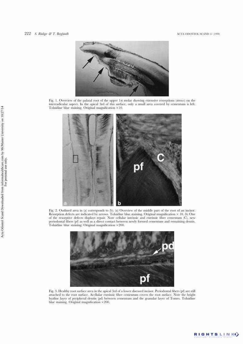
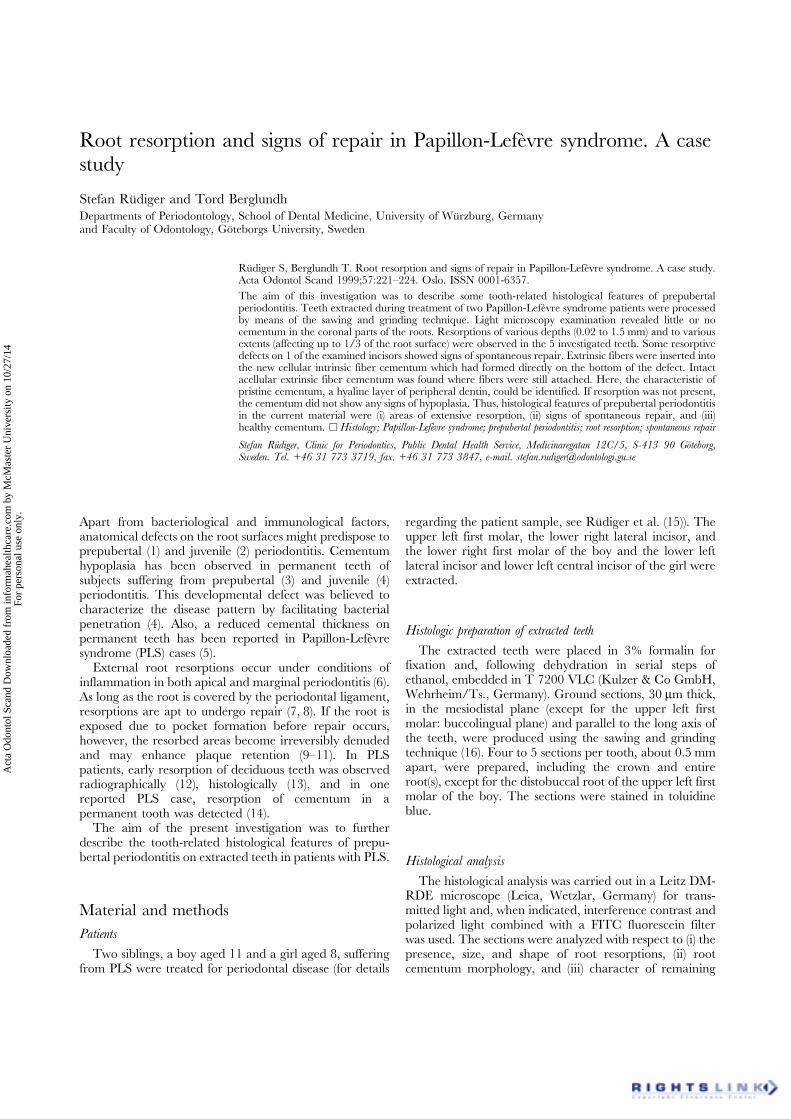
Fig. 1. Overview of the palatal root of the upper 1st molar showing extensive resorptions (arrows) on theinterradicular aspect. In the apical 3rd of this surface, only a small area covered by cementum is left.Toluidine blue staining. Original magni®cation�10.
Fig. 2. Outlined area in (a) corresponds to (b). (a) Overview of the middle part of the root of an incisor.Resorption defects are indicated by arrows. Toluidine blue staining. Original magni®cation� 10. (b) Oneof the resorptive defects displays repair. Note cellular intrinsic and extrinsic ®ber cementum (C), newperiodontal ®bers (pf) as well as a direct contact between newly formed cementum and remaining dentin.Toluidine blue staining: Original magni®cation�200.
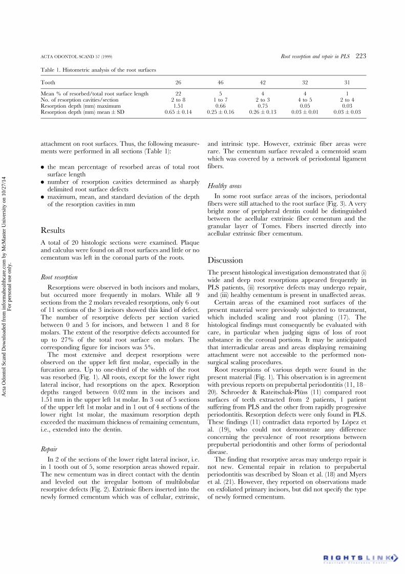
Fig. 3. Healthy root surface area in the apical 3rd of a lower diseased incisor. Periodontal ®bers (pf) are stillattached to the root surface. Acellular extrinsic ®ber cementum covers the root surface. Note the brighthyaline layer of peripheral dentin (pd) between cementum and the granular layer of Tomes. Toluidineblue staining. Original magni®cation�200.
222 S. RuÈdiger & T. Berglundh ACTA ODONTOL SCAND 57 (1999)
Act
a O
dont
ol S
cand
Dow
nloa
ded
from
info
rmah
ealth
care
.com
by
McM
aste
r U
nive
rsity
on
10/2
7/14
For
pers
onal
use
onl
y.
attachment on root surfaces. Thus, the following measure-ments were performed in all sections (Table 1):
. the mean percentage of resorbed areas of total rootsurface length
. number of resorption cavities determined as sharplydelimited root surface defects
. maximum, mean, and standard deviation of the depthof the resorption cavities in mm
Results
A total of 20 histologic sections were examined. Plaqueand calculus were found on all root surfaces and little or nocementum was left in the coronal parts of the roots.
Root resorption
Resorptions were observed in both incisors and molars,but occurred more frequently in molars. While all 9sections from the 2 molars revealed resorptions, only 6 outof 11 sections of the 3 incisors showed this kind of defect.The number of resorptive defects per section variedbetween 0 and 5 for incisors, and between 1 and 8 formolars. The extent of the resorptive defects accounted forup to 27% of the total root surface on molars. Thecorresponding figure for incisors was 5%.
The most extensive and deepest resorptions wereobserved on the upper left first molar, especially in thefurcation area. Up to one-third of the width of the rootwas resorbed (Fig. 1). All roots, except for the lower rightlateral incisor, had resorptions on the apex. Resorptiondepths ranged between 0.02 mm in the incisors and1.51 mm in the upper left 1st molar. In 3 out of 5 sectionsof the upper left 1st molar and in 1 out of 4 sections of thelower right 1st molar, the maximum resorption depthexceeded the maximum thickness of remaining cementum,i.e., extended into the dentin.
Repair
In 2 of the sections of the lower right lateral incisor, i.e.in 1 tooth out of 5, some resorption areas showed repair.The new cementum was in direct contact with the dentinand leveled out the irregular bottom of multilobularresorptive defects (Fig. 2). Extrinsic fibers inserted into thenewly formed cementum which was of cellular, extrinsic,
and intrinsic type. However, extrinsic fiber areas wererare. The cementum surface revealed a cementoid seamwhich was covered by a network of periodontal ligamentfibers.
Healthy areas
In some root surface areas of the incisors, periodontalfibers were still attached to the root surface (Fig. 3). A verybright zone of peripheral dentin could be distinguishedbetween the acellular extrinsic fiber cementum and thegranular layer of Tomes. Fibers inserted directly intoacellular extrinsic fiber cementum.
Discussion
The present histological investigation demonstrated that (i)wide and deep root resorptions appeared frequently inPLS patients, (ii) resorptive defects may undergo repair,and (iii) healthy cementum is present in unaffected areas.
Certain areas of the examined root surfaces of thepresent material were previously subjected to treatment,which included scaling and root planing (17). Thehistological findings must consequently be evaluated withcare, in particular when judging signs of loss of rootsubstance in the coronal portions. It may be anticipatedthat interradicular areas and areas displaying remainingattachment were not accessible to the performed non-surgical scaling procedures.
Root resorptions of various depth were found in thepresent material (Fig. 1). This observation is in agreementwith previous reports on prepubertal periodontitis (11, 18±20). Schroeder & Rateitschak-PluÈss (11) compared rootsurfaces of teeth extracted from 2 patients, 1 patientsuffering from PLS and the other from rapidly progressiveperiodontitis. Resorption defects were only found in PLS.These findings (11) contradict data reported by LoÂpez etal. (19), who could not demonstrate any differenceconcerning the prevalence of root resorptions betweenprepubertal periodontitis and other forms of periodontaldisease.
The finding that resorptive areas may undergo repair isnot new. Cemental repair in relation to prepubertalperiodontitis was described by Sloan et al. (18) and Myerset al. (21). However, they reported on observations madeon exfoliated primary incisors, but did not specify the typeof newly formed cementum.
Table 1. Histometric analysis of the root surfaces
Tooth 26 46 42 32 31
Mean % of resorbed/total root surface length 22 5 4 4 1No. of resorption cavities/section 2 to 8 1 to 7 2 to 3 4 to 5 2 to 4Resorption depth (mm) maximum 1.51 0.66 0.75 0.05 0.03Resorption depth (mm) mean� SD 0.65� 0.14 0.25� 0.16 0.26� 0.13 0.03� 0.01 0.03� 0.03
ACTA ODONTOL SCAND 57 (1999) Root resorption and repair in PLS 223
Act
a O
dont
ol S
cand
Dow
nloa
ded
from
info
rmah
ealth
care
.com
by
McM
aste
r U
nive
rsity
on
10/2
7/14
For
pers
onal
use
onl
y.

Cellular intrinsic fiber cementum grows more rapidlythan acellular extrinsic fiber cementum (22), which mightexplain the predominance of intrinsic fibers in reparativecementum and the exclusive cellular character of thepresent material. Spontaneous reparative cementum asobserved in our findings resembled therapeutically in-duced reparative cementum (23) in 2 ways. Firstly,extrinsic fibers inserted into the peripheral part of thenew cellular cementum formed a network with intrinsicfibers. Secondly, peripheral dentin, a characteristic findingof pristine cementum (8), could not be observed.
In healthy areas of the present material, however,peripheral dentin was found. This hyaline layer betweenthe granular layer of Tomes, a poorly calcified layer ofdentin, and the cementum, has frequently been subjectedto discussion because of its developmental origin. The lackof agreement on this matter is reflected in the unclearterminology of this anatomical structure, which is knownas peripheral dentin, layer of Hopewell-Smith, andintermediate cementum (8).
In the present material, small cemental defects wereobserved. Developmental malformation reported to be apossible predisposing factor for early onset forms ofperiodontal disease, cannot be distinguished from resorp-tions using light microscopy (4). The observation ofreparative cementum might provide indirect evidence thatthe root surface defects in the present material areresorption cavities.
Early onset periodontitis in the presence of PLS, as inthis case, may be described as a very aggressive form ofdisease with an annual progression rate of up to 3 to 4 mm(15, 24). Resorptions, as observed in the present material,may be associated with periodontal breakdown. There is,however, no clear evidence of their etiologic role. It issuggested that root resorption is a conspicuous feature ofearly onset periodontitis in the presence of PLS.
References
1. Watanabe K. Prepubertal periodontitis: a review of diagnosticcriteria, pathogenesis, and differential diagnosis. J Periodont Res1990;25:31±48.
2. Page RC, Baab DA. A new look at the etiology and pathogenesisof early-onset periodontitis. Cementopathia revisited. J Period-ontol 1985;56:748±51.
3. Waldrop TC, Hallmon WW, Mealey BL. Observations of rootsurfaces from patients with early-onset periodontitis andleukocyte adhesion efficiency. J Clin Periodontol 1995;22:168±78.
4. BlomloÈf L, HammarstroÈm L, Lindskog S. Occurrence and
appearance of cementum hypoplasia in localized and generalizedjuvenile periodontitis. Acta Odontol Scand 1986;44:313±20.
5. Vrahopoulos TP, Barber P, Liakoni H, Newman HN. Ultra-structure of the periodontal lesion in a case of Papillon-LefeÁvresyndrome (PLS). J Clin Periodontol 1988;15:17±26.
6. Schroeder HE. Pathobiologie oraler Strukturen. 3rd ed. Basel:Karger; 1997. p. 146, 187.
7. Henry JL, Weinman JP. The pattern of resorption and repair ofhuman cementum. J Am Dent Assoc 1951;42:270±90.
8. Schroeder HE. The periodontium. Berlin: Springer; 1986. p. 23±129.
9. Kerr DA. The cementum: its role in periodontal health anddisease. J Periodontol 1961;32:183±9.
10. Sottosanti JS. A possible relationship between occlusion, rootresorption, and the progression of periodontal disease. J WestSoc Periodontol Periodont Abstr 1977;25:69±74.
11. Schroeder HE, Rateitschak-PluÈss EM. Focal root resorptionlacunae causing retention of subgingival plaque in periodontalpockets. Schweiz Monatsschr Zahnmed 1983;93:1033±41.
12. Goepferd SJ. Advanced alveolar bone loss in the primarydentition. A case report. J Periodontol 1981;52:753±7.
13. Cocchia CT, McDonald RE, Mitchell DF. Papillon-LefeÁvresyndrome: precocious periodontitis with palmar-plantar hyper-keratosis. J Periodontol 1966;37:408±14.
14. LoÈst C, Haubner C. Histologische Studien und klinischeVerlaufskontrolle einschlieûlich vorlaÈufigem Behandlungsergeb-nis in einem Fall mit Papillon-LefeÁvre-Syndrom. Dtsch ZahnaÈrztlZ 1985;40:778±82.
15. RuÈdiger S, Petersilka G, Flemmig TF. Combined systemic andlocal antimicrobial therapy of periodontal disease in Papillon-LefeÁvre syndrome. J Clin Periodontol 1999. In press.
16. Donath K, Breuner G. A method for the study of undecalcifiedbones and teeth with attached soft tissues. The SaÈge-Schliff(sawing and grinding) technique. J Oral Pathol 1982;11:318±26.
17. Kressin S, Herforth A, Preis S, Wahn V, Lenard HG. Papillon-LefeÁvre syndromeÐsuccessful treatment with a combination ofretinoid and concurrent systematic periodontal therapy: casereports. Quintessence Int 1995;26:795±803.
18. Sloan P, Soames JV, Murray JJ, Jenkins WMM. Histopatholo-gical and ultrastructural findings in a case of Papillon-LefeÁvresyndrome. J Periodontol 1984;55:482±5.
19. LoÂpez NJ, Gigoux C, Canales ML. Histological differencesbetween teeth with adult periodontitis and prepubertal period-ontitis. J Periodontol 1990;61:87±94.
20. Bimstein E, Wagner M, Nauman RK, Abrams RG, Shapira L.Root surface characteristics of primary teeth from children withprepubertal periodontitis. J Periodontol 1998;69:337±47.
21. Myers DR, O'Dell NL, Clark JW, Cross RL. Localizedprepubertal periodontitis: literature review and report of case. JDent Child 1989;56:107±11.
22. Bosshardt DD, Luder HU, Schroeder HE. Rate and growthpattern of cementum apposition as compared to dentine and rootformation in a fluorochromeÐlabelled monkey (Macaca fasci-cularis). J Biol Buccale 1989;17:3±13.
23. ArauÂjo M, Berglundh T, Lindhe J. The periodontal tissues inhealed degree III furcation defects. An experimental study indogs. J Clin Periodontol 1996;23:532±41.
24. Soskolne WA, Stabholz A, van Dyke TE, Hart TC, Meyle J.Partial expression of the Papillon-LefeÁvre syndrome in twounrelated families. J Clin Periodontol 1996;23:764±9.
Received for publication 6 April 1999
Accepted 28 June 1999
224 S. RuÈdiger & T. Berglundh ACTA ODONTOL SCAND 57 (1999)
Act
a O
dont
ol S
cand
Dow
nloa
ded
from
info
rmah
ealth
care
.com
by
McM
aste
r U
nive
rsity
on
10/2
7/14
For
pers
onal
use
onl
y.


