
Reproductive System
• Gametes
• Specialized reproductive
• or sex cells.
• Female: ova
• Male: sperm
• These gametes fuse to form a single celled zygote, which will develop into a new individual.

Commonalities
• Although there are different structures involved, it is important to understand that a common general structure and function can be identified between the systems in both sexes and that both sexes contribute in uniquely important ways to overall reproductive success.
• Both male and female reproductive systems permit development of sperm or ova followed by successful fertilization and then the normal development and birth of a baby.
• Essential organs of reproduction in men and women are called gonads
• In addition, production of hormones that permits development of secondary sex characteristics, such as breast development in women and beard growth in men, occurs as a result of normal reproductive system activity.
Male Reproductive System
• The gonads of men consist of a pair of main sex glands called the testes.
• The testes produce the male sex cells or
spermatozoa.
Testes
• Located in the pouch like scrotum, which is suspended outside the body cavity.
• This exposed location provides an environment about 1 degree C or 3 degrees F cooler than normal body temperature. This is an important requirement for the normal production and survival of sperm.

Seminiferous tubules
• Coiled structures that form the bulk of the testicular tissue mass.
• Each tubule is a long duct with a central passageway.
• Sperm develop in the walls of the tubule and are then released into the central passageway.
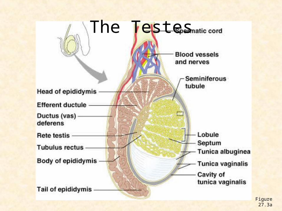
The Testes
Figure 27.3a
Testes Function
• Sperm production is called spermatogenesis.
• From puberty on, seminiferous tubules continuously form sperm, although, the number of sperm produced each day diminishes with increasing age.
Spermatogonium
• Sperm are created through meiosis from spermatogonia cells located near the outer edge of each seminiferous tubule.
• The cells will reduce down to 23 chromosomes in each of 4 spermatids from 46 chromosomes.
Sperm
• Each sperm has all the characteristics a baby will inherit from their father in a condensed nuclear (genetic) package in the sperm head.
• They are equipped with tails for motility and are designed to penetrate the outer membrane of the ovum when contact occurs with it.
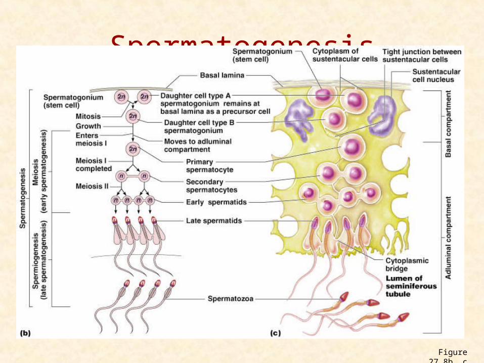
Spermatogenesis
Figure 27.8b, c
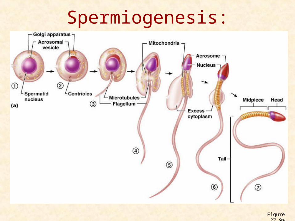
Spermiogenesis: Spermatids to Sperm
Figure 27.9a
Testosterone
• Another function of the testes is to secrete the male hormone, testosterone.
• Testosterone serves the general functions:• Masculinizes (various male characteristics)• Promotes and maintains development of the
male accessory organs • Stimulating effect on protein anabolism which is
responsible for the greater muscular development and strength of the male.
Accessory Organs
• A series of passageways or ducts that carry the sperm from the testes to the exterior:
• Epididymis (two), vas deferens (two), ejaculatory duct (two), and urethra
Ducts
• Epididymis: single and very tightly coiled tube about 6 meters or 20 feet in length.
• Sperm mature and develop their ability to move or swim as they pass through.

Ducts
• Ductus deferens or vas deferens permits exit from the epididymis and pass from the scrotal sac upward into the abdominal cavity.
• Ejaculatory duct: Moves sperm from vas deferens to the urethra.It will pass through the substance from the prostate gland on its way to the urethra.
Accessory Sex Glands
• Semen or seminal fluid: mixture of sex cells and secretions from the 2 seminal vessicles, 2 Cowper’s glands and one prostate gland.
• Semen is alkaline and protects sperm from acidic environment of the female reproductive tract.
Female Reproductive System
• Essential organs of reproduction in women are the paired ovaries where the female sex cells, the ova, are produced.
Ovaries
• Ovaries resemble large almonds in size and shape and are attached to ligaments in the pelvic cavity on each side of the uterus.
• Baby girls are born with about 1 million ovarian follicles; each contains an oocyte, an immature stage of the female sex cell.
• By the time a girl reaches puberty, further development has resulted in the formation of a reduced number (about 400,000) of what are now called primary follicles.
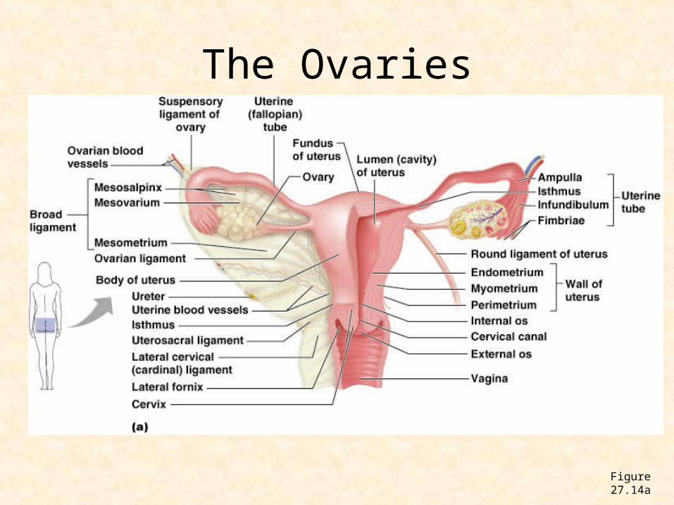
The Ovaries
Figure 27.14a
Ova formation
• During the reproductive lifetime of most women, only about 350 to 500 of these primary follicles fully develop into mature follicles, which ovulate and release an ovum for potential fertilization.
• Follicles that do not mature will degenerate and be reabsorbed by the ovarian tissue.
Ovary functions
• Oogenesis: production or female gametes
• Meiosis reduces the number of chromosomes into 23, however instead of 4 ova produced, there is 1 ova and 3 polar bodies. The ova will develop into the embryo and the polar bodies will add to nutritional structures.
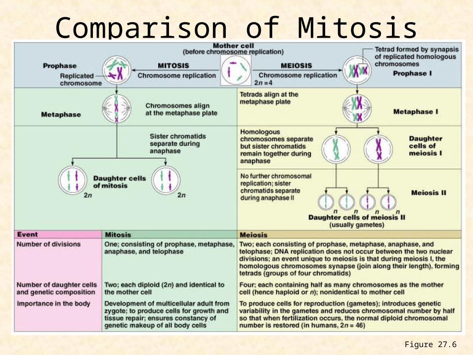
Comparison of Mitosis and Meiosis
Figure 27.6
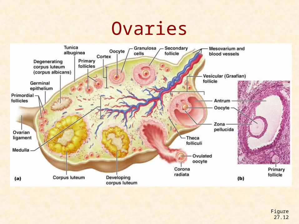
Ovaries
Figure 27.12
Estrogen and Progesterone
• The second major function of the ovaries, in addition to oogenesis, is the secretion of the sex hormones estrogen and progesterone.
• Hormone production begins at puberty with the cyclic development and maturation of the ovum.
Estrogen
• Sex hormone that causes the development and maintenance of the female secondary sex characteristics and stimulates growth of the epithelial cells lining the uterus.
• Includes: Development of external genitals, pubic hair and breast development, female body contours (fat deposits) and initiation of the first menstrual cycle.
Progesterone
• Produced by the corpus luteum, which is a glandular structure that develops from a follicle that has just released an ovum.
• Progesterone is produced for about 11 days after ovulation.
• It stimulates growth and blood vessels of the epithelial lining of the uterus and acts with estrogen to initiate the menstrual cycle in girls entering puberty.
Reproductive Ducts
• The two uterine tubes, also called fallopian tubes or oviducts, serve as ducts for the ovaries, even though they are not attached to them.
• The outer end of each tube is a funnel shaped structure with fringelike projections called fimbriae along its edge. This part curves over the top of each ovary.
• The fallopian tubes, which are about 10 cm or 4 inches in length lead to the uterus.
Reproductive ducts
• After ovulation, the discharged ovum first enters the abdominal cavity and then enters the fallopian tube assisted by the wavelike movement of the fimbriae and the beating of the cilia on their surface.
• Once in the tube, the ovum begins its journey to the uterus.
• Some ova never find their way and are reabsorbed by the body.
Uterus
• The uterus is a small organ – only the size of a pear – but it is extremely strong.
• It is almost all muscle or myometrium, with only a small cavity inside.
• It is composed of two parts:
• Body: upper portion
• Cervix: lower narrow section
Menstruation
• Corpus luteum stops secreting progesterone and decreases estrogen about 11 days after ovulation.
• 3 days later, menstruation starts
• Mucous membrane lining of the uterus, the endometrium pulls loose and sheds.
• Immediately afterwards, it repairs itself to prepare for possible pregnancy.
Menstruation
• Usually occurs at puberty, around 12 yrs old.
• Normally repeats itself every 28 days or 13 times a year for 30 – 40 years before it ceases at menopause, when a woman is somewhere around 50 years old.
Vagina
• The vagina is a tube about 10 cm or 4 inches long.
• It lies in the pelvic cavity between the urinary bladder and the rectum.
• Leads from cervix to exterior.
Breasts
• Breasts lie over pectoral muscles and are attached to them by connective tissue ligaments.
• Breast size is determined more by the amount of fat around the glandular tissue than the amount of milk secreting tissue, hence size has little to do with the ability to secrete adequate amounts of milk after the birth of a baby.
Breasts
• Each breast consists of 15 to 20 lobes radially arranged. Each lobe consists of several lobules, and each lobule consists of milk-secreting glandular cells.
• The milk secreting cells are arranged in grapelike clusters called alveoli.
• Small lactiferious ducts drain the alveoli and converge toward the nipple.


