(Relates to Chapter 32, “Nursing Assessment: Cardiovascular System,” in the textbook)
Copyright © 2011, 2007 by Mosby, Inc., an affiliate of Elsevier Inc.

HeartFour chambersComposed of three layers Endocardium Myocardium Epicardium
Copyright © 2011, 2007 by Mosby, Inc., an affiliate of Elsevier Inc.
2

Blood Flow Through the Blood Flow Through the HeartHeart
Copyright © 2011, 2007 by Mosby, Inc., an affiliate of Elsevier Inc.
3
Fig. 32-1. Schematic representation of blood flow through the heart. Arrows indicate direction of flow. 1, The rightatrium receives venous blood from the inferior and superior venae cavae and the coronary sinus. The blood thenpasses through the tricuspid valve into the right ventricle. 2, With each contraction, the right ventricle pumpsblood through the pulmonic valve into the pulmonary artery and to the lungs. 3, Oxygenated blood flows from thelungs to the left atrium by way of the pulmonary veins. 4, It then passes through the mitral valve and into the leftventricle. 5, As the heart contracts, blood is ejected through the aortic valve into the aorta and thus enters thesystemic circulation.
Copyright © 2011, 2007 by Mosby, Inc., an affiliate of Elsevier Inc.
4
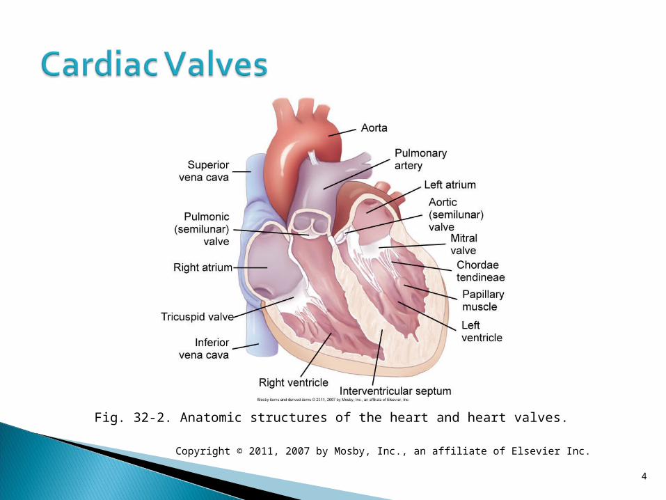
Fig. 32-2. Anatomic structures of the heart and heart valves.

Copyright © 2011, 2007 by Mosby, Inc., an affiliate of Elsevier Inc.
5
Fig. 32-3. Coronary arteries and veins.

Copyright © 2011, 2007 by Mosby, Inc., an affiliate of Elsevier Inc.
6
Fig. 34-4. Location of pain during angina or myocardial infarction.

Copyright © 2011, 2007 by Mosby, Inc., an affiliate of Elsevier Inc.
7
Fig. 34-4. Location of pain during angina or myocardial infarction.

• Systole: Contraction of myocardium
• Diastole: Relaxation of myocardium
• Cardiac output: Amount of blood pumped by each ventricle in 1 minuteCO = SV × HR
• Cardiac index: CO divided by body surface area
Mechanical SystemMechanical System
Copyright © 2011, 2007 by Mosby, Inc., an affiliate of Elsevier Inc.
8

• PreloadVolume of blood in ventricles at the end of diastole
• Contractility• Afterload
Peripheral resistance against which the left ventricle must pump
Factors Affecting Cardiac Factors Affecting Cardiac OutputOutput
Copyright © 2011, 2007 by Mosby, Inc., an affiliate of Elsevier Inc.
9

A patient is receiving a drug that decreases afterload. To evaluate the effect of the drug, the nurse monitors the patient’s:
1. Heart rate.2. Lung sounds.3. Blood pressure.4. Jugular vein distention.
Audience Response Question
Copyright © 2011, 2007 by Mosby, Inc., an affiliate of Elsevier Inc.
10

• Vascular systemBlood vessels
• Arteries, arterioles• Veins, venules• Capillaries
Copyright © 2011, 2007 by Mosby, Inc., an affiliate of Elsevier Inc.
11

Copyright © 2011, 2007 by Mosby, Inc., an affiliate of Elsevier Inc.
12
Fig. 32-5. Comparative thickness of layers of the artery, vein, and capillary.

• Regulation of the cardiovascular systemAutonomic nervous systemBaroreceptorsChemoreceptors
Copyright © 2011, 2007 by Mosby, Inc., an affiliate of Elsevier Inc.
13

Blood pressure• Measurement of arterial blood
pressure• Pulse pressure • Mean arterial pressure
Copyright © 2011, 2007 by Mosby, Inc., an affiliate of Elsevier Inc.
14

• Age alters the cardiovascular response to physical and emotional stress.
• Heart valves become thick and stiff.
• Frequent need for pacemakers• Less sensitive to β-adrenergic agonist drugs
• Increase in SBP; decrease or no change in DBP
Copyright © 2011, 2007 by Mosby, Inc., an affiliate of Elsevier Inc.
15

• Subjective dataHealth information
• History of present illness• Past health history• Past and current medications• Surgery or other treatments
Copyright © 2011, 2007 by Mosby, Inc., an affiliate of Elsevier Inc.
16

• Functional health patterns (Table 32-3)Health perceptionhealth management pattern
Nutritional-metabolic patternElimination patternActivity-exercise patternSleep-rest patternCognitive-perceptual pattern
Copyright © 2011, 2007 by Mosby, Inc., an affiliate of Elsevier Inc.
17

• Functional health patternsSelf-perceptionself-concept pattern
Role-relationship patternSexuality-reproductive patternCopingstress tolerance patternValues-belief pattern
Copyright © 2011, 2007 by Mosby, Inc., an affiliate of Elsevier Inc.
18

• Objective dataPhysical examination Vital signs Peripheral vascular system
Inspection Palpation Auscultation
Copyright © 2011, 2007 by Mosby, Inc., an affiliate of Elsevier Inc.
19

Copyright © 2011, 2007 by Mosby, Inc., an affiliate of Elsevier Inc.
20
Fig. 32-6. Common sites for palpating arteries.

• Physical examination (cont’d)Thorax
• Inspection • Palpation• Percussion• Auscultation
Copyright © 2011, 2007 by Mosby, Inc., an affiliate of Elsevier Inc.
21

Copyright © 2011, 2007 by Mosby, Inc., an affiliate of Elsevier Inc.
22
Fig. 32-7. Orientation of the heart within the thorax and cardiac auscultatory areas. Red lines indicate the midsternal line (MSL), midclavicular line (MCL), and anterior axillary line (AAL). ICS, intercostal space; PMI, point of maximal impulse.

Copyright © 2011, 2007 by Mosby, Inc., an affiliate of Elsevier Inc.
23
Fig. 32-8. Relationship of electrocardiogram, cardiac cycle, and heart sounds.
• Noninvasive studiesBlood studiesChest x-rayElectrocardiogram Resting ECG Ambulatory ECG monitoring Event monitor or loop recorder Exercise or stress testing 6-minute walk test
Copyright © 2011, 2007 by Mosby, Inc., an affiliate of Elsevier Inc.
24
Chest X-rayChest X-ray
Copyright © 2011, 2007 by Mosby, Inc., an affiliate of Elsevier Inc.
25
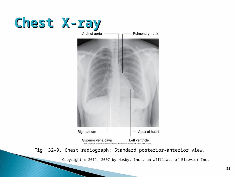
Fig. 32-9. Chest radiograph: Standard posterior-anterior view.

A patient arrives at an urgent care center after experiencing unrelenting substernal and epigastric pain and pressure for about 12 hours. The nurse reviews laboratory results with the understanding that at this point in time, a myocardial infarction would by indicated by peak levels of:
1. Troponin T.2. Homocysteine.3. Creatine kinase-MB.4. Type b natriuretic peptide.
Audience Response Question
Copyright © 2011, 2007 by Mosby, Inc., an affiliate of Elsevier Inc.
26
• Noninvasive studiesEchocardiogramNuclear cardiology
Copyright © 2011, 2007 by Mosby, Inc., an affiliate of Elsevier Inc.
27

Copyright © 2011, 2007 by Mosby, Inc., an affiliate of Elsevier Inc.
28
Fig. 32-10. Apical four-chamber two-dimensional echocardiographic view in a normal patient. LA, Left atrium; LV, left ventricle; MV, mitral valve; RA, right atrium; RV, right ventricle; TV, tricuspid valve.
• Noninvasive studies (cont’d)Magnetic resonance imagingComputed tomography
Copyright © 2011, 2007 by Mosby, Inc., an affiliate of Elsevier Inc.
29
Computed TomographyComputed Tomography
Copyright © 2011, 2007 by Mosby, Inc., an affiliate of Elsevier Inc.
30
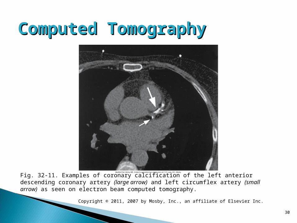
Fig. 32-11. Examples of coronary calcification of the left anterior descending coronary artery (large arrow) and left circumflex artery (small arrow) as seen on electron beam computed tomography.

• Invasive studiesCardiac catheterization and coronary angiography
Intracoronary ultrasoundFractional flow reserveElectrophysiology studyBlood flow and pressure measurements
Copyright © 2011, 2007 by Mosby, Inc., an affiliate of Elsevier Inc.
31

Copyright © 2011, 2007 by Mosby, Inc., an affiliate of Elsevier Inc.
32
Fig. 32-12. Normal left coronary artery angiogram.
A patient returns to the cardiac observation area following a cardiac catheterization with coronary angiography. Which of the following assessments would require immediate action by the nurse?
1. Pedal pulses are 2+ bilaterally.2. Apical pulse is 54 beats/minute. 3. Mean arterial pressure is 72 mm Hg.4. ST-segment elevation develops on the ECG.
Audience Response Question
Copyright © 2011, 2007 by Mosby, Inc., an affiliate of Elsevier Inc.
33


