
Protein-protein interactions and western blotting
MCB 130LLecture 3

Antibodies in the Immune System
Structure:
2 heavy chains + 2 light chains
Disulfide bonds
2 antigen binding sites
Isotypes: IgG, IgM, IgA, IgE, IgD

Antibodies are produced by B lymphocytes
Clonal Selection
• Millions of B cell clones w/ specific cell-surface
receptors
• Activation of B cell clones by specific target antigen
• Activated B cells secrete specific antibodies
EM of resting and activated B cells
Activated: Extensive rough ER for antibody production/secretion

Antibody Production
1. Inject antigen (i.e. purified protein) into animal (i.e. mouse, rabbit, chicken)
2. Animal produces antibodies that recognize antigen
Antigen injected more than once: response heightened in subsequent injections

collect blood serum
purify antibodies w/ affinity chromatographyusing antigen attached to beads
Producing antibodies to a specific antigen
Polyclonal antibodies: Derived from multiple B-cell clones,recognize multiple epitopes on antigens
Linear epitope
Conformationalepitope
Inject with antigen
“epitope” = unique part of antigen recognized by antibody

Producing antibodies to a specific antigen
Monoclonal antibodies:• Derived from B-cell clone “Hybridoma”
• Recognize single epitope on antigen

Uses of antibodies in molecular biology
Applications:
Western blotting (Immunoblotting) - Identification of protein antigen following SDS-PAGE
Immunoprecipitation - Isolation of specific proteins + binding partners
Immunofluorescence microscopy- Localization of specific proteins in cells
ELISA (Enzyme-Linked Immunosorbent Assay)- Detection of proteins in a sample

Detection of specific proteins:SDS-PAGE and Western blot
Western blottingFrom Lodish et al. Molecular Cell Biology 4th edition.
Indirect immunodetection
1. Separate proteins by SDS PAGE2. Transfer proteins to membranes (i.e. Nitrocellulose)3. Block non-specific sites on membrane4. Incubate with primary antibody, wash5. Incubate with secondary antibody, wash6. Detect secondary antibody

Detection of HRP labeled secondary antibody by chemiluminescence• Electrochemiluminescence (ECL) reagent: H2O2 + luminol • HRP catalyzes breakdown of H2O2 to H2O and O2, • Luminol is oxidized• Light from oxidized luminol is detected using film
Figures from Amersham Biosciences
Detection of specific proteins:SDS-PAGE and Western blot

Immunopreciptation: Identification of protein-protein interactions
bead
protein A
primary antibody
Steps:1. Attach antibody to beads via protein A2. Lyse cells to release antigen and its binding partners3. Mix cell lysate + antibody-coated beads (antibody binds antigen)4. Purify antigen and its binding partners by centrifugation

Immunofluorescence Microscopy

ELISA(Enzyme-Linked Immunosorbent Assay)
Detection of proteins (i.e. cytokines, HIV antigens) in samples

This Week’s Lab:Protein-protein interactions in synaptic vesicle fusion
Release of acetylcholine at presynaptic plasma membrane

Disruption of synaptic vesicle release by Tetanus toxin
Clostridum tetani • Anaerobic soil bacterium• Responsible for 350,000 cases/year of tetanus (spastic paralysis)
worldwide• Tetanus toxin blocks release of neurotransmitters from the presynaptic
membranes; Cleaves VAMP2
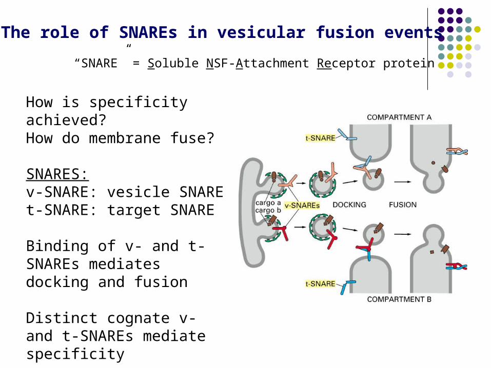
The role of SNAREs in vesicular fusion events
How is specificity achieved?How do membrane fuse?
SNARES:v-SNARE: vesicle SNAREt-SNARE: target SNARE
Binding of v- and t-SNAREs mediates docking and fusion
Distinct cognate v- and t-SNAREs mediate specificity
“SNARE” = Soluble NSF-Attachment Receptor protein

The role of SNAREs in vesicular fusion events
Structure of the SNARE complex: Sb = VAMP (synaptobrevin)Sx = syntaxinSn1, Sn2 = SNAP25.
(VAMP)
Jahn and Scheller Nature Reviews Molecular Cell Biology 7, 631–643 (2006) | doi:10.1038/nrm2002
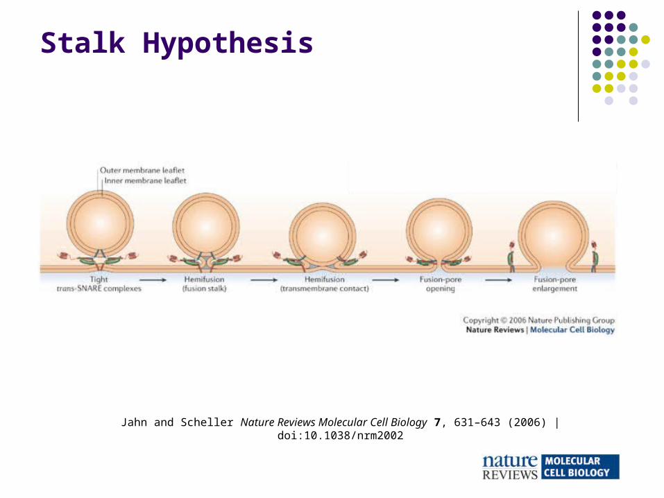
Stalk Hypothesis

Jahn and Scheller Nature Reviews Molecular Cell Biology 7, 631–643 (2006) | doi:10.1038/nrm2002
SNAREs in intracellular membrane-trafficking pathways

SNARE Domains
Chen and Scheller Nature Reviews Molecular Cell Biology 2, 98-106 (2001)
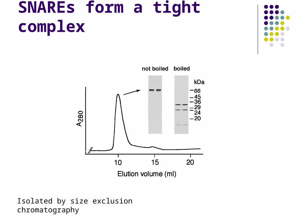
SNAREs form a tight complex
Isolated by size exclusion chromatography

Identification of protein-protein interactionsby GST-pulldown assays
GST
bead
glutathione
protein of interest
bead
add binding partner
wash
elute with glutathione
SDS-PAGE, Western blottingPurpose: to determine which protein domains are necessary for SNARE interactions

Hey, where’d all the mice go?

Botulinum toxin
JAMA. 2001;285:1059-1070


